artificial intelligence and the future of diagnostic and
TRANSCRIPT

Artificial intelligence and the future of diagnostic and therapeutic radiopharmaceutical development: in Silico
smart molecular design
Bahar Ataeinia, MD-MPH† and Pedram Heidari, MD*†
†Department of Radiology, Massachusetts General Hospital, Boston, MA
* Correspondence to:
Pedram Heidari, MD
55 Fruit St, Wht 427, Boston, MA 02114
[email protected], 617-726-5121
Disclosure: The authors have nothing to disclose.
Keywords: Artificial intelligence, Computer-aided drug design, in silico, positron emission
tomography, radiopharmaceuticals
Key Points:
• Artificial intelligence (AI) is a novel way for development of new
radiopharmaceuticals.
• AI methods enable designing lead radiotracers with favorable pharmacokinetics
and pharmacodynamics.
• Using AI methods reduces the radiopharmaceutical development time and cost.
• AI does not obviate the need for in vivo testing of the lead compounds, given that
the accuracy of the methods is greatly affected by the assumptions put in the
models.
• Radiopharmaceuticals developed by AI have a wide range of applications including
central nervous system (CNS), cancer, infection and inflammation imaging.

Synopsis
Novel diagnostic and therapeutic radiopharmaceuticals are increasingly becoming
a central part of personalized medicine. Continued innovation in the development of new
radiopharmaceuticals is key to sustained growth and advancement of precision medicine.
Artificial intelligence (AI) has been used in multiple fields of medicine to develop and
validate better tools for patient diagnosis and therapy, including in radiopharmaceutical
design. In this review, we first discuss common in silico approaches and focus on their
utility and challenges in radiopharmaceutical development. Next, we discuss the practical
applications of in silico modeling in design of radiopharmaceuticals in various diseases.
Abbreviation list:
Aβ: Amyloid β
ADMET: Absorption, Distribution, Metabolism, Excretion, and Toxicity
AD: Alzheimer’s Disease
AI: Artificial Intelligence
ANN: Artificial Neural Networks
AR: Adenosine Receptor
α-syn: α-synuclein
BBB: Blood Brain Barrier
CAAD: Computer-Aided Drug Design
CCK-2R: Cholecystokinin Receptor Subtype 2
CNS: Central Nervous System
64Cu: 64Copper
CXCR4: Chemokine Receptor-4
11 C: 11 Carbon

DL: Deep Learning
FAK: Focal Adhesion Kinase
FBDD: Fragment-Based Drug Design
18 F: 18 Fluorine
GGT: Gamma Glutamyl Transferase
68 Ga: 68 Gallium
HFPPS: Human Farnesyl Pyrophosphate Synthase
HYNIC: Hydrazinonicotinic acid
131 I: 131Iodine
LBDD: Ligand Based Drug Design
ML: Machine Learning
NMR: Nuclear Magnetic Resonance
NOP: Nociceptin Opioid Peptide
PD: Parkinson’s Disease
PET: Positron Emission Tomography
PDE: Phosphodiesterase
PSMA: Prostate-Specific Membrane Antigen
QM: Quantum Mechanical
QSAR: Quantitative Structure-activity Relationships Analysis
SAR: Structure-activity Relationships Analysis
SB: styrylbenzoxazole
SBDD: Structure-Based Drug Design
SBP: Structure-based Pharmacophore Modeling

SSTR2: Somatostatin Receptor Subtype 2
SPECT: Single-photon Emission Computed Tomography
99m Tc: 99m technetium
VAP-1: Vascular Adhesion Protein-1
2-D: two Dimensional
3-D: Three Dimensional

Introduction:
Radiopharmaceuticals play a pivotal role in the rapidly emerging field of
personalized medicine. 1 In disease states, altered expression or aggregation of specific
molecules allow for non-invasive diagnosis and/or targeted radiotherapy using
radiopharmaceuticals. 2-4 Thus, development of new radiopharmaceuticals is key to
improved diagnosis and therapy in a broader range of diseases. However, design,
validation, and translation of these compounds are time-consuming and require
considerable effort and financial investment. 5,6
A translatable radiopharmaceutical has structural and functional characteristics
that result in high affinity for its target, low non-specific binding, and favorable
pharmacokinetics. Whether the structure of a compound is novel or similar to an existing
compound, substantial effort is required to ensure optimal radiopharmaceutical
performance. Despite all the care in designing a compound, there is high likelihood that
it fails to adequately engage its target in vivo, due to unpredicted parameters that were
not considered during the development phase. 7
The term “in silico medicine” refers to using computer modeling and simulation for
conducting biomedical research. In silico approaches can predict outcomes for crucial
variables that are quit taxing by conventional in vitro and in vivo radiopharmaceutical
development methods. 7,8 Computational models can predict target-binding properties of
a compound, and critical pharmacokinetic characteristics such as absorption, distribution,
metabolism, excretion, and toxicity (ADMET). 9 Therefore, incorporation of in silico
methods could facilitate faster and more cost-effective design and testing of new

radiopharmaceuticals, by informing in vitro and in vivo studies which reduces the need
for animal models to evaluate the lead compounds. 10-14
Artificial intelligence (AI) methods have been used to improve drug discovery since
the 90s. 15 With ever growing rapid expansion of our knowledge about the structure and
function of biological targets, AI can expedite radiopharmaceutical design research
resulting in more rapid incorporation of these compounds in routine medical practice
(Figure 1). 16-18 It is important to note that computational modeling is not a replacement
for in lab experiments but rather a complementary assist tool to facilitate
radiopharmaceutical development as currently no modeling can emulate the complexity
of human body.12,19
Herein, we summarize the current computer-aided drug design (CADD) methods,
their potential application, challenges in radiopharmaceutical development research and
provide real-world examples of radiopharmaceutical design using the input from CADD
methods.
Approach
In general, in silico approaches focus on structure and behavior of compounds for
modeling (Figure 2). Structural models predict binding affinity of a radiopharmaceutical
for its target, while behavioral approaches focus on its pharmacokinetics. Both
approaches are complimentary and essential since a translatable radiopharmaceutical
should have high specificity and affinity for the target, optimal biodistribution, stability,

practical effective half-life and a desirable clearance kinetic based on its intended
diagnostic or therapeutic applications.20
Structure-based computational modeling approaches are classified into two main
categories, structure-based drug design (SBDD) and ligand-based drug design (LBDD).
SBDD is used to predict radiopharmaceutical-target interaction when the structure of the
target is known. For instance, chemical structure of some of the most studied targets such
as prostate-specific membrane antigen (PSMA) and chemokine receptor-4 (CXCR4) is
known and can be used for modeling in SBDD. 21,22 If the structure of the target is not
available LBDD models can be used, which analyzes the structure of known ligands for
the target (Figure 3). 12,23,24 Here we discuss structural and behavioral approaches in
more detail.
Structural computational modeling
1. Structure-based drug design (SBDD) (direct approach)
1.1. Molecular docking
Molecular docking models predict candidate ligands' interaction to a target
with a known three-dimensional (3-D) structure. The model predicts various
orientations and conformations of ligand and target to determine the ideal pose
with minimum energy for best-fit interaction and complex stability. Moreover, each
ligand's binding affinity can be computed based on binding energetics by the
algorithm's scoring functions to rank the ligands. 25,26 Scoring functions are
categorized as classical, and machine learning (ML) functions. 27,28 Determination

of the appropriate approach depends on the target and data availability. 25,29
Different AI techniques such as swarm intelligence, ant colony optimization
algorithms, and DL have been used for molecular docking.30-32 Selecting the most
appropriate software and scoring function is a critical step and can significantly
affect outcomes.33 Docking models cannot be reliably used if chemical structure
of the ligand or the specific binding site are unknown, unless protein homology
modeling and binding site identification is performed prior to docking. 34-36
Molecular docking models are commonly used in radiopharmaceutical
development studies to predict the effect of labeling with different radionuclides
and chelators on ligand's binding affinity to the target. 37-41
1.2. Fragment-based drug design (FBDD)
This method is an alternative for the modeling of compounds with high
molecular weight and low solubility. In FBDD, low molecular weight fragments that
interact with biological targets are first identified, and appropriate fragments are
then expanded by adding chemical groups or merging with other fragments to
design the final compound. 42,43
1.3. Pharmacophore modeling
Pharmacophore analysis is applicable in both SBDD and LBDD, although
is more commonly used with the latter. This method mainly focuses on the
molecular features, important for the target-ligand interaction. Such features
include hydrophobic groups, aromatic rings, polar groups, and hydrogen bond
vectors. 23,44 Structure-based pharmacophore modeling (SBP) is a SBDD

pharmacophore analysis which is used if the 3-D structure of the target is known.
The model can include structural properties of both target and ligand or the target
exclusively. 45,46 LBDD pharmacophore modeling is discussed in more details in
section 2.2.
2. ligand-based drug design (LBDD) (indirect approach)
2.1. Ligand chemical similarity search
In this method, two-dimensional (2-D) molecular fingerprints of an already
known ligand are used to identify candidate compounds from a database of
chemicals with a similar structure to a known ligand. Molecular fingerprints are
binary codes that record the presence or lack of specific chemical groups and can
be utilized by screening software to perform the chemical similarity search. 23,47
Given that a compound's complete structure is used in chemical similarity search
in contrast to other LBDD methods, such as pharmacophore analysis, it may
provide a more accurate estimation of in vivo biological activity.
2.2. Pharmacophore analysis
As previously mentioned, this approach incorporates crucial features for
ligand-target interaction into the model. Initially, a wide range of biologically active
ligands is studied to train the model. The main two goals are to distinguish
overlapping structures for optimal ligand-target interaction and create
conformational space for the studied ligands to determine structural flexibility in
the model. However, there are several challenges in designing a pharmacophore
model including the modeling of ligand flexibility, molecular alignment and proper

selection of training set compounds, which is even more challenging in designing
radiopharmaceuticals.23,44 Even when a radionuclide is added to the backbone
structure and not the active binding site, it can alter the radiopharmaceutical's
biological behavior. 48-53 Pharmacophore analyses is mainly utilized to screen
potential ligands or to design a new radiopharmaceutical. 44 Pharmacophore
models can also be used in SBDD approaches as previously discussed.
2.3. Structure-activity relationships analysis (SAR)
SAR is based on the assumption that molecules with similar chemical
structures are more likely to demonstrate a similar biological activity. 54,55 Using a
regression model that correlates structural properties of a known group of similar
ligands to their biological activity, such as binding to the desired target or inhibiting
an enzyme, SAR can predict a pharmaceutical’s biological behavior. 56
Quantitative structure-activity relationship (QSAR) is a SAR that quantifies
bioactivity level of a pharmaceutical. Using artificial neural networks (ANN) and
deep learning (DL) algorithms, QSAR has been used for drug design. 15,57 The
crucial step in SAR is carefully choosing molecular properties used in the
regression model to avoid overfitting. An overfitted model has optimal
performance on the training dataset but fails when used on new data. 15 Note
should be made that structural similarity does not necessarily imply similar activity
indicating the need for cautious use of this assumption given the complexity of in
vivo biochemical interactions. 58 SAR models can predict whether adding
chelators, radionuclides or chemical groups to a bioactive compound will alter the

binding properties and potency of the proposed radiopharmaceutical or cause
undesired interactions leading to toxicity or off-target binding. 59,60
Behavioral computational modeling
Absorption, Distribution, Metabolism, Excretion, and Toxicity (ADMET)
modeling is used to predict the ligand's in vivo behavior based on the
pharmacokinetics of similar ligands and modeling interaction of the candidate
compound with enzymes involved in toxicity, insolubility, and undesired metabolism
61. Inputs from SBDD models and SAR, as well as other in silico approaches are used
to predict ADMET characteristics. 62 A fundamental step to ensure optimal accuracy
of ADMET models is using a chemically diverse data set of ligands to train the model
63.
In addition to proper target specificity and affinity, a translatable
radiopharmaceutical should have appropriate pharmacokinetics and stability for
optimal performance in vivo. Even slight alterations in radiopharmaceutical structure,
such as changes in chelator and linker, can still dramatically affect biodistribution and
stability. Hence, using computational ADMET modeling prior to in vivo experiments
can help predict serum stability and target organ uptake. 64 Absorption is often not an
issue for radiopharmaceuticals as they are generally administrated intravenously. In
certain situation such as targeting a receptor in the CNS or an intracellular target
ADMET algorithms can be additionally beneficial to model the passage of a
radiopharmaceutical through these natural barriers. 7,13,14,65 ADMET models can also

help predicting the effect of individual components of the radiopharmaceutical on the
overall pharmacokinetics. In this regard, using computer aided drug design (CAAD)
models, one can alter the design of a radiopharmaceutical to improve the
pharmacokinetics without adversely affecting the binding affinity to the target. 66 This
strategy has been successfully implemented for radiolabeled analogs of somatostatin,
integrins, bombesin, cholecystokinin and vasoactive intestinal peptide analogues.
Lastly, ADMET can help in ensuring that the radiopharmaceutical has minimal toxicity,
especially if the ligand is not a naturally occurring molecule or an established
pharmaceutical. 67
Applications
To date, molecular docking, SAR and ADMET models are the most commonly
used approaches in radiopharmaceutical development. These methods can predict a
bioactive compound’s interaction with its target after radiolabeling and whether altering
radionuclides and chelators affects radiopharmaceutical’s performance (figure 1). In this
section, we review some of the applications of these methods in radiopharmaceutical
design in caner, neurological disorders, and other disease processes.
Cancer
Radiopharmaceuticals are extensively used for imaging various nodes in cancer
pathways. One common approach is radiolabeling chemotherapy agents to study their
pharmacokinetics and their ability to engage the target in the tumor. Molecular docking
models have been used to ensure addition of a chelator or a radionuclide does not
adversely affect the binding affinity to the target. For instance, a recent study investigated

99mtechnetium (99mTc) labeled Ifosfamide, an alkylating chemotherapy agent, for solid
tumor imaging. Molecular docking showed that 99mTc labeling did not affect the
radiotracer's affinity for the binding site. 38 A similar approach was used with iodine labeled
Cladribine, a chlorinated purine analogue, showing that its binding affinity to DNA
polymerase was not changed. 68 Another example of using molecular docking is to
evaluate effect of changing chelators on binding properties of a commonly used
radiopharmaceutical. For instance, Cai et al. showed the in vivo binding affinity of a
somatostatin receptor subtype 2 (SSTR2) agonist was not affected by using new
generation of chelators for 64copper (64Cu) (Figure 4). 69
Some studies used a combination of approaches to develop radiopharmaceuticals
targeting receptors or enzymes, overexpressed by cancer cells or other cells in tumor
microenvironment. An example is development of PET tracers for imaging focal adhesion
kinase (FAK), a tyrosine kinase overexpressed in a number of cancers. Inputs from
molecular docking and molecular dynamics were utilized to ensure binding of designed
inhibitory tracers to FAK as well as the effect of structural modifications, such as altering
chain length on inhibitory potency of designed compounds. 70-72 Similar approach was
used for targeting gamma glutamyl transferase (GGT) and human farnesyl
pyrophosphate synthase (HFPPS). 73,74
CNS
Developing radiopharmaceuticals for imaging different receptors, enzymes and
pathologic aggregates in CNS has been of great interest and an area of intense research.
In additional to specificity for the target, a potent radiopharmaceutical for CNS imaging
has to cross the blood brain barrier (BBB) and wash out from the non-target tissue fairly
rapidly posing an additional challenge. Therefore, the use of in silico methods has gained
a lot of attention in CNS radiopharmaceutical development. Novel PET-tracers for
cerebral adenosine receptors (AR), phosphodiesterase 2A (PDE2A) and serotonin
transporter (SERT) were among the first attempts of using in silico models in CNS
radiopharmaceutical design. 41,75,76
PDE2A is mainly expressed in limbic area and basal ganglia and could be of
importance for cognitive function through modulating the signal transduction by a
regulating cGMP and cAMP levels. Therefore, development of selective PDE2A PET
ligands has been of interest using the in silico models. For this purpose, properties of a
large database of successful and failed CNS PET tracers was created and screened to
identify favorable ADMET properties for CNS PET imaging. Using these ADMET features,
enabled researchers to easily narrow down a library of more than thousand PDE2A
inhibitors to less than ten compounds. Next, a SAR model was used to design more potent
analogs from the identified lead compound prior to in vivo studies. As predicted by in silico
models, the final compound demonstrated optimal performance in vivo. 75 A similar
approach was used to design promising radiotracers for PDE4B and nociceptin opioid
peptide (NOP) receptor. 77,78
AR is widely distributed in the brain and involved in different signaling pathways.
Therefore, AR PET imaging can be useful for diagnostic and treatment monitoring in a
broad range of psychiatric, neurovegetative diseases and Parkinson’s disease. A number
of 11carbon (11C) labeled tracers and a 18Fluorine (18F) labeled AR tracers have been

developed using in silico methods.79 For instance, 18F-FESCH which is specific for AR
subtype 2A (AR2A) receptor has been developed using silico metabolite analysis. This
tracer showed high binding affinity for AR2A and favorable pharmacokinetics in preclinical
studies. PET imaging of rat brains showed this tracer is accurate for mapping AR2A
expression in the brain.41,80 In silico methods although very powerful, are not always
predictive of in vivo behavior of radiopharmaceuticals. Most recently, using QSAR and
molecular docking 18F-TOZ1 was developed based on the structure of tozadenant, an
AR2A antagonist with excellent binding affinity. Despite its ability to pass the BBB and high
brain uptake, AR2A specific binding was insufficient.81
When placing a chelator for radiometal labeling of a lead molecule to image
enzymes or cell surface receptors, molecular docking is a useful tool to predict if it affects
the binding site to target or adversely affects the binding affinity. Examples are
radiotracers developed for imaging cholinesterase, for imaging Alzheimer's disease (AD)
or histamine receptor type 1. 37,39
Pathologic aggregates, such as tau protein, amyloid β (Aβ) and α-synuclein
aggregates (α-syn), have been extensively investigated in the pathophysiology of
neurodegenerative diseases. 82-84 The complexity of their structure and different
molecular isoforms can make tracer design challenging for these targets. 3,85 With the
microscopic structure of more aggregates now being discovered, in silico design can
accelerate developing high-affinity radiotracers for PET imaging of neurodegenerative
diseases. 86,87 In addition, off target binding of these tracers to other targets such as
monoamine oxidase B (MAO-B) can be predicted by molecular docking approaches. 88-90
Such information can help optimize binding specificity of next generation of PET

radiopharmaceuticals for CNS imaging. Combining different modeling methods to design
a merged workflow can improve the ultimate accuracy of modeled data. 83 Further
optimization of modeling strategy and variable definition is required to ensure maximal
correlation of modeled predictions with in vivo performance. 91 For instance, when
different simulation approaches were used to predict binding affinity of styrylbenzoxazole
(SB) based tracers for Aβ it highlighted shortcomings of some of the model predictions;
molecular dynamics, a complex approach that incorporates particle movements in the
model, had higher accuracy than conventional docking models, while quantum
mechanical (QM) methods significantly improved prediction accuracy compared to both.
Despite higher accuracy, the downside of molecular dynamics and QM is increased
complexity and cost of simulations.83
Infection and Inflammation
Developing non-invasive imaging probes to detect infection has been gaining more
attention in the recent years, as structural imaging modalities are insensitive for detection
of the site of infection at an early stage. Anti-bacterial antibiotics are attractive compounds
for developing radiotracers as they are specific for bacterial targets, allowing for
distinction between bacterial infection and inflammation, and have favorable tissue
biodistribution for reaching the infected tissue. In addition, their minimal interaction with
eukaryotic cells and wash out from normal tissue makes them desirable for infection
imaging. 92,93 As the structure of the antibiotics are usually available, chemical similarity
search and SAR modeling can be used to screen libraries of antibiotic analogs that have
an appropriate binding affinity for imaging purposes. SBDD methods can also be used as
the structure of antibiotics’ target is commonly known. 92 Ordonez et al. screened

commercially available libraries of random radiolabeled small molecules to identify
potential substrates involved in bacterial metabolism or interacting with bacteria but with
minimal mammalian cell interaction. The identified molecules demonstrated high level of
in vitro accumulation in a wide range of bacterial species. Candidate compounds were
then labeled with 18F and successfully identified infection from sterile inflammation in vivo
murine models. 94
Molecular docking has also been used for designing the inflammation PET tracer,
68Ga-Siglec-9, which targets vascular adhesion protein-1 (VAP-1). Siglec-9 was identified
by phage display as a ligand for VAP-1. Molecular docking aided to assess the binding of
Siglec-9 to VAP-1 before proceeding with in vivo experiments. 95
Other
SAR models were utilized to predict in vivo performance of three potential
radiopharmaceuticals for lung perfusion scan. 99mTc-Hexoprenaline, a β2 adrenergic
agonist, 99mTc-Zolmitriptan, a selective serotonin receptor agonist and 131I-Dapoxetine, a
selective serotonin reuptake inhibitor. Lungs are the reservoirs for Zolmitriptan and
Dapoxetine. In the case of Hexoprenaline, the most energetically favored confirmation
required addition of 99mTc to a moiety essential for appropriate interaction of
Hexoprenaline with its target. Hence, poor in vivo binding was predicted for this
radiotracer. On the other hand, the position of 131I in Dapoxetine and 99mTc in Zolmitriptan
did not affect vital sites for tracer-target interaction, and appropriate target binding was
predicted. In vivo experiments in mice confirmed modeled predictions for all three
radiopharmaceuticals. 60,96
Summary
In silico approaches are novel tools that guide conventional in vitro and in vivo
radiopharmaceutical design experiments and accelerate novel radiopharmaceuticals’
bench to bedside translation if used and interpreted appropriately. Various examples of
successful incorporation of in silico approaches in radiopharmaceutical design and
confirmed validation of modeled data in vivo in a wide range of diseases highlights the
additional value of AI integration in this field. However, successful modeling depends on
careful inclusion of appropriate variables in the model, the modeling approach, choice of
software and availability of accurate structure of the ligands and targets. Thus, developing
a systematic workflow could help incorporating computational modeling in routine
radiopharmaceutical design process and overcome the current challenges of this valuable
technology.
Clinics Care Points
• AI can greatly utilize the advances in structural chemistry to accelerate the design
of a broad range of radiopharmaceuticals for clinical use.
• In silico approaches can markedly shorten the time frame and reduce the cost
associated with radiopharmaceutical development to make them more accessible
for non-invasive imaging and therapy in a wide range of targets.
• AI methods have been particularly helpful for CNS radiotracer design, where BBB
poses an additional challenge for the radiopharmaceutical to reach its target.
• Currently a well-established systematic approach for incorporating in silico
methods in the radiopharmaceutical design workflow is lacking.

• AI is complimentary to the conventional radiopharmaceutical design methods and
does not replace them
• In vivo studies and clinical trials are required to confirm utility of in silico designed
radiopharmaceuticals.
Figure legends:
Figure 1. Incorporation of in silico drug design methods in routine approach toward developing novel radiopharmaceuticals for unmet clinical needs. NMR: Nuclear magnetic resonance

Figure 2. Categories of computer aided drug design methods. A) Structural computational modeling, B) Behavioral computational modeling. SBDD: structure-based drug design, LBDD: ligand-based drug design, ADMET: Absorption, Distribution, Metabolism, Excretion, and Toxicity

Figure 3. Basic workflow of structural computational modeling technics in radiopharmaceutical design and optimization
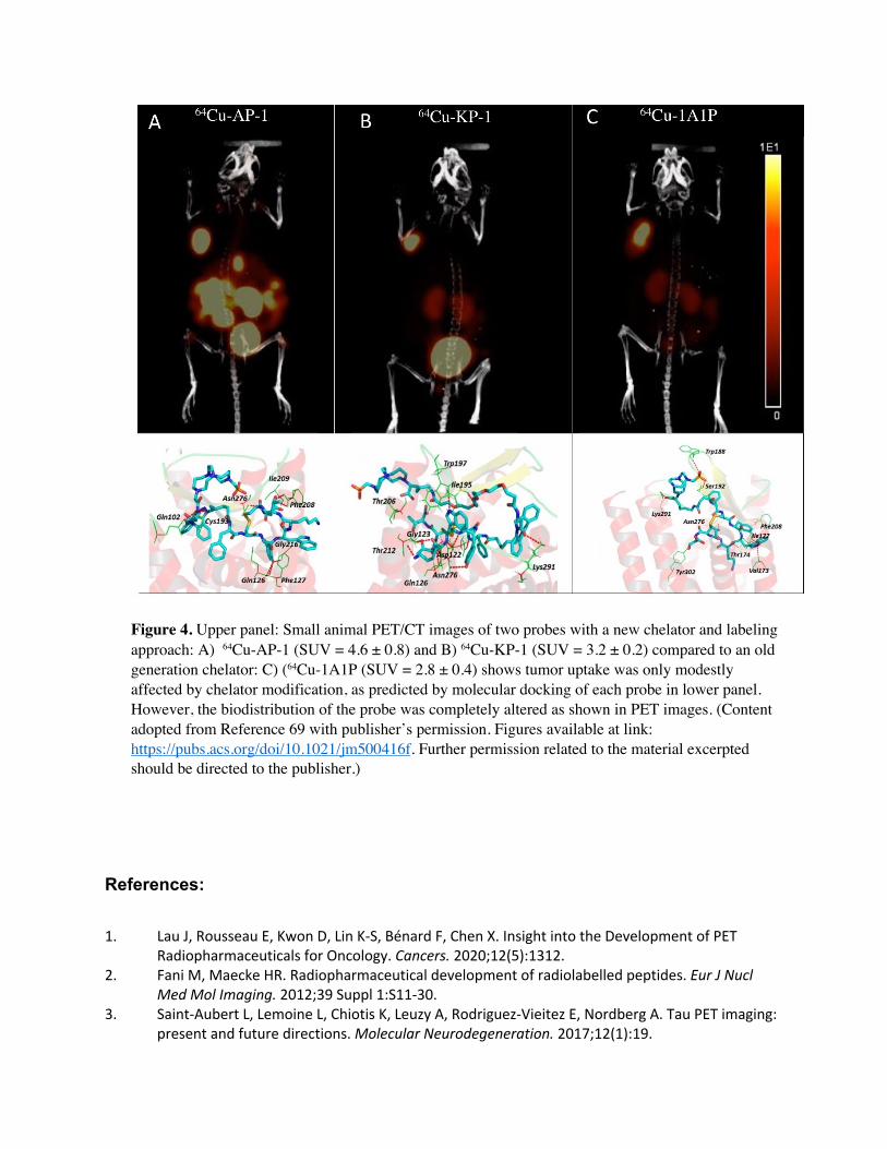
Figure 4. Upper panel: Small animal PET/CT images of two probes with a new chelator and labeling approach: A) 64Cu-AP-1 (SUV = 4.6 ± 0.8) and B) 64Cu-KP-1 (SUV = 3.2 ± 0.2) compared to an old generation chelator: C) (64Cu-1A1P (SUV = 2.8 ± 0.4) shows tumor uptake was only modestly affected by chelator modification, as predicted by molecular docking of each probe in lower panel. However, the biodistribution of the probe was completely altered as shown in PET images. (Content adopted from Reference 69 with publisher’s permission. Figures available at link: https://pubs.acs.org/doi/10.1021/jm500416f. Further permission related to the material excerpted should be directed to the publisher.)
References:
1. Lau J, Rousseau E, Kwon D, Lin K-S, Bénard F, Chen X. Insight into the Development of PET Radiopharmaceuticals for Oncology. Cancers. 2020;12(5):1312.
2. Fani M, Maecke HR. Radiopharmaceutical development of radiolabelled peptides. Eur J Nucl Med Mol Imaging. 2012;39 Suppl 1:S11-30.
3. Saint-Aubert L, Lemoine L, Chiotis K, Leuzy A, Rodriguez-Vieitez E, Nordberg A. Tau PET imaging: present and future directions. Molecular Neurodegeneration. 2017;12(1):19.

4. Tornesello AL, Buonaguro L, Tornesello ML, Buonaguro FM. New Insights in the Design of Bioactive Peptides and Chelating Agents for Imaging and Therapy in Oncology. Molecules. 2017;22(8).
5. George GPC, Pisaneschi F, Nguyen Q-D, Aboagye EO. Positron Emission Tomographic Imaging of CXCR4 in Cancer: Challenges and Promises. Molecular Imaging. 2015;14(1):7290.2014.00041.
6. Nguyen QD, Aboagye EO. Imaging the life and death of tumors in living subjects: Preclinical PET imaging of proliferation and apoptosis. Integr Biol (Camb). 2010;2(10):483-495.
7. Vermeulen K, Vandamme M, Bormans G, Cleeren F. Design and Challenges of Radiopharmaceuticals. Semin Nucl Med. 2019;49(5):339-356.
8. Alonso H, Bliznyuk AA, Gready JE. Combining docking and molecular dynamic simulations in drug design. Med Res Rev. 2006;26(5):531-568.
9. Chandrasekaran B, Abed SN, Al-Attraqchi O, Kuche K, Tekade RK. Chapter 21 - Computer-Aided Prediction of Pharmacokinetic (ADMET) Properties. In: Tekade RK, ed. Dosage Form Design Parameters: Academic Press; 2018:731-755.
10. Jean-Quartier C, Jeanquartier F, Jurisica I, Holzinger A. In silico cancer research towards 3R. BMC Cancer. 2018;18(1):408.
11. Doke SK, Dhawale SC. Alternatives to animal testing: A review. Saudi Pharm J. 2015;23(3):223-229.
12. Kleynhans J, Kruger HG, Cloete T, Zeevaart JR, Ebenhan T. In Silico Modelling in the Development of Novel Radiolabelled Peptide Probes. Curr Med Chem. 2020;27(41):7048-7063.
13. Zhang L, Villalobos A. Strategies to facilitate the discovery of novel CNS PET ligands. EJNMMI Radiopharm Chem. 2017;1(1):13.
14. Zhang L, Villalobos A, Beck EM, et al. Design and Selection Parameters to Accelerate the Discovery of Novel Central Nervous System Positron Emission Tomography (PET) Ligands and Their Application in the Development of a Novel Phosphodiesterase 2A PET Ligand. Journal of Medicinal Chemistry. 2013;56(11):4568-4579.
15. Chang M. Artificial Intelligence for Drug Development, Precision Medicine, and Healthcare (1st ed.) CRC Press 2020.
16. Ilem Ozdemir D, Asikoglu M. Radio imaging and diagnostic applications. Ed. Senyigit T., Ozcan I., Ozer O. Nanotechnology in Progress: Pharmaceutical Applications. 2012.
17. Schmidt BJ, Papin JA, Musante CJ. Mechanistic systems modeling to guide drug discovery and development. Drug Discov Today. 2013;18(3-4):116-127.
18. Emine Selin Demir EO, Meliha Ekinci, Evren Atlihan Gundogdu, Derya İlem Özdemir and Makbule Asikoglu. Computational Study of Radiopharmaceuticals. In: Stefaniu A, ed. Molecular Docking and Molecular Dynamics: IntechOpen; 2019.
19. Honarparvar B, Govender T, Maguire GE, Soliman ME, Kruger HG. Integrated approach to structure-based enzymatic drug design: molecular modeling, spectroscopy, and experimental bioactivity. Chem Rev. 2014;114(1):493-537.
20. Brandt M, Cardinale J, Aulsebrook ML, Gasser G, Mindt TL. An Overview of PET Radiochemistry, Part 2: Radiometals. J Nucl Med. 2018;59(10):1500-1506.
21. Davis MI, Bennett MJ, Thomas LM, Bjorkman PJ. Crystal structure of prostate-specific membrane antigen, a tumor marker and peptidase. Proc Natl Acad Sci U S A. 2005;102(17):5981-5986.
22. Wu B, Chien EY, Mol CD, et al. Structures of the CXCR4 chemokine GPCR with small-molecule and cyclic peptide antagonists. Science. 2010;330(6007):1066-1071.
23. Makrynitsa GI, Lykouras M, Spyroulias GA, Matsoukas M-T. In silico Drug Design. eLS:1-7. 24. Yu W, MacKerell AD, Jr. Computer-Aided Drug Design Methods. Methods Mol Biol.
2017;1520:85-106.

25. Meng XY, Zhang HX, Mezei M, Cui M. Molecular docking: a powerful approach for structure-based drug discovery. Curr Comput Aided Drug Des. 2011;7(2):146-157.
26. Kitchen DB, Decornez H, Furr JR, Bajorath J. Docking and scoring in virtual screening for drug discovery: methods and applications. Nat Rev Drug Discov. 2004;3(11):935-949.
27. Grinter SZ, Zou X. Challenges, applications, and recent advances of protein-ligand docking in structure-based drug design. Molecules. 2014;19(7):10150-10176.
28. Ain QU, Aleksandrova A, Roessler FD, Ballester PJ. Machine-learning scoring functions to improve structure-based binding affinity prediction and virtual screening. Wiley interdisciplinary reviews. Computational molecular science. 2015;5(6):405-424.
29. Li H, Sze K-H, Lu G, Ballester PJ. Machine-learning scoring functions for structure-based drug lead optimization. WIREs Computational Molecular Science. 2020;10(5):e1465.
30. Ant Colony Optimization and Swarm Intelligence, 5th International Workshop, ANTS 2006, Brussels, Belgium, September 4-7, 2006, Proceedings. 2006.
31. Pereira JC, Caffarena ER, Dos Santos CN. Boosting Docking-Based Virtual Screening with Deep Learning. J Chem Inf Model. 2016;56(12):2495-2506.
32. Fu Y, Wu X, Chen Z, Sun J, Zhao J, Xu W. A New Approach for Flexible Molecular Docking Based on Swarm Intelligence. Mathematical Problems in Engineering. 2015;2015:540186.
33. Batool M, Ahmad B, Choi S. A Structure-Based Drug Discovery Paradigm. Int J Mol Sci. 2019;20(11).
34. Somarowthu S, Ondrechen MJ. POOL server: machine learning application for functional site prediction in proteins. Bioinformatics. 2012;28(15):2078-2079.
35. Huang B. MetaPocket: a meta approach to improve protein ligand binding site prediction. Omics. 2009;13(4):325-330.
36. Muhammed MT, Aki-Yalcin E. Homology modeling in drug discovery: Overview, current applications, and future perspectives. Chem Biol Drug Des. 2019;93(1):12-20.
37. Gniazdowska E, Koźmiński P, Halik P, et al. Synthesis, physicochemical and biological evaluation of tacrine derivative labeled with technetium-99m and gallium-68 as a prospective diagnostic tool for early diagnosis of Alzheimer's disease. Bioorg Chem. 2019;91:103136.
38. Motaleb MA, El-Safoury DM, Abd-Alla WH, Awad GAS, Sakr TM. Radiosynthesis, molecular modeling studies and biological evaluation of (99m)Tc-Ifosfamide complex as a novel probe for solid tumor imaging. Int J Radiat Biol. 2018;94(12):1134-1141.
39. Sanad MH, Ibrahim AA. Preparation and biological evaluation of 99mTc N-histamine as a model for brain imaging: in silico study and preclinical evaluation. Radiochimica Acta. 2018;106(3):229-238.
40. Khedr M, Rashed HM, Farag H, Sakr T. Rational design of some substituted phenyl azanediyl (bis) methylene phosphonic acid derivatives as potential anticancer agents and imaging probes: Computational inputs, chemical synthesis, radiolabeling, biodistribution and gamma scintigraphy. Bioorganic chemistry. 2019;92:103282.
41. Khanapur S, Paul S, Shah A, et al. Development of [18F]-labeled pyrazolo[4,3-e]-1,2,4- triazolo[1,5-c]pyrimidine (SCH442416) analogs for the imaging of cerebral adenosine A2A receptors with positron emission tomography. J Med Chem. 2014;57(15):6765-6780.
42. Scott DE, Coyne AG, Hudson SA, Abell C. Fragment-based approaches in drug discovery and chemical biology. Biochemistry. 2012;51(25):4990-5003.
43. Kumar A, Voet A, Zhang KY. Fragment based drug design: from experimental to computational approaches. Curr Med Chem. 2012;19(30):5128-5147.
44. Yang S-Y. Pharmacophore modeling and applications in drug discovery: challenges and recent advances. Drug Discovery Today. 2010;15(11):444-450.

45. Katsila T, Spyroulias GA, Patrinos GP, Matsoukas M-T. Computational approaches in target identification and drug discovery. Computational and structural biotechnology journal. 2016;14:177-184.
46. Vlachakis D, Fakourelis P, Megalooikonomou V, Makris C, Kossida S. DrugOn: a fully integrated pharmacophore modeling and structure optimization toolkit. PeerJ. 2015;3:e725.
47. Stumpfe D, Bajorath J. Similarity searching. WIREs Computational Molecular Science. 2011;1(2):260-282.
48. Boudreau RJ, Efange SM. Computer-aided radiopharmaceutical design. Invest Radiol. 1992;27(8):653-658.
49. Li H, Sutter J, Hoffmann R. HypoGen: an automated system for generating 3D predictive pharmacophore models. Pharmacophore perception, development, and use in drug design. 2000;2:171.
50. Martin YC. and What We Missed. Pharmacophore perception, development, and use in drug design. Vol 22000:49.
51. Dixon SL, Smondyrev AM, Knoll EH, Rao SN, Shaw DE, Friesner RA. PHASE: a new engine for pharmacophore perception, 3D QSAR model development, and 3D database screening: 1. Methodology and preliminary results. Journal of computer-aided molecular design. 2006;20(10):647-671.
52. Güner O, Clement O, Kurogi Y. Pharmacophore modeling and three dimensional database searching for drug design using catalyst: recent advances. Curr Med Chem. 2004;11(22):2991-3005.
53. Wolber G, Seidel T, Bendix F, Langer T. Molecule-pharmacophore superpositioning and pattern matching in computational drug design. Drug Discov Today. 2008;13(1-2):23-29.
54. Macalino SJ, Gosu V, Hong S, Choi S. Role of computer-aided drug design in modern drug discovery. Arch Pharm Res. 2015;38(9):1686-1701.
55. Zhang S. Computer-aided drug discovery and development. Methods Mol Biol. 2011;716:23-38. 56. Lešnik S, Štular T, Brus B, et al. LiSiCA: A Software for Ligand-Based Virtual Screening and Its
Application for the Discovery of Butyrylcholinesterase Inhibitors. J Chem Inf Model. 2015;55(8):1521-1528.
57. Ghasemi F, Mehridehnavi A, Pérez-Garrido A, Pérez-Sánchez H. Neural network and deep-learning algorithms used in QSAR studies: merits and drawbacks. Drug Discov Today. 2018;23(10):1784-1790.
58. Martin YC, Kofron JL, Traphagen LM. Do structurally similar molecules have similar biological activity? J Med Chem. 2002;45(19):4350-4358.
59. Rashed HM, Ibrahim I, Motaleb M. 99mTc-hexoprenaline and 131I-dapoxetine: preparation, in silico modeling and biological evaluation as promising lung scintigraphy radiopharmaceuticals. Journal of Radioanalytical and Nuclear Chemistry. 2017;314:1297-1307.
60. Rashed HM, Ibrahim IT, Motaleb MA. 99m Tc-hexoprenaline and 131 I-dapoxetine: preparation, in silico modeling and biological evaluation as promising lung scintigraphy radiopharmaceuticals. Journal of Radioanalytical and Nuclear Chemistry. 2017;314(2):1297-1307.
61. van de Waterbeemd H, Gifford E. ADMET in silico modelling: towards prediction paradise? Nat Rev Drug Discov. 2003;2(3):192-204.
62. Moroy G, Martiny VY, Vayer P, Villoutreix BO, Miteva MA. Toward in silico structure-based ADMET prediction in drug discovery. Drug Discov Today. 2012;17(1-2):44-55.
63. Norinder U, Bergström CA. Prediction of ADMET Properties. ChemMedChem. 2006;1(9):920-937. 64. Price EW, Orvig C. Matching chelators to radiometals for radiopharmaceuticals. Chem Soc Rev.
2014;43(1):260-290.
65. Clark DE. In silico prediction of blood–brain barrier permeation. Drug discovery today. 2003;8(20):927-933.
66. Sun X, Li Y, Liu T, Li Z, Zhang X, Chen X. Peptide-based imaging agents for cancer detection. Advanced drug delivery reviews. 2017;110-111:38-51.
67. Evans BJ, King AT, Katsifis A, Matesic L, Jamie JF. Methods to Enhance the Metabolic Stability of Peptide-Based PET Radiopharmaceuticals. Molecules. 2020;25(10).
68. Bayoumi NA, Amin AM, Ismail NSM, Abouzid KAM, El-Kolaly MT. Radioiodination and biological evaluation of Cladribine as potential agent for tumor imaging and therapy. Radiochimica Acta. 2015;103(11):777-787.
69. Cai Z, Ouyang Q, Zeng D, et al. 64Cu-labeled somatostatin analogues conjugated with cross-bridged phosphonate-based chelators via strain-promoted click chemistry for PET imaging: in silico through in vivo studies. J Med Chem. 2014;57(14):6019-6029.
70. Fang Y, Wang D, Xu X, et al. Synthesis, biological evaluation, and molecular dynamics (MD) simulation studies of three novel F-18 labeled and focal adhesion kinase (FAK) targeted 5-bromo pyrimidines as radiotracers for tumor. Eur J Med Chem. 2017;127:493-508.
71. Wang D, Fang Y, Wang H, Xu X, Liu J, Zhang H. Synthesis and evaluation of novel F-18-labeled pyrimidine derivatives: potential FAK inhibitors and PET imaging agents for cancer detection. RSC Advances. 2017;7(36):22388-22399.
72. Fang Y, Wang D, Xu X, et al. Preparation, in vitro and in vivo evaluation, and molecular dynamics (MD) simulation studies of novel F-18 labeled tumor imaging agents targeting focal adhesion kinase (FAK). RSC Advances. 2018;8(19):10333-10345.
73. Khurana H, Meena VK, Prakash S, et al. Preclinical Evaluation of a Potential GSH Ester Based PET/SPECT Imaging Probe DT(GSHMe)₂ to Detect Gamma Glutamyl Transferase Over Expressing Tumors. PloS one. 2015;10(7):e0134281-e0134281.
74. Sakr TM, Khedr MA, Rashed HM, Mohamed ME. In Silico-Based Repositioning of Phosphinothricin as a Novel Technetium-99m Imaging Probe with Potential Anti-Cancer Activity. Molecules. 2018;23(2):496.
75. Zhang L, Villalobos A, Beck EM, et al. Design and selection parameters to accelerate the discovery of novel central nervous system positron emission tomography (PET) ligands and their application in the development of a novel phosphodiesterase 2A PET ligand. J Med Chem. 2013;56(11):4568-4579.
76. Wellsow J, Kovar KA, Machulla HJ. Molecular modeling of potential new and selective PET radiotracers for the serotonin transporter. Positron Emission Tomography. J Pharm Pharm Sci. 2002;5(3):245-257.
77. Zhang L, Drummond E, Brodney MA, et al. Design, synthesis and evaluation of [(3)H]PF-7191, a highly specific nociceptin opioid peptide (NOP) receptor radiotracer for in vivo receptor occupancy (RO) studies. Bioorg Med Chem Lett. 2014;24(22):5219-5223.
78. Zhang L, Chen L, Beck EM, et al. The Discovery of a Novel Phosphodiesterase (PDE) 4B-Preferring Radioligand for Positron Emission Tomography (PET) Imaging. J Med Chem. 2017;60(20):8538-8551.
79. Vuorimaa A, Rissanen E, Airas L. In Vivo PET Imaging of Adenosine 2A Receptors in Neuroinflammatory and Neurodegenerative Disease. Contrast Media Mol Imaging. 2017;2017:6975841.
80. Khanapur S, van Waarde A, Dierckx RA, Elsinga PH, Koole MJ. Preclinical Evaluation and Quantification of (18)F-Fluoroethyl and (18)F-Fluoropropyl Analogs of SCH442416 as Radioligands for PET Imaging of the Adenosine A(2A) Receptor in Rat Brain. J Nucl Med. 2017;58(3):466-472.

81. Lai TH, Toussaint M, Teodoro R, et al. Synthesis and Biological Evaluation of a Novel (18)F-Labeled Radiotracer for PET Imaging of the Adenosine A(2A) Receptor. Int J Mol Sci. 2021;22(3).
82. Okamura N, Harada R, Ishiki A, Kikuchi A, Nakamura T, Kudo Y. The development and validation of tau PET tracers: current status and future directions. Clin Transl Imaging. 2018;6(4):305-316.
83. Balamurugan K, Murugan NA, Ågren H. Multistep Modeling Strategy To Improve the Binding Affinity Prediction of PET Tracers to Aβ42: Case Study with Styrylbenzoxazole Derivatives. ACS Chemical Neuroscience. 2016;7(12):1698-1705.
84. Uzuegbunam BC, Librizzi D, Hooshyar Yousefi B. PET Radiopharmaceuticals for Alzheimer's Disease and Parkinson's Disease Diagnosis, the Current and Future Landscape. Molecules (Basel, Switzerland). 2020;25(4):977.
85. Leuzy A, Chiotis K, Lemoine L, et al. Tau PET imaging in neurodegenerative tauopathies-still a challenge. Mol Psychiatry. 2019;24(8):1112-1134.
86. Lemoine L, Gillberg P-G, Svedberg M, et al. Comparative binding properties of the tau PET tracers THK5117, THK5351, PBB3, and T807 in postmortem Alzheimer brains. Alzheimer's research & therapy. 2017;9(1):96-96.
87. Murugan NA, Nordberg A, Ågren H. Different Positron Emission Tomography Tau Tracers Bind to Multiple Binding Sites on the Tau Fibril: Insight from Computational Modeling. ACS Chem Neurosci. 2018;9(7):1757-1767.
88. Murugan NA, Chiotis K, Rodriguez-Vieitez E, Lemoine L, Ågren H, Nordberg A. Cross-interaction of tau PET tracers with monoamine oxidase B: evidence from in silico modelling and in vivo imaging. European journal of nuclear medicine and molecular imaging. 2019;46(6):1369-1382.
89. Ng KP, Pascoal TA, Mathotaarachchi S, et al. Monoamine oxidase B inhibitor, selegiline, reduces (18)F-THK5351 uptake in the human brain. Alzheimers Res Ther. 2017;9(1):25.
90. Lemoine L, Gillberg PG, Svedberg M, et al. Comparative binding properties of the tau PET tracers THK5117, THK5351, PBB3, and T807 in postmortem Alzheimer brains. Alzheimers Res Ther. 2017;9(1):96.
91. Shaw RC, Tamagnan GD, Tavares AAS. Rapidly (and Successfully) Translating Novel Brain Radiotracers From Animal Research Into Clinical Use. Frontiers in Neuroscience. 2020;14(871).
92. Mota F, Ordonez AA, Firth G, Ruiz-Bedoya CA, Ma MT, Jain SK. Radiotracer Development for Bacterial Imaging. Journal of Medicinal Chemistry. 2020;63(5):1964-1977.
93. Signore A, Artiko V, Conserva M, et al. Imaging Bacteria with Radiolabelled Probes: Is It Feasible? Journal of Clinical Medicine. 2020;9(8):2372.
94. Ordonez AA, Weinstein EA, Bambarger LE, et al. A Systematic Approach for Developing Bacteria-Specific Imaging Tracers. J Nucl Med. 2017;58(1):144-150.
95. Aalto K, Autio A, Kiss EA, et al. Siglec-9 is a novel leukocyte ligand for vascular adhesion protein-1 and can be used in PET imaging of inflammation and cancer. Blood. 2011;118(13):3725-3733.
96. Rashed HM, Marzook FA, Farag H. 99m Tc-zolmitriptan: radiolabeling, molecular modeling, biodistribution and gamma scintigraphy as a hopeful radiopharmaceutical for lung nuclear imaging. La radiologia medica. 2016;121(12):935-943.