asc mediates caspase-1 independent activation of map ... · t. bergstralh1,4, irving c. allen1, yu...
TRANSCRIPT

THE NLR ADAPTOR ASC/PYCARD REGULATES DUSP10, MAP kinase (MAPK) AND
CHEMOKINE INDUCTION INDEPENDENT OF THE INFLAMMASOME*
Debra J. Taxman1, Elizabeth A. Holley-Guthrie
1, Max Tze-Han Huang
1, Chris B. Moore
1,3, Daniel
T. Bergstralh1,4
, Irving C. Allen1, Yu Lei
2, Denis Gris
1 and Jenny Pan-Yun Ting
1
From 1Department of Microbiology and Immunology and 2NC Oral Health Institute; Lineberger
Comprehensive Cancer Center; University of North Carolina, Chapel Hill, NC 27599 3Current Address: GlaxoSmithKline, Virology, Research Triangle Park, North Carolina, 27709
4Current Address: The Wellcome Trust / Cancer Research UK Gurdon Institute, Tennis Court Road,
Cambridge, UK, CB2 1QN
Running head: ASC mediates MAPK induction by P. gingivalis
Address correspondence to: Jenny Pan-Yun Ting, Ph.D, Fax: 919-966-8212; Tel: 919-966-5538; E-mail:
ASC/PYCARD is a common adaptor for a
diverse set of inflammasomes which activate
caspase-1, most prominently the NLR-based
inflammasome. Mounting evidence indicates
that ASC and these NLRs also elicit non-
overlapping functions, but the molecular
basis for this difference is unclear. To address
this, we performed microarray and network
analysis of ASC shRNA knockdown cells. In
pathogen-infected cells, an ASC-dependent
interactome is centered on the MAP kinase
(MAPK), ERK, and on multiple chemokines.
ASC did not affect expression of MAPK, but
affected its phosphorylation by pathogens
and TLR agonists via suppression of the dual-
specificity phosphatase, DUSP10/MKP5.
Chemokine induction, DUSP function and
MAPK phosphorylation were independent of
caspase-1 and IL-1. MAPK activation by
pathogen was abrogated in Asc-/-
but not
Nlrp3-/-
, Nlrc4-/-
, or Casp1-/-
macrophages.
These results demonstrate a function for ASC
that is distinct from the inflammasome in
modulating MAPK activity and chemokine
expression, and further identifies DUSP10 as
a novel ASC target.
Secretion of proinflammatory cytokines
and chemokines by macrophages in response to
pathogens is an important innate immune event
orchestrated by a complex signaling network.
Pathogenic signaling leads to the formation of an
intracellular protein complex termed the
inflammasome. A conventional inflammasome
is composed of caspase-1, which promotes
cleavage and maturation of the inflammatory
cytokines IL-1 and IL-18, ASC (apoptotic
speck protein, also known as TMS1 or pycard),
and a nucleotide-binding domain leucine-rich
repeat/NBD-LRR protein (NLR) (1). ASC
serves as a bridge between caspase-1 via CARD-
CARD interactions, and NLRs via pyrin/pyrin
interactions (2,3). Mutations in several NLRs are
associated with inflammatory diseases,
underscoring their importance in innate
immunity (4,5). More recently, AIM2 and RIG-I
have also been identified in separate
inflammasome complexes that rely on the ASC
adaptor (6-10).
Association of specific NLRs within the
inflammasome may be dictated by the type and
dose of infectious microorganism, whereas ASC
is thought to assume a broader role as an NLR
adaptor. For example, ASC and Nlrc4 each have
defined roles for caspase-1 activation and cell
death by Salmonella typhimurium (11,12), while
ASC and caspase-1 are required for T cell
activation and protective immunity against flu
challenge (13), however each of these processes
are independent of Nlrp3. Moreover, both
Francisella tularensis-mediated IL-1
processing and sensitivity to lethal doses of
lipopolysaccharide (LPS) are ASC-dependent,
but Nlrc4 and Nlrp3 independent (11,12). The
pyrin and HIN domain-containing protein,
AIM2 recognizes cytosolic DNA within an
NLR-free ASC inflammasome, further
supporting a mechanism for ASC independent of
NLRs (6-9). Thus, ASC may play a role as a
common downstream factor for different sets of
NLRs and may also function within an
inflammasome complex exclusive of NLR
family proteins.
http://www.jbc.org/cgi/doi/10.1074/jbc.M111.221077The latest version is at JBC Papers in Press. Published on April 12, 2011 as Manuscript M111.221077
Copyright 2011 by The American Society for Biochemistry and Molecular Biology, Inc.
by guest on February 1, 2019http://w
ww
.jbc.org/D
ownloaded from

2
Additional evidence suggests that ASC
function extends beyond the NLR/caspase-1
inflammasome. ASC was identified by its
unique ability to condense into cytosolic speck
structures and induce apoptosis in tumor cell
lines (14). It also is silenced in certain cancers
(15). Moreover, a caspase-independent type of
necrosis induced by high dose Shigella (≥50
MOI) (16), Neisseria gonorrhoeae (17) or
Porphyromonas gingivalis (Pg) (18) is ASC-
dependent. Asc-/- mice exhibit increased
susceptibility to Mycobacterium tuberculosis
without reduction in IL-1, implying additional
ASC function that is distinct from cytokine
cleavage (19). Two recent studies of antigen-
induced murine arthritis show dependence on
Asc, but caspase-1, NLRP3, and NLRC4
independence (20,21). A requirement for Asc,
but not Nlrp3 or Caspase-1, was also recently
demonstrated for antigen-specific humoral
immunity after vaccination with MF59-
adjuvented influenza (22). ASC has been
proposed to regulate cytokine transcription
through activation of NF-B (23,24). AP1,
STAT3, ISGF3 and NF-AT have also been
identified as transcriptional ASC targets in a
reconstituted cell system with exogenously
expressed ASC and a chimeric CARD12/NOD2
protein (25). Given the complexity of
inflammatory signaling it is likely that additional
signaling pathways contribute to inflammasome-
independent ASC function in macrophages.
One crucial feature of pathogenic
signaling is the activation of the mitogen-
activated protein kinase kinase kinases
(MAP3Ks), a family of signaling proteins that
regulate a variety of physiological processes
including proliferation, cell death, stress
response, and differentiation. The MAP3Ks act
as nodes in the toll-like receptor (TLR) signaling
cascade for both the NF-B and MAPK
pathways (26,27). Transcription factors
downstream of these signaling pathways then
collaborate to regulate the expression of immune
and inflammatory mediators. By contrast, the
role of components of the inflammasome
complex in MAP3K signaling has not been
directly explored. MAPK is at the center of
many innate immune responses, thus the link
between ASC and MAPK is an important topic
to explore.
Porphyromonas gingivalis (Pg) is a
gram negative oral pathogen associated with
chronic adult periodontal disease. Pg surface
components including LPS, fimbriae, and
hemagglutinin B induce host inflammatory
responses that result in breakdown of
periodontal ligaments and destruction of the
local alveolar bone (28-30). Although
periodontal disease is localized to the tissues
surrounding the tooth, Pg infection predisposes
people to more serious systemic conditions such
as cardiovascular disease and delivery of
preterm infants (29,31,32). Recently, we showed
that during Pg infection, ASC exhibits
inflammasome-independent functions, including
TNF- and NF-B activation (23). We therefore
elected to use this pathogen to reveal new ASC
functions that might be separable from that of
the inflammasome. Results described herewithin
provide a novel microbial pathogen-induced
mechanism for ASC in activating chemokine
expression through MAPK activation
independent of the conventional caspase-1
inflammasome.
Experimental Procedures
Generation of cell lines, isolation of mouse
macrophages and cell culture- THP1 monocytic
cells (ATCC) were cultured in RPMI, 10% FCS.
ShASC#1, shASC#2, and shASC#1mut THP1
cell lines were generated using retroviral vectors
and have been characterized previously (18,23).
DUSP10-expressing THP1 cells were generated
using the full-length ORF cloned into lentiviral
vector pLex-JRed (catalog #OHS4493-
98905681; Open Biosystems). A control empty
vector, pLex-EV, was generated by digesting
pLex-DUSP10 with Xho I to remove the
DUSP10 cDNA and religating the empty vector.
DUSP10 shRNA plasmid pLKO-shDUSP10 was
obtained from the TRC collection (Open
Biosystems). Lentivirus was packaged in 293T
cells using vectors pMD2.G and psPAX2
(Addgene plasmids 12259 and 12260) as
described (33). Cells were selected with 1 g/ml
puromycin for 2 weeks, and JRed expression
confirmed by FACS. Asc-/- and Nlrc4-/- mice
were obtained from Dr. Vishva Dixit at
Genetech; Nlrp3-/- mice from Millenium Inc.;
Casp1-/- mice from Dr. Richard Flavell, Yale
by guest on February 1, 2019http://w
ww
.jbc.org/D
ownloaded from

3
University (12); and MyD88-/- mice from Dr.
Shizuo Akira. All mice were backcrossed for a
minimum of nine generations to C57BL/6 mice.
Bone marrow derived macrophages were
harvested and cultured in DMEM 10% fetal calf
serum, M-CSF without replating for 6-7 days.
Cells were maintained at low density and serum-
starved 16 h prior to infection. Where indicated,
cells were stimulated with 10 MOI Pg, 0.5 MOI
E. coli, or pharmacological agents detailed in
Supplementary Table 1.
Bacterial culture- Pg strain A7436 was cultured
anaerobically, and E. coli strain DH5
aerobically until late exponential phase (OD 0.8
to 1.2 at 660 nm). Aliquots were stored in media
containing 20% glycerol at -80C and used
within 3-4 months of preparation. Replating of
frozen cultures confirmed accuracy of bacterial
counts to within 2-3 CFU.
Microarray analysis- RNA was isolated using
Qiagen RNeasy columns following 2 h infection
with Pg. Two-color microarray analysis was
performed at the Duke Microarray facility using
34,000 spot custom chips based on the version 3
Human oligo set (Operon). All samples were
compared to a universal control created by
pooling RNA over a timecourse of LPS
treatment. Gene lists were generated from the
averages from 3 independent experiments using
GeneSpring 7.0. To identify Pg-regulated genes
the following criteria were used: minimum raw
signal of 50 in the universal control and 100 in
at least one condition; and ≥5-fold difference
between expression in control vs Pg-infected
cells. The criteria for ASC-dependent genes was
as follows: minimum raw signal of 60 in the
universal control and 100 in at least one
condition; ≥3-fold difference between
expression in Pg-infected THP1 vs shASC#1
cells and shASC#1mut vs shASC#1 cells; and
≤50% difference between Pg-infected THP1 and
shASC#1mut cells. Genes were further analyzed
in GeneSpring 11 for statistical validation using
modernized normalization methods, as presented
in Supplementary Tables 3 and 4 (Lanes 5-7).
Genes which passed the filtering schemes were
uploaded into the Ingenuity Pathways analysis
application, which overlaid them onto a global
molecular network developed from information
within the Ingenuity Pathways Knowledge Base.
Each network was algorithmically generated
based on the connectivity of genes. P values,
which indicate the likelihood that the same
number of genes taken from a random set would
appear in the network, were calculated using
Fischer's exact test.
Real time PCR and PCR-based expression
profiling- Realtime PCR was performed as
described (23) using the primers listed in
Supplementary Table 2. Mouse RNA was
quantified using Taqman® Assays on Demand
(Applied Biosystems). Values represent
averages ±SD of triplicates for RNA isolated on
different days unless otherwise stated. All values
were standardized to 18s rRNA expression.
PCR-based expression profiling was performed
according to manufacturer recommendation
using equal amounts of RNA from six mice with
the Mouse Inflammatory Cytokines and
Receptors array (SABiosciences). Values were
standardized to an average of 5 housekeeping
genes.
Assessment of RNA stability and de novo mRNA
synthesis- To assess RNA stability, cells were
treated for 2 h with LPS, washed to remove the
LPS, and treated with 50 M 5,6-dichloro-1-β-
-ribofuranosylbenzimidazole (DRB; Sigma) for
30 min. RNA was quantified by realtime PCR
over a timecourse as described above. To assess
de novo transcription, RNA was isolated from
nuclear extracts using the PARIS kit (Ambion)
and assessed by realtime PCR using primers that
target nascent transcripts. Efficient separation of
nuclei was confirmed by the absence of GAPDH
on Western blots.
Antibody array- Cell culture supernatants were
collected 24 h following exposure to Pg and
applied to RayBio® Human Cytokine Antibody
Array 5 glass slides according to manufacturer's
protocols. The signal strength for each cytokine
was normalized to the average signal strength of
spiked, internal controls. Data for each treatment
group were assessed as change compared to
uninfected samples.
ELISA Analysis- Supernatants were assessed 18-
24 h following stimulation using human ELISA
Sets for TNF and IL-1 (BD Biosciences),
DuoSets for CCL3 and CCL20(R&D Systems)
or the human quantikine ELISA kit for IGF-1
(R&D Systems). Samples were assayed within
linear range.
by guest on February 1, 2019http://w
ww
.jbc.org/D
ownloaded from

4
Western and IP/IB analyses- Cells were washed
in 1X PBS and lysed for 20 min in ice-cold 1X
Lysis Buffer (20 mM Tris, pH 7.5, 150 mM
NaCl, 1 mM EDTA, 1 mM EGTA, 1% triton X)
supplemented with Complete EDTA-free
Protease Inhibitor and PhosSTOP Phosphatase
Inhibitor cocktail (Roche). Lysates were
centrifuged for 10 min and supernatants boiled
for 5 min in 1/3 volume 3X SDS Sample Buffer
(187.5 mM Tris, pH 6.8, 6% SDS, 30% glycerol,
150 mM DTT, 0.03% bromphenol blue).
Immunoblots were processed using Abs sc-7383
for p-ERK, equal volumes of sc-93 (ERK1) and
sc-154 (ERK2) for total ERK, sc-474 for JNK,
sc-1615 for actin (Santa Cruz Biotechnology),
#4668 for pJNK (Cell Signaling Technology),
and MAB374 for GAPDH (Millipore). For
caspase-1 IP/western analyses, cells were plated
at 107/ml and lysed following infection by
addition of 0.1% NP-40 and 1X Complete
EDTA-free Protease Inhibitors (Roche). 1 ml of
supernatant was recovered and incubated
overnight with 25 l anti-caspase-1 (sc-515,
Santa Cruz Biotechnology) and 30 l protein
A/G UltraLink resin (Thermo Scientific).
Immunoprecipitates were washed and then
boiled in 3X SDS Sample Buffer. Immunoblots
were performed using antibody IMG-5028
(Imgenex).
RESULTS
Identification of Pg interactomes by
microarray and bioinformatics. We selected Pg
for this study because a previous study
suggested that it activates ASC-mediated
functions that are distinct from IL-1 processing
and inflammasome activation (23). To identify
biological pathways activated by Pg, RNA from
untreated THP1 cells and cells treated with 10
MOI Pg for 2 h was assessed using human
genome microarray chips comprising over
35,000 oligonucleotide probes and representing
approximately 25,000 unique genes (Operon
Biotechnologies, Inc). Using GeneSpring 7.0
microarray analysis software, a list was
compiled of approximately 150 genes that Pg
modulates by ≥5-fold (Supplementary Table 3).
Additional statistical analysis using GeneSpring
11.0 verified the inclusion of the majority of the
genes in this table (Columns 5-7). Unbiased
software analysis was performed to identify
interactomes of genes that encode physically or
functionally interacting proteins (Ingenuity
Systems). Interactomes are networks of genes
that are interconnected based on information
derived from the literature, a textbook, or
canonical knowledge. Three primary networks
of Pg-modulated genes were identified. The first
interactome encompasses the inflammatory
cytokine TNF-. TNFA -
160 fold by Pg infection (Fig. 1A). Other genes
in the TNF- interactome were modulated from
5-319 fold by Pg. The extremely low p value of
this interactome (P < 10-46) suggests very high
likelihood of functionality. Consistently, TNF-
pathways are known to contribute to Pg-
associated pathogenesis during periodontitis
(34,35). The second interactome (Fig. 1B; P <
10-46) encompasses IL1Bwhich itself is
activated more than 200-fold. Both interactomes
show high induction for multiple chemokines.
The third interactome (P < 10-41) is unique in
that its central molecule, NF-B, does not appear
to be significantly regulated at the
transcriptional level but was identified based on
the genes that it is known to modulate (Fig. 1C).
This is consistent with the role of the NF-B
members as early signaling transcriptional
regulators (36). Induction of the transcript for
the CARD-containing serine/threonine kinase
RIPK2 by 15-17-fold (Fig. 1A) could potentially
contribute to the enhanced NF-B pathway
activity by Pg (37,38). The identification of each
of these Pg interactomes is consistent with
known biological effects of Pg in the activation
of cellular signaling pathways toward
inflammation.
Identification of an ASC-dependent Pg
interactome. ASC is an adaptor molecule
important in the induction of apoptosis and
inflammatory response (2,11,15). Our previous
studies demonstrate that in addition to regulating
IL-1 processing through caspase-1 activation,
ASC regulates the transcription of a panel of
cytokines including IL-6, IL-8, IL-10 and TNF-
(23). Because induction of these cytokines is
thought to be NLR and caspase-1 independent,
their regulation by ASC implies ASC-mediated
functions that are distinct from the
inflammasome. For example, several reports
by guest on February 1, 2019http://w
ww
.jbc.org/D
ownloaded from

5
have shown that TNF expression is not altered
by the deletion of Nlrp3 (12,39). Thus we
reasoned that Pg represents a good system to
reveal differences between ASC-associated
inflammasome-dependent and inflammasome-
independent signaling.
To identify additional genes and
pathways regulated by ASC, two different ASC
knockdowns were utilized with reduced ASC
expression. The lentiviral shRNA construct
shASC#1 caused approximately 90%
knockdown and shASC#2 approximately 70%
knockdown when compared to controls,
including non-transfected THP1 cells, and cells
bearing an empty vector (EV) or a mutated
target site (shASC#1mut) (23) (Fig. 1D). The
shRNAs reduced IL-1 cytokine induction,
verifying that the knockdowns reduce ASC
function as previously observed (Fig. 1E).
Microarray analysis was performed to identify
genes that were differentially regulated by ASC.
Approximately 80 genes were modulated by ≥3-
fold in shASC#1 cells as compared to control
cells (Supplementary Table 4). This set of ASC-
dependent genes was evaluated by Ingenuity
Pathways Analysis and an ASC-dependent
interactome was identified (Fig. 1F, P < 10-42).
As confirmation of the technology, ASC was
reduced 8.8 to 12.6-fold in shASC cells. Several
chemokines, including CCL3, CCL3L1, and
CCL4 were reduced in the absence of ASC,
while CXCL10 was increased approximately 4-
fold. NF-B was also identified as an ASC-
dependent regulator of these genes, consistent
with our previous findings linking ASC-
dependent gene expression to NF-B activation
(23). Since the expression and activity of NF-B
and several chemokines within this interactome
are also highly regulated by Pg (Fig. 1A-C), the
role of ASC may extend to many of the same
pathways that Pg induces. All of the genes
within the ASC-dependent interactome could be
interconnected to the signaling molecule
MAPK1/ERK2, albeit expression of the latter,
like NF-B, was not affected by ASC. Because
MAPKs are primarily regulated post-
transcriptionally, the central position of MAPK1
within this interactome provides novel insight
into potential post-transcriptional mechanisms of
ASC function.
Assessment of ASC-dependent cytokine
and chemokine expression. To verify ASC-
dependent chemokine expression, RNA levels in
ASC knockdown THP1 cells were measured
following a timecourse of infection with Pg.
Consistent with the microarray results, TNFA
was induced by Pg in control cells, with a peak
at 2-4 h post infection, and this induction was
reduced in the ASC knockdown cells (Fig. 2A).
CCL3, CCL4, and CXCL3 each showed a similar
pattern of ASC-dependent RNA expression (Fig.
2B-D). These findings support the identification
of the ASC interactome and show a role for ASC
in Pg-dependent induction of chemokines.
Regulation of mRNA expression can
occur either at the level of transcription or RNA
stability. To distinguish these possibilities, RNA
stability was examined following LPS treatment
using DRB to block de novo transcription (Fig.
2E). The RNA decay profiles were similar in
shASC#1mut and shASC#1 cells, indicating that
regulation of RNA stability is not a major
determinant for ASC-dependent differences in
chemokine expression. Conversely, assessment
of de novo mRNA synthesis using nuclear
extracts and primers that target unspliced
message indicates that regulation is primarily
transcriptional (Fig. 2F).
To further verify a role for ASC in
chemokine induction by Pg we performed
protein array analysis using antibody chips
comprising 79 cytokines, chemokines and
growth factors (RayBiotech, Inc). Supernatants
were assayed from shASC#1mut and shASC#1
cells before and following 24 h infection with
Pg. Six proteins were differentially induced by
Pg in shASC#1mut vs shASC#1 cells (Fig. 3A).
This includes IL-1 which is a known target for
ASC regulation via cleavage by caspase-1
within the inflammasome complex (2). TNF-
was also identified, further confirming our
realtime results. The chemokines from Figs. 2B-
D either were not detected at high levels (for
CCL4), or were not included on this array (for
CCL3 and CXCL3). However, the chemokines,
GRO/GRO-, CCL13 and CCL20 were
identified, further supporting a broad role for
ASC in regulating chemokine expression. The
latter proteins are not thought to be regulated by
caspase-1, suggesting a potential role for ASC in
by guest on February 1, 2019http://w
ww
.jbc.org/D
ownloaded from

6
caspase-1-independent regulation of chemokine
expression.
To further verify these findings,
supernatants were collected from control and
ASC knockdown lines 24 h following infection
with Pg and assessed by ELISA. Levels of
secreted TNF-, CCL3, and CCL20 were
reduced in shASC#1 cells, with somewhat less
reduction in shASC#2 cells suggesting dose-
dependent regulation (Fig. 3B-D). IGF-1
expression was not ASC-dependent,
demonstrating specificity of this effect (Fig. 3E).
These findings further support ASC-dependent
chemokine expression suggested by the
interactome described in Fig. 1F.
MAPK activation is ASC dependent in
THP1 cells. MAPKs are activated by
phosphorylation by upstream kinases (26,27). To
provide support for the central position of
MAPK1/ERK2 within the ASC interactome
(Fig. 1F), we assessed phosphorylation levels of
ERK in ASC knockdown cells following Pg
infection. Consistent with the microarray results,
total expression of ERK1 and ERK2 was not
significantly different in control shASC#1mut vs
shASC#1 knockdown cells and was not
regulated by Pg infection. Levels of phospho-
ERK (p-ERK) induction peaked at
approximately 60-90 minutes for both cell lines,
but were quantitatively reduced in ASC
knockdown cells over a 120 min timecourse of
infection (Fig. 4A). These findings are
consistent with a role for ASC in ERK
activation.
To determine whether effects of ERK
activation by ASC have broad significance
during microbial infection, control and ASC
knockdown cells were treated for 60 min with E.
coli and a variety of agents known to activate
cells through different TLRs (Fig. 4B). For each
of these treatments, levels of p-ERK were
reduced in the absence of ASC. These findings
indicate that in addition to Pg, ASC mediates
ERK activation through E. coli and a variety of
TLR agonists.
ERK is one of three related MAPK
pathways activated in stimulated cells. To test
whether the JNK and p38 pathways also display
reduced activation in ASC knockdown cells,
phosphoblots were repeated. Levels of phospho-
p38 in THP1 cells were too low to measure
accurately (data not shown). However, similar to
p-ERK, levels of p-JNK activation by Pg were
delayed and reduced in ASC knockdown cells,
indicating that the role of ASC in MAPK
activation extends to other MAPK pathways
(Fig. 4C).
To determine whether reduction in ERK
and JNK activation might explain the reduced
chemokine levels in ASC knockdown THP1
cells, ELISA assays were repeated in the
presence of specific inhibitors of ERK and JNK
pathways (Fig. 4D). Levels of CCL3 and CCL20
following Pg infection were reduced by both
inhibitors, either alone or in combination, while
the carrier, DMSO, did not affect expression.
This effect was specific because levels of IGF-1
were unchanged (Fig. 4E). These results confirm
the importance of the ERK and JNK pathways in
chemokine induction by Pg.
MAPK activation and chemokine
induction by Pg in primary mouse macrophages
is ASC-dependent. To confirm our findings in a
primary cell system, macrophages were
harvested from wildtype C57BL/6 mice and
matched Asc-/- mice and infected with Pg over a
timecourse. Levels of p-ERK activation were
nearly eliminated in macrophages from Asc-/-
mice, while levels of p-JNK and p-p38 were
modestly reduced (Fig. 5A-C). These results
confirm the role for ASC in Pg-induced MAPK
phosphorylation in a primary cell system.
To determine whether reduced MAPK
activity in mouse macrophages correlates with
reduced chemokine activation, RNA was pooled
from six WT and Asc-/- mice following Pg
infection and assessed by PCR-based expression
profiling. Several chemokines were identified
(Fig. 5D). This list of candidate genes includes
Ccl3, which was decreased, and Cxcl10 which
was increased both in the RNA from pooled
Asc-deficient mouse macrophages and THP1
shASC#1 cells (Fig. 1F). Other chemokines
identified in the two model system differed,
mostly likely explained by differences between
species and the transformed or nontransformed
state of the cells. Nonetheless, the regulation of
these chemokines would be consistent with our
overall finding that ASC modulates chemokine
expression.
To verify Asc-dependent expression of
chemokines in mouse macrophages, Taqman®
by guest on February 1, 2019http://w
ww
.jbc.org/D
ownloaded from

7
PCR was performed before and following 2h Pg
infection and fold induction was calculated.
Consistent with the above results, Asc was
required for high level inducbility of Ccl3,
Ccl19 and Ccl25, while Cxcl10 was increased in
the absence of Asc (Fig. 5E). Ccl3 expression
was dependent upon ERK, JNK and p38 (Fig.
5F), verifying an essential role for MAPKs in
chemokine expression in primary mouse
macrophages.
The dual-specificity phosphatase
DUSP10/MKP5 negatively regulates MAPK
phosphorylation and chemokine activation by
Pg. The modulation of multiple MAPKs by ASC
suggests that ASC might lie upstream of a
common regulator of MAPKs. Examination of
ASC-dependent genes identified by microarray
analysis (Supplementary Table 4) revealed that
DUSP10/MKP5 is approximately 3.5-fold higher
in shASC-containing cells. The dual-specificity
phosphatases (DUSPs) are known to negatively
regulate the activity of multiple MAPKs through
dephosphorylation (40,41). To verify the effect
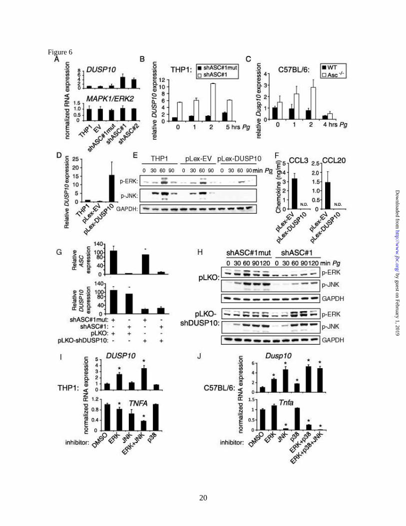
of ASC on DUSP10 expression, realtime PCR
analysis was performed. DUSP10 levels were
increased in both ASC knockdown lines relative
to the controls (Fig. 6A, top panel). As a control,
levels of transcript for MAPK1/ERK2 were
ASC-independent (Fig. 6A, bottom panel).
Increased DUSP10 in ASC knockdown cells
was observed over a timecourse of infection
with Pg in THP1 cells (Fig. 6B) and mouse
macrophages (Fig. 6C). Because DUSP10 is
known to correlate negatively with the activity
of MAPKs (42), these findings are consistent
with the reduced post-transcriptional activation
of MAPKs in ASC knockdown cells following
Pg stimulation and provide a potential
mechanism for diminished MAPK
phosphorylation.
To directly assess the role of DUSP10 in
MAPK phosphorylation, DUSP10 was
exogenously expressed in THP1 cells (Fig. 6D).
Cells were infected with Pg, and MAPK
phosphorylation levels were measured by
immunoblotting. ERK phosphorylation was
significantly reduced and JNK phosphorylation
nearly eliminated in pLex-DUSP10 cells as
compared to non-transfected THP1 cells and the
empty vector control, pLex-EV (Fig. 6E). These
results verify that DUSP10 can negatively
regulate MAPK phosphorylation following Pg
infection. To determine whether increased
DUSP10 expression can regulate chemokine
induction, supernatants from pLex-EV control
cells and pLex-DUSP10 cells were collected
following infection with Pg and assessed by
ELISA. Levels of secreted CCL3 and CCL20
were ablated in pLex-DUSP10 cells (Fig. 6F).
These results suggest a potential mechanism for
reduced MAPK activation and chemokine
activation in ASC knockdown cells through the
increased expression of DUSP10.
To further examine the capacity of
DUSP10 to negatively regulate ERK
phosphorylation, double knockdown THP1
shRNA cell lines were produced with reduced
ASC and DUSP10 expression (Fig. 6G). Cells
expressing the control vector, pLKO showed
ASC-dependent pERK and pJNK
phosphorylation, however in cells containing
shDUSP10, MAPK phosphorylation was no
longer dependent on ASC (Fig. 6H). These
results suggest that DUSP10 expression is
required for ASC-mediated ERK
phosphorylation.
To determine how DUSP10 expression
is regulated, THP1 cells were treated with a
panel of inhibitors for specific MAPK pathways.
Inhibition of ERK caused increased DUSP10
expression with the greatest increase achieved
using a combination of ERK and JNK inhibitors
(Fig. 6I, top panel). The converse results were
observed for TNFA as expected (bottom panel).
Inhibition of p38 did not affect DUSP10 nor
TNFA expression. Since the p38 inhibitor
SB203580 also can potently inhibit RIPK2,
these results could suggest that RIPK2 is not
involved in the short term regulation of DUSP10
in THP1 cells (43). For primary mouse
macrophages, all three inhibitors affected
Dusp10 expression, though the greatest
enhancement of expression was observed
following inhibition of either JNK or ERK and
p38/RIPK2 in combination (Fig. 6J). These
findings suggest that a negative feedback loop
occurs in which DUSP10 regulates MAPK
activation, and MAPKs in turn regulate DUSP10
expression. The complement of the specific
MAPKs involved appears to vary for THP1 cells
vs mouse macrophages. However, a similar
by guest on February 1, 2019http://w
ww
.jbc.org/D
ownloaded from

8
pattern of reciprocal regulation occurs in each
system.
Induction of chemokines and ERK
phosphorylation by Pg is inflammasome
independent. ASC is a central component of the
inflammasome that is responsible for processing
of mature IL-1 from pro-IL-1 (2,11). To test
the possibility that reduction in chemokine
levels in ASC knockdown cells is due to
autocrine activation by secreted IL-1, THP1
cells were treated with the IL-1 receptor
antagonist Kineret® under conditions shown
previously to block IL-1 signaling (16,23).
Realtime PCR analysis (Fig. 7A) and ELISA
assay (Fig. 7B) showed that Kineret® did not
affect levels of induction of a panel of
chemokines by Pg. The caspase-1 inhibitor
YVAD-cmk reduced the secretion of mature IL-
1 as expected, but did not affect levels of
CCL3 or CCL20 (Fig. 7B). Levels of activation
of p-ERK following Pg infection also remained
high in the presence of either Kineret® (Fig. 7C)
or YVAD (Fig. 7D), further ruling out a
significant contribution for IL-1/caspase-1
signaling in ASC-dependent ERK activation by
Pg. These findings suggest that the induction of
MAPKs and chemokines by Pg is IL-1 and
caspase-1 independent, and furthermore suggest
that the reduced chemokine induction in ASC
knockdown cells is not explained by autocrine
IL-1 signaling or signaling through a caspase-1
containing inflammasome complex.
To directly test whether caspase-1
activation is dependent on DUSP10 expression,
immunoprecipitation/immunoblot experiments
were performed to assess levels of cleaved
caspase-1 following Pg infection (Fig 7E).
Activation of caspase-1 was ablated in shASC#1
cells as expected (lane 6 vs lane 5). However,
expression of DUSP10 did not reduce caspase-1
activation following Pg infection (lane 8 vs lane
7). These findings suggest that the regulation of
MAPK signaling by DUSP10 constitutes a
distinct ASC-dependent activity that is
independent of caspase-1 and the conventional
inflammasome. Further studies will be necessary
to determine whether the inflammasome
independence extends to other genes and
pathways within the ASC-dependent
inflammasome identified in Figure 1F.
Macrophages from wild-type B6 and
gene deletion mice were tested to confirm
inflammasome independence of MAPK
activation in a primary cell system. Following
Pg infection, phosphorylation of p-ERK was
dramatically reduced in the positive control,
macrophages from MyD88 -/- mice. This is
expected due to the prominent role of TLR/IL-
1R in this pathway (26,27) (Fig. 7F). However
an examination of macrophages from Casp1-/-,
Nlrp3/-, and Nlrc4-/- mice show that these genes
do not affect ERK activation. The persistence of
ERK activation in the Nlrp3-/- and Nlrc4-/-
macrophages shows independence from Nlrp3
and Nlrc4 inflammasome function. The use of
Casp1-/- mice most clearly indicates that p-ERK
activation is independent of the caspase-1
inflammasome. These findings further argue for
a role for ASC-dependent MAPK activation that
is exclusive of the caspase-1 inflammasome.
DISCUSSION
Using a combination of gene profiling
analysis and bioinformatics we identified several
pathways activated by Pg. The finding that Pg
activates NF-B and TNF-related signaling is
consistent with other microarray studies of Pg
infection (28,34). Our further assessment of
ASC-modulated genes revealed several
chemokines that are ASC-dependent. These
findings are novel, and led us to the
identification of MAPK1/ERK2 as a potential
ASC-regulated protein within a complex
interactome of chemokines, signaling molecules,
and transcriptional regulators. MAPK regulation
was not transcriptional, but rather at the level of
phosphorylation. This work demonstrates the
power of combining microarray technology with
software-based network analysis to study cell
signaling pathways. In this instance the relevant
proteins are regulated post-transcriptionally and
would therefore be missed by microarray
analysis alone.
Western analysis confirmed the role of
MAPKs in ASC-mediated signaling in THP1
cells and Asc-/- mice following Pg infection.
ASC also was important in the activation of
ERK in response to E. coli, and to agonists of
TLR2, 4, and 5. The contribution of MAPKs to
the activation of chemokines by Pg correlates
by guest on February 1, 2019http://w
ww
.jbc.org/D
ownloaded from

9
with these results and provides one potential
functional outcome for reduced MAPK
activation in ASC deficient cells. The
chemokines identified and the dependency on
specific MAPKs in each model system had
partial overlap. Differences in the two model
system might be explained by human vs mouse
differences, the transformed vs nontransformed
state of the cell, or perhaps by differences in the
stage of differentiation; however, the
fundamental finding of ASC-dependent MAPK
activation and chemokine induction was
conserved.
Our results identify DUSP10 as a key
regulator in ASC-dependent MAPK activation.
The DUSPs function by reversing the tyrosine
and serine/threonine phosphorylation of the
MAPKs that occurs upon activation (41).
DUSP10 expression was enhanced in the
absence of ASC. Furthermore, exogenous
expression of DUSP10 reduced ERK
phosphorylation and chemokine induction, and
conversely, reduction of DUSP10 by RNA
interference reversed the ASC-dependent ERK
phosphorylation. Interestingly, DUSP10
expression also was regulated reciprocally by
the MAPKs in a classic negative feedback loop.
The reciprocal regulation of DUSP10 and
MAPKs could explain the profound effect on the
expression of chemokines within the ASC-
dependent interactome.
It is noteworthy that a connection
between ASC and the transcription factor AP1
has recently been established using a
reconstituted cell system that is engineered to
respond to the bacterial cell wall component
muramyl dipeptide (25). AP1 is among a large
number of transcriptional regulators that are
downstream targets of MAPKs (26). The
regulation of AP1 described in the former study
appears is at the level of transcription, whereas
our results reveal a post-transcriptional
regulatory mechanism. Further studies will be
necessary to define effects of ASC-dependent
DUSP repression and MAPK activation on AP1
activity. ASC-dependent post-transcriptional
activation of AP1 could provide a
complementary mode of controlling AP1
activity. Given the broad role of MAPKs in a
number of biological pathways, it is likely that
ASC-dependent MAPK activation controls the
activity of multiple additional transcription
factors and pathways.
Initial studies of Asc-/- mouse
macrophages failed to show a difference in
MAPK activation (11). Differences between
these findings and ours could reflect the use of
different stimuli to activate p-ERK, or different
methods of macrophage isolation or culture.
MAPKs can be readily activated by a wide array
of stimuli. In our protocol, macrophages were
plated for at least 6 days without disruption
because MAPKs can be easily induced by
changes in adherence. There are multiple types
of proliferative stimuli and pathways that can
lead to MAPK activation (41), and for this
reason we have serum-starved the primary cell
macrophages to reduce background stimulation
from serum components. Serum starvation is one
major difference between our study and earlier
studies, and is typically required to see ERK
activation.
Classically, ASC acts within an
inflammasome complex that also contains
caspase-1 and one of several different NLR
proteins (1) or the HIN domain protein AIM2
for cytosolic DNA viruses (6-9). In contrast to
IL-1 processing, activation of MAPK and
chemokines via an ASC-dependent pathway did
not require caspase-1. Furthermore, exogenous
DUSP10 expression reduced MAPK
phosphorylation and chemokine activation
without affecting caspase-1 processing. ERK
phosphorylation was not reduced in Casp1-/-,
Nlrp3-/- or Nlrc4-/- mice. These findings suggest
that the MAPK pathway of chemokine
activation is caspase-1 and NLR-independent,
providing a novel inflammasome-independent
function for ASC. ASC also is required for
caspase-independent activation of necrosis (16-
18) and for antigen-induced arthritis (20,21).
Previously we showed that NF-B activation by
Pg is caspase-1 independent (23). The present
study provides an additional caspase-
independent mechanism of ASC that could
explain ASC functionality in the absence of the
inflammasome.
In summary, ASC has important roles in
both inflammasome-dependent and
inflammasome-independent signaling cascades.
We have revealed a novel mechanism for ASC
in the activation of MAPKs that is regulated
by guest on February 1, 2019http://w
ww
.jbc.org/D
ownloaded from

10
through DUSP10 suppression, and a seminal
result of this activation is the alteration of
chemokines necessary for host response to
microbial pathogens.
REFERENCES
1. Pedra, J. H., Cassel, S. L., and Sutterwala, F. S. (2009) Curr Opin Immunol 21, 10-16
2. Martinon, F., Burns, K., and Tschopp, J. (2002) Mol Cell 10, 417-426
3. Srinivasula, S. M., Poyet, J. L., Razmara, M., Datta, P., Zhang, Z., and Alnemri, E. S.
(2002) J Biol Chem 277, 21119-21122
4. Ting, J. P., Kastner, D. L., and Hoffman, H. M. (2006) Nat Rev Immunol 6, 183-195
5. Becker, C. E., and O'Neill, L. A. (2007) Semin Immunopathol 29, 239-248
6. Hornung, V., Ablasser, A., Charrel-Dennis, M., Bauernfeind, F., Horvath, G., Caffrey, D.
R., Latz, E., and Fitzgerald, K. A. (2009) Nature 458, 514-518
7. Fernandes-Alnemri, T., Yu, J. W., Datta, P., Wu, J., and Alnemri, E. S. (2009) Nature
458, 509-513
8. Burckstummer, T., Baumann, C., Bluml, S., Dixit, E., Durnberger, G., Jahn, H.,
Planyavsky, M., Bilban, M., Colinge, J., Bennett, K. L., and Superti-Furga, G. (2009) Nat
Immunol 10, 266-272
9. Roberts, T. L., Idris, A., Dunn, J. A., Kelly, G. M., Burnton, C. M., Hodgson, S., Hardy,
L. L., Garceau, V., Sweet, M. J., Ross, I. L., Hume, D. A., and Stacey, K. J. (2009)
Science 323, 1057-1060
10. Poeck, H., Bscheider, M., Gross, O., Finger, K., Roth, S., Rebsamen, M.,
Hannesschlager, N., Schlee, M., Rothenfusser, S., Barchet, W., Kato, H., Akira, S.,
Inoue, S., Endres, S., Peschel, C., Hartmann, G., Hornung, V., and Ruland, J. Nat
Immunol 11, 63-69
11. Mariathasan, S., Newton, K., Monack, D. M., Vucic, D., French, D. M., Lee, W. P.,
Roose-Girma, M., Erickson, S., and Dixit, V. M. (2004) Nature 430, 213-218
12. Sutterwala, F. S., Ogura, Y., Szczepanik, M., Lara-Tejero, M., Lichtenberger, G. S.,
Grant, E. P., Bertin, J., Coyle, A. J., Galan, J. E., Askenase, P. W., and Flavell, R. A.
(2006) Immunity 24, 317-327
13. Ichinohe, T., Lee, H. K., Ogura, Y., Flavell, R., and Iwasaki, A. (2009) J Exp Med 206,
79-87
14. Masumoto, J., Taniguchi, S., Ayukawa, K., Sarvotham, H., Kishino, T., Niikawa, N.,
Hidaka, E., Katsuyama, T., Higuchi, T., and Sagara, J. (1999) J Biol Chem 274, 33835-
33838
15. Conway, K. E., McConnell, B. B., Bowring, C. E., Donald, C. D., Warren, S. T., and
Vertino, P. M. (2000) Cancer Res 60, 6236-6242
16. Willingham, S. B., Bergstralh, D. T., O'Connor, W., Morrison, A. C., Taxman, D. J.,
Duncan, J. A., Barnoy, S., Venkatesan, M. M., Flavell, R. A., Deshmukh, M., Hoffman,
H. M., and Ting, J. P. (2007) Cell Host Microbe 2, 147-159
17. Duncan, J. A., Gao, X., Huang, M. T., O'Connor, B. P., Thomas, C. E., Willingham, S.
B., Bergstralh, D. T., Jarvis, G. A., Sparling, P. F., and Ting, J. P. (2009) J Immunol 182,
6460-6469
18. Huang, M. T., Taxman, D. J., Holley-Guthrie, E. A., Moore, C. B., Willingham, S. B.,
Madden, V., Parsons, R. K., Featherstone, G. L., Arnold, R. R., O'Connor, B. P., and
Ting, J. P. (2009) J Immunol 182, 2395-2404
by guest on February 1, 2019http://w
ww
.jbc.org/D
ownloaded from

11
19. Mayer-Barber, K. D., Barber, D. L., Shenderov, K., White, S. D., Wilson, M. S.,
Cheever, A., Kugler, D., Hieny, S., Caspar, P., Nunez, G., Schlueter, D., Flavell, R. A.,
Sutterwala, F. S., and Sher, A. J Immunol 184, 3326-3330
20. Kolly, L., Karababa, M., Joosten, L. A., Narayan, S., Salvi, R., Petrilli, V., Tschopp, J.,
van den Berg, W. B., So, A. K., and Busso, N. (2009) J Immunol 183, 4003-4012
21. Ippagunta, S. K., Brand, D. D., Luo, J., Boyd, K. L., Calabrese, C., Stienstra, R., Van de
Veerdonk, F. L., Netea, M. G., Joosten, L. A., Lamkanfi, M., and Kanneganti, T. D. J
Biol Chem
22. Ellebedy, A. H., Lupfer, C., Ghoneim, H. E., Debeauchamp, J., Kanneganti, T. D., and
Webby, R. J. Proc Natl Acad Sci U S A 108, 2927-2932
23. Taxman, D. J., Zhang, J., Champagne, C., Bergstralh, D. T., Iocca, H. A., Lich, J. D., and
Ting, J. P. (2006) J Immunol 177, 4252-4256
24. Stehlik, C., Fiorentino, L., Dorfleutner, A., Bruey, J. M., Ariza, E. M., Sagara, J., and
Reed, J. C. (2002) J Exp Med 196, 1605-1615
25. Hasegawa, M., Imamura, R., Motani, K., Nishiuchi, T., Matsumoto, N., Kinoshita, T.,
and Suda, T. (2009) J Immunol 182, 7655-7662
26. Banerjee, A., and Gerondakis, S. (2007) Immunol Cell Biol 85, 420-424
27. Krishnan, J., Selvarajoo, K., Tsuchiya, M., Lee, G., and Choi, S. (2007) Exp Mol Med 39,
421-438
28. Zhou, Q., and Amar, S. (2007) J Immunol 179, 7777-7790
29. Darveau, R. P., Tanner, A., and Page, R. C. (1997) Periodontol 2000 14, 12-32
30. Zhang, P., Martin, M., Michalek, S. M., and Katz, J. (2005) Infect Immun 73, 3990-3998
31. Gibson, F. C., 3rd, and Genco, C. A. (2007) Curr Pharm Des 13, 3665-3675
32. Lin, D., Moss, K., Beck, J. D., Hefti, A., and Offenbacher, S. (2007) J Periodontol 78,
833-841
33. Moore, C. B., Guthrie, E. H., Huang, M. T., and Taxman, D. J. Methods Mol Biol 629,
141-158
34. Milward, M. R., Chapple, I. L., Wright, H. J., Millard, J. L., Matthews, J. B., and Cooper,
P. R. (2007) Clin Exp Immunol 148, 307-324
35. Boyce, B. F., Li, P., Yao, Z., Zhang, Q., Badell, I. R., Schwarz, E. M., O'Keefe, R. J., and
Xing, L. (2005) Keio J Med 54, 127-131
36. Hayden, M. S., and Ghosh, S. (2008) Cell 132, 344-362
37. Moreira, L. O., El Kasmi, K. C., Smith, A. M., Finkelstein, D., Fillon, S., Kim, Y. G.,
Nunez, G., Tuomanen, E., and Murray, P. J. (2008) Cell Microbiol 10, 2067-2077
38. Tigno-Aranjuez, J. T., Asara, J. M., and Abbott, D. W. Genes Dev 24, 2666-2677
39. Hsu, L. C., Ali, S. R., McGillivray, S., Tseng, P. H., Mariathasan, S., Humke, E. W.,
Eckmann, L., Powell, J. J., Nizet, V., Dixit, V. M., and Karin, M. (2008) Proc Natl Acad
Sci U S A 105, 7803-7808
40. Chi, H., Barry, S. P., Roth, R. J., Wu, J. J., Jones, E. A., Bennett, A. M., and Flavell, R.
A. (2006) Proc Natl Acad Sci U S A 103, 2274-2279
41. Jeffrey, K. L., Camps, M., Rommel, C., and Mackay, C. R. (2007) Nat Rev Drug Discov
6, 391-403
42. Theodosiou, A., Smith, A., Gillieron, C., Arkinstall, S., and Ashworth, A. (1999)
Oncogene 18, 6981-6988
43. Argast, G. M., Fausto, N., and Campbell, J. S. (2005) Mol Cell Biochem 268, 129-140
by guest on February 1, 2019http://w
ww
.jbc.org/D
ownloaded from

12
FOOTNOTES
*This work was supported by RO1-DE016326 (JT). We thank Drs. Richard Flavell, Vishva M. Dixit,
Fayyaz Sutterwala, Shizuo Akira, and Millenium Pharmaceuticals for supplying gene deficient mice.
Lentiviral packaging vectors pMD2.G and psPAX2 were kindly provided by Dr. Didier Trono (Addgene).
The abbreviations used are: MAPK, MAP kinase; ASC, apoptotic speck protein; NLR, nucleotide-binding
domain leucine-rich repeat /NBD-LRR protein; LPS, lipopolysaccharide; Pg, Porphyromonas gingivalis;
MAP3K, mitogen-activated protein kinase kinase kinase; TLR, toll-like receptor; DRB, 5,6-dichloro-1-β-
-ribofuranosylbenzimidazole; DUSP, dual-specificity phosphatase
FIGURE LEGENDS
Fig. 1. Network analysis of Pg-stimulated genes in control and ASC knockdown cells. (A-C)
Interactomes identified by Ingenuity analysis of the GeneSpring 7.0 microarray genes in Supplementary
Table 3. Values represent fold stimulation for Pg-stimulated vs control THP1 cells. Positive values and
red color represent increase in Pg-stimulated cells, while green color and negative values represent
decrease. Bold lines represent physical interactions between proteins and dotted lines represent functional
interactions. (A) Cell to Cell Signaling Interactome, P<10E-46; (B) Cellular Growth and Proliferation
Interactome, P<10E-46; (C) Gene Expression Interactome, P< 10E-41. (D) Assessment of ASC
knockdown in shASC THP1 cell lines. Control cells include THP1 and THP1 expressing an empty vector
(EV) or a mutated target site (shASC#1mut). ASC knockdown cell lines, shASC#1 and #2, encode
different shRNAs for ASC. ASC expression was measured by realtime PCR and normalized to an average
of 100 in control cell lines. Data represent averages ±SD for 3 independent experiments. (E) IL-1 ELISA
of supernatants from control and ASC knockdown cells following Pg infection. Data represent averages
±SD for 3 independent experiments. (F) Ingenuity analysis of genes differentially activated by Pg in the
presence and absence of ASC. The “Inflammatory and Immunological Disease” Interactome depicted
(P<10E-42) was based on GeneSpring 7.0 microarray values from Supplementary Table 4. Values
represent fold stimulation for Pg-infected shASC#1 vs shASC#1mut cells. Negative values and green
color represent decrease in shASC#1 cells, while positive values and red color represent increase.
Fig. 2. Chemokine transcription is reduced in shASC-containing THP1 cell lines. (A) TNFA expression
over a timecourse of infection with Pg. Realtime PCR values were normalized to 1 in uninfected THP1
cells. Representative of 3 independent experiments. (B-D) Expression of CCL3, CCL4, and CXCL3as
measured by realtime PCR. Data were normalized to 100 in Pg-induced THP1 cells. N.D., not detectable.
Data represent averages ±SD for at least 3 independent experiments. (E) RNA decay following DRB
treatment as measured by realtime PCR. Starting values were normalized to 100. Data represent averages
±SD and are representative of 3 independent experiments. (F) Realtime PCR of nascent transcripts in
control and shASC cells. Data were normalized to 1 in uninfected cells. Representative of 3 independent
experiments.
Fig. 3. Analysis of secreted cytokine and chemokine levels in control and shASC-containing THP1 lines.
(A) Fold induction of cytokines and chemokines in supernatants 24h following Pg infection. Values
represent ratios of binding on RayBio® Human Cytokine Antibody Array 5 chips for 3-fold or more
difference between shASC #1mut and shASC#1 cells. (B-E) ELISA of TNF-, CCL3, CCL20, and IGF-1
prior to or following 24 h infection with Pg. Data represent averages ±SD for at least 3 independent
experiments.
Fig. 4. MAPK activation is reduced in shASC-containing THP1 cells. (A) Western analysis of p-ERK,
total ERK (ERK1 and ERK2) and GAPDH in shASC#1mut and shASC#1 cells following a timecourse of
infection with Pg. Representative of at least 5 independent experiments. (B) Western analysis of p-ERK
by guest on February 1, 2019http://w
ww
.jbc.org/D
ownloaded from

13
and GAPDH in shASC#1mut and shASC#1 cells following 60 min treatment with E. coli or TLR agonist.
Representative of 3 independent experiments. P3C, Pam3Cys-Ser-(Lys)4-trihydrochloride. (C) Western
analysis of p-JNK and total JNK in shASC#1mut and shASC#1 cells following a timecourse of infection
with Pg. Representative of at least 3 independent experiments. (D) ELISA of chemokine levels in THP1
cells treated with Pg, DMSO solvent, ERK and/or JNK inhibitor. Data represent average ±SD for at least
3 independent experiments. N.D., not detectable. (E) ELISA of IGF-1 levels in Pg-treated cells following
addition of DMSO, ERK or JNK inhibitor. Data represent averages ±SD for 3 independent experiments.
Fig. 5. MAPK activation and chemokine induction in primary mouse macrophages is Asc-dependent. (A-
C) Western blot of p-ERK, p-JNK and p-p38 in primary mouse macrophages from WT C57BL/6 and Asc-
/- mice following a timecourse of infection with Pg. Blotting for the Asc protein is shown as a verification
of the knockout, and GAPDH as a loading control. Representative of at least 3 independent experiments.
(D) Chemokines modulated in Asc-/- mice as assessed by pathway-focused gene expression profiling of
pooled RNA from six mice. Values represent fold expression for Pg-infected Asc-/- vs WT macrophages.
Negative values represent decrease in Asc-/- macrophages, and positive values increase. A complete list of
genes is provided in Supplementary Table 5. (E) Induction of chemokines following 2h Pg infection as
assessed by Taqman® PCR. Data represent averages ±SD for 4 independent experiments. *, p<0.05; **,
p<0.05 (F) Effects of MAPK inhibitors on Ccl3 expression as determined by Taqman® Assays.
Expression levels were normalized to 100 in control cells. Data represent averages ±SD for 3 independent
experiments.
Fig. 6. Elevated DUSP10 levels negatively regulate Pg-induced chemokine expression. (A) Realtime
analysis of DUSP10 and MAPK1/ERK2 RNA in control and shASC-containing cell lines following 2 h
Pg infection. Expression was normalized to 1 in THP1 cells. Representative of at least 3 independent
experiments (B-C) Realtime PCR of DUSP10 RNA over a timecourse of infection with Pg for (B)
shASC#1mut vs shASC#1 THP1 cells and (C) WT C57BL/6 and Asc-/- murine primary macrophages.
Expression was normalized to 1 in uninfected control cells. Data represent averages ±SD for 3
independent experiments. (D) Realtime PCR of DUSP10 RNA levels in THP1, pLex-EV, and pLex-
DUSP10 cells. Values were normalized to 1 in THP1 cells. Data represent average +SD for three
independent experiments. (E) Western analysis of p-ERK and p-JNK in non-transfected THP1 cells,
pLex-EV- and pLex-DUSP10-containing cells following a timecourse of infection with Pg.
Representative of 3 independent experiments. (F) ELISA of CCL3 and CCL20 in supernatants from cells
expressing pLex-EV or pLex-DUSP10 18 h following infection with Pg. N.D., not detectable. Data
represent averages ±SD for 3 independent experiments. (G) Realtime PCR of ASC and DUSP10 RNA
levels in single or double knockdown THP1 cells created by transduction with lentivirus expressing
shASC#1 or shASC#1mut and shDUSP10 (pLKO-shDUSP10) or an empty vector (pLKO). Values were
normalized to an average of 100 in control cells. Data represent average +SD for three independent
experiments. (H) Western analysis of pERK and pJNK in shASC#1mut- and shASC#1-containing THP1
cells transduced with pLKO control or pLKO-shDUSP10 expressing lentivirus. Representative of 3
independent experiments. (I-J) Realtime PCR analysis of DUSP10 expression following pretreatment
with MAPK inhibitors in (I) THP1 cells and (J) primary mouse macrophages. TNFA is shown as a
control. Expression was normalized to 1 in control cells. Data represent averages ±SD for 3 independent
experiments. *, p<0.05.
Fig. 7. Induction of chemokines and ERK phosphorylation by Pg is IL-1 caspase-1, and NLRP3
independent. (A) Realtime PCR of chemokine RNA in THP1 cells treated with Pg and/or the IL-1
receptor antagonist, Kineret®. Data normalized to an average of 100 in Pg-treated THP1 cells and
represent averages ±SD for 3 independent experiments. N.D., not detectable. (B) ELISA of IL-1, CCL3,
and CCL20 in supernatant from THP1 cells treated with Pg, Kineret®, and/or the caspase-1 inhibitor
YVAD-cmk. (C-D) Western analysis of p-ERK in THP1 cells following a timecourse of infection with
Pg. Kineret® (C) or YVAD-cmk (D) was added as indicated. GAPDH is shown as a loading control.
by guest on February 1, 2019http://w
ww
.jbc.org/D
ownloaded from

14
Representative of 3 independent experiments. (E) Caspase-1 activation in shASC#1 and pLex-DUSP10
cells following Pg infection. IP/IB was performed in uninfected cells (lanes 1-4) and following 2.5 h Pg
infection (lanes 5-8). An immunoblot for actin in cell lysates is shown as a loading control.
Representative of 3 independent experiments. (F-I) Western blot of p-ERK following a timecourse of
infection with Pg in primary mouse macrophages from WT C57BL/6 vs MyD88-/-, Casp1-/-, Nlrp3-/-, or
Nlrc4-/- mice. GAPDH is shown as a loading control. Representative of 3 independent experiments. (J)
Western blot of p-ERK following a timecourse of infection with Pg in primary mouse macrophages.
Supernatant from Pg-infected WT or ASC-/- mouse macrophages was applied to WT and ASC-/- mouse
macrophages 5 minutes prior to infection.
by guest on February 1, 2019http://w
ww
.jbc.org/D
ownloaded from


Daniel T. Bergstralh, Irving C. Allen, Yu Lei, Denis Gris and Jenny Pan-Yun TingDebra J. Taxman, Elizabeth A. Holley-Guthrie, Max Tze-Han Huang, Chris B. Moore,
chemokine induction independent of the inflammasomeThe NLR adaptor ASC/pycard regulates DUSP10, MAP kinase (MAPK) and
published online April 12, 2011J. Biol. Chem.
10.1074/jbc.M111.221077Access the most updated version of this article at doi:
Alerts:
When a correction for this article is posted•
When this article is cited•
to choose from all of JBC's e-mail alertsClick here
Supplemental material:
http://www.jbc.org/content/suppl/2011/04/26/M111.221077.DC1
by guest on February 1, 2019http://w
ww
.jbc.org/D
ownloaded from