ascending granule cell axon: an important component of ... · an important component of cerebellar...
TRANSCRIPT

Ascending Granule Cell Axon:An Important Component of Cerebellar
Cortical Circuitry
GIRIJA GUNDAPPA-SULUR,1 ERIK DE SCHUTTER,2 AND JAMES M. BOWER3*1Department of Pathology, University of California Los Angeles,
Los Angeles, California 900242Born-Bunge Foundation, University of Antwerp-UIA, 2610, Antwerp, Belgium
3Division of Biology, 216–76, California Institute of Technology, Pasadena, California 91125
ABSTRACTPhysiologic evidence suggests that local activation of the cerebellar granule cell layer
produces a much more restricted spatial activation of overlying Purkinje cells than would beexpected from the parallel fiber system. These results have led to the suggestion that synapsesassociated with the ascending granule cell axon may provide a large, direct, excitatory input toPurkinje cells, whereas parallel fiber synapses may be more modulatory in nature. In thecurrent experiments, serial electron microscopy was used to reconstruct synapses associatedwith these two segments of the granule cell axons in the cerebellar cortex of albino rats. Theresults indicate that there are significantly more presynaptic vesicles in ascending segmentsynapses than in parallel fiber synapses. Furthermore, a first-order linear regression analysisrevealed positive correlations between all measures of pre- and postsynaptic morphology forparallel fibers, but not for ascending segment synapses. Perhaps most surprisingly, serialreconstructions of postsynaptic spines and their associated dendrites demonstrated thatspines contacted by ascending segment synapses are located exclusively on the smallestdiameter distal regions of the Purkinje cell dendrites, whereas parallel fiber synapses arefound exclusively on intermediate- and large-diameter regions of the spiny branchlets. Basedon two independent calculations, we estimate that 20% of the granule cell synapses onto aPurkinje cell are actually made by the ascending segment. By using computer simulations of asingle Purkinje cell dendrite, we have also demonstrated that synchronous activation of thesedistal ascending segment inputs could produce a substantial somatic response. Takentogether, these results suggest that the two different regions of granule cell axons may playvery different physiologic roles in cerebellar cortex. J. Comp. Neurol. 408:580–596, 1999.r 1999 Wiley-Liss, Inc.
Indexing terms: cerebellum; Purkinje cells; synapses; electron microscopy; parallel fibers
The cerebellar cortex is often described as the bestanatomically and physiologically understood circuit in themammalian central nervous system. Starting with thework of Ramon y Cajal (1911) at the turn of the century, itsnearly crystalline structure has allowed highly detaileddescriptions of cerebellar network architecture to be ob-tained (Palkovits et al., 1971a–c, 1972; Mugnaini, 1972;Palay and Chan-Palay, 1974; Schild, 1980; Bloedel andCourville, 1981; Laine and Axelrad, 1994; Mugnairi andFloris, 1994). The resulting description of basic cerebellaranatomy and physiology has had a strong influence ontheories and models of cerebellar function (Eccles et al.,1967; Marr, 1969; Albus, 1971; Fujita, 1982; Szenth-agothai, 1983; Ito, 1984; Kawato and Gomi, 1992; Killeenand Fetterman, 1993). In turn, several of these theories,
such as the Marr/Albus model of cerebellar plasticity andmotor control (Marr, 1969; Albus, 1971), have stronglyinfluenced modern physiologic, anatomic, and behavioralexperiments (Thompson, 1988; Ito, 1989, 1996; Karachotet al., 1994; De Schutter and Maex, 1996).
The feature of cerebellar circuitry that has most influ-enced speculations on cerebellar function is the stereo-
Grant sponsor: The National Institutes of Health; Grant numbers:NS22205 and NS37109; Grant sponsor: The Human Frontier ScienceProgram; Grant sponsor: The Fund for Scientific Research (Flanders).
*Correspondence to: Dr. James M. Bower, Division of Biology 216–76,Caltech, Pasadena, CA 91125. E-mail: [email protected]
Received 7 August 1997; Revised 19 August 1998; Accepted 1 December1998
THE JOURNAL OF COMPARATIVE NEUROLOGY 408:580–596 (1999)
r 1999 WILEY-LISS, INC.

typed synaptic relation between the parallel fiber axons ofthe granule cells and the dendrites of the Purkinje cells(Braitenberg and Atwood, 1958; Eccles et al., 1967; Marr,1969; Albus, 1971; Sabah, 1971; Eccles, 1973; Mittenthal,1974; Gilbert, 1975; Fujita, 1982; Ito, 1984; Lisberger,1988; Kawato and Gomi, 1992; Killeen and Fetterman,1993; Keifer and Houk, 1994; Heck, 1995). Very largenumbers of parallel fibers run through the molecular layerat right angles to the isoplanar Purkinje cells (Fig. 1),where, collectively, they make up to 150,000 synapticcontacts on each Purkinje cell in the rat cerebellum(Harvey and Napper, 1991). It was demonstrated manyyears ago that direct molecular layer stimulation of theseparallel fibers results in a sequentially activated ‘‘beam’’ ofcerebellar Purkinje cells (Eccles et al., 1966). The resulting‘‘beam hypothesis’’ (Braitenberg and Atwood, 1958; Eccleset al., 1967) has been a dominant component of many, if notmost, theories of cerebellar cortical function (for recentexamples, see Gabbiani et al., 1994; Braitenberg et al.,1997), including the influential Marr/Albus hypothesis(Albus, 1971; Marr, 1969).
Although the physiologic basis for the beam hypothesisis based on electrical stimulation experiments (Eccles etal., 1966), it was a natural extension of this hypothesis toassume that any focal activation of the granule cell layeralso would set up a ‘‘beam’’ of activated Purkinje cells upand down the folium (Eccles et al., 1967). However, the fewPurkinje cell recording experiments that have been con-ducted using natural peripheral stimuli have failed todemonstrate beams of activation (Eccles et al., 1972;Bower and Woolston, 1983; Bell and Grimm, 1969). In-stead, peripheral stimuli produce ‘‘clumps’’ or ‘‘patches’’ of
activated Purkinje cells (Eccles et al., 1972). Althoughprevious authors have assumed that the clumps andpatches were a result of a complex interaction of parallelfiber beams (Eccles et al., 1972), experiments we con-ducted a number of years ago (Bower and Woolston, 1983)suggested that this patchy pattern of Purkinje cell re-sponse directly reflected the patchy pattern of tactileperipheral inputs to the granule cell layer (Shambes et al.,1978; Welker, 1987; Bower and Kassel, 1990). In otherwords, a comparison between the spatial extent of granulecell layer and Purkinje cell responses to tactile stimulisuggested that there was a previously unsuspected verti-cal influence of granule cells on Purkinje cells (Bower andWoolston, 1983). More recent in vivo and in vitro intracel-lular experiments in our laboratory have confirmed thatgranule cells do have a profound physiologic influence onimmediately overlying Purkinje cells (Jaeger and Bower,1994).
Shortly after our original description of the verticalorganization of cerebellar cortex (Bower et al., 1980),Llinas (1982) proposed that the restricted activation ofPurkinje cells might reflect synapses made by granule cellaxons as they ascend into the molecular layer past thePurkinje cell dendrite. Although Mugnaini (1972) previ-ously described these synapses anatomically, several sub-sequent anatomic studies have questioned their impor-tance (Napper and Harvey, 1988a,b). To date, the presenceof synapses on the ascending segments of the granule cellaxon is not included in most descriptions of cerebellarcircuitry (Ito, 1984; Kandel et al., 1991), nor is it taken intoaccount in theories of cerebellar function (c.f. Gabbiani etal., 1994; Braitenberg et al., 1997). Instead, synapses
Fig. 1. This schematic drawing shows the two planes of sectionused to distinguish between ascending and parallel fiber synapses.The horizontal plane of section was used to characterize synapsesassociated with parallel fibers, whereas the parasagittal plane of
section was used to characterize synapses associated with the ascend-ing segment. Although only one horizontal plane is shown, data wereobtained from two planes: one high in the molecular layer and one low,as described in Materials and Methods.
THE ASCENDING GRANULE CELL AXON 581

associated with the granule cell parallel fibers are stillconsidered to provide the primary excitatory drive onPurkinje cells.
In the experiments described here, we used serial elec-tron microscopic reconstruction techniques to directly com-pare the ultramorphology of ascending and parallel fibersegment synapses in the cerebellar cortex of the albino rat.We examined the structure of the pre- and postsynapticcomponents of both types of synapses as well as the regionsof Purkinje cell dendrites they contact. The results revealclear differences in the ultramorphology of these two typesof synapses as well as differences in the locations of theirsynaptic terminations on the Purkinje cell dendrite. Thedata suggest that the ascending segment synapses consti-tute a much larger fraction of granule cell input toPurkinje cells than was thought previously (cf. Napper andHarvey, 1988a,b). When these results are incorporatedinto a computer simulation of the cerebellar Purkinje cell(De Schutter and Bower, 1994a), simulations of ascendingsegment synaptic activation produce Purkinje cell re-sponses very similar to our previous physiologic resultswith tactile peripheral stimuli (Bower and Woolston, 1983;De Schutter and Bower, 1994c). Taken together, theseresults suggest that the ascending and parallel fibersegments of granule cell axons subserve distinctly differ-ent physiologic functions within cerebellar cortical cir-cuitry and that the ascending segment synapses constitutean important feature of cerebellar cortical circuitry. Theseresults were published previously in abstract form (Gun-dappa-Sulur and Bower, 1990).
MATERIALS AND METHODS
Electron microscopy procedures
Animal preparation. Two adult female Sprague-Dawley rats were deeply anesthetized with ketaminehydrochloride (50 mg/kg) and sodium pentobarbital (12mg/kg) and then perfused transcardially with 0.1 M phos-phate buffer at physiological pH, followed by 2% parafor-maldehyde, 2% glutaraldehyde, and 2 mM CaCl in 0.1cacodylate buffer, pH 7.35. The brains were left undis-turbed in the cranium for 1 hour, and then the cerebellumwas removed. The intact cerebellum was postfixed for 2hours in the same aldehyde mixture followed by anovernight soak in buffer. All research reported here wascarried out in accordance with the guidelines for animaluse established by the National Institutes of Health andwith approval of the Caltech animal use committee.
Tissue sectioning. The tissue staining procedures usedwere generally similar to those used previously by Harrisand colleagues to study the dendritic spines and synapsesof cerebellar Purkinje cells (Harris and Stevens, 1988).Prior to sectioning of the tissue, several small areas of thecerebellar vermis were removed from the fixed brain andcut either sagittally or horizontally, immersed for 1 hour in1% OsO4, and then rinsed in cacodylate and acetate buffers(these two different sectioning angles, as described in moredetail below, aided in distinguishing between synapsesassociated with the parallel fiber and ascending segmentsof the granule cell axon). After these rinses and dehydra-tion through graded alcohols and propylene oxide, thetissue was embedded in Epon. Serial sections were thencut from these thin sections to be between pale gold andsilver on an LKB ultramicrotome (LKB Ultrascan XL,Bromma, Sweden), following the same sagittal or horizon-
tal planes of section described above. These interferencecolors are consistent with an average section thickness of0.07 µm.
Serial reconstructions. Stained and mounted tissuewas viewed by using a Phillips 201 electron microscope(Phillips, Mahwah, NJ). All sets of serial sections weremounted on Formar-coated slot grids (Ted Pella Inc.,Redding, CA.) and stained for 1 minute with 1% uranylacetate followed by 30 seconds in lead citrate. Each grid ofeach series was mounted in a grid cassette (Stevens andTrogadis, 1984) and stored in a numbered gelatin capsule.The series identification numbers reflected the sequentialnumber assigned to each series. A mark was placed on eachgrid to serve as a reference with which to align particularsections at each use. Photomicrographic negatives wereexposed for each section in each series. The photomicro-graphs used for the figures were obtained by scanningpositive photographic prints with a 600 3 1,200 dots perinch (DPI) resolution scanner. Slight retouching of thedigital images was performed when needed for the solepurpose of removing imperfections resulting from thephotographic process.
Analysis procedures
Distinguishing ascending and parallel fiber syn-
apses. Synapses made by the ascending and parallelfiber segments of the granule cell axon were distinguishedby making use of the well-known geometry of the granulecell axon in the cerebellar cortex (Palay and Chan-Palay,1974). Specifically, parallel fiber synapses were identifiedin brain tissue cut in a horizontal plane of section, whereasascending segment synapses were identified in sectionscut with a sagittal orientation (see Fig. 1). In horizontalsections, all granule cell axons running in the plane of thesection are associated with the parallel fibers. On the otherhand, granule cell axons running in the plane of sagittalsections are associated with the ascending segment.
Synaptic ultramorphology. The ultramorphology ofeach type of synapse was quantified by tracing the outlinesof synaptic features through successive serial sectionswith computer-aided reconstruction software of our owndesign. Structures quantified in each section included thepre- and postsynaptic terminal regions, the location ofpostsynaptic densities, and the position and numbers ofpresynaptic vesicles. Pre- and postsynaptic regions wereincluded in the measures of area and volume if the sectionbeing scored contained presynaptic vesicles. The outlinesof terminal regions, the locations of individual presynapticvesicles, and the location of the postsynaptic density weredigitized for each section by using electron microscopicphotomicrographs and a custom-mounted Science Accesso-ries Corp. (Norwalk, CT) GP-8 sonic digitizer connected toa Silicon Graphics work station (Iris, Mountainview, CA).The total number of presynaptic vesicles, the volume ofpresynaptic and postsynaptic regions, and the area of thepostsynaptic densities were determined for each synapseby using custom-designed software. The volumes of thepre- and postsynaptic terminal regions were calculated byusing procedures similar to those described by Harris andStevens (1988).
Dendritic origins of synaptic spines. The diameterof the Purkinje cell dendrite of origin for spines contactingeither the ascending segment (from sagittal sections) orthe parallel fiber (from horizontal sections) segments ofthe granule cell axon was determined by following thecontacting spine through consecutive serial sections to the
582 G. GUNDAPPA-SULUR ET AL.
parent dendrite. This dendrite was then reconstructedfrom serial sections until a round dendritic profile could bereconstructed. This procedure was necessary to ensurethat the true dendritic diameter was determined indepen-dent of the orientation of the dendrite with respect to theplane of section. Cross-sectional areas were measureddirectly, omitting any contribution by the spine necks. Themeasured dendrite cross sections were then converted intodiameters under the simplifying assumption that the crosssection was perfectly circular. All diameters were in-creased by 10% to compensate for tissue shrinkage (Rappet al., 1994).
Distributions of different dendritic diameters. Inaddition to the serial reconstructions, a series of experi-ments was also conducted to determine the orientationand distribution of Purkinje cell dendrites of differentcross-sectional areas in different section planes and depthswithin the molecular layer. To collect these data, 250dendritic cross sections were sampled randomly in sec-tions cut in both the sagittal and the horizontal planes. Inhorizontal sections, data were collected for both superficial(50 µm below the surface) and deep (200 µm below thesurface) regions of the molecular layer. In all cases, rounddendritic profiles were digitally traced and used to calcu-late the distribution of dendritic diameters. Again, all finaldiameters were adjusted by 10% to compensate for tissueshrinkage (Rapp et al., 1994).
Purkinje cell simulations
To determine the possible physiologic significance of theresults presented here, we performed several computersimulations by using a previously described Purkinje cellcompartmental model (De Schutter and Bower, 1994a).Space does not permit a complete description of this model,but full details can be found in the referenced publications(De Schutter and Bower, 1994a–c; Jaeger and Bower,1997). For the results described here, the only change inpublished model parameters involved modifying the distri-bution of synaptic inputs over the dendrite in a wayconsistent with the results of the current study. Specifi-cally, modeled parallel fiber synapses were made to contactthe 909 compartments with a diameter between 1.3 µmand 3.2 µm, whereas modeled ascending segment syn-apses contacted the 563 compartments with diameterssmaller than 1.3 µm (see Results). Similar to previoussimulations (De Schutter and Bower, 1994b,c), the pri-mary synaptic activation pattern of the parallel fibersystem was asynchronous at a mean frequency of 46 Hz.Also similar to previous simulations, asynchronous inhibi-tion representing stellate cell firing was provided at 1.0 Hz(De Schutter and Bower, 1994b,c). Together, these asynchro-nous excitatory and inhibitory inputs resulted in a back-ground, irregular Purkinje cell firing frequency of 78.6Hz 6 4.5 Hz. To assess the physiologic consequences of thedistal dendritic location of ascending segment synapticcontacts (see Results), we contrasted the response of themodel to synchronous input with more distal and proximaldendrites. The model has suggested previously that thePurkinje cell dendrite specifically amplifies such synchro-nous inputs (De Schutter and Bower, 1994c). The responseof the Purkinje cell model, as found previously, wasquantified as the number of spikes in the 10-msec intervalfollowing the synchronous input. All simulations wereperformed with GENESIS software (Bower and Beeman,1995).
RESULTS
Electron microscopy
The organization of granule cell axons. The axon ofthe granule cell axon emerges from the cell body in thegranule cell layer and then courses vertically through themolecular layer until it bifurcates into its parallel fibersegment (Palay and Chan-Palay, 1974). In the molecularlayer, multiple ascending segments often course togetherin axonal bundles (Mugnaini, 1972; Palay and Chan-Palay,1974). Parallel fibers course in a plane orthogonal to theascending granule cell axons. The regular vertical courseof the ascending segment axons made them relatively easyto identify and track in prepared tissue.
Differences in synaptic ultrastructure. Figure 2 com-pares single electron photomicrographic images of typicalsynaptic contacts made by the ascending segments (Fig.2A) and the parallel fibers (Fig. 2B). The image in Figure2A, as described in Materials and Methods, was takenfrom tissue cut in the sagittal plane, whereas that inFigure 2B was taken from a horizontal plane of section.Note the overall similarity in appearance between thesetwo types of synapses, except for the larger number ofvesicles associated with the ascending segment. Thiscomparison is quantified in Figure 3 based on pooledmeasurements of the presynaptic volumes, the number ofpresynaptic vesicles, the postsynaptic volumes, and thearea of the postsynaptic densities for 20 serially recon-structed synapses of each type. The data show that, in fact,the only statistically significant difference between thepooled synaptic values for these synapses was in the largernumber of presynaptic vesicles associated with the ascend-ing segment (standard t test; P , 0.001). Although it washarder to quantify, the only other ultramorphologic differ-ence we noted in this tissue was a tendency for thepresynaptic vesicles of the ascending segment to stainmore darkly than those of the parallel fiber system.
Differences in correlation between synaptic compo-
nents. Beyond comparing the mean values of pre- andpostsynaptic components (Fig. 3), we were also interestedin examining correlations between these values in indi-vidual synapses. Table 1 shows the results of a linearregression analysis made between all measured values ofsynaptic structure. The data demonstrate that there arepositive correlations in all measures for synapses associ-ated with the parallel fibers, with r2 values ranging from0.294 to 0.536. In contrast, the r2 value for the ascendingsegment synapses are below 0.01, except for a relativelyweak value of 0.115 obtained when comparing the presyn-aptic volume with the number of presynaptic vesicles.Figure 4A,B shows that this same comparison resulted inthe largest r2 value (0.536) for the parallel fiber synapses.Figure 4C,D illustrates the correlation between the area ofthe postsynaptic density and the number of presynapticvesicles in each synapse. This correlation, as discussedbelow, may be particularly important to synaptic function,because the number of presynaptic vesicles presumablyreflects available transmitter for release (Murthy et al.,1997), and the area of the postsynaptic density reflects thelocation of postsynaptic transmitter receptors (Korn andFaber, 1991). For this comparison, the parallel fiber datashowed a highly significant correlation (r2 5 0.481),whereas there was no correlation whatsoever for theascending segment synapses (r2 5 0.000).
THE ASCENDING GRANULE CELL AXON 583

Dendritic origins of spines contacting ascending and
parallel fiber synapses. In these studies, we examinedthe dendritic sites of termination for the two segments ofthe granule cell axons on Purkinje cells. To do this, serialsections were used to trace contacted postsynaptic spinesback to their parent dendrites. The cross-sectional areas ofthese dendrites were then estimated, as described inMaterials and Methods. The results were compiled from 32ascending segment synaptic contacts and from 60 parallelfiber synapses. To ensure that the horizontal plane ofsection did not bias the parallel fiber data, 30 synapseswere analyzed from sections obtained from the first 50 µmof the surface of the molecular layer, and 30 synapses wereanalyzed from sections obtained at depths below 150 µm.Ascending segment synapses were sampled throughoutthe vertical extent of the molecular layer.
Figure 5 presents two electron photomicrographs takenfrom this analysis. Figure 5A shows two ascending seg-ment synapses that were made on spines associated with asingle Purkinje cell dendrite. Figure 5B shows the samearrangement between two parallel fibers and a differentPurkinje cell dendrite. Keeping in mind that the scale ofthe photomicrograph of the ascending segment synapses(Fig. 5A) is twice that for the parallel fibers (Fig. 5B), it isclear that these synapses are made on dendritic shafts ofvery different sizes. The cross-sectional areas of the den-drites associated with all of the reconstructed synapses areshown in Figure 6. This graph indicates that there is aclear difference in the location of termination of these two
types of synapses. More specifically, ascending segmentswere found to terminate only on Purkinje cell dendrites,1.3 µm in diameter (,1.2 µm2 cross-sectional area),whereas parallel fiber synapses were found only on den-drites of 1.5–5.0 µm diameter (.1.2 µm2 cross-sectionalarea). The figure also shows that no molecular layerdepth-related differences were seen between dendritesassociated with either type of synapse. In our data, there isno evidence for any overlap between these two terminationzones. The dendritic profiles shown in the electron photomi-crographs of Figure 5 are close to the mean for the datapresented in Figure 6.
Depth distributions of dendrites with small diam-
eters. The result that ascending and parallel segmentsterminate on different regions of the Purkinje cell dendritewas surprising and unexpected. Although these data wereobtained from serial analysis of a relatively large numberof synapses (n 5 90), we felt it prudent to test this result byusing a second, independent measure. Given the knowngeometry of granule cell axons, if ascending segmentsynapses terminate exclusively on Purkinje cell dendrites,1.3 µm in diameter, then one would expect Purkinje celldendrites in deeper regions of the molecular layer to have alarger percentage of these very small, spiny branchletsthan dendrites in superficial layers. This prediction isbased on the assumptions that the ascending segmentsbifurcate into parallel fibers in roughly equal proportion atall horizontal levels of the molecular layer (Palay andChan-Palay, 1974) and that ascending segment axons
Fig. 2. High-power electron photomicrographs of two Purkinje cellspines characteristic of those associated with synapses of either theascending segment or the parallel fiber segments of the granule cellaxon. A: Image from a sagittal section showing a Purkinje cell dendritein cross section and a synapse associated with an ascending segmentaxon. B: Image from a horizontal section showing a synapse associated
with a parallel fiber segment axon. Serial reconstruction, as describedin Materials and Methods, confirmed that the axons associated withthe indicated synapses were coursing in each plane of section. Note thelarger number of presynaptic vesicles associated with the ascendingsegment synapse. Scale bar 5 0.5 µm.
584 G. GUNDAPPA-SULUR ET AL.

make an equal number of synapses along their entirevertical length prior to branching (Pichitpornchai et al.,1994). If both of these assumptions are correct, then there
should be many more ascending segment synapses and,thus, more Purkinje cell dendrites ,1.3 µm in diameter indeeper than superficial regions of the molecular layer.
To test this conjecture, we measured cross-sectionalareas for a large number of randomly selected, round-profile Purkinje cell dendrites taken at different depths ofthe molecular layer (see Materials and Methods). Onlyround dendritic profiles were used in this comparison onthe assumption that round profiles are more likely to havebeen sectioned perpendicular to their long axis. Figure 7Adirectly compares the distribution of round dendritic pro-files at depths of 50 µm and 200 µm below the pia. Figure7B,C shows the same data in the form of pie charts foreasier comparison of the percentage of dendritic sizes ateach depth. All three graphs clearly demonstrate thatthere are substantially larger numbers of dendrites ,1.3
TABLE 1. Regression Analysis of Synaptic Morphology1
Correlation
r2,Ascendingsegment
r2,Parallelfibers
Presynaptic volume, number of presynaptic vesicles 0.115 0.536Presynaptic volume, postsynaptic volume 0.000 0.314Presynaptic volume, postsynaptic density 0.006 0.423Postsynaptic volume, number of presynaptic vesicles 0.001 0.411Postsynaptic volume, postsynaptic density 0.000 0.294Postsynaptic density, number of presynaptic vesicles 0.000 0.481
1Linear regression analysis made between the pre- and postsynaptic volumes, the areaof the postsynaptic densities, and the number of presynaptic vesicles for ascending andparallel fiber synapses.
Fig. 3. Quantitative comparison of the ultrastructural features ofascending segment and parallel fiber synapses based on serial recon-structions of entire synapses. This figure compares presynaptic vol-umes (A), the number of presynaptic vesicles (B), the postsynaptic
volumes (C), and the area of the postsynaptic densities (D) for eachtype of synapse. The only significant difference between these mea-sures (as indicated in B) was in the number of presynaptic vesicles (t-test; P 5 0.0006). All other measures were statistically identical.
THE ASCENDING GRANULE CELL AXON 585

µm in cross-sectional area at deeper levels of the molecularlayer. In our random sample, dendrites in this size rangeconstituted 27% of the dendrites 200 µm below the surfaceand only 8% in the upper region. The reader should notethat these differences are matched inversely to the percent-age of dendrites of intermediate size, which are moreprevalent in the higher regions of the molecular layer. Itshould also be noted that this result is the opposite of whatwould be expected in most neurons, in which dendritic sizeusually decreases with increased distance from the soma.In the case of the Purkinje cell, the small-diameter den-drites at the base of the molecular layer arise from thosesecondary dendrites that first branch off of the maindendritic stem. Thus, the actual distance along the den-drite from the soma to these small, deep dendrites israther short. The important point here, however, is thatthese results are completely consistent with our serialelectron microscopic reconstructions showing that ascend-ing segments synapse on the smallest diameter dendrites.
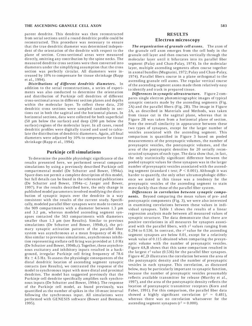
Orientation of small-diameter dendrites. Finally, asa second independent check of the serial reconstructiondata, we were interested in determining whether thesmallest diameter Purkinje cell dendrites might have aparticular spatial orientation within the molecular layer.
Specifically, consistent with previous reports (Mugnaini,1972), we have found that ascending segment axonspursue an essentially straight vertical course through themolecular layer, usually in bundles of several axons. Giventhe isoplanar orientation of the Purkinje cell dendrite andthe density of dendritic branches within this plane (Palayand Chan-Palay, 1974), it seemed reasonable to predictthat the small-diameter dendrites contacting the ascend-ing axons might extend horizontally outward, toward theascending segment axons. Comparing the number of round(i.e., perpendicular), small Purkinje cell dendritic profilesin sagittal sections with those found in horizontal sectionstested this prediction. Figure 8 shows that 56% of theround dendritic profiles traced in sagittal sections were,1.3 µm in diameter. In contrast, only 27% of the profilesfound in horizontal sections were this small. These resultssuggest that the smallest diameter dendrites tend to beoriented perpendicular to the sagittal plane of the Pur-kinje cell dendrite and into the plane of the ascendinggranule cell axon bundles.
Comparison of electron and light microscopic diam-
eter distributions. The results described above suggestthat the restriction of ascending segment synapses to thesmallest diameter Purkinje cell dendrites results in an
Fig. 4. Two sets of regression comparisons between ultrastructuralfeatures of ascending segment and parallel fiber synapses.A,B: Comparison of presynaptic volume and the number of presynap-tic vesicles for ascending and parallel fiber synapses, respectively.
C,D: Comparison of the area of the postsynaptic density with thenumber of presynaptic vesicles in each synapse. Full analysis of thedata for all combinations of comparisons is shown in Table 1.
586 G. GUNDAPPA-SULUR ET AL.

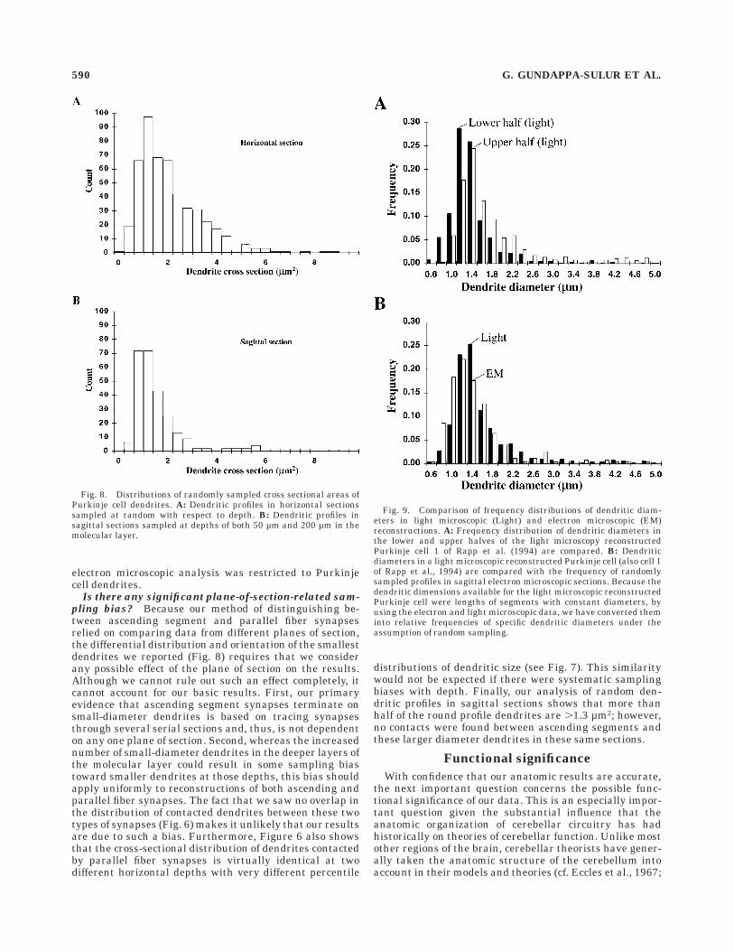
increase in small-diameter dendrites in the deeper regionsof the molecular layer (Fig. 7). However, this conclusion isbased on a random sample of specifically round dendriticprofiles in our electron microscopic material. If this conclu-sion is correct, then this differential distribution alsoshould be reflected in morphology of single Purkinje celldendrites. To determine whether this is the case, wecalculated the distribution of dendritic diameters in previ-ously published (Rapp et al., 1994) light microscopicreconstructions of guinea pig Purkinje cells (data kindlyprovided by M. Rapp, Y. Yarom, and I. Segev). Specifically,we divided the reconstructed Purkinje cell dendrites intoupper and lower halves and then calculated the frequencyof dendritic diameters as the ratio of the sum of the lengthsof all segments with a particular diameter divided by thesum of the lengths of all segments. The results of thisanalysis are shown in Figure 9A. Like the electron micro-scopic data (Fig. 7), we found many more small-diameterdendrites in the lower half of the dendrite than in theupper half. A second comparison, which is shown in Figure9B, indicates that, overall, the dendritic diameters of thelight data closely match those in the electron microscopicdata. The frequency distributions from both samples werestatistically similar (paired t test; P , 0.10). Thus, theresults of the electron microscopic analysis are in completeagreement with a completely independent data set ob-tained from light microscopic reconstruction in a separatelaboratory.
Simulation of Purkinje cell responsesto ascending segment inputs
Finally, as described in Materials and Methods, we usedrealistic computer simulations to begin to explore thefunctional consequences of the distal termination sites ofthe ascending segment synapses. These simulations arebased on a realistic single-cell model we used previouslyused to study Purkinje cell responses to granule cell inputs(De Schutter and Bower, 1994b,c). One unexpected resultof these initial simulations was that even a relativelysmall number of synchronously activated granule cellinputs results in a somatic action potential due to acalcium-dependent amplification mechanism (De Schutterand Bower, 1994c). We previously proposed that the ascend-ing granule cell axon is the most likely source of thissynchronous activation of granule cell synapses (Jaegerand Bower, 1994; Bower, 1997a–c) and that this synchro-nous activation might account for the vertical organizationof cerebellar cortex (Bower and Woolston, 1983). However,in models of other neuronal cell types, the passive proper-ties of a neuron can reduce substantially the effectivenessof synaptic inputs on small-diameter distal dendrites(Rall, 1964; Jack et al., 1975). Although Purkinje celldendrites are clearly active, we were still surprised to findthe ascending segment synapses on the distal most regionsof the Purkinje cell dendritic tree, given the apparentpotency of their inputs (Jaeger and Bower, 1994).
Fig. 5. Direct comparison of two electron photomicrographs ofsynapses made by ascending (A) and parallel fiber (B) segments ofgranule cell axons. Arrowheads highlight the Purkinje cell dendrite ineach case. Asterisks indicate the spines associated with each synapse.Note the characteristic appearance of mitochondria in the dendrite ofthe Purkinje cell as well as the similar shape of the Purkinje cellspines in each case. The scale bar in each figure corresponds to a
length of 1.2 µm, indicating an almost twofold difference in magnifica-tion between the two photographs. Taking this scale difference intoaccount, the size of the parent dendrite contacting the ascendingsegment is much smaller. Also note the appearance of several otherascending segment axons in a ‘‘bundle’’ with the axon whose synapse ishighlighted. Scale bars 5 1.2 µm.
THE ASCENDING GRANULE CELL AXON 587

We used our Purkinje cell model to specifically comparesomatic responses to small synchronous inputs on largerdiameter and smaller diameter dendritic branches. Forthese simulations, the balance of asynchronous excitatoryand inhibitory input was adjusted to produce a mean firingfrequency of 78 Hz, which is within the physiologic rangefor this cell (Murphy and Sabah, 1970). Previous modelingresults have shown that, under these conditions, synchro-nous granule cell inputs produce somatic spiking re-sponses that are very similar to those recorded experimen-tally (De Schutter and Bower, 1994c). This amplificationmechanism is characterized by a near-linear relationbetween response amplitude and the number of synchro-nously activated inputs (De Schutter and Bower, 1994c).Figure 10 compares this relation for synchronously acti-vated synapses on the distal (Fig. 10, open circles) andmore proximal (Fig. 10, solid circles) Purkinje cell den-drites. The simulations demonstrate that there is verylittle if any difference in the amplification mechanism ineach case. Statistically, the somatic responses to distal andproximal activation were identical (paired t-test; P , 0.04).
DISCUSSION
We have described and contrasted the ultramorphologicproperties of synapses associated with the ascending andthe parallel fiber segments of the cerebellar granule cellaxon. To our knowledge, this is the first time that these twosynaptic populations have been compared specifically.Harris and Stevens (1988) previously described the ultra-morphology of parallel fiber synapses on Purkinje cells,and their calculated values for the number of presynapticvesicles (485 6 246) and the area of the postsynapticdensity (0.15 6 0.08) are comparable to data presentedhere for granule cell synapses in general. Their findingthat different measured pre- and postsynaptic features ofthe reconstructed parallel fiber synapses were correlatedpositively is also consistent with our results for parallelfibers (Table 1). However, those authors did not specificallyidentify or describe the synapses made by the ascendinggranule cell axons.
The fact that the ascending segment was not taken intoaccount in earlier electron microscopic studies is notsurprising, because these synapses were not consideredpreviously to be an important component of the cerebellarcortical circuit (Eccles et al., 1967; Ito, 1984; Napper andHarvey, 1988a,b). However, physiologic studies in ourlaboratory have suggested that the ascending segmentsynapses may have a substantial and more obvious physi-ologic effect on Purkinje cells than the parallel fibers(Bower and Woolston, 1983; Jaeger and Bower, 1994). It isour view that the distinct morphologic differences betweenthese two types of synapses reported here also suggest thatthese synapses are functionally distinct.
Accuracy of anatomic procedures
Before considering the possible functional implicationsof our anatomical data, it is necessary to consider whetherthe anatomic techniques used in this study could havebiased our basic results. There are several importantmethodological issues that must be addressed.
Are we certain that reconstructed synapses belonged
to granule cell axons? All measured synapses wereconfirmed to be associated with granule cell axons by usingserial electron microscopic reconstruction techniques. Thecharacteristic asymmetric morphology of excitatory gran-ule cell synapses (Figs. 2, 5) clearly distinguishes themfrom the symmetric profiles made by inhibitory synapsesassociated with other cerebellar interneurons (Palay andChan-Palay, 1974; Sultan et al., 1995). Excitatory granulecell synapses also are clearly distinguishable from theexcitatory contacts made by climbing fibers on Purkinjecells, because granule cell synapses occur on spines associ-ated with the spiny branchlets, whereas climbing fibersynapses occur on spines with a very different morphology(e.g., with a spine stem) occurring on the much largerdiameter ‘‘smooth’’ Purkinje cell dendrites (Palay andChan-Palay, 1974).
Can we really distinguish between ascending and
parallel fiber synapses? These experiments used differ-ent planes of section to distinguish between synapsesassociated with the ascending and parallel fiber segments.This approach relies on the fact that the general geometryof the granule cell axon is both regular and consistentthroughout the cerebellar cortex (Palay and Chan-Palay,1974). Although parallel fibers generally run in a horizon-
Fig. 6. Graph showing the diameters of the parent dendrites forspines associated with ascending segment and parallel fiber synapses.For the parallel fibers, the analysis was performed at two differentdepths of the molecular layer. Thirty measurements were made inhorizontal sections 50 µm below the pial surface, and 30 measure-ments were made at a depth of 150 µm. The 32 measurements for theascending segment synapses were made throughout the molecularlayer. The horizontal bar in each data set indicates the mean value ofdendritic cross section in each case.
588 G. GUNDAPPA-SULUR ET AL.

tal plane, and ascending segments run in a vertical(sagittal) plane (Fig. 1), the trajectories of individualgranule cell axons are never as straight as they arerepresented schematically. However, in our analysis, serialreconstructions were used to assure that the axon associ-ated with each measured synapse actually ran in thecorrect plane of section. In the case of ascending segmentsynapses, this was aided by the fact that these axons tendto run in vertical bundles containing several axons.
Are we certain that all measured synaptic termina-
tions are on Purkinje cell dendrites? Another possiblesource of error in our measurements involves the specificidentification of granule cell synapses on Purkinje celldendrites. In fact, it has been demonstrated previouslythat ascending segment axons contact Golgi cell dendritesin the molecular layer (Hamori, 1981). We recently showedthat ascending segment synapses also contact molecularlayer interneurons (Sultan et al., 1995). Nevertheless, weare confident that the data presented here reflect synapticcontacts only with Purkinje cells. First, Purkinje celldendrites can be distinguished unambiguously from otherdendrites in the molecular layer on the basis of their
characteristic dendritic spines (Figs. 2, 5). None of theother neurons with dendrites in the molecular layer havespines of this type (Palay and Chan-Palay, 1974). Second,Purkinje cell dendrites are well known to contain exten-sive amounts of endoplasmic reticulum (cf. Fig. 5). This istrue even for dendrites of the smallest diameter (Martoneet al., 1993). The existence of both spines and endoplasmicreticulum have been used by other neuroanatomists toidentify synaptic contacts on Purkinje cells (Harris andStevens, 1988). However, in this study, we further testedour electron microscopic level identification of Purkinjecell dendrites by comparing the measured dendritic diam-eters in the electron microscopic material with diametersobtained by using single cell light microscopic reconstruc-tion techniques (Rapp et al., 1994). Despite the fact thatthis reconstruction was performed in a separate laboratoryand under different histologic conditions, the diameterprofiles in the light and electron microscopic data werevirtually the same (Fig. 9B). Given the known differencesbetween Purkinje cell dendrites and the dendrites of otherneurons in the molecular layer (Palay and Chan-Palay,1974), this correspondence strongly suggests that our
Fig. 7. Comparison of the distribution of 250 round dendritic crosssections for randomly sampled dendrites in horizontal sections at eachof two depths in the molecular layer. A: Comparison of the totalnumber of profiles of different diameter for each horizontal level. The
striped bars correspond to sampled dendritic cross sections at a depthof 50 µm, and the solid bars represent randomly sampled dendriticcross sections found 200 µm below the pial surface. B,C: Chartsshowing the percentage of dendrites of each diameter at each level.
THE ASCENDING GRANULE CELL AXON 589
electron microscopic analysis was restricted to Purkinjecell dendrites.
Is there any significant plane-of-section-related sam-
pling bias? Because our method of distinguishing be-tween ascending segment and parallel fiber synapsesrelied on comparing data from different planes of section,the differential distribution and orientation of the smallestdendrites we reported (Fig. 8) requires that we considerany possible effect of the plane of section on the results.Although we cannot rule out such an effect completely, itcannot account for our basic results. First, our primaryevidence that ascending segment synapses terminate onsmall-diameter dendrites is based on tracing synapsesthrough several serial sections and, thus, is not dependenton any one plane of section. Second, whereas the increasednumber of small-diameter dendrites in the deeper layers ofthe molecular layer could result in some sampling biastoward smaller dendrites at those depths, this bias shouldapply uniformly to reconstructions of both ascending andparallel fiber synapses. The fact that we saw no overlap inthe distribution of contacted dendrites between these twotypes of synapses (Fig. 6) makes it unlikely that our resultsare due to such a bias. Furthermore, Figure 6 also showsthat the cross-sectional distribution of dendrites contactedby parallel fiber synapses is virtually identical at twodifferent horizontal depths with very different percentile
distributions of dendritic size (see Fig. 7). This similaritywould not be expected if there were systematic samplingbiases with depth. Finally, our analysis of random den-dritic profiles in sagittal sections shows that more thanhalf of the round profile dendrites are .1.3 µm2; however,no contacts were found between ascending segments andthese larger diameter dendrites in these same sections.
Functional significance
With confidence that our anatomic results are accurate,the next important question concerns the possible func-tional significance of our data. This is an especially impor-tant question given the substantial influence that theanatomic organization of cerebellar circuitry has hadhistorically on theories of cerebellar function. Unlike mostother regions of the brain, cerebellar theorists have gener-ally taken the anatomic structure of the cerebellum intoaccount in their models and theories (cf. Eccles et al., 1967;
Fig. 8. Distributions of randomly sampled cross sectional areas ofPurkinje cell dendrites. A: Dendritic profiles in horizontal sectionssampled at random with respect to depth. B: Dendritic profiles insagittal sections sampled at depths of both 50 µm and 200 µm in themolecular layer.
Fig. 9. Comparison of frequency distributions of dendritic diam-eters in light microscopic (Light) and electron microscopic (EM)reconstructions. A: Frequency distribution of dendritic diameters inthe lower and upper halves of the light microscopy reconstructedPurkinje cell 1 of Rapp et al. (1994) are compared. B: Dendriticdiameters in a light microscopic reconstructed Purkinje cell (also cell 1of Rapp et al., 1994) are compared with the frequency of randomlysampled profiles in sagittal electron microscopic sections. Because thedendritic dimensions available for the light microscopic reconstructedPurkinje cell were lengths of segments with constant diameters, byusing the electron and light microscopic data, we have converted theminto relative frequencies of specific dendritic diameters under theassumption of random sampling.
590 G. GUNDAPPA-SULUR ET AL.

Marr, 1969; Albus, 1971; Fujita, 1982; Ito, 1984; Kawatoand Gomi, 1992; Killeen and Fetterman, 1993). For thisreason, any substantial change in our understanding ofthe anatomic organization of this circuit will have asubstantial effect on existing theories.
How many ascending segment synapses are there?
These anatomic investigations were motivated by thesuggestion that our previous physiologic results showing avertical organization in cerebellar cortical circuitry (Boweret al., 1980; Bower and Woolston, 1983) might reflect thesynaptic influence of the ascending segment of the granulecell axon (Llinas, 1982). The predominance of the verticaleffects of granule cell layer activation on Purkinje cells wasproposed at that time to result from the multiple synapseslikely to be made by the ascending segments on singlePurkinje cell dendrites (Llinas, 1982). However, since thisoriginal proposal was made, there has been a generalreluctance to consider the importance of the ascendingsegment in cerebellar physiology. One possible reason forthis lack of interest is the calculation published by Harveyand Napper (1988a) purporting to demonstrate that theascending segment synapses represent at most 3% of thetotal granule cell synaptic contacts with Purkinje cells.Although those authors did note subsequent to the firstpreliminary report of the results in this paper that anychange in the assumptions used in their indirect calcula-tions would require a modification of this estimate (Harveyand Napper, 1991), no efforts have been made to recalcu-late these numbers or explore this issue. By using the
anatomic data from the current experiments, as describedbelow, we estimate that the ascending segment synapsesmay actually constitute up to 20% of the total granule cellsynaptic input to Purkinje cells.
Estimates based on granule cell counts. Harvey andNapper’s (1988a) 3% estimate for the percentage of ascend-ing segment synapses was calculated indirectly by multi-plying the estimated number of granule cells underneatheach Purkinje cell (274) by the average length of ascendingaxon segments (125 µm), and then dividing by the numberof synapses per unit length of the parallel fiber segment ofthe axon (one synapse every 7.3 µm). However, several ofthese values have been altered by more recent anatomicdata. For example, it has been reported recently (Pichit-pornchai et al., 1994) that, on average, there is oneascending segment synapse for every 4.0 µm of axonlength, which is considerably higher than the rate of oneevery 5.2 µm for parallel fibers near the axon bifurcationpoint. This number drops to one synapse per 7.4 µm ofaxon length farther along the parallel fiber. These revisedmeasurements immediately raise the estimated number ofascending segment synapses to 5.5% of the total. Second,Korbo et al. (1993) have reported a value of 434 granulecells per Purkinje cell, which is between the Harvey andNapper estimate of 274 and the much higher earlierestimate of 897 by Lange (1975). If one also considers thatbetween 11% (Pichitpornchai et al., 1994) and 19% (Harrisand Stevens, 1988) of granule cell axon varicosities actu-ally make two synaptic contacts, then these values esti-mate that the numbers of ascending segment synapsesrange between 10,000 and 35,000 per Purkinje cell. Assum-ing that there are between 100,000 and 150,000 granulecell synapses per each Purkinje cell (Napper and Harvey,1988b), these indirect calculations give us a percentage ofascending segment synapses ranging anywhere between7% and 24% of the total. Either number is substantiallyhigher than previous estimates.
Estimates based on dendritic profiles. Our finding thatthe ascending segment synapses contact only the smallestdiameter Purkinje cell dendrites allows us to make a muchmore direct estimate of the number of ascending segmentsynapses per Purkinje cell. Specifically, the total length ofdendrites ,1.3 µm in diameter on a particular cell multi-plied by the frequency of dendritic spines per length ofdendrite should provide a reasonable estimate of thenumber of ascending segment synapses. Such an estimateis shown in Table 2 for the three Purkinje cells that werereconstructed completely by Rapp et al. (1994) using lightmicroscopic techniques. The first column in Table 2 showsthe total length of the spinous dendrite for each neuron,
Fig. 10. Comparison of the response of the Purkinje cell model tosynchronous activation of different numbers of synapses on regions ofthe dendrite that our data show are occupied by parallel fiber (opencircles) and ascending segment (solid circles) inputs. The number ofsimple spikes in a 10-msec interval after the synchronous input isplotted against the number of inputs activated synchronously. Eachdata point represents the average response of the model to 200stimulus trials. Background asynchronous input frequency for thesesimulations was 46 Hz for parallel fiber inputs and 1 Hz for stellatecell inputs.
TABLE 2. Estimates of the Number of Ascending Segment Synapses1
Purkinjecell
Totallengthspiny
dendrite(mm)
Percent,1.3 µmdiametersegments
Estimatednumber ofascending
synapse spines(density; 8/µm)
Estimatedtotal number
of spines(density;13/µm)
1 11.11 37.4 33,222 144,4562 8.27 69.1 45,690 107,4733 7.92 29.6 18,190 102,985Average 9.10 45.4 32,367 118,305
1Estimates of the number of spines on three morphologically reconstructed guinea pigPurkinje cells. See Rapp et al. (1994) for drawings of the cells. The estimates of thenumber of ascending segment synapses are based on the assumption that all dendriticbranches with a diameter smaller than 1.3 µm receive such inputs and that the densityof spines is constant and identical for all cells.
THE ASCENDING GRANULE CELL AXON 591

and the second column contains the percentage of eachcell’s dendrite ,1.3 µm in diameter. Somewhat surpris-ingly, dendrites of this diameter average 45% of the totaldendrite. Converting this percentage into an estimate forthe number of ascending segment synapses requires onlyan accurate estimate of the density of spines per unitlength of the dendrite. However, the previously publishedvalue of 13 spines per 1.0 µm probably applies only to theparallel fibers (Harris and Stevens, 1988). Given thesmaller size of the dendrites receiving ascending projec-tions, we have calculated an adjustment factor for thepublished density estimates based on comparing the num-ber of spines on 30 dendritic profiles contacted by theascending segment with the number of spines on 30profiles contacted by the parallel fibers. We found anaverage of 2.5 6 1.3 spines per profile for ascendingsegments and 3.9 6 1.4 spines per profile for parallelfibers. Given this ratio, we estimate that the dendritesreceiving ascending segment synapses have a spine den-sity of 8 spines per 1.0 µm length. In Table 2, the thirdcolumn shows that, when even this conservative estimateis multiplied by the total length of the small dendrites forthese three reconstructed cells, we calculate an average of32,000 ascending segment synapses per Purkinje cell.Despite the fact that this number is calculated fromentirely different numbers than the modified Harvey andNapper estimate discussed above, it still falls within therange of the previous calculation. In addition, when theaverage reported spine density is multiplied by the calcu-lated total length of the dendrite for each neuron (Table 2,second column), we obtain a range of values from 100,000to l50,000 for the total number of granule cell synapse perPurkinje cell (Table 2, last column). These numbers arewell within the range usually reported for Purkinje cells inthe rat (Napper and Harvey, 1988b). Because the overallnumber of synapses that we calculate fits with previousestimates, we have increased confidence in our new methodof estimating the number of ascending segment synapses.Accordingly, we suggest that, on average, ascending seg-ment synapses are likely to contribute at least 20% of thegranule cell synapses on each Purkinje cell.
What are the functional implications of differences in
synaptic morphology?
Presynaptic physiology. Beyond the apparently largenumbers of contacts made by the ascending segmentsynapses, the results reported here also demonstratepotentially important differences in synaptic morphology.For example, our data show that the average ascendingsegment synapse contains significantly more presynapticvesicles per synapse than the average parallel fiber syn-apse. In several other systems, the number of presynapticvesicles per synapse has been correlated directly with theprobability of transmitter release (Murthy et al., 1997).Although there is no evidence to date that ascendingsegment synapses may be more potent than those ofparallel fibers, our data suggest that this might be aworthy subject for further investigation.
Although it is harder to quantify, we have also consis-tently observed that the staining density of vesicles in theascending segment is much darker than for the parallelfibers. This is not likely to be an effect of fixation or tissuepreparation, because the same blocks of tissue were usedfor analysis of the two types of synapses, and the process-ing procedures were identical in all cases. In other sys-tems, differences in vesicle staining density have been
taken as indications of possible differences in the pharma-cology of synaptic transmission (Schmidle et al., 1991).Obviously, any difference in the transmitter pharmacologyof these two synaptic populations could have importantphysiologic consequences.
Correlation in pre- and postsynaptic features. Anothersignificant difference between ascending and parallel fibersynapses involves correlations between the values ob-tained for the pre- and postsynaptic features of singlesynapses. All measures made of pre- and postsynapticultramorphology were correlated highly positively in paral-lel fibers, whereas only the volume of the presynapticterminal and the number of presynaptic vesicles in theascending segment synapses showed any tendency to becorrelated. In their previous electron microscopic analysisof granule cell synapses on Purkinje cell spiny branchlets,Harris and Stevens (1988) also noted the correlation inpre- and postsynaptic features of parallel fiber synapsesand speculated that this correlation might reflect thepresence of a mechanism responsible for regulating synap-tic efficacy. The same authors found similar pre- andpostsynaptic ultramorphologic correlations in the CA1region of the hippocampus (Harris and Stevens, 1989) inwhich mechanisms responsible for long-term synapticpotentiation (LTP) are now generally considered to requirecoordination between the pre- and postsynaptic terminals(Gustafsson et al., 1987; Markam and Tsodyks, 1996). Inthe cerebellum, parallel fiber synapses have been shown toundergo a form of long-term synaptic depression (LTD)under certain stimulus conditions (Ito, 1989; Linden, 1994;De Schutter 1995a; De Schutter and Maex, 1996). Al-though initial descriptions of LTD emphasized postsynap-tic effects (De Schutter, 1995a), more recent studies sug-gest that LTD may also involve molecular interactionsbetween pre- and postsynaptic terminals (Lev-Ram et al.,1997). The correlation in pre- and postsynaptic ultramor-phology shown here for the parallel fibers is consistentwith these results.
Whatever the relationship may be between the morpho-logic correlations described here for the parallel fibersystem and LTD, the lack of any pre- or postsynapticcorrelation in the synapses of the ascending segmentssuggests that pre- and postsynaptic morphologic proper-ties are not coordinated in these synapses. Consistent withthis idea, De Schutter (1995a) has recently proposed thatLTD specifically may regulate parallel fiber synapticstrength in relation to the overall level of stellate cellinhibition. Our recent modeling studies, as described inmore detail below, suggest that maintaining a balancebetween these two types of synaptic input may be impor-tant in controlling the overall level of Purkinje cell den-dritic excitability (De Schutter and Bower, 1994b; Jaegerand Bower, 1997; Jaeger and Bower, 1996). In fact, wehave proposed that this is the primary role of the parallelfiber inputs within cerebellar cortex (Bower, 1997a,b). Webelieve that it is the ascending segment synapses thatprovide the more classical excitatory synaptic drive on thePurkinje cell soma (Bower and Woolston, 1983; Jaeger andBower, 1994).
Differences in dendritic location of synaptic termina-tions. Perhaps the most surprising finding of this study isthat ascending and parallel fiber synapses appear toterminate on completely separate regions of the Purkinjecell dendrite. It has been known for a long time thatgranule cell synapses in general are restricted to the
592 G. GUNDAPPA-SULUR ET AL.

smaller diameter Purkinje cell ‘‘spiny branchlets’’ ratherthan the large central dendritic shafts (Palay and Chan-Palay, 1974). These ‘‘smooth’’ regions of the Purkinje celldendrite are reserved for the other major excitatory inputto the Purkinje cell, the climbing fiber (Eccles et al., 1967).However, the current results indicate a further suborgani-zation of the spiny branchlets into those associated withascending segment and parallel fiber synapses. This re-sults in a spatially organized sequence of excitatory synap-tic connections to any particular branch of a Purkinje celldendrite that starts with the climbing fiber, progressesthrough parallel fiber inputs, and terminates with syn-apses from the ascending segment of the granule cell axon.
One interesting aspect of this spatial pattern of synapticorganization is its possible correlation to the developmentof the Purkinje cell dendrite itself (for review, see Altmanand Bayer, 1997). Specifically, the first contact made on thenewly developing Purkinje cell dendrite is from the climb-ing fiber, the synapses of which in the adult are found onthe large central dendritic shafts. The unusual developmen-tal pattern of the cerebellum, however, makes it very likelythat the second axonal contact made on the developingPurkinje cell dendrite is from the parallel fiber and notfrom not the ascending segment. The reason for this is thatthe granule cells, which, in the adult, are located beneaththe Purkinje cells, actually are generated by mitoticallyactive precursors located above the developing Purkinjecells. After these precursor cells divide, the neophytegranule cell first elaborates its parallel fiber axon and thenmigrates past the Purkinje cells into the internal granulecell layer. For this reason, elaborating parallel fibers arelikely to contact newly forming intermediate dendrites(Landis, 1987), whereas newly formed ascending segmentsare found in regions of the molecular layer that alreadycontain parallel fibers. Although it is important to notethat granule cell axons do not appear to make adult formsynapses until the granule cell contacts mossy fibers in thegranule cell layer at the end of their development (Altmanand Bayer, 1997), they do form morphologically distinctcontacts at earlier stages. When considered in the contextof the data reported in this paper, it is likely that theascending segment is available to make contacts with thePurkinje cell dendrite as its last dendritic extensions aredeveloping. In this way, the climbing fiber-parallel fiber-ascending segment innervation sequence in the adultdendrite may mirror directly the temporal sequence ofdevelopment of the Purkinje cell dendrite and its innerva-tion.
What are the implications for the physiologic organi-
zation of cerebellar cortical circuits? These anatomicexperiments were motivated by our previous physiologicfindings suggesting that ascending segment synapses havea powerful excitatory effect on overlying Purkinje cells(Bower and Woolston, 1983; Jaeger and Bower, 1994). Thefinding that ascending segment synapses are numerousand contain large numbers of presynaptic vesicles isconsistent with their having a strong influence on overly-ing Purkinje cells. However, the lack of ‘‘beams’’ reportedin previous physiologic experiments using peripheralstimulation also imply that parallel fibers are much lesspotent than previously suspected (Eccles et al., 1972;Bower and Woolston, 1983; Bell and Grimm, 1969). Onepossibility raised by our data is that differences in thedendritic location of these two types of synapses mightaccount for this difference in synaptic effectiveness. In
addition, we have already discussed the likelihood that themolecular or cellular properties of this synapses mightdiffer, given the differences in the structural correlationsbetween pre- and postsynaptic morphologies. In this re-gard, there is considerable evidence that synaptic segrega-tion on dendrites can be associated with biophysical ormolecular specializations in either pre- or postsynapticmembranes (cf. Hasselmo and Bower, 1993). Within thecerebellum, some preliminary results suggest that metabo-trophic glutamate receptors may be associated with paral-lel fiber synapses and not with those of the ascendingsegments (T. Knopfel, personal communication). Given thepossible importance of these receptors in mechanisms ofsynaptic modification like LTD (De Schutter, 1995a), thisfinding is consistent with our previous speculation thatLTD may not occur in ascending segments.
It is our hope that molecular and cellular biologistsinterested in the cerebellum will make a point in futureexperiments to distinguish between synapses on ascend-ing and parallel fiber axonal segments in their studies.However, it is also worth considering whether eitherdifferential dendritic location or differential activation dueto the normal operation of cerebellar cortical circuitry alsomight account for the apparent differences in the physi-ologic effects of these two synaptic populations. In thisregard, it has been known for many years that the relativeposition of synapses on the dendrite of a cell can directlyeffect their contribution to action potential generation.However, in the current case, the apparent physiologiceffects of the two populations of synapses are the reverse ofwhat classically would be expected from their spatialpositions. Specifically, if Purkinje cell dendrites acted assimple electrical cables, then one would expect that theparallel fiber synapses closer to the cell body would have agreater effect on the soma than the more distal ascendingsynapses (Rall, 1964). The reverse appears to be the case.Accordingly, something more complicated apparently isgoing on.
Synaptic effects of parallel fibers in the Purkinje celldendrite. There is abundant evidence, of course, thatPurkinje cell dendrites are not simple cables but, instead,contain a complex combination of voltage- and calcium-gated conductances (Llinas and Sugimori, 1980, 1992).Over the last several years, we have developed a detailedsingle-cell model of a cerebellar Purkinje cell to provide atool to unravel this complexity (De Schutter and Bower,1994a–c; Jaeger and Bower, 1996). These simulations haveproduced several unexpected results relevant to the synap-tic effects of ascending and parallel fiber synapses.
First, the model has predicted that the large, intrinsic,dendritic and somatic voltage- and calcium-gated conduc-tances primarily control the timing of somatic spikes, notthe relatively small conductances associated with theexcitatory parallel fiber inputs (Jaeger and Bower, 1996).Although a complete discussion of the underlying mecha-nisms is well beyond the scope of the current paper, themodel suggests that parallel fiber synaptic effects onsomatic spiking are indirect at best and, instead, that thisinput provides one half of a partial voltage clamp of localregions of the Purkinje cell dendrite (Jaeger and Bower,1996). The other half of the local voltage clamp is providedby the inhibitory synaptic effects of molecular layer inter-neurons. In the model, these inhibitory neurons, workingin opposition to the parallel fibers, modulate local den-dritic voltage levels and, thus, have a large effect on the
THE ASCENDING GRANULE CELL AXON 593

state of activation of the intrinsic voltage-dependent den-dritic conductances. Recent electrophysiologic experi-ments support these modeling predictions (Jaeger andBower, 1996), as do our current network simulations,which demonstrate that stellate cell-mediated, feed-forward inhibition effectively blocks beam-like activationof Purkinje cells by parallel fibers (Santamaria and Bower,1997). Thus, the lack of parallel fiber beams would appearto be a consequence of the biophysical properties of thePurkinje cell dendrite combined with the anatomic andphysiologic correlations within the cerebellar cortical cir-cuitry itself. Given the probable functional importance ofthe correlation between the synapses of molecular layerinterneurons and the parallel fibers, it would be veryinteresting to know whether inhibitory synapses also werefound selectively on the intermediate-sized Purkinje cellspiny branchlets.
Synaptic effects of ascending synapses on Purkinje cells.The second relevant result from our modeling effortsrelates to the possible effect on single Purkinje cells of thesimultaneous activation of a large number of ascendingsynapses. The data presented in this paper suggest thatactivation of the granule cell layer will result in activationof a large number of excitatory synapses on overlyingPurkinje cells. In addition, at least some of these synapticinputs will be nearly synchronous, especially if singleascending segments make multiple contacts on the samePurkinje cell. Recent multisite recording experiments inour laboratory have demonstrated that the granule celllayer is activated synchronously during exploratory behav-ior in rats (Hartmann and Bower, 1998). In our Purkinjecell model, this type of synaptic input evokes a subthresh-old dendritic calcium activation, resulting in a substantialsomatic depolarization and, usually, an action potential(De Schutter and Bower, 1994c). Therefore, it appears thatthe Purkinje cell dendrite has a mechanism that specifi-cally detects the presence of ascending segment inputs. Ofcourse, given the enormous numbers of parallel fibersynapses per Purkinje cell, it is reasonable to expect thatparallel fibers also would generate synchronous inputsunder the right conditions. At least one recent model ofcerebellar cortical function, in fact, relies on such a mecha-nism (Braitenberg, et al., 1997). However, as discussedabove, cortical circuitry would seem to assure that anyparallel fiber activity is counterbalanced by inhibition(Santamaria and Bower, 1997). The ascending synapses,in contrast, probably influence the Purkinje cell dendritebefore inhibition can be established (Santamaria andBower, 1997). Thus, again, the unique response of thePurkinje cell to ascending synapses also appears to rely ona combination of the biophysical properties of the Purkinjecell dendrite and the neuronal connections within thecerebellar cortex.
Functional correlations between ascending and parallelfiber synaptic inputs. Finally, it also is clearly importantto determine how the two components of the granule cellinfluence on Purkinje cells interact. Originally, we pro-posed (Bower and Woolston, 1983) that the parallel fibersmight be more modulatory in function. By using ourmodels, in fact, we have found that the level and timing ofbackground parallel fiber excitatory and molecular layerinhibitory synaptic inputs can modulate Purkinje cellresponses to synchronous ascending segment inputs (DeSchutter, 1995b). Again, it is not possible here to describethe detailed mechanisms underlying this modulation; how-ever, it is very likely to be mediated by the large calcium-
related conductances in the Purkinje cell dendrite. Such amodulatory effect also is consistent with the data pre-sented here showing that parallel fibers synapse in aposition between the ascending branch input on the distaldendrites and the large proximal dendrites and the Pur-kinje cell somata. In this way, parallel fiber synapses are ina position between the ascending synapses and the somataand, the model suggests, modulate the very calcium conduc-tances responsible for the subthreshold activation thatdrives somatic responses to synchronous ascending inputs(De Schutter and Bower, 1994c). Thus, the anatomicposition of these two different types of synapses very wellmay have an important influence on their apparent differ-ential physiologic effects.
CONCLUSIONS
It is our view that the anatomic results reported heredemonstrate that the cerebellar cortical neuronal circuit,long believed to be the best understood in the mammalianbrain, actually includes a previously under-consideredcomponent. This component, the ascending segment of thegranule cell axon, actually may have a dominant influenceon the response properties of the cerebellar Purkinje cell.These experiments also have uncovered an additionalstructural complexity in the synaptic organization of thePurkinje cell dendrite. We propose that there are actuallythree distinctly different afferent excitatory influences onPurkinje cells: climbing fibers, parallel fibers, and theascending segment of the granule cell axon. Each influ-ences its own region of the Purkinje cell dendrite, and eachhas its own physiologic consequences. Although numerousauthors have recognized that any such change in ourunderstanding of the cerebellar cortex has importantconsequences for interpretations of Purkinje cell function(cf. Epema et al., 1985; Sharp and Gonzales, 1985; Arm-strong, 1986; Campbell and Hesslow, 1986; Edgley andLidierth, 1987; Boegman et al., 1988; Theunnissen et al.,1989), all existing theories of cerebellar function exceptour own (Bower, 1997a,b) continue to assume that parallelfibers provide the primary excitatory drive on cerebellarPurkinje cells.
ACKNOWLEDGMENTS
We gratefully acknowledge the help of Fahad Sultan,Chris Assad, Inge Bats, and Mike Wijnants in preparationof the electron photomicrographs; Erika Oller for produc-tion of the first illustration; and Judy Macias for help, asalways, in keeping things organized and on track. Weacknowledge the entire laboratory for ongoing discussionson the significance of these experimental results. In particu-lar, we acknowledge Mitra Hartmann, John Thompson,Brian Rasnow, Brigitte Stricanne, Mark Nelson, MikePaulin, and, for their skepticism in particular, FahadSultan and Chris Assad. Outside the laboratory, we areindebted to Herbert Axelrad, Idan Segev, and Yosi Yaromfor numerous discussions and insights.
LITERATURE CITED
Albus JS. 1971. A theory of cerebellar function. Math Biosci 10:25–61.Altman J, Bayer SA. 1997. Development of the cerebellum in relation to its
evolution, structure, and functions. Boca Raton, FL: CRC Press.Armstrong DM. 1986. Supraspinal contributions to the initiation and
control of locomotion in the cat. Progr Neurol 26:273–361.
594 G. GUNDAPPA-SULUR ET AL.

Bell CC, Grant K, Serrier J. 1992. Sensory processing and corollarydischarge effects in the mormyromast regions of the mormyrid electro-sensory lobe. 1. Field potentials, cellular-activity in associated struc-tures. J Neurophysiol 68:843–858.
Bell CC, Grimm RJ. 1969. Discharge properties of Purkinje cells recordedon single and double microelectrodes. J Neurophysiol 32:1044–1055.
Bloedel JR, Courville J. 1981. Cerebellar afferent systems. In: Brooks VB,editor. Handbook of physiology: the nervous system, vol II. Bethesda,MD: American Physiological Society. p 735–829.
Boegman RJ, Parent A, Hawkes R. 1988. Zonation in the rat cerebellarcortex: patches of high acetylcholinesterase activity in the granularlayer are congruent with Purkinje cell compartments. Brain Res448:237–251.
Bower JM. 1997a. Is the cerebellum sensory for motor’s sake, or motor forsensory’s sake: the view from the whiskers of the rat. Progr Brain Res114:483–516.
Bower JM. 1997b. The cerebellum and the control of sensory data acquisi-tion. In: Schmahmann J, editor. The cerebellum and cognition. SanDiego: Academic Press. p 489–513.
Bower JM. 1997c. What do parallel fibers do? [commentary on ‘‘Thedetection and generation of sequences as a key to cerebellar function:experiments and theory’’ by Braitenberg V, Heck D, Sultan F]. BehavBrain Sci 20:247.
Bower JM, Beeman D. 1995. The book of GENESIS. New York: Springer-Verlag.
Bower JM, Kassel J. 1990. Variability in tactile projection patterns tocerebellar folia crus IIa of the Norway rat. J Comp Neurol 302:768–778.
Bower JM, Woolston DC. 1983. Congruence of spatial organization of tactileprojections to granule cell and Purkinje cell layers of cerebellarhemispheres of the albino rat: vertical organization of cerebellar cortex.J Neurophysiol 49:745–766.
Bower JM, Woolston DC, Gibson JM. 1980. Congruence of spatial patternsof receptive field projections to Purkinje cell and granule cell layers inthe cerebellar hemispheres of the rat. Soc Neurosci Abstr 6:511.
Braitenberg V, Atwood RP. 1958. Morphological observations on the cerebel-lar cortex. J Comp Neurol 109:1–33.
Braitenberg V, Heck D, Sultan F. 1997. The detection and generation ofsequences as a key to cerebellar function: experiments and theory.Behav Brain Sci 20:235–240.
Campbell NC, Hesslow G. 1986. The secondary spikes of climbing fibreresponses recorded from Purkinje cell somata in cat cerebellum. JPhysiol 377:207–224.
De Schutter E. 1995a. Cerebellar long-term depression might normalizeexcitation in Purkinje cells. Trends Neurosci 18:291–295.
De Schutter E. 1995b. Dendritic calcium channels amplify the variability ofpostsynaptic responses. Soc Neurosci Abstr 21:586.
De Schutter E, Bower JM. 1994a. An active membrane model of thecerebellar Purkinje cell I. Simulation of current clamp in slice. JNeurophysiol 71:375–400.
De Schutter E, Bower JM. 1994b. An active membrane model of thecerebellar Purkinje cell. II. Simulation of synaptic responses. J Neuro-physiol 71:401–419.
De Schutter E, Bower JM. 1994c. Simulated responses of cerebellarPurkinje cells are independent of the dendritic location of granule cellsynaptic inputs. Proc Natl Acad Sci USA 91:4736–4740.
De Schutter E, Maex R. 1996. The cerebellum: cortical processing andtheory. Curr Opin Neurobiol 6:759–764.
Eccles JC. 1973. The cerebellum as a computer: patterns in space and time.J Physiol (Lond) 229:1–32.
Eccles JC, Llinas R, Sasaki K. 1966. Parallel fiber stimulation and theresponses induced thereby in the Purkinje cells of the cerebellum. ExpBrain Res 1:17–39.
Eccles JC, Ito M, Szentagothai J. 1967. The cerebellum as a neuronalmachine. Berlin: Springer.
Eccles JC, Sabah NH, Schimdt RF, Taborikova H. 1972. Integration ofPurkinje cells of mossy and climbing fiber inputs from cutaneousmechanoreceptors. Exp Brain Res 15:498–520.
Edgley SA, Lidierth M. 1987. The discharges of cerebellar Golgi cells duringlocomotion in the cat. J Physiol 392:315–332.
Epema AH, Guldemond JM, Voogd J. 1985. Reciprocal connections betweenthe caudal vermis and the vestibular nuclei in the rabbit. Neurosci Lett57:273–278.
Fujita M. 1982. Adaptive filter model of the cerebellum. Biol Cybern45:195–206.
Gabbiani F, Midtgaard J, Knopfel T. 1994. Synaptic integration in a modelof cerebellar granule cells. J Neurophysiol 72:999–1009.
Gilbert P. 1975. How the cerebellum could memorize movements. NatureLondon 254:688–689.
Gundappa-Sulur G, Bower JM. 1990. Differences in ultramorphology anddendritic termination sites of synapses associated with the ascendingand parallel fiber segments of granule cell axons in the cerebellar cortexof the albino rat. Soc Neurosci Abstr 16:896.
Gustafsson B, Wigstrom H, Abraham WC, Huang YY. 1987. Long-termpotentiation in the hippocampus using depolarizing current pulses asthe conditioning stimulus to single volley synaptic potentials. J Neuro-sci 7(3):774–780.
Hamori J. 1981. Synaptic input to the axon hillock and initial segment ofinhibitory interneurons in the cerebellar cortex of the rat. Cell TissueRes 217:553.
Harris KM, Stevens JK. 1988. Dendritic spine of rat cerebellar Purkinjecells: serial electron microscopy with reference to their biophysicalcharacteristics. J Neurosci 8:4455–4469.
Hartmann M, Bower JM. 1998. Oscillatory activity (spindling) in thecerebellum of unrestrained rats. J Neurophysiol 80:1598–1604.
Harvey R J, Napper RMA. 1991. Quantitative studies of the mammaliancerebellum. Progr Neurobiol 36:437–463.562.
Hasselmo ME, Bower JM. 1993. Acetylcholine and memory. Trends Neuro-sci 16:218–222.
Heck D. 1995. Sequential input to Guinea pig cerebellar cortex in vitrostrongly affects Purkinje cells via parallel fibers. Naturwissenschafen82:210–203.
Ito M. 1984. The cerebellum and neural control. New York: Raven Press.Ito M. 1989. Long-term depression. Annu Rev Neurosci 12:85–102.Ito M. 1996. Cerebellar long-term depression. Trends Neurosci 19:11–12.Jack JJ, Noble D, Tsien RW. 1975. Electrical current flow in excitable cells.
Oxford: Clarendon.Jaeger D, Bower JM. 1994. Prolonged responses in rat cerebellar Purkinje
cells following activation of the granule cell layer: an intracellular invitro and in vivo investigation. Exp Brain Res 100:200–214.
Jaeger D, Bower JM. 1996. The function of background synaptic input incerebellar Purkinje cells explored with dynamic current clamping. SocNeurosci Abstr 22: 949.
Jaeger D, De Schutter E, Bower JM. 1997. The role of synaptic andvoltage-gated currents in the control of Purkinje cell spiking: a model-ing study. J Neurosci 17:91–106.
Kandel ER, Schwartz JHS, Jessell TM. 1991. Principles of neural science,3rd ed. Norwalk, CT: Appleton and Lange.
Karachot L, Kado RT, Ito M. 1994. Stimulus parameters for indication oflong-term depression in in vitro rat Purkinje cells. Neurosci Res21:161–168.
Kawato M, Gomi H. 1992. A computational model of 4 regions of thecerebellum based on feedback-error learning. Biol Cybern 68:95–103.
Keifer J, Houk JC. 1994 Motor function of the cerebello-rubro-spinalsystem. Physiol Rev 74:509–542.
Killeen PR, Fetterman JG. 1993. The behavioral theory of timing: transi-tion analysis. J Exp Anal Behav 59:411–422.
Korbo L, Andersen BB, Ladefoged O, Moller K. 1993. Total numbers ofvarious cell types in rat cerebellar cortex estimated using an unbiasedstereological method. Brain Res 609:262–268.
Korn H, Faber DS. 1991. Quantal analysis and synaptic efficacy in the CNS.Trends Neurosci 10:439–445.
Laine J, Axelrad H. 1994. The candelabrum cell: a new interneuron in thecerebellar cortex. J Comp Neurol 339:159–173.
Landis DMD. 1987. Initial junctions between developing parallel fibers andPurkinje cells are different from mature synaptic junctions. J CompNeurol 260:513–525.
Lange W. 1975. Cell number and cell density in the cerebellar cortex of manand some other mammals. Cell Tissue Res 157:115–124.
Lev-Ram V, Jiang T, Wood J, Lawrence DS, Tsien RY. 1997. Synergies andcoincidence requirements between NO, cGMP, and Ca21 in the induc-tion of cerebellar long-term depression. Neuron 18:1025–1038.
Linden DJ. 1994. Long-term synaptic depression in the mammalian brain.Neuron 12:457–472.
Lisberger SG. 1988. The neural basis for motor learning in the vestibulo-occular reflex in monkeys. Trends Neurosci 11:147–152.
Llinas R. 1982. General discussion: radial connectivity in the cerebellarcortex: a novel view regarding the functional organization of themolecular layer. In: Palay SL, Chan-Palay V, editors. The cerebellum:
THE ASCENDING GRANULE CELL AXON 595

new vistas [Exp Brain Res, vol 6(Suppl)]. New York: Springer Verlag. p189–194.
Llinas R, Sugimori M. 1980. Electrophysiological properties of in vitroPurkinje cell dendrites in mammalian cerebellar slices. J Physiol(London) 305:197–213
Llinas R, Sugimori M. 1992. The electrophysiology of the cerebellarPurkinje cell revisited. In: Llinas R, Soleto C, editors. The cerebellumrevisited. New York: Springer. p 167–181.
Markram H, Tsodyks M. 1996. Redistribution of synaptic efficacy betweenneocortical pyramidal neurons. Nature 382:807–810.
Marr D. 1969. A theory of cerebellar cortex. J Physiol (Lond) 202:437–471.Martone ME, Zhang Y, Simpliciano VM, Carragher BO, Ellisman MH.
1993. Three-dimensional visualization of the smooth endosplasmaticreticulum in Purkinje cell dendrites. J Neurosci 13:4636–4646.
Mittenthal JE. 1974. Reliability of pattern separation by the cerebellarmossy fiber-granule cell system. Kybernetik 16:93–101
Mugnaini E. 1972. The histology and cytology of the cerebellar cortex. In:Larsell O, Jansen J, editors. The comparative anatomy and histology ofthe cerebellum: the human cerebellum, cerebellar connections, andcerebellar cortex. Minneapolis: University of Minnesota Press. p 201–262.
Mugnaini E, Floris A. 1994. The unipolar brush cell: a neglected neuron ofthe mammalian cerebellar cortex. J Comp Neurol 339:174–180.
Murphy JT, Sabah NH. 1970. The inhibitory effect of climbing fiberactivation on cerebellar Purkinje cells. Brain Res 19:486–490.
Murthy VN, Sejnowski TJ, Stevens DF. 1997. Heterogeneous releaseproperties of visualized individual hippocampal synapses. Neuron17:4382–4388.
Napper RMA, Harvey RJ. 1988a. Number of parallel fiber synapses on anindividual Purkinje cell in the cerebellum of the rat. J Comp Neurol274:168–177.
Napper RMA, Harvey RJ. 1988b. Quantitative study of the Purkinje celldendritic spines in the rat cerebellum. J Comp Neurol 274:158–167.
Palay SL, Chan-Palay V. 1974. Cerebellar cortex: cytology and organiza-tion. Berlin: Springer.
Palkovits M, Magyar P, Szentagothai J. 1971a. Quantitative histologicalanalysis of the cerebellar cortex in the cat. I. Number and arrangementin space of the Purkinje cells. Brain Res 32:1–13.
Palkovits M, Magyar P, Szentagothai J. 1971b. Quantitative histologicalanalysis of the cerebellar cortex in the cat. II. Cell numbers anddensities in the granular layer. Brain Res 32:15–30.
Palkovits M, Magyar P, Szentagothai J. 1971c. Quantitative histologicalanalysis of the cerebellar cortex in the cat. III. Structural organizationof the molecular layer. Brain Res 34:1–18.
Palkovits M, Magyar P, Szentagothai J. 1972. Quantitative histologicalanalysis of the cerebellar cortex in the cat. IV. Mossy fiber-Purkinje cellnumerical transfer. Brain Res 45:15–29.
Pichitpornchai C, Rawson JA, Rees S. 1994. Morphology of parallel fibers inthe cerebellar cortex of the rat: an experimental light and electron-microscopic study with biocytin. J Comp Neurol 342:206–220.
Rall W. 1964. Theoretical significance of dendritic trees for neuronalinput-output relations. In: Reiss RF, editor. Neuronal theory andmodeling. Stanford, CA: Stanford University Press. p 73–97.
Ramon y Cajal S. 1911. Histologie du System Nerveux de l’Homme et desVertebres. Paris: Maloine.
Rapp M, Segev I, Yarom Y. 1994. Physiology, morphology and detailedpassive models of guinea-pig cerebellar Purkinje cells. J Physiol (Lond)474:101–118.
Sabah NH. 1971. Reliability in computation in the cerebellum. Biophys J11:429–445.
Santamaria F, Bower JM. 1997. A realistic cerebellar network: the temporaland spatial, direct and indirect influences of granule cell axons onPurkinje cells. Soc Neurosci Abstr 23:2364.
Schild RF. 1980. Length of the parallel fibers in rat cerebellar cortex. JPhysiol (Lond) 303:25P.
Schmidle T, Weiler R, Desnos C, Scherman D, Fishercolbrie R, Floor E,Winkler H. 1991. Synaptin synaptophysin, P65 and SV2: their presencein adrenal chromaffin granules and sympathetic large dense corevesicles. Biochim Biophys Acta 1060:3.
Shambes GM, Gibson JM, Welker W. 1978. Fractured somatotopy ingranule cell tactile areas of rat cerebellar hemispheres revealed bymicromapping. Brain Behav Evol 15:94–140.
Sharp FR, Gonzalez MF. 1985. Multiple vibrissae sensory regions in ratcerebellum: a 14C 2-deoxyglucose study. J Comp Neurol 234:489–500.
Stevens JK, Trogadis M. 1984. Computer assisted reconstruction fromserial electron micrographs: a tool for systematic study of neuronal formand function. Annu Rev Neurobiol 5:341–369.
Sultan F, Ellisman MH, Bower JM. 1995 Quantitative anatomical aspectsof the inhibitory interneurons of the rat cerebellar molecular layer: alight and electron microscopic Golgi study. Soc Neurosci Abstr 21:1191.
Szenthagothai J. 1983. The modular architectonic principle of neuralcenters. Rev Physiol Biochem Pharmacol 98:11–61.
Thompson RF. 1988. The neural basis of basic associative learning ofdiscrete behavioral responses. Trends Neurosci 11:152–155.
Theunnissen IE, Epema AH, Gerrits NM. 1989. Secondary vestibulocerebel-lar mossy fiber projection to the caudal vermis in the rabbit. J CompNeurol 190:262–277.
Welker W. 1987. Spatial organization of somatosensory projections togranule cell cerebellar cortex: functional and connectional implicationsof fractured somatotopy [summary of Wisconsin studies]. In: King JS,editor. New concepts in cerebellar neurobiology. New York: Alan R. Liss.p 239–280.
596 G. GUNDAPPA-SULUR ET AL.