asn neuro-2016-nikolakopoulou-
TRANSCRIPT

Original Article
Astrocytic Ephrin-B1 RegulatesSynapse Remodeling FollowingTraumatic Brain Injury
Angeliki M. Nikolakopoulou1, Jordan Koeppen1,2, Michael Garcia1,Joshua Leish1, Andre Obenaus3, and Iryna M. Ethell1,2
Abstract
Traumatic brain injury (TBI) can result in tissue alterations distant from the site of the initial injury, which can trigger
pathological changes within hippocampal circuits and are thought to contribute to long-term cognitive and neuropsycho-
logical impairments. However, our understanding of secondary injury mechanisms is limited. Astrocytes play an important
role in brain repair after injury and astrocyte-mediated mechanisms that are implicated in synapse development are likely
important in injury-induced synapse remodeling. Our studies suggest a new role of ephrin-B1, which is known to regulate
synapse development in neurons, in astrocyte-mediated synapse remodeling following TBI. Indeed, we observed a transient
upregulation of ephrin-B1 immunoreactivity in hippocampal astrocytes following moderate controlled cortical impact model
of TBI. The upregulation of ephrin-B1 levels in hippocampal astrocytes coincided with a decline in the number of vGlut1-
positive glutamatergic input to CA1 neurons at 3 days post injury even in the absence of hippocampal neuron loss. In
contrast, tamoxifen-induced ablation of ephrin-B1 from adult astrocytes in ephrin-B1loxP/yERT2-CreGFAP mice accelerated the
recovery of vGlut1-positive glutamatergic input to CA1 neurons after TBI. Finally, our studies suggest that astrocytic ephrin-
B1 may play an active role in injury-induced synapse remodeling through the activation of STAT3-mediated signaling in
astrocytes. TBI-induced upregulation of STAT3 phosphorylation within the hippocampus was suppressed by astrocyte-
specific ablation of ephrin-B1 in vivo, whereas the activation of ephrin-B1 in astrocytes triggered an increase in STAT3
phosphorylation in vitro. Thus, regulation of ephrin-B1 signaling in astrocytes may provide new therapeutic opportunities
to aid functional recovery after TBI.
Keywords
astrocytes, hippocampus, ephrinB1, STAT3, synapse, traumatic brain injury
Received August 6, 2015; Received revised December 30, 2015; Accepted for publication December 31, 2015
Introduction
Traumatic brain injury (TBI) occurs as a result of aclosed or penetrating head injury by an external mechan-ical force, including a blast wave, impact, or penetrationby a bullet (Maas et al., 2008). While advanced diagnosticand monitoring approaches have lead to a steady increasein the survival rate following brain injury, TBI triggerslong-term neuropsychological changes and physical dis-abilities affecting nearly 1.5 million individuals in theUnited States each year. Considerable efforts have beendevoted to developing treatments that enhance neuronalsurvival following brain injury. However, our under-standing of the mechanisms that regulate injury-inducedbrain rewiring is limited. Brain injury can cause dramaticchanges in brain synaptic connectivity that may promote
functional recovery, but may also lead to cognitive andneuropsychological impairment (Langlois et al., 2004;Nortje and Menon, 2004). In addition to neuronaldamage to the neocortex at the time of injury, other
1Biomedical Sciences Division, School of Medicine, University of California
Riverside, CA, USA2Cell, Molecular, and Developmental Biology graduate program, University
of California Riverside, CA, USA3Department of Pediatrics, School of Medicine, Loma Linda University, CA,
USA
Corresponding Author:
Iryna M. Ethell, Biomedical Sciences Division, School of Medicine, University
of California, 900 University Avenue, Riverside, CA 92521, USA.
Email: [email protected]
ASN Neuro
January-February 2016: 1–18
! The Author(s) 2016
DOI: 10.1177/1759091416630220
asn.sagepub.com
Creative Commons CC-BY: This article is distributed under the terms of the Creative Commons Attribution 3.0 License
(http://www.creativecommons.org/licenses/by/3.0/) which permits any use, reproduction and distribution of the work without further permission
provided the original work is attributed as specified on the SAGE and Open Access pages (https://us.sagepub.com/en-us/nam/open-access-at-sage).
by guest on March 3, 2016asn.sagepub.comDownloaded from
areas of the brain, such as the hippocampus, are suscep-tible to long-term anatomical and functional changes.Alterations in hippocampal circuits may contribute tomemory loss and long-term behavioral changes followinginjury (Baldwin et al., 1997; Scheff et al., 2005; Yu andMorrison, 2010; Atkins, 2011).
Astrocytes can facilitate brain repair after injury byprotecting neurons from glutamate excitotoxicity andregulating the blood-brain barrier; however, the role ofastrocytes in rewiring neuronal networks is not wellunderstood (Ito et al., 2006; Myer et al., 2006; Brownand Murphy, 2008; Benowitz and Carmichael, 2010;Mostany et al., 2010; Shields et al., 2011). Astrocytesundergo substantial changes in response to brain andspinal cord injury (Sofroniew, 2009), including cell hyper-trophy (Wilhelmsson et al., 2006), enhanced proliferationand scar formation, increased expression of glial-fibrillaryacidic protein (GFAP), and reexpression of the progeni-tor markers vimentin and nestin (Eddleston and Mucke,1993; Pekny and Nilsson, 2005; Sofroniew, 2005).Although it is elusive whether these changes are beneficialor detrimental for brain recovery, STAT3 signaling hasbeen implicated in reactive astrogliosis and astrocyte-specific STAT3 KO mice exhibited attenuated upregula-tion of GFAP, failure to exhibit astrocytic hypertrophy,and disruption of astroglial scar formation after spinalcord injury (Herrmann et al., 2008; O’Callaghan et al.,2014). STAT3 is also known to regulate astrocytic differ-entiation (Bonni et al., 1997), the formation of perineur-onal astrocytic processes, and the expression ofsynaptogenic molecule TSP-1 (Tyzack et al., 2014).
EphB receptor tyrosine kinases and their ligands,ephrin-Bs, play an important role in neuronal connectivityand synaptogenesis (Ethell and Pasquale, 2005).More pre-cisely, ephrin-B/EphB signaling pathways participate incell–cell interaction and regulate a plethora of biologicalprocesses during development and in adulthood, such asaxon guidance (Zimmer et al., 2003), synaptogenesis(Moeller et al., 2006; Segura et al., 2007), dendritic spineformation (Henkemeyer et al., 2003), and neurogenesis(Conover et al., 2000; Catchpole and Henkemeyer,2011). Recent studies have linked several Ephs and ephrinsto neurodevelopmental disorders, neurodegenerative dis-eases, and central nervous system injuries (Cisse et al.,2011; Georgakopoulos et al., 2011; Sanders et al., 2012;Coulthard et al., 2012; Overman et al., 2012; Van Hoeckeet al., 2012; Ren et al., 2013; Barthet et al., 2013), but themechanisms of ephrin-B/EphB signaling in neurologic dis-eases remain largely unexplored. Both ephrins and EphBreceptors are shown to be upregulated in reactive astro-cytes following injury (Sofroniew, 2009). An increase inephrin-B1 expression has been reported in reactive astro-cytes in the hippocampus after transection of entorhinalafferents (Wang et al., 2005). Similarly, ephrin-B2 and Ephreceptor levels increase in astrocytes after spinal cord
injury (Goldshmit et al., 2006) and the ablation of astro-cytic ephrin-B2 leads to increased motor axon regener-ation (Chen et al., 2013). Ephrin-B2 expression is alsoupregulated in microglia and astrocytes at the head ofthe optic nerve in glaucomatous DBA/2 J mice coincidingwith the loss of retina ganglia cell axons (Du et al., 2007).The upregulation of ephrins in astrocytes most likelyaffects the Eph receptor activity in neurons. Indeed,increased EphB3 has been reported in regenerating axonsafter optic nerve injury and EphB3 loss impeded axonalregeneration (Liu et al., 2006). In stroke, ephrin-A5 upre-gulation in reactive astrocytes is associated with a signifi-cant increase in the phosphorylation of EphA receptors inperi-infarct tissue (Overman et al., 2012) and EphB1 levelsare increased during axonal sprouting following stroke (Liand Carmichael, 2006). Although ephrin-A/EphA recep-tor interactions are implicated in astrocyte regulation ofsynaptic maintenance and remodeling after injury (Muraiet al., 2003; Carmona et al., 2009; Filosa et al., 2009;Muraiand Pasquale, 2011), the functional significance of astro-cytic ephrin-B1 signaling in the regeneration of brain cir-cuits after injury has not been investigated.
Here we report a transient upregulation of ephrin-B1in the adult hippocampal astrocytes, but not microglia,following moderate TBI using a controlled corticalimpact (CCI) model, concomitant with reactiveastrogliosis. The upregulation of ephrin-B1 levels inreactive astrocytes in stratum radiatum (SR) area ofCA1 hippocampus coincides with a decline in thenumber of vGlut1-positive glutamatergic input to CA1neurons at 3 days post injury (dpi), followed by a signifi-cant downregulation of astrocytic ephrin-B1 at 7 dpi.Furthermore, targeted ablation of ephrin-B1 from adultastrocytes accelerates the recovery of vGlut1-positive glu-tamatergic input to CA1 neurons after TBI, suggestingthat astrocytic ephrin-B1 may play an active role ininjury-induced synapse remodeling. Finally, our studiessuggest that astrocytic ephrin-B1 may act through theactivation of STAT3-mediated signaling in astrocytes asthe activation of ephrin-B1 in cultured astrocytes inducesphosphorylation of STAT3, whereas astrocyte-specificdeletion of ephrin-B1 suppresses STAT3 activation inthe hippocampus following TBI in vivo.
Materials and Methods
Mice and Tamoxifen Injections
GFAP-ERT2Cre/þ (Jax#012849) male mice were crossedwith ephrinB1loxP/þ female mice (exons 2 through 5 ofefnb1 gene are floxed by loxP sites, Jax#007664) toobtain GFAP-ERT2Cre/þephrinB1loxP/y (KO) or GFAP-ERT2Cre/þ (WT) male mice. WT and KO littermates malemice older than 8 weeks received tamoxifen intraperitone-ally (1mg; dissolved at 5mg/ml in 1:9 ethanol/sunflower
2 ASN Neuro
by guest on March 3, 2016asn.sagepub.comDownloaded from

oil mixture) once a day for 7 consecutive days. Animalsreceived surgery (see later) 1 to 2 weeks after the first tam-oxifen injections as we observed a significant downregula-tion of ephrin-B1 expression in hippocampalastrocytes, but not neurons, of tamoxifen-injected GFAP-ERT2Cre/þephrinB1loxP/y during this period. We did notdetect any changes in ephrin-B1 levels in astrocytes or neu-rons in GFAP-ERT2Cre/þephrinB1loxP/y noninjected orinjected with sunflower oil without tamoxifen. Cre andephrin-B1 immunoreactivities were analyzed in GFAP-ERT2Cre/þephrinB1loxP/y (KO) and GFAP-ERT2Cre/þ
(WT) mice. GFAP-ERT2Cre/þephrinB1loxP/y animalsshowed a detectable Cre immunoreactivity and at leasttwofold reduction of ephrin-B1 immunoreactivity in astro-cytes following tamoxifen injection. For expression of YFPin GFAPþ cells, Rosa-STOPloxPYFP animals were alsocrossed with GFAP-ERT2Cre/þephrinB1loxP/y or GFAP-ERT2Cre/þ mice. All genotypes were confirmed by poly-merase chain reaction (PCR) analysis of genomic DNAisolated from mouse tails. Mice were maintained in anAAALAC accredited facility under 12-h light/dark cyclesand fed standard mouse chow. All mouse studies were doneaccording to NIH and Institutional Animal Care and UseCommittee guidelines.
Surgery
Two-month-old C57BL/6 wild-type, GFAP-ERT2Cre/þ
ephrinB1loxP/y (KO) or GFAP-ERT2Cre/þ (WT) malemice received a unilateral CCI after a 4-mm craniotomyover the right parietal cortex (impactor center at Bregma:anterior-posterior, �1.50mm, medial lateral 1.50mm)using a stereotaxically positioned 3mm diameter stainlesssteel tipped piston (Figure 1(d)). CCI (3mm diameter tip,1mmdepth, 6.0m/s speed, 250ms dwell) was then deliveredto the cortical surface using an electromagnetically drivenpiston, resulting in cortical compression and some lossof cortical tissue. Moderate TBI in C57Bl6 mice results ina lesion volume of 2.51� 0.52% and 2.40� 0.46%(mean�SEM; % of brain volume) at 3 and 7 days postinjury (dpi), respectively, showing no hippocampal damage.Involvement of the hippocampus irrespective of the depthwas considered severe TBI. Mice recovered quickly afterTBI and demonstrated full activity within few hours aftersurgery, Shammice received craniotomywithout TBI.Micewith moderate CCI were analyzed in these studies. Animalswere sacrificed at 1, 3, and 7 days post TBI or craniotomy.Untreated animals were sacrificed at similar timepoints asTBI animals for comparison.
Immunohistochemistry
Animals were anesthetized with isoflurane and transcar-dially perfused first with 0.9% NaCl followed by with 4%paraformaldehyde in 0.1M phosphate-buffered saline
(PBS), pH¼ 7.4. Brains were postfixed overnight in4% paraformaldehyde/0.1M PBS and 100 -mm coronalbrain sections were cut with a vibratome. We observedno neuronal loss or apoptotic nuclear morphology in thehippocampus of these animals at 1, 3, or 7 dpi by assessingnuclear morphology with 4’6-diamidino-2-phenylindole(DAPI) staining. To determine ephrin-B1 immunoreactiv-ity in the hippocampus, double immunostaining was per-formed using goat antibodies against ephrin-B1 (1:50, BDPharmingen). The specificity of the ephrin-B1 immunor-eactivity in astrocytes was confirmed by the depletion ofanti-ephrin-B1 antibody against ephrin-B1-Fc coupled toprotein-A agarose (not shown). Astrocytes were labeledwith conjugated Cy3-anti-GFAP (1:500, Sigma) andmicroglia with rabbit anti-Iba1 (1:1,000, Wako) antibo-dies to identify changes in glial phenotype. Cre expressionwas analyzed with mouse anti-Cre antibody (1:100, EMDMillipore). Presynaptic boutons were labeled by immu-nostaining for the excitatory synapse marker vesicular glu-tamate transporter 1 (vGLUT1) using rabbit anti-vGLUT1 (1mg/4ml; 482400; Invitrogen). Secondary anti-bodies used were Alexa Fluor 594-conjugated donkeyanti-mouse IgG (4 mg/ml; Molecular Probes), AlexaFluor 647-conjugated donkey anti-rabbit IgG (4 mg/ml;Molecular Probes), or Alexa Fluor 488-conjugateddonkey anti-goat IgG (4mg/ml; Molecular Probes).Sections were mounted on slides with Vectashield mount-ing medium containing DAPI (Vector Laboratories Inc.).
Imaging
Confocal images from the ipsilateral and contralateralhemispheres were captured with a Leica SP2 confocallaser-scanning microscope using a series of high-resolu-tion optical sections (1,024� 1,024-pixel format) thatwere captured with a 20� (immunohistochemistry) or63� (synaptogenesis) water-immersion objective (1.2numerical aperture), 1� zoom at 1 -mm step intervals(z-stack of 11 optical sections). All images were acquiredunder identical conditions. Each z-stack was collapsedinto a single image by projection, converted to a tiff file,encoded for blind analysis, and analyzed using Image JSoftware. Three adjacent projections from SR were ana-lyzed per brain slice from at least three animals/group.Cell area, integrated fluorescent intensity, and cell per-imeter were determined for each GFAP-positive andephrinB1-positive cells (100–300 astrocytes, 5–11images, 3–4 mice per group). For the analysis ofvGlut1 immunolabeling, at least six sequential imageswere captured for selected area at 1-mm step intervals,each image in the series was threshold-adjusted toidentical levels and the puncta were measured usingImageJ. Three adjacent areas from SR were imagedand analyzed per brain slice from at least three ani-mals/group.
Nikolakopoulou et al. 3
by guest on March 3, 2016asn.sagepub.comDownloaded from
Cell Culture
Astrocytes were isolated from WT or ephrinB1loxP mousehippocampi at P0-P1 as previously described (Barkeret al., 2008). Briefly, hippocampi were treated with0.1%trypsin/ethylenediaminetetraacetic acid (EDTA)solution for 25min at 37�C and mechanically dissociated.Cells were plated on cell culture flasks and cultured inDMEM containing 10% fetal bovine serum (FBS) and1% pen-strep, under 10% CO2 atmosphere at 37�C. Toachieve purified astrocyte cultures (>95% astrocytes)cells were shaken after 4 days in vitro (DIV) for 1 h.After shaking, the media were removed and cells werewashed twice with 0.1M PBS (pH 7.4). Cells were thentreated with 0.1% trypsin/EDTA solution for 20min at37�C and plated on 10-cm Petri dishes with DMEMcontaining 10% FBS. Once confluent astrocytes weretrypsinized and plated on six-well plates at a density of1.2� 106 per plate and cultured for 2 days before beingtransfected with pEGFP, pEGFP and pcDNA-ephrinB1,or pEGFP and pcDNA-Cre plasmids usingLipofectamine according to the manufacturer’s instruc-tions (Invitrogen, 11668-019). Astrocytes were treated
with preclustered EphB2-Fc to activate ephrin-B1 or con-trol Fc and processed for Western blot as described later.
Ephrin-B1 Induction In Vitro
Preclustered EphB2-Fc or Fc were generated by incubatingEphB2-Fc (R&D Systems) or Fc (R&D Systems) with goatanti-human IgG (Jackson ImmunoResearch) for 1 h at 4�C.Transfected astrocytes were stimulated with 2.5mg/mlEphB2-Fc or 2.5ug/ml Fc for 15min. Cells were thenlysed with lysis buffer: (in mM) 25 Tris-HCl, 150mMNaCl, 5 EDTA, 1% Triton-X, 1 sodium pervanadate,and protease inhibitor mixture (1:100, Sigma, P8340).
Western Blotting
The hippocampi were removed from each mouse (n¼ 4mice per group), frozen, and stored at �80�C. Braintissues were homogenized in cold lysis buffer: 50mMTris-HCl (pH¼ 7.4), 150mM NaCl, 1mM EDTA(pH¼ 8.0), 1% Triton X-100, 0.1% sodium dodecyl sul-fate (SDS) containing protease inhibitor cocktail (1:100,Sigma, P8340), and 0.5mM sodium pervanadate. The
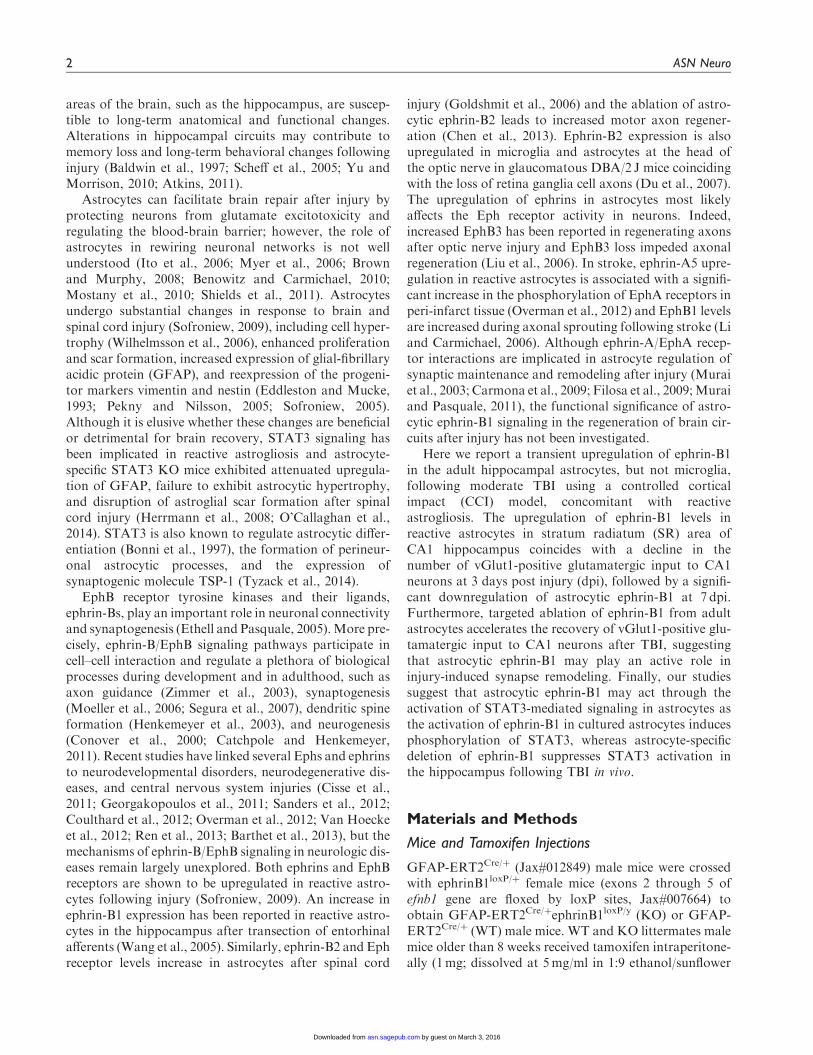
Figure 1. Reactive astrogliosis is observed in the hippocampus at 3 and 7 days following moderate CCI. Fluorescent images show GFAP
immunoreactivity (a) and DAPI labeling (b) in coronal brain sections of control mice at 1, 3, or 7 dpi. The location of the impact area is
indicated by a white bar. Note that reactive astrogliosis was observed in the ipsilateral cortex and hippocampus after moderate CCI, which
leads to neuronal loss near cortical impact but not in the hippocampus. (c) 3D brain reconstruction shows the size and location of the
impact. SR, stratum radiatum area of CA1 hippocampus; hp, hippocampus; CA1, CA1 layer; CA3, CA3 layer. (d) TBI location and size.
4 ASN Neuro
by guest on March 3, 2016asn.sagepub.comDownloaded from

samples were rotated at 4�C for at least 1 hr to allow forcomplete cell lysis and then cleared by centrifugation at10,000� g for 15min at 4�C. Hippocampal and cell lys-ates were boiled in reducing sample buffer (Laemmli 2�concentrate, Sigma, S3401), and proteins separated on8% to 16% Tris-glycine SDS-PAGE precast gels (LifeTechnologies, EC6045BOX). Proteins were transferredonto Protran BA 85 Nitrocellulose membrane (GEHealthcare) and blocked for 1 hr at room temperaturein 5% skim milk (BioRad, #170-6404). Primary antibodyincubations were done overnight at 4�C with antibodiesdiluted in tris-buffered saline (TBS)/0.1% Tween-20/5%bovine serum albumin (BSA). Primary antibodies usedwere anti-STAT3 (1:2,000; Cell Signaling; 4904S) andanti-p-STAT3 (1:2,000; Cell Signaling; 9145P). Blotswere washed 3� 10min with TBS/0.1% tween-20 andincubated with the appropriate horseradish peroxidase(HRP)-conjugated secondary antibodies for an hour atroom temperature in a TBS/0.1% tween-20/5% BSAsolution. The secondary antibodies used were a-rabbit-HRP at 1:5,000 and a-mouse-HRP at 1:5,000 (GEHealthcare). After secondary antibody incubations,blots were washed 3� 10min in TBS/0.1% tween-20and developed with ECL Detection reagent (ThermoScientific, #80196). For reprobing, membrane blots werewashed in stripping buffer (2% SDS, 100mM b-mercap-toethanol, 50mM Tris-HCl, pH¼ 6.8) for 30min at56�C, then rinsed repeatedly with TBS/0.1% tween-20,finally blocked with 5% skim milk, and then reprobed.Developed films were then scanned and protein levelsquantified by comparing band density values obtainedusing ImageJ. Two samples per group were run per blotand pSTAT3/STAT3 ratio was calculated for EphB2-Fc-treated samples and normalized to pSTAT3/STAT3 ratioof Fc-treated samples. Statistical analysis was performedusing two-way analysis of variance (ANOVA) followedby post hoc pair-by-pair comparisons with Fisher’s LeastSignificant Difference (LSD) method.
Quantitative Real-Time PCR
RNA was isolated from mouse hippocampi using Trizolreagent (Invitrogen), precipitated in isopropanol, andRNA concentration (ng/ml) was identified from theabsorbance at 260 and 280 nm, detected by NanoDropND-1000 spectrophotometer (NanoDrop Technologies).cDNA was transcribed using the Reverse TranscriptionSystem (Promega) according to manufacturer’s instruc-tions. To examine mRNA expression of ephrinB1 (F:ACCCTAAGTTCCTAAGTGGGA, R: CTTGTAGTACTCGTAGGGC), EphB2 (F: TACATCCCCCATCAGGGTGG, R: GCCGGATGAATTTGGTCCGC,GFAP (F: GCCACCAGTAACATGCAAGA, R:GCTCTAGGGACTCGTTCGTG), and vimentin (F:ATGCTTCTCTGGCACGTCTT, R: AGCCACGC
TTTCATACTGCT), specific forward and reverse pri-mers were used. Real-time PCR was carried out on aniCycler (Bio-Rad laboratories). Each reaction mixturecontained 1�Power SYBR Green PCR Master Mix(Life Technologies), and all the reactions were run intriplicate. The PCR amplification protocol was as fol-lows: initial DNA Polymerase activation at 95�C for10min, followed by 40 cycles with denaturation at 95�Cfor 15 s, and annealingþ extension at 60�C for 1min.Amplification was performed in a StepOne Real TimePCR System (96-well format) (Life Technologies) andanalyzed by normalizing the expression of each gene toGAPDH within each tissue sample.
Statistical Analysis
Both for in vivo and in vitro studies, the groups werecompared using one-way or two-way ANOVA withTukey’s or Bonferroni post hoc analysis, respectively,or paired Student’s t-test, and p values< .05 were takenas statistically significant.
Results
The aim of this study was to determine whether ephrin-B1 levels are regulated in hippocampal astrocytes follow-ing moderate CCI and to investigate the role of astrocyticephrin-B1 in synapse recovery after CCI in vivo usingconditional ephrin-B1 KO mice.
Moderate CCI Triggers a Transient Upregulation ofEphrin-B1 Immunoreactivity in Reactive Astrocytesin the Hippocampus
To induce TBI, mice received moderate unilateralCCI after craniotomy over the right parietal cortex(Figures 1(a) to (d)). As anticipated, we observed anincrease in GFAP-immunoreactivity suggesting reactiveastrogliosis at the site of injury (Figure 1(a)). Althoughwe observed no neuronal loss or apoptotic nuclearmorphology in the hippocampus of these animals at 1,3, or 7 dpi by assessing nuclear morphology with DAPIstaining (not shown), an increase in GFAP immunoreac-tivity was noted in the ipsilateral hippocampus at 3 dpi,with a significant upregulation of GFAP immunoreactiv-ity at 7 dpi (Figures 1(a) and 2(c) and (e)). To examinewhether ephrin-B1 levels were also upregulated in thereactive astrocytes after TBI, we performed immunos-taining against ephrin-B1 and analysis of GFAP andephrin-B1 immunoreactivity per area of astrocyte(Figure 2(a) to (f)). Our results show a significant increaseof ephrin-B1 immunoreactivity in astrocytes at 3 dpi inthe SR area of CA1 hippocampus as compared with con-trols (p< .05; Figure 2(f)) but not in cortical astrocytesaround the impact area (not shown). Surprisingly, at
Nikolakopoulou et al. 5
by guest on March 3, 2016asn.sagepub.comDownloaded from

7 dpi, ephrin-B1 immunoreactivity in hippocampal astro-cytes had subsided to control levels and was significantlylower than at 3 dpi (p< .01; Figure 2(f)). Interestingly,although ephrin-B1 immunoreactivity was reduced inastrocytes at 7 dpi, astrocytes remained reactive and
showed higher GFAP immunoreactivity as comparedwith control astrocytes (p< .01; Figure 2(e)). Ephrin-B1immunoreactivity was also slightly upregulated in CA1neurons, but we could not detect ephrin-B1 immunoreac-tivity in microglial cells (blue, Figure 2(a)). To further
Figure 2. Ephrin-B1 immunoreactivity was significantly upregulated in reactive astrocytes in the hippocampus following moderate CCI.
(a–d) Fluorescent images show GFAP-positive astrocytes (GFAP, red in a and d, gray in c), Iba1-positive microglia (Iba1, blue in a), and
ephrin-B1 immunoreactivity (ephrin-B1, green in a, gray in b) in the SR area of the CA1 hippocampus in control, 1, 3, and 7 dpi. (d) High
magnification images show examples of ephrin-B1-positive astrocytes. Note that ephrin-B1-positive immunoreactivity is found in the
dendrites of CA1 neurons and astrocytes in SR area of CA1 hippocampus. (e–g) Graphs show GFAP immunoreactivity per GFAP-positive
astrocyte (e) or ephrin-B1 immunoreactivity per ephrin-B1-positive astrocyte (f) in control (n¼ 682 cells, 9 images, 3 mice), 1 dpi (n¼ 385
cells, 5 images, 3 mice), 3 dpi (n¼ 732 cells, 11 images, 4 mice), and 7 dpi (n¼ 1217 cells, 9 images, 3 mice) or post-sham (n¼ 300–500 cells,
4–6 images, 3 mice). Note that ephrin-B1 immunoreactivity was not detected in all GFAP-positive cells. Error bars indicate SEM. Statistical
analysis was performed using one-way ANOVA followed by Tukey’s post hoc analysis (e,f, n¼ 3–4 mice, *p< .05, **p< .01) or two-way
ANOVA, followed by Bonferroni post hoc analysis (g, n¼ 3–4 mice, *p< .05).
6 ASN Neuro
by guest on March 3, 2016asn.sagepub.comDownloaded from
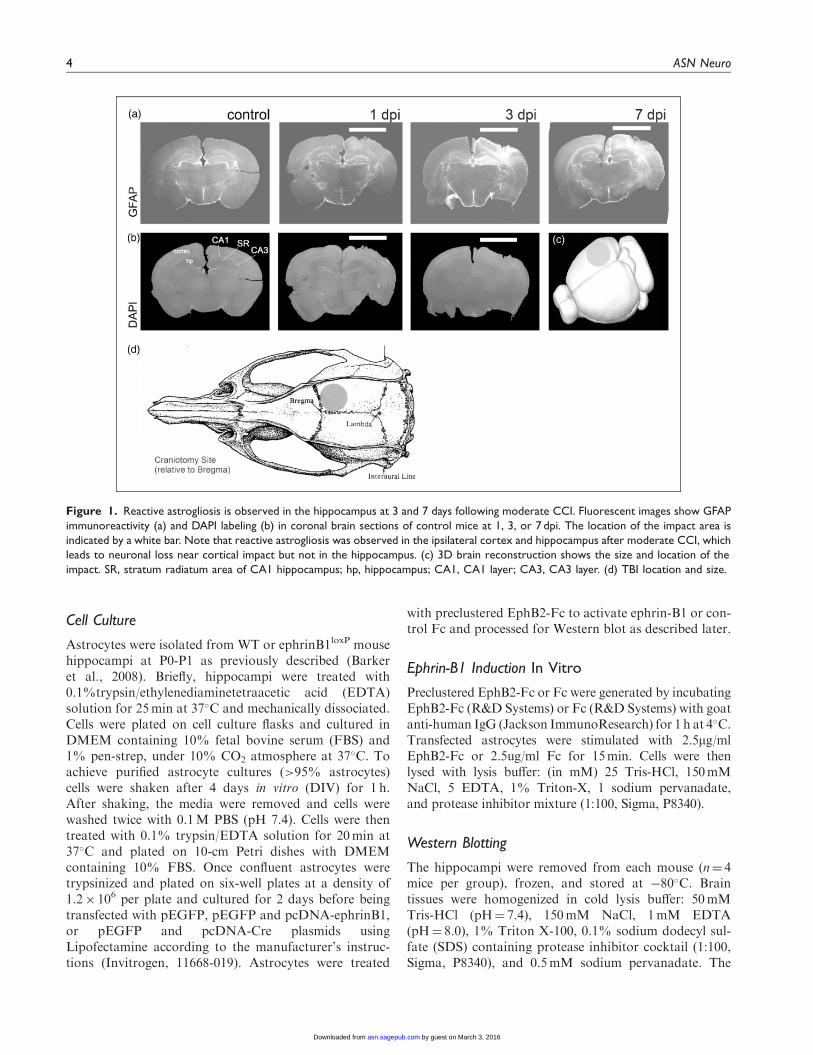
verify that the changes described earlier are induced as aconsequence of the brain injury, we compared TBI ani-mals to animals that had received craniotomy without theimpact (sham). Our data clearly show that craniotomyalone does not promote upregulation of ephrin-B1 immu-noreactivity at any timepoint (Figure 2(g)). In contrast,ephrin-B1 immunoreactivity was significantly higher inhippocampal astrocytes at 3 dpi as compared with 3days sham animals (p< .01; Figure 1(g)). In addition toimmunohistological studies, we examined gene expressionlevels of ephrin-B1 and its receptor EphB2 in control andTBI animals. Our results show an upregulation of bothephrin-B1 and EphB2 expression in the hippocampusafter TBI compared with control animals (Figure 3).The changes in the levels of ephrin-B1 and EphB2 recep-tor may affect transynaptic ephrin-B/EphB signaling andsynaptic rewiring following TBI.
Ephrin-B1 Upregulation in Hippocampal AstrocytesCoincides With a Decline in vGlut1-PositiveGlutamatergic Innervation of CA1Hippocampal Neurons
Although we observed reactive astogliosis, there was nosignificant neuronal loss noted in the hippocampus fol-lowing CCI. Previously published results demonstratethat while most neurons are spared following moderateor mild CCI, the hippocampus undergoes substantialchanges in synaptic organization (Scheff et al., 2005;Norris and Scheff, 2009; Gao et al., 2011). Therefore,we examined the changes in presynaptic Schaffercollateral input from CA3 to CA1 hippocampal neuronswithin the SR area of CA1 hippocampus following TBIby immunostaining against the excitatory presynapticmarker vGlut1 (Figure 4(a) to (d)). Our results show
Figure 3. Moderate CCI causes an upregulation in gene expression of ephrinB1 and its receptor, EphB2 in the hippocampus. qPCR data from
the hippocampi of control and TBI animals show that TBI causes an increase in ephrinB1 at all timepoints postinjury in the ipsilateral hemisphere;
however, in the contralateral hemisphere, this increase is noticeable at 3 dpi. Similarly, we observe a concomitant increase in EphB2 levels at 1, 3,
and 7 dpi both in the ipsi- and contralateral hemispheres. Graphs show mean� SEM (n¼ 3, two-way ANOVA, *p< .05, **p< .01).
Nikolakopoulou et al. 7
by guest on March 3, 2016asn.sagepub.comDownloaded from

that at 3 dpi, when ephrin-B1 is significantly upregu-lated, the number of vGlut1-positive presynaptic bou-tons was significantly decreased in SR area of CA1hippocampus (p< .05; Figure 4(e)). Furthermore, therewas no further decline in the density of presynapticboutons in the SR at 7 dpi, when ephrin-B1 levelswere similar to controls (Figure 4(e)). Our results alsoshow that there is a negative correlation between thelevels of ephrin-B1 in astrocytes and the number ofexcitatory glutamatergic presynaptic input to CA1 neu-rons in SR at 3 dpi (Figure 4(f)), suggesting that astro-cytic ephrin-B1 may play a role in synapse removalfollowing TBI.
Targeted Ablation of Ephrin-B1 From AstrocytesPromotes Fast Recovery of vGlut1-PositiveGlutamatergic Innervation of CA1Hippocampal Neurons Following CCI
To further establish a causal link between the upregu-lation in the levels of astrocytic ephrin-B1 and synapsereorganization after TBI, we developed a mouse modelwhere ephrin-B1 was specifically ablated from adultastrocytes. A significant reduction in ephrin-B1 immu-noreactivity was detected by immunostaining in GFAP-positive astrocytes of ephrinB1loxP/yGFAP-ERT2Cre/þ
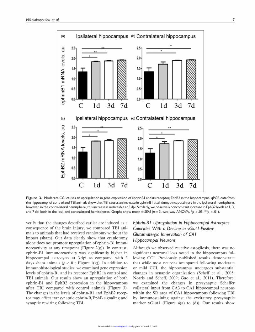
(KO) but not GFAP-ERT2Cre/þ (WT) mice treatedwith tamoxifen (Figure 5). The reduction in ephrin-B1levels was also confirmed in adult astrocytes in Rosa-STOPloxPYFP/ephrin-B1Flox/yERT2-CreGFAP mice butnot Rosa-STOPloxPYFP/ERT2-CreGFAP mice followingtamoxifen treatment (p< .001; Figure 6). Ephrin-B1was still detected in the cell bodies and dendrites ofCA1 neurons but not in YFP-positive astrocytes inSR area of CA1 hippocampus (Figure 6(a)). Wefound that astrocyte-specific deletion of ephrin-B1 trig-gered about three- to fivefold decrease in ephrin-B1immunoreactivity in GFAP-positive astrocytes (Figures5(b) and 6(c)).
Next, we examined whether the deletion of ephrin-B1from astrocytes can prevent the loss of excitatory pre-synaptic innervation of CA1 neurons following TBI.TBI was performed 7 to 14 days after the first tamoxifeninjection, at which point we see a selective ablation ofephrin-B1 from astrocytes (Figure 5(a)). Astrocyticephrin-B1 levels were significantly reduced in SR areaof CA1 hippocampus of ephrin-B1 KO as comparedwith WT mice at 1 dpi (p< .01), 3 dpi (p< .05), and7 dpi (p< .01; Figure 5(a) and (b)). Although both WTand ephrin-B1 KO mice showed a significant loss ofvGlut1-positive presynaptic innervation of CA1 neuronsat 3 dpi as compared with control animals (p< .01), a
significant recovery of vGlut1-positive presynaptic siteswas observed in ephrin-B1 KO but not WT mice(Figure 5(c)). At 7 dpi, the number of vGlut1-positivepresynaptic sites reached control levels in ephrin-B1 KOmice (p> .05), whereas the number of vGlut1-positivepresynaptic sites remained low in WT mice (p< .001;Figure 5(c)). A higher number of vGlut1-positive pre-synaptic boutons was also seen at 3 dpi in ephrin-B1KO mice as compared with WT mice (p< .05;Figure 5(c)). This difference was most likely a result ofan overall increase in the number of vGlut1-positive pre-synaptic sites in ephrin-B1 KO mice as compared withWT mice prior to TBI (p< .01). Our results show thatwhile a selective ablation of ephrin-B1 from astrocytesdid not prevent the initial loss of vGlut1-positive pre-synaptic innervation of CA1 neurons in SR followingCCI, it accelerated recovery of vGlut1-positive excitatoryinput to CA1 neurons at 7 dpi, suggesting negative effectsof astrocytic ephrin-B1 on new synapse formation in CA1hippocampus following TBI.
Ephrin-B1 Regulates STAT3 Phosphorylationin Astrocytes Following TBI
STAT3 signaling plays an important role in reactiveastrocytosis and may regulate protein expression in astro-cytes following injury (Herrmann et al., 2008). Similar toprevious work, our studies demonstrate an increase in thelevels of phosphorylated STAT3 (pSTAT3) followingbrain injury (Figure 7(a)). Interestingly, the changes inpSTAT3 levels also coincided with increased ephrin-B1immunoreactivity levels in astrocytes, showing increasedpSTAT3 at 1 and 3 dpi as compared with control (p< .01and p< .001), followed by its downregulation at 7 dpi(p< .01; Figure 7(d)). To establish a causal link betweenan upregulation in the levels of astrocytic ephrin-B1 andan increase in STAT3 phosphorylation, we examined theeffects of ephrin-B1 activation in astrocytes on STAT3phosphorylation in vitro. Our results show higherSTAT3 phosphorylation (pSTAT3) in cultured astrocytesfollowing ephrin-B activation with preclustered EphB2-Fc (p< .05; Figure 7(b) and (e)). We see a greater increasein pSTAT3 in astrocytes overexpressing ephrin-B1, sug-gesting that injury-induced increases in STAT3 phos-phorylation may be mediated through the activation ofephrin-B1. Indeed, astrocyte-specific deletion of ephrin-B1 affected STAT3 phosphorylation following TBI. Ourdata show significantly lower levels of pSTAT3 in theipsilateral hippocampus of ephrinB1-KO mice at 3 dpiwhen compared with WT animals (p< .05; Figure 7(c)and (f)). These studies suggest that ephrin-B1-dependentactivation of STAT3 in astrocytes may contribute toastrocyte-mediated synapse remodeling following TBI.
8 ASN Neuro
by guest on March 3, 2016asn.sagepub.comDownloaded from

Figure 4. Moderate CCI triggers a significant downregulation of vGlut1-positive glutamatergic innervation of CA1 hippocampal neurons.
We have analyzed vGlut1-positive puncta in SR area of the CA1 hippocampus, which immunolabels terminals of CA3 neurons innervating
dendrites of CA1 neurons. (a–d) Fluorescent images show a reduced vGlut1-positive puncta in SR at 3 (c) and 7 dpi (d) as compared with
control (a), suggesting a reduction in excitatory innervation of CA1 neurons following TBI. (e) Graph shows vGlut1-positive puncta per
100 mm2 area (mean� SEM; n¼ 3–4 mice, one-way ANOVA followed by Tukey’s post hoc analysis, *p< .05). (f) Graph shows a negative
correlation between the mean levels of ephrin-B1 per ephrin-B1-positive astrocyte (x axis, n¼ 100–300 astrocytes per animal) and the
average number of vGlut1-positive puncta per 100 mm2 area (y axis, n¼ 50–100 images per animal) in SR area of CA1 hippocampus of four
mice at 3 dpi. SR¼ stratum radiatum.
Nikolakopoulou et al. 9
by guest on March 3, 2016asn.sagepub.comDownloaded from

Discussion
The main findings of this study are as follows: (a)there is an increase in ephrin-B1 immunoreactivity in
hippocampal astrocytes during synapse remodeling fol-lowing moderate CCI in the absence of hippocampalneuron loss (middle panel, Figure 8), (b) specific deletionof ephrin-B1 from adult astrocytes accelerates the
Figure 5. Astrocyte-specific deletion of ephrin-B1 triggers accelerated recovery of vGlut1-positive excitatory presynaptic sites in SR area
of CA1 hippocampus at 7 dpi. (a) Confocal images show increased ephrin-B1 immunoreactivity (green) in WT (left panel), but not ephrin-
B1 KO astrocytes (right panel) in the SR at 3 dpi. Note that the remaining ephrin-B1-positive immunoreactivity (green) in ephrin-B1 KO
(right panel) represent ephrin-B1 expression in dendrites of CA1 neurons. (b, c) Graphs show integrated intensity of ephrin-B1 immu-
noreactivity per GFAP-positive astrocyte (b) and vGlut1-positive puncta per 100 mm2 area (mean� SEM; n¼ 3–4 mice, two-way ANOVA,
followed by Bonferroni post hoc analysis *p< .05, **p< .01, ***p< .001).
10 ASN Neuro
by guest on March 3, 2016asn.sagepub.comDownloaded from
recovery of vGlut1-positive excitatory innervation ofCA1 hippocampal neurons following injury (rightpanel, Figure 8), and (c) ephrin-B1 regulates STAT3phosphorylation in hippocampal astrocytes and astro-cyte-specific deletion of ephrin-B1 suppresses STAT3activation in the hippocampus following CCI. Our resultssuggest the involvement of ephrin-B1 signaling in astro-cyte-mediated remodeling of excitatory synapses
following injury, which can be accomplished throughephrin-B1-mediated regulation of STAT3 signaling inastrocytes.
The CCI model of TBI is a well-established model thathas been extensively used by many research groupsto study both neuronal degeneration and plasticity fol-lowing brain injury (Albensi et al., 2000; Immonen et al.,2009; Villapol et al., 2014). Numerous studies have
Figure 6. Specific ablation of ephrin-B1 from adult hippocampal astrocytes but not CA1 neurons in vivo. (a, b) Confocal images show YFP
(green), GFAP (red), and ephrin-B1 (blue) in the SR area of CA1 hippocampus of ephrin-B1Flox/yGFAP-ERT2Cre/þSTOPFloxYFP (KO, A) and
control GFAP-ERT2Cre/þSTOPFloxYFP (WT, B) mice. GFAP and ephrin-B1 levels were detected by immunostaining. YFP-positive WT
astrocytes (black arrow, b) but not YFP-positive KO astrocytes (a) express ephrin-B1. Note that the ephrin-B1 deletion is specific to
astrocytes and CA1 neurons express ephrin-B1 in both WT and KO mice. (c) Graph shows mean integrated intensity of ephrin-B1
immunoreactivity per GFAP-positive astrocyte in WT (n¼ 552 cells, 9 images, 3 mice) and KO (n¼ 520 cells, 9 images, 3 mice) groups
(mean� SEM; n¼ 3 mice). Statistical analysis was performed using paired Student’s t test (*p< .05, **p< .01).
Nikolakopoulou et al. 11
by guest on March 3, 2016asn.sagepub.comDownloaded from

documented the cellular and metabolic consequencesof TBI (for a review, see Giza and Hovda, 2014).Quantitative magnetic resonance imaging has been usedto assess the temporal evaluation of injury, where in the
acute phase there is edema formation (Badaut et al.,2011) that peaks at 3 to 5 dpi. This edematous phasethen resolves and results in increased astrogliosis as aconsequence of ongoing neuronal cell death at the
Figure 7. STAT3 phosphorylation in astrocytes is regulated by ephrin-B1. (a) Western blot analysis of pSTAT3 and STAT3 in the
hippocampus of control, 1, 3, or 7 dpi. (b) Western blot analysis of pSTAT3 and STAT3 in the primary cultures of untransfected astrocytes
(WT) and astrocytes overexpressing GFP (GFP) or ephrin-B1 (ephrin-B1) following the treatment with control Fc or EphB2-Fc for 15 min.
(c) Western blot analysis of pSTAT3 and STAT3 in the ipsilateral (Ipsi) or contralateral (Cont) hippocampus of ephrin-B1 KO or WT at
3 dpi. The blots were first probed against pSTAT3 and then reprobed for total STAT3. (d–f) Graphs show pSTAT3/STAT3 (mean� SEM;
n¼ 3–4 cultures for E, Student’s t test, *p< .05; n¼ 3–4 mice for d and f, one-way ANOVA followed by Tukey’s post hoc analysis, *p< .05,
**p< .01, ***p< .001). The levels of pSTAT3 and total STAT3 in EphB2-Fc-treated cultures were normalized to Fc-treated cultures (e).
Figure 8. Visual depiction of hypothesis. Increased ephrin-B1 immunoreactivity in astrocytes coincide with a reduction in vGlut1-positive
glutamatergic innervation of CA1 hippocampal neurons in SR at 3 dpi (middle panel), suggesting that astrocytic ephrin-B1 may regulate
synapse reorganization in the hippocampus following CCI. Astrocyte-specific deletion of ephrin-B1 accelerates recovery of vGlut1-positive
glutamatergic innervation of CA1 hippocampal neurons in SR after CCI (right panel).
12 ASN Neuro
by guest on March 3, 2016asn.sagepub.comDownloaded from
injury site. The predominant secondary injury mechan-isms are believed to include increased [Ca2þ]i, loss ofmetabolic regulation, and altered blood flow mostprominently at the site of the injury (cortex in ourmodel). However, here we studied the changes withinthe hippocampus away from the injury site in animalswith moderate CCI when the hippocampus was not dir-ectly injured by the CCI. Although the roles of neurode-generation and neuroplasticity are likely interwoven andare not mutually exclusive as the brain attempts to rees-tablish homeostasis, our moderate CCI model of TBIclearly results in cortical tissue loss independent ofdirect hippocampal tissue loss. Transient upregulationof ephrin-B1 immunoreactivity in hippocampal astro-cytes, which is not observed in cortical astrocytes, is unli-kely to be solely a direct response to the lesion, but rathera response within the hippocampus to deafferenationthat occurs after cortical neuronal loss to establishhomeostatic controls. Although astrocytic roles in watermovement and glutamate recycling certainly can andlikely do play a role in both neurodegeneration and plas-ticity, we propose that synaptic plasticity and reorganiza-tion are most likely mechanisms of astrocyte-derivedephrin-B1 reported here, considering its role in thenormal synapse development.
Our studies suggest that astrocytic ephrin-B1 is a nega-tive regulator of vGlut1-positive innervation in the CA1hippocampus and ephrin-B1 expressing astrocytes mayinhibit new synapse formation by competing with neur-onal ephrin-B1. Our previous work demonstrated theimportance of trans-synaptic neuronal ephrin-B/EphBinteractions in the formation and maintenance of syn-apses in vitro and within the developing mouse brain(Henderson et al., 2001; Ethell et al., 2001; Takasuet al., 2002; Henkemeyer et al., 2003). In the adultbrain, several EphB receptors, including EphB1, EphB2,and EphB3, are expressed at synapses (Torres et al., 1998;Buchert et al., 1999; Grunwald et al., 2001; Hendersonet al., 2001). Inhibition of EphB receptor signaling by theexpression of a kinase-inactive, dominant-negative formof EphB2 interferes with the maturation of postsynapticdendritic spines in cultured hippocampal neurons (Ethellet al., 2001). Further supporting a physiological role ofEphB receptors in synapse development, hippocampalneurons lacking multiple EphB receptors develop abnor-mal synapses in vivo (Henkemeyer et al., 2003). On theother hand, activation of EphB receptors in cultured hip-pocampal or cortical neurons with ephrin-B2 triggers thematuration of dendritic spines (Henkemeyer et al., 2003;Penzes et al., 2003) and induces clustering of EphB2receptors together with NMDA receptors and other post-synaptic components (Dalva et al., 2000; Penzes et al.,2003). Although EphB2 contains a PDZ domain-bindingsite at its intracellular carboxy-terminal end, its associ-ation with the NR1 subunit of the NMDA receptor is
direct and is mediated by the extracellular domain ofEphB2 (Takasu et al., 2002; Nolt et al., 2011). ThePDZ domain-binding motif of the EphB receptors mayinstead contribute to the localization of several PDZdomain-containing components to the postsynapticdensity.
Postsynaptic proteins that interact with EphB recep-tors and possibly regulate their postsynaptic localizationinclude the glutamate receptor-binding protein 1(GRIP1), the protein interacting with C kinase 1(Pick1), and the Ras-binding protein AF6 (Hock et al.,1998; Torres et al., 1998; Hoogenraad et al., 2005). Dalvaet al. demonstrated that EphB2 also controls AMPA-typeglutamate receptor localization through PDZ-bindingdomain interactions and also triggers presynaptic differ-entiation via its ephrin binding domain (Kayser et al.,2006). Thus, EphB receptors may promote the assemblyof both pre- and postsynaptic proteins following theirtrans-synaptic interactions with ephrin-B ligand. Sinceboth EphB receptors and ephrin-B ligands are mem-brane-bound, EphB/ephrin-B trans-interactions requirecell–cell adhesion between cells expressing Eph receptorsand those expressing ephrins. Interestingly, the EphBreceptor may be either pre- or postsynaptic dependingon the brain region (Contractor et al., 2002; Grunwaldet al., 2004). EphB2 was found to be expressed in bothCA1 and CA3 neurons and the disruption of interactionsbetween presynaptic EphB2 on Schaffer collaterals andephrin-B ligand on dendrites of CA1 hippocampal neu-rons was implicated in impaired hippocampal long-termpotentiation observed in EphB2 KO mice (Grunwaldet al., 2004). Therefore, it is possible that astrocyticephrin-B1 detects presynaptic terminals that express theEphB2 receptor and prevent excessive innervation ofCA1 hippocampal neurons by inhibiting growth ofEphB2-containing sprouting fibers following injury.EphB receptor activation in axon fibers with ephrin-Bligand is well known to trigger the repulsion of ipsilateralretinal projections at the optic chiasm (Petros et al.,2009), to regulate growth of spinal motor axons (Wangand Anderson, 1997), and to induce collapse of axonalgrowth cones of embryonic hippocampal and corticalneurons in vitro (Lin et al., 2008; Srivastava et al.,2013). Ephrin-B/EphB receptor signaling has been alsosuggested to guide medial lateral motor column axonsinto the ventral limb (Luria et al., 2008) and to regulatethe formation of intercortical connections through theguidance of axon fibers within the corpus callosumin vivo (Innocenti et al., 1995; Mendes et al., 2006).Indeed, we see an increase in the number of vGlut1-posi-tive presynaptic fibers in SR area of CA1 hippocampus ofastrocyte-specific ephrin-B1 KO mice as compared withWT mice following TBI, suggesting that astrocyticephrin-B1 may prevent sprouting of CA3 axon fibers byrepulsion.
Nikolakopoulou et al. 13
by guest on March 3, 2016asn.sagepub.comDownloaded from
It is also possible that ephrin-B1 expressing astrocytesdirectly target EphB-expressing presynaptic boutons.Ephrin-B-expressing astrocytes were shown to engulfneuronal EphB2 in mixed neuron/astrocyte coculturesat the sites of neuron-glial contacts (Lauterbach andKlein, 2006). However, functional significance ofephrin-B/EphB trans-endocytosis is not clear. Our studiesare the first to show that specific deletion of ephrin-B1from astrocytes accelerates v-Glut-positive glutamatergicinnervation of CA1 hippocampal neurons, possibly dueto reduced removal of vGlut1-positive terminals ofsprouting fibers by astrocytes following TBI. Althoughthe factors or molecular signals triggering astrocyte-mediated engulfment of synapses are not known, con-focal microscopy revealed that both excitatory andinhibitory synapses are engulfed by astrocytes in thedeveloping and adult mouse brain through phagocyticpathways (Chung et al., 2013). Other studies alsoreported a decrease in the number of synapses in CA1hippocampus accompanied by decreased dendritic com-plexity at 3 dpi (Gao et al., 2011) and reduced excitabilityof CA1 hippocampal neurons at 7 dpi (Cohen et al.,2007). Thus, a vacant presynaptic EphB receptor fromdisassembled synaptic connections and a reduced synap-tic activity may serve as an eat me signal to recruit ephrin-B1 expressing astrocytes and to initiate synapse engulfingprocess.
It is also possible that astrocytic ephrin-B1 may not beacting through neuronal EphB but activate EphB recep-tors in microglia to trigger microglia-mediated synapticpruning. Microglia has been suggested to prune synapticconnections in the brain, and clathrin-coated spinuleswere observed at sites of contact between microglia anddendritic spines, axon terminals, or astrocytic processes(Tremblay et al., 2010; Schafer et al., 2012). The role ofmicroglia in the developmental pruning of synapses issupported by studies showing reduced number of micro-glia and decreased synaptic pruning in the hippocampusof mice lacking the fractalkine receptor (Cx3cr1)(Paolicelli et al., 2011). However, mechanisms of glial-mediated synapse pruning are still not clear. Microglialactivation was shown to be reduced following nerveinjury in EphB1 KO mice (Cibert-Goton et al., 2013)and macrophages expressing EphB3 accumulated at theinjury site after optic nerve crush injury (Liu et al., 2006).Future studies will establish the effects of astrocyte-specific overexpression of ephrin-B1 on microglial-dependent synapse pruning.
In addition to regulating EphB receptor forward sig-naling in neurons, ephrin-B1 is know to activate reversesignaling in ephrin-B-expressing cells through the activa-tion of STAT3 (Bong et al., 2007). Therefore, it is pos-sible that the effects of astrocytic ephrin-B1 on synapsesare mediated through the activation of STAT3 signalingin astrocytes. Indeed, STAT3 is known to regulate
astrocyte differentiation (Bonni et al., 1997), the forma-tion of perineuronal astrocytic processes, and the expres-sion of synaptogenic molecule TSP-1 (Tyzack et al.,2014). STAT3 signaling also plays an important role inreactive astrocytosis and may regulate protein expressionin astrocytes following TBI (Herrmann et al., 2008). Infact, STAT3 phosphorylation is upregulated in astrocytesfollowing TBI (Oliva et al., 2012) and astrocyte-specificdeletion of STAT3 attenuated reactive astrogliosis andglial scar formation after injury (Wanner et al., 2013;O’Callaghan et al., 2014). Our results also show higherSTAT3 phosphorylation (pSTAT3) in the hippocampusfollowing TBI. Furthermore, we observed that astrocyte-specific ablation of ephrin-B1 suppressed injury-inducedupregulation of STAT3 phosphorylation in the hippo-campus in vivo and the activation of ephrin-B1 triggeredan increase in STAT3 phosphorylation in culturedastrocytes.
Ephrin-B1-mediated activation of STAT3 may lead tosynapse pruning through phagocytosis, as astrocyte-mediated pruning of synapses involves the activation ofMEGF10 and MERTK phagocytic pathways (Chunget al., 2013), and STAT3 has been implicated in the com-plement-dependent phagocytosis (Pietrocola et al., 2013).Interestingly, the role of Rac-mediated pinocytosis inephrin-B-dependent axon pruning was previously sug-gested via the activation of Grb4/DOCK180/Rac path-way (Marston et al., 2003; Parker et al., 2004; Xu andHenkemeyer, 2009; reviewed in Xu and Henkemeyer,2012). The cytoplasmic tail of ephrin-B can become phos-phorylated by the Src family of nonreceptor tyrosinekinases and recruits SH2/SH3 domain adaptor proteinGrb4 following its interaction with EphB receptor.Grb4 adaptor protein links ephrin-B to Rac GTPaseguanine exchange factor Dock 180. This pathway playsa critical role in ephrin-B3-mediated pruning of mossyfibers, axons of dentate granular cells (Xu andHenkemeyer, 2009), and EphB receptor-induced cluster-ing and internalization of ephrin-B1 through a clathrin-mediated endocytotic pathway (Parker et al., 2004).As glial cells expressing ephrin-B are able to trans-endo-cytose full-length EphB2 receptor from the neighboringneurons in neuron/glial cocultures, it is possible thatreactive astrocytes expressing ephrin-B1 are also involvedin trans-endocytosis of EphB2 receptor containing pre-synaptic boutons following brain injury. Future studieswill determine whether ephrin-B1 activation in astrocytesfollowing its interaction with neuronal EphB receptor caninduce engulfing of EphB/ephrin-B complex by astrocytesin vivo through Rac-mediated endocytosis.
However, the long-term effects of ephrin-B1 upregula-tion in astrocytes following TBI are still unclear. Thereduction in vGlut1-positive innervation of CA1 neuronsmay contribute negatively to TBI recovery by eliminatingexisting connections that are necessary for appropriate
14 ASN Neuro
by guest on March 3, 2016asn.sagepub.comDownloaded from

hippocampal function. Conversely, elimination of syn-apses may improve recovery by reducing the amount ofexcitatory synapses and preventing glutamate excitotoxi-city. In addition, accelerated restoration of excitatorysynaptic innervation that we see in astrocyte-specificephrin-B1 KO mice can lead to an early activation ofcognitive and other processes during the window of vul-nerability, which may actually be detrimental to long-term recovery (Griesbach et al., 2012; Shen et al., 2013).Long-term functional analysis will be preformed in futurestudies to determine if the short-term changes observedhere lead to long-term benefits or deficits.
In summary, our studies show the role of astrocyticephrin-B1 in the regulation of synapse remodeling inthe hippocampus following brain injury. As the changesin synaptic circuits contribute to long-term neuropsycho-logical changes and cognitive deficits observed in humansfollowing brain injury, the regulation of ephrin-B1/EphBreceptor signaling may provide new therapeutic opportu-nities to moderate synaptic connectivity and aid func-tional recovery after TBI.
Acknowledgments
The authors thank members of the Ethell and Obenaus laboratories
for helpful discussions and comments. The authors also thank Sima
Mortazavi and Mary Hammer for technical support.
Declaration of Conflicting Interests
The authors declared no potential conflicts of interest with respect
to the research, authorship, and/or publication of this article.
Funding
The authors disclosed receipt of the following financial support for
the research, authorship, and/or publication of this article: The
work was supported by MH67121 grant from NIMH (to I. M. E.)
and PP1903 grant from NMSS (to A. M. N.).
References
Albensi, B. C., Knoblach, S. M., Chew, B. G. M., O’Reilly, M. P.,
Faden, A. I., & Pekar, J. J. (2000). Diffusion and high resolution
MRI of traumatic brain injury in rats: Time course and correl-
ation with histology. Experimental Neurology, 162, 61–72.
Atkins, C. M. (2011). Decoding hippocampal signaling deficits
after traumatic brain injury. Translational Stroke Research, 2,
546–555.
Badaut, J., Ashwal, S., & Obenaus, A. (2011). Aquaporins in cere-
brovascular disease: A target for treatment of brain Edema?
Cerebrovascular Diseases, 31, 521–531.
Baldwin, S. A., Gibson, T., Callihan, C. T., Sullivan, P. G., Palmer,
E., & Scheff, S. W. (1997). Neuronal cell loss in the CA3
subfield of the hippocampus following cortical contusion utiliz-
ing the optical disector method for cell counting. Journal of
Neurotrauma, 14, 385–398.
Barker, A. J., Koch, S. M., Reed, J., Barres, B. A., & Ullian, E. M.
(2008). Developmental control of synaptic receptivity. The
Journal of Neuroscience, 28, 8150–8160.
Barthet, G., Dunys, J., Shao, Z., Xuan, Z., Ren, Y., Xu,
J., . . . Robakis, N. K. (2013). Presenilin mediates neuroprotec-
tive functions of ephrinB and brain-derived neurotrophic factor
and regulates ligand-induced internalization and metabolism of
EphB2 and TrkB receptors. Neurobiology of Aging, 34(2):
499–510.
Benowitz, L. I., & Carmichael, S. T. (2010). Promoting axonal
rewiring to improve outcome after stroke. Neurobiology of
Disease, 37, 259–266.
Bong, Y. S., Lee, H. S., Carim-Todd, L., Mood, K., Nishanian, T.
G., Tessarollo, L., & Daar, I. O. (2007). EphrinB1 signals from
the cell surface to the nucleus by recruitment of STAT3.
Proceedings of the National Academy of Sciences, 104(44):
17305–17310.
Bonni, A., Sun, Y., Nadal-Vicens, M., Bhatt, A., Frank, D. A.,
Rozovsky, I., . . . Greenberg, M. E. (1997). Regulation of
gliogenesis in the central nervous system by the JAK-STAT
signaling pathway. Science, 278(5337): 477–483.
Brown, C. E., & Murphy, T. H. (2008). Livin’ on the edge: Imaging
dendritic spine turnover in the peri-infarct zone during ischemic
stroke and recovery. The Neuroscientist, 14(2): 139–146.
Buchert, M., Schneider, S., Meskenaite, V., Adams, M. T.,
Canaani, E., Baechi, T., . . . Hovens, C. M. (1999). The junc-
tion-associated protein AF-6 interacts and clusters with specific
Eph receptor tyrosine kinases at specialized sites of cell–cell
contact in the brain. The Journal of Cell Biology, 144, 361–371.
Carmona, M. A., Murai, K. K., Wang, L., Roberts, A. J., &
Pasquale, E. B. (2009). Glial ephrin-A3 regulates hippocampal
dendritic spine morphology and glutamate transport.
Proceedings of the National Academy of Sciences of the
United States of America, 106, 12524–12529.
Catchpole, T., & Henkemeyer, M. (2011). EphB2 tyrosine kinase-
dependent forward signaling in migration of neuronal progeni-
tors that populate and form a distinct region of the dentate niche.
The Journal of Neuroscience, 31, 11472–11483.
Chen, X., Yang, J., Kress, B., T., Tong, J., Liu, H., Takano,
T., . . . Ren, Z. (2013). Improved axonal regeneration after
spinal cord injury in mice with astrocytic specific deletion of
ephrin B2. Neuroscience, 241, 89–99.
Chung, W. S., Clarke, L. E., Wang, G. X., Stafford, B. K., Sher, A.,
Chakraborty, C., . . . Barres, B. A. (2013). Astrocytes mediate
synapse elimination through MEGF10 and MERTK pathways.
Nature, 504(7480): 394–400.
Cibert-Goton, V., Yuan, G., Battaglia, A., Fredriksson, S.,
Henkemeyer, M., Sears, T., & Gavazzi, I. (2013).
Involvement of EphB1 receptors signalling in models of inflam-
matory and neuropathic pain. PLoS One, 8(1): e53673.
Cisse, M., Halabisky, B., Harris, J., Devidze, N., Dubal, D. B., Sun,
B., . . . Mucke, L. (2011). Reversing EphB2 depletion rescues
cognitive functions in Alzheimer model. Nature, 469, 47–52.
Cohen, A. S., Pfister, B. J., Schwarzbach, E., Grady, M. S.,
Goforth, P. B., & Satin, L. S. (2007). Injury-induced alterations
in CNS electrophysiology. Progress in Brain Research, 161,
143–169.
Conover, J. C., Doetsch, F., Garcia-Verdugo, J. M., Gale, N. W.,
Yancopoulos, G. D., & Alvarez-Buylla, A. (2000). Disruption of
Eph/ephrin signaling affects migration and proliferation in the
adult subventricular zone. Nature Neuroscience, 3, 1091–1097.
Contractor, A., Rogers, C., Maron, C., Henkemeyer, M., Swanson,
G. T., & Heinemann, S. F. (2002). Trans-synaptic Eph receptor-
Nikolakopoulou et al. 15
by guest on March 3, 2016asn.sagepub.comDownloaded from
ephrin signaling in hippocampal mossy fiber LTP. Science,
296(5574): 1864–1869.
Coulthard, M. G., Morgan, M., Woodruff, T. M., Arumugam, T. V.,
Taylor, S. M., Carpenter, T. C., . . . Boyd, A. W. (2012). Eph/
Ephrin signaling in injury and inflammation. American Journal
of Pathology, 181(5): 1493–1503.
Dalva, M. B., Takasu, M. A., Lin, M. Z., Shamah, S. M., Hu, L.,
Gale, N. W., & Greenberg, M. E. (2000). EphBs interact with
NMDA receptors and regulate excitatory synapse formation.
Cell, 103, 945–956.
Du, J., Fu, C., & Sretavan, D. W. (2007). Eph/ephrin signaling as a
potential therapeutic target after central nervous system injury.
Current Pharmaceutical Design, 13, 2507–2518.
Eddleston, M., & Mucke, L. (1993). Molecular profile of reactive
astrocytes – Implications for their role in neurologic disease.
Neuroscience, 54, 15–36.
Ethell, I. M., & Pasquale, E. B. (2005). Molecular mechanisms of
dendritic spine development and remodeling. Progress in
Neurobiology, 75, 161–205.
Ethell, I. M., Irie, F., Kalo, M. S., Couchman, J. R., Pasquale, E. B.,
& Yamaguchi, Y. (2001). EphB2/syndecan-2 signaling in den-
dritic spine morphogenesis. Neuron, 31, 1001–1013.
Filosa, A., Paixao, S., Honsek, S. D., Carmona, M. A., Becker, L.,
Feddersen, B., . . . Klein, R. (2009). Neuron-glia communication
via EphA4/ephrin-A3 modulates LTP through glial glutamate
transport. Nature Neuroscience, 12, 1285–1292.
Gao, X., Deng, P., Xu, Z. C., & Chen, J. (2011). Moderate trau-
matic brain injury causes acute dendritic and synaptic degener-
ation in the hippocampal dentate gyrus. PLoS One, 6(9):
e24566.
Georgakopoulos, A., Xu, J., Xu, C., Mauger, G., Barthet, G., &
Robakis, N. K. (2011). Presenilin1/gamma-secretase promotes
the EphB2-induced phosphorylation of ephrinB2 by regulating
phosphoprotein associated with glycosphingolipid-enriched
microdomains/Csk binding protein. FASEB Journal, 25(10):
3594–3604.
Giza, C. C., & Hovda, D. A. (2014). The new neurometabolic
cascade of concussion. Neurosurgery, 75, S24–S33.
Goldshmit, Y., McLenachan, S., & Turnley, A. (2006). Roles of
Eph receptors and ephrins in the normal and damaged adult
CNS. Brain Research Reviews, 52, 327–345.
Griesbach, G. S., Tio, D. L., Vincelli, J., McArthur, D. L., &
Taylor, A. N. (2012). Differential effects of voluntary and
forced exercise on stress responses after traumatic brain
injury. Journal of Neurotrauma, 29, 1426–1433.
Grunwald, I. C., Korte, M., Wolfer, D., Wilkinson, G. A., Unsicker,
K., Lipp, H. P., . . . Klein, R. (2001). Kinase-independent
requirement of EphB2 receptors in hippocampal synaptic plas-
ticity. Neuron, 32, 1027–1040.
Grunwald, I. C., Korte, M., Adelmann, G., Plueck, A., Kullander,
K., Adams, R. H., . . . Klein, R. (2004). Hippocampal plasticity
requires postsynaptic ephrinBs. Nature Neuroscience, 7(1):
33–40.
Henderson, J. T., Georgiou, J., Jia, Z., Robertson, J., Elowe, S.,
Roder, J. C., & Pawson, T. (2001). The receptor tyrosine kinase
EphB2 regulates NMDA-dependent synaptic function. Neuron,
32(6): 1041–1056.
Henkemeyer, M., Itkis, O. S., Ngo, M., Hickmott, P. W., & Ethell,
I. M. (2003). Multiple EphB receptor tyrosine kinases shape
dendritic spines in the hippocampus. The Journal of Cell
Biology, 163(6): 1313–1326.
Herrmann, J. E., Imura, T., Song, B., Qi, J., Ao, Y., Nguyen, T.
K., . . . Sofroniew, M. V. (2008). STAT3 is a critical regulator of
astrogliosis and scar formation after spinal cord injury. The
Journal of Neuroscience, 28, 7231–7243.
Hock, B., Bohme, B., Karn, T., Yamamoto, T., Kaibuchi, K.,
Holtrich, U., . . . Strebhardt, K. (1998). PDZ-domain-mediated
interaction of the Eph-related receptor tyrosine kinase EphB3
and the ras-binding protein AF6 depends on the kinase activity
of the receptor. Proceedings of the National Academy of
Sciences, 95(17): 9779–9784.
Hoogenraad, C. C., Milstein, A. D., Ethell, I. M., Henkemeyer, M.,
& Sheng, M. (2005). GRIP1 controls dendrite morphogenesis by
regulating EphB receptor trafficking. Nature Neuroscience, 8,
906–915.
Immonen, R. J., Kharatishvili, I., Niskanen, J. P., Grohn, H.,
Pitkanen, A., & Grohn, O. H. J. (2009). Distinct MRI pattern
in lesional and perilesional area after traumatic brain injury in
rat—11 months follow-up. Experimental Neurology, 215,
29–40.
Innocenti, G. M., Aggoun-Zouaoui, D., & Lehmann, P. (1995).
Cellular aspects of callosal connections and their development.
Neuropsychologia, 33, 961–987.
Ito, U., Kuroiwa, T., Nagasao, J., Kawakami, E., & Oyanagi, K.
(2006). Temporal profiles of axon terminals, synapses and
spines in the ischemic penumbra of the cerebral cortex:
Ultrastructure of neuronal remodeling. Stroke: A Journal of
Cerebral Circulation, 37, 2134–2139.
Kayser, M. S., McClelland, A. C., Hughes, E. G., & Dalva, M. B.
(2006). Intracellular and trans-synaptic regulation of glutama-
tergic synaptogenesis by EphB receptors. The Journal of
Neuroscience, 26(47): 12152–12164.
Langlois, J. A., Rutland-Brown, W., & Thomas, K. E. (2004).
Traumatic brain injury in the United States. Emergency depart-
ment visits, hospitalizations, and deaths. Atlanta, Georgia:
Centers for Disease Control and Prevention, National Center
for Injury Prevention and Control.
Luria, V., Krawchuk, D., Jessell, T. M., Laufer, E., & Kania, A.
(2008). Specification of motor axon trajectory by ephrin-B:
EphB signaling reveals symmetrical control of axonal patterning
in the developing limb. Neuron, 60, 1039–1053.
Lauterbach, J., & Klein, R. (2006). Release of full-length EphB2
receptors from hippocampal neurons to cocultured glial cells.
The Journal of Neuroscience, 26(45): 11575–11581.
Li, S., & Carmichael, S. T. (2006). Growth-associated gene and
protein expression in the region of axonal sprouting in the aged
brain after stroke. Neurobiology of Disease, 23, 362–373.
Lin, K. T., Sloniowski, S., Ethell, D. W., & Ethell, I. M. (2008).
Ephrin-B2-induced cleavage of EphB2 receptor is mediated by
matrix metalloproteinases to trigger cell repulsion. The Journal
of Biological Chemistry, 283(43): 28969–28979.
Liu, X., Hawkes, E., Ishimaru, T., Tran, T., & Sretavan, D. W.
(2006). EphB3: An endogenous mediator of adult axonal plas-
ticity and regrowth after CNS injury. The Journal of
Neuroscience, 26, 3087–3101.
Maas, A. I., Stocchetti, N., & Bullock, R. (2008). Moderate and
severe traumatic brain injury in adults. The Lancet Neurology, 7,
728–741.
16 ASN Neuro
by guest on March 3, 2016asn.sagepub.comDownloaded from
Marston, D. J., Dickinson, S., & Nobes, C. D. (2003). Rac-depen-
dent trans-endocytosis of ephrinBs regulates Eph-ephrin contact
repulsion. Nature Cell Biology, 5(10): 879–888.
Mendes, S. W., Henkemeyer, M., & Liebl, D. J. (2006). Multiple
Eph receptors and B-class ephrins regulate midline crossing of
corpus callosum fibers in the developing mouse forebrain. The
Journal of Neuroscience, 26, 882–892.
Moeller, M. L., Shi, Y., Reichardt, L. F., & Ethell, I. M. (2006).
EphB receptors regulate dendritic spine morphogenesis through
the recruitment/phosphorylation of FAK and RhoA activation.
Journal of Biological Chemistry, 281(3): 1587–1598.
Mostany, R., Chowdhury, T. G., Johnston, D. G., Portonovo, S. A.,
Carmichael, S. T., & Portera-Cailliau, C. (2010). Local hemo-
dynamics dictate long-term dendritic plasticity in peri-infarct
cortex. The Journal of Neuroscience, 30, 14116–14126.
Murai, K. K., Nguyen, L. N., Irie, F., Yamaguchi, Y., & Pasquale,
E. B. (2003). Control of hippocampal dendritic spine morph-
ology through ephrin-A3/EphA4 signaling. Nature
Neuroscience, 6, 153–160.
Murai, K. K., & Pasquale, E. B. (2011). Eph receptors and ephrins
in neuron-astrocyte communication at synapses. Glia, 59,
1567–1578.
Myer, D. J., Gurkoff, G. G., Lee, S. M., Hovda, D. A., &
Sofroniew, M. V. (2006). Essential protective roles of reactive
astrocytes in traumatic brain injury. Brain: A Journal of
Neurology, 129, 2761–2772.
Nolt, M. J., Lin, Y., Hruska, M., Murphy, J., Sheffler-Colins, S. I.,
Kayser, M. S., . . . Dalva, M. B. (2011). EphB controls NMDA
receptor function and synaptic targeting in a subunit-specific
manner. The Journal of Neuroscience, 31(14): 5353–5364.
Norris, C. M., & Scheff, S. (2009). Recovery of afferent function
and synaptic strength in hippocampal CA1 following traumatic
brain injury. Journal of Neurotrauma, 26(12): 2269–2278.
Nortje, J., & Menon, D. K. (2004). Traumatic brain injury:
Physiology, mechanisms, and outcome. Current Opinion in
Neurology, 17, 711–718.
O’Callaghan, J. P., Kelly, K. A., VanGilder, R. L., Sofroniew, M.
V., & Miller, D. B. (2014). Early activation of STAT3 regulates
reactive astrogliosis induced by diverse forms of neurotoxicity.
PLoS One, 9(7): e102003.
Oliva, Jr. A. A., Kang, Y., Sanchez-Molano, J., Furones, C., &
Atkins, C. M. (2012). STAT3 signaling after traumatic brain
injury. Journal of Neurochemistry, 120(5): 710–720.
Overman, J. J., Clarkson, A. N., Wanner, I. B., Overman, W. T.,
Eckstein, I., Maguire, J. L., . . . Carmichael, S. T. (2012). A role
for ephrin-A5 in axonal sprouting, recovery, and activity-depen-
dent plasticity after stroke. Proceedings of the National
Academy of Sciences of the United States of America, 109,
E2230–E2239.
Paolicelli, R. C., Bolasco, G., Pagani, F., Maggi, L., Scianni, M.,
Panzanelli, P., . . . Gross, C. T. (2011). Synaptic pruning by
microglia is necessary for normal brain development. Science,
333(6048): 1456–1458.
Parker, M., Roberts, R., Enriquez, M., Zhao, X., Takahashi, T., Pat
Cerretti, D., . . . Chen, J. (2004). Reverse endocytosis of trans-
membrane ephrin-B ligands via a clathrin-mediated pathway.
Biochemical and Biophysical Research Communications,
323(1): 17–23.
Pekny, M., & Nilsson, M. (2005). Astrocyte activation and reactive
gliosis. Glia, 50, 427–434.
Penzes, P., Beeser, A., Chernoff, J., Schiller, M. R., Eipper, B. A.,
Mains, R. E., & Huganir, R. L. (2003). Rapid induction of den-
dritic spine morphogenesis by trans-synaptic ephrinB-EphB
receptor activation of the Rho-GEF kalirin. Neuron, 37,
263–274.
Petros, T. J., Shrestha, B. R., & Mason, C. (2009). Specificity and
sufficiency of EphB1 in driving the ipsilateral retinal projection.
The Journal of Neuroscience, 29(11): 3463–3474.
Pietrocola, F, Izzo, V., Niso-Santano, M., Vacchelli, E., Galluzzi,
L., Maiuri, M. C., & Kroemer, G. (2013). Regulation of autop-
hagy by stress-responsive transcription factors. Seminars in
Cancer Biology, 23(5): 310–322.
Ren, Z., Chen, X., Yang, J., Kress, B. T., Tong, J., Liu,
H., . . . Nedergaard, M. (2013). Improved axonal regeneration
after spinal cord injury in mice with conditional deletion of
ephrin B2 under the GFAP promoter. Neuroscience, 241, 89–99.
Sanders, S. J., Murtha, M. T., Gupta, A. R., Murdoch, J. D.,
Raubeson, M. J., Willsey, A. J., . . . State, M. W. (2012).
De novo mutations revealed by whole-exome sequencing are
strongly associated with autism. Nature, 485, 237–241.
Schafer, D. P., Lehrman, E. K., Kautzman, A. G., Koyama, R.,
Mardinly, A. R., Yamasaki, R., . . . Stevens, B. (2012).
Microglia sculpt postnatal neural circuits in an activity and
complement-dependent manner. Neuron, 74(4): 691–705.
Scheff, S. W., Price, D. A., Hicks, R. R., Baldwin, S. A., Robinson,
S., & Brackney, C. (2005). Synaptogenesis in the hippocampal
CA1 field following traumatic brain injury. Journal of
Neurotrauma, 22, 719–732.
Segura, I., Essmann, C. L., Weinges, S., & Acker-Palmer, A.
(2007). Grb4 and GIT1 transduce ephrinB reverse signals mod-
ulating spine morphogenesis and synapse formation. Nature
Neuroscience, 10, 301–310.
Shen, X., Li, A., Zhang, Y., Dong, X. M., Shan, T., Wu, Y., . . . Hu,
Y. (2013). The effect of different intensities of treadmill exer-
cise on cognitive function deficit following a severe controlled
cortical impact in rats. International Journal of Molecular
Sciences, 14, 21598–21612.
Shields, J., Kimbler, D. E., Radwan, W., Yanasak, N., Sukumari-
Ramesh, S., & Dhandapani, K. M. (2011). Therapeutic targeting
of astrocytes after traumatic brain injury. Translational Stroke
Research, 2, 633–642.
Sofroniew, M. V. (2005). Reactive astrocytes in neural repair and
protection. The Neuroscientist: A Review Journal Bringing
Neurobiology, Neurology and Psychiatry, 11, 400–407.
Sofroniew, M. V. (2009). Molecular dissection of reactive astro-
gliosis and glial scar formation. Trends in Neurosciences, 32,
638–647.
Srivastava, N., Robichaux, M. A., Chenaux, G., Henkemeyer, M.,
& Cowan, C. W. (2013). EphB2 receptor forward signaling
controls cortical growth cone collapse via Nck and Pak.
Molecular and Cellular Neuroscience, 52, 106–116.
Takasu, M. A., Dalva, M. B., Zigmond, R. E., & Greenberg, M. E.
(2002). Modulation of NMDA receptor-dependent calcium
influx and gene expression through EphB receptors. Science,
295(5554): 491–495.
Torres, R., Firestein, B. L., Dong, H., Staudinger, J., Olson, E. N.,
Huganir, R. L., . . . Yancopoulos (1998). PDZ proteins bind,
cluster, and synaptically colocalize with Eph receptors and
their ephrin ligands. Neuron, 21(6): 1453–1463.
Nikolakopoulou et al. 17
by guest on March 3, 2016asn.sagepub.comDownloaded from

Tremblay, M. -E., Lowery, R. L., & Majewska, A. K. (2010).
Microglial interactions with synapses are modulated by visual
experience. PLoS Biology, 8(11): e1000527.
Tyzack, G. E., Sitnikov, S., Barson, D., Adams-Carr, K. L., Lau, N.
K., Kwok, J. C., . . . Lakatos, A. (2014). Astrocyte response to
motor neuron injury promotes structural synaptic plasticity via
STAT3-regulated TSP-1 expression. Nature Communications,
5, 4294.
Villapol, S., Byrnes, K. R., & Symes, A. J. (2014). Temporal
dynamics of cerebral blood flow, cortical damage, apoptosis,
astrocyte–vasculature interaction and astrogliosis in the pericon-
tusional region after traumatic brain injury. Frontiers in
Neurology, 5, 82.
Wang, H. U., & Anderson, D. J. (1997). Eph Family
Transmembrane Ligands Can Mediate Repulsive Guidance of
Trnk Neural Crest Migration and Motor Axon Outgrowth.
Neuron, 18, 383–396.
Wang, Y., Ying, G. X., Liu, X., Wang, W. Y., Dong, J. H., Ni, Z.
M., & Zhou, C. F. (2005). Induction of ephrin-B1 and EphB
receptors during denervation-induced plasticity in the adult
mouse hippocampus. The European Journal of Neuroscience,
21, 2336–2346.
Wanner, I. B., Anderson, M. A., Song, B., Levine, J., Fernandez,
A., Gray-Thompson, Z., . . . Sofroniew, M. V. (2013). Glial scar
borders are formed by newly proliferated, elongated astrocytes
that interact to corral inflammatory and fibrotic cells via
STAT3-dependent mechanisms after spinal cord injury. The
Journal of Neuroscience, 33(31): 12870–12886.
Wilhelmsson, U., Bushong, E. A., Price, D. L., Smarr, B. L., Phung,
V., Terada, M., . . . Pekny, M. (2006). Redefining the concept of
reactive astrocytes as cells that remain within their unique
domains upon reaction to injury. Proceedings of the National
Academy of Sciences of the United States of America, 103,
17513–17518.
Xu, N. J., & Henkemeyer, M. (2009). Ephrin-B3 reverse signaling
through Grb4 and cytoskeletal regulators mediates axon prun-
ing. Nature Neuroscience, 12(3): 268–276.
Xu, N. J., & Henkemeyer, M. (2012). Ephrin reverse signaling in
axon guidance and synaptogenesis. Seminars in Cell &
Developmental Biology, 23(1): 58–64.
Van Hoecke, A., Schoonaert, L., Lemmens, R., Timmers, M.,
Staats, K. A., Laird, A. S., . . . Robberecht, W. (2012). EPHA4
is a disease modifier of amyotrophic lateral sclerosis in animal
models and in humans. Nature Medicine, 18(9): 1418–1422.
Yu, Z., & Morrison, 3rd. B. (2010). Experimental mild traumatic
brain injury induces functional alteration of the developing
hippocampus. Journal of Neurophysiology, 103, 499–510.
Zimmer, M., Palmer, A., Kohler, J., & Klein, R. (2003). EphB-
ephrinB bi-directional endocytosis terminates adhesion allowing
contact mediated repulsion. Nature Cell Biology, 5, 869–878.
18 ASN Neuro
by guest on March 3, 2016asn.sagepub.comDownloaded from