asociación de un potexvirus como agente causal de … · laños b y de la torre almaraz r. 2015....
TRANSCRIPT

75Volumen 33, Número 1, 2015
Alonso Barrera B, Mora Aguilera G, Valdovinos Ponce G, Ochoa Martínez DL, Rodríguez Leyva E, Tlapal Bo-laños B y De la Torre Almaraz R. 2015. Asociación de un Potexvirus como Agente Causal de Manchas Cloró-ticas en Opuntia ficus-indica. Revista Mexicana de Fito-patología 33: 75-86.
Resumen. El nopal verdura (Opuntia ficus-in-dica) se introdujo recientemente a Cuautepec de Hinojosa, Hidalgo. En esta región se detectó un virus ocasionando halos cloróticos y manchas irre-gulares en los cladodios. En infecciones severas los cladodios pierden turgencia en postcosecha, lo cual causa mermas comerciales significativas. El virus se transmitió mecánicamente en seis de 11 plantas indicadoras. En Chenopodium quinoa causó infec-ción sistémica consistente en venas cloróticas y manchas amarillas intervenales. El virus se trans-mitió en 43 % de los cladodios inoculados indu-ciendo halos cloróticos y manchas cloróticas irre-gulares a los 7 y 25 días después de la inoculación, respectivamente. El análisis electroforético mostró que el genoma del virus es de ARN de cadena sen-cilla, mientras que al microscopio electrónico de
Asociación de un Potexvirus como Agente Causal de Manchas Cloróticas en Opuntia ficus-indica
Association of a Potexvirus as a Causal Agent of Chlorotic Spots on Opuntia ficus-indica
Berenice Alonso Barrera, Gustavo Mora Aguilera, Guadalupe Valdovinos Ponce, Daniel Leobardo Ochoa Martínez, Esteban Rodríguez Leyva, Postgrado en Fitosanidad, Colegio de Postgraduados-Campus Montecillo, km 36.5 Carretera México-Texcoco. Montecillo, Estado de México, C.P. 56230; Bertha Tlapal Bolaños, Departamento de Parasitología Agrícola, Universidad Autónoma Chapingo, km 38.5 Carretera México-Texcoco. Chapingo, Estado de México, C.P. 56230; Rodolfo De la Torre-Almaraz, FES-Iztacala-UNAM. Unidad de Biotecnología y Prototipos. Avenida De los Barrios 1, Los Reyes Iztacala. Tlalnepantla, Estado de México, C.P. 54090. Correspondencia: ([email protected]).
Recibido: Julio 4, 2013 Aceptado: Octubre 15, 2014
Abstract. The cactus pear (Opuntia ficus-indica) was recently introduced in Cuautepec de Hinojosa, Hidalgo. In this region, a virus was detected causing chlorotic halos and irregular spots on the cladodes. In severe infections, postharvest cladodes lose turgidity, which causes significant commercial losses. The virus was mechanically transmitted to six out of eleven indicator plants. In Chenopodium quinoa, it caused systemic infection showed as chlorotic veins and intervenal yellow spots. The virus was transmitted in 43 % of the inoculated cladodes, inducing chlorotic halos and irregular chlorotic spots at 7 and 25 days after inoculation, respectively. The electrophoretic analysis showed that the virus is an RNA single stranded genome, and flexible rod particles were observed using a transmission electron microscope. The RT-PCR diagnostic test indicated that the virus is a species of Potexvirus. In the cactus pear production units that were evaluated, the virus caused a 47-60 % and 51-79 % of incidence and severity, respectively, with an aggregated spatial pattern with strong row directionality, suggesting virus dispersion through pruning and harvesting.

Revista Mexicana de FITOPATOLOGÍA
Volumen 33, Número 1, 2015 76
transmisión se observaron partículas virales en for-ma de varilla flexible. El diagnóstico por RT-PCR indicó que el virus corresponde a una especie del género Potexvirus. En las unidades de producción evaluadas, el virus causó una incidencia del 47-60 % y severidad del 51-79 %, con distribución espa-cial en agregados y con fuerte direccionalidad en sentido de los surcos, debido posiblemente al ma-nejo de poda y cosecha.
Palabras clave: nopal verdura, cactáceas, virus ARN, varilla flexible.
México cuenta con diferentes especies de cactá-ceas que tienen importancia alimenticia, ecológica y cultural. De las cactáceas comestibles destacan los nopales (Opuntia spp.) los cuales se producen como cladodios jovenes para consumo humano, siendo O. ficus-indica (L.) Miller la especie más importante. Actualmente, México es el primer pro-ductor y consumidor de nopal verdura en el mun-do. En el 2009, el país contaba con 13,123.91 ha (SIAP-SAGARPA, 2013), siendo la zona centro la principal región productora y consumidora. En Cuautepec de Hinojosa, Hidalgo, en donde se culti-va la variedad Atlixco-Puebla, se detectaron plantas con manchas anulares de color amarillo a blanque-cino localizadas alrededor de las areolas. Cladodios con síntomas más severos presentaron coloración amarillenta generalizada, adelgazamiento y even-tual marchitez. Los cladodios que se cortan para la venta se oscurecen progresivamente y en 24 h pueden mostrar una coloración cobriza que deme-rita su comercialización. En México, Venezuela y Estados Unidos de Norteamérica se reportan en algunas cactáceas al Cactus virus X (Lastra et al., 1976), Saguaro cactus virus (Nelson y Tremaine, 1975), Sammons’Opuntia virus (Milbrath y Nel-son, 1972), Cactus virus 2 (Brandes and Wetter, 1959), Zygocactus virus (Fauquet et al., 2005) y al
Key words: cactus pear, cacti, RNA virus, flexible rod.
Mexico has several different species of cacti that are of alimentary, ecological and cultural importance. Of those that are edible, the cactus pearscactus (Opuntia spp.) are the most important. These species are produced as young cladodes for human consumption, beeing O. ficus-indica (L.) Miller the most important one. Currently, Mexico is the first producer and consumer of cactus pear in the world. In 2009, the country had 13,123.91 hectares (SIAP-SAGARPA, 2013), beeing the central zone the main region for production and consumption. In Cuautepec de Hinojosa, Hidalgo, where the Atlixco-Puebla variety is cultivated, plants were detected with yellow to whitish ringspots around the areoles. Cladodes with more severe symptoms showed a generalized yellow color, thinning and eventual wilt. The cladodes that are harvested for sale become progressively dark and within 24 hours could showed a copper-like color that reduced its commercialization. In Mexico, Venezuela and the United States of North America: Cactus virus X (Lastra et al., 1976), Saguaro cactus virus (Nelson and Tremaine, 1975), Sammons’ Opuntia virus (Milbrath and Nelson, 1972), Cactus virus 2 (Brandes and Wetter, 1959), Zygocactus virus (Fauquet et al., 2005), and Tobacco mosaic virus are reported in some cacti (Giri and Chessin, 1975). In Cuautepec de Hinojosa, the symptoms on the cladodes of the cactus pear cause economic losses to the producer, which justifies the importance of generating knowledge regarding the etiology of this disease. According to this information, the objectives of this research were to determine the causal agent of the chlorotic spots on the cactus pear in Cuautepec de Hinojosa, Hidalgo, and to estimate the incidence and severity in commercial plants in this region.

Volumen 33, Número 1, 2015 77
Revista Mexicana de FITOPATOLOGÍA
Tobacco mosaic virus (Giri y Chessin, 1975). En Cuautepec de Hinojosa, los síntomas que se mani-fiestan en los cladodios de nopal verdura ocasionan pérdidas económicas al productor, lo que justifica la importancia de generar conocimiento acerca de la etiología de esta enfermedad. Con base en lo anterior, los objetivos del presente trabajo fueron determinar el agente causal de las manchas cloró-ticas del nopal verdura en Cuautepec de Hinojo-sa, Hidalgo, y estimar la incidencia y severidad en plantaciones comerciales de esta localidad.
Se examinaron tres unidades de producción de nopal verdura de 102 x 46 m cada una, ubicadas en Puerta de Yolo en Cuautepec de Hinojosa, Hidalgo. Las plantas estaban sembradas en surcos a 35 cm entre plantas y a 60 cm entre hileras, y se fertiliza-ron con estiércol vacuno. El productor incluyó po-das intensas en el manejo de plantación y cosecha. En otoño 2009, se recolectaron 50 cladodios jóve-nes de 50 plantas de nopal localizadas en estas uni-dades; 25 tenían halos cloróticos alrededor de las areolas (Figura 1A) y el resto fueron asintomáticos. Las muestras se trasladaron al invernadero y labo-ratorio de Virus Fitopatógenos del Colegio de Post-graduados. Para descartar la presencia de hongos y bacterias fitopatógenas, los cladodios sintomáti-cos se observaron directamente en un microscopio estereoscópico y se disectaron en fragmentos de 1 cm2 que se sembraron en PDA y B de King. Para cada medio se sembraron 40 fragmentos de clado-dio distribuidos en cuatro cajas Petri. Este análisis se repitió dos veces, y al obtener resultados negati-vos para estos microorganismos se hizo la extrac-ción de ARN bicatenario (ARNbc) para determinar la presencia de un posible virus. La extracción se hizo independientemente a partir del tejido epidér-mico de cuatro cladodios sintomáticos y de cuatro asintomáticos (testigo), siguiendo el protocolo de Dodds et al., (1987).
La purificación parcial del posible virus se hizo de acuerdo con el protocolo propuesto por Lastra
Three units for the production of cactus pear were examined, each 102 x 46 m, located in Puerta de Yolo in Cuautepec de Hinojosa, Hidalgo. The plants were sown in furrows with 35 cm between plants and 60 cm between rows, and were fertilized with cattle manure. The producer included intensive pruning as part of the management of the plantation and harvest. In fall of 2009, 50 young cladodes were collected from 50 cactus plants located in these units; 25 had chlorotic spots around the areoles (Figure 1A) and the rest were asymptomatic. The samples were transported to the greenhouse and to the laboratory of Plant pathogenic virus at the Postgraduate College. In order to discard the presence of fungi and phytopathogenic bacteria, the symptomatic cladodes were directly observed through a stereo microscope and were dissected in 1 cm2 fragments that were spread on PDA and King B media. For each medium, 40 fragments of cladodes were placed and, distributed on four Petri dishes. This analysis was repeated twice, and due to they were obtained negative results for these microorganisms, the double-stranded RNA (dsRNA) was extracted in order to determine the presence of a possible virus. The extraction was done independently from the epidermal tissue of four symptomatic and four asymptomatic (control), following the protocol of Dodds et al., (1987).
The partial purification of the possible virus was performed in according to the protocol proposed by Lastra et al., (1976). Furthermore, the partial purification of the virus and the maceration of the epidermis that covered the chlorotic spots of the sick cladodes were independently placed on formvar coated copper 400 mesh grids. They were contrasted with 2 % uranyl acetate and observed under a JEOL transmission electron microscope, models JEM-1200 EXII and JEM-100 CX II of the Central de Instrumentación de Microscopía de la Escuela Nacional de Ciencias Biológicas del IPN and the Facultad de Estudios Superiores Unidad Iztacala de la UNAM, respectively.

Revista Mexicana de FITOPATOLOGÍA
Volumen 33, Número 1, 2015 78
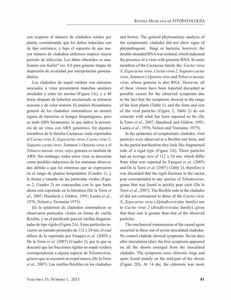
Figura 1. Cladodios de nopal verdura en campo con manchas anulares cloróticas alrededor de las aereolas o entre ellas (A). Síntomas en plantas indicadoras inoculadas con la savia de cladodios de nopal verdura con manchas anulares cloróticas (B). Lesiones locales necróticas en Nicotiana tabacum var. Xanthi (B1). Lesiones locales cloróticas en Chenopodium quinoa (B2). Lesiones locales cloróticas en N. benthamiana (B3).
Figure 1. Cladodes of cactus pear in the field with chlorotic ringspots around the areoles or between them (A). Symptoms on indicator plants inoculated with sap of cladodes of cactus pear with chlorotic ringspots (B). Necrotic local injuries on Nicotiana tabacum var. Xanthi (B1). Chlorotic local injuries on Chenopodium quinoa (B2). Chlorotic local injuries on N. benthamiana (B3).
A
B1 B2 B3

Volumen 33, Número 1, 2015 79
Revista Mexicana de FITOPATOLOGÍA
et al., (1976). Además, el purificado parcial y el macerado de la epidermis que cubría los halos clo-róticos de cladodios enfermos se colocaron inde-pendientemente en rejillas de cobre de malla 400 con formvar, se contrastaron con acetato de uranilo al 2 % y se observaron en un microscopio electrónico de transmisión JEOL modelos JEM-1200 EXII y JEM-100 CX II de la Central de Instrumentación de Microscopía de la Escuela Nacional de Ciencias Biológicas del IPN y de la Facultad de Estudios Superiores Unidad Iztacala de la UNAM, respec-tivamente.
La transmisión del posible virus se hizo median-te inoculación mecánica en tres grupos de 11 plan-tas indicadoras cada uno (Nicotiana tabacum cv. Xanthi, N. rustica, N. glutinosa, N. occidentalis, N. benthamiana, N. clevelandii, Capsicum annuum, Chenopodium quinoa, Ch. amaranticolor, Datura stramonium y Solanum lycopersicum). Como fuen-te de inóculo se utilizó la epidermis de cladodios sintomáticos recolectados en Cuautepec de Hinojo-sa. Las plantas se mantuvieron en invernadero a 25-30 °C con humedad relativa del 70±5 % y 12 horas-luz, y se revisaron durante 15 días para registrar la aparición de síntomas. La transmisión mecánica a plantas de nopal se hizo también mecánicamente en siete cladodios asintomáticos de plantas diferentes. Como fuente de inóculo se utilizó la epidermis de cladodios sintomáticos, la cual se maceró en amor-tiguador de fosfatos + DIECA 0.01M pH 7.2. Se espolvoreó carborundum de 600 mallas sobre una superficie aproximada de 5 cm2 en la porción supe-rior del cladodio, y con un hisopo se tomó el inó-culo y se frotó sobre dicha superficie. Como testigo negativo se inocularon siete plantas asintomáticas con un hisopo humedecido en el amortiguador an-tes indicado. Todas las plantas se mantuvieron en invernadero durante 45 días y se observaron cada 72 h para registrar la aparición de síntomas. Las plantas utilizadas en esta fase se obtuvieron en
The transmission of the possible virus was performed through mechanical inoculation on three groups of eleven indicator plants (Nicotiana tabacum cv. Xanthi, N. rustica, N. glutinosa, N. occidentalis, N. benthamiana, N. clevelandii, Capsicum annuum, Chenopodium quinoa, Ch. amaranticolor, Datura stramonium y Solanum lycopersicum). The epidermis of symptomatic cladodes collected in Cuautepec de Hinojosa was used as a source of inoculum. The plants were kept in greenhouse at 25-30 ºC with relative humidity of 70 ± 5 % and 12 daylight-hours; they were examined during 15 days in order to record the appearance of symptoms. Mecanical transmission to cactus plants was also done mechanically on seven asymptomatic cladodes of different plants. The epidermis of symptomatic cladodes was used as a source of inoculum, which was macerated in phosphate buffer + DIECA 0.01M pH 7.2. 600 mesh carborundum was sprinkled over a surface of approximately 5 cm2 on the upper portion of the cladode; the inoculum was taken with a cotton swab and was rubbed over the surface. As a negative control, seven asymptomatic plants were inoculated with a cotton swab previously moistened with the buffer mentioned above. All the plants were kept in greenhouse for 45 days and were observed every 72 hours in order to record the appearance of symptoms. The plants used in this phase were obtained from Tlalnepantla, Morelos; a region where no viral symptoms have been observed, in order to have natural growth conditions.
The extraction of RNA was done with the epidermis of symptomatic and asymptomatic cladodes with the commercial product Trizol© following the manufacturer’s instructions, and with the silica method. The RT-PCR was done with primers for Tobacco mosaic virus (TMV), Carnation mottle virus (CarMV) and for the Potexvirus and Tobamovirus genera. For TMV, the RT-PCR was

Revista Mexicana de FITOPATOLOGÍA
Volumen 33, Número 1, 2015 80
Tlalnepantla, Morelos, región en donde no se han observado síntomas virales, a fin de tener condicio-nes naturales de crecimiento.
La extracción de ARN se hizo de la epidermis de cladodios sintomáticos y asintomáticos con el pro-ducto comercial Trizol© siguiendo las instrucciones del fabricante, y con el método de sílica. La RT-PCR se hizo con iniciadores para el Tobacco mo-saic virus (TMV), Carnation mottle virus (CarMV) y para los géneros Potexvirus y Tobamovirus. Para el TMV la RT-PCR se realizó en un solo paso con los iniciadores específicos TMVCPF y TMVCPR (Ortega et al., 2007) que amplifican un fragmento de 240 pb. El fragmento de la cubierta proteica del CarMV (1.05 kb) se amplificó con los iniciadores CarMV y CarMV/F (Singh et al., 2005). Para el gé-nero Potexvirus (Van der Vlugt et al., 2000) se uti-lizaron los iniciadores POTEX1RC, POTEX2RC y POTEX5RC. Las combinaciones POTEX1RC / POTEX5RC y POTEX2RC / POTEX5RC amplifi-can un fragmento de 735 y 584 pb, respectivamen-te, correspondientes a la replicasa viral. Para algún miembro del género Tobamovirus se utilizaron los iniciadores TMV5vAccl / TMV3vc y TMVcp5 / TMVcp3, que amplifican un fragmento de 360-419, y 300 pb, respectivamente. Para cada par de iniciadores se realizó la RT-PCR en un solo paso.
Para evaluar la distribución de la incidencia de síntomas en las unidades de producción, se deter-minó el número de plantas con manchas cloróticas irregulares o circulares, y la presencia de anillos cloróticos alrededor de las areolas. La primera uni-dad se dividió en tres secciones de cinco surcos cada una (localizadas a la entrada, parte media y final del invernadero, respectivamente) para eva-luar 50 plantas/surco/sección (250 plantas en total). En cada una de las otras dos unidades se evaluaron 20 plantas continuas en cinco surcos (100 plantas en total). La severidad de la enfermedad se estimó cuantificando el número de cladodios sintomáticos
done in a single step with the specific primers TMVCPF and TMVCPR (Ortega et. al, 2007) that amplify a fragment of 240 pb. The fragment of the protein cover of the CarMV (1.05 kb) was amplified with the CarMV and CarMV/F (Singh et al., 2005). For the Potexvirus genus (Van der Vlugt et al., 2000) used the POTEX1RC, POTEX2RC. The PTEX1RC / POTEX5RC and POTEX2RC / pairs POTEX5RC amplify a fragment of 735 and 584 pb, respectively, corresponding to the viral replicase. For any member of the Tobamovirus genus, the primers used were TMV5vAccl / TMV3vc and TMVcp5 / primers TMVcp3, which amplify a fragment of 360-419, and 300 pb, respectively. For each pair of primers, the RT-PCR was done in a single step.
In order to evaluate the distribution of the incidence of symptoms in the production units, the number of plants with irregular or circular chlorotic spots and the presence of chlorotic rings around the areoles were determined. The first unit was divided in three sections of five furrows each (located at the entrance, middle and end of the greenhouse, respectively) in order to evaluate 50 plants/furrow/section (250 plants total). In each of the other two units, 20 continuous plants were evaluated in five furrows (100 plants total). The severity of the disease was estimated quantifying the number of symptomatic cladodes with regard to the number of total cladodes per plant, considering that the induced damages are of a systemic type, and under the supposition that greater number of infected cladodes implied a greater period of infection. The data obtained was analyzed the using Surfer® program ver. 8.0 in order to generate dispersion maps of severity with geostatistic interpolation.
The cladodes of cactus pear with symptoms associated to virus showed ringspots around and between the areoles (Figure 1A), and 48 hours after having been collected they turned aqueous
Volumen 33, Número 1, 2015 81
Revista Mexicana de FITOPATOLOGÍA
con respecto al número de cladodios totales por planta, considerando que los daños inducidos son de tipo sistémico, y bajo el supuesto de que ma-yor número de cladodios enfermos implicó mayor periodo de infección. Los datos obtenidos se ana-lizaron con Surfer® ver. 8.0 para generar mapas de dispersión de severidad por interpolación geoesta-dística.
Los cladodios de nopal verdura con síntomas asociados a virus presentaron manchas anulares alrededor y entre las areolas (Figura 1A), y a 48 horas después de haberlos recolectado se tornaron acuosos y de color marrón. El análisis fitosanitario general de los cladodios sintomáticos no mostró signos de bacterias ni hongos fitopatógenos, pero se aisló ARN bicatenario, lo que indicó la presen-cia de un virus con ARN genómico. En algunos miembros de la familia Cactaceae están reportados el Cactus virus X, Zygocactus virus, Cactus virus 2, Saguaro cactus virus, Sammon´s Opuntia virus y el Tobacco mosaic virus, cuyo genoma es también de ARN. Sin embargo, todos estos virus se descartan como posibles inductores de los síntomas observa-dos debido a que los síntomas que se expresaron en el rango de plantas hospedantes (Cuadro 1), y la forma y tamaño de las partículas virales (Figu-ra 2, Cuadro 2) no concuerdan con lo que hasta ahora está reportado en la literatura (De la Torre et al., 2007; Hausbeck y Gildow, 1991; Lastra et al., 1976; Nelson y Tremaine 1975).
En la epidermis de cladodios sintomáticos se observaron partículas virales en forma de varilla flexible, y en el purificado parcial varillas fragmen-tadas de tipo rígido (Figura 2A). Estas partículas tu-vieron un tamaño promedio de 112 x 24 nm, el cual difiere de lo reportado por Fauquet et al. (2005) y De la Torre et al. (2007) (Cuadro 2), por lo que se descartó que las fracciones rígidas en nopal verdura correspondieran a alguna especie de Tobamovirus, género que se encontró en nopal tunero (De la Torre et al., 2007). Las varillas flexibles en los cladodios
and brown. The general phytosanitary analysis of the symptomatic cladodes did not show signs of phtypathogenic fungi or bacteria; however, the double-stranded RNA was isolated, which indicated the presence of a virus with genomic RNA. In some members of the Cactaceae family the: Cactus virus X, Zygocactus virus, Cactus virus 2, Saguaro cactus virus, Sammon’s Opuntia virus and Tobacco mosaic virus, whose genome is also RNA. However, all of these viruses have been reported discarded as possible causes for the observed symptoms due to the fact that the symptoms showed in the range of the host plants (Table 1), and the form and size of the viral particles (Figure 2, Table 2) do not coincide with what has been reported so far (De la Torre et al., 2007; Hausbeck and Gildow, 1991; Lastra et al., 1976; Nelson and Tremaine, 1975).
In the epidermis of symptomatic cladodes, viral particles were observed in a flexible rod form, and in the partial purification they look like fragmented rods of a rigid type (Figure 2A). These particles had an average size of 112 x 24 nm, which differ from what was reported by Fauquet et al. (2005) and De la Torre et al. (2007) (Table 2), therefore it was discarded that the rigid fractions in the cactus pear corresponded to any species of Tobamovirus, genus that was found in prickly pear cacti (De la Torre et al., 2007). The flexible rods in the cladodes of did not correspond to those of the Cactus virus X, Zygocactus virus (Alphaflexiviridae family) nor to Cactus virus 2 (Betaflexiviridae family), given that their size is greater than that of the observed particles.
The mechanical transmission of the causal agent occurred in three out of seven inoculated cladodes. No control cladode showed symptoms. Seven days after inoculation (dai), the first symptoms appeared on all the shoots emerged from the inoculated cladodes. The symptoms were chlorotic rings and spots found mainly on the mid-part of the shoots (Figure 2D). At 14 dai, the chlorosis was more

Revista Mexicana de FITOPATOLOGÍA
Volumen 33, Número 1, 2015 82
de nopal no correspondieron a las del Cactus virus X, Zygocactus virus (familia Alphaflexiviridae) ni al Cactus virus 2 (Familia Betaflexiviridae), ya que su tamaño es mayor al de las partículas observadas.
La transmisión mecánica del agente causal ocu-rrió en tres de los siete cladodios inoculados. Nin-gún cladodio testigo mostró síntomas. A los siete días después de la inoculación (ddi), se presentaron los primeros síntomas en todos los brotes emitidos de los cladodios inoculados. Los síntomas fueron anillos y halos cloróticos localizados principalmen-te en la parte media de los brotes (Figura 2D). A los 14 ddi, la clorosis fue más evidente e incrementaron el número y tamaño de halos cloróticos (1-1.5cm), principalmente rodeando las areolas (Figura 2D). A los 25 ddi, los halos se convirtieron en manchas cloróticas circulares (1.5-2cm) e irregulares debi-do a su coalescencia (Figura 2D). Estos síntomas contrastan con los reportados por De la Torre et al. (2007), quienes observaron manchas cloróticas de 0.5-1cm distribuidas uniformemente fuera de la zona areolar en los cladodios de nopal tunero (O. amyclaea). De los virus reportados en cactáceas, únicamente en la asociación Cactus virus X – No-palea cochenillifera (Lastra et al., 1976), se pre-sentan síntomas similares a los observados en el presente trabajo. En cactáceas silvestres este virus induce manchas anulares amarillas simples o con-céntricas de 2-4 cm entorno a las areolas. Con base en la identificación por RT-PCR, los cladodios sin-tomáticos estuvieron infectados con alguna especie del género Potexvirus (Figura 2D). Este resultado es congruente con el análisis en microscopía elec-trónica de transmisión, en donde se observaron partículas virales flexibles, por lo que los síntomas asociados a cladodios de nopal verdura podrían co-rresponder a alguna especie del género Potexvirus, a excepción del Cactus virus X y Zygocactus virus, cuyas partículas son más grandes que las del virus aislado de los cladodios de nopal verdura; además,
Cuadro 1. Síntomas en plantas indicadoras inoculadas con el virus aislado de cladodios de nopal verdura con manchas anulares cloróticas. Cuautepec de Hinojosa, Hidalgo.
Table 1. Symptoms on indicator plants inoculated with the vi-rus isolated from cladodes of cactus pear with chlo-rotic ringspots. Cuautepec de Hinojosa, Hidalgo.
Hospedante Síntomas
Capsicum annuum var. Ancho AsintomáticaChenopodium amaranticolor Lesiones locales cloróticasC. quinoa Lesiones locales cloróticasDatura stramonium Lesiones locales cloróticasNicotiana benthamiana Lesión sistémicaN. clevelandii AsintomáticaN. glutinosa Lesiones locales cloróticasN. occidentalis AsintomáticaN. rustica AsintomáticaN. tabacum var. Xanthi Lesiones locales cloróticasSolanum lycopersicum Asintomática
evident and the number and size of the chlorotic spots increased, mainly circling the areoles (Figure 2D). At 25 dai, the rings became chlorotic spots, circular and irregular due to their coalescence (Figure 2D). These symptoms contrast with those reported by De la Torre et al. (2007), who observed chlorotic spots of 0.5-1cm uniformly distributed outside the areolar zone on the cladodes of the prickly pear cacti (O. amyclaea). Of the viruses reported in cacti, only in the Cactus virus X – Nopalea cochenillifera interaction (Lastra et al., 1976), the symptoms showed are similar to to those observed in this work. In wild cacti, this virus induces simple or concentric yellowish ringspots of 2-4 cm surrounding the areoles. According to identification by RT-PCR, the symptomatic cladodes were infected with a species of the genus Potexvirus (Figure 2D). This result is congruent with the transmission electron microscopy analysis, where flexible viral particles were observed, so the symptoms associated to cladodes of cactus pear could correspond to some specie of the genus

Volumen 33, Número 1, 2015 83
Revista Mexicana de FITOPATOLOGÍA
Figura 2. Partículas virales presentes en el purificado parcial obtenido de cladodios de nopal verdura con síntomas putativos a virus (A). Bandas de RNA bicatenario obtenidas a partir de plantas de nopal verdura (B). Carril 1: marcador molecular de 1Kb; carril 2: cladodios asintomáticos; carril 3: cladodios con manchas anulares cloróticas alrededor de las areolas. Productos de amplificación de RT-PCR para el género Potexvirus (C). Carril 1: marcador molecular de 1 Kb (Plus Invitrogen); carriles 2-6 y 8-12: cladodios de nopal verdura con manchas anulares cloróticas; carril 13: tejido foliar de orquídea infectada con un Potexvirus; carril 14: testigo negativo; carril 7: sin muestra. Manchas anulares cloróticas en brotes de cladodios inoculados con savia infectiva a los 7 (D1), 14 (D2) y 25 (D3) días después de la inoculación.
Figure 2. Viral particulates found in the partial purification obtained from cladodes of cactus pear with putative virus symptoms (A). Bands of double-stranded RNA obtained from the cactus pear (B). Line 1: molecular marker of 1Kb; line 2: asymptomatic cladodes; line 3: cladodes with chlorotic ringspots around the areoles. Products of RT-PCR amplification for the genus Potexvirus (C). Line 1: molecular marker 1 Kb (Plus Invitrogen); line 2-6 and 8-12: cladodes of cactus pear with chlorotic ringspots; line 13: foliar tissue of orchid infected with Potexvirus; line 14: negative control; line 7: without sample. Chlorotic ringspots in growths of cladodes inoculated with infected sap at 7 (D1), 14 (D2) and 25 (D3) days after inoculation.
A B
100 nm
1 2 3
2 3 4 5 6 7 81 9 10 11 12 13 14
600 pb
C
D1 D2 D3

Revista Mexicana de FITOPATOLOGÍA
Volumen 33, Número 1, 2015 84
los síntomas inducidos por Zygocactus virus en C. quinoa son distintos a los encontrados en el presen-te trabajo.
La incidencia de plantas enfermas y la severi-dad promedio fue de 47-60 y 51-79 %, respecti-vamente, lo que sugiere una infección crónica y la transmisibilidad vegetativa del agente causal. En Taiwán, el Cactus virus X causó en pithaya (Hylo-cereus undatus) una incidencia de 90, 50 y 60-70 % en Pingtung, Kinman y en otras áreas, respec-tivamente (Liao et al., 2003). El patrón de distri-bución espacial de plantas enfermas fue agregado con direccionalidad de los surcos (Figura 3). Los agregados fueron de 2-5 plantas implicando po-sible diseminación por medio de poda y cosecha, las cuales se realizan con cuchillo sin desinfestar. Esta posibilidad se reafirma con resultados de la transmisión mecánica (43 %), por lo que el manejo de este virus debe ser preventivo mediante uso de material propagativo sano y con desinfestación de herramientas. Debido a que el altiplano mexicano es la principal región productora de nopal verdura, es necesario determinar la especie encontrada del género Potexvirus y conducir estudios epidemioló-gicos y de manejo.
Cuadro 2. Tipo y tamaño de partícula viral en el purificado parcial obtenido de cladodios de nopal verdura con manchas anulares cloróticas. Cuautepec de Hinojosa, Hidalgo.
Table 2. Type and size of a viral particle in a partial purified obtained from cactus pear cladodes with clhorotic ringspots. Cuatepec de Hinojosa, Hidalgo.
Virus Tipo de partícula Tamañonm Referencias
Potexvirus Varilla flexible En esta investigaciónPotexvirus Varilla rígida 112 x 24 En esta investigaciónSammon´s Opuntia Virus Varilla rígidaSaguaro virusZygocactus virus Varilla flexibleCactus virus XCactus virus 2TSWVTMV
Tobamovirus Varilla rígida 325 x ¿?317 x 18
De la Torre et al., 2007Fauquet et al., 2005
Potexvirus, except Cactus virus X and Zygocactus, whose particles are larger than those isolated from the cactus cladodes; furthermore, the induced symptoms by Zygocactus virus on C. quinoa are different to those found in this work.
The incidence of diseased plants and the average severity was of 47-60 and 51-79 %, respectively, which suggest a chronic infection and the vegetative transmission of the causal agent. In Taiwan, the Cactus virus X caused on the pitahaya (Hylocereus undatus) an incidence of 90, 50 and 60-70% in Pingtung, Kinman and in other areas, respectively (Liao et al., 2003). The spatial distribution pattern of diseased plants was added with directionality of the furrows (Figure 3). The aggregates were 2-5 plants implicating possible dissemination by means of pruning and harvest tools, which are carried out using a non-desinfested knife. This possibility agrees with the results of the mechanical transmission (43%), therefore the management of this virus must be preventive through the use of healthy material and desinfested tools. Due to the Mexican plateau is the most important cactus pear production region, it is necessary to determine the species of the Potexvirus that was found and

Volumen 33, Número 1, 2015 85
Revista Mexicana de FITOPATOLOGÍA
CONCLUSIONES
En este trabajo se reporta un nuevo virus afec-tando a la familia Cactaceae en México. Los sín-tomas de halos y manchas cloróticas irregulares y circulares presentes en O. ficus-indica se rela-cionaron con un virus de ARN de cadena sencilla con partícula de tipo varilla flexible. Con base en la RT-PCR, se confirmó que el virus pertenece al género Potexvirus. La incidencia de la enfermedad fue del 47-60 %, con distribución agregada y fuerte tendencia en dirección de surcos posiblemente por efecto del manejo y cosecha.
Figura 3. Mapa de distribución espacial de la severidad de la enfermedad asociada a manchas cloróticas en nopal verdura en Cuautepec de Hinojosa, Hidalgo.
Figure 3. Spatial distribution map of the severity of the disease associated with chlorotic spots on cactus pear in Cuautepec de Hinojosa, Hidalgo.
Plan
tas
Dire
cció
n de
l sur
co
SurcosUnidad de producción 1 Unidad de
producción 2Unidad de producción 3
to carry out epidemiological and management studies. Given that the Mexican plateau is the main region that produces cactus pear, it is necessary to determine the species found in the genus Potexvirus and carry out epidemiological and management studies.
CONCLUSIONS
In this work, a new virus is reported affecting the Cactaceae family in Mexico. The irregular

Revista Mexicana de FITOPATOLOGÍA
Volumen 33, Número 1, 2015 86
Agradecimientos
A la M.C. Ma. Esther Sánchez Espíndola, de la Central de Ins-
trumentación de Microscopía de la ENCB-IPN, por su apoyo
en la observación y fotografías al microscopio electrónico de
transmisión (MET). Al técnico Jesús Espinoza de la FES-Iz-
tacala UNAM, por las fotografías en el MET. Al Sr. Fermín
Cerón Téllez, por permitir hacer este estudio en las unidades
productivas de nopal verdura. Al CONACyT, por proporcionar
la beca de postgrado.
LITERATURA CITADA
Brandes J, Wetter C. 1959. Classification of elongated plant vi-ruses on the basis of particle morphology. Virology 8: 99.
De la Torre AR, Salazar SM y Ruiz MR. 2007. Ocurrencia de un Tobamovirus asociado con manchas anulares amarillas en nopal tunero en México. Agrociencia 41:763-773.
Dodds J, Morris T and Jordan R. 1987. Plant viral double stran-ded RNA. Annual Review of Phytopathology 22:151-168.
Fauquet CM, Mayo MA, Maniloff J, Desselberger U and Ball LA. 2005. Virus taxonomy Eighth Report of the Interna-tional Committee on Taxonomy of Virus. Academic Press, San Diego, CA. 1259 p.
Giri L and Chessin M. 1975. A severe strain of Tobacco mosaic virus from cactus. Phytopathology 65: 824-825.
Hausbeck MK and Gildow FE. 1991. First report of Tomato spotted wilt virus on thanksgiving cactus. Plant Disease 75: 215.
Lastra JR, Gaskin D. and Uzcategui RC. 1976. Virus X del cactus en Venezuela. Agronomía Tropical 26: 303-310.
Liao JY, Chang CA, Yan CR, Chen YC, and Deng TC. 2003. Detection and incidence of Cactus virus X on pitaya in Taiwan. Plant Pathology Bulletin 12: 225-234.
Milbrath MG and Nelson MR. 1972. Isolation and characte-rization of a virus from saguaro cactus. Phytopathology 62:739-742.
Nelson MR and Tremaine JH. 1975. Physiochemical and sero-logical properties of virus from saguaro cactus. Virology 65: 309-319.
Ortega E, Zambrano K, Carballo O, Romano M. and Edgloris M. 2007. Caracterización de un Tobamovirus aislado de plantas de Canavalia (Canavalia ensiformis L.) Aragua, Venezuela. INCI. 32:202-205.
and circular chlorotic rings and spots symptoms found on O. ficus-indica were related to a virus of a simple RNA chain with particles of a flexible rod type. Based on the RT-PCR results, it was confirmed that the virus belongs to the Potexvirus genus. The incidence of the disease was of 47-60%, with aggregated distribution and a strong tendency to row direction possibly due to management and harvest effects.
Acknowledgements
To M.C. Ma. Esther Sánchez Espíndola, of the Central de
Instrumentación de Microscopía de la ENCB-IPN, for her aid
in the observation and photographs using the transmission
electron microscope (TEM). To the technician Jesús Espinoza
of the FES-Iztacala UNAM, for the photographs in the
TEM. To Fermín Cerón Téllez, for allowing this study in
the production units of cactus pear. To the CONACYT for
providing the postgraduate grant.
Finaliza la versión en Inglés
SIAP-SAGARPA. 2013. Anuario estadístico de la producción agrícola. Servicio de información agroalimentaria y pes-quera. www.siap.gob.mx/index (consulta, junio del 2015).
Singh HP, Hallan V, Raikhy G, Kulshrestha S, Sharma ML, Ram R, Garg ID and Zaidi AA. 2005. Characterization of an Indian isolate of Carnation mottle virus infecting carna-tions. Current Science 88: 25.
Van Der Vlugt RAA, Stijger CM, Verhoeven JThJ and Lese-mann DE. 2000. First report of Pepino mosaic virus on tomato. Plant Disease 84: 103.