aspects of brassica juncea meal toxicity: allyl isothiocyanate
TRANSCRIPT

1
ASPECTS OF BRASSICA JUNCEA MEAL TOXICITY: ALLYL ISOTHIOCYANATE
RELEASE AND BIOASSAY
A Thesis Submitted to the College of
Graduate Studies and Research
in Partial Fulfillment of the Requirements
for the Degree of Master of Science
in the Department of Food and Bioproduct Sciences
University of Saskatchewan
Saskatoon
By
Akal Rachna Kaur Saini
March, 2009
© Copyright Akal Rachna Kaur Saini, 2009, All rights reserved.

i
PERMISSION TO USE
In presenting this thesis in partial fulfillment of the requirements for a Masters
degree from the University of Saskatchewan, I agree that the Libraries of this University
may make it freely available for inspection. I further agree that permission for copying
of this thesis in any manner, in whole or in part, for scholarly purposes may be granted
by the professor or professors who supervised my thesis work or, in their absence, by the
Head of the Department or the Dean of the College in which my thesis work was done.
It is understood that any copying or publication or use of this thesis or parts thereof for
financial gain shall not be allowed without my written permission. It is also understood
that due recognition shall be given to me and to the University of Saskatchewan in any
scholarly use which may be made of any material in my thesis.
Requests for permission to copy or to make other use of material in this thesis in
whole or part should be addressed to:
Head of the Department of Food and Bioproduct Sciences
University of Saskatchewan
Saskatoon SK, S7N 5A8, Canada

ii
ABSTRACT
Oilseed and oilseed meal extracted from members of Brassicaceae release broad-
spectrum biocidal isothiocyanate when ground and exposed to moisture. The compounds
are released when the seed enzyme myrosinase catalyzes the hydrolysis of
glucosinolates producing glucose, sulfate, and pesticidal isothiocyanates.
Allylisothiocyanate (AITC), the predominant isothiocyanate of Brassica juncea, has
broad-spectrum biological activities against plants, animals and fungi. Knowledge of the
concentration of AITC arising from a treatment with mustard and AITC toxicity to many
target and non-target species is not known. Therefore, factors affecting AITC release
and assays of mustard toxicity were conducted. The rate of AITC release from mustard
meal was affected by temperature and pH. Current isothiocyanate extraction and
quantification methods measure a change in the concentration of glucose (a predominant
product of myrosinase-catalysed glucosinolate hydrolysis) to determine myrosinase
activity. The objectives of this work were to study: 1) factors affecting myrosinase
activity in mustard (Brassica juncea), 2) the effects of AITC on seed germination and 3)
the toxicity of AITC and mustard meal.
Attempts were made to improve the Herb and Spice Method, the only available
industrial method to measure total isothiocyanate production in mustard meal. The
effects of a wide range of reaction temperatures (7 to 97°C) and incubation times (0 min
to 2 h) on myrosinase-catalyzed conversion of sinigrin (a glucosinolate) to allyl
isothiocynate (AITC) were studied. Significant inhibition of enzyme activity was
observed at all temperatures over 57°C, and at 97°C no myrosinase activity was found. It
was concluded that myrosinase-catalyzed conversion of sinigrin to AITC was a rapid
process and detectable amounts of AITC could be found in samples in two min, and that
higher temperatures inhibited myrosinase activity. The pH of the reaction mixture
significantly affected myrosinase-catalyzed conversion of sinigrin to AITC. A change in
pH did not affect the substrate, but severely affected the activity of myrosinase.
Furthermore, other compounds viz., boric acid (H3BO3), succinic acid (C2H4(COOH)2),
calcium chloride (CaCl2) and ethanol (C2H5OH), were explored for their ability to
inhibit myrosinase activity. Calcium chloride and ethanol were particularly effective.

iii
It was hypothesized that AITC might act as a plant growth promoter/regulator based
on the fact that AITC and ethylene, a plant growth regulator, exhibit structural similarity
(R-CH=CH2, where R is -CH2SCN and -H in AITC and ethylene, respectively).
Therefore, AITC might act as an ethylene analogue. Ethylene is known to promote seed
germination and overcome seed dormancy in a dose- and species-dependent manner.
Flax and tomato seeds were used as model systems to test the germination enhancing
properties of AITC. It was concluded that AITC promoted flax and tomato seed
germination and thus might be used for this application in agricultural practice.
An assay was developed for testing AITC toxicity in ground seed by
exploring HSP70 expression in Caenorhabditis elegans as a marker of toxicity. C.
elegans strain N2 was exposed to different concentrations (0 to 10 µM) of AITC for 2 h
at room temperature. Western blotting with anti-HSP70 antibody showed a marked
increase in the expression of HSP70 protein in a dose-dependent manner. Assays of the
expression of HSP70A mRNA by quantitative real time reverse transcriptase (RT) PCR
revealed no significant change in the expression of HSP70A mRNA at low
concentrations of AITC (< 0.1 µM). However, treatment with higher concentrations (>1
µM) resulted in four- to five - fold increase in expression of HSP70A mRNA over the
control. To understand if mustard toxicity was due to AITC alone, or if other compounds
in mustard ground seed affected HSP70 transcript production, C. elegans was exposed to
AITC or Brassica juncea cv. Arrid ground seed (Arrid is a mustard variety with a lower
level of sinigrin (<3 µM per gram of seed), or both. ELISA revealed increased
expression of HSP70 protein in C. elegans treated with AITC + ground seed, but the
level of protein was less than that observed with AITC alone. These results indicated
that mustard ground seed toxicity was contributed primarily by AITC, and that some
ground seed components antagonized AITC toxicity in C. elegans.

iv
ACKNOWLEDGEMENTS
I will like to thank my God for giving me the best in life.
With a deep sense of feelings, I express my utmost gratitude and appreciation to my
supervisor, Dr. Martin J. T. Reaney, for his inspiration, moral support and
encouragement during the entire period of this research.
It is my pleasure to express my gratitude to my co-supervisor, Prof. Robert Tyler, for
his guidance in completing this research. I would like to thank my committee members,
Dr. Mark Wickstorm, and graduate chair, Prof. Phyllis Shand, for their timely help and
valuable suggestions.
My special thanks go to the Agriculture Development Fund, Government of
Saskatchewan, for providing financial support for this project. I am grateful to the
University of Saskatchewan for providing the University Devolved Scholarship, the
Blake John Memorial Scholarship, the ReneVandeveld Bursary, Travel Award and
Education Enhancement Award, College of Agriculture and Biosources to travel during
my M.Sc. program. I am also thankful to Dr. Baljit Singh, Veterinary Biomedical
Sciences, for allowing me to use equipment for the molecular work. Thanks are due to
Ms Sarah Caldwell.
I would like to express my deepest gratitude to my parents for their strong
encouragement of my academic endeavors over the years. Thanks to my brother and
sister for being patient with me at all times. Last but not least, thanks are due to my
beautiful daughter, Amishi, for being there in our life and for bringing laughter and
happiness in life. The completion of this thesis would not be possible without the
support, encouragement, and understanding of my husband Sarabjeet who gave me the
confidence to do research.
I also express my heart felt thanks to my lab members for their support and my
colleagues for their marvelous help and support during this research work.
Akal Rachna K. Saini

v
Dedication
To my parents, for teaching me to do the best I can, my Husband and my cute daughter Amishi

vi
TABLE OF CONTENTS
ABSTRACT
ii
ACKNOWLEDGEMENTS
.
iv
DEDICATION
v
TABLE OF CONTENTS
vi
LIST OF TABLES
x
LIST OF FIGURES
xi
LIST OF ABBREVATIONS
xiii
1. INTRODUCTION
1
2. LITERATURE REVIEW 6
2.1 Mustard 6
2.2 Glucosinolates (GLS) 7
2.2.1 Chemical structure 8
2.2.2 Biosynthesis 12
2.2.3 Hydrolysis 12
2.2.4 Extraction 16
2.2.5 Purification and desulfation 16
2.2.6 Detoxification 17
2.2.7 Methods of GLS determination 18
2.2.7.1 High Pressure Liquid Chromatography (HPLC) 18
2.2.7.2 Gas chromatography (GC) 20

vii
2.2.7.3 Capillary Electrophoresis 21
2.2.7.4 Nuclear Magnetic Resonance (NMR) 23
2.3 Myrosinase 23
2.3.1 Occurrence 23
2.3.2 Distribution 24
2.3.3 Genetics 25
2.3.4 Purification 25
2.4 Allyl isothiocynate (AITC) 25
2.4.1 A potent anti-microbial agent 25
2.4.2 Herbicidal activity 26
2.4.3 As a biofumigant 27
2.4.4 Nematocidal effects 27
2.4.5 Anti-cancer properties 28
2.4.6 Toxicity of other GLS hydrolysis products 28
2.4.7 Time release of AITC 29
2.4.8 Extraction 29
2.4.9 Quantification 29
2.4.9.1 HPLC 30
2.4.9.2 Colorimetric assay 30
2.4.9.3 Ion-pair electrospray mass spectroscopy 30
2.4.10 Thermal degradation 30
2.4.11 Heat shock proteins as markers of stress 31
2.5 Research needs
31
3. MATERIALS AND METHODS 32
3.1 Biological materials 32
3.2 Measurement of AITC in seed meal 32
3.3 Kinetics of myrosinase 34
3.4 Inhibition of myrosinase activity 34
3.5 Seed germination 35
3.6 Maintainance of C. elegans cultures 35

viii
3.7 Chemical treatment of nematodes 35
3.8 Sequence alignment and phylogenetic study 35
3.9 Protein extraction 36
3.10 Protein quantification 36
3.11 Qualitative analysis of HSP70 proteins by Western blotting 37
3.12 Sequence alignments 40
3.13 Quantification of HSP70 by an Enzyme Linked Immunosorbant Assay
(ELISA)
41
3.14 RNA isolation 41
3.15 Formaldehyde-agarose gel electrophoresis 42
3.16 Quantification of RNA 43
3.17 cDNA synthesis 43
3.18 Determination of HSP70 transcript levels by qualitative reverse
transcriptase PCR (RT-PCR)
44
3.19 Electrophoresis of DNA in agarose gels 44
3.20 Determination of HSP70 transcript levels by quantitative real time RT-
PCR
45
3.21 Excision and elution of DNA from agarose gels 45
3.22 Gene sequencing 47
3.23 Statistical analysis of the data
57
4. RESULTS 48
4.1 Factors affecting myrosinase activity 48
4.1.1 Kinetics of myrosinase 48
4.1.2 Inhibition of myrosinase activity in mustard meal 52
4.2 Biological activity of AITC 56
4.2.1 Effect of AITC on the germination of flaxseed 56
4.2.2 Effect of AITC on the germination of tomato seed 62
4.3 HSP70 as an indicator of stress/toxicity in C. elegans 67
4.3.1 Sequence alignments and phylogenetic relationships among HSP70s in C.
elegans
67
ix
4.3.2 Expression of HSP70 protein as an indicator of stress 71
4.3.2.1 Expression of HSP70A transcripts in response to AITC 71
4.3.2.2 Reverse transcriptase polymerase chain reaction (RTPCR) 71
4.3.2.3 Quantitative real time RTPCR 73
4.3.2.4 Western blotting 78
4.4 Mustard toxicity is contributed by AITC
81
5. DISCUSSION
83
6. SUMMARY AND CONCLUSIONS
90
7. REFERENCES 93

x
LIST OF TABLES
Table 2-1. Common names and the side chain of some GLS
11
Table 4-1. Effect of reaction pH on the yield of AITC
53
Table 4-2. Accession numbers of various known HSP70s in C. elegans and their functions
69
Table 4-3. Properties of primers used for real time RT-PCR. 74

xi
LIST OF FIGURES
Figure 2-1. Structure of Glucosinolate 9
Figure 2-2. Chemical structure of GLS 10
Figure 2-3. Biosynthesis of GLS 13
Figure 2-4. Outline of GLS hydrolysis 15
Figure 3-1. Apparatus used for quantification of volatile oil in mustard meal 33
Figure 3-2. Apparatus used for polyacrylamide gel electrophoresis and
Western blotting. (Source: Biorad Laboratories)
38
Figure 3-3. Real time PCR machine used for the study 46
Figure 4-1. Effect of temperature and incubation time on the yield of AITC,
which is a measure of myrosinase activity
49
Figure 4-2. Myrosinase attains near-maximum activity in 5 min at 27 oC 50
Figure 4-3. Myrosinase activity measured as % volatile oil at 27 ºC 51
Figure 4-4. Inhibition of myrosinase activity by HCl 54
Figure 4-5. Effect of different compounds on myrosinase activity 55
Figure 4-6. Structures of AITC and ethylene 57
Figure 4-7. Effect of AITC on the germination of flaxseeds 58
Figure 4-8. Effect of Ethephon® on the germination of flaxseeds 59
Figure 4-9. Effect of IAA on the germination of flaxseeds 60
Figure 4-10. Ten-days-old flaxseedling grown in different concentrations of
IAA, Ethephon® and AITC
61
Figure 4-11. Effect of AITC on the germination of tomato seeds 63
Figure 4-12. Effect of Ethephon® on the germination of tomato seeds 64
Figure 4-13. Effect of IAA on the germination of tomato seeds 65
Figure 4-14. Ten-days-old tomato seedlings grown in different concentrations
of IAA, Ethephon® and AITC
66
Figure 4-15. Sequence alignment of HSP70A, HSP70C, HSP70D and HSP70F
showing identity in peptide sequences
68
Figure 4-16. Phylogenetic relationships among various HSP70 proteins in C.
elegans.
70

xii
Figure 4-17. RTPCR showing the expression of HSP70A (Accession No.
M18540, NCBI) in response to stress caused by different
concentrations of AITC
72
Figure 4-18. Nucleotide sequence alignment of various isoforms of HSP70A
and position of primers for the amplification of HSP70A showed
primer specificity.
75
Figure 4-19. Fluorescence (dRn) values of the SYBR green and normalizing
ROX obtained for GAPDH and HSP70A from a single sample
showed a sigmoid curve.
76
Figure 4-20. Real time RTPCR for the expression of HSP70A in response to
stress caused by different concentrations of AITC
77
Figure 4-21. Aminoacid sequence alignment of human (Accession No.
NM_005346) and C. elegans (Accession No. M18540) HSP70
protein, showed 80% identity
79
Figure 4-22. Western blot analysis showing the induced expression of HSP70
protein in response to stress caused by AITC
80
Figure 4-23. Expression of HSP70 protein in C.elegans in response to exposure
to AITC, meal and AITC+meal
82

xiii
LIST OF ABBREVATIONS
ACN Acetonitrile
AITC Allyl isothiocyanate
ANDSA 7-aminonaphthalene-1, 3-disulfonic acid
AP-1 Activator protein 1
APS Ammonium persulphate
BITC Benzyl isothiocyanate
CE Capillary Electrophoresis
CTAB Cetyltrimethylammonium bromide
ERK Extracellular signal- regulated kinase
ERN Erucin
GAPDH Glyceraldehyde-3-phosphate dehydrogenase
GC Gas chromatography
GLS Glucosinolate
HPLC High Pressure Liquid Chromatography
HPLC-MS HPLC coupled with mass spectroscopy
HRP Horseradish peroxidase
HSP Heat shock protein
IBN Iberin
ITC Isothiocyanate
JNK c-Jun N-terminal kinase
MAPK Mitogen-activated protein kinase
MECC Micellar Electrokinetic Capillary Chromatography
NAC N-acetylcysteine
NCRR Nationa l Centre for Research Resources
NIH National Institutes of Health
NMR Nuclear Magnetic Resonance
PAGE Polyacylamide gel electrophoresis
PBS Phosphate buffered saline

xiv
PEITC Phenethyl isothiocyanate
PVDF Polyvinylidene difluoride
RT-PCR Reverse transcriptase polymerase chain reaction
SDS Sodium dodecyl sulfate
SFN Sulforaphane
TAE Tris-acetate/EDTA
TBA Tetrabutylammonium
TEMED Tetramethylethylenediamine
TMAB Tetramethylammonium bromide
TNF-α Tumour necrosis factor-α
1
Chapter 1: Introduction
Canada is a major producer of condiment mustard seed. Mustard is the only
oilseed crop adapted to the drier regions of the Canadian prairies and provides a suitable
crop for rotation with wheat. Furthermore, mustard is resistant to many pests and
diseases that affect canola and thus can be grown with fewer inputs. Covercropping with
mustard has been tested recently for weed management (Haramoto and Gallandt, 2004).
Seed producing crops from the plant family Brassicaeae are used for food and non-food
applications. The oil produced from these crops has increasing demand in non-food
applications such as industrial lubricants (Heath and Earle, 1995), biodiesel fuel (Kimber
and McGregor, 1995) and biopesticides (Bones, and Rossiter, 1996; Brown and Morra,
2005). The protein-rich oilseed meal from canola may be used in animal feed (Sakorn, et
al., 1999) and human food (Hiron et al., 2006).
In response to biotic challenges, mustard has evolved a broad spectrum of natural
defence mechanisms, such as physical and chemical barriers. The glucosinolate (GLS) -
myrosinase system referred to as ‘The Mustard Bomb’ is a primary chemical defence
used by mustard species against a wide range of biotic challenges (Bones and Rossiter,
1996). Specific GLS (glucoraphanin, glucoerucin, gluconasturtiin, sinigrin,
glucotropaeolin, glucoraphenin, glucoraphasatin, glucomoringin and glucobrassicin) are
hydrolysed by the myrosinase enzyme (thioglucosidase) to produce an aglycone, which
undergoes spontaneous non-enzymatic rearrangement to produce organic
isothiocyanates (ITCs), thiocyanates, nitriles, epithionitriles, oxazolidinethiones, and
organic cyanates (Fahey et al., 2001; Mithen, 2001; Fenwick et al., 1983; Chew, 1988).
Many GLS products are of interest because of their broad spectrum of biological
activities.

2
Mustard seed flour contains 28% to 36% protein. The presence of tocopherols in
mustard contributes to its shelf life (Cahoon et al., 2003). Mustard is widely known for
its sharp taste and is an essential component of many dressings and sauces. The volatile
oil in mustard inhibits the growth of certain yeasts, moulds and bacteria, enabling
mustard to function as a natural preservative. GLS hydrolysis products have biocidal
activity against a wide variety of organisms such as insects, plants, fungi and bacteria
(Vaughn and Berhow, 2005; Ware, 2000). Certain ITCs interfere with iodine
availability, and are responsible for morphological and physiological changes in the
thyroid gland (Tripathi and Mishra, 2006). The fungicidal property of allyl isothiocynate
(AITC) vapour against wild type and thiabendazole-resistant strains of Penicillium
expansum has been demonstrated (Tunc et al., 2007; Kiyoshi 2005; Mari et al., 2002).
The use of AITC produced from pure sinigrin or from Brassica juncea defatted meal
may be an economically viable alternative to synthetic fungicides against P. expansum.
The relative toxicity of different GLS hydrolysis products is dependant both on
the target organism and the chemical structure. Thus, it is important to determine
toxicity of GLS hydrolysis products inorder to maximize the likelihood of effective pest
suppression in pesticide applications (Sarwar et al., 1998). These products possess
potential biodegradable and biofriendly insect fumigant properties (Tsao et al., 2002a)
and nematocidal properties (Mitarai et al., 1997). They may act on the insect respiratory
system.
Specific ITCs are well-known cancer- inhibitory phytochemicals (Hu et al., 2007;
Thejass and Kuttan, 2007; Hwang and Lee 2006; Tang et al., 2006; Jakubikova et al.,
2005; Smith et al., 2004; Zhang, 2004; Myzak et al., 2004; Thornalley, 2002). Allyl
isothiocyanate (AITC), benzyl- ITC (BITC), phenethyl- ITC (PEITC), sulforaphane
(SFN), erucin (ERN) and iberin (IBN) induce time- and dose-dependent G2/M arrest in
leukemia cells (Jakubikova et al., 2005). Mustard ITCs are mitotic inhibitors and/or
apoptosis inducers. This activity suggests that they might be chemotherapeutic agents
against cancer cells with multi-drug resistance phenotypes. Tang et al. (2006) recently
demonstrated that naturally occurring ITCs, including AITC, BITC, PEITC and
sulforaphane, strongly inhibited the growth of both human bladder and drug-resistant
bladder cancer cell lines. Anti-proliferative mechanisms include causing the cleavage of

3
caspase-3, -8 and -9 in apoptosis induction and arresting cells in the cell cycle (S and
G2/M phases). Sulforaphane [1- isothiocyanoto-4-(methylsulfinyl) butane], a degradation
product of the GLS, glucoraphanin, is a potent inducer of detoxification enzymes, which
are strongly correlated with the prevention of certain types of cancer (Brooks et al.,
2001; Matusheski et al., 2001).
Due to the toxic nature of GLS degradation products, it is important to develop
quick and efficient bioassays for screening traces of AITC in food products, animal
feeds and oils. Stress caused by ITC toxicity could be used as a measure of toxicity.
Caenorhabditis elegans, a microscopic soil roundworm or nematode, is a model
system for studying stress responses. They are readily transformed, small, transparent,
and their genome has been sequenced (Leitz et al., 2002; Candido and Jones, 1996; Stein
et. al., 2001). The stress response in C. elegans, and most other organisms, is
characterized by a rapid activation of heat shock genes and the synthesis and
accumulation of heat shock proteins (HSPs). There is now extensive evidence in the
literature that HSPs play important roles in tolerance to a variety of biotic and abiotic
stresses (Vierling, 1991; Parsell and Lindquist, 1994; Hamilton and Coleman, 2001;
Cranfield et al., 2004). Thus, HSPs appear to be general stress proteins that are involved
in maintaining cell function and stress survival or facilitating recovery from stress.
Members of the 70 kDa heat shock protein (HSP70) family are ubiquitous in plants,
animals and microorganisms and their structure and function are highly conserved
among diverse organisms - from algae to mammals (Wu et al., 1994). Some HSPs are
constitutively expressed at low levels and are believed to act as molecular chaperones,
proteins that assist proper protein folding, found in the cytosol and most cell organelles
(Guy and Li, 1998; Parsell and Lindquist, 1994). HSP70 is involved in preventing
protein aggregation and in refolding of denatured proteins produced in response to
cellular stress. Furthermore, HSP70 is involved in regulating the heat shock response
and other stresses through mitogen-activated protein kinase (MAPK) signaling (Hirt,
2000; Suri et al., 2007). Thus, it is a hypothesis of this thesis that HSP70 protein may be
used as an indicator of stress induced by AITC. DNA promoter sequences for HSP70 in
C. elegans have been characterized (Snutch et al., 1988).

4
The present study was aimed at inhibiting myrosinase activity in mustard seed
meal, establishing the biological properties of AITC, and understanding its toxicity
aspects using C. elegans as a model system.
The objectives of this research were as follows:
Objective 1. To study the factors affecting myrosinase activity.
Hypothesis: Myrosinase is known to convert sinigrin to AITC in the presence of water,
and this leads to toxicity in mustard seed meal. Therefore, inhibiting myrosinase activity
might be helpful in reducing AITC toxicity. As the activity of an enzyme depends upon
reaction conditions such as temperature and pH, efforts were made to inhibit or reduce
myrosinase activity by altering such reaction conditions.
Objective 2. To investigate the biological activity of allyl isothiocyanate.
Hypothesis: Structurally, AITC might act as an ethylene analogue due to a common
CH2=CH-R motif. Ethylene is known to promote seed germination in a dose-dependent
manner (Kepczynski et al., 1997). Therefore, it was hypothesized that AITC might
promote seed germination.
Objective 3. To identify molecular indicators of AITC-induced stress in C. elegans.
Hypothesis: The anti- fungal and anti-bacterial properties of AITC are well established.
However, to date, no literature is available to support its toxicity or stress inducing
ability in living organisms. Efforts were made to establish such properties of AITC in C.
elegans, an experimental model for this study. HSPs are synthesized under stressful
conditions. Many HSPs are involved in the process of protein renaturation, folding and
activation in a cell. Members of the HSP70s family play a major protective role against
stresses (Cranfield et al., 2004; Suri and Dhindsa, 2007) and are among the most

5
conserved molecules in the phylogeny. Different isoforms of HSP70 are localized in
different cell organelles, and among these isoforms, HSP70A is known to be expressed
in the cytoplasm. Therefore, this protein was selected for further investigations.
Bioassays were developed to quantify stress responses and to provide insight into the
mechanism of AITC action.

6
Chapter 2: Literature Review
2.1 Mustard
Mustard is an annual herb that belongs to the division Magnoliophyta, class
Magnoliopsida, order Brassicales and family Brassicaceae. Three types of mustard are
used as a source of seed viz., yellow mustard (Sinapis alba), brown or oriental mustard
(Brassica juncea) and black mustard (B. nigra). B. nigra possess a very strong and
distinctive flavour. Saskatchewan accounts for nearly 90 per cent of Canadian mustard
production. Members of the Brassicaceae family are characterized by the presence of an
enzyme, myrosinase (thioglucoside glucohydrolase, EC 3.2.1.147, formerly 3.2.3.1) that
hydrolyzes glucosinolates to form an aglucone and D-glucose. The aglucone is unstable
and spontaneously decomposes into nitriles, thiocynates, isothiocynates, oxazolindine-2-
thiones or indoles, depending on the side chain, pH, presence of iron ions and proteins
such as epithiospecifier proteins. Some of these hydrolysis products contribute to the
characteristic flavours and odours of Brassica vegetables (McNaughton and Marks,
2003).
Mustard is rich in protein. Rapeseed is closely related to mustard and proteins of
the two species are similar. Recently, rapeseed protein has been shown to have better
emulsification capacity than that of whole egg (Yoshie-Stark et al., 2007). Therefo re, it
could be used as a replacement for animal proteins. Yoshie-Stark et al. (2007) also
showed health benefits of these proteins such as inhibition of angiotensin I converting
enzyme which is beneficial for patients suffering from hypertension. These proteins also
have been shown to have bile-acid-binding and free-radical-scavenging activities.

7
2.2 Glucosinolates
Glucosinolates (GLS) are a class of secondary metabolites that contain sulfur,
nitrogen and a group derived from glucose. They are naturally occurring ß-D-
thioglucosides N-hydroxysulphates found in fifteen families of dicotyledonous plants
including Brassicaceae and related families of the order Capparales. These fifteen
families are the Akaniaceae, Bataceae, Brassicaceae, Bretschneideraceae, Capparaceae,
Caricaceae, Euphorbiaceae, Gyrostemonaceae, Limnanthaceae, Moringaceae,
Pentadiplantdraceae, Resedaceae, Salvodoraceae, Tropaeolaceae and Tovariaceae
(Rodman et al., 1996, Fahey et al. 2001). By 2000, about 120 GLS had been identified
(Rask et al., 2000). GLS types and abundance in plant species are highly variable. For
example, the main GLS in radish seed (Raphanus sativus) is 4-methylsulphinyl-3-
butenyl glucosinolate, while mustard seed (B. juncea) predominantly contains propenyl
glucosinolate. Cabbage seed (B. oleracea) contains mainly propenyl and 2-hydroxy-3-
butenyl glucosinolate; rapeseed (B. napus) contains 4 major glucosinolates: 2-hydroxy-
3-butenyl, 3-butenyl, 4-pentenyl, and 2-hydroxy-4-pentenyl. GLS are found in all parts
of the plant and up to fifteen different GLS have been found in the same plant.
Generally, levels are high in the seed i.e., up to ten per cent of the dry weight, whereas
the levels in the leaf, stem and root are approximately ten times lower. The
concentration varies within plants of single species as this depends on the tissue type,
physiological age, plant health and nutrient availability (Brown and Morra, 2005). Plants
use substances derived from GLS as natural pesticides and as a defense against
herbivores. These substances are also responsible for the bitter or sharp taste of many
common foods such as mustard, radish, horseradish, cress, cabbage, Brussels sprouts,
kohlrabi, kale, cauliflower, broccoli, turnip and rapeseed.
In the past, successions of reviews have addressed the biology and chemistry of
GLS (Fahey et al., 2001; Halkier and Gershenzon, 2006). GLS are grouped into a
number of chemical classes on the basis of structural similarities. The most extensively
studied GLS are the aliphatic, methylthioalkyl, aromatic and heterocyclic (e.g., indole)
glucosinolates.

8
2.2.1 Chemical structure
GLS are water-soluble anions and belong to the glucosides. Every GLS contains
a central carbon atom which is bound via a sulfur atom to the glycone group, and via a
nitrogen atom to a sulfonated oxime group (Figures 2-1 and 2-2). In addition, the central
carbon is bonded to a side group; different GLS have different side groups. The structure
is composed of a thioglucosidase link to the carbon of sulphonate oxide. The sulphate
group and the R group are present in the anti-stereochemical configuration. The structure
of the R- group may be aliphatic, cyclic or heterocyclic (Kimber and McGregor, 1995).
The properties of the R-group may vary from lipophilic to hydrophilic (Ino lates, 2005).
The approximately 120 described GLS share a chemical structure consisting of a ß-D-
glucopyranose residue linked via a sulfur atom to a (Z)-N-hydroximinosulfate ester, plus
a variable R group derived from one of eight amino acids (Fahey et al., 2001). GLS can
be classified by their precursor amino acid and the types of modification to the R group.
Compounds derived from Alanine (Ala), Leucine (Leu), Isoleucine (Ile), Methionine
(Met), or Valine (Val) are called aliphatic GLS, those derived from Phenylalanine (Phe)
or Tyrosine (Tyr) are called aromatic GLS, and those derived from Tryptophan (Trp) are
called indole glucosinolates. The R groups of most glucosinolates are extensively
modified from these precursor amino acids, with methionine undergo ing an especially
wide range of transformations (Fahey et al., 2001). Most of the R groups are elongated
by one or more methylene moieties. Both elongated and non-elongated R groups are
subject to a wide variety of transformations, including hydroxylation, O-methylation,
desaturation, glycosylation, and acylation.
When GLS were first discovered they were named after the plants in which they
were found. With the discovery of more GLS, a semi-systematic system for their naming
arose, based on the structure of the side chain. Table 2.1 shows common names for some
GLS and indicates their side chain. The name of the side chain followed by the word
"glucosinolate" gives the semi-systematic name. The suffix "ate" indicates the anionic
nature of GLS.

9
Figure 2-1. Structure of a GLS.
OH
HO HO
O
OH S
R
N
O S
O-
O O
Sulfonated oxime
Glycone

10
s
o
Allylglucosinolate
Benzylglucosinolate
2-Hydroxy-3-butenyl glucosiniolate
4-Methylsulfinylbutyl glucosinolate
R=
s
o
s
o
Allylglucosinolate
Benzylglucosinolate
2-Hydroxy-3-butenyl glucosiniolate
4-Methylsulfinylbutyl glucosinolate
R=
Figure 2-2. Chemical structure of GLS.
N
R S Gluc
OSO3
-
GLS

11
Table 2-1. Trivial names and the side chains of some GLS.
Trival name(s) Side chain Type
Gluconasturtin 2-Phenethyl Cyclic
Glucobrassicin 3-indoly methyl Heterocyclic
Progoitrin, epiprogoitrin 2-hydroxy-3-butenyl Aliphatic
Sinigrin 2-propenyl Aliphatic
(Gluco)sinalbin p-Hydroxybenzyl Cyclic
12
2.2.2 Biosynthesis
The formation of GLS can be conveniently divided into three separate phases.
First, certain aliphatic and aromatic amino acids are elongated by inserting methylene
groups into their side chains. Second, the amino acid moiety itself, whether elongated or
not, is metabolically reconfigured to give the core structure of the glucosinolate. Third,
the initially formed GLS is modified by various secondary transformations (Wittstock
and Halkier, 2002; Figure 2-3).
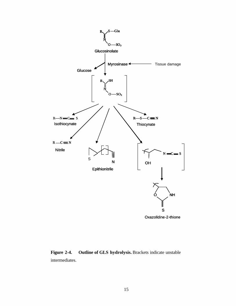
2.2.3 Hydrolysis
Plants accumulating GLS always possess a thioglucoside glucohydrolase activity
known as myrosinase, which hydrolyzes GLS to D-glucose and allelochemicals (Rask et
al., 2000). The products are glucose and an unstable aglycone that can rearrange to form
isothiocyanates, nitriles and other products. GLS hydrolysis in intact plants appears to be
hindered by the spatial separation of GLS and myrosinase or the inactivation of
myrosinase, but these components mix together upon tissue damage, leading to the rapid
formation of glucosinolate hydrolysis products. Myrosinase is present in myrosin cells
and is separated from the GLS pool. Most of the biological activities of GLS are
attributed to the actions of their hydrolysis products (Wittstock and Halkier, 2002).
GLS are degraded upon plant damage to a variety of hydrolysis products that are
likely responsible for much of the biological activities of this class of compounds. The
process begins with myrosinase-catalyzed hydrolysis of the thioglucoside linkage,
leading to the formation of glucose and an unstable aglycone (Bones and Rossiter, 1996;
Rask et al., 2000). Depending on the structure of the side chain and the presence of
additional proteins and cofactors, the aglycone then rearranges to form different
products, including ITCs, oxazolidine- 2-thiones, nitriles, epithionitriles, and
thiocyanates (Figure 2-4).

13
R COOH
NH2
R
NHO
Amino acid Aldoxime
Nitrile oxide
R
NHO O
R
NO
Aci-nitrocompound
CysteineNADPH + O2NADPH + O2R
NHO
SCOOH
NH2
alkyl-thiohydroximate
R
NHO
SHR
NO SO3
S Glu R
NOH
S Glu
Thiohydroximicacid
UDPH
Desulpho-glucosinolate
Glucosinolate
PAPS
R COOH
NH2
R COOH
NH2
R
NHO
R
NHO
Amino acid Aldoxime
Nitrile oxide
R
NHO
R
NHO O
R
NO
R
NO
Aci-nitrocompound
CysteineNADPH + O2NADPH + O2R
NHO
SCOOH
NH2
R
NHO
SCOOH
NH2
alkyl-thiohydroximate
R
NHO
SHR
NHO
SHR
NO SO3
S GluR
NO SO3
S Glu R
NOH
S Glu
Thiohydroximicacid
UDPH
Desulpho-glucosinolate
Glucosinolate
PAPS
Figure 2-3. Biosynthesis of GLS.

14
The most common GLS hydrolysis products in many species are ITCs, which are
formed from the aglycone by a Lossen rearrangement involving the migration of the side
chain from the oxime carbon to the adjacent nitrogen. When the GLS side chain bears a
hydroxyl group at C-2, the ITCs formed are unstable and cyclize to oxazolidine-2-
thiones, a class of substances known to cause goiter. In other plants, a major percentage
of GLS hydrolysis products are nitriles (Coles, 1976; Lambrix et al., 2001). The
formation of nitriles in vitro is favored at a pH of less than 3 or in the presence of Fe2+
ions (Galletti et al., 2001; Gil and MacLeod, 1980). However, protein factors, such as
the epithiospecifier protein (ESP), may be involved in nitrile formation in vivo,
(Bernardi et al., 2000; Foo et al., 2000; MacLeod and Rossiter, 1985; Tookey 1973).
When the glucosinolate side chain has a terminal double bond, ESP promotes the
reaction of the sulfur atom of the thioglucoside linkage with the double bond to
form a thirane ring, giving an epithionitrile. The process occurs only in the presence of
myrosinase, and ESP is not known to have any catalytic abilities. The recent isolation of
an Arabidopsis gene encoding an ESP showed that this protein not only promotes the
formation of epithionitriles, but also the formation of simple nitriles from a large variety
of GLS (Lambrix et al., 2001). Other hydrolysis products include ITCs, which are
formed from only three GLS: benzyl-, allyl-, and 4-methylsulfinylbutyl-glucosinolate
(Figure 2-4), all of which form stable side-chain cations. Like nitrile formation,
thiocyanate production is also associated with specific protein factors (Hasapis and
MacLeod, 1982), but these have not yet been identified. The hydrolysis of indole
glucosinolates is somewhat different from that of the other GLS types, because the
initially formed ITCs are unstable at neutral or slightly acidic pH, and are converted to
further metabolites, including indole-methanols, ascorbic acid conjugates, and
oligomeric mixtures (Agerbirk et al., 1998; Buskov et al., 2000; Latxague et al., 1991).
15
R
NO SO3
S Glu
Glucosinolate
Myrosinase
Glucose
R
NO SO3
SH
R S C NR N C S
R C N
ThiocynateIsothiocynate
Nitrile
SN
Epithionitrile
OH
N C S
O NH
S
Oxazolldine-2-thione
R
NO SO3
S Glu
Glucosinolate
Myrosinase
Glucose
R
NO SO3
SH
R
NO SO3
S GluR
NO SO3
S Glu
Glucosinolate
Myrosinase
Glucose
R
NO SO3
SH
R S C NR S C NR N C S
R C NR C N
ThiocynateIsothiocynate
Nitrile
SN
Epithionitrile
SN
Epithionitrile
OH
N C S
OH
N C S
O NH
S
O NH
S
Oxazolldine-2-thione
Figure 2-4. Outline of GLS hydrolysis. Brackets indicate unstable
intermediates.
Tissue damage

16
GLS hydrolysis products play important roles in plant defense systems against
pests, fungi and bacterial infections (Manici et al., 1997; Vaughn et al., 2005, 2006;
Vaughn and Berhow, 2005; Ware, 2000). Certain GLS breakdown products are volatile
and, therefore, good candidates for insect fumigants. Propenyl isothiocynate released
from the GLS sinigrin in B. nigra, B. carinata and B. juncea, is an effective fumigant for
suppression of the growth of soil-borne fungal pathogens (Manici et al., 1997; Rosa and
Rodrigues, 1999). Although they are insecticidal, the mode of action of such natural
products is not clear.
2.2.4 Extraction
Due to the hardy nature of myrosinase, the extraction of GLS is a complex
procedure. Several methods of GLS extraction have been adopted, including
denaturation of myrosinase by boiling or inactivation at -20ºC or by solvent extraction.
Extracts for HPLC/GC are prepared in boiling water (Rangkadilok et al., 2001;
Matusheski and Jeffery, 2001; Szmigielska et al., 2000) or by autoclaving samples (Jen
et al., 2001) or in boiling methanol (Zrybko et al., 1997; Verkerk et al., 2002), or in 50%
acetonitrile (ACN; Tsao et al., 2002b). After extraction, debris is removed either by
filtration or centrifugation. The clarified extract is then concentrated and used for HPLC
analysis. Other researchers have used liquid nitrogen to arrest myrosinase activity for
storage of samples, and before analysis, frozen samples are added directly to boiling
water or methanol (Karcher et al., 1999). Any of the methods of extraction described
above can be used, as the basic purpose is to inactivate myrosinase while extracting GLS
from plant samples.
2.2.5 Purification and desulfation
After extraction of GLS, they are further purified as described by Szmigielska et
al. (2000). In this method, GLS from the extract are bound to an anion exchange
membrane. Initially, anion exchange membranes were prepared for use by shaking three
times with saturated sodium bicarbonate solution (1 h each time). Membranes were then
thoroughly washed and stored in distilled water for further use. The total exchange
capacity of the membranes was 1.1mequiv/g (0.04mequiv/cm2). GLS may also be

17
purified using a DEAE Sephadex A-25 anion exchange column (Oerlemans et al., 2006).
The column is washed with 1 mL Millipore water, loaded with 2 mL of the GLS extract,
and then washed twice with 1 mL of 20 mM sodium acetate solution. Sulphatase enzyme
is added to the column to cleave the sulfate by incubation overnight at room
temperature. Finally, GLS are eluted with reverse-osmosis-purified water (3 x 0.5 mL).
2.2.6 Detoxification
Tripathi and Mishra (2007) reviewed the recent development of various
treatment methods to reduce GLS content in mustard meal to minimize GLS -associated
deleterious effects. Most of these methods involved hydrolysis or decomposition of GLS
before consumption by humans or animals. Some of these methods are as follows.
1. Microwaving
Microwave irradiation at 2450 MHz for 2.5 min is known to inactivate myrosinase and
decompose GLS (Verkerk and Dekker, 2004).
2. Metal solutions
Treatment of meal with copper sulphate solution inactivates ITC in meals (Das and
Singhal, 2001, 2005) due to rearrangement reactions and the production of amines such
as allylamine or thiourea (Rouzaud et al., 2003).
3. Fermentation
The fermentation of mustard meal using Rhizopus oligosporus and Aspergillus sp.
(meal: water ratio of 1:3, 25ºC, under aerobic conditions, 10 days) inactivated
myrosinase and reduced total GLS (Vig and Walia, 2001). The complete degradation of
GLS occurred after 60 h of fermentation at 30ºC (Rakariyatham and Sakorn, 2002).
Decomposition of GLS was greater with longer fermentation periods. The reduction in
GLS and their metabolites during fermentation may be due to the utilization of glucose
and sulphur moieties of these compounds by microbial enzymes.
18
4. Heat treatment
Wet heating is more effective than dry heating (Burel et al., 2000; Tripathi et al., 2001;
Leming et al., 2004) for decomposing GLS.
2.2.7 Methods of GLS determination
Several methods have been used for the quantification of GLS. These are as follows.
1. High Pressure Liquid Chromatography (HPLC)
2. Gas chromatography (GC)
3. Capillary Electrophoresis (CE)
4. Determining total GLS by capillary electrophoresis via enzymatically released
gluconic acid labeled with 7-aminonaphthalene-1,3-disulfonic acid (ANDSA)
5. Nuclear Magnetic Resonance (NMR)
6. Near IR spectroscopy
7. X-ray fluorescence
Some of the most commonly used methods are described below.
2.2.7.1 High Pressure Liquid Chromatography (HPLC)
HPLC has been used as an efficient analytical tool for accurate quantification of
many metabolites. HPLC methods take advantage of the sophistication and automation
of the instrument, along with its precision and accuracy. Reverse-phase HPLC has
generally been the method of choice in recent years because it has the versatility of
analyzing GLS in both intact and desulfonated forms. Spinks et al. (1984) developed the
reverse-phase HPLC method for quantitative analysis of desulphoglucosinolates which
is the most widely used method today. This method utilizes an on-column enzymatic
desulfation treatment of plant extracts followed by HPLC detection of the resultant
desulfoglucosinolates. Adaptation of the sulfohydrolase desulfation method as HPLC
method, although the most widely used method for GLS separation, is still subject to
difficulties in interpretation because of the effect of pH, time and enzyme activation on
the desulfation products (Spinks et al., 1984; Sang et al., 1984). Typically, this method
uses response factors determined with purified desulfosinigrin and uses desulfobenzyl
glucosinolate as an internal standard. Correspondence of GLS retention times, and
19
comparison to standardized rapeseed extracts, are typically used to validate the
chromatographic profile. Unfortunately, the biological activity of these molecules is
compromised by the removal of the sulfate. After desulfation, they can no longer serve
as substrates for myrosinase and thus their cognate ITCs are not available for bioassay or
for direct measurement by cyclocondensation, a key tool in the study of the
pharmacokinetics, pharmacodynamics and bioactivity of these compounds.
Therefore, several authors have made attempts to improve the HPLC method for
GLS analysis. Zrybko et al. (1997) developed an HPLC method for separation and
quantification of GLS in mustard using a Phenomenex 5 µm ODS column (40ºC) with
the mobile phase water: methanol, with an ion pairing reagent cons isting of 0.15%
triethylamine and 0.18% formic acid. The mobile phase gradient was 100% water for 10
min, which was then increased to 100% methanol over the next 60 min. Using this
method, the retention time for sinigrin detected at 235 nm was 10.5 min. Sinigrin was
detected at 235 nm because the contribution to the UV absorption at this wavelength
arises predominately from the GLS group and not from the R groups (Helboe et al.,
1980; Björkqvist and Hase, 1988). Later, Szmigielska et al. (2000) reported another
HPLC method for separation and quantification of naturally occurring GLS from canola.
They separated GLS on a Supelcosil LC-18 column using methanol and 0.1 N
ammonium acetate (NH4OAc) (3:97; v/v) as the mobile phase. In this way, detection
was proportional to the molar concentrations of the GLS.
Jen et al. (2001) further improved the HPLC method for GLS analysis. They
separated GLS with a reverse phase C-18 column using ACN-water (20:80; v/v)
containing 0.02 M tetrabutylammonium (TBA) at pH 7.0. The retention time for sinigrin
detected at 227 nm was 8.8 min. Rangkadilok et al. (2001) also used a C-18 reverse
phase column. The column was saturated with 5 mM tetramethylammonium bromide
(TMAB) before loading the sample and TMAB (5 mM) was used as the mobile phase.
Sinigrin was detected in 8.84 min at 230 nm. This method is simple, efficient and quick.
The method developed by Tsao et al. (2002b) is the most commonly used
method in industry. They used a Sphereclone ODS-2 column and the mobile phase was
composed of 0.025 M ammonium acetate (NH4OAc, pH 6.75) and ACN. The mobile
phase was run isocratically for 2 min, then linearly increased to 50% NH4OAc (0.025 M,
20
pH 6.75) and 50% ACN in 0.5 min, held for 7.5 min, and then brought back to 99%
NH4OAc (0.025 M, pH 6.75) and 1% ACN at 12 min. Sinigrin (228 nm) and AITC (242
nm) were detected as early as 2.8 min and 9.8 min, respectively.
The HPLC method for determining desulfonated GLS provides a simple means
of determining the GLS profile (Verker et al., 2002; Oerlemans et al., 2006). Oerlemans
et al. (2006) separated de-sulfonated GLS using a C-18 reverse phase column. Elution of
desulfonated GLS was performed by a gradient system of water and ACN/water (20:80;
v/v). Sinigrin was detected at 229 nm and was eluted as early as 4.8 min. Recently,
Trenerry et al. (2005) used a C-18 column and a mobile phase of 0.005 M TMAB
dissolved in 2% methanol/water for the separation of glucoraphanin in broccoli. The
chromatograms were monitored at 230 and 270 nm.
Ion exchange and desulfonation methods are more sensitive and time consuming,
and sometimes can lead to loss of GLS. Because the separated compounds are not
suitable for physiological or biological studies, the use of such methods as a preparative
technique is limited to specific purposes (Betz and Fox, 1994). Reverse phase HPLC
methods for direct analysis of intact and non-derivatized GLS are advantageous.
Due to the importance of GLS and their hydrolysis products, there is a need for a
quick, economic, efficient and simple HPLC method for detection of GLS in various
sample types. The total GLS content is generally measured by adding up all of the
individual GLS (Linsinger et al., 2001). HPLC coupled with mass spectroscopy (HPLC-
MS) also has shown promise.
2.2.7.2 Gas chromatography (GC)
Gas chromatography is useful for the separation and quantification of GLS,
provided the GLS of interest is volatile after derivatization. Brown et al., (1994)
extracted GLS from plant tissues with methanol. The extract was placed on a DEAE
Sephadex A-25 column that was first treated with 6 M imidazole in 5 M formic acid and
rinsed with water. Other compounds were separated from the sample by rinsing with
67% methanol and water. GLS were desulfated overnight, removed from the columns
using 60% methanol, and silylated in acetone. Silylated derivatives were separated and
identified using a GC equipped with a quadrupole mass selective detector (MSD).
21
GCMSD operating conditions were: injector 260°C; interface 320°C; initial oven
temperature 130°C for 1 min, ramped 15°C/min to 320°C and held for 10 min; purge
(splitless injection) 0.5 min; helium flow of 1.14 mL/min; emission at 50 µA; repeller at
30 V; and scan range of m/z 25-470. The column coating was 5% phenyl-substituted
methylpolysiloxane (HP-5MS; 30 m, 0.25 mm inside diameter, and 0.250-µm film).
Gas chromatography chemical ionization mass spectrometry has been found to
possess advantages over gas chromatography electron impact mass spectrometry for the
structural elucidation of GLS, separated as the volatile per-trimethylsilyldesulpho
derivates (Eagles et al., 2005). The technique demonstrates the versatility of mass
spectrometry in GLS identification.
2.2.7.3 Capillary Electrophoresis
The separation of GLS and related desulfoglucosinolates has also been achieved
by Micellar Electrokinetic Capillary Chromatography (MECC). MECC is a highly
efficient separation technique that is complementary to HPLC and GC and is especially
suited to the analysis of low to medium molecular weight ionic compounds. MECC
separations are often faster and more cost effective than corresponding HPLC or GC
procedures. MECC was introduced by Terabe et al. (1984) and has become one of the
most widely used capillary electrophoresis (CE) methods due to the ability to separate
both charged and neutral analytes. MECC separates compounds by using a surfactant as
part of the separation buffer. This surfactant forms a micelle and neutral analytes
separate based on their affinity for the micelle and their hydrophobicity. The amount of
surfactant present in the system needs to be above the critical micelle concentration to
allow for the micelles to form. Most MECC systems for GLS analysis are based on the
cationic surfactant cetyltrimethylammonium bromide (CTAB) (Karcher and Rassi, 1999;
Michaelsen et al., 1992; Morin et al., 2005). The surfactant coats the wall of the bare
fused silica capillary and covers the silica with a layer of positive charges. The net result
of this adsorption is the reversal of the direction of the electro-osmotic flow. The CTAB
micelle is advantageous because it forms an ion-pair with the negatively charged sulfate
group of the intact GLS, thus imparting strong analyte-micelle interactions. Michaelsen
et al. (1992) successfully used CTAB to separate eleven intact GLS. The optimum

22
separation conditions were 18 mM borate, 30 mM phosphate, 50 mM CTAB at pH 7.0
with an applied voltage of 20 KV. Under these conditions, the CTAB micelle migrates
against the electro-osmatic flow, whereas the negatively charged glucosinolate migrates
with the electro-osmatic flow. Thus, stronger the ion-pair formation and hydrophobic
interactions of a given GLS with the CTAB micelle, longer the migration time of that
GLS. Bjergegaard et al. (1995) also separated a number of desulfoglucosinolates using
the cationic surfactant sodium cholate. Paugman et al. (1995) used sodium dodecyl
sulfate (SDS) as the surfactant to separate glucobrassicin and methoxyglucobrassicin,
with tetramethylammonium hydroxide as the ion-pair reagent and methanol as an
organic modifier. Bringmann et al. (2005) recently developed a capillary zone
electrophoresis method for the analysis of GLS from Arabidopsis thaliana. The
electrolyte used with the fused-silica capillaries was a 100 mM ammonium acetate
buffer (pH 5.4) containing 30% ACN. The pH of the buffer was adjusted with acetic
acid. The sample was loaded by applying a pressure injection of 50 mbar (=0.725 psi)
for 10 sec on the anodic side and detected at 225 nm. Electrophoresis was conducted at
30oC and 25 KV. Optimization of CE is required for rapid separation of GLS. Research
is needed to identify possibly more GLS in mustard and to develop a quick, efficient and
economic method of determining these GLS. The use of capillary electrophoresis is
potentially the most efficient and fastest method for determination of GLS.
A selective and sensitive method for the determination of total GLS in plants by
capillary electrophoresis- laser- induced fluorescence (CE-LIF) detection was developed
by Karcher and Rassi (1999). The method was based on the enzymatic release of glucose
from GLS in the presence of myrosinase. The released glucose was converted to
gluconic acid by glucose oxidase. The resulting gluconic acid was then labeled
selectively with the fluorescent tag 7-aminonaphthalene-1,3-disulfonic acid (ANDSA).
Peak area resulting from gluconic acid-ANDSA was derived from the free glucose in the
sample. This allowed quantitation of the total GLS in the sample. The peak area was
normalized to the internal standard, N-acetlyneuraminic acid derivatized with ANDSA.
Detection of GLS was performed at 228 nm. For Laser Induced Fluorescence detection
of the ANDSA derivatives, a fluorescence emission bandpass filter of 380 ± 2 nm was
23
used. A 360 nm cut-off filter was used to reject the laser beam. Sodium phosphate (50
mM, pH 3.0) was used as an electrolyte.
2.2.7.4 Nuclear Magnetic Resonance (NMR)
Prestera et al. (1996) identified and isolated GLS by using mass spectrometery
and NMR spectroscopy. Proton NMR spectra were obtained in D2O at 600 MHz.
Ammonium salts of GLS were dissolved in D2O and dried in a vacuum centrifuge, and
then dissolved in 625 µL of D2O at a concentration of 1 to 10 mM and one-dimensional
(1-D) proton spectra were obtained in high quality NMR tubes. Proton resonance
assignments were initially based on the covalent structure of the R group as well as on
the analysis of the coupling patterns in the 1-D proton NMR spectra.
2.3 Myrosinase
2.3.1 Occurrence
Myrosinases (Thioglucoside glucohydrolase; EC 3.2.3.1) are typical enzymes of
the Brassicaceae family. Although the myrosinase- GLS system is always present in the
organs of Brassicaceae in various arrangements and concentrations, it is only activated
following tissue damage. It plays a defensive role against pathogens in general (Bones
and Roostier, 1996; Chew, 1998; Louda and Mole, 1991; Rosa et al,. 1997). Myrosinase
and GLS were first discovered in mustard seed by Bussy (1840). Myrosinase activity
always appears to be accompanied by one or more GLS. GLS occur in all Brassicaceae
(Cruciferae) species and have also been found in Akaniaceae, Bataceae,
Bretschneideraceae, Capparaceae, Caricaceae, Drypetes (Euphorbiaceae),
Gyrostemonaceae, Limnanthaceae, Moringaceae, Pentadiplantdraceae, Resedaceae,
Salvodoraceae, Tovariaceae and Tropaeolaceae (Rodman, 1991). Enzymes with
myrosinase activity have also been found in the fungi Aspergillus sydowi (Reese et al.,
1958; Othsuru et al., 1969) and Aspergillus niger (Ohtsuru et al., 1973), in the intestinal
bacteria Enterobacter cloacae (Tani et al., 1974) and Paracolobactrum aerogenoides
(Oginsky et al., 1965), in mammalian tissues (Goodman et al., 1959) and in the
cruciferous aphids Brevicoryne brassicae and Lipaphis erisimi (MacGibbon and
Beuzenberg, 1978). The amount of myrosinase activity found in seed from cultivars of

24
Sinapis alba L., Brassica campestris L. and Brassica napus L. has been examined by
Henderson and McEwen (1972), Bjørkman and Lønnerdal (1973) and Bones (1990).
Myrosinase activity was found to be about ten times higher in S. alba than in B.
campestris and the activity in B. napus was slightly higher than in B. campestris (Bones,
1990).
2.3.2. Distribution
The distribution of myrosinase isoenzymes appears to be both organ-specific and
species-specific. Electrophoretic examination of isoenzymes from many plants, organs
and tissues demonstrates that the pattern may vary with species, organ and age of the
plant (MacGibbon and Allison, 1970; Henderson and McEwen, 1972; Buchwaldt et al.,
1986). Little is known about the physiological reasons for this difference. It has been
postulated that the particular isoenzymes correspond to endogenous conditions found in
that plant, or to conditions found in the target organism, or to particular GLS that
dominate the profile of that tissue. It is not known if electrophoretic separation is
separating isoenzymes with distinct amino acid sequences.
A systematic analysis of the variation in myrosinase activity in plants at different
developmental stages and organs has been reported. Bones (1990) examined the
myrosinase activity at different developmental stages and in different plant parts
throughout the life cycle. The reported activities in different tissues varied, but all tested
organs/tissues contained some myrosinase activity. Myrosinase activity could also be
detected in callus cultures and in in vitro cultured plants (Bones, 1990). Except for the
roots of fully grown plants where a high activity was observed, other organs of mature
plants normally contained low myrosinase activity.
Bones (1990) used enzymatic assays and ultrastructural observations of calli to
examine the correlation between myrosinase and myrosin cells. A lack of observable
myrosin cells by light microscopy and transmission electron microscopy was associated
with low myrosinase activity.

25
2.3.3 Genetics
Myrosinase is not properly identified as a single enzyme, but rather as a family
or group of similar enzymes. Multiple forms of the enzyme exist, both among species
and within a single plant (Bones and Slupphaug, 1989; Falk et al., 1995a; Lenman et al.,
1993a; Xue et al., 1993), and all perform a similar function (Björkman, 1976). Although
their genetic sequences are similar to those of other ß-glycosidases (Lenman et al.,
1993b), mostly myrosinases are specific toward GLS (Durham and Poulton, 1990).
These enzymes cleave the sulfur-glucose bond regardless of either the enzyme or
substrate source. However, the particular enzyme and GLS substrate do influence
reaction kinetics (Bones, 1990).
2.3.4 Purification
Myrosinase from white mustard (Sinapis alba) has been purified as early as in
1986 (Palmieri et al., 1986). They used single step affinity chromatography on Con A-
Sepharose for the isolation of myrosinase. The binding capacity of Con A-Sepharose
was 6.6 mg/mL gel bed which corresponds to 150,000 U/mL of chromatographic bed.
Moreover, the enzyme bound to Con A-Sepharose remained active towards GLS.
2.4 Allyl isothiocynate (AITC)
Allyl isothiocynate (AITC, CH2=CH-CH2-N=C=S) is a hydrolysis product of
GLS which contributes to the peculiar odour of mustard oil. It is a colourless to pale
yellow, volatile, highly flammable liquid with a molecular weight of 99.15 g. It is
slightly soluble in water (0.1 mg/mL) and is soluble in organic solvents. AITC possess
various biochemical and physiological properties (Keum et al., 2005), which are
discussed below.
2.4.1 A potent anti-microbial agent
Mustard seed flour contains 28% to 36% protein. The presence of tocopherols in
mustard contributes to its shelf life (Cahoon et al., 2003; Winther and Nielsen, 2006).
Mustard is widely known for its sharp taste and it is an essential component of dressings
and sauces. The oil in mustard inhibits the growth of certain yeasts, moulds and bacteria.

26
enabling mustard to function as a natural preservative. AITC vapour significantly
reduced the growth of Penicillium expansum, the causal agent of blue mould on pears
(Mari et al., 2002). Therefore, AITC may have potential as an economically viable
alternative to synthetic fungicides.
GLS hydrolysis products may have biocidal activity against a wide variety of
organisms such as insects, plants, fungi and bacteria (Tunc et al., 2007; Vaughn and
Berhow, 2005; Ware, 2000); some may have human health benefits. ITCs are among the
most potent products and are suspected to be the major inhibitors of microbial activity.
Benzyl ITC is sometimes used as an antibiotic to treat infections of the respiratory and
urinary tract (Mennicke et al., 1988). Bacteriostatic, bactericidal, and fungicidal effects
of many ITCs are well documented (Brown and Morra, 2005). The fungicidal property
of AITC vapour against wild type and thiabendazole-resistant strains of Penicillium
expansum has been demonstrated (Tunc et al., 2007; Kiyoshi 2005; Mari et al., 2002).
Bacteriocidal effects of mustard flour against Escherichia coli, Listeria monocytogenes,
and Salmonella enterica have also been documented (Rhee et al., 2003). The use of
AITC produced from pure sinigrin or from defatted Brassica juncea meal may be an
economically viable alternative to synthetic fungicides against P. expansum. AITC has
been shown to be effective against cheese-related fungi such as Penicillium commune, P.
roqueforti and Aspergillus flavus (Winther and Nielsen, 2006), but the incorporation of
AITC in the packing material contributed to unacceptable mustard flavour in cheese.
2.4.2 Herbicidal activity
GLS-containing plants have a reputation for inhibiting the growth of nearby
weeds and other crops such as wild oat (Avena sterilis), wheat and pea (Jones, 1992).
Stands of wild B. nigra appeared to inhibit germination of annual grasses, and broccoli
(B. oleracea) residues amended to soil reduced germination and growth of lettuce for
10-21d (Patrick et al., 1963). Sinapis alba seed meal applied to the soil is also known to
suppress weed emergence (Ascard and Jonasson, 1991; Johansson, 1992; Johansson and
Ascard, 1994; Oleszek et al., 1994). For example, mustard meal or “cake” applied in
rows between cole crops with the intent of attracting enemies of the cabbage root fly
also reduced numbers of several annual weeds (Ascard and Jonasson, 1991).

27
2.4.3 As a biofumigant
AITC has been shown to have toxic effects on insects and other invertebrates
(Brown and Morra, 2005). Noble et al. (2002) demonstrated larval mortality by AITC.
The relative toxicity of different GLS hydrolysis products such as ITCs and nitriles is
important in maximizing the likelihood of effective pest suppression in pesticide
applications (Sarwar et al., 1998). ITCs have been used as biofumigants for the control
of soil-borne pests. These products possess potential biodegradable and bio-friendly
insect fumigant (Tsao et al., 2002a) and nematocidal properties (Mitarai et al., 1997).
They may act on the insect respiratory system in their mode of action. Leaching of ITCs
and their degradation in soil has also been studied (Gimsing et al., 2005, 2006 and
2007). The rates of ITC formation and degradation are critical to the toxicity and
leaching of GLS and ITCs in soil. Degradation of GLS is much faster in a clay soil (half-
life, 3.5-6.8 h) than in a sandy soil (half- life, 9.2-15.5 h). AITC is the most toxic
compound formed from allyl GLS (sinigrin) hydrolysis and is possibly most important
for biofumigation (Noble et al., 2002). Additionally, ITCs can be lost by volatilization.
However, for fumigants, volatilization is significant for spreading in soil. Therefore,
volatilization can be a concern for health and environmental reasons.
2.4.4 Nematocidal effects
Nematocidal effects of GLS hydrolysis products on the sugar beet cyst nematode
Heterodera schachii were shown (Lazzeri et al., 1993). AITC, gluconapin,
glucotropeolin and glucodehydroerucin had strong nematocidal effects, whereas GLS
alone, glucoraphenin and sinalbin had no toxic effect. The potato cyst nematode
(Globodera rostochiensis) has been controlled by the use of GLS hydrolysis products
(Buskov et al., 2002). They reported 100% mortality by adding active myrosinase to
phenethylglucosinolate, benzyl-enylglucosinolate and prop-2-enylglucosinolate.

28
2.4.5 Anti-cancer properties
The ITCs are known to have cancer-preventive properties (Zhang, 2004; Myzak
et al., 2004). Anti-cancer properties of glucosinolates have also been reported (Hu et al.,
2007; Thejass and Kuttan, 2007; Hwang and Lee, 2006; Tang et al., 2006; Jakubikova et
al., 2005; Smith et al., 2004; Zhang, 2004; Myzak et al., 2004; Thornalley, 2002). For
example, suplforaphane [1- isothiocyanoto-4-(methylsulfinyl) butane], a degradation
product of the glucosinolate, glucoraphanin, is a potent inducer of phase II
detoxification enzymes, which are strongly correlated with the prevention of certain
types of cancer (Brooks et al., 2001; Matusheski et al., 2001). Allyl isothiocynate
(AITC), benzyl-ITC (BITC), phenethyl-ITC (PEITC), sulforaphane (SFN), erucin
(ERN) and iberin (IBN) induce time- and dose-dependant G[2]/M arrest in HL60 cells
(Jakubikova et al., 2005). Mustard ITCs are mitotic inhibitors and/or apoptosis
inductors, which suggest that they could be chemotherapeutic agents in cells with
multidrug resistance phenotypes. Dietary isothiocyanates inhibit the growth of human
bladder carcinoma cells (Tang and Zhang, 2004). Tang et al. (2006) recently
demonstrated that naturally occurring ITCs, including AITC, BITC, PEITC and SFN,
potently inhibited the growth of cells in human bladder cancers and drug-resistant
bladder cancer cells. They also demonstrated a mechanism for the anti-proliferative
property of AITC. The AITC breaks down caspase-3, -8 and -9 which are known
inducers of apoptosis and cell arrest, thereby inhibiting apoptosis. Suplforaphane [1-
isothiocyanoto-4-(methylsulfinyl) butane], a degradation product of the GLS,
glucoraphanin, is a potent inducer of detoxification enzymes, which are strongly
correlated with the prevention of certain types of cancer (Brooks et al., 2001;
Matusheski et al., 2001).
2.4.6 Toxicity of other GLS hydrolysis products
Other than AITC, gluconapin, glucotropeolin and glucodehydroerucin have been
shown to possess strong nematocidal properties against sugar beet cyst nematode
Heterodera schachii (Lazzeri et al., 1993). GLS-derived nitriles have the ability to
increase the phase 2 detoxification enzyme glutathione S-transferase, quinine reductase
and glutathione in the mouse (Tanii et al., 2005). Maximum potency was observed in

29
lung and stomach, which is of interest in light of epidemiological studies demonstrating
an inverse association between Brassica intake and the incidence of lung and stomach
cancers.
2.4.7 Time release of AITC
The beneficial effects of AITC were discussed above. However, the application
of AITC in food systems is limited due to its volatility and strong odour which affect the
taste of food (Chacon et al., 2006). Its application in food packaging systems is also
limited by its existence as an oil form and its volatility. Recently, AITC coupled with α-
and β-cyclodextrin has shown potential in the food industry (Li et al., 2007). A
controlled release of AITC from the complexes was achieved, which is of great benefit
in masking the strong odour, prolonging the antimicrobial time and enhancing the
antimicrobial effect of AITC. AITC release was accelerated by increased relative
humidity and the release rate of AITC from the α-cyclodextrin-AITC complex was
much slower than that from the β-cyclodextrin-AITC complex. This opens many
avenues of research with target to the mafucture of storage bags for increased shelf life
of fruits and vegetables.
2.4.8 Extraction
Jogdeo et al. (2000) demonstrated a steam distillation method for recovery of
AITC from mustard meal by using Amberlite XAD-4 adsorbent lining in the condenser.
Using a pure AITC standard, recovery was 95% using this steam distillation method. A
solvent extraction method for AITC was described by Padukka et al. (2000), and was
also used by Li et al. (2007) and other researchers. Mustard meal was mixed with
distilled water and hexane (5:7, v/v) and heated at 85ºC for 20 min using an upright
glass condenser. This extraction process was repeated four times.
2.4.9 Quantification
Various methods of AITC quantification have been developed. Some of these
methods are described as follows.

30
2.4.9.1 HPLC
Reverse phase HPLC methods have been explored for the analysis of ITCs
(Mathaus and Fiebig, 1996). Later, Jogdeo et al. (2000) developed an HPLC method for
quantification of AITC using a stationary phase of Lichrospher 100 PR-18. The detector
was set at 245 nm and methanol was used as solvent (Jogdeo et al., 2000). In successive
years, methods were further improved for better resolution, recovery and detection. Tsao
et al. (2002b) developed a reverse phase HPLC method for direct and simultaneous
detection of sinigrin and AITC in mustard samples. The detection limit for AITC was
0.1µg/mL. A Sphereclone ODS-2 column was employed and the detector was set at 242
nm for AITC and 228 nm for sinigrin. Compounds were separated using a step gradient
of mobile phase composed of 0.025 M ammonium acetate (pH 6.75) and acetonitrile
(AcN).
2.4.9.2 Colorimetric assay
Recently, a colorimeteric method based on the colour reaction between allyl
isothiourea and potassium ferricyanide in dilute acetic acid was developed for the
determination of AITC in mustard meal (Mukhopadhya and Bhattacharyya, 2006). The
colour developed by this reaction was measured at 600 nm. This method requires 3 h for
analysis and can detect AITC in the range of 2.5 to 7.5 µg. In the method described by
Li et al. (2007), the absorbance of a hexane extract was measured at 248 nm.
2.4.9.3 Ion-pair electrospray mass spectroscopy
Mellon et al. (2002) developed a voltage electrospray LC/MS method to analyze
GLS in plant extracts. This method has been shown to be effective for all the GLS by
many workers (Bennett et al., 2004). A significant concentration of 4-hydroxy-3-
indolylmethylglucosinolate was found in the majority of Brassica species.
2.4.10 Thermal degradation
It has been shown that AITC is unstable and gradually degrades into other
compounds having a garlic- like odour. AITC in aqueous solution, when heated at 100ºC
for 1 h, disintegrates into N,N-diallylthiourea, a major degradation product (Chen and

31
Ho, 1998); other products obtained were diallyl sulphide, diallyl disulphide, diallyl
trisulphide, diallyl tetrasulphide, allyl thiocyanate, 3H-1,2-dithiolene, 2-vinyl-4H-1,3-
dithiin, 4H-1,2,3-trithiin and 5-methyl-1,2,3,4-tetrathiane.
2.4.11 Heat shock proteins as markers of stress
Heat shock proteins (HSP) play important roles in the tolerance of living organisms to a
variety of biotic and abiotic insults (Vierling, 1991; Parsell and Lindquist, 1994;
Hamilton and Coleman, 2001). Thus, HSPs appear to be general stress proteins that are
involved in the maintenance of proper cell function and, thereby, help in cell survival by
facilitating recovery from stress (Vierling, 1991; Parsell and Lindquist, 1994; Downs
and Heckathorn, 1998; Guy and Li, 1998; Heckathorn et al., 1998). Most commonly,
HSP70, HSP110, HSP52 and small HSPs (for example, HSP27), play vital roles in
protecting cells in stressful conditions. Members of the 70 KDa heat shock protein
(HSP70) family are ubiquitous in plants, animals and microorganisms. Their structure
and function are highly conserved among diverse organisms, from algae to mammals
(Wu et al., 1994). HSP70 is involved in preventing protein aggregation and degradation
in response to cellular stress caused by an insult. Therefore, HSP70 can be used as a
marker of AITC toxicity.
2.5 Research needs
To improve knowledge of AITC production from mustard meal and the biological
activity of whole mustard meal, it is important to develop rapid analytical methods.
AITC is released by the action of myrosinase on sinigrin and thus it is also important to
study the enzymatic production of AITC. This requires the development of methods to
inhibit myrosinase activity in mustard meal, while allowing accurate measurement of
AITC. It is also important to investigate whether AITC possesses any plant growth
regulation properties. Research is needed to develop bioassays for screening AITC
bioactivity and toxicity.
32
Chapter 3: Materials and Methods
3.1 Biological materials
Germination trials were conducted with flax (Linum usitatissimum cv. Vimy)
seed which was obtained from Dr. Gordon Rowland, Crop Development Centre,
University of Saskatchewan, Saskatoon, SK, and tomato (Solanum lycopersicum cv.
Roma) seed purchased from Early's Home and Garden Centre, Saskatoon, SK.
Caenorhabditis elegans strain N2 used in the toxicity study was provided by
Caenorhabditis Genetics Center, which is funded by the National Institutes of Health
(NIH), the National Centre for Research Resources (NCRR), Bethesda, MD. Brassica
juncea cv. Arrid seed was obtained from Dr. Kevin Falk, Agriculture and Agri-Food
Canada, Saskatoon Research Centre, Saskatoon, SK. Seed was produced on plots near
Saskatoon in 2006.
3.2 Measurement of AITC in seed meal
Allyl isothiocynate (AITC) in seed meal was quantified using a modified
American Spice Trade Association method where AITC is recovered by steam
distillation (Figure 3-1). B. juncea seed (5g) was ground in a coffee grinder and then
extracted in 100 mL of deionized water containing a few drops of anti- foam (Sigma-
Aldrich Canada Ltd., Oakville, ON). This mixture was incubated at 37ºC for 2 h with
continuous stirring. After adding ethanol (95%, 20 mL) and a few boiling chips, the
mixture was distilled in the dark by covering the distillation flask with aluminium foil.
Sixty millilitres of distilled solution was collected in a flask containing 10 mL of 33.5%

33
Figure 3-1. Apparatus used for quantification of volatile oil in mustard meal.

34
ammonium hydroxide solution. To this mixture, 20mL of 0.1N silver nitrate (Sigma-
Aldrich) was added and the final volume was adjusted to 100 mL with distilled water
before incubating overnight in the dark by covering the flask with aluminium foil. This
resulted in the formation of a black precipitate which was removed by filtration with
Whatman grade No. 4 filter paper (GE Health Care, Piscataway, NJ). Two titrations
were performed, each using 50 mL of this filtrate. Fifty millilitres of the filtrate was
acidified with 5 mL of concentrated nitric acid (analytical grade, Sigma-Aldrich) and
was titrated with 0.1N ammonium thiocyanate (analytical grade, Sigma-Aldrich) after
adding 5 mL of 8% FeNH4 (SO4)2.12H2O indicator (Sigma-Aldrich). Per cent volatile oil
was calculated using the formula below:
[(mL x N) AgNO3 – 2(mL x N) NH4SCN] x 0.04958 x 100
Volatile oil (%) = ------------------------------------------------------------------------------
Weight of sample (g)
3.3 Kinetics of myrosinase
To understand the kinetics of myrosinase activity in Brassica juncea seed, 5 g
ground seed was extracted in water at different temperatures (7 to 97ºC) for various
times (0-5 min at intervals of 30 seconds and 5, 15, 30, 60 and 120 min). Myrosinase
activity was determined as percent volatile oil released using the method described in
section 3.2.
3.4 Inhibition of myrosinase activity
Efforts were made to inhibit myrosinase activity by altering reaction conditions
(pH and incubation temperature). The pH of the reaction mixture was changed by adding
sodium hydroxide (NaOH), hydrochloric acid (HCl), acetic acid (CH3COOH), succinic
acid (C2H4(COOH)2) or boric acid (H3BO3). The pH of the reaction mixture was varied
from 1.5 to 13. Further, the effects of calcium chloride (CaCl2) and ethanol (C2H5OH)
were also studied. Myrosinase activity was determined as percent volatile oil released
using the method described in section 3.2. Data presented are averages of four replicates.

35
3.5 Seed germination
Effects of AITC, IAA and Ethephon (2-Chloroethyl-dioxido-oxophosphorane)
on the germination of flaxseed and tomato seed were studied. Flaxseed and tomato seed
were treated with different concentrations (0, 0.01, 0.05, 0.1, 0.5 and 1 mM) of AITC,
IAA and Ethephon and incubated in the dark. Observations were recorded from day 0 to
day 6. Data presented are averages of three replicates of 20 seeds per plate. Each study
was replicated twice.
3.6 Maintainance of C. elegans cultures
C. elegans N2 strain was grown in sterile plates (10 mm; VWR, Edmonton, AB)
on 10% bacteriological agar (Sigma-Aldrich) layered with 1 mL of autoclaved Baker’s
yeast (1%, w/v). Cultures were incubated in the dark at room temperature and sub-
cultured to fresh plates every 15 days. For chemical treatments, two-week-old
nematodes from three plates were pooled in a 10 mL sterile tube. Movements of
nematodes were examined 30-60 min after each treatment under a microscope at 10X
magnification.
3.7 Chemical treatment of nematodes
To study the toxicity or stress effects of AITC, C. elegans N2 strain was used as
a model system. C. elegans was treated with different concentrations (0.001-10 µM) of
AITC (Sigma-Aldrich) and incubated in the dark at room temperature for 2 h. Controls
were untreated organisms maintained under the same incubation conditions. After
treatment and incubation, cultures were centrifuged at 400 x g for 10 min at 4°C, and the
pellet was quickly rinsed with sterile distilled water and centrifuged at 400 x g for 10
min at 4°C. After removing the supernatant, the pellet was stored at -80°C until analyzed
further.
3.8 Sequence alignment and phylogenetic study
The GenBank data base was searched to identify sequences of isoforms of
HSP70. Amino acid sequences of different isoforms of HSP70 in C. elegans viz.,
HSP70A (GenBank Accession No. M18540), HSP70C (GenBank Accession No.

36
T15513), HSP70D (GenBank Accession No. P20163) and HSP70F (GenBank Accession
No. P11141) were aligned using the ClustalW multiple sequence alignment program
(http://www.ebi.ac.uk). The phylogenetic tree was generated using the same software to
determine the proximity of these HSPs in C. elegans.
3.9 Protein extraction
C. elegans samples stored at -80°C were ground in liquid nitrogen using a sterile
mortar and pestle. Ground tissues were lysed in 200 µL of freshly prepared lysis buffer
(50 mM Tris-HCl, pH 7.4, 150 mM sodium chloride (NaCl), 1% NP-40, 1 mM
phenylmethylsulfonyl fluoride, 5 µg/mL antipain, 5 µg/mL aprotinin, 5 µg/mL leupeptin,
7.5% polyvinylpolypyrrolidone). Extracts were kept on ice and mixed for one minute
using a vortex mixture (Mandel Scientific, Guelph, ON) at maximum speed. Lysed
tissues were maintained on ice for 30 min with occasional mixing for 30 seconds. The
lysate was centrifuged to remove cell debris, and the cell- free lysate was divided into
aliquots of 50 µL and stored immediately at -80°C until further use, at which time
aliquots were thawed on ice and used within an hour. Aliquots were not reused after
thawing.
3.10 Protein quantification
Protein was quantified using the Bradford dye-binding assay (Bio-Rad
Laboratories, Hercules, CA) with albumin from bovine serum (BSA, Sigma-Aldrich) as
a standard. Standard 96-well microplates (BD Falcon, Mississuaga, ON) were used in
this assay. A standard BSA stock solution of 400 µg/mL concentration was made in
sterile distilled water and was diluted in sterile water so as to generate concentrations of
6.25, 12.5, 25, 50, 100, 200 and 400 µg/mL. Unused BSA standards were stored at -
20ºC. Standard and sample solutions (10 µL) were added to separate microplate wells
and 200 µL of diluted dye reagent was added to each well. After mixing using a multi-
channel micropipette, the microplate was incubated at room temperature for 5 min and
the optical density of each well was measured using a microplate reader (NOVOstar
BMG Labtech Inc., Durham, NC) set to 595 nm absorbance and with the aid of NOVO
Star version 1.20-0 software. The average of the duplicate readings for each standard

37
was used to generate a standard curve by plotting the mean absorbance on the x-axis,
and a best fit curve through the points was made using Microsoft Excel (Microsoft
Canada Co., Mississauaga, ON). The regression equation and the r2 value of the slope
were generated using this program.
3.11 Qualitative analysis of HSP70 proteins by Western blotting
The level of expression of HSP70 was determined in extracts of control or
variously-treated C. elegans. Proteins (20µg) from each sample were separated on 12%
polyacrylamide gels (BioRad Laboratories) and blotted onto polyvinylidene difluoride
(PVDF; BioRad Laboratories) membranes using the apparatus shown in Figure 3-2.
Phosphate buffer saline (PBS) was prepared by dissolving NaCl (8 g), KCl (0.2 g),
Na2HPO4 (1.44 g) and KH2PO4 (0.24 g) in 1 L of distilled water, and the pH was
adjusted to 7.4. This buffer was sterilized using a 0.2-micron filter and stored in an
autoclaved glass bottle at 4ºC. To prevent non-specific binding, the Hybond ECL PVDF
membrane (Amersham Biosciences UK Ltd., Bucks, UK; Product No. RPN1010D) was
blocked with 5% skim milk in 0.1% Tween-20 in PBS buffer (PBST; pH 7.4) for 1 h at
room temperature and incubated overnight with goat anti-human HSP70 antibody
(AbCam, Cambridge, MA) diluted to 1:5,000 with PBST. Unbound antibody was
removed by vigorous washings with PBS + 0.2% Tween-20 followed by PBS (pH 7.4).
This was followed by 1 h exposure to horseradish peroxidase (HRP)- labeled rabbit anti-
goat secondary antibody (1:5,000, v/v; AbCam). Following washings as described
above, the antigen-antibody complexes were detected by chemiluminescence (GE
Health Care, Piscataway, NJ) and exposed on X-ray films (GE Health Care). Detailed
protocols are described below:
Protein gels:
The BioRad Mini gel system (BioRad Laboratories) was used for SDS polyacrylamide
gel electrophoresis (PAGE). Glass plates were cleaned with ethanol and the glass
assembly was set up on the gel casting system according to the manufacturer’s
instructions. The compositions of the resolving and stacking gels are provided below:

38
Figure 3-2. Apparatus used for polyacrylamide gel electrophoresis and Western blotting.
39
Resolving gel (for 20 mL): 5 mL 40% acrylamide (BioRad Laboratories), 5 mL 1.5 M
Tris pH 8.8, 200 µL 10% (w/v) sodium dodecyl sulfate (SDS), 10 µL N,N,N',N'-
tetramethylethylenediamine (TEMED), 100 µL 10% (w/v) ammonium persulphate
(APS) and 9.7 mL sterilized distilled water.
Stacking gel (For 20 mL): 1 mL 40% acrylamide (BioRad), 2.5 mL 0.5 M Tris pH 6.8,
100 µL 10% (w/v) SDS, 10 µL TEMED, 50 µL 10% (w/v) APS and 6.4 mL sterilized
distilled water.
Electrophoresis buffer:
Electrophoresis buffer (10 X) was made by dissolving 30.3 g/L Tris base, 144
g/L glycine and 10 g/L SDS in distilled water. The buffer was sterilized by autoclaving
(121ºC for 20 min) and stored at room temperature.
Western blotting:
The blotting cassette was placed in blotting buffer to ensure that the pads were
completely saturated with buffer. In sequential order, starting from the side facing the
positive electrode, a sandwich of pads, filter paper, gel, PVDF membrane, filter paper
and pads was assembled. Air bubbles were avoided in preparing the sandwich assembly.
The assembled cassette was closed and placed in the electro-blotting tank (BioRad
Laboratories). Charge-mediated transfer of proteins was performed at 100 volts for 60
min. After transfer, the cassette was disassembled and the PVDF membrane was
removed, trimmed and blocked with blocking buffer (5% (w/v) skim milk in 1X PBST)
at room temperature for 60 min. Following three washes with PBS, each for 10 min,
goat anti-human HSP70 monoclonal antibody (AbCam) was added at a 1:1000 (v/v)
dilution as recommended by the manufacturer. The membrane was incubated overnight
at 4ºC with gentle rocking. Unbound antibody was removed by washing three times for
30 min with PBST. This was followed by three rinses x 10 min with PBS. The
membrane was incubated with rabbit anti-goat HRP secondary antibody (1:5000, v/v;
AbCam) for 60 min and excess unbound antibody was removed by washing with PBST

40
and PBS as described above. The chemical compositions of the various buffers and
solutions mentioned above are shown below:
Blotting buffer: Blotting buffer was made by dissolving 3g/L Tris base, 14.4 g/L glycine
and 10% (v/v) methanol in ice-chilled, sterile, distilled water to a final volume of 1 L.
This buffer was prepared fresh and stored on ice until used.
10 X Phosphate buffer saline (PBS): PBS buffer (10X) stock was prepared by dissolving
87 g/L NaCl, 22.5 g/L Na2HPO4.2H2O and 2 g/L KH2PO4 in distilled water. The pH of
the buffer was adjusted with 1 M KOH and then the buffer was autoclaved. PBS buffer
was stored at 4ºC. 1X working concentration of the buffer was made by diluting with
sterile distilled water prior to use.
PBS-Tween (PBST): Tween-20 (0.25%, v/v) was added to PBS buffer.
Immunofluoroscence detection:
After washing the membrane with PBS, bands were detected using an ECL
Western blotting immunofluoroscence detection kit (GE Health Care UK Ltd.,
Bockinghamshire, UK, Product No. RPN2106) according to the manufacturer’s
instructions. The membrane was exposed to high performance chemiluminescence 5x7″
X-ray films (GE Health Care, Product No. 28906835) in a dark room and developed.
3.12 Sequence alignments
Based on the information provided by AbCam, antibody peptide was aligned
with the HSP70A peptide of C. elegans (GenBank accession No. M18540) using web
based ClustalW version 1.83 (http://www.ebi.ac.uk). This antibody was raised against a
synthetic peptide corresponding to residues near the C terminus of human HSP70
(GenBank accession No. NM_005346). The significance of this work was to show
specificity of the commercial HSP70 antibody with its complementary sequences in C.
elegans.

41
3.13 Quantification of HSP70 by an Enzyme-Linked Immunosorbant Assay
(ELISA)
Enzyme-Linked Immunosorbant Assay is a quantitative sandwich immunoassay.
Inducible HSP70 was quantified using a HSP70 ELISA Kit (Stressgen Biotechnologies,
Ann Arbor, MI; Product No. EKS-700) according to the manufacturer’s instructions.
Briefly, a standard curve was made using 0.78-50 ng/mL recombinant human HSP70
and absorbance was measured at 450 nm using a microplate reader (NOVOstar BMG
Labtech Inc., Durham, NC). Equal amounts of cell lysate (20 µg total proteins) were
loaded into wells coated with an immobilized mouse monoclonal antibody for inducible
HSP70 and detected with a rabbit anti-human HSP70 polyclonal antibody. The rabbit
polyclonal antibody was subsequently bound by a horseradish peroxidase (HRP)-
conjugated goat anti-rabbit IgG secondary antibody. The assay was performed with
tetramethylbenzidine (TMB) substrate that resulted in development of a blue colour. The
colour development was stopped with acid stop solution which converted the endpoint
colour to yellow. The absorbance was read at 450 nm. The HSP70 concentrations of the
samples were quantified by interpolating absorbance readings from the standard curve.
3.14 RNA isolation
Total RNA was isolated from C. elegans by using the RNeasy mini kit (Qiagen,
Mississauga, ON) followed by treatment with RNase-free DNase (Qiagen) following the
manufacturer's instructions. Nematodes were disrupted in Buffer RLT® and
homogenized by vigorously mixing for 2 min with a vortex mixer at maximum speed
following incubation on ice for 20 min. Ethanol was added to the lysate, creating
conditions that promoted selective binding of RNA to the RNeasy column. The sample
was then applied to the RNeasy Mini spin column, and incubated for 2 min at room
temperature before removal of contaminants that were washed away during
centrifugation. Total RNA bound to the membrane, and was eluted in 25 µL of RNase-
free water. All binding, washing and elution steps were performed by centrifugation at
9000 x g, 4ºC, for 1 min.

42
3.15 Formaldehyde-Agarose Gel Electrophoresis
Integrity of RNA was confirmed by agarose gel electrophoresis. Agarose gels
(1%, w/v) were prepared in 1X TAE buffer (2 M Tris base, 5.71% v/v glacial acetic acid
and 0.05 M EDTA, pH 8.0) by boiling for one minute. After the gel cooled to 50ºC,
formaldehyde (1.2%, v/v) was added maintain single strand nature of RNA. Ethidium
bromide (1 µg) was added to 30 mL of agarose gel and the gel was cast in a gel casting
tray cleaned with hydrogen peroxide.
One volume of 5X loading buffer was added to 4 volumes of RNA sample (for
example, 10 µL of loading buffer and 40 µL of RNA) and mixed thoroughly by pipetting
several times. This mixture was incubated at 65°C for 3 to 5 min, chilled on ice, and
loaded onto the equilibrated Formaldehyde Agarose gel.
Agarose gel was electrophoresed in 1X formaldehyde gel running buffer at 100
volts for about 20 min using a mini gel electrophoresis system (GibcoBRL, Burlington,
ON). A stock of 50X solution of TAE buffer was prepared and was diluted to 1X using
sterile distilled water. Gels were viewed under a UV transilluminator.
10X formaldehyde agarose gel buffer:
200 mM 3-[N-morpholino] propanesulfonic acid (MOPS) (free acid)
50 mM sodium acetate
10 mM EDTA
pH to 7.0 with NaOH
1X formaldehyde agarose gel running buffer:
100 mL 10X formaldehyde agarose gel buffer
20 mL 37% (12.3 M) formaldehyde
880 mL RNase-free water
5X RNA loading buffer:
16 µL saturated aqueous bromophenol blue solution
80 µL 500 mM EDTA, pH 8.0
720 µL 37% (12.3 M) formaldehyde

43
2 mL 100% glycerol
3084 µL formamide
4 mL 10X formaldehyde agarose gel buffer
RNase-free water to 10 mL
Stability: Approximately 3 months at 4°C
3.16 Quantification of RNA
RNA samples were diluted in RNase-free water and the concentration of RNA
was quantified by Nano drop spectrophotometer (Thermo Fisher Scientific, Ottawan
ON) at 260 nm. An absorbance of 1 unit at 260 nm corresponded to 40 µg RNA/mL. To
check the purity of the RNA, the ratio between the absorbance values at 260 nm (for
RNA) and 280 nm (for protein contamination) was calculated. Cuvettes were washed
with 10% hydrogen peroxide following several rinses with sterile distilled water.
3.17 cDNA synthesis
mRNA was reverse transcribed at 42ºC for 40 min using the QuantiTect reverse
transcription kit (Qiagen) and a mixture of universal oligo dT and random primers
according to the manufacturer’s instructions. The RNA template was incubated with 1X
genomic DNA wipeout buffer for 2 min at 42ºC. Quantiscript reverse transcriptase (with
RNase inhibitor), 1X Quantiscript RT buffer and the RT primer mixture were added to
the RNA. The mixture was incubated at 42ºC for 40 min to synthesize cDNA. Reverse
transcriptase was inactivated by heating the reaction mixture at 95ºC for 3 min. The
cDNA was stored at -20ºC until used.
Concentration of RNA (µg/mL) = 40 µg/mL x A260nm x dilution factor
Total amount of RNA = concentration x volume of sample in mL

44
3.18 Determination of HSP70 transcript levels by semi-quantitative reverse
transcriptase PCR (RT-PCR)
In order to determine the changes in HSP70 mRNA levels during AITC
exposure, reverse transcriptase PCR (RT-PCR) was performed on the total RNA
extracted from C. elegans subjected to AITC for different times. The SuperScript III
one-step RT-PCR system with Platinum Taq DNA polymerase (Invitrogen, Burlington,
ON) was used for semi-quantitative analysis of the expression of HSP70A (Gen-Bank
Accession No. M18540) and glyceraldehyde-3-phosphate dehydrogenase (GAPDH;
GenBank Accession No. X04818). The reactions were performed using the following
primer pairs; 5’-ATGAGTAAGCATAACGCTGTT-3’ and 5’-
ACAGTGTTATGTGGGTTCATG-3’ for HSP70A and 5’-
AACCATGAGAAGTACGAC-3’ and 5’-CTGTCTTCTGGGTTGCGG-3’ for GAPDH.
Each reaction was performed using 100 ng of total RNA and reverse transcribed at 55°C
for 30 min. After initial denaturation of the cDNA at 94°C for 2 min, 30 amplification
cycles (94°C for 15 sec, 57°C for 30 sec, and 68°C for 1 min) were performed, followed
by a final extension at 68°C for 5 min. Two units of Platinum Taq DNA polymerase
were substituted for the SuperScript III RT/Platinum Taq mixture in the reaction. A
negative control reaction consisted of all of the components of the reaction mixture
except RNA. Reactions were amplified and PCR products were subjected to
electrophoresis on a 1% 1X Tris-acetate/EDTA (TAE) agarose gel stained with ethidium
bromide and imaged under UV light. The intensity of the amplified bands was analyzed
by densitometry of the photograph and all results were normalized to GAPDH.
3.19 Electrophoresis of DNA in agarose gels
To resolve the products of PCR, 1% (w/v) agarose gel in 1X TAE buffer was
prepared containing 1 µL of ethidium bromide in 30 mL agarose. Agarose gel was
subjected to electrophoresis conditions of 1X TAE buffer at 100 volts for about 20 min
using a mini gel electrophoresis system (Invitrogen Canada Inc.). A stock of 50 X
solution of TAE buffer (2 M Tris base, 5.71% v/v glacial acetic acid and 0.05 M EDTA,
pH 8.0) was prepared and was diluted to 1X using sterile distilled water.

45
3.20 Determination of HSP70 transcript levels by quantitative real time RT-PCR
(qRT-PCR)
Complementary DNA (cDNA) was used for qRTPCR analysis for the expression
of HSP70A (GenBank Accession No. M18540) gene using the QuantiFast SYBR Green
PCR kit (Qiagen; Produc t No. f204054). GAPDH (GenBank Accession No. X04818)
was used as the reference housekeeping gene. The reactions were performed using the
primer pairs 5’-ATGAGTAAGCATAACGCTGTT-3’ and 5’-
ACAGTGTTATGTGGGTTCATG-3’ for HSP70A and 5’-
AACCATGAGAAGTACGAC-3’ and 5’-CTGTCTTCTGGGTTGCGG-3’for GAPDH.
A negative control reaction consisted of all of the components of the reaction mixture
except the RNA. Real-Time PCR analysis was performed using a LightCycler
(Stratagene, La Jolla, CA, Figure 3-3) according to the manufacturer’s instructions. The
cDNA was denatured at 95°C for 5 min. This was followed by amplification of the
target DNA through 45 cycles of denaturation at 95°C for 1 min, annealing at 55°C for
30 seconds and elongation at 60°C for 30 seconds. Relative expression levels were
calculated after correction for expression of GAPDH.
3.21 Excision and elution of DNA from agarose gels
To confirm if the correct gene was being amplified by real time qRT-PCR, the
amplified product was sequenced. The PCR product was separated on 0.8% low melting
agarose. The appropriate band was excised with a sterile scalpel blade while observing
with a UV transilluminator. The DNA was eluted and purified from the agarose gel
using the QIAprep gel extraction kit (Qiagen) following the manufacturer’s instructions.
Briefly, 1 volume of gel was added to three volumes of buffer QG and incubated at 50ºC
for about 5 to 10 min on a dry hot plate to completely dissolve the gel. The sample was
mixed using a vortex mixer (Mandel Scientific) set at maximum speed to further
enhance solubility. DNA was precipitated by adding an equal volume of isopropanol.
Briefly, 1 volume of gel was diluted with three volumes of buffer QG and incubated at
50ºC for about 5 to 10 min on a dry hot plate to completely dissolve the gel. The sample
was mixed using a vortex mixer (Mandel Scientific) set at maximum speed to further
enhance solubility. DNA was precipitated by adding an equal volume of isopropanol.
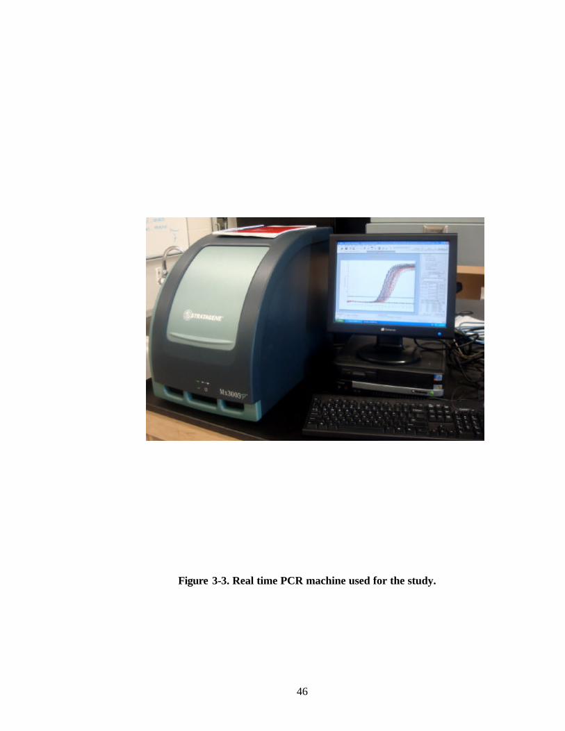
46
Figure 3-3. Real time PCR machine used for the study.

47
The contents of the tube were loaded onto a QIAquick spin column and centrifuged at
maximum speed for 1 min. QIAquick columns were washed with 500 µL of Buffer QG
to remove traces of agarose. Columns were then washed with 500 µL of PE buffer to
remove salts, and columns were air dried by spinning for 1 min. The column was then
placed in a sterile microfuge tube and the purified DNA was eluted using 50 µL of 10
mM Tris.HCl, pH 8.5. Samples were stored at -20ºC until further use.
3.22 Gene sequencing
Purified DNA was sequenced us ing an automated gene sequencer. The
sequencing PCR reaction mixture (10µL) contained 10 ng of the PCR-amplified product,
2 µL terminator ready reaction mixture, 1X sequencing buffer (80 mM Tris.HCl pH 8.0
and 2 mM MgCl2) and 0.32 pM of primers used to amplify the product. Amplifications
were performed in a thermocycler which was programmed for 25 cycles of denaturation
at 94ºC for 15 sec, annealing at 56ºC for 5 sec and extension at 60ºC for 2 min.
Amplified DNA was precipitated using 95% non-denatured ethanol at room temperature.
Before sequencing, the DNA pellet was dissolved in 12µL of template suspension
reagent, denatured at 95ºC for 2 min and loaded onto an Automatic DNA sequencer
(Applied Biosystems, Foster City, CA).
3.23 Statistical analysis of the data
Data were analyzed using SigmaStat version 3.1 (Systat Software Inc., Chicago,
IL). Groups were compared by one-way or two-way analysis of variance (ANOVA) and
differences were considered significant at P<0.05.

48
Chapter 4: Results
4.1 Factors affecting myrosinase activity
4.1.1 Kinetics of myrosinase
Myrosinase is known to release allyl isothiocynate (AITC) by acting on sinigrin
in the presence of water. AITC contributes to the toxicity of mustard meal. Therefore,
inhibiting myrosinase activity might be helpful in reducing AITC toxicity. Attempts
were made to improve the “Herb and Spice Method”, the only available industrial
method to measure total AITC production in mustard meal. This method is imprecise
and time consuming (requires 2 h of extraction for the quantification of one sample). To
develop an improved method for measuring AITC production in mustard powders, a
study of the effect of temperature on the kinetics of myrosinase activity was conducted.
A variety of reaction temperatures (7 to 97ºC) and incubation times (30 sec to 2
h) were studied. The seed meal was continuously agitated at various temperatures for 30
sec to 2 h and total AITC was measured as % volatile oil. A large proportion of the
AITC was released in 5 min of incubation at all temperatures below 87ºC (Figure 4-1).
Significant inhibition of enzyme activity was observed at all temperatures over 57ºC,
and at 97ºC no myrosinase activity was detected. Hence, it was concluded that: 1)
myrosinase catalyzed conversion of sinigrin to AITC was a rapid process since
detectable amounts of AITC could be found in samples as early as 5 min, and 2) higher
temperatures inhibited myrosinase activity. Incubation time was reduced to less than 5
min in a subsequent investigation of enzyme kinetics. No AITC was found at zero
incubation time. AITC yield increased with time and attained a maximum at 5 min.
Thus, an incubation time of 5 min at 27ºC (Figures 4-2 and 4-3) was used for all
subsequent studies.

49
Figure 4-1. Effect of temperature and incubation time on the yield of AITC, which is a measure of myrosinase activity.
5min15min30min60min120min
0.0
0.2
0.4
0.6
0.8
1.0
1.2
1.4
7 17 27 37 47 57 67 77 87 97
Temperature (°C)
% V
olat
ile o
il

50
0
0.1
0.2
0.3
0.4
0.5
0.6
0.7
0.8
0.9
1
0 20 40 60 80 100 120
Time (min)
% V
ola
tile
oils
a
d
b
c c c
Figure 4-2. Myrosinase attains near-maximum activity in 5 min at 27oC. Data was analysed by one way ANOVA. Values marked with different letters are significantly different from each other (P<0.05).
c

51
Figure 4-3. Myrosinase activity measured as % volatile oil at 27 ºC.
30 60 90 120 150 180 210 240 270 300
0
0.1
0.2
0.3
0.4
0.5
0.6
0.7
1 2 3 4 5 6 7 8 9 10
Time (seconds)
% V
olat
ile
oils
a a a
b
c
c
c c
c
c
30 60 90 120 150 180 210 240 270 300

52
4.1.2 Inhibition of myrosinase activity in mustard meal
To study reaction kinetics in response to temperature, pH and other factors, it
was necessary to have an effective method of starting and stopping the reaction. It was
known that pH could significantly alter the rate of myrosinase activity, and a study was
conducted to determine the effect of pH on myrosinase kinetics.
Enzyme activity was determined at pH ranging from 1.5 to 13, and the results
were compared with the control (water at pH 6.0). The reaction was conducted at 27°C
for 5 min. An increase or decrease in pH resulted in a reduction in enzyme activity as
compared to the control (Table 4-1), indicating that the enzyme had optimal near-neutral
conditions. When the reaction pH was brought back to 6.0 in the mixture containing
acetic acid or NaOH, further AITC was released due to residual enzyme activity, but the
treatment initiated in the presence of HCl did not exhibit residual enzyme activity.
Further, the addition of 1 g of ground Sinapis alba seed (a source of enzyme, but devoid
of substrate i.e., sinigrin) to the neutralized treatments resulted in the release of AITC,
presumably from unreacted sinigrin. Thus, it may be concluded that the change in pH
did not affect the substrate but severely affected the activity of myrosinase.
Furthermore, different concentrations of HCl were used to inhibit the activity of
myrosinase. Two milliliters of 2 N HCl per 100 mL reaction mixture (yielding a final
HCl concentration of 0.04 N) significantly inhibited myrosinase activity compared to the
control (Figure 4-4). Results were significant at P<0.05.
Other compounds viz., H3BO3, C2H4(COOH)2, CaCl2 and C2H5OH were
explored for their ability to inhibit myrosinase activity. The addition of C2H4(COOH)2,
calcium chloride or ethanol significantly reduced myrosinase activity (Figure 4-5). One-
way ANOVA revealed significant differences in all treatments (P<0.05). Boric acid
slightly increased myrosinase activity.

53
Table 4-1. Effect of pH reaction on the yield of AITC.
Neutralize to
pH 6.0
Addition of 1 g
powdered Sinapis alba
seed
Alkali/Acid pH % volatile oil
% volatile oil % volatile oil
Water (Control) 6.0 0.793 -- 0.821
NaOH 13 0.079 0.158 (HCl) 0.872
CH3COOH 3.0 0.039 0.178 1.150
HCl 1.5 0.118 0.079 1.189
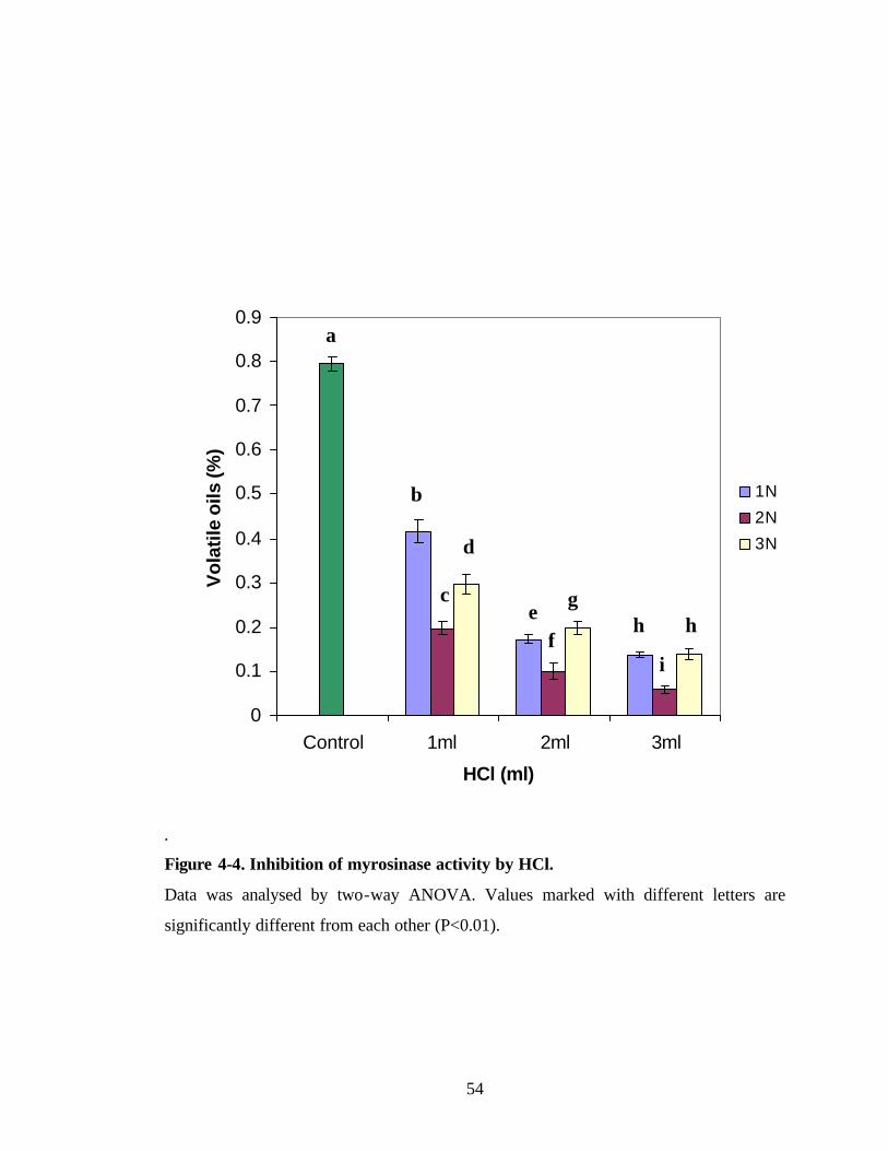
54
.
Figure 4-4. Inhibition of myrosinase activity by HCl.
Data was analysed by two-way ANOVA. Values marked with different letters are
significantly different from each other (P<0.01).
0
0.1
0.2
0.3
0.4
0.5
0.6
0.7
0.8
0.9
Control 1ml 2ml 3ml
HCl (ml)
Vo
lati
le o
ils (%
)
1N
2N
3N
b
d
e f
g h h
i
a
c

55
Values are the average of four replicates per treatment. Data were analysed by one-way
ANOVA. Values marked with different letters were significantly different from each
other (P<0.05).
Figure 4-5. Effect of different compounds on myrosinase activity.
0.3
0.4
0.5
0.6
0.7
0.8
0.9
1
Contro
l
C2H4(C
OOH)2H3B
O3CaC
l2
C2H5O
H
Vol
atile
oils
(%)
e
a
b
c
d

56
4.2 Biological activity of AITC
In the present study, it was hypothesized that AITC might act as a plant growth
promoter/regulator based on the structural similarity of AITC to the plant growth
regulator, ethylene, exhibit structural similarity (Figure 4-6). Therefore, AITC might act
as an ethylene analogue. AITC and ethylene share a common general formula which is
R-CH=CH2, where R is -CH2SCN and -H in AITC and ethylene, respectively. Ethylene
is a well known plant growth regulator known to promote seed germination and to
overcome seed dormancy in a dose- and species-dependent manner (Esashi et al.,, 1988;
Abeles et al., 1992; Kepczynski et al., 1997; Nascimento 2003). Therefore, it was
hypothesized that AITC might promote seed germination. Flaxseed and tomato seed
were used as model systems to test the germination promoting properties of AITC, along
with Ethephon® and IAA.
4.2.1 Effect of AITC on the germination of flaxseed
The effects of different concentrations (0 to 1 mM) of AITC, IAA and
Ethephon® were tested on the germination of flaxseed. Observations were taken every
24 h up to 6 days after treatment, and the data presented are averages of three replicates
of 20 seeds per treatment. Higher concentrations (0.1 to 1 mM) of AITC showed
stimulatory effects and promoted flaxseed germination (Figure 4-7). The effect of
Ethephon® (0.5 mM) was comparable to the control; all other concentrations of
Ethephon® tested inhibited flaxseed germination (Figure 4-8). All concentrations of
IAA tested inhibited germination of flaxseed as compared to the control, and
concentrations higher than 1 mM completely inhibited germination (Figure 4-9). IAA is
known to inhibit lettuce seed germination (Khan and Tolbert, 1966).
Growth of seedlings in AITC was comparable to that of the control, whereas
Ethephon® and IAA inhibited seedling growth (Figure 4-10). It may be concluded that
AITC at a higher concentration (1 mM) promoted flaxseed germination.

57
R-CH=CH2 R-CH=CH2
(R = -H) (R =CH2-N=C=S) Ethylene AITC
Figure 4-6. Formulas of AITC and ethylene.

58
0
20
40
60
80
100
120
0 1 2 3 4 5 6
Days
% G
erm
inat
ion 0
0.01
0.05
0.1
0.51
Figure 4-7. Effect of AITC on the germination of flaxseed. Values are averages of six replicates per treatment.
AITC (mM)

59
0
10
20
30
40
50
60
70
80
90
100
0 1 2 3 4 5 6
Days
% G
erm
inat
ion 0
0.01
0.05
0.1
0.51
Figure 4-8. Effect of Ethephon® on the germination of flaxseed. Values are averages of six replicates per treatment.
Ethephon (mM)

60
0
10
20
30
40
50
60
70
80
90
100
0 1 2 3 4 5 6
Days
% G
erm
inat
ion 0
0.01
0.05
0.1
0.51
Figure 4-9. Effect of IAA on the germination of flaxseed. Values are averages of six replicates per treatment.
IAA (mM)
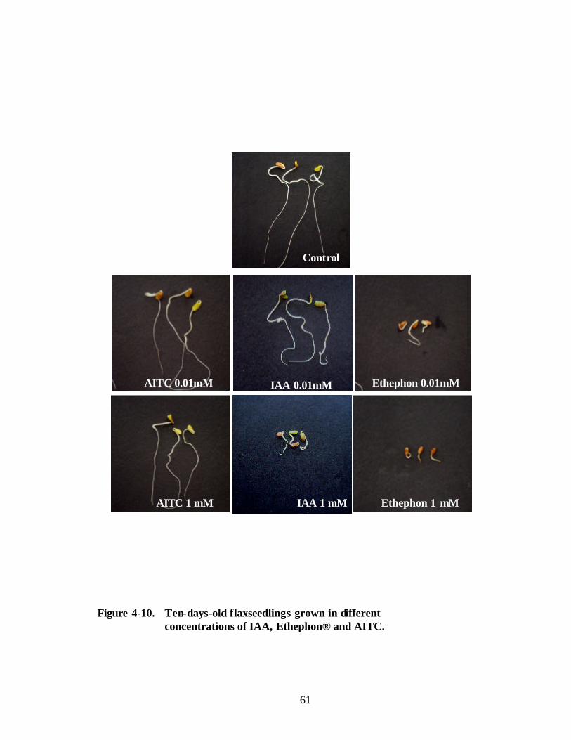
61
Control
AITC 0.01mM Ethephon 0.01mM
AITC 1 mM
IAA 0.01mM
Ethephon 1 mM IAA 1 mM
Figure 4-10. Ten-days-old flaxseedlings grown in different concentrations of IAA, Ethephon® and AITC.

62
4.2.2 Effect of AITC on the germination of tomato seed
To further evaluate the effect of AITC on seed germination, the effects of
different concentrations (0 to 1 mM) of AITC, IAA and Ethephon® on the germination
of tomato seed were tested. Observations were taken from 0h to 6 days after treatment,
and data presented are averages of three replicates of 20 seeds per treatment.
Concentrations of AITC higher that 0.1 mM enhanced seed germination and lower
concentrations were comparable to the control (Figure 4-11). Most concentrations of
Ethephon® tested had an inhibitory effect on tomato seed germination (Figure 4-12).
Unlike with flaxseed, lower concentrations (0.01 to 0.1 mM) of IAA enhanced tomato
seed germination (Figure 4-13). Growth of seedlings was inhibited by Ethephon® and
IAA as compared to AITC and the control (Figure 4-14). Therefore, it was concluded
that AITC enhanced seed germination and thus might have use in agricultural practice.

63
0
20
40
60
80
100
120
0 1 2 3 4 5 6
Days
% G
erm
inat
ion 0
0.01
0.050.1
0.5
1
Figure 4-11. Effect of AITC on the germination of tomato seed. Values are the average of six replicates per treatment.
AITC (mM)
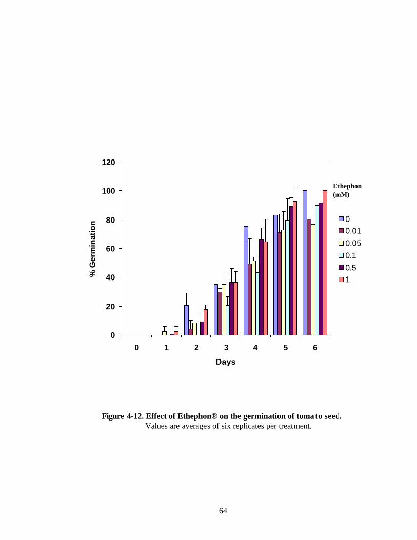
64
0
20
40
60
80
100
120
0 1 2 3 4 5 6
Days
% G
erm
inat
ion 0
0.01
0.05
0.1
0.51
Figure 4-12. Effect of Ethephon® on the germination of tomato seed. Values are averages of six replicates per treatment.
Ethephon (mM)

65
0
20
40
60
80
100
120
0 1 2 3 4 5 6
Days
% G
erm
inat
ion 0
0.01
0.05
0.1
0.51
Figure 4-13. Effect of IAA on the germination of tomato seed. Values are averages of six replicates per treatment.
IAA (mM)

66
Control
AITC 0.01 mM IAA 0.01 mM Ethephon 0.01 mM
AITC 1 mM IAA 1 mM Ethephon 1 mM
Figure 4-14. Ten-days-old tomato seedlings grown in different concentrations of IAA, Ethephon® and AITC.

67
4.3 HSP70 as an indicator of stress/toxicity in C. elegans
Heat shock proteins play important roles in the tolerance of living organisms to a
variety of biotic and abiotic stresses (Vierling, 1991; Parsell and Lindquist, 1994;
Hamilton and Coleman, 2001). Thus, HSPs appear to be general stress proteins that are
involved in maintenance of proper cell function, and thereby help in cell survival during
stress by facilitating recovery from stress (Vierling, 1991; Parsell and Lindquist, 1994;
Downs and Heckathorn, 1998; Guy and Li, 1998; Heckathorn et al., 1998). Members of
the 70 kDa heat shock protein (HSP70) family are ubiquitous in plants, animals and
microorganisms, and their structure and function are highly conserved among diverse
organisms, from algae to mammals (Wu et al., 1994). HSP70s are involved in
preventing protein aggregation and in refolding of denatured proteins produced in
response to cellular stress caused by an insult. In the present research, methods of
screening AITC toxicity were developed and the following questions were addressed:
1. How different isoforms of HSP70 in C. elegans are phylogenetically related?
2. Does AITC induce expression of HSP70?
3. Is toxicity contributed by AITC alone or is there synergistic effects of other
components of mustard seed meal?
4.3.1 Sequence alignments and phylogenetic relationships among HSP70s in C.
elegans
The HSP70 multigene family in C. elegans has been characterized (Heschl and
Baillie, 1998). Bioinformatic studies were done on the different forms of HSP70 viz.,
HSP70A, HSP70C, HSP70D and HSP70F, to understand level of sequence identity and
phylogenetic relationships. These isoforms of HSP70 showed high homology (Figure 4-
15), but they have been shown to perform different functions (Table 4-2). The
phylogenetic tree made with Clustal-W showed that among the various isoforms of
HSP70 in C. elegans, HSP70C and HSP70D were the most closely related (Figure 4-16).
Phylogenetic analysis showed that HSP70A and HSP70F of C. elegans are distantly
related. Because HSP70A is a cytoplasmic protein known to be expressed in response to
stress or toxicity, further studies were focused on HSP70A.

68
Figure 4-15. Sequence alignment of HSP70A, HSP70C, HSP70D and HSP70F showing high identity in peptide sequences.

69
Table 4-2. Accession numbers of various known HSP70s in C. elegans and their
Functions (Heschi and Baillie, 1998).
.
Gene Accession No. (NCBI) Function
HSP70A M18540 Expressed in response to variety of stresses
HSP70C T15513 Expressed in the Endoplasmic reticulum (ER), not
increased in response to heat stress
HSP70D P20163 Prevents secretion from the ER
HSP70F P11141 Expressed in the mitochondria

70
Figure 4-16. Phylogenetic relationships among various HSP70s in C. elegans.

71
4.3.2 Expression of HSP70 protein as an indicator of stress
Western blotting is a technique to understand the accumulation of a protein in
response to treatments, because the protein of interest can be detected in whole extracts
by using a protein specific antibody. Western blotting can also be used to confirm the
size of the protein of interest.
4.3.2.1 Expression of HSP70A transcripts in response to AITC
It is important to understand the transcription of genes within the HSP70 domain.
The bioinformatic research discussed above provided a reason for selecting one of the
various isoforms of HSP70 in C. elegans. Therefore, expression of HSP70A transcripts
in response to AITC treatment was investiga ted. Preliminary trials were performed using
reverse transcriptase polymerase chain reaction (RT-PCR) technique.
4.3.2.2 Reverse transcriptase polymerase chain reaction (RT-PCR)
The heat-shock- induced accumulation of HSP70A transcripts is shown in Figure
4-17. In order to determine the level of HSP70A transcripts, RT-PCR was conducted on
total RNA isolated from C. elegans exposed to different concentrations of AITC for 1 h.
Although HSP70A transcripts were constitutively present at low levels, there was a
considerable increase in expression at all AITC concentrations over 1 µM (Figure 4-17).
Based on densitometeric scoring of bands obtained by RT-PCR, the maximum level of
HSP70A transcription was observed in C. elegans exposed to AITC at a concentration of
5 µM. A slight decline in the expression of HSP70A at the highest concentration (10
µM) of AITC might be due to its lethality. No change in the expression of
glyceraldehyde-3-phosphate dehydrogenase (GAPDH), a housekeeping gene, was
observed, which indicated that the same amount of RNA was taken in each sample.
These results confirm that AITC exposure increased the expression of the HSP70A gene
in C. elegans.

72
4.3.2.3 Uantitative real time RtPCR
AITC (µM)
Hsp70A GAPDH
Con
trol
0.00
1
0.1
1 5 10
Figure 4-17. RT-PCR showing the expression of HSP70A (Accession No. M18540, NCBI) in response to stress caused by different concentrations of AITC. GAPDH (Accession No. X04818, NCBI), a house keeping gene was used as control.

73
4.3.2.3 Quantitative real time RT-PCR
Real time RT-PCR is one of the most sensitive techniques available to quantify
levels of gene transcripts in response to a treatment. The GAPDH gene was used as the
reference housekeeping gene and relative expression levels were calculated after
correction for expression of GAPDH.
Specificity of the primer sets was evident from higher annealing temperature i.e.
58°C and 60°C for forward and reverse primers, respectively (Table 4-3). These primers
were 21 bases long. Higher temperature and longer length of primers are two well
known factors that determine specificity. Alignment of nucleotide sequences and
position of these primers (Figure 4-18) also showed specificity of primers for HSP70A.
Specificity of primers was confirmed by sequencing amplified product which was
identical to HSP70A transcript (Accession No. M18540).
Fluorescence (dRn) values of the SYBR green and normalizing ROX obtained
for GAPDH and HSP70A from a single sample when plotted against number of
amplification cycles showed showed a sigmoid curve (Figure 4-19).
No significant change in the levels of HSP70A transcripts over the control was
observed at concentrations of AITC of 0.1 µM or lower (Figure 4-20). However, C.
elegans treated with higher concentrations of AITC (1 to 10 µM) responded with a 4- to
5-fold increase in expression of HSP70A transcripts over the control. These results
clearly indicate that AITC causes stress in C. elegans and HSP70A could be used as an
indicator of this property of AITC.

74
Table 4-3: Properties of primers used for real time RT-PCR.
Gene Primer sequences (5’ to 3’) Primer
length
%
GC
Tm
(ºC)
Product
size
Forward:
ATGAGTAAGCATAACGCTGTT
21 38.09 58 HSP70A
Reverse:
ACAGTGTTATGTGGGTTCATG
21 42.85 60
200 bp

75
Figure 4-18. Nucleotide sequence alignment of various isoforms of HSP70A and
position of primers for the amplification of HSP70A showing primer specificity.
HSP70C ATGAAAACCCTGTTTCTGCTGGGCCTGATTGCGCTGAGCGCGGTGAGCGTGTATTGCGAA 60 HSP70D ATGAAAG---TGTTTAGCCTGATTCTGATTGCGTT---TGTGGCGAACGCGTATTGCGAT 54 HSP70F ATGCTGAGCGCGCGCAGCTTTCTGAGCAGCGCGCGCACCATTGCGCGCA-GCAGCCTGAT 59 HSP70A ------------------------------------------------------------ HSP70C GAAGAAGAAAAAACCGAAAAAAAAGAAACCAAATATGGCACCATTATTGGCATTGATCTG 120 HSP70D GAAGGCGCGAGCACCGAAAAAGAAGAAA--AAATG-GGCACCATTATTGGCATTGATCTG 111 HSP70F GAGCGCGCGCAGCCTGAGCGATAAACCG--AAA---GGCCATGTGATTGGCATTGATCTG 114 HSP70A ------------------------ATGAGTAAG---CATAACGCTGTTGGAATCGATTTG 33 ** **** ** *** ** HSP70C GGCACCACCTATAGCTGCGTGGGCGTGTATAAAAACGGCCGCGTGGAAATTATTGCGAAC 180 HSP70D GGCACCACCTATAGCTGCGTGGGCGTGTTTAAAAACGGCCGCGTGGAAATTATTGCGAAC 171 HSP70F GGCACCACCAACAGCTGCGTGAGCATTATGGAAGGCAAAACCCCGAAAGTGATTGAAAAC 174 HSP70A GGAACTACCTACTCCTGCGTGGGAGTTTTCATGCACGGAAAGGTAGAAATCATTGCCAAC 93 ** ** *** * ******* * * * ** * **** *** HSP70C GATCAGGGCAACCGCATTACCCCGAGCTATGTGGCGTTTAGCGGCGATCAGGGCGATCGC 240 HSP70D GATCAGGGCAACCGCATTACCCCGAGCTATGTGGCGTTTAGCGGCGATCAGGGCGAACGC 231 HSP70F GCGGAAGGCGTGCGCACCACCCCGAGCACCGTGGCGTTTACCGCGGAT---GGCGAACGC 231 HSP70A GATCAAGGAAACCGTACAACTCCATCATATGTGGCTTTCACCGACACC------GAGCGT 147 * * ** ** * ** ** ***** ** * ** ** ** HSP70C CTGATTGGCGATGCGGCGAAAAACCAGCTGACCATTAACACGGAAAACACCATTTTTGAT 300 HSP70D CTGATTGGCGATGCGGCGAAAAACCAGCTGACCATTAACACGGAAAACACCATTTTTGAT 291 HSP70F CTGGTGGGCGCGCCGGCGAAACGCCAGGCGGTGACCAACAGCGCGAACACCCTGTTTGCG 291 HSP70A CTCATCGGAGATGCTGCCAAGAATCAAGTTGCCATGAACCCACATAACACTGTTTTCGAT 207 ** * ** * * ** ** ** * *** ***** * ** *

76
Figure 4-19. Fluorescence (dRn) values of the SYBR green and normalizing ROX obtained for GAPDH and HSP70A from a single sample showed a sigmoid curve.
Fluo
resc
ence
(dR
n)
1.6
1.4
1.2
1.0
0.8
0.6
0.4
0.2
0
Cycles
2 4 6 8 10 12 14 16 18 20 22 24 26 28 30 32 34 36 38 40

77
0
0.2
0.4
0.6
0.8
1
1.2
1.4
1.6
1 2 3 4 5 6
Rel
ativ
e Q
uant
ity
(dR
n)
0 0.001 0.1 1 5 10 AITC (µM)
Figure 4-20. Real time RT-PCR for the expression of HSP70A in response to stress caused by different concentrations of AITC. C. elegans was treated with different concentrations of AITC (0-10 µ M) for 2 h at room temperature. Real time RT-PCR was performed to amplify HSP70A. Bars on the columns represent standard deviations of three values. Data was analysed by one-way ANOVA. Values marked with different letters were significantly different from each other (P<0.01).
b
a a a
c c

78
4.3.2.4 Western blotting
Although the transcription of the HSP70A gene in response to AITC was
confirmed by real time RT-PCR, all the transcripts are not necessarily translated to
mature, completely functional protein. Therefore, it was necessary to confirm the
expression of HSP70 proteins using Western blotting.
C. elegans was exposed to different concentrations of AITC (0-20 µM) for 2 h at
room temperature and the expression of HSP70 protein was investigated by Western
blotting followed by immunoblotting with anti-HSP70 antibody. The specificity of the
anti-HSP70 monoclonal antibody to the HSP70 protein in C. elegans was tested by
sequence alignment. Based on the information provided by AbCam, antibody was raised
against a synthetic peptide corresponding to residues near the C-terminus of human
HSP70 (GenBank accession No. NM_005346). Precise sequences against which
antibody was raised is their commercial confidfentility. Then, human HSP70 amino acid
sequences were aligned with HSP70A (GenBank accession No. M18540) in C. elegans
by using web-based ClustalW version 1.83 software (EMBL-EBI). Results showed 80%
identity in these sequences (Figure 4-21).
Western blotting followed by immunodetection with anti-HSP70 antibody
revealed the expression of a 70 kDa band that represents HSP70 protein. These results
clearly showed accumulation of HSP70 protein in C. elegans exposed to concentrations
of AITC above 1 µM for 2h at room temperature. The highest accumulation of HSP70
protein was observed in C. elegans exposed to 5 µM AITC (Figure 4.22). These results
showed a concentration- and time-dependent increase in expression of the HSP70
protein(s), which confirmed the toxic effect of AITC on C. elegans. Therefore, it was
concluded that AITC causes stress/toxicity in C. elegans and this system can be used to
test toxicity of AITC in ground seed.

79
Human MAKAAAIGIDLGTTYSCVGVFQHGKVEIIANDQGNRTTPSYVAFTDTERLIGDAAKNQVA 60 C. elegans MSKHNAVGIDLGTTYSCVGVFMHGKVEIIANDQGNRTTPSYVAFTDTERLIGDAAKNQVA 60 *:* *:************** ************************************** Human LNPQNTVFDAKRLIGRKFGDPVVQSDMKHWPFQVIN-DGDKPKVQVSYKGETKAFYPEEI 119 C. elegans MNPHNTVFDAKRLIGRKFDDPAVQSDMKHWPFKVISAEGAKPKVQVEYKGENKIFTPEEI 120 :**:**************.**.**********:**. :* ******.****.* * **** Human SSMVLTKMKEIAEAYLGYPVTNAVITVPAYFNDSQRQATKDAGVIAGLNVLRIINEPTAA 179 C. elegans SSMVLLKMKKTAEAFLEPTVKDAVVTVPTYFNDSQRQATKDAGAIAGLNVLRIINEPTAA 180 ***** ***: ***:* .*.:**:***:**************.**************** Human AIAYGLDRTGKGERNVLIFDLGGGTFDVSILTIDDGIFEVKATAGDTHLGGEDFDNRLVN 239 C. elegans AIAYGLDKKGHGERNVLIFDLGGGTFDVSILTIEDGIFEVKSTAGDTHLGGEDFDNRMVN 240 *******:.*:**********************:*******:***************:** Human HFVEEFKRKHKKDISQNKRAVRRLRTACERAKRTLSSSTQASLEIDSLFEGIDFYTSITR 299 C. elegans HFCAEFKRKHKKDLASNPRALRRLRTACERANETLSSSCQASIEIDSLFEGIDFYTNITR 300 ** *********::.* **:**********:.***** ***:*************.*** Human ARFEELCSDLFRSTLEPVEKALRDAKLDKAQIHDLVLVGGSTRIPKVQKLLQDFFNGRDL 359 C. elegans ARFEELCADLFRSTMDPVEKSLRDAKMDKSQVHDIVLVGGSTRIPKVQKLLSDLFSGKEL 360 *******:******::****:*****:**:*:**:****************.*:*.*::* Human NKSINPDEAVAYGAAVQAAILMGDKSENVQDLLLLDVAPLSLGLETAGGVMTALIKRNST 419 C. elegans NKSINPDEALAYGAAVQAAILSGDKSEAVQDLLLLDVAPLSLGIETAGGVMTALIKRNTT 420 *********:*********** ***** ***************:**************:* Human IPTKQTQIFTTYSDNQPGVLIQVYEGERAMTKDNNLLGRFELSGIPPAPRGVPQIEVTFD 479 C. elegans IPTKTAQTFTTYSDNQPGVLIQVYEGERAMTKDNNLLGKFELSGIPPAPRGVPQIEVTFD 480 **** :* ******************************:********************* Human IDANGILNVTATDKSTGKASKITITNDKGRLSKEEIERMVQEAEKYKAEDEVQRERVSAK 539 C. elegans IDANGILNVSATDKSTGKAKQITITNDKDRFSKDDIERMVNEAEKYKADDEAQKDRIGAK 540 *********:*********.:*******.*:**::*****:*******:**.*::*:.** Human NALESYAFNMKSAVEDEGLKGKISEADKKKVLDKCQEVISWLDANTLAEKDEFEHKRKEL 599 C. elegans NGLESYAFNLKQTIEDEKLKDKISPEDKKKIEDKCDEILKWLDSNQTAEKEEFESQQKDL 600 *.*******:*.::*** **.*** ****: ***:*::.***:* ***:*** ::*:* Human EQVCNPIISGLYQGAGGPGPGGFGAQGPKGGSGSGPTIEEVD 641 C. elegans EGLAKPDLSKLYQSAGGAPPG--AAPGGAAGGAGGPTIEEVD 640 * :.:* :* ***.***. ** .* * .*...********
Figure 4-21. Amino acid sequence alignment of human (Accession No. NM_005346) and C. elegans (Accession No. M18540) HSP70 protein, showed 80% identity. Sequences were aligned using ClustalW version 1.83 (EMBL-EBI) software.

80
.
Hsp70
Con
trol
0.00
1
0.1
1 5 10
20
AITC (µM)
Figure 4-22. Western blot analysis showing induced expression of HSP70 protein in response to stress caused by AITC.
C. elegans was treated with different concentrations of AITC (0-10 µM) for 2 h at room temperature. Proteins in cell lysates were separated on SDS-PAGE gel and HSP70 protein was detected using anti-HSP70 antibody.
81
4.4 Mustard toxicity is contributed by AITC
From the above experiments on the toxicity aspects of AITC, it was concluded
that all concentrations of AITC over 1 µM caused significant stress in C. elegans.
However, it is important to know whether compounds in ground mustard seed, other
than AITC, also contributed to mustard toxicity. The advantage of protein expression
over the expression of mRNA transcripts was discussed earlier. At the same time, it is
important to have some quantitative method of analysis of protein. Therefore, the
expression of HSP70s was measured by an enzyme-linked immunosorbant assay
(ELISA).
B. juncea cv. Arrid does not contain sinigrin, a substrate for myrosinase, and was
used for treating C. elegans with or without AITC. The dose was based on AITC levels
of 1% the total seed meal. C. elegans strain N2 was treated with AITC (0-10 µM), B.
juncea cv. Arrid (0-114.5 µg/mL) or AITC (0-10 µM) + B. juncea cv. Arrid (0-114.5
µg/mL) for 2 h and the total protein extract was analyzed by ELISA. Lower
concentrations of B. juncea cv. Arrid (0-72.25 µg/mL) had no effect on the expression of
HSP70s, only the highest concentration (114.5 µg/mL) resulted in elevated expression of
HSP70s (Figure 4-23). All of the concentrations of AITC alone resulted in significant
increases in the expression of HSP70. AITC + ground mustard seed resulted in an
increased expression of HSP70s, but the levels were still less than those observed with
AITC alone.
These results indicate that 1) ground mustard seed toxicity is contributed by
AITC and not by other compounds present in the meal, and 2) some components of the
meal might act as AITC antagonists or the oil content in the meal inhibits the absorption
of AITC, thereby reducing toxicity in C. elegans.

82
Figure 4-23. Expression of HSP70 protein in C. elegans in response to exposure to AITC, ground seeds and AITC+ground seeds . C. elegans strain N2 was treated with AITC (0-10 µM), B. juncea cv. Arrid (0-114.5 µg/mL) or AITC (0-10 µM) + B. juncea cv. Arrid (0-114.5 µg/mL) for 2 h at room temperature and the total protein extract was analyzed by ELISA for the presence of HSP70. Bars on the columns represent standard deviations of three values. Data was analysed by multiple ANOVA. Values marked with different letters were significantly different from each other (P<0.01).
-20
0
20
40
60
80
100
120
Control AITC 0.001 AITC 0.1 AITC 1 AITC 5 AITC 10
a a a aa
a a a
b b
c
d
d d
e
f g
d
HS
P 7
0 (n
g/m
g)
AITC (µM) Control 0.001 0.1 1 5 10 Ground seeds (µg) Control 0.01445 1.445 14.45 72.25 114.5 Ground seeds + AITC Control 0.01445 1.445 14.45 72.25 114.5 (µg) (µM) + 0.001 + 0.1 +1 + 5 + 10

83
Chapter 5: Discussion
Western Canada has been one of the major producers of mustard seed since
World War II, when supplies from Europe were disrupted. Canadian mustard seed
production is concentrated in the Prairie Provinces, particularly in Saskatchewan.
Mustard seed has the advantage of being tolerant to drought, heat and frost. Canada
produces three mustard types: yellow mustard (Sinapis alba), brown mustard (Brassica
juncea), and oriental mustard (Brassica juncea). Over the last ten years, research stations
such as the Saskatoon Research Centre (SRC) of Agriculture and Agri-Food Canada
have bred all three types of mustard for the condiment market, and are working on
developing mustard varieties suited to non-food applications such as biodiesel and other
industrial products. The SRC has released progressively improved varieties of this crop
and Agriculture and Agri-Food Canada (AAFC), the Natural Sciences and Engineering
Research Council of Canada (NSERC), the Agriculture Development Fund (ADF) and
other major funding agencies have significantly supported improvement of mustard and
mustard-based products. According to the Saskatchewan Ministry of Agriculture, the
main funding for mustard research is contributed by AAFC, the Saskatchewan Mustard
Development Commission (SDMC) and Saskatchewan Agriculture.
Mustard is the only cultivated oilseed crop adapted to the drier regions of the
Canadian prairies and provides a suitable crop for rotation with wheat (Kirkegaard et al.,
1994; Marcia et al., 1999). Mustard is also resistant to many pests and fungal diseases
and thus requires fewer inputs for its cultivation (Bones and Rossiter, 1996). Co-
cropping Brassica has been tested for weed management (Haramoto and Gallandt, 2004,
2005).

84
Mustard seed, in the form of either seed or oil, is primarily used in the condiment
and food industries. It is also used as an emulsifier or a water binding agent, and for
texture control, in meat products. According to Red Coat REDA Inc., Gravelbourg, SK,
the oil content of yellow mustard seed is the lowest (27%) of the three types of mustard,
and it is the mildest in flavour of the three types. Therefore, yellow mustard is suitable
for a wide range of applications, including flour, mustard pastes, spice mixes, meat
processing, and other food products. Brown and Oriental mustard seed are ingredients of
hot spices. Brown mustard has the highest oil content (36%; Skrypetz, 2003). Oriental
mustard seed is often used to produce spicy cooking oils. There are Oriental mustard
seed varieties grown in Canada that have oil contents of up to 50%, although the average
oil content is approximately 39% according to AAFC. There has been a fluctuating trend
in Canadian mustard seed production in recent years from a low of 105,000 tonnes in
2001 to a high of 306,000 tonnes in 2004. Exports increased nearly 8 percent from 2004
to 2005. Canada exports mustard seed mainly to the United States of America (46%),
Belgium (22%) and Japan (7%) (Jacques, 2005). Demand for mustard seed is expected
to grow during the next decade as world population and the use of spices increases.
Canada’s ability to meet this growing international demand, as well as continued
research into new crop varieties, will contribute to Canada’s continued success in the
mustard industry (Jacques, 2005).
According to the SMDC, since 1936 plant breeders in Canada, mainly in
Saskatchewan, have worked to improve the quality of mustard. During the 1950s and
1960s, mustard production migrated east, and today, Saskatchewan accounts for nearly
90 per cent of Canadian mustard production.
The myrosinase-glucosinolate system is characteristic of members of the
Brassicaceae family (Bones and Rossiter, 1996). This system is only activated following
tissue damage and plays a defensive role against pathogens (Rask et al., 2000).
Glucosinolate hydrolysis leads to the rapid formation of hydrolysis products such as
allyl isothiocyanate (AITC) (Fahey et al., 2001; Mithen 2001; Fenwick et al., 1983).
Mustard is characterized by its typical flavour and taste, which is contributed by
glucosinolate hydrolysis products. However, glucosinolate hydrolysis products comprise
a large family of compounds. A subset of these compounds has been shown to have

85
possible health promoting effects (Verhoeven et al., 1997), they may inhibit
tumorigenesis (Smith et al., 1998) and may play an important role in preventing heart
disease (Wu et al., 2004). AITC, one of the glucosinolate hydrolysis products, may be
widely used in applications where its broad spectrum of toxicity and volatility are
advantageous viz., anti- fungal, anti-bacterial and bio-fumigant, which are described in
Chapter 2. Due to the toxic effects of AITC, it is important to develop quick and
efficient AITC detection methods and to determine myrosinase activity in mustard seed
meal.
In the present investigation, an attempt was made to improve the “Herb and
Spice Method”, the only available “official” method to measure total isothiocyanate
production in mustard meal. The “Herb and Spice Method” is crude, imprecise, time
consuming (requires 2 h of extraction for the quantification of one sample) and requires
incubation at 37ºC (ASTA 1997).
The method developed in the current study was rapid and more reliable and was
used to indirectly measure myrosinase activity and to understand the kinetics of the
enzyme. A wide range of conditions (temperatures of 7 to 97ºC and incubation times of
30 sec to 2 h) were studied to determine optimal conditions for myrosinase activity.
Rapid evolution of AITC was observed at five min of incubation at all temperatures
tested. Higher temperatures (>57ºC) resulted in significant inhibition of enzyme activity,
and at 97ºC, no myrosinase activity was found. It was concluded that: 1) hydrolysis of
glucosinolates is a rapid process and a detectable amount of AITC can be found as early
as 5 min; and 2) higher temperatures inhibit myrosinase activity.
Eylen et al. (2006) studied the thermal and pressure stability of myrosinase at
temperatures from 60 to 75ºC and pressures from ambient to 1000 MPa. Myrosinase was
found to be stable at 600 MPa pressure at temperatures up to 60ºC. At low pressures,
there was an antagonistic effect between temperature and pressure. They suggested that
the high pressure stability of myrosinase may be a valuable alternative to thermal
treatments if one wants to retain myrosinase activity. Matuscheki et al. (2004) have
shown inactivation of myrosinase following thermal treatment. The kinetics of
myrosinase inactivation during thermal treatment has also been investigated in crude

86
broccoli extract (Ludikhuyze et al., 1999) and in red cabbage juice (Verkerk et al.,
2002).
The conditions for inactivation of myrosinase using microwave energy were
studied with a response surface design. Inactivation of myrosinase was dependent on
moisture content and exposure time to microwaves, and there was a significant increase
in both the yellow colour and sulfur content of the microwave-treated seed oils (Owusu-
Ansah and Marianchuk, 1991). Microwave irradiation at 2450 MHz for 2.5 min is
known to inactivate myrosinase and decompose glucosinolates (Verkerk and Dekker,
2004).
Attempts were made to inhibit or reduce myrosinase activity by manipulating the
pH of the extraction medium over the range of 1.5 to 13. Results indicated that
myrosinase required a pH of 6.0 for optimum activity, and that a change in pH did not
impact the substrate but altered the activity of the enzyme. Other compounds viz.,
C2H4(COOH)2, H3BO3, CaCl2 and C2H5OH were also explored for their ability to inhibit
myrosinase activity. Succcinic acid, calcium chloride and ethanol significantly reduced
myrosinase activity. Different acids were tested for a rapid inactivation of myrosinase
without hydrolysis of the GLS. Mild acids (like succnic acid and borate) and harsh acid
(hydrochloric acid) inhibited myrosinase activity in mustard grounded seeds. A base was
also tesetd to inhibit myrosinase activity because the gycosidic bond is comparatively
more resilient to base. Furthermore, ethanol which can denature protein without altering
pH was also used that would leave the GLS intact. An extract from Arctostaphylos uvae-
ursi has been reported to inhibit myrosinase activity (Mykytyn and Demjanchuk, 1999).
In the present research, it was hypothesized that AITC might act as a plant
growth promoter/regulator because AITC and ethylene share a common chemical
structure (R-CH=CH2, where R is -CH2SCN and -H in AITC and ethylene, respectively).
Therefore, AITC might act as an ethylene analogue. Ethylene is a well known plant
growth regulator known to promote seed germination and overcome seed dormancy in a
dose- and species-dependent manner (Esashi et al., 1988; Abeles et al., 1992;
Kepczynski et al., 1997; Nascimento 2003). Based on the similarities between AITC and
ethylene, AITC may also regulate seed germination. Ethephon® and AITC were tested
to determine their impact on flaxseed and tomato seed germination. Interestingly, higher

87
concentrations (0.1 to 1 mM) of AITC enhanced seed germination. The present study
showed the seed germination enhancing properties of AITC, and thus AITC could be
used in agricultural practice.
There may be more than a single mode of action for glucosinolate toxicity and
biological activity. For example, AITC possesses mutagenic and DNA-modifying
activity with formation of reactive oxygen species (Kassie and Knasmuller, 2000). In
addition, benzyl isothiocynate has mutagenic properties (Yamaguchi, 1980) and caused
DNA strand breaks (Musk et al., 1995). AITC inhibits growth of PC-3 human cancer
cell xenografts by inducing apoptosis and reducing mitotic activity (Srivastava et al.,
2003). The apoptosis and cell cycle arrest properties of AITC have been documented in
leukemia cells (Xu and Thornalley, 2000), prostate cancer cells (Xiao et al., 2003) and
colon cancer cells (Gamet-Payrastre et al., 2000). AITC induces apoptosis and rapid
transient activation of Caspase-3 and Caspase-8 (Yu et al., 1998; Xu and Thornalley,
2000; Xu and Thornalley, 2001). AITC is also known to arrest the cell cycle in G2/M
phase (Xiao et al., 2003; Smith et al., 2004). In the present investigation, C. elegans was
used to study AITC-mediated stress or toxicity.
Heat shock proteins play an important role in acclimatization of living organisms
to a variety of biotic and abiotic stresses (Vierling, 1991; Parsell and Lindquist, 1994;
Hamilton and Coleman, 2001). HSPs are general stress proteins and are involved in
maintenance of proper cell function and, thereby, help in cell survival during stress by
facilitating recovery from stress (Vierling, 1991; Parsell and Lindquist, 1994; Downs
and Heckathorn, 1998; Guy and Li, 1998; Heckathorn et al., 1998). HSP70 family
proteins are ubiquitous in plants, animals and microorganisms and their structure and
function are highly conserved among diverse organisms, from algae to mammals (Wu et
al., 1994). These proteins are invo lved in preventing protein aggregation and in
refolding of denatured proteins produced as a result of cellular stress caused by an insult.
In C. elegans, four isoforms of HSP70 are known: HSP70A, HSP70C, HSP70D and
HSP70F. Each of these isoforms of HSP70 performs a different function. HSP70A is
expressed in response to a variety of stresses, and HSP70C is expressed in the ER and its
expression is not increased in response to heat stress (Heschl and Baillie, 1998).

88
However, HSP70D prevents secretion of proteins from the ER and HSP70F is expressed
in the mitochondria.
In the present investigation, methods of screening AITC toxicity were developed.
Western blotting revealed expression of a 70 kDa protein in C. elegans due to exposure
to different concentrations of AITC in a dose-dependent manner. Nematodes were
visibly injured by the treatments with AITC, as was indicated by their moment.
Nematodes were actively moving in all the concentrations below 10µM. High
concentrations appeared to be lethal as their moment stopped. Detection of HSP70
protein by ELISA confirmed the above observations. Methods were developed to screen
AITC toxicity by real time RT-PCR to accomplish quantitative analysis of the
expression of HSP70A transcripts. It is important to understand whether compounds in
ground mustard seed, other than AITC, also contribute to mustard toxicity. The
advantage of protein expression over expression of mRNA transcripts was discussed
earlier. At the same time, it is important to have a quantitative method of analysis of
protein. Therefore, the expression of HSP70 proteins was measured by an ELISA.
C. elegans strain N2 was treated with B. juncea cv. Arrid and/or AITC and
expression of HSP70 proteins was quantified by ELISA. The dose of B. juncea cv. Arrid
was determined based on the fact that AITC is 1% of the total seed meal. Lower
concentrations of B. juncea cv. Arrid (0-72.25 µg/mL) had no effect on the expression of
HSP70 proteins, whereas the highest concentration (114.5 µg/mL) resulted in elevated
expression of HSP70 proteins. All of the concentrations of AITC alone resulted in a
significant increase in the expression of HSP70 protein. AITC + ground mustard seed
resulted in an increased expression of HSP70 proteins but the levels were still less than
those observed with AITC alone.
These results indicate that 1) ground mustard seed toxicity is contributed by
AITC, and 2) some components of the ground mustard seed act as AITC antagonists or
the oil content in the meal inhibits the absorption of AITC, thereby reducing toxicity in
C. elegans. These findings suggest that HSP70 could be used as a universal tool for
studying mustard toxicity. The present work could help the mustard industry open a new
market. Industry needs the tools for screening toxicity of mustard meal or oil-based
products. A company in Saskatchewan plans to use mustard for nematode control.

89
Therefore, the possibility for a totally new market and new acreage for mustard is
supported.
HSP70 expression can also be used as a universal biomarker in ecotoxicological
studies. It can also be a helpful tool for the monitoring of freshwater ecological systems.
Genetically modified zebra fish containing enhanced green fluorescent protein (eGFP)
under the control of a 1.5 kb HSP70 promoter have been developed (Blechinger et al.,
2002) which could be used for screening toxicity of mustard-based fish meals. The
methods of screening AITC toxicity developed in this study are fast, reliable and
quantitative.
Use of mustard as a biopesticide is one of the applications based on toxicity of
AITC. Toxicity of Indian mustard and AITC to masked chafer Beetle larvae has been
demonstrated (Ryan et al., 2002). Mustard used in biopesticide production may also
yield protein meal, fertilizer and biodiesel. Bio-pesticidal (Brown and Morra, 2005),
fungicidal (Brown and Morra, 2005), antibiotic (Mari et al., 2002; Kiyoshi 2005;
Winther and Nielsen, 2006; Tunc et al., 2007), biofumigant (Brown and Morra, 2005;
Tsao et al., 2002a; Noble et al., 2002) and nematocidal (Buskov et al., 2002; Lazzeri et
al., 1993) properties of AITC have been studied. This suggests the potential importance
of AITC and other GLS hydrolysis products in agricultural applications.
If AITC and mustard are to be used more widely in new pesticide markets,
questions will arise regarding both the risks to non-target organisms and the efficacy of
the compound on target organisms. The regulatory environment will often require
extensive testing for compounds in the environment, yet the presence of a compound
does not imply either impact or toxicity. The need for expensive analytical tests may
slow the introduction of new products with potent toxicity. Conversely, simple and
robust tests may aid in the development of suitable policy and regulations regarding the
deployment of the compound or material. The HSP approach of using a sentinel
organism could be a useful tool to allow the determination of toxicity in situ and to
potentially enable the deployment of mustard as a biopesticide. The HSP test data has
been shared with BioGreen Inc. of Hague, SK and the company is interested in using the
test for both product development and in their presentations to regulatory agencies,
including the US Environmental Protection Agency.

90
Chapter 6: Summary and Conclusions
The present study was aimed to study: 1) factors affecting myrosinase activity in
mustard (Brassica juncea L.), 2) the effect of allyl isothiocynate (AITC) on the
germination of flax and tomato seed, and 3) the toxicity of AITC and ground mustard
seed using C. elegans as a model system.
Myrosinase hydrolyzes sinigrin to produce AITC in the presence of water.
Spatial separation of the myrosinase and sinigrin in the intact plant prevents this
hydrolytic reaction. However, milling and grinding processes result in the hydrolysis of
sinigrin. As a result, AITC contributes to the toxicity of ground mustard seed and
mustard-based products. Therefore, a quick and efficient method for measuring AITC
was developed and used to measure myrosinase activity indirectly. It was revealed that
sinigrin hydrolysis was a rapid process and can be inhibited by 1) higher incubation
temperatures (>57ºC), 2) a pH less than or greater than 6.0, and 3) adding succinic acid,
calcium chloride or ethanol to the reaction mixture. Myrosinase preferred near-neutral
conditions for its activity and a change in pH did not affect the substrate but severely
affected the activity of myrosinase. These results may lead to the improvement of
mustard processing methods to reduce toxicity caused by sinigrin hydrolysis products.
It was hypothesized that AITC might act as a plant growth promoter/regulator
based on the structural similarity of AITC to ethylene (R-CH=CH2, where R is -
CH2SCN and -H in AITC and ethylene, respectively). Therefore, AITC might act as an
ethylene analogue and promote seed germination. The effects of Ethephon®, IAA and
AITC on the germination of flaxseed and tomato seed were investigated. It was
concluded that AITC could be useful in agricultural practice.
The third objective of this study was to investigate the toxicity of AITC and
ground mustard seed using C. elegans as an experimental model and to identify

91
molecular indicators of AITC-induced stress in C. elegans. Members of the 70 kDa heat
shock protein (HSP70) family are ubiquitous in plants, animals and microorganisms, and
their structure and function are highly conserved among diverse organisms, from algae
to mammals. HSP70 proteins are known to be involved in preventing protein
aggregation and in refolding of denatured proteins produced as a result of cellular stress
caused by an insult. Bioinformatic tools used in this study revealed high identity in the
four isoforms of HSP70 in C. elegans viz., HSP70A, HSP70C, HSP70D and HSP70F.
Phylogenetic analysis showed that HSP70C and HSP70D were the most closely related
and that HSP70A and HSP70F were distinctly related. Because HSP70A is a cytosolic
protein known to be expressed in response to stress or toxicity, expression of HSP70A
was used as a marker of stress/toxicity in C. elegans.
Real-time RT-PCR results confirmed over-expression of the HSP70A gene in C.
elegans strain N2 following AITC treatment. Translation of HSP70 transcripts was
confirmed by Western blotting which showed accumulation of HSP70 in C. elegans
exposed to AITC (>1 µM) for two hours at room temperature. The highest accumulation
of HSP70 was observed in C. elegans exposed to 5 µM AITC. Therefore, expression of
HSP70A could be used as an indicator of the stress-inducing properties of AITC and to
test toxicity of AITC in ground mustard seed.
Expression of HSP70 proteins was quantified by ELISA. C. elegans was treated
with B. juncea cv. Arrid ground seed and/or AITC. Results showed that the highest
concentration of B. juncea ground seed (114.5µg/mL) caused an elevated expression of
HSP70. All of the concentrations of AITC alone resulted in a significant increase in the
expression of HSP70s. AITC + ground mustard seed resulted in an increased expression
of HSP70s, but the levels were less than those observed with AITC alone. Therefore, it
was concluded that it is AITC alone and no other component from mustard ground seed
that contributes to mustard toxicity. Some components of the ground seed either act as
AITC antagonists or it is may be due to presence of other compounds, which may
interact rather than oil alone. Therefore, presence of other compounds in the ground seed
inhibits the absorption of AITC, thereby reducing toxicity in C. elegans.

92
This work may lead to the development of better industrial mustard processing
methods or new methods for testing the toxicity of mustard-based products, and has
demonstrated that AITC may have practical application in enhancing seed germination.
Agro- industries may benefit from these findings.

93
Chapter 7: References
Abeles F.B., Morgan P.W. and Saltveit M.E. 1992. Ethylene in plant biology. Academic
Press, London.
Agerbirk N., Olsen C.E. and Sorensen H. 1998. Initial and final products, nitriles, and
ascorbigens produced in myrosinase-catalyzed hydrolysis of indole glucosinolates. J.
Agri. Food Chem. 46: 1563–1571.
Amit S. and Ben-Neriah Y. 2003. NF-kappaB activation in cancer: a challenge for
ubiquitination- and proteasome-based therapeutic approach. Semin. Cancer Biol. 13: 15-
28.
Ascard J. and Jonasson T. 1991. White mustard meal interesting for weed control. In:
Weeds and Weed Control. Reports. 32nd Swedish Crop Protection Conference, Swedish
University of Agricultural Sciences, Uppsala, pp. 139-155.
"American Herb and Spice Trade Association". 1997. Volatile Oil in Mustard Seed and
Flour (Alternate revised Method); Method No. 15.1.
Bandella M., Story G.M., Hwang S.W., Viswanath V., Eid S.R., Petrus M.J., Earley T.J.
and Patapoutain A. 2004. Noxious cold ion channel TRPA1 is activated by pungent
compounds and bradykinin. Neuron 41: 849-857.

94
Bautista D.M., Movahed P., Hinman A., Axelsson H.E., Sterner O., Hogestatt E.D.,
Julius D., Jordt S.E. and Zygmunt P.M. 2005. Pungent products from garlic activate the
sensor ion channel TRPA1. Proc. Natl. Acad. Sci., USA 102: 12248-12252.
Bautista D.M., Jordt S.E., Nikai T., Tsuruda P.R., Read A.J., Poblete J., Yamoah E.N.,
Basbaum A.J. and Julius D. 2006. TRPA1 mediates the inflammatory actions of
environmental irritants and proalgesic agents. Cell 124: 1269-1282.
Bennette R.N., Mennol F.A. and Kroon P.A. 2004. Screening crucifer seeds as source of
specific intact glucosinolates using ion-pair high performance liquid chromatography
negative ion electrospray mass spectrometry. J. Agric. Food Chem. 52: 428-438.
Bernardi R., Negri A., Ronchi S. and Palmieri S. 2000. Isolation of the epithio-specifier
protein from oil-rape (Brassica napus ssp. oleofera) seed and its characterization. FEBS
Lett. 467: 296-298.
Betz J.M. and Fox. W.D. 1994. High performance liquid chromatographic determination
of glucosinolates in Brassica vegetables. In: Food Phytochemicals for Cancer
Prevention: Fruits and vegetables, Huang M.T., Osawa T., Ho C.T. and Rosen R.T.
(Eds.), American Chemical Society, Washington, DC, pp. 180-196.
Bjergegaard C., Møller P., Sørensen H., Sørensen H. and Sørensen S. 1999. Micellar
electrokinetic capillary chromatography of thiocarbamoyl derivatives produced in
reactions between isothiocyanates and amino acids. J. Chrom. 836: 115-127.
Björkman, R. 1976. Properties and function of plant myrosinases. In The Biology and
Chemistry of the Cruciferae. Vaughan J.G., MacLeod A.J. and Jones B.M.G. (Eds.);
Academic Press: London 1976, pp. 191-205.

95
Bjorkman R. and Lonnerdal B. 1973. Studies on myrosinases. III. Enzymatic properties
of myrosinases from Sinapis alba and Brassica napus seeds. Biochim. Biophys. Acta
327: 121-131.
Björkqvist B. and Hase A. 1988. Separation and determination of the intact
glucosinolates in rapeseed by high performance liquid chromatography. J. Chrom. 435:
501-507.
Blechinger S.R., Evans T.G., Tang P.T., Kuwada J.Y., Warren J.T. and Krone P.H.
2002. The heat- inducible zebrafish HSP70 gene is expressed during normal lens
development under non-stress conditions. Mech. Develop. 112: 213-215.
Bones A.M. 1990. Distribution of ß-thioglucosidase activity in intact plants, cell and
tissue cultures and regenerant plants of Brassica napus. J. Exp. Bot. 41: 737-744.
Bones A.M. and Rossiter J.T. 1996. The myrosinase-glucosinolate system, its
organization and biochemistry. Physiol. Plant. 97: 194-208.
Bones A. and Slupphaug G. 1989. Purification, characterization and partial amino acid
sequencing of ß-thioglucosidase from Brassica napus. J. Plant Physiol. 134: 722-729.
Bremner P. and Heinrich M. 2002. Natural products as targeted modulator of the nuclear
factor-kappa B pathway. J. Pharm. Pharmacol. 54: 453-472.
Bringmann G., Kajahn I., Neusü C., Pelzing M., Laug S., Unger M. and Holzgrabe U.
2005. Analysis of the glucosinolate pattern of Arabidopsis thaliana seeds by capillary
zone electrophoresis coupled to electrospray ionization-mass spectrometry.
Electrophoresis 26: 1513-1522.

96
Brooks J.D., Paton V.G. and Vidanes G. 2001. Potent induction of phase 2 enzymes in
Human prostate cells by sulforaphane. Cancer Epidemiol. Biomarkers Prev. 10: 949-
954.
Brown J. and Morra M.J. 2005. Glucosinolate-containing seed meal as a soil amendment
to control plant pests. Subcontract Report NREL/SR-510-35254.
Brown P.D., Morra M.J. and Borek V. 1994. Gas chromatography of allelochemicals
produced during glucosinolate degradation in soil. J. Agric. Food Chem. 42: 2029-2034.
Buchwaldt L., Larsen L.M., Ploger A. and Sørensen H. 1986. Fast polymer liquid
chromatography isolation -thioglucoside glucohydrolase, and characterization of plant
myrosinase, isoenzymes. J. Chrom. 363: 71-80.
Burel C., Boujard T., Tulli F. and Kaushik S.J. 2000. Digestibility of extruded peas,
extruded lupin and rapeseed meal in rainbow trout (Oncorhynchus mykiss) and turbot
(Psetta maxima). Aquaculture 188: 285–298.
Buskov, S., Hasselstrøm, J., Olsen, C.E., Sørensen, H., Sørensen, J.C., Sørensen, S.,
2000. Supercritical- fluid chromatography as a method of analysis for the determination
of 4-hydroxybenzylglucosinolate degradation products. J. of Biochem. Biophys.
Methods 43, 157–174.
Buskov S., Serra B., Rosa E., Sorenses H. and Sorensen J.C. 2002. Effects of intact
glucosinolates and products produced from glucosinola tes in myrosinase-catalyzed
hydrolysis on the potato cyst nematode (Globodera rostochiensis Cv. Woll). J. Agric.
Food Chem. 50: 690-695.
Bussy A. 1840. Sur la formation de l`huile essentielle de moutarde. J. Pharm. 27: 464-
471.

97
Cahoon E.B., Hall S.E., Ripp K.G., Ganzke T.S., Hitz W.D. and Coughlan S.J. 2003.
Metabolic redesign of vitamin E biosynthesis in plants for tocotrienol production and
increased antioxidant content. Nature Biotech. 21: 1082 – 1087.
Candido E.P. and Jones D. 1996. Transgenic Caenorhabditis elegans strains as
biosensors. Tibtech. 14: 125-129.
Cao J., Chiarelli C., Kozarekar P. and Adler H.L. 2005. Membrane type-I matrix
metalloproteinase promotes human prostate cancer invasion and metastasis. Thromb.
Haemost. 93: 770-778.
Chacon P.A., Buffo R.A. and Holley R.A. 2006. Inhibitory effects of microencapsulated
allyl isothiocynate (AIT) against Escherichia coli O157:H7 in refrigerated, nitrogen
packed, finely chopped beef. Int. J Microbiol. 107: 231-237.
Chen C. and Ho C. 1998. Thermal degradation of allyl siothiocynate in aqueous
solution. J. Agric. Food Chem. 46: 220-223.
Chew F.S. 1988. Biological effects of glucosinolates. In: Biologically Active Natural
Products: Potential Use in Agriculture; Cutler HG (Ed.), American Chemical Society:
Washington, D.C., pp 155-181.
Coles R. 1976. Isothiocynates, nitriles and thiocynates as products of autolysis of
glucosinolate in Cruciferae. Phytochem. 15; 759-762.
Cranfield C.G., Dawe A., Karloukovski V. and Dunin-Borkowski R.E. 2004. Proc Bio l
Sci 271 Suppl 6: S436-439.
Das M.M. and Singhal K.K. 2001. Influence of chemical treatment of mustard oil cake
on its glucosinolate content and myrosinase activity. Ind. J. Anim. Sci. 71: 793–796.

98
Das M.M. and Singhal K.K. 2005. Effect of feeding chemically treated mustard cake on
growth, thyroid and liver function and carcass characteristics in kids. Small Rumin. Res.
56: 31–38.
Downs C.A. and Heckathorn S.A. 1998.The mitochondrial small heat-shock protein
protects NADH:ubiquinone oxidoreductase of the electron transport chain during heat
stress in plants. FEBS Lett. 430(3): 246-250.
Durham P.L. and Poulton J.E. 1990. Enzymic properties of purified myrosinase from
Lepedium sativum Seedlings. Z. Naturforsch 45: 173-178.
Eagles J., Fenwick G.R. and Heaney R.K. 2005. Gas chromatography chemical
ionization mass spectrometry of glucosinolate derivatives. Biol. Mass Spec. 8: 278 –
282.
Esashi Y., Kawabe K., Isuzugawa K. and Ishizawa K. 1988. Interrelations between
carbon dioxide and ethylene on the sStimulation of cocklebur sed germination. Plant
Physiol. 86(1): 39-43.
Eylen D.V., Hendrickx I.M. and Loey A.V. 2006. Temperature and pressure stability of
mustard seed (Sinapis alba L.) myrosinase. Food Chem. 97: 263-271.
Fahey J.W., Zalcmann A.T. and Talalay P. 2001. The chemical diversity and distribution
of glucosinolates and isothiocyanates among plants. Phytochem. 56: 5-51.
Falk A., Ek B. and Rask L. 1995. Characterization of a new myrosinase in Brassica
napus. Plant Mol. Biol. 27: 863-874.
Fenwick G.R. and Heaney R.K. 1983. Glucosinolates and their break-down products in
cruciferous crops, foods and feeding stuffs. Food Chem. 11: 249-271.

99
Foo H.L., Gronning L.M., Goodenough L., Bones A.M., Danielsen B., Whiting D.A.
and Rossiter J.T. Purification and characterisation of epithiospecifier protein from
Brassica napus: enzymic intramolecular sulphur addition within alkenyl
thiohydroximates derived from alkenyl glucosinolate hydrolysis. FEBS Lett. 468(2-3):
243-246.
Galletti S., Bernardi R., Leoni O., Rollin P. and Palmieri S. 2001. Preparation and
biological activity of four epiprogoitrin myrosinase-derived products. J. Agric. Food
Chem. 49(1): 471-476.
Gamet-Payrastre L., Li P., Lumeau S., Cassar G., Dupont M.A., Chevolleau S., Gasc N.,
Tulliez J. and Tercé F. 2000. Sulforaphane, a naturally occurring isothiocyanate, induces
cell cycle arrest and apoptosis in HT29 human colon cancer cells. Cancer Res. 60(5):
1426-1433.
Gil V. and MacLeod A.J. 1980. The effects of pH on glucosinolate degradation by a
thioglucoside glucohydrolase preparation. Phytochem. 19: 2547–2551.
Gimsing A.L., Kirkegaard J.A. and Hansen H.C. 2005. Extraction and determination of
glucosinolates from soil. J. Agric. Food Chem. 53: 9663-9667.
Gimsing A.L., Sørensen J.C., Tovgaard L., Jørgensen A.M., Hansen H.C. 2006.
Degradation kinetics of glucosinolates in soil. Environ. Toxicol. Chem. 25: 2038-2044.
Gimsing A.L., Poulsen J.L., Pedersen H.L. and Hansen H.C. 2007. Formation and
degradation kinetics of the biofumigant benzyl isothiocyanate in soil. Environ. Sci.
Technol. 41: 4271-4276.
Goodman I., Fouts J.R., Bresnick, E., Menegas R. and Hitchings G.H. 1959. A
mammalian thioglucosidase. Science 130: 450-451.

100
Guy C.L. and Li Q.B. 1998. The organization and evolution of the spinach stress 70
molecular chaperone gene family. Plant Cell. 10: 539-556.
Halkier B.A. and Gershenzon J. 2006. Biology and biochemistry of glucosinolates. Ann.
Rev. Plant Biol. 57: 303-333.
Hamilton E.W. and Coleman J.S. 2001. Heat-shock proteins are induced in unstressed
leaves when distant leaves are stressed. Amer. J. Bot. 88: 950-955.
Haramoto E.R. and Gallandt E.R. 2004. Brassica cover cropping for weed management:
A review. Renewable Agric. Food Syst. 19: 187-198.
Haramoto E.R. and Gallandt E.R. 2005. Brassica cover cropping: I. Effects on weed and
crop establishment. Weed Sci. 53: 695-701.
Heschl M.F. and Baillie D.L. 1998. Characterization of the hsp70 multigene family of
Caenorhabditis elegans. DNA 8: 233-243.
Hasapis X. and MacLeod A.J. 1982. Benzylglucosinolate degradation in heat-treated
Lepidium sativum seeds and detection of a thiocyanate forming factor. Phytochem. 21:
291
Heath D.W. and Earle E.D. 1995. Synthesis of low linolenic acid rapeseed (Brassica
napus L.) through protoplast fusion. Euphytica 93(3): 339-343.
Heckathorn S.A., Downs C.A., Sharkey T.D. and Coleman J.S. 1998 The small,
methionine-rich chloroplast heat-shock protein protects photosystem II electron
transport during heat stress. Plant Physiol. 116: 439-444.

101
Heiss E., Herhaus C., Klimo K., Bartsch H. and Gerhauser C. 2001. Nuclear factor
kappa B is a molecular target for sulforaphane-mediated anti- inflammatory mechanisms.
J. Biol. Chem. 276: 32008-32015.
Helboe P., Olsen O. and Sorensen H. 1980. Separation of glucosinolates by high
performance liquid chromatography. J. Chrom. 197: 199-205.
Henderson H.M. and McEwen T.J. 1972. Effect of ascorbic acid on thioglucosidases
from different crucifers. Phytochemistry 11: 3127-3133.
Heschl M.F., Baillie D.L. 1998. Characterization of the HSP70 multigene family of
Caenorhabditis elegans. DNA 8(4):233-243.
Hiron S., Martns R.W. and Murray E.D. 2006. Canola protein isolate functionality II.
United States Patent No. 7, 001, 990.
Hirt H. 2000. MAP kinases in plant signal transduction. Cell Differ. 27: 1-9.
Hsu T.C., Young M.R., Cmarik J. and Colburn N.H. 2000. Activator protein 1 (AP-1)-
and nuclear factor kappa B (NF-kappaB)-dependent tyranscriptionall events in
carcinogenesis. Free. Radic. Biol. Med. 28: 1338-1348.
Hu Y., Urig S., Koncarevic S., Wu X., Fischer M., Rahlfs S., Mersch-Sundermann V.
and Becker K. 2007. Glutathione- and thioredoxin-related enzymes are modulated by
sulfur-containing chemopreventive agents. Biol. Chem. 388: 1069-1081.
Hwang E. and Lee H.J. 2006. Allyl isothiocyanate and its N-acetylcysteine conjugate
suppress metastasis via inhibition of invasion, migration, and matrix metalloproteinase-
2/-9 activities in SK-Hep1 human hepatoma cells. Expt. Biol. Med. 231: 421-430.

102
Jacques J. 2005. Canada’s Agriculture, Food and Beverage INDUSTRY: Canada’s
Mustard Seed Industry, Agriculture and Agri-Food Canada, Ottawa, Ontario
Jakubikova J., Yongping B. and Sedlak J.. 2005. Isothiocyanates induce cell cycle arrest,
apoptosis and mitochondrial potential depolarization in HL-60 and multidrug-resistant
cell lines. Anticancer Res. 25: 3375-3386.
Jen J., Lin T., Huang J. and Chung W. 2001. Direct determination of sinigrin in mustard
seed without desulfatation by reverse-phase ion-pair liquid chromatography. J. Chrom.
A 912: 363-368.
Jogdeo D.A., Niranjan K. and Pangarkar V.G. 2000. Recovery of allyl isothiocynate
from steam distillation condensate using adsorption. J. Chem. Technol. Biotech. 75:
673-680.
Johansson H. 1992. Ogräsbekämpning I Grönsaksodling med Vitsenapsexpeller - en
Naturlig Herbicid. SLU Info/Trädgård Rapporter 371. Swedish University of
Agricultural Sciences.
Johansson H. and Ascard J. 1994. Ogräsbekämpning med Senapsexpeller Bland Träd
och Buskar. Försök med Äpple- och Plommonträd, Svarta Vinbär, Prydnadsbuskar och
Gräs. SLU Z
Jones C.E. 1992. Crop rotation for the control of wild oats in wheat. In: Proceedings 6th
Australian Society of Agronomy Conference UNE Armidale, February, pp. 438-441.
Jordt S.E., Bautista D.M., Chuang H.H., McKemy D.D., Zygmunt P.M., Hogestatt E.D.,
Meng I.D. and Julius D. 2004. Mustard oils and cannabinoids excite sensory nerve fibres
through the TRP channel ANKTM1. Nature 427: 260-265.

103
Karcher A. and Rassi Z.E. 1999. Capillary electrophoresis of glucosinolates and their
degradation products. Electrophoresis 20: 3181-3189.
Kassie F. and Knasmuller S. 2000. Genotoxic effects of allyl isothiocyanate (AITC) and
phenethyl isothiocyanate (PEITC). Chem. Biol. Interact. 127: 163-168.
Kepczynski J., Bihun M. and Kepczynska E. 1997. Ethylene involvement in the
dormancy and germination of Amaranthus seeds. In Biology and Biotechnology of the
Plant Hormone Ethylene, A.K. Kanelis, C. Chang, H. Kende, and D. Grierson (Eds),
Dordrecht, The Netherlands: Kluwer Academic Publishers, pp. 113–122.
Keum Y., Jeong W. and Kong A.T. 2005. Chemopreventive functions of isothiocynates.
Drug News Perspect 18: 445.
Khan A.A. and Tolbert N.E. 1966. Inhibition of lettuce seed germination and root
elongation by derivatives of auxin and reversal by derivatives of cycocel. Physiol. Plant.
19 (1): 81–86.
Kim D. and Cavanaugh E.J. 2007. requirement of a soluble intracellular factor for
activation of transient receptor potential A1 by pungent chemicals: Role of inorganic
polyphosphates. J. Neurosci. 27: 6500-6509.
Kimber D.S. and McGregor D.I. 1995. The species and their origin, cultivation and
world production. In: Brassica oilseeds, Cambridge Press, Kimber DS and McGregor DI
(Eds.), pp. 1-9.
Kirkegaard J.A., Gardner P.A., Angus J.F. and Koetz E. 1994. Effect of Brassica break
crops on the growth and yield of wheat. Aust. J. Agri. Res. 45(3) 529 – 545.
Kiyoshi K. 2005. Application of antibacterial and antifungal films containing mustard
extract for food packing. JPI J. 43: 166-169.

104
Lambrix V., Reichelt M., Mitchell-Olds T., Kliebenstein D.J., Gershenzon J. 2001. The
Arabidopsis epithiospecifier protein promotes the hydrolysis of glucosinolates to nitriles
and influences Trichoplusia ni herbivory. Plant Cell. 2001 13(12): 2793-2807.
Latxague L, Gardrat C, Coustille JL, Viaud MC and Rollin P. 1991. Identification of
enzymatic degradation products from synthesized glucobrassicin by gas
chromatography-mass spectrometry. J. Chrom. A 586: 166–170.
Lasserzi L., Tacconi R. and Palmieri S. 1993. In vitro activity of some glucosinolates
and their reaction products towards a population of the nematode Heterodera schachtii. J
Agric. Food Chem. 41: 825-829.
Leitz G., Fällman E., Tuck S. and Axner O. 2002. Stress Response in Caenorhabditis
elegans Caused by Optical Tweezers: Wavelength, Power, and Time Dependence
Biophys. J. 82: 2224-2231.
Leming R., Lember A. and Kukk T. 2004. The content of individual glucosinolates in
rapeseed and rapeseed cake produced in Estonia. Agraaeteadus 15: 21-27.
Lenman M., Falk A., Rodin J., Hoglund A., Ek B. and Rask L. 1993a. Differential
Expression of Myrosinase Gene Families. Plant Physiol. 103: 703-711.
Lenman M., Falk A., Xue J. and Rask L. 1993b Characterization of a Brassica napus
Myrosinase Psuedogene: Myrosinases Are Members of the BGA Family of ? -
Glycosidases. Plant Mol. Biol. 21: 463-474.
Li X., Jin Z. and Wang J. 2007. Complexation of allyl isothiocynate by α- and β-
cyclodextrin and its controlled release characterstics. Food Chem. 103: 461-466.

105
Linsinger T., Kristiansen N., Beloufa N., Schimmel H. and Pauwels J. 2001. EUR
19764: The certification of the total glucosinolate and sulfur contents of three rapeseed
(colza) materials BCR-190R, -366R and -367R.
Louda S. and Mole S. 1991. Glucosinolates, chemistry and ecology. In: Herbivores.
Their interactions with secondary plant metabolites, Rosenthal GA, Berenbaum MR
(Eds.), 2nd ed. vol.1. Academic Press, San Diego, CA, pp. 123-164.
Ludikhuyzee L., Ooms V., Weemaes C. and Hendrickx M. 1999. Kinetic study of the irr
eversible thermal and pressure inactivation of myrosinase from broccoli (Brassica olera
cea L. cv. Italica). J. Agri. Food Chem. 47: 1794-1800.
MacGibbon D.B. and Allison R.M. 1970. A method for the separation and detection of
plant glucosinolases (myrosinases). Phytochem. 9: 541-544
MacGibbon D.B. and Beuzenberg E.J. 1978. Location of glucosinolase in Brevicoryne
brassicae and Lipaphis erysimi (Aphididae). New Zealand J. Sci. 21: 389-392.
MacLeod A.J. and Rossiter T. 1985. The occurrence and activity of epithiospecifier
protein in some Cruciferae seeds. Phytochem. 24: 1895–1898.
Manici L.M., Lazzeri L. and Palmieri S. 1997. In vitro fungitoxic activity of some
glucosinolates and their enzyme-derived products toward plant pathogenic fungi. J. Agri.
Food Chem. 45: 2768-2773.
Marcia P., McMullen H. and Lamey H.A. 1999. Crop Rotations for Managing Plant
Disease. Exten. Plant Pathol. pp. 705.
Mari M., Leoni O., Lori O. and Cembali T. 2002. Antifungal vapour-phase activity of
allyl- isothiocyanate Against Penicillium expansum on pears. Plant Pathol. 51: 231-236.

106
Mathaus B. and Fiebig H.L. 1996. Simultaneous determination of isothiocynates,
indoles, and oxazolidinethiones in myrosinase digests of rapeseeds and rapeseed meal by
HPLC. J Agric. Food Chem. 44: 3894-3899.
Matusheski N.V., Wallig M.A., Juvik J.A, Klein B.P., Kushad M.M. and Jaffery E.H.
2001. Preparative HPLC Method for the Purification of Sulforaphane and Sulforaphane
Nitrile from Brassica oleracea. J. Agric. Food Chem. 49: 1867-1872.
Matusheski N. and Jeffery E.H. 2001. Comparison of the bioactivity of two
glucoraphanin hydrolysis products found in broccoli, sulforaphane and sulforaphane
nitrile. J. Agric. Food Chem. 49: 5743-5749.
McNaughton S.A. and Marks G.C. 2003. Development of a food composition database
for the estimation of dietary intakes of glucosinolates, the biologically active
constituents of cruciferous vegetables. Br. J. Nutr. 90: 687-697.
Mellon F.A., Bennett R.N., Holst B. and Willianson G. 2002. Intact glucosinolate
analysis in plant extracts by programmed cone voltage electrospray LC/MS:
Performance and comparison with LC/MS/MS methods. Anal. Biochem. 306: 83-91.
Mennicke W.H., Gorler K., Krumbiegel G., Lorenz D. and Rittmann N. 1988. Studies on
the Metabolism and Excretion of Benzyl Isothiocyanate in Man. Xenobiotica 18: 441-
447.
Michaelsen S., Møller P. and Sørensen H. 1992. Factors influencing the separation and
quantification of intact glucosinolates and desulphoglucosinolates by Micellar
eletrokinetic capillary chromatography. J Chrom. 608: 363-374.
Mitarai M., Sicat J.C.V, Uchida Y., Okada Y. and Nigata M. 1997. Effect of AITC on
the control of nematodes and its optimum application method. Bull. Fac. Agric.
Miyazaki Univ. 44: 35-44.

107
Mithen R. 2001. Glucosinolates - biochemistry, genetics and biological activity. Plant
Growth Reg. 34: 91-103.
Morin P.H., Villard F., Quinsac A. and Dreux M. 2005. Micellar electrokinetic capillary
chromatography of glucosinolates and desulfoglucosinolates with a cationic surfactant.
J. High Res. Chrom. 15: 271-275.
Mukhopadhya S. and Bhattacharyya D.K. 2006. Colorimetric estimation of allyl
isothiocynate content in mustard and rapeseed oils. Wiley interscience 85(8): 309-311.
Musk S.R.R., Astley S.B., Edwards S.M., Stephenson P., Hubert R.B. and Johnson I.T.
1995. Cytotoxic and clastogenic effects of benzyl isothiocyanate towards cultured
mammalian cells. Food Chem. Toxicol. 33 (1): 31-37.
Mykytyn M.S. and Demjanchuk G.T. 1999. Inhibition of myrosinase prepration by an ex
trat from Arctostaphylos uvae-ursi. Proc. 10th Int. Rapeseed Congress, Canberra.
Myzak M.C., Karplus P.A., Chung F.L. and Dashwood R.H. 2004. A novel mechanism
of chemoprotection by sulforaphane: inhibition of histone deacetylase. Cancer Res. 64:
5767-5774.
Nascimento W.M. 2003. Ethylene and lettuce seed germination. Scientia Agricola 60:
601-606.
Noble R.R.P., Harvey S.G. and Sams C.E. 2002. Toxicity of Indian mustard and
allylisothiocynate to masked chafer beetle larvae. Plant Health Progress,
doi:10.1094/PHP-2002-0610-01-RS.
Oerlemans K., Barrett D.M., Suades C.B., Verkerk R. and Dekker M. 2006. Thermal
degradation of glucosinolates in red cabbage. Food Chem. 95: 19-29.

108
Oginsky E.L., Stein A.E. and Greer M.A. 1965. Myrosinase activity in bacteria as
demonstrated by the conversion of progoitrin to goitrin. Soc. Exp. Biol. Med. Proc. 119:
360-364.
Ohtsuru M., Tsuruo I. and Hata T. 1973. The production and stability of intracellular
myrosinase from Aspergillus niger. Agr. Biol. Chem. 37: 967-971.
Oleszek W., Ascard J. and Johansson H. 1994. Cruciferae as Alternative Plants for
Weed Control in Sustainable Agriculture. In: Abstract Book from the International
Symposium “Allelopathy in Sustainable Agriculture, Forestry and Environment.”
Narwal, S.S., Tauro, P., Dhaliwal, G.S., and Prakash, J., Ed.; Indian Society of
Allelopathy: New Delhi, p. 160.
Owusu-Ansah Y.J. and Marianchuk M. 1991. Microwave Inactivation of Myrosinase in
Canola Seeds: A Pilot Plant Study. J. Food Sci. 56: 1372-1374.
Oya M., Takayanagi A., Horiguchi A., Mizuno R., Ohtsubo M., Marumo K., Shimizu N.
and Murai M. 2003. Increased nuclear factor-kappa B activation is related to the tumor
development of renal cell carcinoma. Carcinogenesis 24, 377-384.
Padukka I., Bhandari B. and D’Arcy B. 2000. Evaluation of various extraction methods
of decomposition of encapsulated oil from β-cyclodextrin-melon oil complex powder. J
Food Comp. Anal. 13: 59-70.
Palmieri S., Iori R. and Leoni O. 1986. Myrosinase from Sinapsis alba L.: A new
method of purification for glucosinolate analysis. J. Agric. Food Chem. 34: 138-140.
Parsell D.A. and Lindquist A.S. 1994. In: The Biology of heat shock proteins and
molecular chaperones, Morimoto, R.I, Tissieres, A. and Georgopoulos, C. (Eds.), Cold
Spring Harbour Laboratory Press, New York pp. 457-494.
109
Patrick Z.A., Toussoun T.A. and Snyder W.C. 1963. Phytotoxic substances in arable
soils associated with decomposition of plant residues. Phytopathol. 53: 152-161.
Paugman L., Menard R., Larue J.P. and Thouvenot D. 1999. Optimization of
glucosinolate separation by micellar electrokinetic capillary chromatography using a
Doehlerts experimental design. J. Chrom. 864: 155–162.
Prestera T., Fahey J.W., Holtzclaw W.D., Abeygunawardana C., Kachinski J.L. and
Talalay P. 1996. Comprehensive chromatographic and spectroscopic methods for the
separation and identification of intact glucosinolates. Anal. Biochem. 239: 168-179.
Rakariyatham N. and Sakorn P. 2002. Biodegradation of glucosinolates in brown
mustrad meal (Brassica juncea) by Aspergillus sp. NR-4201 in liquid and solid culture.
Biodegradation 3: 395-409.
Rangkadilok N., Nicolas M.E., Bennett R.N., Premier R.R., Eagling D.R. and Taylor
P.W.J. 2001. Determination of sinigrin and glucoraphanin in Brassica species using a
simpl;e extraction method combined with ion-pair HPLC analysis. Scientia Hort. 96: 27-
41.
Rask L., Andreasson E., Ekbom B., Eriksson S., Pontoppidan B. and Meijer J. 2000.
Myrosinase: gene family evolution and herbivore defense in Brassicaceae. Plant Mol.
Biol. 42: 93-113.
Reese E.T., Clapp R.C. and Mandels M. 1958. A thioglucosidase in fungi. Arch.
Biochem. Biophys. 75: 228-242.
Rhee M., Lee S., Dougherty R.H. and Kang D. 2003. Antimicrobial ffects of mustard
flour and acetic acid against Escherichia coli O157:H7, Listeria monocytogenes, and
Salmonella enterica serovar typhimurium. Appl. Environ. Micro. 69: 2959-2963.

110
Riddick A.C., Shukla C.J., Pennington C.J., Bass R., Nuttall R.K., Hogan A., Sethia
K.K., Ellis V., Collins A.T., Maitland N.J., Mall R.Y. and Edwards D.R. 2005.
Identification of degradome components associated with prostate cancer progression by
expression analysis of human prostate tissues. Br. J. Cancer 92: 2171-2180.
Rodman J.E., Karol K.G., Price R.A. and Sytsma K.J. 1996. Molecules, Morphology,
and Dahlgren's Expanded Order Capparales. System. Bot. 21(3): 289-307.
Rodman J.E. 1991. A taxonomic analysis of glucosinolate-producing plants, Part 1:
Phenetics. System.c Bot. 16: 598-618.
Rosa E.A.S., Heaney R., Fenwick G. and Portas C. 1997. Glucosinolates in crop plants.
Hort. Rev. 19: 99-215.
Rosa, E.A.S. and Rodrigues, P.M.F. 1999. Review towards a more sustainable
agriculture system: the effect of glucosinolates on the control of soil-borne disease. J.
Hort. Sci. Biotechnol. 74 (6): 667–674.
Rouzaud G., Rabot S., Ratcliffe B. and Duncan A.J. 2003. Influence of plant and
bacterial myrosinase activity on the metabolic fate of glucosinolates in gnotobiotic rats.
Brit. J. Nutr. 90: 395-405.
Sakorn P., Rakariyatham N., Niamsup H. and Kovitaya P. 1999. Sinigrin degradation by
Aspergillus sp. NR-4201 in liquid culture. Science Asia 25: 189-194.
Sambrook J., Fritsch E.F. and Maniatis T. 1989. Molecular Cloning: A Laboratory
Handbook, 2nd edn. Cold Spring Harbor Laboratory Press, Cold Spring Harbor, NY.
111
Sang J.P., Minchinton I.R., Johnstone P.K. and Truscott R.J.W. 1984. Glucosinolate
profiles in the seed, root and leaf tissue of cabbage, mustard, rape seed, radish and
swede. Can. J. Plant Sci. 64 : 77-93.
Sarwar M., Kirkegaard J.A., Wong P.T.W. and Desmarchelier J.M. 1998.
Biofumigation potential of brassicas.III In-vitro toxicity of isothiocyanates to soil-borne
fungal pathogens. Plant Soil 201: 103-112.
Shauian E. and karin M. 2002. AP-1 as a regulator of cell life and death. Nature cell.
Biol. 4: E131-E136.
Skrypetz S. 2003. Mustard seed: situation and outlook. Bi-weekly Bulletin, Agriculture
and Agri-Food Canada, Winnipeg 16 ( 4).
Smith T.K., Lund E.K., Johnson I.T. 1998. Inhibition of dimethylhydrazine induced
aberrant crypt foci and induction of apoptosis in rat colon following oral administration
of the glucosinolate sinigrin. Carcinogenesis 19 (2): 267–273.
Smith T.K., Lund E.K., Parker M.L., Clarke R.G. and Johnson I.T. 2004. Allyl-
isothiocyanate causes mitotic block, loss of cell adhesion and disrupted cytoskeletal
structure in HT29 cells. Carcinogenesis 25(8): 1409-1415.
Snutch T.P., Heschl M.F. and Baillie D.L. 1988. The Caenorhabditis elegans hsp70
gene family: A molecular genetic characterization. Gene 64: 241-255.
Spinks A., Sones K. and Fenwick G. 1984. The quantitative analysis of glucosinolates in
cruciferous vegetables, oilseeds and forage crops using high performance liquid
chromatography. Fette. Seifen. Anstrichmittel 86: 228-231.
Srivastava S.K., Xiao D., Lew K.L., Hershberger P., Kokkinakis D.M., Johnson C.S.,
Trump D.L. and Singh S.V. 2003. Allyl isothiocynate, a constituent of Cruciferous

112
vegetables, inhibits growth of PC-3 human prostate cancer xenographs in vivo.
Carcinogenesis 24 (10): 1665-1670.
Stein L., Sternberg P., Durbin R., Meig J.T. and Spieth J. 2001. Worm Base: network
access to the genome and biology of Caenorhabditis elegans. Nuc. Acids Res. 29: 82-
86.
Suri S.S. and Dhindsa R.S. 2008. A heat-activated MAP kinase (HAMK) as a mediator
of heat shock response in tobacco cells. Plant, Cell Environ. 31(2): 218-226.
Shofran B.G., Purrington S.T., Breidt F. and Fleming H.P. 1998. Antimicrobial
Properties of Sinigrin and its Hydrolysis Products. J. Food Sci. 63 (4): 621.
Szmigielska A.M. and Schoenau J.J. 2000. Use of anion exchange membrane extraction
for the high performance liquid chromatographic nalysis of mustard seed glucosinolates.
J. Agri. Food Chem. 48: 5190–5194.
Tang L. and Zhang Y. 2004. Isothiocyanates in the chemoprevention of bladder
carcinoma cells. J. Nutr. 134: 2004-2010.
Tang L., Guolin L., Song L. and Zhang Y. 2006. The principal urinary metabolites of
dietary isothiocyanates, N-acetylcysteine conjugates, elicit the same anti-proliferative
response as their parent compounds in human bladder cancer cells. Anti-Cancer Drugs.
17(3): 297-305.
Tani N., Ohtsuru M. and Hata I., 1974. Isolation of myrosinase producing
microorganism. Agric. Biol. Chem. 38, 1617–1622.
Tanii H., Higashi T., Nishimura F., Higuchi Y. and Saijoh K. 2005. Induction of
detoxication enzymes in mice by naturally occurring allyl nitriles. J. Agric. Food Chem.
53: 8993-8996.

113
Terabe S., Otsuka K., Ichikawa K., Tsuchiya A. and Ando T. 1984. Micellar
Electrokinetic Chromatography. In: Capillary Electrophoresis Guidebook: Principles,
Operation, and Applications, pp.125-155.
Thejass P. and Kuttan G. 2006. Antimetastatic activity of Sulforaphane. Life Sci. 78:
3043–3050.
Thejass P. and Kuttan G. 2007. Allyl isothiocyanate (AITC) and phenyl isothiocyanate
(PITC) inhibit tumour-specific angiogenesis by downregulating nitric oxide (NO) and
tumour necrosis factor-a (TNF-a) production. Nitric Oxide 16(2): 247-257.
Thornalley P.J. 2002. Isothiocyanates: mechanism of cancer chemopreventive action.
Anti-Cancer Drugs. 13: 331-338.
Tookey H.L. 1973. Crambe thioglucoside glucohydrolase (EC 3.2.3.1): separation of a
protein required for epithiobutane formation. Can. J. Biochem. 51 : 1654-1660.
Trenerry V.C., Caridi D., Elkins A., Donkor O. and Jones R. 2006. The determination of
glucoraphanin in broccoli seeds and florets by solid phase extraction and micellar
electrokinetic capillary chromatography. Food Chem. 98 (1): 179-187.
Tripathi M.K. and Mishra A.S. 2007. Glucosinolates in animal nutrition: A review.
Animal Feed Sci.Technol. 132: 1-27.
Tripathi M.K., Agrawal I.S., Sharma S.D. and Mishra D.P. 2001. Effect of substitution
of soybean meal with treated or untreated high glucosinolate mustard (Brassica juncea)
meal on intake, digestibility, growth performance and body composition of calves.
Anim. Feed Sci. Technol. 94: 137-146.

114
Tsao R., Peterson C.J. and Coats J.R. 2002a. Glucosinolate breakdown products as
insect fumigants and their effect on carbon dioxide emission of insects. BMC Ecology 2;
5.
Tsao G., Yu Q., Potters J. and Chiba M. 2002b. Direct and simultaneous analysis of
sinigrin and allyl isothiocynates in mustard samples by high-performance liquid
chromatography. J. Agric. Food Chem. 50: 4749-4753.
Tunc S.E., Chollet P., Chalier L., Preziosi-Belloy and Gontard N. 2007. Combined effect
of volatile antimicrobial agents on the growth of Penicillium notatum. Int. J. Food
Microbiol. 113(3): 263-270.
Vaughn S.F. and Berhow M.A. 2005. Glucosinolate Hydrolysis Products from Various
Plant Sources: Ph Effects, Isolation, and Purification. Ind. Crops Prod. 21: 193-202.
Verhoeven DTH, Verhagen H, Goldbohm RA, Van den Brandt PA. And van Poppel G.
1997. A review of mechanisms underlining anticarcinogenicity by Brassica vegetables.
Chem. Biol. Int. 103” 79-129.
Vaughn T., Cavato T., Brar G., Coombe T., DeGooyer T., Ford S., Groth M.,
Howe A., Johnson S., Kolacz K. 2005. A method of controlling corn
rootworm feeding using a Bacillus thuringiensis protein expressed in
transgenic maize. Crop Sci. 45: 931–938.
Vaughn S.F, Palmquist DE, Duval SM and Berhow MA. 2006. Herbicidal activity of
glucosinolate-containing seedmeals. Weed Sci. 54: 743-748.
Verker R and Dekker M. 2004. Glucosinolates and myrosinase activity in red cabbage
(Brassica oleraces L. Var. Captita f. rubra DC.) after various microwave treatments. J.
Agric. Food Chem. 52: 7318-7323.

115
Verkerk R, van Boekel M.A.J.S., Jongen W.M.F. and Dekker M. 2002. Predictive model
ing of the glucosinolatemyrosinase system during cooking of cabbage. In: Verkerk R.
(Eds.) Evaluation of glucosinolate levels throughout the production chain of Brassica ve
getables; towards a novel predictive modeling approach, Ph.D thesis, Wageningen Unive
rsity. pp 87110.
Vierling E. 1991. The roles of heat shock proteins in plants. Ann. Rev. Plant Physiol.
Plant Mol. Biol. 42: 579-620.
Vig A.P. and Walia A. 2001. Beneficial effects of Rhizopus oligosporus fermentation on
reduction of glucosinolates, fiber and phytic acid in rapeseed (Brassica napus) meal.
Bioresource Technol. 78: 309-312.
Vincent L., Chen W., Hong L., Mirshahi F., Mishal Z., Mirshahi-Khorassani T., Vannier
J.P., Soria J. and Soria C. 2001. Inhibition of endothelial cell migration by cerivastatin,
an HMG-CoA reductase inhibitor: contribution to its anti-angiogenic effect. FEBS Lett..
495(3):159-166.
Ware G.W. 2000. In: The Pesticide Book. Fresno C.A. (Ed.) Thomson Pub., pp 418.
Winther M and Nielsen PV. 2006. Active packaging of cheese with allyl isothiocynate,
an alternative to modified atmosphere packaging. J. Food Prod. 69(10): 2430-2435.
Wittstock U. and Halkier B.A. 2002. Glucosinolate research in the Arabidopsis era.
Trends Plant Sci. 7: 263–270.
Wu S.H., Wang C., Chen J. and Lin B.L. 1994. Isolation of a cDNA encoding a 70 kDa
heat-shock cognate protein expressed in vegetative tissues of Arabidopsis thaliana. Plant
Mol. Biol. 25: 577-583.
Wu Y.R., Wang C.K. and Chen C.M. 2004. Analysis of heat-shock protein 70 gene
polymorphisms and the risk of Parkinson's disease. Hum. Genet. 114: 236–241.

116
Xiao D., Srivastava S.K., Lew K.L., Zeng Y., Hershberger P., Johnson C.S., Trump D.L.
and Singh S.V. 2003. Allyl isothiocynate, a constituent of Cruciferous vegetables,
inhibits proliferation of human prostate cancer cells by causing G2M arrest and inducing
apoptosis. Carcinogenesis 24 (5): 891-897.
Xu K. and Thornalley P.J. 2000. Studies on the mechanism of the inhibition of human
leukaemia cell growth by dietary isothiocyanates and their cysteine adducts in vitro.
Biochem. Pharm. 60: 221-231.
Xu K. and Thornalley P.J. 2001. Involvement of glutathione metabolism in the
cytotoxicity of the phenethyl isothiocyanate and its cysteine conjugate to human
leukaemia cells in vitro. Biochem. Pharmacol. 61(2):165-177.
Xu C, Shen G., Yuan X., Kim J., Gopalkrishnan A., Keum Y., Nair S. and Kong AT.
2006. ERK and JNK signalinmg pathways are involved in the regulation of activator
protein 1 and cell death elicited by three isothiocynates in human prostate cancer PC-3
cells. Carcinogenesis 27(3): 437-445.
Xue J., Pihlgren U. and Rask L. 1993. Temporal, Cell-Specific, and Tissue-Preferential
Expression of Myrosinase Genes during Embryo and Seedling Development in Sinapis
alba. Planta 191: 95-101.
Yamaguchi T. 1980. Mutagenicity of isothiocyanates, thiocyanates and thioureas on
Salmonella typhimurium. Agric. Biol. Chem. 44: 3017-3018.
Yoshie-Stark Y., Wada Y. and Wasche A. 2008. Chemical composition, functional
properties, and bioactivities of rapeseed protein isolates. Food Chem. 107: 32-39.
117
Yu R., Mandlekar S., Harvey K.J., Ucker D.S. and Kong A.N. 1998. Chemopreventive
isothiocyanates induce apoptosis and caspase-3-like protease activity. Cancer Res.
58(3):402-408.
Zhang Y. 2004. Cancer-preventive isothiocyanates: measurement of human exposure
and mechanism of action. Mutat. Res. 555: 173-190.
Zucker S., Cao J. and Malloy C.J. 2002. Role of matrix metalloproteinases and
plasminogen activators in cancer invasion and metastasis: Therapeutic strategies—
Chapter 6, In: Baguley B.C., Kerr D.J. (Eds): Anticancer Drug Development. San Diego,
CA, Academic Press, pp 91-122