assessing rv thickness, size and function€¦ · assessing rv thickness, size and function joe m....
TRANSCRIPT
Assessing RV Thickness, Size
and Function
Joe M. Moody, Jr, MD
UTHSCSA and ALMMVAH
October 2001
Ares Pasipoularides

Assessing RV Performance
• Normal RV anatomy and physiology
• Assessing RV size
• Assessing RV function

Normal RV Anatomy
• Inflow, body, ouflow (conus, infundibulum)
– Angle inflow to outflow: 60 degrees
• Crescent in cross section
– Large surface/volume ratio (small changes in septal to free wall distance can cause large volume displacement)
– Less myocardial shortening for stroke volume
– Inefficient if afterload increases
• Triangular in lateral projection
• Irregular inner walls, 4-5 mm thick, heavier trabeculation than LV, infundibulum smooth
Structurally complex

Netter, 1978

Netter, 1978
Right atrial
appendage is
trabeculated,
body has
relatively
smooth walls,
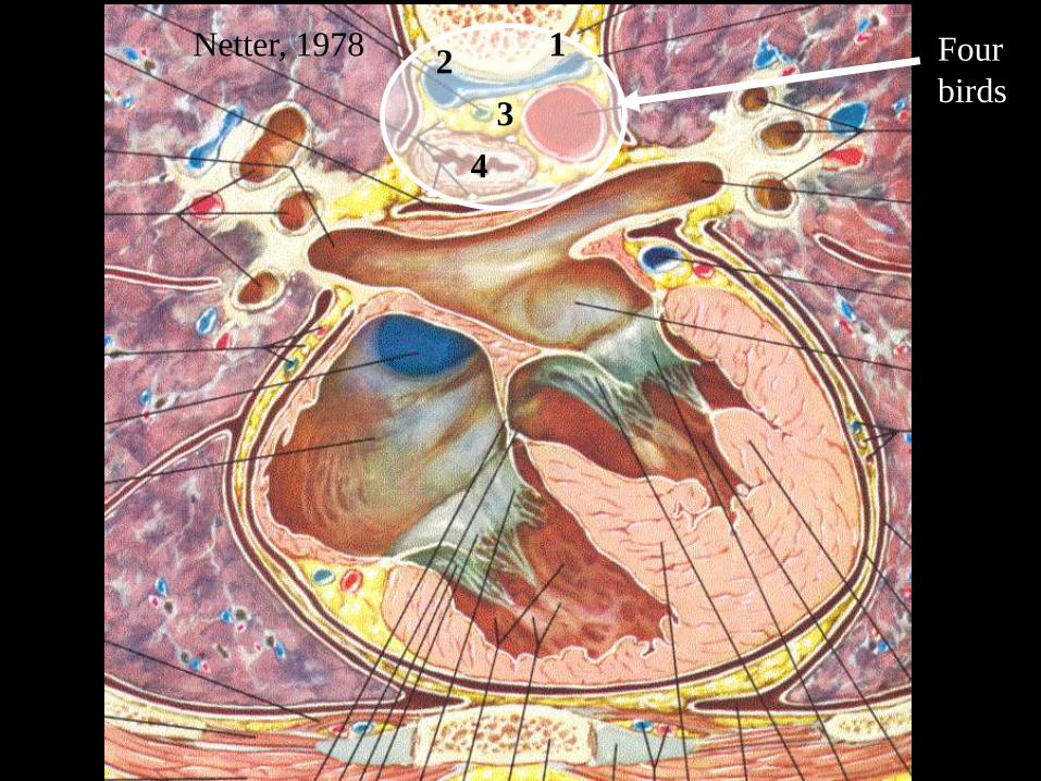
Netter, 1978 Four
birds
12
3
4

Netter, 1978

Netter, 1978
Medial Papillary
muscle
Anterior
Papillary
muscle
Anterior
Septal
Inferior
Supraventricular crest
Parietal
Band
The four highlighted
structures form an almost
circular structure separating
the inflow and outflow
portions of the right ventricle
The RV has
a conus
Septomarginal
Trabeculation
Moderator
band

RV trabeculations are
coarser than LV
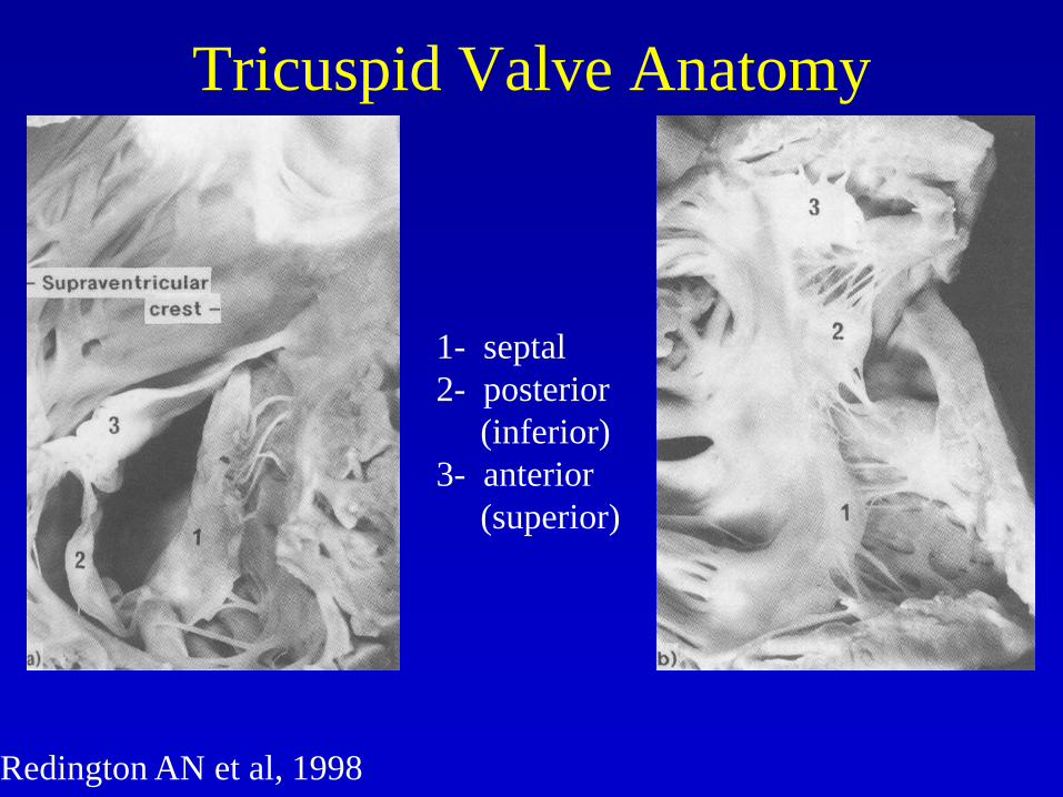
Redington AN et al, 1998
1- septal
2- posterior
(inferior)
3- anterior
(superior)
Tricuspid Valve Anatomy

Large
white
arrow
indicates
RV
outflow

Netter, 1978

Netter, 1978
Moderator band
Septal band
Supraventricular crest

Fiber Geometry
Bovine LV Apex
RV free wall
Imbrication inward to the right Porcine cardiac base
Posterior
Pulmonary conus
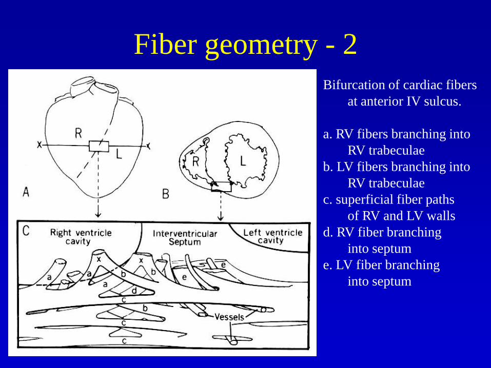
Fiber geometry - 2
Bifurcation of cardiac fibers
at anterior IV sulcus.
a. RV fibers branching into
RV trabeculae
b. LV fibers branching into
RV trabeculae
c. superficial fiber paths
of RV and LV walls
d. RV fiber branching
into septum
e. LV fiber branching
into septum
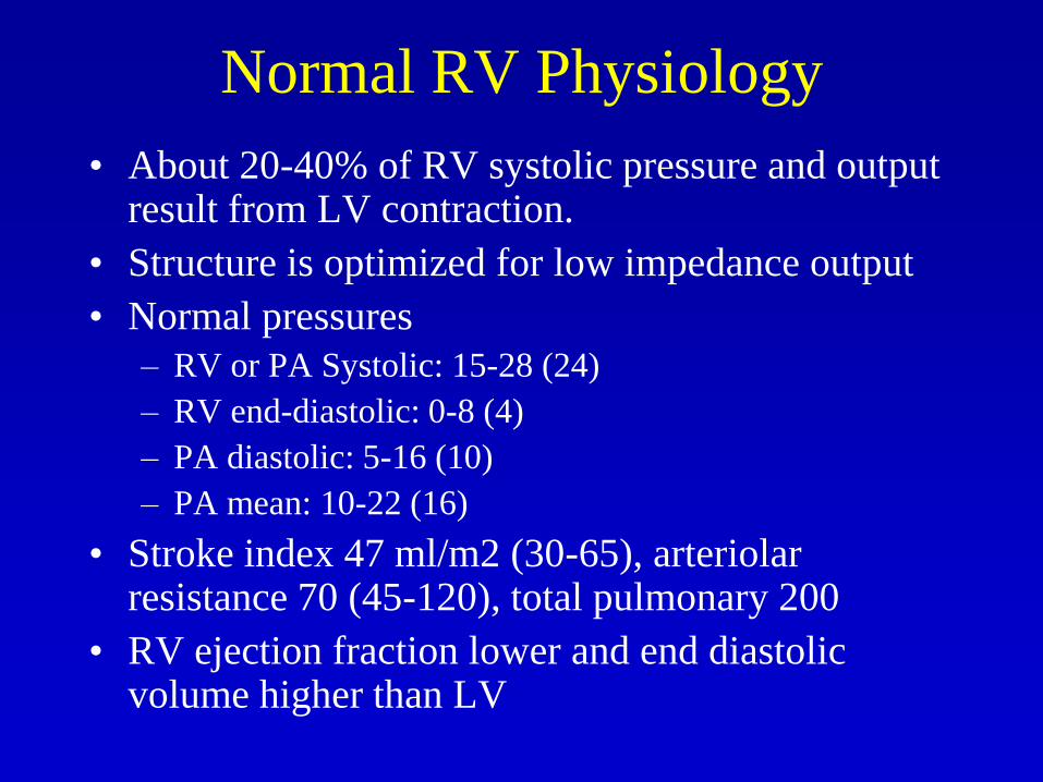
Normal RV Physiology
• About 20-40% of RV systolic pressure and output result from LV contraction.
• Structure is optimized for low impedance output
• Normal pressures
– RV or PA Systolic: 15-28 (24)
– RV end-diastolic: 0-8 (4)
– PA diastolic: 5-16 (10)
– PA mean: 10-22 (16)
• Stroke index 47 ml/m2 (30-65), arteriolar resistance 70 (45-120), total pulmonary 200
• RV ejection fraction lower and end diastolic volume higher than LV

PCD, 1998
Dell’Italia LJ, Walsh RA.
Am Heart J 1988;116:1289-97.

Normal RV Physiology
• Low afterload system
– Mean PA pressure, pulse pressure and vascular
resistance are 1/6 systemic
– Pulse wave velocity is about half of systemic
• Ventricular interdependence
– Common pericardial constraint
– Common myocardial fiber bundles
– Common septal wall
– Both diastole and systole are affected
PCD, 1998

PCD, 1998
Simultaneous Right and Left Heart Waveforms

Acute Response to Pressure Load
PCD, 1998

RV Pressure-Volume Loop

Diastolic Ventricular Interaction
• Increasing RV volume shifts the LV diastolic pressure-volume curve upward (making the LV appear noncompliant)
• Increasing LV volume and pressure shifts the RV pressure-volume curve upward and to the left (making the RV appear noncompliant)
• This occurs even if the pericardium is absent, but more pronounced if pericardium present, and still more if pericardial constraint is augmented by constriction or tamponade
PCD, 1998
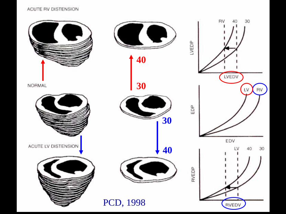
PCD, 1998
30
40
30
40

Systolic Ventricular Interaction
• Increased RV systolic pressure augments LV systolic pressure a little (and vice versa)
• RV dP/dt is affected by LV dP/dt, accentuated by RV or LV endocardial pacing
• One experimental preparation calculated that the LV contributed > 50% of RV systolic pressure and > 50% of PA flow
• Interaction augmented by compliant septum, and decreased by stiff septum, probably not affected by pericardium (in contrast to diastolic interaction)
PCD, 1998

Panel B and D: dashed line is pressure on beat after
constriction release. Blue arrow: pressure release. PCD, 1998
RV
LV

Right ventricular blood supply
Baim DS, Grossman W, 1996

Right ventricular blood supply
• From the RV branch of the right coronary
artery, and the acute marginal branch
Hurst, 1998

RV Response to a Pressure Load
• Acquired pressure load
– Pulmonary emboli
– Primary pulmonary hypertension
– Secondary pulmonary hypertension
• LV dysfunction, mitral disease
• COPD, emphysema (cor pulmonale results in increased wall thickness, myocyte diameter and fibrosis amount in both LV and RV)
• Eisenmenger’s physiology (ASD, VSD, PDA)
• Congenital pressure load
– Congenitally corrected transposition of the great vessels (L-transposition)
– PS (any level), ToF
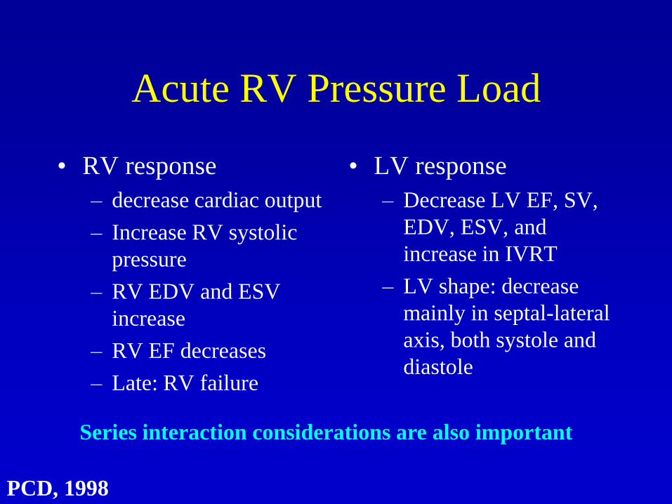
Acute RV Pressure Load
• RV response
– decrease cardiac output
– Increase RV systolic
pressure
– RV EDV and ESV
increase
– RV EF decreases
– Late: RV failure
• LV response
– Decrease LV EF, SV,
EDV, ESV, and
increase in IVRT
– LV shape: decrease
mainly in septal-lateral
axis, both systole and
diastole
PCD, 1998
Series interaction considerations are also important

RV response to a volume load
• Congenital volume load
– ASD, partial anomalous pulmonary venous
return
• Acquired volume load
– TR
– PR

Hemodynamic Effects of RVVOASD, TR, or PR
• RV volume increased, RV EF normal
• LV
– volume decreased, shape more distorted in diastole, more normal in systole
– EF normal or slightly decreased
– LVEDP normal
– atrial kick diminished
– SV and LV SWI decreased (myocyte function probably normal)
– PEP increased and LVET decreased, and LV response to exercise may be decreased
PCD, 1998

Management Considerations in
RV Loads
• If the RV is failing and there is shock, perhaps increasing LV afterload with vasoconstrictor may help
– Increase RV coronary perfusion
– Increase RV function by septal interaction
• RV infarction hemodynamics may be improved by augmenting LV function
– Dopamine, etc are rational

Assessing RV Systolic Function
• Preload: surrogate is RA pressure
• Afterload: surrogate is PA pressure
• Contractility
– Echo: visual estimate
– Cath:visual estimate
– Nuclear: calculation
• First pass, RAO projection, usually higher value
• Gated blood pool EF >40-45%, lower due to RA
overlap (LAO projection to eliminate LV overlap)

AP Projection of RV

Lateral Projection of RV

Lateral Projection of RV with PS
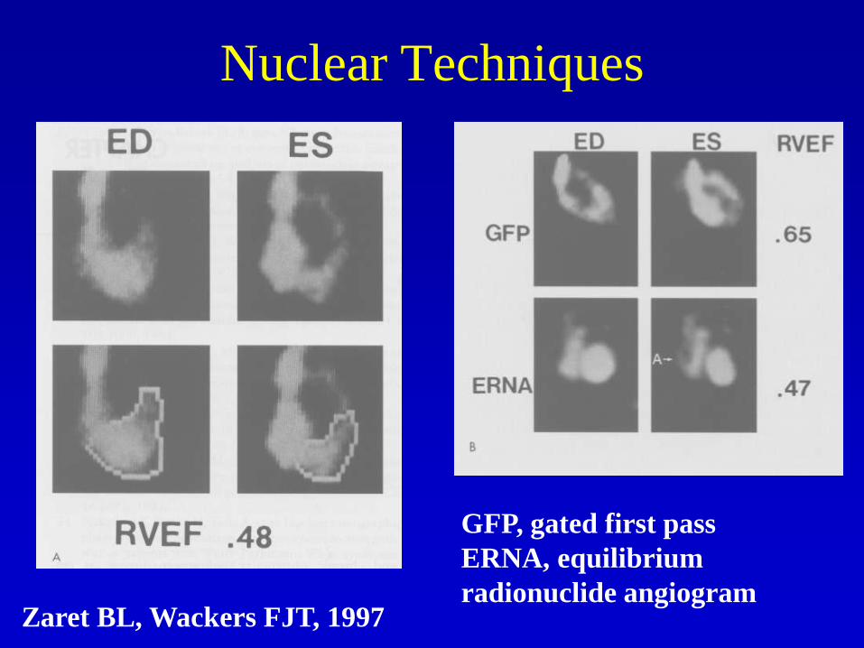
Nuclear Techniques
Zaret BL, Wackers FJT, 1997
GFP, gated first pass
ERNA, equilibrium
radionuclide angiogram

Nuclear Techniques
Berger HJ et al, AJC 1978 Brent BN et al, AJC 1984

Echocardiographic Assessment
of the Right Ventricle
• Transthoracic views: use all possible
– Parasternal RVIT view and RVOT view
– Parasternal short axis
– Apical four -chamber
– Subcostal four -chamber
• Transesophageal views
– Transgastric short axis and long axis RV
– Transesophageal four-chamber

RV Inflow View Structures• RV anterior
free wall and
inferior wall
• Tricuspid
valve: anterior
and posterior
leaflets
• IVC
Otto, 1997, p. 567

RV Outflow Tract Structures
• RVOT
• Pulmonic Valve
• Main PA
• Parasternal
short-axis view
is best for
bifurcation of
PA
Otto, 1997, P. 567

RV Assessment in Four-Chamber
View
• Geometry
– Enlargement
– Wall thickness
– IV septum
– Interatrial septum
• Systolic function
• Coronary sinus (posterior angulation)

RV Assessment in Subcostal
View
• IVC dimension and respiratory variation
• Atrial situs in congenital heart disease
• Also ASD assessment

Echo Findings in RV Load
• RV Pressure load
– RV walls hypertrophy
– RV dilation occurs early, more evident in short axis than long axis
• RV Volume load
– RV dilation with preserved systolic function
– Later, systolic dysfunction
• Septal motion
– Pressure load: decreased curvature mainly at end systole, normal at end diastole
– Volume load: decreased curvature mainly in mid-to-late diastole

RV
Pressure
Load
11-year-old with
severe pulmonary
hypertension
Otto, 1997, p. 572

RV
Volume
Load
79-year-old with severe
tricuspid regurgitation
Otto, 1997, p. 572

Echo Assessment of the RV
• PS-SA RV should be 60% of LV dimension, Septal-free wall max dimension in diastole is 3.0 (2.5-3.8 cm) and systole is 2.6 (2.0-3.4 cm)
• A4C RV should be 2/3 of LV area
• Volume: no well-validated studies
• Wall thickness: normal is less than 5 mm, best is PS-LA, second is subcostal

Echo
Assessment
of RV
Walls
Weyman, 1994, p. 916

Echo Assessment of RV
3.0 (2.5-3.8)
2.5 (1.8-3.5)
Area 10.9 cm2 (4.5-20)
7.1 (5.5-9.1)
3.0 (2.1-4.2)
3.5 (2.6-4.3)
Weyman, 1994
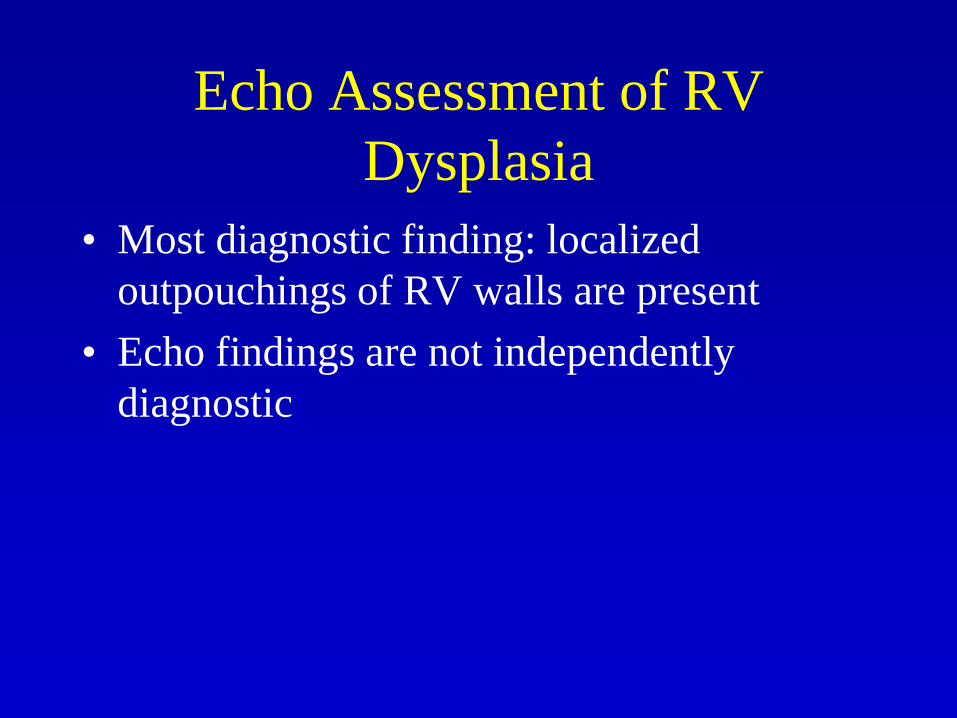
Echo Assessment of RV
Dysplasia
• Most diagnostic finding: localized
outpouchings of RV walls are present
• Echo findings are not independently
diagnostic

Echo Assessment of RV
Diastolic Dysfunction
• Not well defined

Thermodilution Assessment of
RV FunctionRapid response thermistor S-G catheter
Ghio S, JACC, 2001
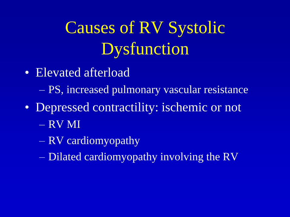
Causes of RV Systolic
Dysfunction
• Elevated afterload
– PS, increased pulmonary vascular resistance
• Depressed contractility: ischemic or not
– RV MI
– RV cardiomyopathy
– Dilated cardiomyopathy involving the RV
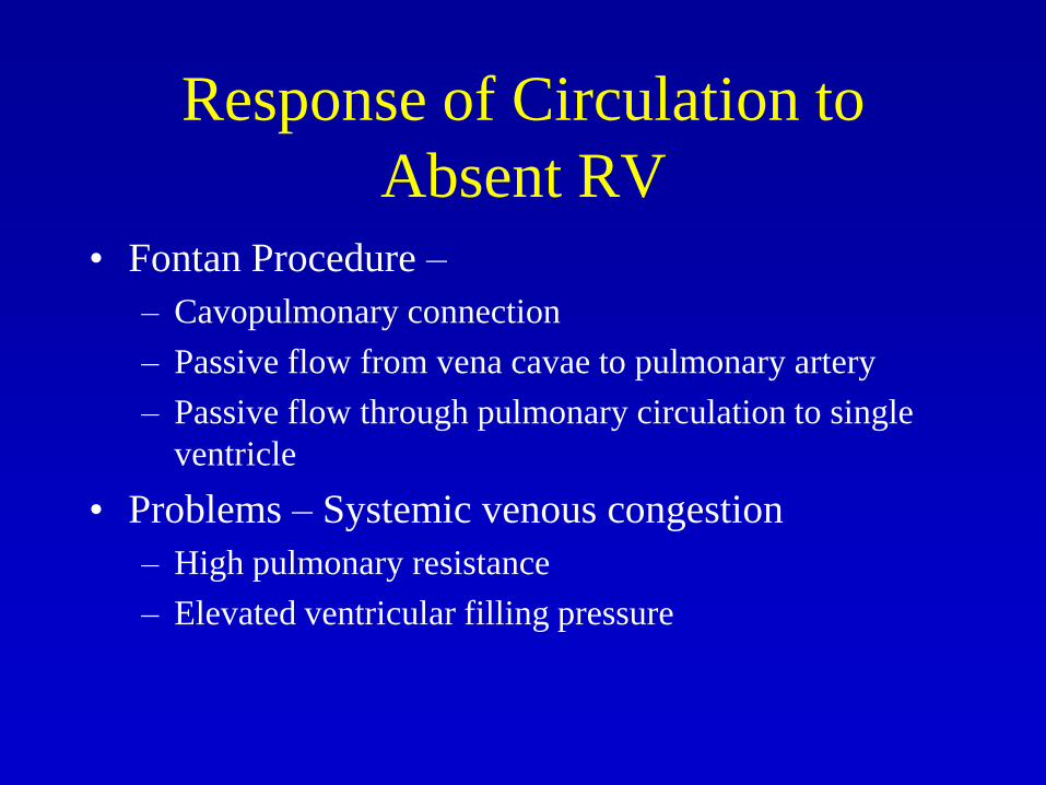
Response of Circulation to
Absent RV
• Fontan Procedure –
– Cavopulmonary connection
– Passive flow from vena cavae to pulmonary artery
– Passive flow through pulmonary circulation to single
ventricle
• Problems – Systemic venous congestion
– High pulmonary resistance
– Elevated ventricular filling pressure

Response of
Circulation to
Absent RV

RV Infarction
Dell’Italia LJ. N Engl J Med, 1998;338:978-980

References• Jiang L, Wiegers SE, Weyman AE. “Chapter 28, Right ventricle” in
Principles and practice of echocardiography 2nd Ed. Lippincott Williams and Wilkins, Philadelphia, 1994.
• Santamore WP, Dell’Italia LJ. Ventricular interdependence: significant left ventricular contributions to right ventricular systolic function. Prog Cardiovasc Dis 1998;40:289-308.
• DellItalia LJ, Santamore WP. Can indices of left ventricular function be applied to the right ventricle? Prog Cardiovasc Dis 1998;40:309-324.
• Cresci SG, Goldstein JA. Chap 36, Hemodynamic manifestations of ischemic right heart dysfunction. In Kern MJ. Hemodynamic Rounds, 2nd Ed. Wiley-Liss, 1999.
• Redington AN, Brawn WJ, Deanfield JE, Anderson RH eds. The right heart in congenital heart disease. Greenwich Medical Media Ltd, 1998.
• Zaret BL, Wackers FJT. Chapter 14. “Measurement of right ventricular function.” In Gerson MC. Cardiac nuclear medicine, 3rd ed.McGraw-Hill, 1997.
• Streeter DD. “Gross morphology and fiber geometry of the heart” Chapter 4 in Handbook of Physiology, Section 2: The Cardiovascular System. Volume 1: The Heart (Ed: Berne RM). American Physiological Society, Bethesda MD, 1979.

PCD, 1998
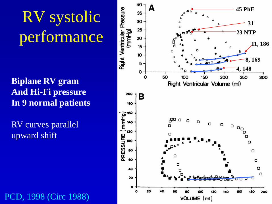
RV systolic
performance
PCD, 1998 (Circ 1988)
Biplane RV gram
And Hi-Fi pressure
In 9 normal patients
RV curves parallel
upward shift
45 PhE
31
23 NTP
11, 186
8, 169
4, 148

RV systolic performance
PCD, 1998

Normal
RVVO
RVPO
Combined
Askah KJ, et al. Canadian J Cardiol 1990