assessment and management of patients with lower gi tract disorders nur 111 common health problems
TRANSCRIPT

Assessment And Management Of Patients With Lower GI Tract Disorders
NUR 111Common Health Problems

Anatomy and Function of Lower GI Tract
GI Tract = 23- 26 feet long: extends from mouth through esophagus, stomach, and intestines to the anus
Small Intestine Function:Small Intestine Function: Longest segment of GI tract, absorption of
nutrients into bloodstream through intestinal walls
3 anatomic parts: duodenum, jejunum, ileum Digestive enzymes and bile in the duodenum
come from pancreas, liver, gallbladder and glands within the intestines
Intestinal glands secrete mucus, hormones, electrolytes and enzymes

Ileal Villi

2 types of contractions: Small Intestine Segmentation contractions: Intestinal peristalsis:
Colonic Function: (Ascending, Transverse, Descending, Sigmoid, and Rectum) Within 4 hrs of eating residual waste material
passes through ileocecal valve into colon Bacteria make up a major part of the contents
of large intestine 2 types of secretions: bicarbonate (neutralize)
and mucus (protects colonic mucosa)

Ileocecal Valve

Colonic Function, Cont.
Slow, weak peristaltic activity moves colonic contents along tract, allowing efficient reabsorption of H2O and electrolytes
Fecal material is approx. 75% fluid, 25% solid, brown in color from breakdown of bile, odor comes from bacteria byproduct

Health Hx. And Clinical Manifestations
Tobacco and alcohol Medications Surgeries Unexplained Wt. Gain or Loss Pain (location, duration, frequency etc.) Indigestion Intestinal Gas Nausea and Vomiting Changes in Bowel Habits and Stool

Physical Assessment & Diagnostic Evaluation Assessment: mouth, abdomen, rectum
Mouth: teeth, gums, tongue Abdomen: look, listen, then feel Anal and perineal area
Diagnostic Evaluation Blood work: CBC, liver panel Stool test: occult blood, parasites, etc.
Hematest: most common for occult blood

Diagnostic Evaluation
Lower GI tract studies: Barium Enema: detect polyps, tumors,
lesions of colon Radiopaque substance instilled rectally Gastrografin (water-soluble iodine
contrast) used in inflammatory disease or perforated colon
Nursing Interventions: May vary according to MD orders, condition of client etc.
Computed Tomography: cross-sectional images Nursing Interventions: NPO for 6-8 hrs prior,
assess for allergies to contrast dye
Diagnostic Evaluation
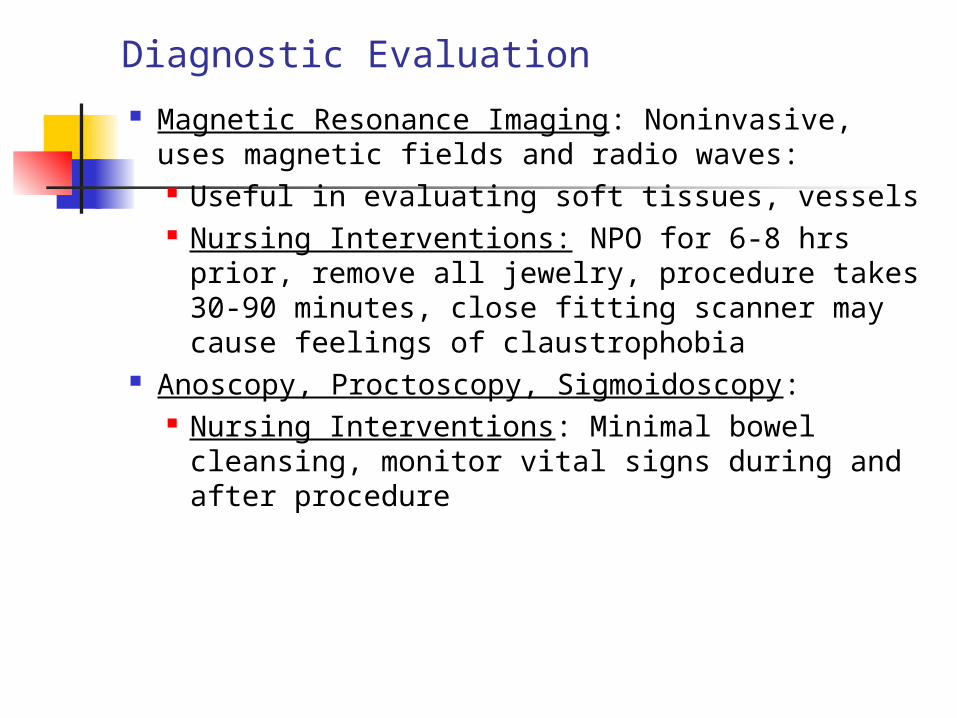
Magnetic Resonance Imaging: Noninvasive, uses magnetic fields and radio waves: Useful in evaluating soft tissues, vessels Nursing Interventions: NPO for 6-8 hrs prior,
remove all jewelry, procedure takes 30-90 minutes, close fitting scanner may cause feelings of claustrophobia
Anoscopy, Proctoscopy, Sigmoidoscopy: Nursing Interventions: Minimal bowel
cleansing, monitor vital signs during and after procedure

Diagnostic Evaluation
Colonoscopy: Direct visual inspection of colon to cecum Flexible fiberoptic colonoscope, can obtain biopsies
and remove polyps Usually takes one hour, pt on left side, legs drawn
toward chest Nursing Interventions: May vary according to MD orders
Bowel cleansing (Colyte, Golytely) clear liquids day before, Informed consent, NPO night before, IV midazolam (Versed) for sedation.
During procedure monitor vital signs, O2 saturation, color and temp of skin, level of consciousness, vagal response

Colonoscopy

Gerontologic Considerations Oral Cavity
Tooth loss or decay Atrophy of taste buds
Esophagus Weakened gag reflex
Stomach Decrease gastric secretions Decrease motility
Small Intestine Atrophy of muscle and mucosal surfaces
Large Intestine Decrease mucus production Decrease tone of anal sphincter

Abnormalities of Fecal Elimination Constipation: irregular, hard stool: may be caused by
certain meds, hemorrhoids, obstructions, neuromuscular diseases
Complications: Hypertension, fecal impaction, hemorrhoids, megacolon
Nursing Management: increase fiber, fluids, laxatives as ordered Diarrhea: Increase in frequency, amount and altered
consistency (looseness): Irritable Bowel Syndrome (IBS), Inflammatory Bowel Disease (IBD), and lactose intolerance are frequently underlying disease processes
Acute or Chronic Complications: dehydration, cardiac dysrhythmias (low potassium)
always be aware of Potassium levels Nursing Management: Stool specimen, bed rest, low bulk diet in acute
phase, advance to bland diet, no caffeine, carbonated drinks, antidiarrheal meds as ordered, diphenoxylate (Lomotil), loperamide (Imodium)

Irritable Bowel Syndrome (IBS) Common GI problem, cause unknown, certain factors
associated with syndrome: heredity, depression, anxiety, high fat diet, smoking, alcohol
Results from functional disorder of intestinal motility Clinical manifestations: constipation, diarrhea, or both,
pain, bloating Assessment and diagnostic findings: diagnosis made
when tests rule out structural or other colon disease Medical management: Treatment aimed at relieving pain,
constipation and diarrhea, reducing anxiety and stress Nursing Management: patient education, reinforce good
diet, not smoking and no alcohol

Zelnorm

Acute Inflammatory Intestinal Disorders
Appendicitis: most common reason for abdominal surgery, appendix becomes inflamed from obstruction, may become pus filled Clinical manifestations: Right lower
quadrant pain, low grade temp, N/V, rebound tenderness, rupture causes diffuse pain and condition worsens
Assessment and Diagnostic: CBC, CT of abdomen, Ultrasound

Opening of Appendix

Appendicitis

Appendicitis Complications: Perforation leading to
peritonitis or abscess Medical Management: Surgery as soon as
possible, IV fluids and antibiotics, analgesics, Appendectomy may be performed with low abdominal incision or by laparoscopy
Nursing Management: Goals include, relieving pain, preventing fluid and electrolyte imbalance, dehydration, and infection
Surgery may be outpatient, if complications of peritonitis are suspected pt may remain in hospital for several days

Acute Inflammatory Intestinal Disorders
Diverticulitis: Diverticulum is saclike pouching of lining of bowel extending through defect in muscle. Most common in sigmoid colon
Clinical Manifestations: Chronic constipation, intervals of diarrhea, left lower quadrant pain, anorexia, fatigue
Assessment and Diagnostic: CT scan procedure of choice, CBC

Diverticula Diverticula seen
on colonoscopy

Diverticula Diverticula seen
on barium enema

Diverticulitis Complications: peritonitis, abscess formation,
bleeding Medical Management: Usually treated outpatient
with diet and medicine therapy, antispasmodics, antibiotics, bulk laxative, clear liquids until inflammation resolved then high fiber, low fat
Acute case may require hospitalization, especially for elderly and immunocompromised
Surgery may be necessary with abscess formation or perforation
Nursing Process: encourage high fiber diet, exercise, bulk laxatives

Peritonitis
Inflammation of the peritoneum, the serous membrane lining the abdominal cavity and covering the organs.
Clinical Manifestations: Affected area of abdomen becomes tender, distended, rigid. Rebound tenderness, paralytic ileus, N/V
Assessment and Diagnostic: CBC, Abdominal CT scan or X-ray, peritoneal aspiration and culture of fluid
Complications: Generalized sepsis (major cause of death), inflammation may cause bowel obstruction
Medical Management: Fluid & electrolyte replacement, analgesics, antiemetics, NG suction, massive antibiotics, surgery to remove infected material
Nursing Management: Ongoing assessment of vital signs, pain, GI function, intake and output

Inflammatory Bowel Disease (IBD)
Refers to 2 chronic inflammatory GI disorders: regional enteritis (Crohn’s disease) and ulcerative colitis. Both have similarities but are ultimately different.
Cause of IBD is unknown. Occurs equally in women and men.
Believed to be triggered by environmental agents such as food additives, tobacco, and radiation, also allergies and immune disorders

Inflammatory Bowel Disease (IBD) Regional Enteritis (Crohn’s disease):
Occurs anywhere along the GI tract most common in distal ileum and colon
Chronic inflammation that extends through all layers of bowel wall
Periods of remission and exacerbation, ulcers form on inflamed mucosa, separated by normal tissue,
Advanced cases the bowel wall thickens and becomes fibrotic, intestines narrow

Crohn’s Disease

Inflammatory Bowel Disease (IBD)
Regional Enteritis (Crohn’s disease): Clinical Manifestations: lower right quadrant pain,
diarrhea unrelieved with defecation, colon spasm, result in decrease PO intake, malnutrition, wt loss, steatorrhea, abscesses and fistulas
Assessment and Diagnostic: Stool sample, barium swallow or enema, CT scan, CBC, albumin
Complications: Intestinal obstruction, fluid & electrolyte imbalance, malnutrition, fistula and abscess formation, increase risk colon cancer

Inflammatory Bowel Disease (IBD)
Ulcerative Colitis: recurrent ulcerative and inflammatory disease of the mucosal and submucosal layers of colon and rectum
Multiple ulcers occurring one after the other, diffuse inflammation, usually begins in rectum and spreads proximally to entire colon; abscesses form and eventually the bowel narrows and shortens

Ulcerative Colitis

Inflammatory Bowel Disease (IBD)
Ulcerative Colitis: Clinical Manifestations: Exacerbation and
remission, diarrhea and left lower quadrant pain, rectal bleeding, anorexia, wt loss, dehydration, 10-20 liquid stools/day
Assessment and Diagnostic: Assess hydration, nutritional status, signs of bleeding, stool specimen, CBC, Sigmoidoscopy, Colonoscopy, CT scan
Complications: Toxic megacolon, perforation, bleeding, vomiting, fatigue

Inflammatory Bowel Disease (IBD) Medical Management of Chronic IBD: Reduction
of inflammation, provide rest for diseased bowel, preventing complications Nutritional Therapy: Oral fluids, low residue,
high protein and calorie diet with vitamin and iron supplements
Pharmacologic Therapy: Sedatives and antidiarrheal meds, Aminosalicylates such as sulfasalazine (Azulfidine), mesalamine (Pentasa) are used for long term maintenance. Corticosteroids (prednisone) also help reduce inflammation

Inflammatory Bowel Disease (IBD)
Surgical Management: May require total colectomy (removal of entire colon) and placement of ileostomy;
Nursing Management: goals; prevention of fluid volume deficit, maintenance of optimal nutrition and wt, avoidance of fatigue, promoting effective coping


Small Bowel Obstruction
Intestinal contents, fluid, gas accumulate above the intestinal obstruction: Adhesions, Intussusception, Volvulus, Hernia, Tumor are all causes of obstruction.
Clinical Manifestations: Cramping pain, pass blood and mucus but no stool, vomiting (intestinal contents), dehydration, abdominal distention
Medical Management: Decompression of bowel through Nasogastric (NG) tube, if obstruction is complete then surgical intervention is warranted
Nursing Management: maintain function of NG tube, assess for fluid and electrolyte imbalance

Large Bowel Obstruction Obstruction of larger bowel is similar to small
bowel obstruction, however the symptoms develop and progress relatively slowly
Constipation may be only symptom for days, eventually abdominal distention and vomiting of fecal contents
Colonoscopy may be performed to untwist and decompress bowel
Usual treatment is bowel resection to remove the obstruction, colostomy may be necessary

Polyps of Colon and Rectum Polyp is a mass of tissue that protrudes
into lumen of bowel Can occur anywhere in colon or rectum Neoplastic (carcinomas) or non-
neoplastic (benign) Most common sign rectal bleeding Diagnosis based on digital rectal exam,
colonoscopy, barium enema Polyps should be removed either through
colonoscopy or laparoscopy

Polyps of the colon

Diseases of the Anorectum Anorectal abscess: obstruction of anal gland,
infection, deep abscesses may result in low abdominal pain and fever Treatment include incision and drainage,
sitz baths and analgesics Anal fistula: Tiny, tubular tract that extends
into the anal cavity from an opening located beside the anus, usually result from an infection Surgery recommended for removal of
fistula, wound is packed with gauze Anal fissure: longitudinal tear or ulceration in
the lining of the anal canal, caused by large firm stool, or childbirth or trauma Most heal with management by stool
softener, sitz baths and increase fluid intake

Diseases of the Anorectum Hemorrhoids: dilated portions of veins
in the anal canal. Increased pressure in the hemorrhoidal tissue due to pregnancy may initiate or aggravate hemorrhoids Two types: internal and external Cause itching and pain, most
common cause of bright red bleeding with defecation
High fiber diet, increase fluids, bulk laxatives, sitz baths, may require rubber band ligation or more extensive surgery

Hemorrhoids

Treatment for hemorrhoids