assessment of buccal bone thickness of aesthetic maxillary
TRANSCRIPT

162 Copyright © 2015 Korean Academy of Periodontology
pISSN 2093-2278eISSN 2093-2286Assessment of buccal bone thickness
of aesthetic maxillary region: a cone-beam computed tomography studyRamón Fuentes1,*, Tania Flores1, Pablo Navarro1, Carlos Salamanca1, Víctor Beltrán1, Eduardo Borie1,2
1Research Centre in Dental Sciences (CICO), Universidad de La Frontera Dental School, Temuco, Chile2Department of Dental Materials and Prosthodontics, University of São Paulo Dental School of Ribeirão Preto, Ribeirão Preto, Brazil
Research ArticleJ Periodontal Implant Sci 2015;45:162-168http://dx.doi.org/10.5051/jpis.2015.45.5.162
Purpose: The aim of this study was to analyze the anatomical dimensions of the buccal bone walls of the aesthetic maxillary region for immediate implant placement, based upon cone-beam computed tomography (CBCT) scans in a sample of adult patients. Methods: Two calibrated examiners analyzed a sample of 50 CBCT scans, performing mor-phometric analyses of both incisors and canines on the left and right sides. Subsequently, in the sagittal view, a line was traced through the major axis of the selected tooth. Then, a second line (E) was traced from the buccal to the palatal wall at the level of the observed bone ridges. The heights of the buccal and palatal bone ridges were determined at the ma-jor axis of the tooth. The buccal bone thickness was measured across five lines. The first was at the level of line E. The second was at the most apical point of the tooth, and the other three lines were equidistant between the apical and the cervical lines, and parallel to them. Statistical analysis was performed with a significance level of P≤0.05 for the bone thick-ness means and standard deviations per tooth and patient for the five lines at varying depths.Results: The means of the buccal wall thicknesses in the central incisors, lateral incisors and canines were 1.14±0.65 mm, 0.95±0.67 mm and 1.15±0.68 mm, respectively. Additionally, only on the left side were significant differences in some measurements of buccal bone thickness observed according to age and gender. However, age and gender did not show significant differences in heights between the palatal and buccal plates. In a few cases, the buccal wall had a greater height than the palatal wall.Conclusions: Less than 10% of sites showed more than a 2-mm thickness of the buccal bone wall, with the exception of the central incisor region, wherein 14.4% of cases were ≥2 mm.
Keywords: Alveolar bone, Buccal bone, Cone-beam computed tomography, Dental esthetics.
Received: Aug. 7, 2015 Accepted: Oct. 7, 2015
*Correspondence: Ramón FuentesUniversidad de La Frontera Dental School, Av. Francisco Salazar 01145, Temuco 4811230, Chile. E-mail: [email protected]: +56452734153Fax: +56452325777
INTRODUCTION
The rehabilitation of the maxillary anterior region with implant-supported prostheses has been reported to be a difficult procedure because of the high aesthetic expectations of patients [1,2]. Soft tissue defects associated with volumetric changes in the alveolar ridge can be unfavorable for both aesthetics and implant placement [3].
A crucial factor during the planning of implant treatment is the loss of bone tissue, which may interfere with the implant placement [4]. Following tooth extraction, the alveo-lar bone is subjected to a high degree of resorption, which is directly associated with the buccal wall thickness [5,6]. Therefore, efforts must be made to preserve bone and soft tis-sues using minimally invasive surgical techniques in accordance with clinical conditions
This is an Open Access article distributed under the terms of the Creative Commons Attribution Non-Commercial License (http://creativecommons.org/licenses/by-nc/3.0/).
Ramón Fuentes et al.
dx.doi.org/10.5051/jpis.2015.45.5.162
www.jpis.org 163
and the results of complementary exams [7]. Beltrán et al. [8] re-ported that the success or failure of implant treatment is fre-quently related to factors specific to each patient. From this per-spective, the thickness of the buccal bone wall in the maxillary an-terior region of each patient is of great importance for implant positioning during treatment planning [6,9].
The dimensions of the buccal bone wall have gained importance in the past few years with the use of immediate implants [10]. The most frequently used methods to measure buccal bone thickness are the use of calipers [10-12] and CBCT scans to compare variations among subjects according to gender, age and tooth type, among other variables [13,14].
Cone-beam computed tomography (CBCT) scans have been widely used in the dental field due to advantages that include low radiation dose, low cost, and the ability to view a detailed three-dimensional image of the regions of interest [15,16]. Alqerban et al. [17] added that CBCT (3D) is more sensitive than the conventional X-ray (2D) both for locating dental structures and for identifying sites of initial resorption. CBCT scans may help in the guidance of treatment plan-ning for the maxillary aesthetic region; specifically, for implant placement, it can contribute to the evaluation of some possible pre- or post-surgical soft or hard tissue complications [14]. Similarly, Vera et al. [18] reported that the presence/absence and thickness of the buccal bone wall may be discerned with a CBCT scan of the interest region, and the tissue biotypes may be taken into consideration for the long-term success of implant treatment.
Huynh-Ba et al. [10] noted that limited information is available about the thickness of the buccal bone wall in the aesthetic zone of humans. Thus, the aim of this study was to analyze the anatom-ical dimensions of the buccal bone wall of the aesthetic maxillary region for immediate implant placement, based on CBCT scans in a sample of adult patients.
MATERIALS AND METHODS
A cross-sectional descriptive study was conducted on 50 CBCT scans from a database available at the Radiology Unit, Dental School, Universidad de La Frontera (UFRO), Temuco, Chile. This study was approved by the UFRO Scientific Ethics Committee (Protocol no. 72/2013).
A convenience sampling procedure was performed, which includ-ed 50 CBCT scans, 100 right and left central and lateral incisors, and canines, taken with a 120×90 mm window during the period be-tween January and June 2014, which fulfilled the following inclu-sion criteria: subjects of both genders, between 15 and 60 years old, Chilean nationality, and the presence of all maxillary central front teeth. Exclusion criteria were as follows: individuals with prostheses, the presence of inflammatory processes at the apical level, tooth misalignment, the presence of restorations, root canal treatment, CBCT with distorted images of the cementoenamel junction (CEJ) or bone crest, and cases with buccal bone loss greater than or equal to 3 mm from the CEJ.
Image acquisition and measurementsAll 3D images were acquired from a PAX Zenith 3D orthopanto-
mograph (Vatech Co., Suwon, Korea). The data were exported in DICOM format to the EZ3D2009 software (E-WOO Technology Co. Ltd., Yongin, Korea), and the reconstructions were generated with 1-mm thickness slices at 1 mm intervals.
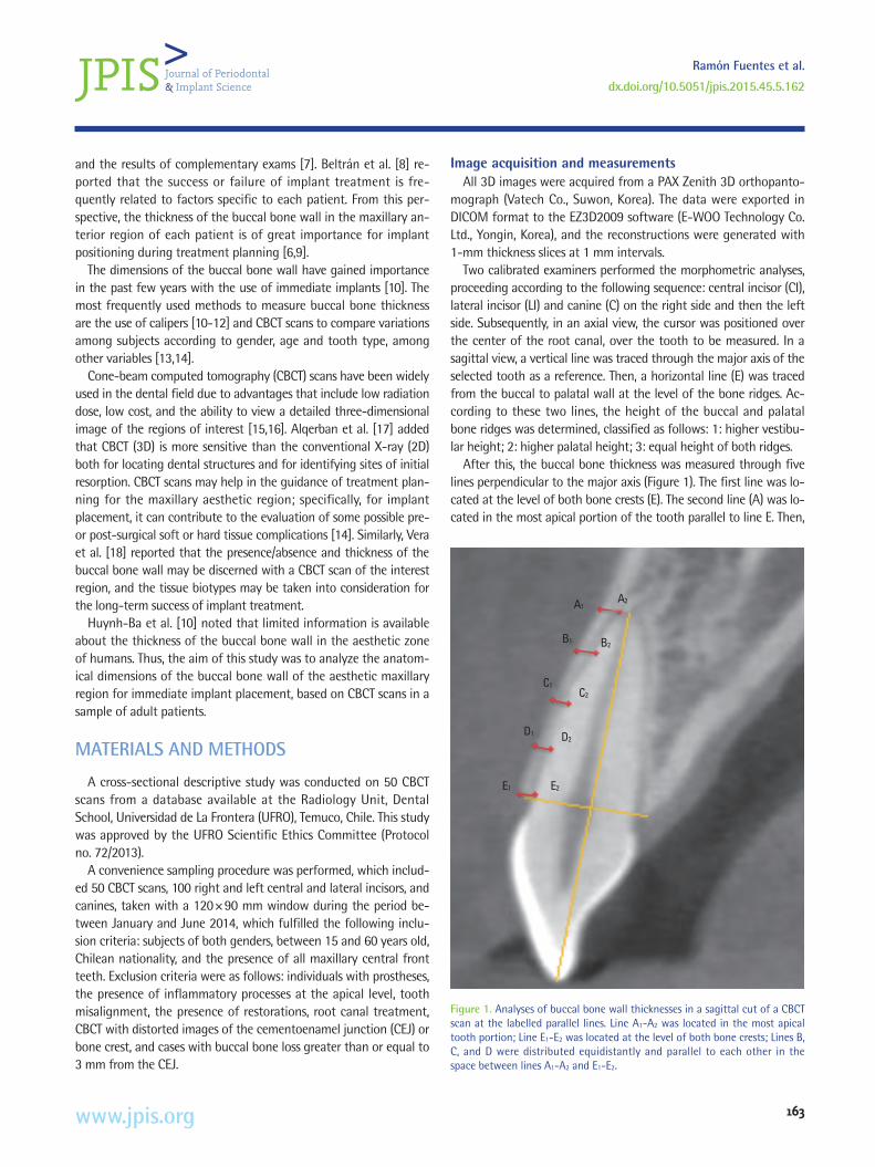
Two calibrated examiners performed the morphometric analyses, proceeding according to the following sequence: central incisor (CI), lateral incisor (LI) and canine (C) on the right side and then the left side. Subsequently, in an axial view, the cursor was positioned over the center of the root canal, over the tooth to be measured. In a sagittal view, a vertical line was traced through the major axis of the selected tooth as a reference. Then, a horizontal line (E) was traced from the buccal to palatal wall at the level of the bone ridges. Ac-cording to these two lines, the height of the buccal and palatal bone ridges was determined, classified as follows: 1: higher vestibu-lar height; 2: higher palatal height; 3: equal height of both ridges.
After this, the buccal bone thickness was measured through five lines perpendicular to the major axis (Figure 1). The first line was lo-cated at the level of both bone crests (E). The second line (A) was lo-cated in the most apical portion of the tooth parallel to line E. Then,
Figure 1. Analyses of buccal bone wall thicknesses in a sagittal cut of a CBCT scan at the labelled parallel lines. Line A1-A2 was located in the most apical tooth portion; Line E1-E2 was located at the level of both bone crests; Lines B, C, and D were distributed equidistantly and parallel to each other in the space between lines A1-A2 and E1-E2.
A1
B1
C1
D1
E1
A2
B2
C2
D2
E2

Buccal wall thickness in adults
dx.doi.org/10.5051/jpis.2015.45.5.162
www.jpis.org164
the space between the lines A and E was divided equidistantly by three more parallel lines (B, C, D). The five measurement lines are la-beled A1-A2, B1-B2, C1-C2, D1-D2, and E1-E2 in Figure 1. Deter-mining the buccal bone wall thickness of each tooth at five differ-ent heights provided a more accurate description of the region to be analyzed. To achieve a more detailed description of the buccal wall region, an analysis of potential significant correlations with study variables, including gender, side and age range, was performed.
Statistical analysisFor the statistical analysis, the average values obtained from the
two examiners were used, and the software SPSS Statistics for Win-dows version 20.0 (IBM Corp., Armonk, NY, USA) was used for all analyses with a significance level of P<0.05. The bone thickness mean and standard deviation per tooth and patient was recorded based on the five lines at varying depths. Normality tests were per-formed using the Kolmogorov-Smirnov test. For the inferential analysis, the Student’s t-test was used for independent samples, and ANOVA was used for samples related to the continuous quanti-tative variables. Confidence intervals were calculated at 95%, and
Fisher's exact test was used for the qualitative variables.
RESULTS
The means of the buccal wall thicknesses in the central incisors, lateral incisors and canines were 1.14±0.65 mm, 0.95±0.67 mm and 1.15±0.68 mm, respectively. The means and standard devia-tions of the buccal bone thicknesses of each tooth at each line measurement are summarized in Table 1. At the central incisors, 42.4% of sites had a buccal wall thickness <1 mm, while 43% showed a thickness between 1 and 2 mm and 14.4% higher than 2 mm. At the lateral incisors, 57% had a buccal wall thickness <1 mm, 36.8% of sites were between 1 and 2 mm, and 6.2% exhibited a thickness of 2 mm or more. At the canines, 41.8% of sites showed a thickness less than 1 mm, with 49.2% between 1 and 2 mm and 9% at a thickness of 2 mm or more.
The results of bone buccal wall thickness by age and side are iden-tified in Figure 2. Additionally, significant differences were found on the left side between the measurements of the lines from the CI (P=0.016), LI (P=0.012), and C (P=0.045) in the three age ranges.
The differences in buccal bone wall thicknesses according to gender and side are summarized in Figure 3. As may be observed, significant differences were exhibited associated with the lines C (P=0.024), D (P=0.03), and E (P= 0.00) of the left CI as well as in relationship to line B (P=0.013) on the left C.
For the height of both bone ridges by gender and tooth (Figure 4), only two cases showed a greater buccal wall value when com-pared to the palatal plate. They were observed at the canine level in both genders. A greater palatal wall value was identified at the central incisors and canines, with both cases of the male gender. No significant differences were found related to ridge height by gender and tooth.
For the variables age and tooth and their relationships with both ridge heights (Figure 5), the buccal wall was the highest in three cas-
Figure 2. Confidence intervals (95%) for buccal bone wall thicknesses on each line and tooth according to age range and side. *Significant differences (ANOVA).
Table 1. Means and standard deviations of buccal wall thicknesses at each measurement line of the maxillary anterior teeth.
ToothLine n Central incisor (mm) Lateral incisor (mm) Canine (mm)
A1-A2 100 2.13±0.67a,c 1.53±0.94a,b 1.81±0.67b,c
B1-B2 100 0.99±0.38d,e 0.60±0.50d 0.75±0.49e
C1-C2 100 0.81±0.34f,g 0.63±0.43f 0.64±0.37g
D1-D2 100 0.95±0.31h 0.98±0.48i 1.28±0.66h,i
E1-E2 100 0.89±0.24j,l 1.00±0.37j,k 1.29±0.44k,l
Mean 500 1.14±0.65 0.95±0.67 1.15±0.68
Values are presented as the mean ±standard deviation. Same letters: Statistically significant differences between groups (Scheffé’s test; P<0.05).

Ramón Fuentes et al.
dx.doi.org/10.5051/jpis.2015.45.5.162
www.jpis.org 165
es in the 15- to 30-year age range and one in the 46- to 60-year age range. A clear tendency towards the palatal wall being the highest was noticed for all three teeth in all age ranges, except for the lateral incisors in the sample between 46–60 years old. Furthermore, no dif-ferences were observed between the wall heights by age or tooth.
DISCUSSION
In the present study, the thickness of the buccal bone in the maxillary aesthetic region was determined through CBCT scans. To achieve an appropriate choice of post-extraction treatment, an
Figure 3. Confidence intervals (95%) for buccal bone wall thicknesses on each line and tooth according to gender and side. *Significant differences (ANOVA).
4035302520151050
12
38
22
28
0 0
Same height Higher palatal Higher buccal
Central incisor
30
25
20
15
10
5
0
25 25
22
28
0 0
Same height Higher palatal Higher buccal
Lateral incisor
35
30
25
20
15
10
5
0
15
33
2
24 24
2
Same height Higher palatal Higher buccal
Canine
MaleFemale
Figure 4. Classifications in height of bone ridges at each tooth according to gender. Note that the buccal bone wall never was higher than the palatal at both incisors.
15-30 years old31-45 years old46-60 years old
50454035302520151050
25
45
0
5
13
04
8
0
Same height Higher palatal Higher buccal
Central incisor
4035302520151050
35 35
0
5
13
0
75
0
Same height Higher palatal Higher buccal
Lateral incisor
454035302520151050
28
39
37
11
04 8
1
Same height Higher palatal Higher buccal
Canine
Figure 5. Classification of the height of the bone ridges at each tooth distributed by age range.

Buccal wall thickness in adults
dx.doi.org/10.5051/jpis.2015.45.5.162
www.jpis.org166
analysis of the alveolar bone dimensions through CBCT scanning, at the region of the tooth to be extracted, may offer valuable in-formation regarding bone volume and the morphology of the fu-ture implant site [19].
Following tooth extraction, the alveolar bone suffers resorption in width and height during the first few months [5,20,21], with a reduction in almost 50% of cases [22]. An additional 2 mm from the original socket dimension may resorb during the socket heal-ing process [13]. Thus, the degree and resorption pattern of the buccal bone wall is directly related to the thickness of this plate [5,21,23,24]. This resorption process may be explained because the marginal portion of the buccal wall contains proportionally higher amounts of bundle bone than the palatal wall [21].
This study describes mean buccal wall thicknesses observed in a sample of Chilean patients, for both incisors and canines, ranging from 0.95 to 1.15 mm, which are worth comparing with some val-ues reported in the literature. Lee et al. [15] analyzed CBCT scans from 20 individuals, identifying the mean buccal wall thickness at 3 mm from the CEJ of 0.68 mm, 0.76 mm, and 1.07 mm, for the central incisors, lateral incisors, and canines, respectively. Januário et al. [13] studied 250 CBCT scans, reporting the mean of the buc-cal plate, which varied from 0.5 to 0.7 mm. Ghassemian et al. [25] examined 66 CBCT scans, reporting values of the buccal bone plate that varied from 1.59 to 1.73 mm for the lateral incisors and 1.47 to 1.6 mm for the canines. These values varied greatly compared with the values found in this study.
Nevertheless, some measurements agree with those of other au-thors. Nowzari et al. [14], after the assessment of 101 CBCT scans, noted a mean value of 1.12 mm for the central incisors at 3 mm be-low the alveolar ridge, similar to our study. Han et al. [12] evaluated five Korean human cadavers, reporting similar values for buccal plate thicknesses from the lateral incisor region (0.98 mm), but low-er values for the central incisors (0.82 mm) and canines (0.72 mm).
On the other hand, there is a consensus among clinicians that a minimum thickness of 2 mm for the buccal plate is an important feature for the maintenance of the vertical dimension of the alve-olar crest [10,26], determining the amount of vertical crestal re-sorption following tooth extraction. In this sense, this minimum dimension is necessary for the optimal healing of soft and hard tissues in cases of immediate implants placed in the aesthetic re-gion [16,19,23,25,27,28]. A thin buccal wall ≤2 mm may be evi-denced in the presence of fenestrations, dehiscences and gingival recession [16,25]. Han et al. [12] added that the buccal bone thick-ness is more critical in cases of flapless surgery, and Younes et al. [29] concluded in their research that there is a moderately positive correlation between the buccal bone thickness and soft tissue. Thus, considering the buccal wall thickness with great caution be-fore tooth extraction and immediate implant placement is neces-sary due to the aesthetic impact and long-term consequences.
A high prevalence of a buccal bone thickness ≥2 mm was noted in the central incisors (14.4%), lateral incisors (6.2%) and canines (9%) compared with the reported literature. These thickness values
were observed generally at the A point, that is, at the most apical point relative to the root. A previous multicenter research study recorded the buccal bone width in 93 extraction sites, and it found that 87.2% of sites measured had a thickness ≤1 mm and only 2.6% showed a thickness over 2 mm [10]. Another recent study concluded there was a low prevalence of buccal wall thicknesses ≥2 mm, reporting 0% for central incisors, 8% for lateral incisors and 2% for canines [29]. Likewise, the research of Nowzari et al. [14] described buccal thicknesses over 2 mm at prevalences of 0%, 1.5%, 2%, 3%, and 2.5% at 1, 2, 3, 4, and 5 mm from the CEJ, re-spectively, while Januário et al. [13] found only one case with buc-cal bone thickness ≥2 mm in 250 CBTC scans. However, Ghassemi-an et al. [25] identified a range that varied from 0% to 28.8%, ob-serving buccal walls over 2 mm with more frequency at 3 mm from the bone crest and with a prevalence higher than 20% in the lateral incisors, which is greater than that reported here. Generally, the occurrence of buccal bone thicknesses ≥2 mm increased as the measurement depth increased, concurring with the findings of other researchers [12,14,15]. However, in most cases, the thickness of the buccal bone wall of the anterior teeth was very thin, which is in accordance with some authors [12,30].
By age, significant differences were found in some measurements of buccal bone thickness in all analyzed teeth, only on the left side, which does not correspond with reports by some authors [13,14], who found no differences between the age ranges. In addition, gender is clearly stated in the literature to be a factor that should show no significant differences in buccal wall thickness measure-ments [14,25,31], but in the present study some significant differ-ences were identified according to gender in a few regions on the left side, mainly at the central incisor. Age and gender differences were not significant for the comparison of heights between the palatal and buccal plates. In a few cases, the buccal wall was of greater height than the palatal wall.
Over the past few years, the buccal wall thickness has gained significance, mainly as a result of its importance for the immediate placement of implants [10]. A multilevel study concluded that thicknesses of the buccal and palatal wall over 1 mm exhibited more bone fill in the gap between the implant surface and the socket plate than did thin (≤1 mm) walls [23]. Thus, in cases with thin buccal bone walls, clinicians must consider the use of bone grafts to reduce the resorption of the buccal plate, without com-promising the aesthetic results and the three-dimensional position of the implant [15].
A potential limitation of this study was the standardization of the distance of the CEJ to the bone crest as always under 3 mm. However, some studies [14,18,25] have assessed the distance from the CEJ to the bone crest, describing values between 2.5 and 2.8 mm. This is why CBCT scans that showed distances greater or equal to 3 mm from the CEJ were excluded in this study, as they could represent a pathological condition of buccal bone wall loss. Addi-tionally, the thickness of the palatal wall and the presence of de-hiscence and fenestrations were not measured because they were

Ramón Fuentes et al.
dx.doi.org/10.5051/jpis.2015.45.5.162
www.jpis.org 167
beyond the aim of the study.The present study provides valuable data on buccal wall thick-
nesses of the aesthetic maxillary region in a Chilean population, to understand how critical this thickness may be for aesthetically pleasing long-term results. The immediate placement of implants without the actual guidelines of gap fill with a bone graft is not recommended. However, further studies with a larger sample size are necessary to compare CBCT scan measurements with fresh ex-traction sockets in the same patients, to determine the buccal wall bone thickness more accurately.
In conclusion, less than 10% of sites showed an ideal condition of a thickness of more than 2 mm of the buccal bone wall, with the exception of the central incisor region, wherein 14.4% of cases had a thickness ≥2 mm. In fact, this study highlighted the great predominance of thin buccal bone thickness in the aesthetic max-illary region of the sample studied.
CONFLICT OF INTEREST
No potential conflict of interest relevant to this article was re-ported.
ORCID
Ramón Fuentes http://orcid.org/0000-0002-5895-024XTania Flores http://orcid.org/0000-0001-7582-6278Pablo Navarro http://orcid.org/0000-0002-5467-5009Carlos Salamanca http://orcid.org/0000-0003-0135-3599Víctor Beltrán http://orcid.org/0000-0001-6046-821XEduardo Borie http://orcid.org/0000-0001-6175-6221
REFERENCES
1. Buser D, Martin W, Belser UC. Optimizing esthetics for implant restorations in the anterior maxilla: anatomic and surgical con-siderations. Int J Oral Maxillofac Implants 2004;19 Suppl:43-61.
2. Rodriguez AM, Rosenstiel SF. Esthetic considerations related to bone and soft tissue maintenance and development around dental implants: report of the Committee on Research in Fixed Prosth-odontics of the American Academy of Fixed Prosthodontics. J Prosthet Dent 2012;108:259-67.
3. Kim HJ, Yu SK, Lee MH, Lee HJ, Kim HJ, Chung CH. Cortical and cancellous bone thickness on the anterior region of alveolar bone in Korean: a study of dentate human cadavers. J Adv Prosthodont 2012;4:146-52.
4. Caubet Biayna J, Heras Rincón I, Sánchez Mayoral J, Morey Mas M, Iriarte Ortabe JI. Manejo de defectos óseos anteroposteriores en el frente estético. Rev Esp Cir Oral Maxilofac 2009;31:81-97.
5. Araújo MG, Sukekava F, Wennström JL, Lindhe J. Tissue modeling following implant placement in fresh extraction sockets. Clin Oral Implants Res 2006;17:615-24.
6. Chen ST, Darby IB, Reynolds EC. A prospective clinical study of non-submerged immediate implants: clinical outcomes and es-thetic results. Clin Oral Implants Res 2007;18:552-62.
7. Engelke W, Beltrán V, Fuentes R, Decco O. Endoscopically Assist-ed Root Splitting (EARS): Method and First Results. Int J Odonto-stomatol 2012;6:313-6.
8. Beltrán V, Matthijs A, Borie E, Fuentes R, Valdivia-Gandur I, Engelke W. Bone healing in transverse maxillary defects with different sur-gical procedures using anorganic bovine bone in humans. Int J Morphol 2013; 31:75-81.
9. Evans CD, Chen ST. Esthetic outcomes of immediate implant place-ments. Clin Oral Implants Res 2008;19:73-80.
10. Huynh-Ba G, Pjetursson BE, Sanz M, Cecchinato D, Ferrus J, Lind-he J, et al. Analysis of the socket bone wall dimensions in the up-per maxilla in relation to immediate implant placement. Clin Oral Implants Res 2010;21:37-42.
11. Botticelli D, Berglundh T, Lindhe J. Hard-tissue alterations fol-lowing immediate implant placement in extraction sites. J Clin Periodontol 2004;31:820-8.
12. Han JY, Jung GU. Labial and lingual/palatal bone thickness of maxillary and mandibular anteriors in human cadavers in Koreans. J Periodontal Implant Sci 2011;41:60-6.
13. Januário AL, Duarte WR, Barriviera M, Mesti JC, Araújo MG, Lind-he J. Dimension of the facial bone wall in the anterior maxilla: a cone-beam computed tomography study. Clin Oral Implants Res 2011;22:1168-71.
14. Nowzari H, Molayem S, Chiu CH, Rich SK. Cone beam computed tomographic measurement of maxillary central incisors to deter-mine prevalence of facial alveolar bone width ≥2 mm. Clin Im-plant Dent Relat Res 2012;14:595-602.
15. Lee SL, Kim HJ, Son MK, Chung CH. Anthropometric analysis of maxillary anterior buccal bone of Korean adults using cone-beam CT. J Adv Prosthodont 2010;2:92-6.
16. González-Martín O, Oteo C, Ortega R, Alandez J, Sanz M, Veltri M. Evaluation of peri-implant buccal bone by computed tomogra-phy: an experimental study. Clin Oral Implants Res. Forthcoming 2015.
17. Alqerban A, Jacobs R, Fieuws S, Willems G. Comparison of two cone beam computed tomographic systems versus panoramic im-aging for localization of impacted maxillary canines and detec-tion of root resorption. Eur J Orthod 2011;33:93-102.
18. Vera C, De Kok IJ, Reinhold D, Limpiphipatanakorn P, Yap AK, Tyn-dall D, et al. Evaluation of buccal alveolar bone dimension of maxillary anterior and premolar teeth: a cone beam computed tomography investigation. Int J Oral Maxillofac Implants 2012;27: 1514-9.
19. Braut V, Bornstein MM, Lauber R, Buser D. Bone dimensions in the posterior mandible: a retrospective radiographic study using cone beam computed tomography. Part 1--analysis of dentate sites. Int J Periodontics Restorative Dent 2012;32:175-84.
20. Johnson K. A study of the dimensional changes occurring in the maxilla following tooth extraction. Aust Dent J 1969;14:241-4.

Buccal wall thickness in adults
dx.doi.org/10.5051/jpis.2015.45.5.162
www.jpis.org168
21. Araújo MG, Lindhe J. Dimensional ridge alterations following tooth extraction. An experimental study in the dog. J Clin Peri-odontol 2005;32:212-8.
22. Schropp L, Wenzel A, Kostopoulos L, Karring T. Bone healing and soft tissue contour changes following single-tooth extraction: a clinical and radiographic 12-month prospective study. Int J Peri-odontics Restorative Dent 2003;23:313-23.
23. Tomasi C, Sanz M, Cecchinato D, Pjetursson B, Ferrus J, Lang NP, et al. Bone dimensional variations at implants placed in fresh ex-traction sockets: a multilevel multivariate analysis. Clin Oral Im-plants Res 2010;21:30-6.
24. Sanz M, Cecchinato D, Ferrus J, Pjetursson EB, Lang NP, Lindhe J. A prospective, randomized-controlled clinical trial to evaluate bone preservation using implants with different geometry placed into extraction sockets in the maxilla. Clin Oral Implants Res 2010; 21:13-21.
25. Ghassemian M, Nowzari H, Lajolo C, Verdugo F, Pirronti T, D'Addona A. The thickness of facial alveolar bone overlying healthy maxillary anterior teeth. J Periodontol 2012;83:187-97.
26. Belser U, Martin W, Jung R, Hämmerle C, Schmid B, Morton D, et
al. ITI Treatment Guide, Volume 1. Implant therapy in the esthet-ic zone: single-tooth replacements. 1st ed. Berlin: Quintessence Publishing Co. Ltd.; 2007.
27. Grunder U, Gracis S, Capelli M. Influence of the 3-D bone-to-implant relationship on esthetics. Int J Periodontics Restorative Dent 2005;25:113-9.
28. Qahash M, Susin C, Polimeni G, Hall J, Wikesjo UM. Bone healing dynamics at buccal peri-implant sites. Clin Oral Implants Res 2008; 19:166-72.
29. Younes F, Eghbali A, Raes M, De Bruyckere T, Cosyn J, De Bruyn H. Relationship between buccal bone and gingival thickness revisit-ed using non-invasive registration methods. Clin Oral Implants Res. Forthcoming 2015.
30. Nevins M, Camelo M, De Paoli S, Friedland B, Schenk RK, Parma-Benfenati S, et al. A study of the fate of the buccal wall of ex-traction sockets of teeth with prominent roots. Int J Periodontics Restorative Dent 2006;26:19-29.
31. Zekry A, Wang R, Chau AC, Lang NP. Facial alveolar bone wall width-a cone-beam computed tomography study in Asians. Clin Oral Implants Res 2014;25:194-206.