assessment of diagnostic reference levels awareness and
TRANSCRIPT

RESEARCH Open Access
Assessment of diagnostic reference levelsawareness and knowledge amongst CTradiographersMuhammad K. Abdulkadir1* , Albert D. Piersson2, Goni M. Musa3, Sadiq A. Audu4, Auwal Abubakar3,Basirat Muftaudeen5 and Josiah E. Umana6
Abstract
Background: Reports indicated that numerous factors, including inadequate personnel knowledge, contributes toinsufficient patient data for setting up diagnostic reference levels (DRLs) in developing countries. This study aims toevaluate the knowledge of DRLs as an optimisation tool amongst computed tomography (CT) radiographers innorthern Nigeria. This is a quantitative cross-sectional study. A structured questionnaire was devised and distributedon site to sixty-two CT radiographers in northern Nigeria. A total of fifteen questions were included in thequestionnaire focusing on DRLs, dose optimisation and dose descriptors generating quantitative data concerningoverall CT radiographers’ perceived knowledge and awareness about DRLs.
Results: A response rate of 77.4% (48/62) was achieved. About 83.3% of the participants declare DRLs awareness,and 37.5% carried out a local dose survey. The percentage correctly perceived knowledge of concepts; DRLs was45.8%, dose optimisation (42%) and CT dose descriptor (39%). Radiographers with work experience ranging from 4-10 years had the highest score.
Conclusion: In this survey, deficiencies were noted in radiographers’ knowledge about DRLs with preciseknowledge gap in the implementation of local dose survey for DRLs and optimisation. There is a need forcontinuous radiographers’ training with greater emphasis on dose optimisation and institutional based doseevaluation.
Keywords: Diagnostic reference levels, Computed tomography, Radiographer education, Dose optimisation
BackgroundThe ongoing technological advancement and improveddiagnostic capabilities of ionising radiation-based im-aging modalities have led to expanding utilisation. Lit-erature shows that there has been rapid increase in totalannual requests for multi-detector computed tomog-raphy (MDCT) imaging over the last few years, globally[1]. Thus, computed tomography (CT) examinationsrepresent about half of the total radiation load for diag-nostic purposes at the moment [1, 2]. This increase in
demand for CT examination is of serious concern to thescientific community, particularly due to potential risksof ionising radiation. The use of ionising radiation onpatients is attributed to radiation-induced malignanciesand death [2, 3]. Besides, a 2019 epidemiological studyin line with findings reported in two other pieces of lit-erature substantiates statistical significant increase riskof malignancy amongst children and adolescent patientsundergoing CT examinations [3–5]. An increase inradiation-induced deoxyribonucleic acid (DNA) injurywas also documented [6]. In this setting, full attention toradiation protection issues and appropriate informationon the knowledge of diagnostic reference levels (DRLs)as an optimisation tool need to be assessed to ensure
© The Author(s). 2021 Open Access This article is licensed under a Creative Commons Attribution 4.0 International License,which permits use, sharing, adaptation, distribution and reproduction in any medium or format, as long as you giveappropriate credit to the original author(s) and the source, provide a link to the Creative Commons licence, and indicate ifchanges were made. The images or other third party material in this article are included in the article's Creative Commonslicence, unless indicated otherwise in a credit line to the material. If material is not included in the article's Creative Commonslicence and your intended use is not permitted by statutory regulation or exceeds the permitted use, you will need to obtainpermission directly from the copyright holder. To view a copy of this licence, visit http://creativecommons.org/licenses/by/4.0/.
* Correspondence: [email protected]; [email protected] of Medical Radiography, Faculty of Basic Medical Sciences,University of Ilorin, Ilorin, NigeriaFull list of author information is available at the end of the article
Egyptian Journal of Radiologyand Nuclear Medicine
Abdulkadir et al. Egyptian Journal of Radiology and Nuclear Medicine (2021) 52:67 https://doi.org/10.1186/s43055-021-00444-x

current CT imaging practice holds fast to the new cri-teria in radiation protection of patients and the ALARA(as low as reasonably achievable) principle [7, 8].DRLs are investigational levels used to identify abnor-
mal radiation doses (unusually high or low) for standarddiagnostic medical X-ray imaging procedures as well asindicators of typical radiological practice in a centre, re-gion or country [9]. DRLs are optimisation tools, and ifthe median value derived from a survey of doses areabove the DRLs for the examination, then the facilityshould review its imaging protocol and determine if ac-ceptable image quality can be achieved at lower doses[9–11]. Dose optimisation aims to establish a balancebetween image quality and patient dose in X-ray im-aging, in addition to available support from the medicalphysicist and radiologist, this also requires the know-ledge and skill of the radiographer to know what actionwill reduce the dose levels.Periodic review of doses (DRLs) led to a substantial
decline in dose levels as documented in national CTdose studies [9, 12]. However, lack of sufficient patientdose data for setting up DRLs is related to lack of quali-fied personnel, tools, appropriate methodology and co-ordination at the national level. These are the mainfactors limiting the setting up of DRLs in low andmiddle-income countries and the reason why practicesin such countries are referenced to other countries’DRLs [8, 13]. Consequently, radiological practices inNigeria are referenced to the United Kingdom (UK)DRLs values since there is no national DRLs at present[14–17]. These findings are of clinical concern becausemonitoring patient dose is a crucial prerequisite towardsdose optimisation [13]. However, increased training andawareness on the relevance of dose monitoring amongstCT radiographers in the affected countries, includingNigeria, may increase the practice of local dose evalu-ation for identification and review or correction of ab-normal doses.General knowledge about CT, radiation protection and
dose optimisation ought to be provided to radiographersbeginning from their undergraduate education with fur-ther updates through continuous professional develop-ment (CPD) whilst in clinical practice [11]. Nigerianradiographers’ training is an undergraduate course thatleads to a bachelor degree in medical radiography. Inaddition, they are mandated to undergo a compulsory 2-week postgraduate CT course, yet the literature indicatesdeficiencies in radiation protection and quality assurancepractices [17–20]; this finding thus obliges an evaluationof the knowledge of DRLs in dose optimisation amongstthis personnel.More so, request for CT imaging has increased in
Nigeria, the most common is the brain CT followed byabdomen and chest CT respectively [14, 15]. Thus,
radiographers must become aware of what might lead tohigh radiation doses and techniques for optimisation.Without this knowledge, the DRLs itself has only limitedvalue. Besides, the relevance of increasing patient safetythrough dose monitoring (survey), dose-comparison andoptimisation of radiological practices has been repeat-edly emphasised [8, 21, 22]. Lately, emphasis on the needfor developing countries to become more aware and de-velop DRLs for radiological practices is advocated [22].More so, patient safety in CT practice cannot be over-stated because of the increasing annual percentage con-tribution of CT imaging to global medical yearlyradiation as a result of its growing demand [1, 4].Hence, this study aims to assess CT radiographers’ de-
gree of perceived knowledge of DRLs as an optimisationtool in CT practice to provoke consideration of specificactions to increase knowledge and awareness amongstradiographers.
MethodsStudy designThe study chose a cross-sectional design for the evalu-ation of radiographers’ awareness and knowledge ofDRLs as an optimisation tool in CT practice. Institu-tional ethics approval for this study was waived, as it didnot include any risk groups. The participation was vol-untary, and the participants’ identity was anonymous.
ParticipantsRecruitment involved purposive sampling, and subjectswere contacted by word of mouth or electronic mail inadvance. The sampled radiographers had a minimum ofa postgraduate certificate in computed tomography fromthe Nigerian institute of radiography in addition to abachelor of medical radiography degree. To be eligiblefor the study, the participant needed to be certified med-ical radiographers practising in a centre (private or pub-lic) where a CT scanner was available. It is relevant toelucidate that a large proportion of radiographers workin centres which do not have a CT scanner, such radio-graphers were excluded in this study. According to a na-tional report on radiology published in 2015, 50 units ofCT scanners were available in the country [23]. How-ever, to the best of the researchers’ knowledge, the exactestimate of CT radiographer’s population in the countryis not available in the literature.
SettingsThe study was conducted between June and November2019, and participants were drawn covering the northernregion of Nigeria only, due to the large geographical sizeof the region and closeness to the researchers. The ques-tionnaire was first piloted with four radiographers in theprimary author’s institution, and this resulted in a few
Abdulkadir et al. Egyptian Journal of Radiology and Nuclear Medicine (2021) 52:67 Page 2 of 8

formatting changes and rephrasing of some questions toimprove the clarity.
QuestionnaireA self-administered questionnaire modified from Pao-liccchi et al. [24] and incorporated with additional ques-tions that addressed the aims being considered in thisstudy was delivered to participants on site by the re-searchers. After a second reminder, fourteen participantsdid not return the questionnaire. The questionnaire wasdesigned to assess the perceived knowledge of DRLs asan optimisation tool in CT practice and radiation pro-tection amongst radiographers who scan patients for CTexaminations. It consisted of 15 questions, divided intothree sections in a multiple-choice, true/false and open-ended format. The first section contained basic ques-tions to establish demographics, whilst the second sec-tion focused on issues to determine awareness aboutessential general radiation protection and optimisationin CT. The third section focused on knowledge andawareness about DRLs and its application as an opti-misation tool. Correct answers to the questions onknowledge of DRLs (n = 8) were given a score of 1,whilst incorrect answers were given a score of 0. Eachperson’s knowledge score was determined by the sum of
the individual’s total score minus the overall availablescore.
Statistical analysisData analysis was performed using the SPSS softwareversion 20 (SPSS Inc., Chicago, IL). Descriptive analysis(frequency and percentage) was used to analyse the re-sponse from the participants. Categorical data were pre-sented in mean and standard deviations. The reliabilityof the questionnaire was assessed for internalconsistency with a Cronbach’s alpha (α) coefficient at a95% confidence interval with the threshold for statisticalsignificance set at P < 0.05. The test was performed tomeasure the extent to which the questions in the surveymeasure the knowledge of DRLs.
ResultsIn total, 62 questionnaires were distributed to radiogra-phers with 48 (77.4%) being returned. The survey hadacceptable internal reliability of α = 0.7075, after ques-tions were subjected to the Cronbach’s alpha (α) test. Fe-male radiographers comprise 12.5% (6 of 48) of the totalnumber of participants. Participants and centres scan anaverage of 4.2±1.25 patients daily. General Electric (GE)CT brand account for 90% whilst Siemens Electronicconstitute the remaining 10% percent of the scannerbrands in the sampled study population. The CT scan-ner slice count (number of rows of detectors) range from4 to 16 slice scanners. Radiographers with work experi-ence (4–10 years) had the highest average knowledgescore compared to radiographers in other categories ofyears of experience, as shown in Table 1. Radiographerswith work experience (10 years and above) obtained theleast average knowledge score.Typical local CT DRL values (75th percentile) for
common CT examinations in the study population forboth adult and children as reported in literature are pre-sented in Table 2. Most of the studies assessed dose inadult CT, whilst only one study assessed and reporteddose values in children CT.
Awareness about general radiation protection and doseoptimisationThe majority of participants (83.3%) declare awarenessof dose display on the console of their CT machines.Participants were asked which of four dosimetry
Table 1 Participants’ average knowledge score according to years of experience
Participants’ experience Number (N) Average score Standard deviation
< 3yrs 19 4.0 1.7
4-10yrs 17 4.1 1.8
>10yrs 12 3.2 0.8
Total 48 3.8 1.6
Table 2 Typical local CT DRL values (75th percentile) forcommon CT examinations in the study population
Study Head Chest Abdomen/pelvic
CTDIvol/DLP CTDIvol/DLP CTDIvol/DLP
Adult
Adejoh et al. [16] 63/1431
Ekpo et al. [25] 61/1310 17/735 20/1486
Zira et al. [26] 67.9/- 18.83/- 19.20/-
Abdulkadir et al. [14] 60/1024 10/407 15/757
Children
Ekpo et al. [27]
Newborn 27/1040
1yr 37/988
5yr 48/1493
≥10yrs 54/1824
Note: Volume weighted computed tomography dose index (CTDIvol in mGy)and dose length product (DLP in mGycm)
Abdulkadir et al. Egyptian Journal of Radiology and Nuclear Medicine (2021) 52:67 Page 3 of 8
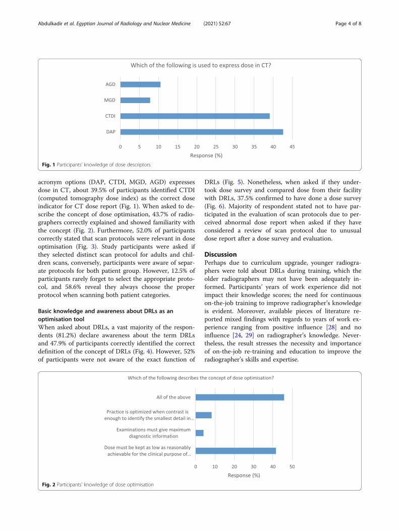
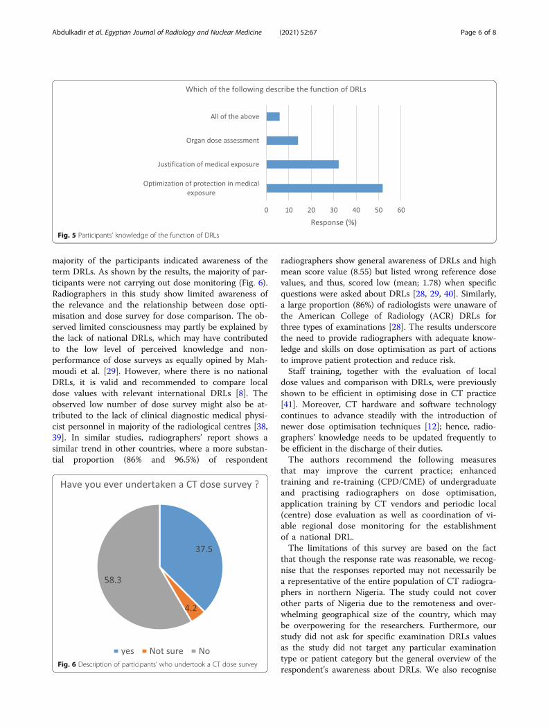
acronym options (DAP, CTDI, MGD, AGD) expressesdose in CT, about 39.5% of participants identified CTDI(computed tomography dose index) as the correct doseindicator for CT dose report (Fig. 1). When asked to de-scribe the concept of dose optimisation, 43.7% of radio-graphers correctly explained and showed familiarity withthe concept (Fig. 2). Furthermore, 52.0% of participantscorrectly stated that scan protocols were relevant in doseoptimisation (Fig. 3). Study participants were asked ifthey selected distinct scan protocol for adults and chil-dren scans, conversely, participants were aware of separ-ate protocols for both patient group. However, 12.5% ofparticipants rarely forget to select the appropriate proto-col, and 58.6% reveal they always choose the properprotocol when scanning both patient categories.
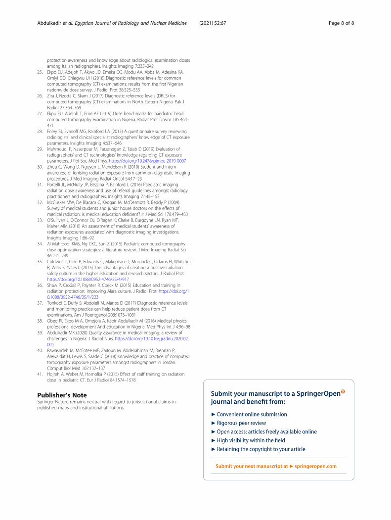
Basic knowledge and awareness about DRLs as anoptimisation toolWhen asked about DRLs, a vast majority of the respon-dents (81.2%) declare awareness about the term DRLsand 47.9% of participants correctly identified the correctdefinition of the concept of DRLs (Fig. 4). However, 52%of participants were not aware of the exact function of
DRLs (Fig. 5). Nonetheless, when asked if they under-took dose survey and compared dose from their facilitywith DRLs, 37.5% confirmed to have done a dose survey(Fig. 6). Majority of respondent stated not to have par-ticipated in the evaluation of scan protocols due to per-ceived abnormal dose report when asked if they haveconsidered a review of scan protocol due to unusualdose report after a dose survey and evaluation.
DiscussionPerhaps due to curriculum upgrade, younger radiogra-phers were told about DRLs during training, which theolder radiographers may not have been adequately in-formed. Participants’ years of work experience did notimpact their knowledge scores; the need for continuouson-the-job training to improve radiographer’s knowledgeis evident. Moreover, available pieces of literature re-ported mixed findings with regards to years of work ex-perience ranging from positive influence [28] and noinfluence [24, 29] on radiographer’s knowledge. Never-theless, the result stresses the necessity and importanceof on-the-job re-training and education to improve theradiographer’s skills and expertise.
Fig. 1 Participants’ knowledge of dose descriptors
Fig. 2 Participants’ knowledge of dose optimisation
Abdulkadir et al. Egyptian Journal of Radiology and Nuclear Medicine (2021) 52:67 Page 4 of 8

Awareness about general radiation protection and doseoptimisationPrevious studies attributed weak radiation protectionculture, which probably emanated from insufficientknowledge and familiarisation during training with thereported inadequate radiation protection knowledge andpractices amongst radiographers [30–33]. Interestingly,it is of concern that a low percentage of participants inthis study were able to identify CT dose descriptors aswell as the correct description of dose optimisation.Thus, it might undermine achievement of potential doseoptimisation. However, it is established that on-the-jobcontinuous medical education (CME) in radiation pro-tection was effective and substantially improved radio-graphers’ performance, radiation protection knowledgeand skills [29, 34–36].On the other hand, slightly above 50% of the partici-
pants were aware that scan protocols are relevant for
dose optimisation. Nevertheless, more awareness is re-quired because scan protocols available to radiographerswere mostly as set up by the vendor application special-ists and then might not be adjusted again if images ap-pear satisfactory for diagnosis. Furthermore, unlikeconventional X-ray imaging, the wide latitude of the CTmodality and advanced inherent image processing cap-abilities makes the adverse effect of overexposure barelydiscern the image quality [28, 29]. Hence, there is a needto periodically monitor, evaluate and compare localdoses with DRLs to detect potential abnormal doses(under and overexposure) that may necessitate a reviewof protocols and optimisation [9]. Equally, a proper un-derstanding of the optimisation principle facilitates skillsand knowledge of dose reduction and the protection ofpatients [24], and the lack of understanding thisprinciple limits the potential of achieving optimised CTpractice.
Knowledge and awareness about DRLs as an optimisationtoolThe evaluation of radiation doses and comparison oflocal practice with DRLs becomes critical, especially inthe light of reported overexposure or higher dose valuesfollowing comparison of some local (Nigerian) CT prac-tices with international DRLs [14–16]. Diagnostic refer-ence levels can assist in reducing patient dose from CTexaminations, once scanning protocol change or im-proved after a review [37]. Consequently, DRLs provedover the years as a useful tool in achieving optimisationof CT practice and should be repeated periodically [17,21, 22]. More so, radiographers’ knowledge and skillsabout dose reduction strategies as well as the cooper-ation from medical physicist and radiologist are equallyessential.Almost two-thirds of participants did not undertake
local dose survey for comparison with DRLs. However,
Fig. 3 Participants’ knowledge of scan protocols
Fig. 4 Participants’ knowledge of the concept DRLs
Abdulkadir et al. Egyptian Journal of Radiology and Nuclear Medicine (2021) 52:67 Page 5 of 8
majority of the participants indicated awareness of theterm DRLs. As shown by the results, the majority of par-ticipants were not carrying out dose monitoring (Fig. 6).Radiographers in this study show limited awareness ofthe relevance and the relationship between dose opti-misation and dose survey for dose comparison. The ob-served limited consciousness may partly be explained bythe lack of national DRLs, which may have contributedto the low level of perceived knowledge and non-performance of dose surveys as equally opined by Mah-moudi et al. [29]. However, where there is no nationalDRLs, it is valid and recommended to compare localdose values with relevant international DRLs [8]. Theobserved low number of dose survey might also be at-tributed to the lack of clinical diagnostic medical physi-cist personnel in majority of the radiological centres [38,39]. In similar studies, radiographers’ report shows asimilar trend in other countries, where a more substan-tial proportion (86% and 96.5%) of respondent
radiographers show general awareness of DRLs and highmean score value (8.55) but listed wrong reference dosevalues, and thus, scored low (mean; 1.78) when specificquestions were asked about DRLs [28, 29, 40]. Similarly,a large proportion (86%) of radiologists were unaware ofthe American College of Radiology (ACR) DRLs forthree types of examinations [28]. The results underscorethe need to provide radiographers with adequate know-ledge and skills on dose optimisation as part of actionsto improve patient protection and reduce risk.Staff training, together with the evaluation of local
dose values and comparison with DRLs, were previouslyshown to be efficient in optimising dose in CT practice[41]. Moreover, CT hardware and software technologycontinues to advance steadily with the introduction ofnewer dose optimisation techniques [12]; hence, radio-graphers’ knowledge needs to be updated frequently tobe efficient in the discharge of their duties.The authors recommend the following measures
that may improve the current practice; enhancedtraining and re-training (CPD/CME) of undergraduateand practising radiographers on dose optimisation,application training by CT vendors and periodic local(centre) dose evaluation as well as coordination of vi-able regional dose monitoring for the establishmentof a national DRL.The limitations of this survey are based on the fact
that though the response rate was reasonable, we recog-nise that the responses reported may not necessarily bea representative of the entire population of CT radiogra-phers in northern Nigeria. The study could not coverother parts of Nigeria due to the remoteness and over-whelming geographical size of the country, which maybe overpowering for the researchers. Furthermore, ourstudy did not ask for specific examination DRLs valuesas the study did not target any particular examinationtype or patient category but the general overview of therespondent’s awareness about DRLs. We also recognise
Fig. 5 Participants’ knowledge of the function of DRLs
Fig. 6 Description of participants’ who undertook a CT dose survey
Abdulkadir et al. Egyptian Journal of Radiology and Nuclear Medicine (2021) 52:67 Page 6 of 8

that as part of the limitations of the questionnaire sur-vey, some participants might not be truthful in responsesconcerning their actual practice and knowledge.
ConclusionThis study strives for better CT practice through an em-phasis on DRLs amongst radiographers in northernNigeria. There is an urgent need for implementation oftraining in CT dose optimisation and the need for radio-graphers to take actions that will improve their know-ledge. The main priorities are on the implementation oflocal dose survey for DRLs and optimisation. Majority ofradiographers in this region have limited awareness ofDRLs as an optimisation tool as well as the need forperiodic dose evaluation. Continuous on the job trainingwill considerably influence radiographers’ knowledge ofCT dose optimisation and thereby reduce patient dosein line with the ALARA principle of radiationprotection.
AbbreviationsDRLs: Diagnostic reference levels; CT: Computed tomography; ALARA: As lowas reasonably achievable; CME: Continuous medical education;CPD: Continuous professional development; CTDI: Computed tomographydose index; CTDIvol: Volume weighted computed tomography dose index;DNA: Deoxyribonucleic acid; DAP: Dose area product; DLP: Dose lengthproduct; MDCT: Multidetector computed tomography; MGD: Mean glandulardose; AGD: Average glandular dose; GE: General electrics; ACR: AmericanCollege of Radiology
AcknowledgementsWe express our appreciation to all the radiographers who participated in thesurvey.
Authors’ contributionsMKA did the conception of the study. All authors participated in the studydesign, questionnaire draft and data collection. First drafting was done byMKA and revised by AA and ADP. All authors read and approved the finalmanuscript.
FundingNone
Availability of data and materialsAll data generated or analysed during this study are included in thispublished article.
Ethics approval and consent to participate(Not applicable) Institutional ethics approval for this study was waived as itdid not include any risk groups, participation was voluntary and theparticipants’ identity was anonymous.
Consent for publicationNot applicable
Competing interestsThe authors declare that they have no competing interests.
Author details1Department of Medical Radiography, Faculty of Basic Medical Sciences,University of Ilorin, Ilorin, Nigeria. 2Department of Imaging Technology andSonography, University of Cape Coast, Cape Coast, Ghana. 3Department ofRadiography, Faculty of Allied Health, College of Medical Sciences, Universityof Maiduguri, Maiduguri, Nigeria. 4Department of Radiography, College ofHealth Sciences, Usmanu Danfodiyo University, Sokoto, Nigeria. 5Radiology
Department, University of Ilorin Teaching Hospital, Ilorin, Nigeria. 6RadiologyDepartment, Obafemi Awolowo University Teaching Hospital, Ile-Ife, Nigeria.
Received: 30 June 2020 Accepted: 17 February 2021
References1. Aroua A, Samara ET, Bochud FO, Meuli R, Verdun FR (2013) Exposure of the
Swiss population to computed tomography. BMC Med Imaging. https://doi.org/10.1186/1471-2342-13-22
2. Shrimpton P, Hillier M (2003) Radiology ML-… journal of, 2006 undefinedNational survey of doses from CT in the UK. birpublications.org
3. UNSCEAR (2000) Effects of ionizing radiation: 2000 report to the generalassembly, with scientific annexes, vol. II: effects. United Nations, New York
4. Bernier MO, Baysson H, Pearce MS et al (2019) Cohort profile: the EPI-CTstudy: a European pooled epidemiological study to quantify the risk ofradiation-induced cancer from paediatric CT. Int J Epidemiol 48:379–381g
5. Brenner DJ, Elliston CD, Hall EJ, Berdon WE (2001) Estimated risks ofradiation. Am Roentgen Ray Soc 176:289–296
6. (UNSCEAR) UNSC on the E of AR (2015) Report of the United NationsScientific Committee on the effects of atomic radiation to the generalassembly. https://doi.org/10.18356/6371bfe8-en
7. Nelson TR (2014) Practical strategies to reduce pediatric CT radiation dose. JAm Coll Radiol 11:292–299
8. International Atomic Energy Agency (2012) Quality assurance programmefor computed tomography: diagnostic and therapy applications, HumanHealth Series No. 19, IAEA, Vienna.
9. American College of Radiology (2013) ACR practice guideline for diagnosticreference levels in medical X-ray imaging.
10. American College of Radiology (2018) ACR – AAPM – SPR practiceparameter for diagnostic reference levels and achievable doses in medicalX-ray imaging, vol 1076, pp 1–12
11. Faggioni L, Paolicchi F, Bastiani L, Guido D, Caramella D (2017) Awareness ofradiation protection and dose levels of imaging procedures among medicalstudents, radiography students, and radiology residents at an academichospital: results of a comprehensive survey. Eur J Radiol 86:135–142
12. Aberle C, Ryckx N, Treier R, Schindera S (2020) Update of national diagnosticreference levels for adult CT in Switzerland and assessment of radiationdose reduction since 2010. Eur Radiol 30:1690–1700
13. Järvinen H, Vassileva J, Samei E, Wallace A, Vano E, Rehani M (2017) Patientdose monitoring and the use of diagnostic reference levels for theoptimization of protection in medical imaging: current status andchallenges worldwide. J Med Imaging 4:1
14. Kabir Abdulkadir M, Schandorf C, Hasford F (2016) Determination ofcomputed tomography diagnostic reference levels in North-Central Nigeria.Pac J Sci Technol 17:341–349
15. Garba I, Engel-Hills P, Davidson F, Tabari AM (2015) Computed tomographydose index for head CT in northern Nigeria. Radiat Prot Dosim 165:98–101
16. Adejoh T, Onwujekwe EC, Abba M, Ali AM, Imo AS, Nzotta CC, Chiegwu HU(2018) Computed tomography scanner census and adult head dose inNigeria. Egypt J Radiol Nucl Med 49:66–70
17. Ogbole G, Obed R (2014) Radiation doses in computed tomography: needfor optimization and application of dose reference levels in Nigeria. WestAfr J Radiol 21:1
18. Eze C, Irurhe N, Njoku J, Olowu O, Abonyi L (2013) Assessment of radiationprotection practices among radiographers in Lagos, Nigeria. Niger Med J 54:386
19. Inyang S, Egbe N, Ekpo E (2015) Challenges in setting up quality control indiagnostic radiology facilities in Nigeria. Niger J Med 4:344–347
20. Inyang SO, Egbe NO, Inyang IS, Oshi DO (2010) Baseline survey of level ofquality control in medical radiology in Cross River State, Nigeria. Pol J MedPhys Eng 16:97–106
21. ICRP (2007) The 2007 recommendations of the international commission onradiological protection. ICRP publication 103. Ann ICRP 37:1–332
22. Vañó E, Miller DL, Martin CJ, Rehani MM, Kang K, Rosenstein M, Ortiz-LópezP, Mattsson S, Padovani R, Rogers A (2017) ICRP Publication 135: diagnosticreference levels in medical imaging. Ann ICRP 46:1–144
23. Soroosh G, Ninalowo H, Hutchens A, Khan S (2015) Nigeria country reportfor use in radiology outreach initiatives
24. Paolicchi F, Miniati F, Bastiani L, Faggioni L, Ciaramella A, Creonti I,Sottocornola C, Dionisi C, Caramella D (2016) Assessment of radiation
Abdulkadir et al. Egyptian Journal of Radiology and Nuclear Medicine (2021) 52:67 Page 7 of 8
protection awareness and knowledge about radiological examination dosesamong Italian radiographers. Insights Imaging 7:233–242
25. Ekpo EU, Adejoh T, Akwo JD, Emeka OC, Modu AA, Abba M, Adesina KA,Omiyi DO, Chiegwu UH (2018) Diagnostic reference levels for commoncomputed tomography (CT) examinations: results from the first Nigeriannationwide dose survey. J Radiol Prot 38:525–535
26. Zira J, Nzotta C, Skam J (2017) Diagnostic reference levels (DRLS) forcomputed tomography (CT) examinations in North Eastern Nigeria. Pak JRadiol 27:364–369
27. Ekpo EU, Adejoh T, Erim AE (2019) Dose benchmarks for paediatric headcomputed tomography examination in Nigeria. Radiat Prot Dosim 185:464–471
28. Foley SJ, Evanoff MG, Rainford LA (2013) A questionnaire survey reviewingradiologists’ and clinical specialist radiographers’ knowledge of CT exposureparameters. Insights Imaging 4:637–646
29. Mahmoudi F, Naserpour M, Farzanegan Z, Talab D (2019) Evaluation ofradiographers’ and CT technologists’ knowledge regarding CT exposureparameters. J Pol Soc Med Phys. https://doi.org/10.2478/pjmpe-2019-0007
30. Zhou G, Wong D, Nguyen L, Mendelson R (2010) Student and internawareness of ionising radiation exposure from common diagnostic imagingprocedures. J Med Imaging Radiat Oncol 54:17–23
31. Portelli JL, McNulty JP, Bezzina P, Rainford L (2016) Paediatric imagingradiation dose awareness and use of referral guidelines amongst radiologypractitioners and radiographers. Insights Imaging 7:145–153
32. McCusker MW, De Blacam C, Keogan M, McDermott R, Beddy P (2009)Survey of medical students and junior house doctors on the effects ofmedical radiation: is medical education deficient? Ir J Med Sci 178:479–483
33. O’Sullivan J, O’Connor OJ, O’Regan K, Clarke B, Burgoyne LN, Ryan MF,Maher MM (2010) An assessment of medical students’ awareness ofradiation exposures associated with diagnostic imaging investigations.Insights Imaging 1:86–92
34. Al Mahrooqi KMS, Ng CKC, Sun Z (2015) Pediatric computed tomographydose optimization strategies: a literature review. J Med Imaging Radiat Sci46:241–249
35. Coldwell T, Cole P, Edwards C, Makepeace J, Murdock C, Odams H, WhitcherR, Willis S, Yates L (2015) The advantages of creating a positive radiationsafety culture in the higher education and research sectors. J Radiol Prot.https://doi.org/10.1088/0952-4746/35/4/917
36. Shaw P, Croüail P, Paynter R, Coeck M (2015) Education and training inradiation protection: improving Alara culture. J Radiol Prot. https://doi.org/10.1088/0952-4746/35/1/223
37. Tonkopi E, Duffy S, Abdolell M, Manos D (2017) Diagnostic reference levelsand monitoring practice can help reduce patient dose from CTexaminations. Am J Roentgenol 208:1073–1081
38. Obed RI, Ekpo M-A, Omojola A, Kabir Abdulkadir M (2016) Medical physicsprofessional development And education in Nigeria. Med Phys Int J 4:96–98
39. Abdulkadir MK (2020) Quality assurance in medical imaging: a review ofchallenges in Nigeria. J Radiol Nurs. https://doi.org/10.1016/j.jradnu.2020.02.005
40. Rawashdeh M, McEntee MF, Zaitoun M, Abdelrahman M, Brennan P,Alewaidat H, Lewis S, Saade C (2018) Knowledge and practice of computedtomography exposure parameters amongst radiographers in Jordan.Comput Biol Med 102:132–137
41. Hojreh A, Weber M, Homolka P (2015) Effect of staff training on radiationdose in pediatric CT. Eur J Radiol 84:1574–1578
Publisher’s NoteSpringer Nature remains neutral with regard to jurisdictional claims inpublished maps and institutional affiliations.
Abdulkadir et al. Egyptian Journal of Radiology and Nuclear Medicine (2021) 52:67 Page 8 of 8