assigned reading - university of california, san...
TRANSCRIPT
Assigned Reading: Review - History of lectins: from hemagglutinins to biological recognition molecules Review - Protein-Carbohydrate Interactions as Part of Plant Defense and Animal Immunity Chapter 24, Essentials to Glycobiology (3rd edition)

History of lectins: from hemagglutinins to biological
recognition molecules
REVIEW
History of lectins: from hemagglutinins to biological recognition molecules
Nathan Sharon1 and Halina Lis
Department of Biological Chemistry, Weizmann Institute of Science,Rehovot 76100, Israel
Editor’s note: Dr. Nathan Sharon is among an elite vanguard ofscientists who pioneered the study of lectin structure and function.Since his early work on soybean agglutinin in the 1960s, Dr. Sharonhas published hundreds of papers covering all aspects of lectin recognition,from the structural basis for lectin–carbohydrate interactions to clinicalapplications. Along with his longtime collaborator at the WeizmannInstitute, Dr. Halina Lis, Dr. Sharon has not only contributed greatlyto our understanding of lectins but has been a tireless and highlyeffective advocate for glycobiology worldwide. It is with great pleasureand appreciation that the editors provide the following historicalperspective of these two leaders in the field.
Received on November 25, 2003; revised on June 28, 2004;accepted on June 28, 2004
The occurrence in nature of erythrocyte-agglutinating pro-teins has been known since the turn of the 19th century. Bythe 1960s it became apparent that such proteins also agglu-tinate other types of cells, and that many of them are sugar-specific. These cell-agglutinating and sugar-specific proteinshave been named lectins. Although shown to occur widely inplants and to some extent also in invertebrates, very fewlectins had been isolated until the early 1970s, and they hadattracted little attention. This attitude changed with thedemonstration that lectins are extremely useful tools forthe investigation of carbohydrates on cell surfaces, in partic-ular of the changes that the latter undergo in malignancy, aswell as for the isolation and characterization of glycopro-teins. In subsequent years numerous lectins have been iso-lated from plants as well as from microorganisms andanimals, and during the past two decades the structures ofhundreds of them have been established. Concurrently, itwas shown that lectins function as recognition molecules incell–molecule and cell–cell interactions in a variety ofbiological systems. Here we present a brief account of 100-plus years of lectin research and show how these proteinshave become the focus of intense interest for biologists andin particular for the glycobiologists among them.
Key words: carbohydrates/functions/glycobiology/microorganisms/plants
Introduction
Toward the end of the 19th century, evidence started toaccumulate for the presence in nature of proteins possessingthe ability to agglutinate erythrocytes. Such proteins werereferred to as hemagglutinins, or phytoagglutinins, becausethey were originally found in extracts of plants. It is gen-erally believed that the earliest description of such a hemag-glutinin was by Peter Hermann Stillmark in his doctoralthesis presented in 1888 to the University of Dorpat (nowTartu, Estonia), one of the oldest universities in czaristRussia (reviewed by Franz, 1988). This hemagglutinin,which was also highly toxic, was isolated by Stillmarkfrom seeds of the castor tree (Ricinus communis) and wasnamed ricin. Subsequently, H. Hellin, also at Tartu, demon-strated the presence of a toxic hemagglutinin, abrin, inextracts of the jequirity bean (Abrus precatorius). Ricinand abrin soon became commercially available, whichprompted Paul Ehrlich, at the Royal Institute of Experi-mental Therapy (Frankfurt), to employ them as modelantigens for immunological studies. Although the prepara-tions available to him were very crude by present criteria(we know now that the ricin and the abrin each contained aweakly agglutinating, powerful toxin and a poorly toxic butstrong agglutinin, all galactose-specific), he was able toestablish with them in the 1890s several of the fundamentalprinciples of immunology. Thus Ehrlich found that micewere rendered immune to a lethal dose of ricin or abrin byrepeated small (sublethal), subcutaneous injections of thelectin and that anti-ricin did not protect the animals againstthe toxic effects of abrin, nor did anti-abrin protect againstricin. This provided clear evidence for the specificity of theimmune response. Ehrlich also showed that immunity to thetoxins is transferred from a mother to her offspring byblood during pregnancy and by milk after birth. By study-ing the inhibitory effect of the anti-ricin immune serum onthe agglutinating activity of ricin, he demonstrated thatthere was a quantitative relationship between the amountof antiserum and that of antigen it could neutralize and onthis basis performed the first quantitative determination ofan antibody in vitro. These studies thus demonstrated thespecificity of the antibody response, the phenomenon ofimmunological memory, and the transfer of humoral immu-nity from a mother to her offspring.
The general public became aware of ricin in 1978, follow-ing its use as a weapon in the notorious politically moti-vated ‘‘umbrella murder’’ of Georgi Markov, Bulgarianopposition writer and broadcaster in exile. Attempts toemploy ricin as a potential weapon of war have been carriedout by the United States during World War I; during World
1To whom correspondence should be addressed; [email protected]
Glycobiology vol. 14 no. 11 # Oxford University Press 2004; all rights reserved. 53R
Glycobiology vol. 14 no. 11 pp. 53R–62R, 2004doi:10.1093/glycob/cwh122Advance Access publication on June 30, 2004
at University of C
alifornia, San Diego on M
arch 24, 2016http://glycob.oxfordjournals.org/
Dow
nloaded from
War II a ricin bomb was developed and tested by the Britishmilitary, but it has never been deployed as a weapon formass destruction. More recently, ricin has found its wayinto the arsenals of extremist individuals, groups, andgovernments.
Sugar binding and blood type specificity
In 1919, James B. Sumner at Cornell University (Ithaca,New York), well known for being the first to crystallize in1926 an enzyme, urease (for which he was awarded theNobel Prize 21 years later), isolated from jack bean(Canavalia ensiformis) a crystalline protein that he namedconcanavalin A and in this way obtained a pure hemagglu-tinin for the first time. However, nearly two decades passedbefore Sumner and Howell (1936) reported that concanav-alin A agglutinates cells such as erythrocytes and yeastsand also precipitates glycogen from solution. They furthershowed that hemagglutination by concanavalin A wasinhibited by sucrose, demonstrating for the first time thesugar specificity of lectins. With much foresight, they sug-gested that the hemagglutination induced by concanavalinA might be a consequence of a reaction of the plant proteinwith carbohydrates on the surface of the red cells.
Already the early results obtained by Stillmark indicatedsome selectivity in the ricin-induced agglutination of redcells from different animals. This observation was corrobo-rated and further extended by Karl Landsteiner from theUniversity of Vienna, the discoverer of the human A, B, andO blood groups in 1900. Nearly a decade later he reportedthat the relative hemagglutinating activities of various seedextracts were quite different when tested with red blood cellsfrom different animals (Landsteiner and Raubitschek,1907). Because of this specificity, Landsteiner concludedthat the actions of plant hemagglutinins ‘‘resemble antibodyreactions in all essentials.’’ He therefore used these proteinsto illustrate the specificity concept in the introductorychapter of his classic book The Specificity of SerologicalReactions (1936).
The 1940s saw the discovery, made independently byWilliam C. Boyd at Boston University and by Karl O.Renkonen at the University of Helsinki, Finland, of thehuman blood group (or blood type) specificity of thehemagglutinins. They found that crude extracts of the limabean, Phaseolus limensis, and the tufted vetch, Vicia cracca,agglutinated blood type A erythrocytes but not blood typeB or O cells, whereas an extract of the asparagus pea, Lotustetragonolobus, agglutinated specifically blood type Oerythrocytes. Olavi M€aakel€aa (1957), a doctoral student ofRenkonen, examined in 1954–56 extracts from seeds repre-senting 743 plant species and 165 genera, all of the familyLeguminosae, and detected hemagglutinating activity inmore than one-third of them; close to one-tenth of thehemagglutinins exhibited blood type specificity. Althoughseveral of the latter were specific either for blood type O ortype A, or both type A and B erythrocytes, and one, fromDolichos biflorus, reacted much better with A1 erythrocytesthan with A2, only the extract from Griffonia simplicifolia(previously known as Bandeiraea simplicifolia) exhibitedalmost exclusively type B specificity. Since then, additional
hemagglutinins specific for blood types A and O (but not B)have been discovered, as well as several for otherblood types, such as N (Vicia graminea lectin), T (peanutagglutinin, PNA) and Tn (the lectins of Vicia villosa andMoluccella laevis).
The blood type–specific hemagglutinins played a crucialrole in early investigations on the structural basis of thespecificity of the antigens associated with the ABO bloodgroup system. In the 1950s, Walter J. T. Morgan andWinifred M. Watkins at the Lister Institute, London,found that the agglutination of type A red cells bylima bean lectin was best inhibited by a-linked N-acetyl-D-galactosamine and that of type O cells by the lectin ofL. tetragonolobus was best inhibited by a-linked L-fucose.They concluded that a-N-acetyl-D-galactosamine anda-L-fucose are the sugar determinants conferring A andH(O) blood group specificity, respectively. Both conclu-sions have been substantiated by subsequent investigations(for a recent review, see Morgan and Watkins, 2000). Thepioneering work of Watkins and Morgan was among theearliest evidence for the presence of sugars on cell surfacesand their potential roles as identity markers, an acceptedtheme in modern glycobiology. It took a while, however,before the counterreceptors for surface sugars, that is, theendogenous lectins that recognize these sugars, were iden-tified, the first being the mammalian hepatic asialoglyco-protein receptor to be described later.
The ability of plant agglutinins to distinguish betweenerythrocytes of different blood types led Boyd andShapleigh (1954) to propose for them the name lectins,from the Latin legere, to pick out or choose. This termwas generalized by us to embrace all sugar-specific aggluti-nins of nonimmune origin, irrespective of source and bloodtype specificity (Sharon and Lis, 1972).
Mitogenic stimulation of lymphocytes and agglutinationof cancer cells
Two major discoveries made in the early 1960s were instru-mental in bringing lectins into the limelight. The first ofthese was by Peter C. Nowell (1960) at the University ofPennsylvania, Philadelphia, who found that the lectin of thered kidney bean (Phaseolus vulgaris), known as phytohe-magglutinin (PHA), is mitogenic, that is, it possesses theability to stimulate lymphocytes to undergo mitosis. Thisdiscovery had a revolutionary impact on immunology inthat it shattered the view, held until then, that lymphocytesare dead-end cells incapable of dividing or differentiatingfurther. Within a short time, several other lectins wereproven to be mitogenic. Of special significance was thefinding that concanavalin A acts as a mitogen because, incontrast to PHA, its activity could be inhibited by lowconcentrations of monosaccharides, for example, mannose.This finding provided proof that mitogenic stimulation isthe result of binding of lectins to sugars on the surface of thelymphocytes and was among the earliest demonstrations fora biological role of cell surface sugars. Mitogenic lectinssoon became tools for the study of signal transmissioninto cells and for the analysis of the biochemical eventsthat occur during lymphocyte stimulation in vitro. A most
N. Sharon and H. Lis
54R
at University of C
alifornia, San Diego on M
arch 24, 2016http://glycob.oxfordjournals.org/
Dow
nloaded from

valuable outcome of such studies was the discovery in the1970s by Robert C. Gallo and his associates at the NationalInstitutes of Health (Bethesda) of T cell growth factor,now known as interleukin-2, in conditioned medium ofnormal human lymphocytes stimulated by PHA (Morganet al., 1976).
The second discovery was made by Joseph C. Aub at theMassachusetts General Hospital in Boston (Aub et al.,1963, 1965). He found that wheat germ agglutinin (WGA)has the ability to preferentially agglutinate malignant cells.This was followed by the reports of Max M. Burger atPrinceton University and Leo Sachs and Michael Inbar atthe Weizmann Institute (Rehovot) that concanavalin Aexhibits the same ability. Together with Sachs and Ben-Ami Sela, we subsequently found that soybean agglutinin(SBA) also possesses the same property. Such investigationsprovided early evidence that changes in cell surface sugarsare associated with the development of cancer and led to theassumption that high susceptibility to agglutination by lec-tins was a property shared by all malignant cells. Unfortu-nately, this is now known not to be generally true.
Lectins galore
Until the early 1970s, the presence of hemagglutinins hadbeen reported in numerous organisms, primarily plants, butonly very few had been purified, almost all by conventionaltechniques. In addition to concanvalin A, they included theplant lectins from soya beans, green peas, Dolichos biflorusseeds, wheat germ, and mushroom (Agaricus campestris)(reviewed in Sharon and Lis, 1972) and the animal lectinsof eel (Springer and Desai, 1971), snail (Hammarstr€oom andKabat, 1969), and horseshoe crab (Marchalonis andEdelman, 1968). The pace of lectin isolation increased dra-matically with the introduction of affinity chromatographyfor lectin purification by Irwin J. Goldstein and Bipin B. L.Agrawal of the University of Michigan, originally for theisolation of concanavalin A on immobilized dextran(Sephadex) (Agrawal and Goldstein, 1967). Numerous lec-tins have thus become available, for a time still mainly fromplants, the number of the latter being now about 500. Theinterest in these lectins was greatly stimulated by thedemonstration that they are invaluable tools for the detec-tion, isolation, and characterization of glycoconjugates,primarily of glycoproteins, for histochemistry of cells andtissues and for the examination of changes that occur on cellsurfaces during physiological and pathological processes,from cell differentiation to cancer (Table I).
The occurrence of hemagglutinins in animals was notedquite early, almost all in invertebrates or lower vertebrates,but until the middle of the 1970s, only the three of thesementioned (of eel, snail, and horseshoe crab) were isolatedand characterized. The first of the animal lectins shown tobe specific for a sugar (L-fucose) was from the eel (Watkinsand Morgan, 1952). The isolation in 1974 of the first mam-malian lectin, the galactose-specific hepatic asialoglyco-protein receptor, was an outcome of the investigation byGilbert Ashwell at the NIH together with Anatol G. Morellat the Albert Einstein Medical School (New York) of themechanisms that control the lifetime of glycoproteins in
blood circulation (Hudgin et al., 1974; Stockert et al.,1974). At the same time, Vivian Teichberg from ourdepartment reported (Teichberg et al., 1975) the isolationfrom the electric eel of the first member of the familyof the b-galactose-specific lectins, designated galectins(Barondes et al., 1994), of which over a dozen membershave by now characterized. Since the beginning of the1980s, the number of purified animal lectins also startedto grow quickly, largely thanks to the advent of recom-binant techniques.
From primary to 3D structures
The 1970s also witnessed the intensification of studies of themolecular properties of individual lectins, a prerequisite fora deep understanding of their activities at the molecularlevel. These studies ranged from the determination of themain physicochemical parameters of lectins to completeamino acid sequencing and elucidation of their 3D struc-ture. Until the advent of recombinant techniques, determi-nation of the primary structure of lectins proceeded ratherslowly, and by the end of that decade the completesequences of only half a dozen lectins, all from plants,were known. In this case, too, concanavalin A led thefield, being the first lectin whose primary sequence hasbeen established (Edelman et al., 1972). Concurrently,Edelman’s group and independently Karl Hardman withClinton F. Ainsworth at Argonne National Laboratories(Argonne, Illinois), solved the 3D structure of concanavalinA by high resolution X-ray crystallography, another firstfor this lectin (Edelman et al., 1972; Hardman andAinsworth, 1972). This was soon followed by the determi-nation of the structure of WGA as well as of its complexeswith its ligands (N-acetylneuraminic acid and b4-linked N-acetylglucosamine oligomers) by Christine Schubert Wrightat the Virginia Commonwealth University (Richmond)even before the complete amino sequence of this lectin hadbecome available (Wright, 1977). The striking differencebetween the structure of concanavalin A and WGA fullycorroborated the suggestion presented by us at the 1973International Glycoconjugate Symposium in Lille, France,that was based largely on compositional data of these two
Table I. Major applications of lectinsa
Cell identification and separation
Detection, isolation, and structural studies of glycoproteins
Investigation of carbohydrates on cells and subcellular organelles; histo-chemistry and cytochemistry
Mapping of neuronal pathways
Mitogenic stimulation of lymphocytesb
Purging of bone marrow for transplantationb
Selection of lectin-resistant mutants
Studies of glycoprotein biosynthesis
aLectins from sources other than plants are rarely in use.
bIn clinical use.
History of lectins
55R
at University of C
alifornia, San Diego on M
arch 24, 2016http://glycob.oxfordjournals.org/
Dow
nloaded from
proteins, that although ‘‘lectins have many biological prop-erties in common, they represent a diversified group ofproteins with respect to size, composition and structure’’(Sharon et al., 1974).
The availability of the primary structure of numerouslectins allowed the identification of homologies betweenthe sequences of lectins from taxonomically related sources,as originally demonstrated for the legume lectins by one ofus (N.S.) in collaboration with Donny Strosberg at the FreeUniversity of Brussels (Foriers et al., 1977). By the end ofthe following decade, homologies were found also for
lectins from other families, such as the galectins and theC-type (Ca2þ requiring) lectins (Drickamer, 1988).
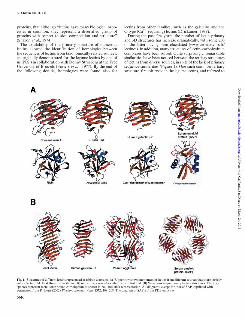
During the past few years, the number of lectin primaryand 3D structures has increase dramatically, with some 200of the latter having been elucidated (www.cermav.cnrs.fr/lectines). In addition, many structures of lectin–carbohydratecomplexes have been solved. Quite surprisingly, remarkablesimilarities have been noticed between the tertiary structuresof lectins from diverse sources, in spite of the lack of primarysequence similarities (Figure 1). One such common tertiarystructure, first observed in the legume lectins, and referred to
Fig. 1. Structures of different lectins represented as ribbon diagrams. (A) Upper row shows monomers of lectins from different sources that share the jellyroll or lectin fold. First three lectins (from left) in the lower row all exhibit the b-trefoil fold. (B) Variations in quaternary lectins structures. The grayspheres represent metal ions; bound carbohydrate is shown in ball-and-stick representation. All diagrams, except for that of SAP, reprinted withpermission from R. Loris (2002) Biochim. Biophys. Acta, 1572, 198–208. The diagram of SAP is from PDB entry sac.
N. Sharon and H. Lis
56R
at University of C
alifornia, San Diego on M
arch 24, 2016http://glycob.oxfordjournals.org/
Dow
nloaded from
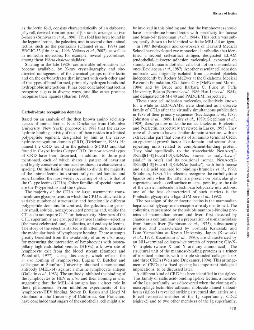
as the lectin fold, consists characteristically of an elaboratejelly roll, derived from antiparallel b-strands, arranged as twob-sheets (Srinivasan et al., 1996). This fold has been found inthe legume lectins, the galectins, and in several other animallectins, such as the pentraxins (Crennel et al., 1994) andERGIC-53 (Itin et al., 1996; Velloso et al., 2002), as well asin nonlectin molecules, for example, several glycosidases,among them Vibrio cholerae sialidase.
Starting in the late 1980s, considerable information hasbecome available, by X-ray crystallography and site-directed mutagenesis, of the chemical groups on the lectinand on the carbohydrates that interact with each other andof the types of bond formed, primarily hydrogen bonds andhydrophobic interactions. It has been concluded that lectinsrecognize sugars in diverse ways, just like other proteinsrecognize their ligands (Sharon, 1993).
Carbohydrate recognition domains
Based on an analysis of the then known amino acid seq-uences of animal lectins, Kurt Drickamer from ColumbiaUniversity (New York) proposed in 1988 that the carbo-hydrate-binding activity of most of them resides in a limitedpolypeptide segment, designated by him as the carbo-hydrate-recognition domain (CRD) (Drickamer, 1988). Henamed the CRD found in the galectins S-CRD and thatfound in C-type lectins C-type CRD. By now several typesof CRD have been discerned, in addition to those justmentioned, each of which shares a pattern of invariantand highly conserved amino acid residues at a characteristicspacing. On this basis it was possible to divide the majorityof the animal lectins into structurally related families andsuperfamilies, the most widely occurring of which is that ofthe C-type lectins (CTLs). Other families of special interestare the P-type lectins and the siglecs.
The majority of the CTLs are large, asymmetric trans-membrane glycoproteins, in which the CRD is attached to avariable number of structurally and functionally differentpolypeptide domains. In contrast, the galectins are gener-ally small, soluble, nonglycosylated proteins and, unlike theCTLs, do not require Ca2þ for their activity. Members of theCTL superfamily are grouped into three families—selectins(the most celebrated one), collectins, and endocytic lectins.The story of the selectins started with attempts to elucidatethe molecular basis of lymphocyte homing. These attemptsgreatly benefited from the availability of an in vitro assayfor measuring the interaction of lymphocytes with postca-pillary high-endothelial venules (HEVs), a known site oflymphocyte exit from the blood stream (Stamper andWoodruff, 1977). Using this assay, which reflects thein vivo homing of lymphocytes, Eugene C. Butcher andcolleagues at Stanford University obtained a monoclonalantibody (MEL-14) against a murine lymphocyte antigen(Gallatin et al., 1983). The antibody inhibited the binding ofthe lymphocytes to HEV in vitro and their homing in vivo,suggesting that the MEL-14 antigen has a direct role inthese phenomena. From inhibition experiments of thelymphocyte-HEV binding, Steven D. Rosen and Lloyd MStoolman at the University of California, San Francisco,have concluded that sugars of the endothelial cell might also
be involved in this binding and that the lymphocytes shouldhave a membrane-bound lectin with specificity for fucoseand Man-6-P (Stoolman et al., 1984). This lectin was sub-sequently shown to be identical with the MEL-14 antigen.
In 1987 Bevilacqua and co-workers of Harvard MedicalSchool have developed two monoclonal antibodies that iden-tified a second cell-surface antigen, designated ELAM(endothelial-leukocyte adhesion molecule)-1, expressed onstimulated human endothelial cells but not on unstimulatedones (Bevilacqua et al., 1987). Another vascular cell adhesionmolecule was originally isolated from activated plateletsindependently by Rodger McEver at the Oklahoma MedicalResearch Foundation, Oklahoma City (McEver and Martin,1984) and by Bruce and Barbara C. Furie at TuftsUniversity, Boston (Berman et al., 1986; Hsu-Lin et al., 1984),and designated GPM-140 and PADGEM, respectively.
These three cell adhesion molecules, collectively knownfor a while as LEC-CAMS, were identified as a discretefamily of CTLs after the virtually simultaneous publicationin 1989 of their primary sequences (Bevilacqua et al., 1989;Johnston et al., 1989; Lasky et al., 1989; Siegelman et al.,1989); these go now under the names L-selectin, E-selectin,and P-selectin, respectively (reviewed in Lasky, 1995). Theywere all shown to have a similar domain structure, with anextracellular part that consists of an amino terminal CRD,an epidermal growth factor–like domain, and several shortrepeating units related to complement-binding protein.They bind specifically to the trisaccharide NeuAca(2-3)Galb(1-4)[Fuca(1-3)]GlcNAc, known as sialyl-Lewisx
(siaLex in brief) and its positional isomer, NeuAca(2-3)Galb(1-3)[Fuca(1-4)]GlcNAc (siaLea), with both fucoseand sialic acid required for binding (Brandley et al., 1990;Stoolman, 1989). The selectins recognize the carbohydrateligands only when the latter are present on particular gly-coproteins, such as cell surface mucins, pointing to the roleof the carrier molecule in lectin-carbohydrate interactions;one of the best characterized of such carriers is theP-selectin glycoprotein ligand (Moore et al., 1992).
The paradigm of the endocytic lectins is the mammalianhepatic asialoglycoprotein receptor already mentioned. Thecollectins, represented by the soluble mannose-binding pro-teins of mammalian serum and liver, first detected bychance as a contaminant of a preparation of a-mannosidasefrom human liver (Robinson et al., 1975), subsequentlypurified and characterized by Toshiaki Kawasaki andIkuo Yamashina at Kyoto University, Japan (Kawasakiet al., 1978; Kozutsumi et al., 1980), are characterized byan NH2-terminal collagen-like stretch of repeating Gly-X-Y- triplets (where X and Y are any amino acid). Thestructural unit of the mannose-binding proteins is a trimerof identical subunits with a triple-stranded collagen helixand three CRDs (Weis and Drickamer, 1994). This arrange-ment of CRDs at a fixed spacing has important biologicalimplications, to be discussed later.
A different kind of CRD has been identified in the siglecs.This family of sialic acid–binding Ig-like lectins, a memberof the Ig superfamily, was discovered when the cloning of amacrophage lectin-like adhesion molecule named siaload-hesin (siglec-1) revealed striking structural similarities to aB cell restricted member of the Ig superfamily, CD22(siglec-2) and to two other members of the Ig superfamily,
History of lectins
57R
at University of C
alifornia, San Diego on M
arch 24, 2016http://glycob.oxfordjournals.org/
Dow
nloaded from
CD33 (siglec-3) and the myelin-associated glycoprotein(siglec-4) (Crocker et al., 1994). Members of this family,11 of which have been identified in humans, are type 1transmembrane proteins with an extracellular part consist-ing of a CRD-containing N-terminal V-set Ig-like domain,followed by variable numbers of C2-set Ig-like domains.Except for myelin-associated glycoprotein (siglec-4), exclu-sively expressed in the nervous system, they are all found oncells of the hematopoietic system. Each siglec has a distinctexpression pattern in different cell types, indicating thatthey perform highly specific functions.
A recent addition to the growing list of mammalian lec-tins is dectin-1, a b-glucan receptor, identified by GordonBrown and Siamon Gordon (2001) at Oxford by screening acDNA library of a macrophage cell line with zymosan. It isa small type II transmembrane receptor containing oneCRD, which recognizes b1,3 and/or b1,6-glucans and intactyeasts.
In protection and symbiosis
The question of the possible physiological role of lectins hasintrigued investigators from the start and focused on plantlectins, which for long time were virtually the only onesknown (reviewed by Etzler, 1986). It was speculated, forexample, that lectins may function as antibodies to protectplants against harmful soil bacteria, control seed germina-tion, or be involved in the transport and storage of sugars,but no evidence for these speculations could be found.However, two proposals put forward in the 1970s stillhold. According to first one, lectins protect plants againstphytopathogenic microorganisms and insects as well asagainst predatory animals. The second theory assumesthat they are involved in the association between legumi-nous plants and their symbiotic nitrogen-fixing bacteria.
Probably the earliest publication on the insecticidal actionof lectins came in 1976 from the laboratory of Irvin E.Liener at the University of Minnesota, at St. Paul(Minnesota) in which it was reported that feeding bruchidbeetles with a diet containing the black bean lectin resultedin the death of the bruchid larvae (Janzen et al., 1976). Onthis basis the authors concluded that the major role oflectins in legumes is to protect them from attack by insectseed predators. In subsequent studies, several other lectinswere shown to be insecticidal, among them WGA,Galanthus nivalis lectin and jacalin.
The proposal that lectins may be involved in the protec-tion of plants against pathogenic microorganisms was orig-inally based on the observation made at Rehovot thatWGA, PNA, and SBA inhibited the sporulation andgrowth of fungi such as Trichoderma viride, Peniciliumnotatum, and Aspergillus niger (see Barkai-Golan et al.,1976). Potato lectin was subsequently shown to act in asimilar manner on Botrytis cinerea, another fungal phyto-pathogen. In an extensive study, 11 purified lectins repre-senting the major carbohydrate specificity groups were allfound to cause growth disruption during germination ofspores of Neurospora crassa, Aspergillus amstelodami, andBotryodiplodia theobromae (Brambl and Gade, 1985). It wasalso shown that recombinant Urtica dioica agglutinin that
has a similar specificity to that of WGA (Broekaert et al.,1989) inhibited the growth of fungal phytopathogens.
The idea that lectins are responsible for the specific asso-ciation between nitrogen-fixing rhizobia and leguminousplants, which provides the plant with the needed nitrogen,was advanced nearly three decades ago (Bohlool andSchmidt, 1974; Hamblin and Kent, 1973). It was based onthe finding that a lectin from a particular legume bound in acarbohydrate-specific manner to the surface polysaccha-rides or lipopolysaccharides of the corresponding rhizobialspecies but not to bacteria that are symbionts of otherlegumes. For instance, SBA agglutinated most strains ofBradyrhizobium japonicum that nodulate soybeans but notnonnodulating bradyrhizobial strains. The suggestion hastherefore been made that rhizobial attachment to plantroots occurs by interaction between the bacterial surfacecarbohydrates and lectins present in the roots of the legu-minous plants. This became known as the lectin recognitionhypothesis, which is still the subject of controversy, becauseof the lack of unequivocal evidence and of some inconsis-tencies. Thus for most host–symbiont systems examined,there is no proof for the presence of lectins and their ligandson plant roots and bacteria, respectively, at precisely theright time and location. Moreover, the correlation betweenthe specificity of the host lectin and its ability to recognizethe nodulating bacteria of that host is not very strict. Also,several lines of soybeans with no detectable lectin in theirseeds or vegetative tissues were nodulated normally by thecorresponding rhizobial symbiont.
Application of the techniques of molecular genetics gaveresults that bolstered the lectin recognition hypothesis butdid not fully settle the controversy (reviewed by Kijne, 1996;Hirsch, 1999).
Recently, a variant of the lectin recognition hypothesishas been proposed, that postulates that the host-specificattachment of the rhizobium is achieved through the inter-action between species-specific lipo-chitooligosaccharidesignal molecules produced by the bacteria, named nodula-tion factors (Nods), and a new type of a plant root lectinfound in different leguminous plants but not in plants ofdifferent families (Kalsi and Etzler, 2000).
Recognition molecules
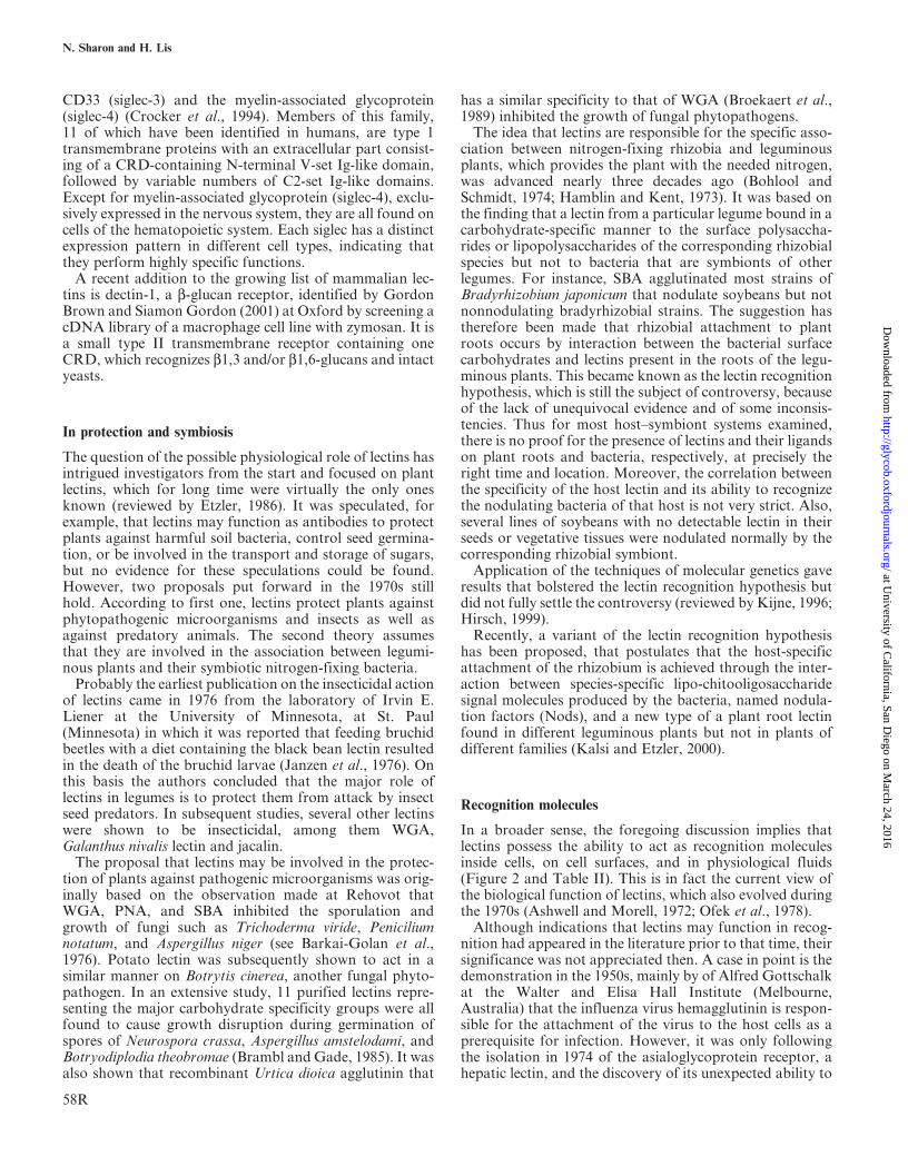
In a broader sense, the foregoing discussion implies thatlectins possess the ability to act as recognition moleculesinside cells, on cell surfaces, and in physiological fluids(Figure 2 and Table II). This is in fact the current view ofthe biological function of lectins, which also evolved duringthe 1970s (Ashwell and Morell, 1972; Ofek et al., 1978).
Although indications that lectins may function in recog-nition had appeared in the literature prior to that time, theirsignificance was not appreciated then. A case in point is thedemonstration in the 1950s, mainly by of Alfred Gottschalkat the Walter and Elisa Hall Institute (Melbourne,Australia) that the influenza virus hemagglutinin is respon-sible for the attachment of the virus to the host cells as aprerequisite for infection. However, it was only followingthe isolation in 1974 of the asialoglycoprotein receptor, ahepatic lectin, and the discovery of its unexpected ability to
N. Sharon and H. Lis
58R
at University of C
alifornia, San Diego on M
arch 24, 2016http://glycob.oxfordjournals.org/
Dow
nloaded from
recognize and to bind terminal galactose residues on serumglycoproteins (reviewed by Ashwell and Morell, 1974) thatthe role of lectins in biological recognition started to gainpopularity. Support came soon with the identification, byWilliam S. Sly at St. Louis University (Kaplan et al., 1977)of the mannose-6-phosphate receptor crucially involvedin intracellular trafficking of lysosomal enzymes. Thedemonstration that hepatic lectins may also mediate theclearance of bacteria from blood in the absence of opsonins(antibodies and complement), was an early indication of theparticipation of lectins in non-immune defense, or innateimmunity (see later discussion).
Another key finding was made in 1979, when our group,together with others, demonstrated that urinary tract infec-tion in mice by mannose-specific Escherichia coli could beprevented by methyl a-D-mannoside (Aronson et al., 1979).It was the first direct evidence for the involvement of bac-terial lectins in the initiation of infection, the basis for thepresent attempts in academia and industry, to apply carbo-hydrates for antiadhesion therapy of such diseases(reviewed by Mulvey et al., 2001).
Together with Itzhak Ofek, we demonstrated at thesame time that the mannose-specific bacterial surface lectinsmay also mediate attachment of the bacteria to phagocyticcells in the absence of opsonins, leading to engulfment andkilling of the bacteria. This process, another example ofinnate immunity, which we named lectinophagocytosis,may be of importance in the clearance of bacteria fromnonimmune patients or from opsonin-poor sites, such asrenal medulla or the peritoneal cavity (Ofek and Sharon,1988). Additional lectins have been implicated in innateimmunity. A prominent example is the mannose-specificreceptor present on the surface of macrophages; it bindsinfectious organisms that expose mannose-containingglycans on their surface, leading to their ingestion andkilling. Another, recently discovered one, is dectin-1, speci-fic for b1,3 and/or b1,6-glucans, present on fungi.
A similar function, albeit by a different mechanism, isperformed by the soluble mannose-binding lectins (MBLs)of mammalian serum and liver (Epstein et al., 1996;Turner, 1996), These proteins bind to oligomannosidesof infectious microorganisms, causing activation of com-plement without participation of antibody, and subse-quent lysis of the pathogens, thus acting in innateimmunity. The spatial arrangement of the CRDs in theMBLs provides a structural basis for their ability to bindligands with repetitive, mannose-rich structures, such as
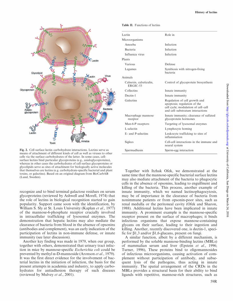
Fig. 2. Cell surface lectin–carbohydrate interactions. Lectins serve asmeans of attachment of different kinds of cell as well as viruses to othercells via the surface carbohydrates of the latter. In some cases, cell-surface lectins bind particular glycoproteins (e.g., asialoglycoproteins),whereas in other cases the carbohydrates of cell surface glycoproteins orglycolipids serve as sites of attachment for biologically active moleculesthat themselves are lectins (e.g. carbohydrate-specific bacterial and planttoxins, or galectins). Based on an original diagram from BioCarbAB(Lund, Sweden).
Table II. Functions of lectins
Lectin Role in
Microorganisms
Amoeba Infection
Bacteria Infection
Influenza virus Infection
Plants
Various Defense
Legumes Symbiosis with nitrogen-fixingbacteria
Animals
Calnexin, calreticulin,ERGIC-53
Control of glycoprotein biosynthesis
Collectins Innate immunity
Dectin-1 Innate immunity
Galectins Regulation of cell growth andapoptosis; regulation of thecell cycle; modulation of cell–celland cell–substratum interactions
Macrophage mannosereceptor
Innate immunity; clearance of sulfatedglycoprotein hormones
Man-6-P receptors Targeting of lysosomal enzymes
L-selectin Lymphocyte homing
E- and P-selectins Leukocyte trafficking to sites ofinflammation
Siglecs Cell-cell interactions in the immune andneural system
Spermadhesin Sperm-egg interaction
History of lectins
59R
at University of C
alifornia, San Diego on M
arch 24, 2016http://glycob.oxfordjournals.org/
Dow
nloaded from
found on fungal and microbial surfaces, but not to theoligomannose units of mammalian glycoproteins (Weisand Drickamer, 1994).
The discovery of the selectins and the demonstrationthat they play a crucial role in the control of lymphocytehoming and of leukocyte trafficking to sites of inflamma-tion was a landmark in lectin research. Indeed, the selec-tins provide the best paradigm for the role of sugar–lectininteractions in biological recognition. They mediate thebinding of leukocytes to endothelial cells and therebyinitiate a rolling phase, in which the lectins interact tran-siently with glycan ligands, leading eventually to theirextravasation. Prevention of adverse inflammatory reac-tions by inhibition of leukocyte–endothelium interactions,another application of antiadhesion therapy, has become amajor aim of the biomedical and pharmacological indus-try. There are also indications that the selectins may func-tion in the spread of cancer cells from the main tumor toother sites in the body and that by blocking their sugar-binding sites it may be possible to prevent the formationof metastases.
From the late 1980s, evidence started to accumulate thatseveral lectins of different types direct intracellular glyco-protein traffic, by acting as chaperones and sortingreceptors in the secretory pathway. Calnexin, a membrane-bound lectin of the endoplasmic reticulum (ER), func-tions in parallel with calreticulin, its soluble homolog, aspart of a quality control system that ensures properfolding of glycoproteins destined to the cell surface. Themannose-specific intracellular lectin, ERGIC-53, firstidentified as a resident of the ER–Golgi intermediate com-partment protein (Schweizer et al., 1988) carries a specificsubset of nascent glycoproteins between the two compart-ments. Two distinct mannose-6-phospate receptors, theonly members of the P-type lectin family, mediate thetargeting of newly synthesized hydrolases from the roughER to the lysosomes (Hoflack and Kornfeld, 1985). Bothreceptors bind their ligands, oligosaccharides bearingterminal Man-6-P residues, most efficiently at pH 6–7,allowing them to interact with hydrolases decoratedwith such oligosaccharides in the trans-Golgi network,and to release them in the more acidic environment ofthe lysosomes.
The galectins are believed to act as modulators of cell–substratum interactions and to be essential for thenormal differentiation and growth of all multicellularanimals. They are capable of inducing cell proliferation,cell arrest, or apoptosis (physiological cell death) andhave been implicated in organ morphogenesis, tumorcell metastasis, leukocyte trafficking, immune response,and inflammation, as well as recognition of extracellularmatrix.
Epilogue
As we have shown in this article, during 120þ years, lectinshave come a long way since their first detection in plants ashemagglutinins to their present status as ubiquitousrecognition molecules with myriad exciting functions andapplications.
Abbreviations
CRD, carbohydrate recognition domain; CTL, C-typelectin; ER, endoplasmic reticulum; HEV, high-endothelialvenule; MBL, mannose-binding lectin; PHA, phytohe-magglutinin; PNA, peanut agglutinin; SBA, soybeanagglutinin; WGA, wheat germ agglutinin.
References
In addition to the references listed here, readers are referred to those inKocourek (1986) and Sharon and Lis (2003).
Agrawal, B.B.I. and Goldstein, I.J. (1967) Specific binding of concanvalinA to cross-linked dextran gels. Biochem. J., 96, 23C–15C.
Aronson, M., Medalia, O., Schori, L., Mirelman, D., Sharon, N., andOfek, I. (1979) Prevention of colonization of the urinary tract of micewith Escherichia coli by blocking of bacterial adherence with methyla-D-mannopyranoside. J. Infect. Dis., 139, 329–332.
Ashwell, G. and Morell, A.G. (1972) Membrane glycoproteins andrecognition phenomena. Trends Biochem. Sci., 2, 76–78.
Ashwell, G. and Morell, A.G. (1974) The role of surface carbohydratesin the hepatic recognition and transport of circulating glycoproteins.Adv. Enzymol. Relat. Areas Mol. Biol., 41, 99–128.
Aub, J.C., Tieslau, C., and Lankester, A. (1963) Reactions of normaland tumor cell to enzymes. I. Wheat-germ lipase and asso-ciated mucopolysaccharides. Proc. Natl Acad. Sci. USA, 50,613–619.
Aub, J.C., Sanford, B.H., and Cote, M.N. (1965) Studies on reactivity oftumor and normal cells to a wheat germ agglutinin. Proc. Natl Acad.Sci. USA, 54, 396–399.
Barkai-Golan, R, Mirelman, D., and Sharon, N. (1978) Studies on growthinhibition by lectins of Penicillia and Aspergilli. Arch. Microbiol., 116,119–121.
Barondes, S.H., Castronovo, V., Cooper, D.N.W., and others. (1994)Galectins—a family of beta-galactoside-binding lectins. Cell, 76,597–598.
Berman, C.L., Yeo, E.L., Wencel-Drake, J.D., Furie, B.C.,Ginsberg, M.H., and Furie, B. (1986) A platelet alpha granulemembrane protein that is associated with the plasma membraneafter activation. Characterization and subcellular localization ofplatelet activation-dependent granule-external membrane protein.J. Clin. Invest., 78, 130–137.
Bevilacqua, M.P., Pober, J.S., Mendrick, D.L., Cotran, R.S., andGimbrone, M.A. Jr. (1987) Identification of an inducible endothelial-leukocyte adhesion molecule. Proc. Natl Acad. Sci. USA, 84,9238–9242.
Bevilacqua, M.P., Stengelin, S., Gimbrone, M.A. Jr., and Seed, B. (1989)Endothelial leukocyte adhesion molecule 1: an inducible receptor forneutrophils related to complement regulatory proteins and lectins.Science, 243, 1160–1165.
Bohlool, B.B. and Schmidt, E.L. (1974) Lectins: a possible basis forspecificity in the Rhizobium-legume root module symbiosis. Science,188, 269–271.
Boyd, W.C. and Shapleigh, E. (1954) Specific precipitation activity ofplant agglutinins (lectins). Science, 119, 419.
Brambl, R. and Gade, W. (1985) Plant seed lectins disrupt growth ofgerminating fungal spores. Physiol. Plant, 64, 402–408.
Brandley, B.K., Swiedler, S.J., and Robbins, P.W. (1990) Carbo-hydrate ligands of the LEC cell adhesion molecules. Cell, 63,861–863.
Broekaert, W.F., Van Parijs, J., Leyns, F., Joos, H., and Peumans, W.J.(1989) A chitin-binding lectin from stinging nettle rhizomes withantifungal properties. Science, 245, 1100–1102.
Brown, G.D. and Gordon, S. (2001) Immune recognition: a new receptorfor beta glucans. Nature, 413, 36–37.
Crennel, S., Garman, E., Laver, G., Vimr, E., and Taylor, G. (1994)Crystal structure of Vibrio cholerae neuraminidase reveals duallectin-like domains in addition to the catalytic domain. Structure, 2,535–544.
N. Sharon and H. Lis
60R
at University of C
alifornia, San Diego on M
arch 24, 2016http://glycob.oxfordjournals.org/
Dow
nloaded from
Crocker, P.R., Mucklow, S., Bouckson, V., McWilliam, A., Willis, A.C.,Gordon, S., Milon, G., Kelm, S., and Bradfield, P. (1994) Sialo-adhesin, a macrophage sialic acid binding receptor for haemopoieticcells with 17 immunoglobulin-like domains. EMBO J., 13, 4490–4503.
Drickamer, K. (1988) Two distinct classes of carbohydrate-recognitiondomains in animal lectins. J. Biol. Chem., 263, 9557–9560.
Edelman, G.M., Cunningham, B.A., Reeke, G.N. Jr., Becker, J.W.,Waxdal, M.J., and Wang, J.L. (1972) The covalent and three-dimensional structure of concanavalin A. Proc. Natl Acad. Sci. USA,69, 2580–2584.
Epstein, J., Eichbaum, Q., Sheriff, S., and Ezekowitz, R.A. (1996) Thecollectins in innate immunity. Curr. Opin. Immunol., 8, 29–35.
Etzler, M.E. (1986) Distribution and function of plant lectins. InLiener, I.E., Sharon, N., and Goldstein, I.J. (Eds.), The lectins:properties, functions and applications in biology and medicine. AcademicPress, Orlando, FL, pp. 371–435.
Foriers, A., Wuilmart, C., Sharon, N., and Strosberg, A.D. (1977)Extensive sequence homologies among lectins from leguminous plants.Biochem. Biophys. Res. Commun., 75, 980–986.
Franz, H. (1988) The ricin story. Adv. Lectin Res., 1, 10–25.
Gallatin, W.M., Weissman, I.L., and Butcher, E.C. (1983) A cell-surfacemolecule involved in organ-specific homing of lymphocytes. Nature,304, 30–34.
Hamblin, J. and Kent, S.P. (1973) Possible role of phytohaemagglutinin inPhaseolus vulgaris L. Nat. New Biol., 245, 28–30.
Hammarstr€oom, S. and Kabat, E.A. (1969). Purification and characteriza-tion of a blood-group A reactive hemagglutinin from the snail Helixpomatia and a study of its combining site. Biochemistry, 8, 2696–2705.
Hardman, K.D. and Ainsworth, C.F. (1972) Structure of concanavalin Aat 2.4-A resolution. Biochemistry, 11, 4910–4919.
Hirsch, A.M. (1999) Role of lectins and rhizobial exopolysaccharides inlegume nodulation. Curr. Opin. Plant Biol., 2, 320–326.
Hoflack, B. and Kornfeld, S. (1985) Lysosomal enzyme binding to mouseP388D1 macrophage membranes lacking the 215-kDa mannose 6-phosphate receptor: evidence for the existence of a second mannose 6-phosphate receptor. Proc. Natl Acad. Sci. USA, 82, 4428–4432.
Hsu-Lin, S., Berman, C.L., Furie, B.C., August, D., and Furie, B. (1984)A platelet membrane protein expressed during platelet activation andsecretion. Studies using a monoclonal antibody specific for thrombin-activated platelets. J. Biol. Chem., 259, 9121–9126.
Hudgin, R.L., Pricer, W.E. Jr., Ashwell, G., Stockert, R.J., and Morell, A.G.(1974) The isolation and properties of a rabbit liver binding proteinspecific for asialoglycoproteins. J. Biol. Chem., 249, 5536–5543.
Itin, C., Roche, A.C., Monsigny, M., and Hauri, H.P. (1996) ERGIC-53 isa functional mannose-selective and calcium-dependent human homo-logue of leguminous lectins. Mol. Biol. Cell, 7, 483–493.
Janzen, D.H., Juster, H.B., and Liener I.E. (1976) Insecticidal action ofthe phytohemagglutinin in black beans on a bruchid beetle. Science,192, 795–796.
Johnston, G.I., Cook, R.G., and McEver, R.P. (1989) Cloning of GMP-140, a granule membrane protein of platelets and endothelium:sequence similarity to proteins involved in cell adhesion andinflammation. Cell, 56, 1033–1044.
Kalsi, G. and Etzler, M.E. (2000) Localization of a Nod factor-bindingprotein in legume roots and factors influencing its distribution andexpression. Plant Physiol., 124, 1039–1048.
Kaplan, A., Achord, D.T., and Sly, W.S. (1977) Phosphohexosyl compo-nents of a lysosomal enzyme are recognized by pinocytosis receptors onhuman fibroblasts. Proc. Natl Acad. Sci. USA, 74, 1016–1030.
Kawasaki, T., Etoh, R., and Yamashina, I. (1978) Isolation andcharacterization of a mannan-binding protein from rabbit liver.Biochem Biophys. Res. Commun., 81, 1018–1024.
Kijne, J.W. (1996) Functions of plant lectins. Chemtracts Biochem. Mol.Biol., 6, 180–187.
Kocourek, J. (1986) Historical background. In Liener, I.E., Sharon, N.,and Goldstein, I.J. (Eds.), The lectins: properties functions andapplications in biology and medicine. Academic Press, Orlando, FL,pp. 1–32.
Kozutsumi, Y., Kawasaki, T., and Yamashina, I. (1980) Isolation andcharacterization of a mannan-binding protein from rabbit serum.Biochem. Biophys. Res. Commun., 95, 658–664.
Landsteiner, K. (1936) The specificity of serological reactions. Bailliere,Tindall and Cox, London, p. 5.
Landsteiner, K. and Raubitschek, H. (1907) Beobachtungen €uuber H€aamolyseund H€aamagglutination. Zbl. Bakt. I. Abt. Orig., 45, 600–607.
Lasky, L.A. (1995) Selectin-carbohydrate interactions and the initiation ofthe inflammatory response. Annu. Rev. Biochem., 64, 113–139.
Lasky, L.A., Singer, M.S., Yednock, T.A., Dowbenko, D., Fennie, C.,Rodriguez, H., Nguyen, T., Stachel, S., and Rosen, S.D. (1989)Cloning of a lymphocyte homing receptor reveals a lectin domain. Cell,56, 1045–1055.
M€aakel€aa O. (1957) Studies in hemagglutinins of Leguminosae seeds. Ann.Med. Exp. Fenn., Suppl. 11.
Marchalonis, J.J. and Edelman, G.M. (1968) Isolation and characterizationof a hemagglutinin from Limulus polyphemus. J. Mol. Biol. 32, 453–465.
McEver, R.P. and Martin, M.N. (1984) A monoclonal antibody to amembrane glycoprotein binds only to activated platelets. J. Biol.Chem., 259, 9799–9804.
Moore, K.L., Stults, N.L., Diaz, S., Smith, D.F., Cummings, R.D., Varki,A., and McEver, R.P. (1992) Identification of a specific glycoproteinligand for P-selectin (CD62) on myeloid cells. J. Cell Biol., 118, 445–456.
Morgan, D.A., Ruscetti, F.W., and Gallo, R. (1976) Selective in vitrogrowth of T lymphocytes from normal human bone marrows. Science,193, 1007–1008.
Morgan, W.T. and Watkins, W.M. (2000) Unraveling the biochemicalbasis of blood group ABO and Lewis antigenic specificity. Glycoconj.J., 17, 501–530.
Nowell, P.C. (1960) Phytohemagglutinin: an initiator of mitosis in cultureof animal and human leukocytes. Cancer Res., 20, 462–466.
Mulvey, G., Kitov, P., Marcato, P., Bundle, D.R., and Armstrong, G.D.(2001) Glycan mimicry as a basis for novel anti-infective drugs.Biochimie, 83, 841–847.
Ofek, I. and Sharon, N. (1988) Lectinophagocytosis: a molecularmechanism of recognition between cell surface sugars and lectins inthe phagocytosis of bacteria. Infect. Immun., 56, 539–547.
Ofek, I., Beachey, E.H., and Sharon, N. (1978) Surface sugars of animalcells as determinants of recognition in bacterial adherence. TrendsBiochem. Sci., 3, 159–160.
Robinson, D., Phillips, N.C., and Winchester, B. (1975) Affinity chromato-graphy of human liver a-D-mannosidase. FEBS Lett., 53, 110–112.
Schweizer, A., Fransen, J.A., Bachi, T., Ginsel, L., and Hauri, H.P. (1988)Identification, by a monoclonal antibody, of a 53-kD proteinassociated with a tubulo-vesicular compartment at the cis-side of theGolgi apparatus. J. Cell Biol., 107, 1643–1653.
Sharon, N. (1993) Lectin-carbohydrate complexes of plants and animals:an atomic view. Trends Biochem. Sci., 18, 221–226.
Sharon, N. and Lis, H. (1972) Lectins: cell-agglutinating and sugar-specific proteins. Science, 177, 949–959.
Sharon, N. and Lis, H. (2003) Lectins, 2nd ed. Kluwer ScientificPublishers, Amsterdam.
Sharon, N., Lis, H., and Lotan, R. (1974) On the structural diversity oflectins. Coll. Int. CNRS, 221, 693–709.
Siegelman, M.H., van de Rijn, M., and Weissman, I.L. (1989) Mouselymph node homing receptor cDNA clone encodes a glycoproteinrevealing tandem interaction domains. Science, 243, 1165–1172.
Springer, G.F. and Desai, P.R. (1971) Monosaccharides as specificprecipitinogens of eel anti-human blood group H (O) antibody.Biochemistry, 10, 3749–3760.
Srinivasan, N., Rufino, S.D., Pepys, M.B., Wood, S., and Blundell, T.L.(1996) A superfamily of proteins with the lectin fold. ChemtractsBiochem. Mol. Biol., 6, 149–164.
Stamper, H.B. Jr. and Woodruff, J.J. (1977) An in vitro model oflymphocyte homing. I. Characterization of the interaction betweenthoracic duct lymphocytes and specialized high-endothelial venules oflymph nodes. J. Immunol., 119, 772–780.
Stockert, R.J., Morell, A.G., and Scheinberg, I.H. (1974) Mammalianhepatic lectin. Science, 186, 365–366.
Stoolman, L.M. (1989) Adhesion molecules controlling lymphocytemigration. Cell, 56, 907–910.
Stoolman, L.M., Tenforde, T.S., and Rosen, S.D. (1984) Phosphomanno-syl receptors may participate in the adhesive interaction betweenlymphocytes and high endothelial venules. J. Cell Biol., 99, 1535–1540.
History of lectins
61R
at University of C
alifornia, San Diego on M
arch 24, 2016http://glycob.oxfordjournals.org/
Dow
nloaded from
Sumner, J.B. and Howell, S.F. (1936) The identification of the hemag-glutinin of the jack bean with concanavalin A. J. Bacteriol., 32, 227–237.
Teichberg, V.I., Silman, I., Beitsch, D.D., and Resheff, G. (1975) A b-D-galactoside binding protein from electric organ tissue of Electrophoruselectricus. Proc. Natl Acad. Sci. USA, 72, 1383–1387.
Turner, M.W. (1996) Mannose-binding lectin: the pluripotent molecule ofthe innate immune system. Immunol. Today, 17, 532–540.
Velloso, L.M., Svensson, K., Schneider, G., Pettersson, R.F., andLindqvist, Y. (2002) Crystal structure of the carbohydrate recognition
domain of p58/ERGIC-53, a protein involved in glycoproteinexport from the endoplasmic reticulum. J. Biol. Chem., 277,15979–15984.
Watkins, W.M. and Morgan, W.T.J. (1952) Neutralization of the anti-Hagglutinin in eel serum by simple sugars. Nature, 169, 825–826.
Weis, W.I. and Drickamer, K. (1994) Trimeric structure of a C-typemannose-binding protein. Structure, 2, 1227–1240.
Wright, C.S. (1977). The crystal structure of wheat germ agglutinin at2–2 A resolution. J. Mol. Biol., 111, 439–457.
N. Sharon and H. Lis
62R
at University of C
alifornia, San Diego on M
arch 24, 2016http://glycob.oxfordjournals.org/
Dow
nloaded from

Protein-Carbohydrate Interactions as Part of Plant Defense and Animal
Immunity
Molecules 2015, 20, 9029-9053; doi:10.3390/molecules20059029
molecules ISSN 1420-3049
www.mdpi.com/journal/molecules Review
Protein-Carbohydrate Interactions as Part of Plant Defense and Animal Immunity
Kristof De Schutter and Els J. M. Van Damme *
Lab Biochemistry and Glycobiology, Department of Molecular Biotechnology, Ghent University, Coupure links 653, B-9000 Ghent, Belgium; E-Mail: [email protected]
* Author to whom correspondence should be addressed; E-Mail: [email protected]; Tel.: +32-9-264-6086; Fax: +32-9-264-6219.
Academic Editor: Derek J. McPhee
Received: 7 April 2015 / Accepted: 14 May 2015 / Published: 19 May 2015
Abstract: The immune system consists of a complex network of cells and molecules that interact with each other to initiate the host defense system. Many of these interactions involve specific carbohydrate structures and proteins that specifically recognize and bind them, in particular lectins. It is well established that lectin-carbohydrate interactions play a major role in the immune system, in that they mediate and regulate several interactions that are part of the immune response. Despite obvious differences between the immune system in animals and plants, there are also striking similarities. In both cases, lectins can play a role as pattern recognition receptors, recognizing the pathogens and initiating the stress response. Although plants do not possess an adaptive immune system, they are able to imprint a stress memory, a mechanism in which lectins can be involved. This review will focus on the role of lectins in the immune system of animals and plants.
Keywords: lectin; carbohydrate; plant defense; animal immunity
1. Introduction
Protein glycosylation is generally recognized as one of the major co- and post-translational modifications. Furthermore, many proteins that specifically recognize and bind these glycosylated structures have been identified in all kingdoms of life. In recent years, it has been clearly established that interactions between carbohydrate chains and their partner proteins can mediate many important
OPEN ACCESS

Molecules 2015, 20 9030
biological events. These carbohydrate-binding proteins will not only interact with the glycan structures on proteins, but will also recognize and bind to carbohydrate chains on glycolipids and proteoglycans, or can interact with polysaccharides and free sugars.
Proteins, of non-immune origin, that can recognize and bind specific carbohydrate structures are classified as lectins. The term lectin (from the Latin ‘lectus’—chosen, selected) was first used by Boyd and Shapleigh [1] to emphasize the selectivity in the carbohydrate binding. More recently, Peumans and Van Damme [2] defined lectins as “all proteins possessing at least one non-catalytic domain which binds reversibly to a specific mono- or oligosaccharide”. The importance of these carbohydrate-binding proteins is shown by their occurrence in all kingdoms of life.
The whole group of lectins can be divided into multiple families of structurally and evolutionary related proteins based on a conserved carbohydrate-recognition domain (CRD). In plants, lectins can be classified into 12 families [3] (Table 1). This classification system proved to be superior to the many attempts to group plant lectins based on sugar specificity and allows to classify the majority of all putative lectin sequences based on the sequence and evolutionary relationships between the CRDs. The assignment of animal lectins to several groups is less straightforward. Originally animal lectins were divided into the C-type (Ca2+ dependent) lectins and the S-type (sulfydryl-dependent or B-galactoside binding) lectins [4], and all remaining lectins were classified in a heterogeneous group, the N-type glycans (not C nor S-type). However, as more knowledge became available with respect to the three-dimensional structure, lectin activity was found to be associated with a diversity of structural motifs. At present, there are at least 14 families grouping structurally related carbohydrate-binding domains (Table 1) and many other “orphan” lectins with a unique structure. Some of these orphan lectins belong to well-established families that are generally not linked to sugar-binding activity [5]. Furthermore, many animal lectins will also interact with macro-molecules through protein-protein, protein-lipid or protein-nucleic acid interactions [6]. Of all lectin domains distinguished in animals only the families with calnexin and calreticulin and the R-type lectins are also present in plants.
In addition to the classification based on structural domains, lectins can be grouped based on their expression patterns. In plants, lectins with a constitutive expression are often present at high concentrations in some specific cells or organs. Examples of these abundant lectins are the storage proteins found in seeds or specialized vegetative tissues. These lectins accumulate during a certain developmental stage and offer a source of nitrogen or amino acids that can readily be degraded when needed [3]. In addition to a role as storage lectins, the localization of these abundant proteins in the vacuoles or extracellular space, suggests a role in plant defense. Most probably, these lectins serve a role in protecting the seeds from pathogens and diseases, or herbivory.
In contrast to the highly abundant constitutively expressed lectins, the inducible plant lectins are present at a low basal level, often too low for detection by western blot analysis. However, in response to biotic or abiotic stresses, the expression of these lectins is upregulated. Although lectin levels rise significantly, they remain low abundant proteins. Interestingly, most of these stress inducible lectins are located in the nucleocytoplasmic compartment of the plant cell and are often expressed throughout the plant. Based on these observations, it is proposed that lectin-mediated protein–carbohydrate interactions in the cytoplasm and the nucleus play an important or possibly even crucial role in the stress physiology of the plant cell [7,8]. Similar to plants, several animal lectins are known to have an inducible expression pattern upon detection of stress [9]. Especially for the lectins with low expression levels, the study of
Molecules 2015, 20 9031
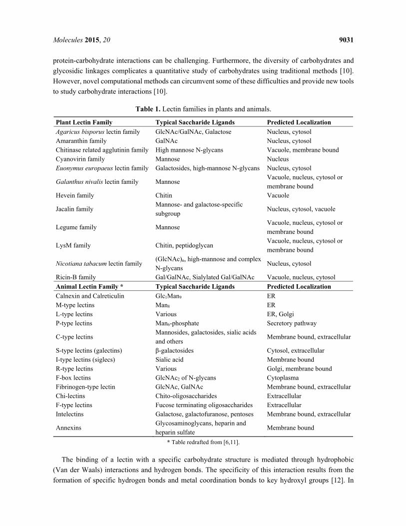
protein-carbohydrate interactions can be challenging. Furthermore, the diversity of carbohydrates and glycosidic linkages complicates a quantitative study of carbohydrates using traditional methods [10]. However, novel computational methods can circumvent some of these difficulties and provide new tools to study carbohydrate interactions [10].
Table 1. Lectin families in plants and animals.
Plant Lectin Family Typical Saccharide Ligands Predicted Localization Agaricus bisporus lectin family GlcNAc/GalNAc, Galactose Nucleus, cytosol Amaranthin family GalNAc Nucleus, cytosol Chitinase related agglutinin family High mannose N-glycans Vacuole, membrane bound Cyanovirin family Mannose Nucleus Euonymus europaeus lectin family Galactosides, high-mannose N-glycans Nucleus, cytosol
Galanthus nivalis lectin family Mannose Vacuole, nucleus, cytosol or membrane bound
Hevein family Chitin Vacuole
Jacalin family Mannose- and galactose-specific subgroup
Nucleus, cytosol, vacuole
Legume family Mannose Vacuole, nucleus, cytosol or membrane bound
LysM family Chitin, peptidoglycan Vacuole, nucleus, cytosol or membrane bound
Nicotiana tabacum lectin family (GlcNAc)n, high-mannose and complex N-glycans
Nucleus, cytosol
Ricin-B family Gal/GalNAc, Sialylated Gal/GalNAc Vacuole, nucleus, cytosol Animal Lectin Family * Typical Saccharide Ligands Predicted Localization Calnexin and Calreticulin Glc1Man9 ER M-type lectins Man8 ER L-type lectins Various ER, Golgi P-type lectins Man6-phosphate Secretory pathway
C-type lectins Mannosides, galactosides, sialic acids and others
Membrane bound, extracellular
S-type lectins (galectins) β-galactosides Cytosol, extracellular I-type lectins (siglecs) Sialic acid Membrane bound R-type lectins Various Golgi, membrane bound F-box lectins GlcNAc2 of N-glycans Cytoplasma Fibrinogen-type lectin GlcNAc, GalNAc Membrane bound, extracellularChi-lectins Chito-oligosaccharides Extracellular F-type lectins Fucose terminating oligosaccharides Extracellular Intelectins Galactose, galactofuranose, pentoses Membrane bound, extracellular
Annexins Glycosaminoglycans, heparin and heparin sulfate
Membrane bound
* Table redrafted from [6,11].
The binding of a lectin with a specific carbohydrate structure is mediated through hydrophobic (Van der Waals) interactions and hydrogen bonds. The specificity of this interaction results from the formation of specific hydrogen bonds and metal coordination bonds to key hydroxyl groups [12]. In
Molecules 2015, 20 9032
addition, unwanted recognition is sometimes excluded by steric exclusion. However, the sequence or the threedimensional conformation of the CRD is no indication for its specificity, since structurally unrelated lectins can recognize similar carbohydrate structures [12]. In addition some lectins with similar CRDs can recognize different carbohydrates. These phenomena can be attributed to the shallowness of the sugar-binding site and the limited number of contacts with the sugar that allows the CRD to recognize multiple carbohydrate structures, further referred to as ‘the promiscuity of the CRD’. Another interesting feature in carbohydrate-binding sites is that within a lectin family, there is a common mechanism for binding of a core monosaccharide in the primary binding site, but diversity in binding of oligosaccharides or glycoconjugates is achieved through extended and secondary binding sites unique to individual lectins [13]. Most likely, a number of lectin domains from animals and plants descended from a common ancestor through divergent evolution [13]. One lectin family that is represented both in mammalia and plants is the R-type lectin family (Ricin-B family in plants). The β-trefoil structure of the R-type lectin was first identified in ricin. In animals a structurally related domain was found in fibroblast growth factors and the cysteine-rich domain of the mannose receptor [14]. Although both ricin and the mannose receptor can bind glycan structures containing galactose ((sialylated) galactose/N-acetylgalactosamine and Lewisa/x structures respectively), their carbohydrate binding site differs significantly [14]. While the β-trefoil structure is conserved, the sequence of their CRDs differs, and amino acids involved in the binding of the carbohydrate structure are not conserved, accounting for the promiscuity of the CRDs.
Animal and plant lectins tend to play a role in a wide variety of biological processes, with some lectins having more than one function. Unfortunately for many lectins their physiological importance remains enigmatic. Several CRDs are involved in the recognition of invaders, and thus are part of the immune system, which consists of various types of cells and molecules that specifically interact with each other to initiate the host defense mechanism. To operate properly, the immune system must be able to detect a wide variety of pathogenic agents, and distinguish them from the organisms own healthy cells. Apart from their role in the immune system CRDs are important for a multitude of cellular processes like cell-cell interactions, self/non-self-recognition and intracellular routing. In addition, lectins also play an important role as molecular chaperones for glycoprotein quality control.
This review wants to offer an anthology of the multitude of functions carried out by lectins in the immune system of plants and animals. We will discuss the role of lectins in the recognition of pathogens in the innate immune system as pattern recognition receptors, their involvement in autophagy and their importance in stress signaling and other processes in the immune system. Furthermore we will also touch on their contribution in the vertebrate adaptive immune response and their potential involvement in epigenetic stress imprinting in plants. However, in view of the vast amount of lectins known today this review will only focus on a few examples to illustrate the functions of lectins in the immune system, and does not claim to present a complete overview of all the lectins involved.
2. Lectins as Pattern Recognition Receptors in the Innate Immune System
The first line of defense against infection by other organisms is the innate immune system. This non-specific immune system consists of cells and molecules that can recognize the pathogens and initiate a generic defense response. This system does not offer long-lasting protective immunity, as does the adaptive immune system, but activation of the innate system is required to initiate an adaptive response.
Molecules 2015, 20 9033
In the innate immune system, pathogenic microorganisms are recognized through highly conserved structures, pathogen associated molecular patterns (PAMPs). These structures are recognized by the pattern recognition receptors (PRRs) of the host [15]. Since many of the PAMPs recognized by the PRRs are carbohydrate structures, lectins play an important role as PRRs. These lectin PRRs are highly variable in structure and can occur in a soluble as well as in a membrane-associated form.
2.1. Animal Lectin PRRs
One of the best studied examples of an inducible lectin with a role as a PRR in the immune response is the mannose-binding protein (MBP), also known as mannose binding lectin (MBL) 2. This lectin is a family member of the collectins which represent a group of soluble lectins belonging to the Ca2+ dependent C-type lectin superfamily [16]. Collectins are organized as oligomers of a trimeric subunit, composed of a C-terminal lectin domain and an N-terminal collagen-like domain which enables the formation of the triplets [16]. Under normal conditions, MBL2 is synthesized at a basal level by the liver and secreted into the serum. However, after exposure to pathogenic microorganisms, the level of MBL2 mRNA transcripts and protein increases [17]. In response to infection, local inflammatory cells secrete cytokines into the bloodstream that stimulate the liver to produce large numbers of acute phase proteins, including MBL2. In the innate immune system, MBL2 functions as a PRR binding a range of carbohydrates including N-acetylglucosamine, mannose, N-acetylmannosamine, fucose and glucose, enabling the lectin to interact with surface glycans on a wide selection of viruses, bacteria, yeasts, fungi and protozoa (Figure 1).
When the C-terminal recognition portion of MBL2 binds to carbohydrates on the pathogen surface, the N-terminal domain can interact with collectin receptors on macrophages, which in turn leads to phagocytosis (Figure 1). In addition, MBL2 can activate the complement pathway through a unique pathway, i.e., the lectin pathway, independent from the classical pathway [18] (Figure 1). In the classical pathway of complement fixation, the binding of antibodies to pathogens leads to the recruitment of the first component of complement, C1q. In turn, C1q associates with two serine proteases, C1r and C1s, which initiates a proteolytic cascade. In the lectin pathway, MBL2 activates the same cascade through direct activation of serine proteases, MBP-associated serine proteases (MASPs), without the involvement of C1q. In essence, MBL2 is substituting for C1q [19]. The structural organization of collectins resembles that of C1q, being composed of an N-terminal collagenous domain and a globular C-terminal domain. However, C1q differs from MBL2 in that it contains a C-terminal immunoglobulin binding domain. However, at present MBL2 is the only collectin known to activate the complement pathway [16]. Activation of C2-C4 complexes through activation of the proteolytic cascade from either pathway leads to the cleavage of the C3 complement component. The resulting C3b fragment will insert into the surface of the target, initiating a lytic pathway in which additional complement components insert into the membrane to form a pore. In addition, C3b mediates phagocytosis of the pathogen through interaction with receptors on macrophages.
The pentraxins (belonging to the L-type lectin superfamily) represent another class of soluble lectins involved in the innate immune response, among these are C-reactive proteins (CRP) and serum amyloid P components (SAP) [20,21]. Similar to MBL2, they are acute phase proteins whose expression levels are increased upon stimulation by cytokines (IL-1 and IL-6). Their biological functions include
Molecules 2015, 20 9034
activation of the complement pathway and stimulation of phagocytic leukocytes [16]. Furthermore ficolins are complement activating soluble PRR able to sense molecular patterns on both pathogens and apoptotic cell surfaces [6].
The mannose receptor is a membrane bound PRR on the surface of macrophages [22]. This glycoprotein belongs to the C-type superfamily of lectins and possesses eight CRDs [23,24], which bind terminal mannose, fucose and N-acetylglucosamine (GlcNAc) residues [25]. Binding of the mannose receptor to components in the cell wall of pathogens leads to the internalization of the pathogen by macrophages (Figure 1). After phagocytosis by the macrophage, the phagosomes will fuse with lysosomes, killing the pathogen. However, Mycobacteria, the causal agent of tuberculosis and leprosy, make use of the mannose receptor to gain access into the cell. They prevent the fusion of the phagosome with lysosomes, creating a habitat to survive and proliferate within macrophages [26].
In addition to its role as a PRR in the innate immune response, several other functions have been attributed to this mannose receptor, including the attachment of sperm to oocytes [27], endocytosis for antigen presentation [22] and clearance of glycoproteins [28,29]. Interestingly, the expression of the mannose receptor is repressed during the early stages of inflammation and is induced during the resolution phase [30]. This expression profile is in accordance with a role in clearing inflammatory agents [30].
Recognition of yeasts through binding of the β-glucans on their surface by Dectin-1, a natural killer (NK)-cell-receptor-like C-type lectin, leads to the uptake and killing of these yeasts. In addition, Dectin-1 will induce a signaling cascade leading to the production of cytokines and chemokines (Figure 1). This signaling occurs cooperatively with Toll-like receptors (TLR) and the use of spleen tyrosine kinases (SYK) [31,32].
The nucleotide-binding oligomerization domain (NOD) proteins NOD1 and NOD2 provide the cell with another level of microbial surveillance. Being cytoplasmic PRRs they recognize conserved fragments in particular muramyl peptides in the cell wall of many types of bacteria [33]. These muramyl peptides are derived from the bacterial peptidoglycan. The mechanisms by which the muramyl fragments enter the cell and activate the NOD-like receptors NOD1 and NOD2 remain poorly understood but multiple routes of entry have been reported [33]. Upon sensing distinct peptidoglycan fragments, NOD1 and NOD2 activate intracellular signaling pathways that drive pro-inflammatory and antimicrobial responses [33]. In addition, polymorphisms in NOD2 have been identified as the strongest genetic risk factor in the development of Crohn’s disease [33].
Molecules 2015, 20 9035
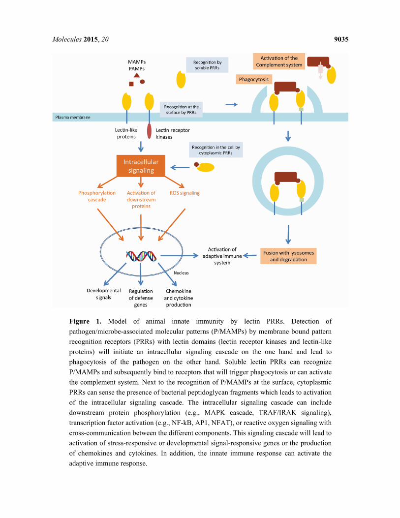
Figure 1. Model of animal innate immunity by lectin PRRs. Detection of pathogen/microbe-associated molecular patterns (P/MAMPs) by membrane bound pattern recognition receptors (PRRs) with lectin domains (lectin receptor kinases and lectin-like proteins) will initiate an intracellular signaling cascade on the one hand and lead to phagocytosis of the pathogen on the other hand. Soluble lectin PRRs can recognize P/MAMPs and subsequently bind to receptors that will trigger phagocytosis or can activate the complement system. Next to the recognition of P/MAMPs at the surface, cytoplasmic PRRs can sense the presence of bacterial peptidoglycan fragments which leads to activation of the intracellular signaling cascade. The intracellular signaling cascade can include downstream protein phosphorylation (e.g., MAPK cascade, TRAF/IRAK signaling), transcription factor activation (e.g., NF-kB, AP1, NFAT), or reactive oxygen signaling with cross-communication between the different components. This signaling cascade will lead to activation of stress-responsive or developmental signal-responsive genes or the production of chemokines and cytokines. In addition, the innate immune response can activate the adaptive immune response.
Molecules 2015, 20 9036
2.2. Plant Lectin PRRs
Despite obvious differences in the immune system of plants and animals, there are also some striking
similarities [34]. While the adaptive immunity is unique to vertebrates, the innate immune response most
probably has ancient origins [35]. In contrast to animals where specialized cell types (macrophages,
neutrophils and dendritic cells) in the blood circulatory system play a key role in the detection of
pathogens and the activation of the immune system, most plant cells are autonomously capable of
sensing the presence of pathogens and activating a defense response [36]. Similar to animals, plants have
evolved systems for non-self-recognition and anti-microbial defense [36]. Like animals, plants have
acquired specialized PRRs for their defense against pathogens [37]. These PRRs are able to recognize
the damage-associated molecular patterns (DAMPs) and the pathogen- or microbe-associated molecular
patterns (PAMPs/MAMPs). As in animals, many of these PRRs carry lectin domains able to recognize
and interact with carbohydrate structures from microbial organisms or saccharides derived from plant
cell wall damage. Upon PAMP/MAMP and DAMP perception by the PRRs, an intracellular response is
activated, referred to as the PAMP/MAMP-triggered immunity (PTI/MTI) (Figure 2). Besides the
components to sense the pathogens, also the building blocks of PAMP-induced signaling cascades
leading to transcriptional activation of response genes are shared between the two kingdoms. In particular,
nitric oxide as well as mitogen-activated protein kinase (MAPK) cascades have been implicated in
triggering innate immune responses, which ultimately lead to the production of anti-microbial
compounds [36]. In addition, this response includes ion fluxes across the plasma membrane, increase of
cytosolic Ca2+
levels, production of reactive oxygen species and protein phosphorylation. This complex
response of the plant finally leads to profound transcriptional changes, stomatal closure and cell wall
reinforcement [38], and will ultimately limit the growth of the pathogen [39].
The best studied example of the perception of PAMPs by plant PRRs is the recognition of bacterial
flagellin, through the conserved flg22 epitope, by the Arabidopsis FLS2 receptor-like kinase [38]. For
more detailed information on FLS2 signaling and some other examples involving recognition through
protein-protein interactions we refer to some recent review papers focused on this topic [40,41]. A
significant part of the patterns recognized by the plant PRRs are carbohydrate structures, which are either
present on the surface of the pathogen (e.g., lipopolysaccharides, peptidoglycans and chitin molecules)
or are derived from plant own molecules (i.e., DAMPs), released during invasion of the pathogen (e.g.,
cellulose fragments, arabinogalactan proteins and oligogalacturonides).
For the recognition of the D/M/PAMPs, a large diversity of membrane-bound or soluble PRRs with
a lectin domain have been identified in plants. However, only a limited number of them have been
functionally characterized. The membrane-bound PRRs carrying one or more extracellular lectin
domains are often coupled to an intracellular Ser/Thr kinase domain (Figure 2). These lectin receptor
kinases (LecRK) can be classified into four types: G-, C-, L- and LysM-type [42,43].
Due to their sessile lifestyle, plants are not only subjected to biotic stresses but also to multitude of
abiotic stress factors (e.g., cold, excess water, increased salt concentrations). To be able to respond to
these detrimental environmental conditions, plants have evolved defense pathways able to sense and
respond to these abiotic stresses. Several of the LecRKs have been reported to act both upon biotic and
abiotic stresses [44–49].
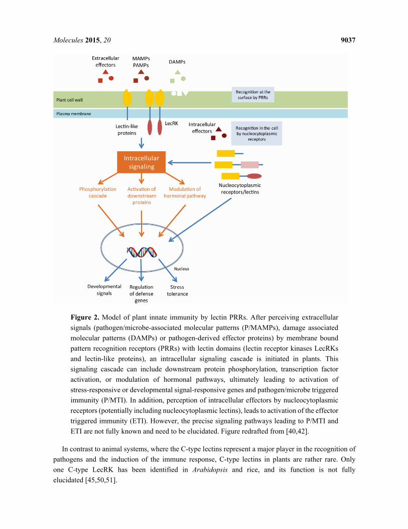
Molecules 2015, 20 9037
Figure 2. Model of plant innate immunity by lectin PRRs. After perceiving extracellular signals (pathogen/microbe-associated molecular patterns (P/MAMPs), damage associated molecular patterns (DAMPs) or pathogen-derived effector proteins) by membrane bound pattern recognition receptors (PRRs) with lectin domains (lectin receptor kinases LecRKs and lectin-like proteins), an intracellular signaling cascade is initiated in plants. This signaling cascade can include downstream protein phosphorylation, transcription factor activation, or modulation of hormonal pathways, ultimately leading to activation of stress-responsive or developmental signal-responsive genes and pathogen/microbe triggered immunity (P/MTI). In addition, perception of intracellular effectors by nucleocytoplasmic receptors (potentially including nucleocytoplasmic lectins), leads to activation of the effector triggered immunity (ETI). However, the precise signaling pathways leading to P/MTI and ETI are not fully known and need to be elucidated. Figure redrafted from [40,42].
In contrast to animal systems, where the C-type lectins represent a major player in the recognition of pathogens and the induction of the immune response, C-type lectins in plants are rather rare. Only one C-type LecRK has been identified in Arabidopsis and rice, and its function is not fully elucidated [45,50,51].

Molecules 2015, 20 9038
The G-type LecRKs carry a lectin domain belonging to the Galanthus nivalis agglutinin (GNA) family of plant lectins. Although 32 G-type LecRKs have been identified in Arabidopsis and 100 in rice [51], it still needs to be confirmed whether the lectin domain plays a role in the interaction with the pathogen. G-type LecRKs have been shown to function in self-incompatibility reactions and defense to both biotic and abiotic stresses [44,46,49].
The 42 L-type LecRKs identified in the genome of Arabidopsis thaliana (72 in rice) are grouped into nine clades on a dendrogram. These kinase receptors with a Legume-like (L-type) lectin domain show variable expression patterns in different tissues and developmental stages in response to stimuli [45]. The amino acid sequence of the legume-like lectin domain is only poorly conserved in the L-type LecRKs, therefore it is doubtful whether it possesses true lectin activity. Despite the fact that it remains to be proven that L-type LecRKs possess lectin activity, several of these proteins were reported to be involved in plant resistance to pathogens [47,52–54]. In addition, some L-type LecRKs have been described to act in hormone signaling and stomatal immunity [47]. Although the overall legume-like lectin domain is poorly conserved, a hydrophobic site within this lectin domain is highly preserved [53]. This site can play a role in the detection of extracellular ATP by L-type LecRKs. Recently, extracellular ATP was shown to play a role as a signaling molecule in plant stress responses [55,56].
The best studied LecRKs in plants belong to the LysM-type LecRKs. These receptors recognize the GlcNAc moieties in various types of bacterial peptidoglycans and fungal chitins [57,58]. Next to their role in recognizing pathogenic microorganisms, the LysM LecRKs are also involved in the recognition of beneficial microorganisms such as mycorrhiza and rhizobacteria [58–60].
2.3. Plant Effector Triggered Immunity
In addition to the PTI/MTI responses, plants have evolved another defense mechanism, referred to as the effector triggered immunity (ETI) (Figure 2). In contrast with the former responses, ETI will mainly act inside the plant cell. Successful pathogens have evolved mechanisms to counter the PTI response of the plant by the delivery of specific elicitors or effectors, also called avirulence (Avr) proteins [61]. These effectors are usually directly secreted into the plant cell by a type III secretion system and suppress or block the PRR-dependent signaling [62]. These effectors are recognized by a class of intracellular plant receptors containing a nucleotide-binding and leucine-rich repeat domain (NB-LRR), thereby activating the ETI response. In the ETI response, plants express specific resistance genes upon recognition of these pathogenic effectors to overcome the action of these effectors. Both the PTI/MTI and the ETI response can lead to programmed cell death of the host cell through activation of the hypersensitive response (HR), but can also result in systemic acquired resistance (SAR) which activates defense mechanisms throughout the plant [38,63].
3. Autophagy
Recognition of pathogens by PRRs leads to the induction of the immune responses and will also result in phagocytosis of the pathogen. These phagocytic vesicles will subsequently fuse with lysosomes leading to the degradation of the pathogen. However, some bacteria are capable of escaping this process by releasing effectors into the cytosol. Mycobacterium tuberculosis secretes an effector (EsxH) that prevents the maturation of the phagosome and fusion with lysosomes, thereby escaping degradation and
Molecules 2015, 20 9039
creating a niche to proliferate [64]. In the case of the bacterium Salmonella enterica serovar Thyphimurium, the major cause of food poisoning, the pathogen can invade and grow in the gut epithelial cells after digestion by its host. S. thyphimurium is recognized by the Toll-like receptor 4 on the gut epithelial cells, which binds to the lipopolysaccharides on the outer membrane of the Gram-negative bacterium [65]. Salmonella uses this interaction and its subsequent phagocytosis to invade the host cell. Early after cell invasion, the bacterium resides in a vesicle known as the Salmonella containing vacuoles (SCV). The bacteria then use a type III secretion system to generate pores in the SCV membrane, through which they can deliver effectors into the cell’s cytoplasm [66]. These effectors modulate the activity of the host cell machinery to prevent degradation and promote growth of the intracellular pathogen. Moreover, some bacteria can escape the damaged SCVs and replicate in the cytosol. However, cells have developed a defense mechanism against this kind of intracellular pathogens. Microbes in damaged vesicles as well as microbes residing in the cytosol are targeted by the autophagy system that will engulf them in autophagosomes and restrict bacterial growth [67,68]. Autophagy is a strictly regulated and mainly non-selective process used by the cell to capture cytoplasmic cargoes in a double membrane vesicle, the autophagosome, to maintain a balance between synthesis and degradation of their own components. The uptake of the intracellular cargo in these vesicles is mediated through evagination of the rough endoplasmatic reticulum associated membrane [69]. When this vesicle fuses with lysosomes its contents become degraded and nutrients and building blocks get recycled. Malfunctioning of the autophagy system has been linked to human diseases such as cancer, neurodegenerative disorders, diabetes and inflammatory bowel disease [66]. In addition to its function in the housekeeping of the cell, autophagy can also help to defend the mammalian cytosol against bacterial infection [70]. Efficient pathogen engulfment is mediated by cargo-selecting autophagy adaptors that rely on pattern recognition or danger receptors to label the invading pathogen as autophagy cargo [71]. This labeling is typically mediated through polyubiquitin coating [72]. Recently the carbohydrate-dependent galectin-8 pathway was discovered, representing a novel mechanism that allows to get rid of bacteria that have been taken up by the cell [71]. Galectins are β-galactoside binding lectins that accumulate in the cytosol before being secreted via a leader peptide-independent pathway [71]. When these galectins occur extracellularly, they bind to cell surface glycans in order to modulate cellular behavior. Inside the cytosol, where complex carbohydrates are absent under normal physiological conditions, galectins play a role as danger and/or pattern recognition receptors. Thurston et al. [71] reported that galectin-8 is recruited to damaged SCVs and the absence of galectin-8 leads to an increase in the growth rate of the intracellular bacteria. However, in contrast to the direct tagging of Shingella flexneri by autophagy related gene 5 (ATG5) [73], it was observed that galectin-8 does not bind to the bacterial glycans but to the host cell’s own complex glycans that become accessible in the damaged vesicles. Supporting this observation, galectin-8 was also found to decorate vesicles with other vesicle damaging bacteria such as Listeria monocytogenes and Shigella flexneri. Moreover, osmotic damage of cytoplasmic vesicles also leads to the recruitment of galectin-8 [71]. Galectin-8 interacts with the autophagy receptor NDP52, which subsequently recruits LC3 and other components of the autophagy machinery to the damaged vesicles. Although, other galectins (galectin-3 and -9) are also recruited to the damaged vesicles, their role in cellular defense to infection remains enigmatic [71].
Similar to animals and yeast, essential autophagic machineries are conserved in plants. The role of autophagy related genes (ATGs) in the lifecycle of plants is similar to that in animals. In Arabidopsis,
Molecules 2015, 20 9040
autophagy is activated in certain stages of development, senescence or in response to starvation, or after environmental stress [74]. In addition, autophagy in plants is suggested to be required for proper regulation of the hypersensitive response programmed cell death [75,76]. However the question remains whether autophagy is involved in the elimination of bacterial pathogens in plants and if any of the cytoplasmic lectins contribute to this process. Compared to animals, not many examples of intracellular microorganisms are known in plant cells since due to their rigid cell wall, plant cells are usually not phagocytic [77]. One well studied example in plants is the endosymbiosis of Rhizobium, where the engulfment of the microorganism by a plant-derived membrane occurs by a phagocytic mechanism [78]. These endosymbionts counteract their degradation [79] but it is unknown if they trigger an autophagosomic response. One indication that cytoplasmic plant lectins can be involved in autophagy comes from the observation of an interaction between the Arabidopsis jacalin-related lectins and proteins of the ER-bodies [80]. These novel ER-derived organelles have been shown to play a role in plant defense against pathogens [81] and are linked to autophagocytosis [82].
Next to a role in the defense against intracellular bacteria, autophagy has also been reported to play a role in the elimination of intracellular and extracellular viruses. Overexpression of beclin 1, a protein required for autophagosome formation and recruitment of autophagosome components, in neurons reduced Sindbis virus replication [83], while in cells deprived of beclin 1, Sindbis virus and Herpes simplex virus replicate to higher titers than in wild-type cells [84,85]. Similarly, the plant tobacco mosaic virus accumulated to a higher level in Beclin 1-silenced plants than in wild-type plants [75]. These results indicate that autophagy functions as an antiviral defense mechanism in which the ATG proteins might target cellular factors or pathways required for virus replication and spread [86].
4. Non-PRR Function of Lectins in the Immune Response
Many of the animal PRRs that detect microbial infection induce the innate immune response by triggering an intracellular signaling cascade which stimulates expression of chemokines, cytokines and other immune mediators. The role of protein-carbohydrate interactions is not limited to the interaction between the pathogen and the host cell, but they can also play an important part in the signaling pathway induced upon pathogen recognition or in other processes of the immune response.
Inflammation is one of the responses of the innate immune system upon detection of stress. Induction of stress signaling in macrophages upon recognition of microorganisms leads to the secretion of chemokines and cytokines which initiate inflammation. A key function of inflammation is the recruitment of leukocytes through vascular tissue, a process in which C-type lectins play a key role. Selectins are present on the surface of leukocytes (L-selectins), endothelial cells (E- and P-selectins) and platelets (P-selectins) and bind to sialyl Lewisx structures (can also bind other carbohydrates such as heparin) [87]. The interaction between these selectins and their ligand will result in a process called leukocyte rolling, which will slow down the movement of the leukocytes in the bloodstream at the sites of inflammation and increases the contact between the leukocyte and the epithelial cells. This allows the tight interaction between Integrin (MAC-1) and its ligand (IMAC-1). In addition, MAC-1 is also able to bind to carbohydrates such as zymosan and lipopolysaccharides [16].
Sialic acid binding Ig-like lectins or Siglecs are a good example of lectins involved in regulating cellular activation within the immune system. Sialoadhesin (siglec-1), a macrophage lectin-like adhesion
Molecules 2015, 20 9041
molecule, has been implicated in key interactions with T-lymphocytes, leading to generation of cytotoxic T-cells [88]. Since siglec-1 lacks the cytoplasmic tyrosine-based motif present in other siglecs, it is
suggested to mediate extracellular rather than intracellular functions [89]. The B-cell restricted Siglec-2 (CD22) plays an important role in regulating B-cell activity [89]. The balance between positive and negative signals is an important factor in the immune response [90]. Upon cellular activation by
receptors, counteracting inhibitory signals can be delivered through distinct mechanisms, resulting in a higher activation threshold [89]. CD22 is described as an inhibitory receptor. Although the precise role of the sialic acid binding activity of CD22 remains to be elucidated, mice lacking CD22 show a higher
prevalence of autoantibodies with increasing age [91]. Similar to animals, several families of plant lectins have been identified with a putative role in plant
defense against biotic or abiotic stress other than the recognition of stress agents. Under non-stress
conditions, the expression levels for these lectins are low, often under the detection limit. But after perception of biotic and/or abiotic stress signals, their transcript levels are upregulated. Although the precise role of these lectins in plant defense is not fully understood, several of them have been reported
to increase plant resistance to biotic or abiotic agents. Lectins with an Euonymus lectin-like (EUL) domain are ubiquitous in plants, indicating a universal
role for these lectins [92]. Despite their widespread distribution, the EUL proteins were only recently
described as a new lectin family. Although all characterized lectins from this family show a very similar localization in the nucleocytoplasmic compartment of the cell, their ligand specificity varies depending on the protein and species studied [93]. Most EULs are chimeric proteins in which the lectin domain is
linked to an unknown domain. In contrast to the Euonymus europaeus agglutinin, the prototype of this family, which is expressed at high concentrations in the arils surrounding the seeds, the EUL proteins from Arabidopsis and rice are low abundant proteins, the expression of which is induced after the plant
was subjected to various biotic (bacterial and fungal infection) and abiotic (dehydration, salinity, osmotic stress and abscisic acid (ABA) treatment) stresses. Arabidopsis EULS3 was shown to interact with the nucleocytoplasmic ABA receptor RCAR1 [94] and a Ca2+ dependent kinase involved in ABA response
in stomata guard cells [95], suggesting a role for ArathEULS3 in ABA signaling and stomatal closure. Jacalin-related lectins can be divided into two subgroups based on their substrate specificity: the
galactose-binding and the mannose-binding jacalins [3]. Structural analysis demonstrated that the
relatively large size of the carbohydrate binding site, which is extended in the galactose-binding jacalins compared to the mannose-binding jacalins, allows this promiscuity of the CRD [96]. There are some striking differences between the two subgroups: First, polypeptides of the mannose-binding jacalins
remain intact whereas those of the galactose-binding subgroup are cleaved into two dissimilar polypeptides [3]. Second, galactose-binding jacalins are synthesized on ER bound ribosomes as a proprotein whereas the mannose-binding lectins are synthesized without signal peptide [3], resulting in
a different subcellular localization for both subgroups of jacalin-related lectins. Whereas the galactose-binding jacalins mainly reside in the vacuole, the mannose-binding lectins are located in the nucleocytoplasmic compartment of the cell [97]. Several jacalin-related lectins have been associated
with disease resistance, abiotic stress signaling, wounding and insect damage [98]. The jacalin domain is often present as part of chimeric proteins, where the lectin domain is fused to an unrelated domain. Interestingly, several of these chimeric proteins contain domains related to stress response and defense.
In Poaceae species (wheat, rice and maize), mannose-binding jacalin-related proteins in which the

Molecules 2015, 20 9042
C-terminal lectin domain is fused to an N-terminal dirigent or disease-response domain have been identified. In wheat (Triticum aestivum), nearly half of the jacalin-related lectins are chimeric proteins containing this dirigent motif, and those already studied in more detail were all reported to have increased expression levels after application of some stress factor [98]. Rice (Oryza sativa) expresses a protein composed of a C-terminal jacalin domain fused to a leucine rich repeat (LRR) and an N-terminal NB-ARC domain. The NB-ARC domain is a novel signaling motif found in bacteria and eukaryotes, and is shared between plant resistance gene products and regulators of cell death in animals. Furthermore, also proteins consisting only of one or more jacalin domain(s) can be upregulated after stress perception. For example, Orysata is a salt inducible mannose-binding lectin from rice shoots. In addition, Orysata is also induced upon hormone treatment (jasmonic acid (JA) and ABA), infection with Magnaporthe grisea and during senescence [99–101]. The expression of the wheat TaJRLL1 lectin, a component of the defense signaling, is induced after fungal infection in a hormone (salicylic acid (SA) and JA) dependent manner [102]. In contrast to the plant hormone dependent regulation, jacalin-related lectins can also be regulated independently from hormones. The Arabidopsis JAX1 offers resistance to potex virus by inhibiting accumulation of viral RNA in a hormone independent manner [103]. In addition to a signaling role, jacalin-related lectins can have an effector function. The Helja lectin from sunflower (Helianthus tuberosus) was shown to possess antifungal properties. Furthermore, it induces morphological changes and reactive oxygen species production in yeast as well as alteration of membrane permeability [104].
Recently, proteins containing the jacalin-related lectin domain were also identified outside the plant kingdom. The human lectin ZG16p is a soluble mammalian lectin with a jacalin-related β-prism fold. ZG16p has been reported to bind both to glycosaminoglycans and α-mannose terminating short glycans [105]. Recognition of a broad spectrum of ligands by ZG16p may account for the multiple functions of this lectin in the formation of zymogen granules via glycosaminoglycan binding, and in the recognition of pathogens in the digestive system through α-mannose-related recognition [105]. Furthermore, the jacalin-related domain has also been reported in a few prokaryotes [3].
The expression of the nucleocytoplasmic tobacco lectin, referred to as the Nicotiana tabacum agglutinin or Nictaba, is enhanced after treatment with JA, a plant hormone important for plant development and involved in signaling of abiotic and biotic stress, such as insect herbivory [40]. Nictaba specifically recognizes GlcNAc oligomers but also reacts with the core structure of N-glycans. The Nictaba domain is widespread in the plant kingdom and several Nictaba-related lectins were shown to have a role in plant defense [40]. For instance, Nictaba was shown to possess entomotoxic activity on Lepidopteran insects and can protect plants against insect herbivory [40]. The phloem proteins (PP) represent a subgroup of the Nictaba-related lectins isolated from Cucurbitaceae. In contrast to Nictaba, the PP2 lectins are constitutively expressed in the companion cells of the phloem and are subsequently translocated to the phloem sap. Expression of the Arabidopsis homolog PP2-A1 is enhanced by ethylene treatment and Pseudomonas infection [40]. In a computational study [106] Arabidopsis PP2-A1 was shown to exhibit specificity for a diverse range of glycans including chitin oligomers and the Man3GlcNAc2 core of high mannose N-glycans [106,107]. Consistent with a role in plant defense, PP2-A1 represses feeding of the aphid Myzus persicae and aphid borne yellow virus transmission [40].
The ricin-B family of lectins contains the first lectin discovered in plants, in particular ricin. Ricin, isolated from the seeds of castor bean (Ricinus communis), is a chimeric protein composed of a lectin
Molecules 2015, 20 9043
domain and a domain with RNA N-glycosidase activity, enabling the protein to cleave an adenine residue from the large rRNA. This enzymatic activity results in the inactivation of ribosomes and the arrest of protein translation. As a result ricin is classified in the group of ribosome inactivating proteins. The uptake of these proteins by the host cell is mediated through the ricin-B lectin domain which binds to specific glycan structures on the cell surface. The ricin-B related proteins accumulate in the vacuole or are secreted to the extracellular space [3]. Similar to the jacalin-related proteins, proteins containing a ricin-B domain are widespread and have also been identified outside the plant kingdom in animals, fungi and bacteria. These lectins are classified as R-type lectins [108]. Although the carbohydrate specificity of the ricin-B lectins can vary [109], evidence supports that many of the ricin-B related lectins play a role in defense against pathogens and insects [40].
5. Adaptive Immunity and Epigenetic Imprinting
5.1. Adaptive Immunity
In vertebrates an adaptive immune response is induced following the innate immune response. When naive T-cells interact with their specific antigen presented on the surface of antigen-presenting cells, they become activated and differentiate into effector T-cells. A subset of these effector T-cells, the T-helper cells, will subsequently stimulate the proliferation and differentiation of B-cells.
The adaptive immune response depends on the innate immune response. It was observed that several PRRs, involved in the innate immunity, also play a role in triggering the adaptive response. Toll-like receptors, nucleotide-oligomerization domain (Nod)-like receptors, retinoic acid-inducible gene-1 (RIG-1)-like receptors and some C-type lectin receptors can trigger both the innate and adaptive immune responses [110]. Whether these receptors contribute to the innate or the adaptive response is dependent of the cell type where they are expressed [111]. Moreover, lectins can have similar functions in the adaptive response and in the innate response. They play a role in cell-cell interactions and can contribute to the activation and differentiation of cells (e.g., Siglecs).
5.2. Epigenetic Imprinting
Plants, unlike animals, lack the adaptive immune mechanisms and rely solely on their multilayered innate immune system to prevent pathogen infection. However, plants are not completely defenseless against reoccurring stresses.
When higher animals are exposed to a specific pathogen, the adaptive immune system creates an immunological memory after the initial response. This leads to an enhanced response to subsequent encounters with that pathogen. Although there is no adaptive immune system in plants, plants must respond and adapt to recurring biotic and abiotic stress factors especially since their sessile lifestyle does not allow to escape from these stresses. It is a well-known phenomenon that stress tolerance in plants can be improved through pre-exposure to an abiotic or biotic stress stimulus. Primed (against biotic stress) or hardened (against abiotic stress) plants display a faster and/or stronger activation to the various defense responses that are induced following infection by pathogens, attack by insects or in response to abiotic stress [112]. This priming can be elicited by either exposure to the stress clues themselves or by the exogenous application of chemical treatments [113]. The latter process can be compared to
Molecules 2015, 20 9044
vaccination in vertebrates. The advantage of priming is that it offers the plant an enhanced protection without the costs of constitutively expressing their defense genes. However, the molecular mechanism of this stress-imprinting on plant immunity is not fully elucidated. Though several mechanisms have been proposed, Conrath et al. [112] suggested that two potential mechanisms can be involved. The first one involves the accumulation of signaling proteins in an inactive configuration that are activated upon stress, potentially through MAPK signaling [112,114]. The second mechanism involves the accumulation of transcription factors that enhance defense gene transcription after stress recognition [112]. Jung et al. [115] proposed a system in which the accumulation of secondary metabolites such as SA and azelaic acid was involved. Another mechanism was described by Bruce et al. [113] in which priming occurs through epigenetic changes. Epigenetics refers to the heritable changes in gene expression that do not involve changes to the DNA sequence. These epigenetic changes consist of DNA methylations, histone modifications and RNA interference that lead to changes in gene expression by activation or silencing. The links and interactions between the different mechanisms for priming are largely unknown but due to the long-lasting stress resistance, it is most likely that epigenetic mechanisms are largely in control [116].
Epigenetic changes in response to cold stress have been reported during vernalisation of Arabidopsis. The long-term exposure to low temperatures in winter is memorized and needed for the plant to be able to flower in the following spring [117]. This ‘memory’ involves a change in the chromatin structure of the flowering locus through modification of histones by several vernalisation (VRN) genes. Recent data suggest that also some of the stress inducible plant lectins play a role in epigenetic priming during stress response. One of the proteins involved in vernalisation in wheat is the VER2 cold inducible jacalin-related protein. Interaction with the O-GlcNAc modified TaGRP2 RNA-binding protein allows accumulation of the VRN1 transcription factor through stabilization of its mRNA [118]. Treatment of tobacco with JA, a plant hormone involved in signaling of biotic and abiotic stress and in plant development, leads to the enhanced expression of the Nictaba protein in the nucleus and the cytosol [119,120]. In addition, this lectin is also induced upon insect herbivory [120]. The accumulation of Nictaba is not limited to the treated leaf but a systemic response is observed in all leaves of the plant [121]. Screening for interaction partners revealed that the core histones H2A, H2B and H4 are the major binding partners for Nictaba in the nucleus [122]. This interaction is established through the specific binding of the lectin with O-GlcNAc modified histone proteins [123]. It was hypothesized that the interaction between Nictaba and histones bound to DNA might result in chromatin remodeling, and subsequent alteration of gene expression as a response to stress or changing environmental conditions [124].
6. Conclusions
Although plants and animals shared their last common ancestor until at least one billion years ago, throughout the eons they have been facing common threats such as bacteria, fungi and viruses trying to invade them. To protect themselves against these threats, both plants and animals evolved a defense system, the immune system. While for a long time it was presumed that these defense systems in plants and animals were very different, it has become clear that both plants and animals respond to infection by pathogens using similar regulatory modules. Common features include receptors that detect molecular
Molecules 2015, 20 9045
signatures of infectious organisms (PAMPs/MAMPs) and of tissue damage (DAMP), conserved signal transduction pathways (e.g., MAPK pathway) and the production of antimicrobial molecules.
Lectins or carbohydrate-binding proteins play an important role in the immune system of animals and plants as PRRs. Carbohydrate structures on the surface of pathogens or released from host cells due to damage provoked by the pathogen are recognized by soluble or membrane-bound lectins that trigger a signaling cascade resulting in the induction of the defense mechanisms, the pathogen/microbial triggered immunity (P/MTI). Although in animals several lectins were found to exert a function as PRR, in plants only the LysM-domain lectins have been shown unambiguously to be dependent on carbohydrate binding for their PRR function. Other identified LecRKs or lectin receptor proteins may depend on protein-protein interactions for the recognition of their target.
In addition to the lectins with a role as PRR, lectins can play a role in stress signaling or as an effector or mediator in the stress response. In plants, several lectin families have been described that are transcriptionally upregulated upon biotic or abiotic stresses. These stress-inducible lectins mainly reside in the cytoplasm and the nucleus, and are suggested to play a role in signal transduction in several stress response pathways.
Successful pathogens manage to overcome the innate immune response through secretion of toxins or effectors into the host cell. Plants have therefore developed another defense layer to detect these effectors via different immune receptors (resistance or R proteins) and initiate effector triggered immunity (ETI). Another method for successful pathogens to overcome the immune response is to escape degradation by the host cell. Recognition of pathogens by PRRs often leads to the phagocytosis of these pathogens by the host cell. These phagocytic vesicles are subsequently fused with lysosomes, resulting in the degradation of the pathogen. This degradation is required in vertebrate systems to activate the adaptive immunity by antigen presenting cells. However, some pathogens are capable of escaping this degradation by the delivery of effector proteins that prevent maturation of the phagocytic vacuoles. These pathogens create a niche inside these (damaged) vesicles or manage to escape these vesicles and proliferate intracellularly. Plant and animal cells have developed a mechanism to defend themselves against these intracellular parasites through autophagy. Lectins are shown to be involved in the recognition of damaged phagocytic vacuoles in animals. Since in plants the essential machineries of autophagy are conserved, lectins are suggested to play a role in this process, but at present their precise function is not fully elucidated.
Although plants lack an adaptive immune system, they are able to create long lasting stress resilience against reoccurring stresses. Epigenetic changes can facilitate quicker and more potent responses to subsequent attacks. One of the stress inducible plant lectins from tobacco, referred to as Nictaba, was shown to interact with O-GlcNAc modified histones and can potentially act as a chromatin remodeler allowing an alteration of gene expression as a result of biotic or abiotic stresses.
Although plants and animals differ significantly, their defense system relies for an important part on comparable mechanisms. This is supported by recent data which demonstrated that lectin domains that descended from common ancestors through divergent evolution appear widely across the kingdoms of life (including prokaryotes, fungi, plants and animals) [13]. However these conserved lectin domains show promiscuity in their sugar binding specificity. These variations might be linked to a directed evolution to recognize carbohydrate structures on the pathogenic agents threatening the plant/animal cell.
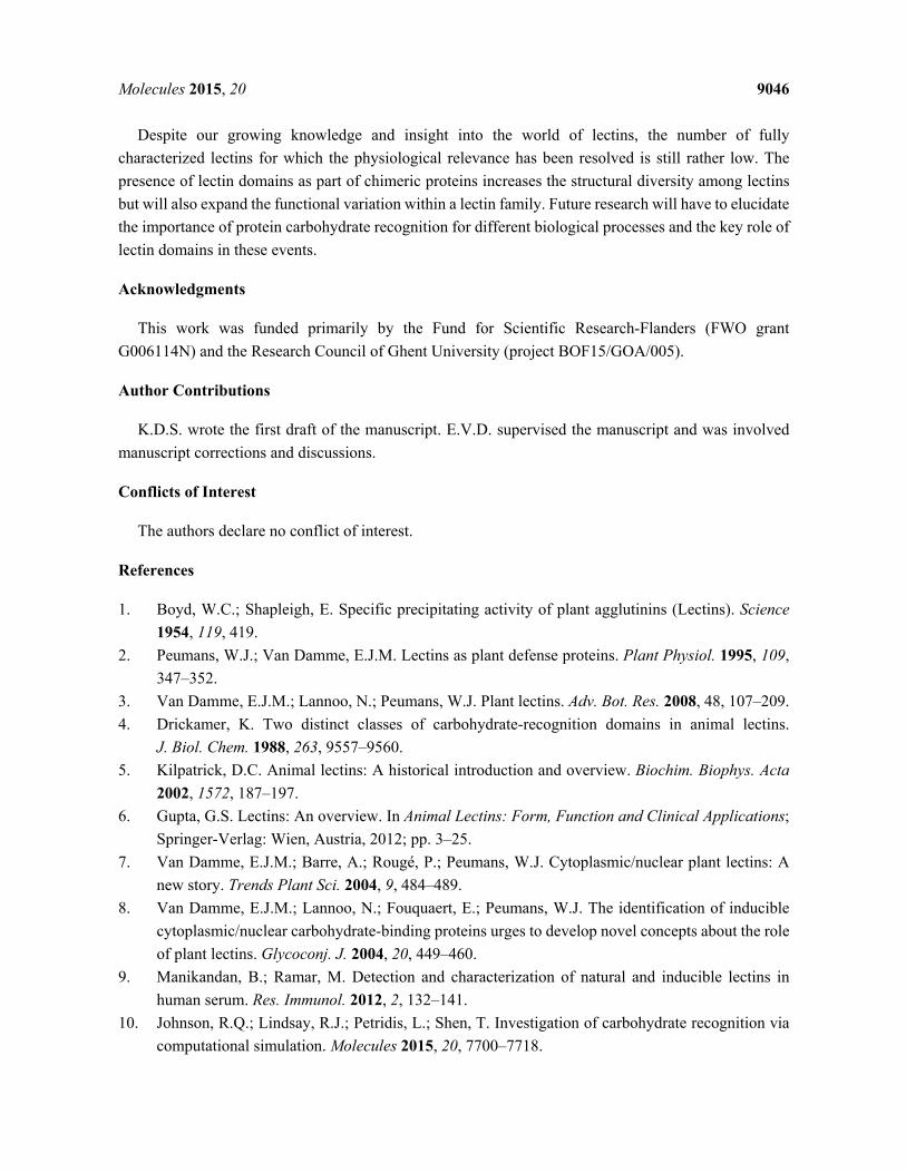
Molecules 2015, 20 9046
Despite our growing knowledge and insight into the world of lectins, the number of fully characterized lectins for which the physiological relevance has been resolved is still rather low. The presence of lectin domains as part of chimeric proteins increases the structural diversity among lectins but will also expand the functional variation within a lectin family. Future research will have to elucidate the importance of protein carbohydrate recognition for different biological processes and the key role of lectin domains in these events.
Acknowledgments
This work was funded primarily by the Fund for Scientific Research-Flanders (FWO grant G006114N) and the Research Council of Ghent University (project BOF15/GOA/005).
Author Contributions
K.D.S. wrote the first draft of the manuscript. E.V.D. supervised the manuscript and was involved manuscript corrections and discussions.
Conflicts of Interest
The authors declare no conflict of interest.
References
1. Boyd, W.C.; Shapleigh, E. Specific precipitating activity of plant agglutinins (Lectins). Science 1954, 119, 419.
2. Peumans, W.J.; Van Damme, E.J.M. Lectins as plant defense proteins. Plant Physiol. 1995, 109, 347–352.
3. Van Damme, E.J.M.; Lannoo, N.; Peumans, W.J. Plant lectins. Adv. Bot. Res. 2008, 48, 107–209. 4. Drickamer, K. Two distinct classes of carbohydrate-recognition domains in animal lectins.
J. Biol. Chem. 1988, 263, 9557–9560. 5. Kilpatrick, D.C. Animal lectins: A historical introduction and overview. Biochim. Biophys. Acta
2002, 1572, 187–197. 6. Gupta, G.S. Lectins: An overview. In Animal Lectins: Form, Function and Clinical Applications;
Springer-Verlag: Wien, Austria, 2012; pp. 3–25. 7. Van Damme, E.J.M.; Barre, A.; Rougé, P.; Peumans, W.J. Cytoplasmic/nuclear plant lectins: A
new story. Trends Plant Sci. 2004, 9, 484–489. 8. Van Damme, E.J.M.; Lannoo, N.; Fouquaert, E.; Peumans, W.J. The identification of inducible
cytoplasmic/nuclear carbohydrate-binding proteins urges to develop novel concepts about the role of plant lectins. Glycoconj. J. 2004, 20, 449–460.
9. Manikandan, B.; Ramar, M. Detection and characterization of natural and inducible lectins in human serum. Res. Immunol. 2012, 2, 132–141.
10. Johnson, R.Q.; Lindsay, R.J.; Petridis, L.; Shen, T. Investigation of carbohydrate recognition via computational simulation. Molecules 2015, 20, 7700–7718.
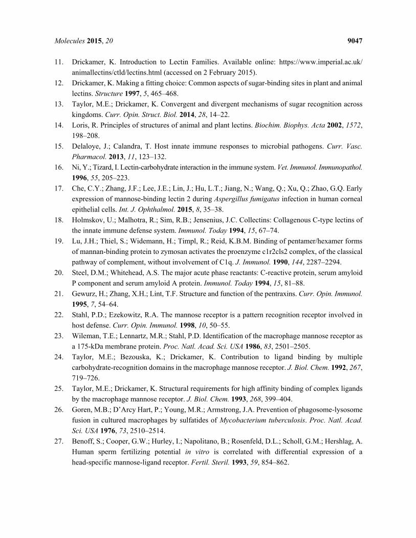
Molecules 2015, 20 9047
11. Drickamer, K. Introduction to Lectin Families. Available online: https://www.imperial.ac.uk/ animallectins/ctld/lectins.html (accessed on 2 February 2015).
12. Drickamer, K. Making a fitting choice: Common aspects of sugar-binding sites in plant and animal lectins. Structure 1997, 5, 465–468.
13. Taylor, M.E.; Drickamer, K. Convergent and divergent mechanisms of sugar recognition across kingdoms. Curr. Opin. Struct. Biol. 2014, 28, 14–22.
14. Loris, R. Principles of structures of animal and plant lectins. Biochim. Biophys. Acta 2002, 1572, 198–208.
15. Delaloye, J.; Calandra, T. Host innate immune responses to microbial pathogens. Curr. Vasc. Pharmacol. 2013, 11, 123–132.
16. Ni, Y.; Tizard, I. Lectin-carbohydrate interaction in the immune system. Vet. Immunol. Immunopathol. 1996, 55, 205–223.
17. Che, C.Y.; Zhang, J.F.; Lee, J.E.; Lin, J.; Hu, L.T.; Jiang, N.; Wang, Q.; Xu, Q.; Zhao, G.Q. Early expression of mannose-binding lectin 2 during Aspergillus fumigatus infection in human corneal epithelial cells. Int. J. Ophthalmol. 2015, 8, 35–38.
18. Holmskov, U.; Malhotra, R.; Sim, R.B.; Jensenius, J.C. Collectins: Collagenous C-type lectins of the innate immune defense system. Immunol. Today 1994, 15, 67–74.
19. Lu, J.H.; Thiel, S.; Widemann, H.; Timpl, R.; Reid, K.B.M. Binding of pentamer/hexamer forms of mannan-binding protein to zymosan activates the proenzyme c1r2cls2 complex, of the classical pathway of complement, without involvement of C1q. J. Immunol. 1990, 144, 2287–2294.
20. Steel, D.M.; Whitehead, A.S. The major acute phase reactants: C-reactive protein, serum amyloid P component and serum amyloid A protein. Immunol. Today 1994, 15, 81–88.
21. Gewurz, H.; Zhang, X.H.; Lint, T.F. Structure and function of the pentraxins. Curr. Opin. Immunol. 1995, 7, 54–64.
22. Stahl, P.D.; Ezekowitz, R.A. The mannose receptor is a pattern recognition receptor involved in host defense. Curr. Opin. Immunol. 1998, 10, 50–55.
23. Wileman, T.E.; Lennartz, M.R.; Stahl, P.D. Identification of the macrophage mannose receptor as a 175-kDa membrane protein. Proc. Natl. Acad. Sci. USA 1986, 83, 2501–2505.
24. Taylor, M.E.; Bezouska, K.; Drickamer, K. Contribution to ligand binding by multiple carbohydrate-recognition domains in the macrophage mannose receptor. J. Biol. Chem. 1992, 267, 719–726.
25. Taylor, M.E.; Drickamer, K. Structural requirements for high affinity binding of complex ligands by the macrophage mannose receptor. J. Biol. Chem. 1993, 268, 399–404.
26. Goren, M.B.; D’Arcy Hart, P.; Young, M.R.; Armstrong, J.A. Prevention of phagosome-lysosome fusion in cultured macrophages by sulfatides of Mycobacterium tuberculosis. Proc. Natl. Acad. Sci. USA 1976, 73, 2510–2514.
27. Benoff, S.; Cooper, G.W.; Hurley, I.; Napolitano, B.; Rosenfeld, D.L.; Scholl, G.M.; Hershlag, A. Human sperm fertilizing potential in vitro is correlated with differential expression of a head-specific mannose-ligand receptor. Fertil. Steril. 1993, 59, 854–862.

Molecules 2015, 20 9048
28. Leteux, C.; Chai, W.; Loveless, R.W.; Yuen, C.T.; Uhlin-Hansen, L.; Combarnous, Y.; Jankovic, M.; Maric, S.C.; Misulovin, Z.; Nussenzweig, M.C.; et al. The cysteine-rich domain of the macrophage mannose receptor is a multispecific lectin that recognizes chondroitin sulfates A and B and sulfated oligosaccharides of blood group Lewisa and Lewisx types in addition to the sulfated N-glycans of lutropin. J. Exp. Med. 2000, 191, 1117–1126.
29. Stahl, P.; Six, H.; Rodman, J.S.; Schlesinger, P.; Tulsiani, D.R.; Touster, O. Evidence for specific recognition sites mediating clearance of lysosomal enzymes in vivo. Proc. Natl. Acad. Sci. USA 1976, 73, 4045–4049.
30. Lee, S.J.; Evers, S.; Roeder, D.; Parlow, A.F.; Risteli, J.; Risteli, L.; Lee, Y.C.; Feizi, T.; Langen, H.; Nussenzweig, M.C. Mannose receptor-mediated regulation of serum glycoprotein homeostasis. Science 2002, 295, 1898–1901.
31. Gantner, B.N.; Simmons, R.M.; Canavera, S.J.; Akira, S.; Underhill, D.M. Collaborative induction of inflammatory responses by dectin-1 and Toll-like receptor 2. J. Exp. Med. 2003, 197, 1107–1117.
32. Brown, G.D. Dectin-1: A signalling non-TLR pattern-recognition receptor. Nat. Rev. Immunol. 2006, 6, 33–43.
33. Caruso, R.; Warner, N.; Inohara, N.; Núñez, G. NOD1 and NOD2: Signaling, host defense, and inflammatory disease. Immunity 2014, 41, 898–908.
34. Nürnberger, T.; Brunner, F. Innate immunity in plants and animals: Emerging parallels between recognition of general elicitors and pathogen-associated molecular patterns. Curr. Opin. Plant Biol. 2002, 5, 318–324.
35. Ausubel, F.M. Are innate immune signaling pathways in plants and animals conserved? Nat. Immunol. 2005, 6, 973–979.
36. Nürnberger, T.; Brunner, F.; Kemmerlinck, B.; Piater, L. Innate immunity in plants and animals: Striking similarities and obvious differences. Immunol. Rev. 2004, 198, 249–266.
37. Nicaise, V.; Roux, M.; Zipfel, C. Recent advances in PAMP-triggered immunity against bacteria: Pattern recognition receptors watch over and raise the alarm. Plant Physiol. 2009, 150, 1638–1647.
38. Boller, T.; Felix, G. A renaissance of elicitors: Perception of microbe-associated molecular patterns and danger signals by pattern-recognition receptors. Annu. Rev. Plant Biol. 2009, 60, 379–406.
39. Wirthmueller, L.; Maqbool, A.; Banfield, M.J. On the front line: Structural insights into plant-pathogen interactions. Nat. Rev. Microbiol. 2013, 11, 761–776.
40. Lannoo, N.; Van Damme, E.J.M. Lectin domains at the frontiers of plant defense. Front. Plant Sci. 2014, 5, doi:10.3389/fpls.2014.00397.
41. Zipfel, C. Plant pattern-recognition receptors. Trends Immunol. 2014, 35, 345–351. 42. Vaid, N.; Macovei, A.; Tuteja, N. Knights in action: Lectin receptor-like kinases in plant
development and stress responses. Mol. Plant 2013, 6, 1405–1418. 43. Singh, P.; Zimmerli, L. Lectin receptor kinases in plant innate immunity. Front. Plant Sci. 2013,
4, doi:10.3389/fpls.2013.00124. 44. Sherman-Broyles, S.; Boggs, N.; Farkas, A.; Liu, P.; Vrebalov, J.; Nasrallah, M.E.; Nasrallah, J.B.
S locus genes and the evolution of self-fertility in Arabidopsis thaliana. Plant Cell 2007, 19, 94–106.
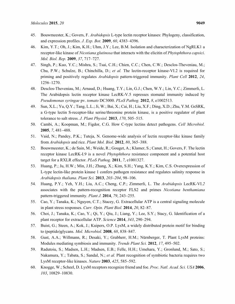
Molecules 2015, 20 9049
45. Bouwmeester, K.; Govers, F. Arabidopsis L-type lectin receptor kinases: Phylogeny, classification, and expression profiles. J. Exp. Bot. 2009, 60, 4383–4396.
46. Kim, Y.T.; Oh, J.; Kim, K.H.; Uhm, J.Y.; Lee, B.M. Isolation and characterization of NgRLK1 a receptor-like kinase of Nicotiana glutinosa that interacts with the elicitin of Phytophthora capsici. Mol. Biol. Rep. 2009, 37, 717–727.
47. Singh, P.; Kuo, Y.C.; Mishra, S.; Tsai, C.H.; Chien, C.C.; Chen, C.W.; Desclos-Theveniau, M.; Chu, P.W.; Schulze, B.; Chinchilla, D.; et al. The lectin-receptor kinase-VI.2 is required for priming and positively regulates Arabidopsis pattern-triggered immunity. Plant Cell 2012, 24, 1256–1270.
48. Desclos-Theveniau, M.; Arnaud, D.; Huang, T.Y.; Lin, G.J.; Chen, W.Y.; Lin, Y.C.; Zimmerli, L. The Arabidopsis lectin receptor kinase LecRK-V.5 represses stomatal immunity induced by Pseudomonas syringae pv. tomato DC3000. PLoS Pathog. 2012, 8, e1002513.
49. Sun, X.L.; Yu, Q.Y.; Tang, L.L.; Ji, W.; Bai, X.; Cai, H.; Liu, X.F.; Ding, X.D.; Zhu, Y.M. GsSRK, a G-type lectin S-receptor-like serine/threonine protein kinase, is a positive regulator of plant tolerance to salt stress. J. Plant Physiol. 2013, 170, 505–515.
50. Cambi, A.; Koopman, M.; Figdor, C.G. How C-type lectins detect pathogens. Cell Microbiol. 2005, 7, 481–488.
51. Vaid, N.; Pandey, P.K.; Tuteja, N. Genome-wide analysis of lectin receptor-like kinase family from Arabidopsis and rice. Plant Mol. Biol. 2012, 80, 365–388.
52. Bouwmeester, K.; de Sain, M.; Weide, R.; Gouget, A.; Klamer, S.; Canut, H.; Govers, F. The lectin receptor kinase LecRK-I.9 is a novel Phytophthora resistance component and a potential host target for a RXLR effector. PLoS Pathog. 2011, 7, e1001327.
53. Huang, P.; Ju, H.W.; Min, J.H.; Zhang, X.; Kim, S.H.; Yang, K.Y.; Kim, C.S. Overexpression of L-type lectin-like protein kinase 1 confers pathogen resistance and regulates salinity response in Arabidopsis thaliana. Plant Sci. 2013, 203–204, 98–106.
54. Huang, P.Y.; Yeh, Y.H.; Liu, A.C.; Cheng, C.P.; Zimmerli, L. The Arabidopsis LecRK-VI.2 associates with the pattern-recognition receptor FLS2 and primes Nicotiana benthamiana pattern-triggered immunity. Plant J. 2014, 79, 243–255.
55. Cao, Y.; Tanaka, K.; Nguyen, C.T.; Stacey, G. Extracellular ATP is a central signaling molecule in plant stress responses. Curr. Opin. Plant Biol. 2014, 20, 82–87.
56. Choi, J.; Tanaka, K.; Cao, Y.; Qi, Y.; Qiu, J.; Liang, Y.; Lee, S.Y.; Stacy, G. Identification of a plant receptor for extracellular ATP. Science 2014, 343, 290–294.
57. Buist, G.; Steen, A.; Kok, J.; Kuipers, O.P. LysM, a widely distributed protein motif for binding to (peptido)glycans. Mol. Microbiol. 2008, 68, 838–847.
58. Gust, A.A.; Willmann, R.; Desaki, Y.; Grabherr, H.M.; Nürnberger, T. Plant LysM proteins: Modules mediating symbiosis and immunity. Trends Plant Sci. 2012, 17, 495–502.
59. Radutoiu, S.; Madsen, L.H.; Madsen, E.B.; Felle, H.H.; Umehara, Y.; Gronlund, M.; Sato, S.; Nakamura, Y.; Tabata, S.; Sandal, N.; et al. Plant recognition of symbiotic bacteria requires two LysM receptor-like kinases. Nature 2003, 425, 585–592.
60. Knogge, W.; Scheel, D. LysM receptors recognize friend and foe. Proc. Natl. Acad. Sci. USA 2006, 103, 10829–10830.

Molecules 2015, 20 9050
61. Grant, S.R.; Fisher, E.J.; Chang, J.H.; Mole, B.M.; Dangl, J.L. Subterfuge and manipulation: Type III effector proteins of phytopathogenic bacteria. Annu. Rev. Microbiol. 2006, 60, 425–449.
62. Block, A.; Toruño, T.Y.; Elowsky, C.G.; Zhang, C.; Steinbrenner, J.; Beynon, J.; Alfano, J.R. The Pseudomonas syringae type III effector HopD1 suppresses effector-triggered immunity, localizes to the endoplasmic reticulum, and targets the Arabidopsis transcription factor NTL9. New Phytol. 2013, 201, 1358–1370.
63. Thomma, B.P.; Nurnberger, T.; Joosten, M.H. Of PAMPs and effectors: The blurred PTI-ETI dichotomy. Plant Cell 2011, 23, 4–15.
64. Mehra, A.; Zahra, A.; Thompson, V.; Sirisaengtaksin, N.; Wells, A.; Porto, M.; Köster, S.; Penberthy, K.; Kubota, Y.; Dricot, A.; et al. Mycobacterium tuberculosis type VII secreted effector EsxH targets host ESCRT to impair trafficking. PLoS Pathog. 2013, 9, e1003734.
65. McCollister, B.D.; Vazquez-Torres, A. Interactions of Salmonella enterica with phagocytic cells. In Salmonella Infections Clinical, Immunological and Molecular Aspects; Mastroeni, P., Maskell, D., Eds.; Cambridge University Press: Cambridge, UK, 2006; pp. 225–278.
66. Huang, J.; Brumell, J.H. A Sweet way of sensing danger. Nature 2006, 482, 316–317. 67. Birmingham, C.L.; Smith, A.C.; Bakowski, M.A.; Yoshimori, T.; Brumell, J.H. Autophagy controls
Salmonella infection in response to damage to the Salmonella-containing vacuole. J. Biol. Chem. 2006, 281, 11374–11383.
68. Kageyama, S.; Omori, H.; Saitoh, T.; Sone, T.; Guan, J.L.; Akira, S.; Imamoto, F.; Noda, T.; Yoshimori, T. The LC3 recruitment mechanism is separate from Atg9L1-dependent membrane formation in the autophagic response against Salmonella. Mol. Biol. Cell 2011, 22, 2290–2300.
69. Blommaart, E.; Luiken, J.; Meijer, A. Autophagic proteolysis: Control and specificity. Histochem. J. 1997, 29, 365–385.
70. Levine, B.; Mizushima, N.; Virgin, H.W. Autophagy in immunity and inflammation. Nature 2011, 469, 323–335.
71. Thurston, T.L.M.; Wandel, M.P.; von Muhlinen, N.; Foeglein, A.; Randow, F. Galectin-8 targets damaged vesicles for autophagy to defend cells against bacterial invasion. Nature 2012, 482, 414–418.
72. Perrin, A.; Jiang, X.; Birmingham, C.; So, N.; Brumell, J. Recognition of bacteria in the cytosol of mammalian cells by the ubiquitin system. Current Biology 2004, 14, 806–811.
73. Ogawa, M.; Yoshimori, T.; Suzuki, T.; Sagara, H.; Mizushima, N.; Sasakawa, C. Escape of intracellular Shigella from autophagy. Science 2005, 307, 727–731.
74. Lv, X.; Pu, X.; Qin, G.; Zhu, T.; Lin, H. The roles of autophagy in development and stress responses in Arabidopsis. Apoptosis 2014, 19, 905–921.
75. Liu, Y.; Schiff, M.; Czymmek, K.; Talloczy, Z.; Levine, B.; Dinesh-Kumar, S.P. Autophagy regulates programmed cell death during the plant innate immune response. Cell 2005, 121, 567–577.
76. Patel, S.; Dinesh-Kumar, S.P. Arabidopsis ATG6 is required to limit the pathogen-associated cell death response. Autophagy 2008, 4, 20–27.
77. Yutin, N.; Wolf, M.Y.; Wolf, Y.I.; Koonin, E.V. The origins of phagocytosis and eukaryogenesis. Biol. Direct 2009, 4, 9:1–9:26.
Molecules 2015, 20 9051
78. Brewin, N.J. Tissue and cell invasion by Rhizobium: The structure and development of infection threads and symbiosisomes. In The Rhizobiaceae; Spaink, H.P., Kondorosi, A., Hooykaas, P.J.J., Eds.; Kluwer academic: Dordrecht, The Netherlands, 1998; pp. 417–429.
79. Parniske, M. Intracellular accommodation of microbes by plants: A common developmental program for symbiosis and disease. Curr. Opin. Plant Biol. 2000, 3, 320–328.
80. Nagano, A.J.; Fukao, Y.; Fujiwara, M.; Nishimura, M.; Hara-Nishimura, I. Antagonistic jacalin-related lectins regulate the size of ER body-type β-glucosidase complexes in Arabidopsis
thaliana. Plant Cell. Physiol. 2008, 49, 969–980. 81. Matsushima, R.; Hayashi, Y.; Yamada, K.; Shimada, T.; Nishimura, M.; Hara-Nishimura, I. The
ER body, a novel endoplasmic reticulum-derived structure in Arabidopsis. Plant Cell. Physiol.
2003, 44, 661–666. 82. Michaeli, S.; Honig, A.; Levanony, H.; Peled-Zehavi, H.; Galili, G. Arabidopsis
ATG8-INTERACTING PROTEIN1 Is Involved in Autophagy-Dependent Vesicular Trafficking of Plastid Proteins to the Vacuole. Plant Cell 2014, 26, 4084–4101.
83. Liang, X.H.; Kleeman, L.K.; Jiang, H.H.; Gordon, G.; Goldman, J.E.; Berry, G.; Herman, B.; Levine, B. Protection against fatal Sindbis virus encephalitis by beclin, a novel Bcl-2-interacting protein. J. Virol. 1998, 72, 8586–8596.
84. Levine, B.; Yuan, J. Autophagy in cell death: An innocent convict? J. Clin. Investig. 2005, 115, 2679–2688.
85. Seay, M.; Dinesh-Kumar, S.P.; Levine, B. Digesting oneself and digesting microbes: Autophagy as a host response to viral infection. In Modulation of Host Gene Expression and Innate Immunity
by Viruses; Palese, P., Ed.; Springer: Dordrecht, The Netherlands, 2005; pp. 245–279. 86. Seay, M.; Patel, S.; Dinesh-Kumar, S.P. Autophagy and plant innate immunity. Cell. Microbiol.
2006, 8, 899–906. 87. Varki, A. Selectin ligands. Proc. Nat. Acad. Sci. USA 1994, 91, 7390–7397. 88. Muerkoster, S.; Rocha, M.; Crocker, P.R.; Schirrmacher, V.; Umanski, V. Sialoadhesin-positive
host macrophages play an essential role in graft-versus-leukemia reactivity in mice. Blood 1999, 93, 4375–4386.
89. Crocker, P.R.; Varki, A. Siglecs in the immune system. Immunology 2001, 103, 137–145. 90. Ravetch, J.V.; Lanier, L.L. Immune inhibitory receptors. Science 2000, 290, 84–89. 91. O’Keefe, T.L.; Williams, G.T.; Batista, F.D.; Neuberger, M.S. Deficiency in CD22, a B
cell-specific inhibitory receptor, is sufficient to predispose to development of high affinity antibodies. J. Exp. Med. 1999, 189, 1307–1313.
92. Fouquaert, E.; Peumans, W.J.; Vandekerckhove, T.; Ongenaert, M.; Van Damme, E.J.M. Proteins with an Euonymus lectin-like domain are ubiquitous in Embryophyta. BMC Plant Biol. 2009, 9,
136:1–136:17. 93. Fouquaert, E.; Van Damme, E.J.M. Promiscuity of the Euonymus carbohydrate-binding domain.
Biomolecules 2012, 2, 415–434. 94. Li, D.; Wang, X.; Yuan, D.; Zhang, L.; Jiang, X.; Tao, Z.; Li, Y.; Wang, J.; Li, X.; Yang, Y.
Over-expression of ArathEULS3 confers ABA sensitivity and drought tolerance in Arabidopsis. Plant Cell Tissue Organ Cult. 2014, 117, 431–442.
Molecules 2015, 20 9052
95. Berendzen, K.W.; Böhmer, M.; Wallmeroth, N.; Peter, S.; Vesić, M.; Zhou, Y.; Tiesler, F.K.;
Schleifenbaum, F.; Harter, K. Screening for in planta protein–protein interactions combining
bimolecular fluorescence complementation with flow cytometry. Plant Methods 2012, 8,
25:1–25:17.
96. Bourne, Y.; Astoul, C.H.; Zamboni, V.; Peumans, W.J.; Menu-Bouaouiche, L.; Van Damme, E.J.M.;
Barre, A.; Rougé, P. Structural basis for the unusual carbohydrate-binding specificity of jacalin
towards galactose and mannose. Biochem. J. 2002, 364, 173–180.
97. Peumans, W.J.; Hause, B.; Van Damme, E.J.M. The galactose-binding and mannose-binding
jacalin-related lectins are located in different sub-cellular compartments. FEBS Lett. 2000, 477,
186–192. 98. Song, M.; Xu, W.; Xiang, Y.; Jia, H.; Zhang, L.; Ma, Z. Association of jacalin-related lectins with
wheat responses to stresses revealed by transcriptional profiling. Plant Mol. Biol. 2014, 84, 95–110.
99. Lee, R.H.; Wang, C.H.; Huang, L.T.; Chen, S.C. Leaf senescence in rice plants: Cloning and
characterization of senescence up-regulated genes. J. Exp. Bot. 2001, 52, 1117–1121.
100. Qin, Q.M.; Zhang, Q.; Zhao, W.S.; Wang, Y.Y.; Peng, Y.L. Identification of a lectin gene induced
in rice in response to Magnaporthe grisea infection. Acta Bot. Sin. 2003, 23, 799–810.
101. De Souza Filho, G.A.; Ferreira, B.S.; Dias, J.M R.; Queiroz, K.S.; Branco, A.T.; Bressan-Smith, R.E.;
Oliveira, J.G.; Garcia, A.B. Accumulation of SALT protein in rice plants as a response to
environmental stresses. Plant Sci. 2003, 164, 623–628.
102. Xiang, Y.; Song, M.; Wei, Z.; Tong, J.; Zhang, L.; Xiao, L.; Ma, Z.; Wang, Y. A jacalin-related
lectin-like gene in wheat is a component of the plant defence system. J. Exp. Bot. 2011, 62,
5471–5483.
103. Yamaji, Y.; Maejima, K.; Ozeki, J.; Komatsu, K.; Shiraishi, T.; Okano, Y.; Himeno, M.;
Sugawara, K.; Neriya, Y.; Minato, N.; et al. Lectin-mediated resistance impairs plant virus
infection at the cellular level. Plant Cell 2012, 24, 778–793.
104. Regente, M.; Taveira, G.B.; Pinedo, M.; Elizalde, M.M.; Ticchi, A.J.; Diz, M.S.S.; Carvalho, A.O.;
de la Canal, L.; Gomes, V.M. A sunflower lectin with antifungal properties and putative medical
mycology applications. Curr. Microbiol. 2014, 69, 88–95.
105. Kanagawa, M.; Liu, Y.; Hanashima, S.; Ikeda, A.; Chai, W.; Nakano, Y.; Kojima-Aikawa, K.;
Feizi, T.; Yamaguchi, Y. Structural basis for multiple sugar recognition of Jacalin-related human
ZG16p lectin. J. Biol. Chem. 2014, 289, 16954–16965.
106. Malik, A.; Lee, J.; Lee, J. Community-based network study of protein-carbohydrate interactions
in plant lectins using glycan array data. PLoS ONE 2014, 9, e95480.
107. Beneteau, J.; Renard, D.; Marché, L.; Douville, E.; Lavenant, L.; Rahbé, Y.; Dupont, D.;
Vilaine, F.; Dinant, S. Binding properties of the N-acetylglucosamine and high-mannose N-glycan
PP2-A1 phloem lectin in Arabidopsis. Plant Physiol. 2010, 153, 1345–1361.
108. R-type lectins. In Essentials of Glycobiology, 2nd ed.; Varki, E., Cummings, R.D., Esko, J.D.,
Freeze, H.H., Stanley, P., Bertozzi, C.R., Hart, G.W., Etzler, M.E., Eds.; Cold Spring Harbor
Laboratory Press: Cold Spring Harbor, NY, USA, 2006; pp. 403–414.
109. Shang, C.; Van Damme, E.J.M. Comparative analysis of carbohydrate binding properties of
Sambucus nigra lectins and ribosome-inactivating proteins. Glycoconj. J. 2014, 31, 345–354.
Molecules 2015, 20 9053
110. Kerringan, A.M.; Brown, G.D. Syk-coupled C-type lectins in immunity. Trends Immunol. 2011, 32, 151–156.
111. Iwasaki, A.; Medzhitov, R. Regulation of adaptive immunity by the innate immune system. Science 2010, 327, 291–295.
112. Conrath, U. Systemic acquired resistance. Plant Signal. Behav. 2006, 1, 179–184.
113. Bruce, T.J.A.; Matthes, M.C.; Napier, J.A.; Pickett, J.A. Stressful ‘‘memories’’ of plants: Evidence and possible mechanisms. Plant Sci. 2007, 173, 603–608.
114. Beckers, G.J.M.; Jaskiewicz, M.; Liu, Y.; Underwood, W.R.; He, S.Y.; Zhang, S.; Conrath, U.
Mitogen-activated protein kinases 3 and 6 are required for full priming of stress responses in Arabidopsis thaliana. Plant Cell 2009, 21, 944–953.
115. Jung, H.W.; Tschaplinski, T.J.; Wang, L.; Glazebrook, J.; Greenberg, J.T. Priming in systemic
plant immunity. Science 2009, 324, 89–91. 116. Conrath, U. Molecular aspects of defence priming. Trends Plant Sci. 2011, 16, 524–531. 117. Sung, S.; Amasino, R.M. Vernalization and epigenetics: How plants remember winter. Curr. Opin.
Plant Biol. 2004, 7, 4–10. 118. Xiao, J.; Xu, S.; Li, C.; Xu, Y.; Xing, L.; Niu, Y.; Huan, Q.; Tang, Y.; Zhao, C.; Wagner, D.; et al.
O-GlcNAc-mediated interaction between VER2 and TaGRP2 elicits TaVRN1 mRNA
accumulation during vernalization in winter wheat. Nat. Commun. 2014, 5, 4572:1–4572:13. 119. Chen, Y.; Peumans, W.; Hause, B.; Bras, J.; Kumar, M.; Proost, P.; Barre, A.; Rougé, P.;
Van Damme, E.J.M. Jasmonic acid methyl ester induces the synthesis of a cytoplasmic/nuclear
chito-oligosaccharide binding lectin in tobacco leaves. FASEB J. 2002, 16, 905–907. 120. Lannoo, N.; Vandenborre, G.; Miersch, O.; Smagghe, G.; Wasternack, C.; Peumans, W.J.;
Van Damme, E.J.M. The jasmonate-induced expression of the Nicotiana tabacum leaf lectin.
Plant Cell Physiol. 2007, 48, 1207–1218. 121. Vandenborre, G.; Van Damme, E.J.M.; Smagghe, G. Natural products: Plant lectins as important
tools in controlling pest insects. In Biorational Control of Arthropod Pests; Ishaaya, I., Horowitz, A.R.,
Eds.; Springer Science: Dordrecht, The Netherlands, 2009; pp. 163–187. 122. Schouppe, D.; Ghesquière, B.; Menschaert, G.; De Vos, W.H.; Bourque, S.; Trooskens, G.; Proost, P.;
Gevaert, K.; Van Damme, E.J.M. Interaction of the tobacco lectin with histone proteins.
Plant Physiol. 2011, 155, 1091–1102. 123. Delporte, A.; De Vos, W.H.; Van Damme, E.J.M. In vivo interaction between the tobacco lectin
and the core histone proteins. J. Plant Physiol. 2014, 171, 986–992.
124. Delporte, A.; Van Holle, S.; Lannoo, N.; Van Damme, E.J.M. The tobacco lectin, prototype of the family of Nictaba-related proteins. Curr. Prot. Pept. Sci. 2015, 16, 5–16.
© 2015 by the authors; licensee MDPI, Basel, Switzerland. This article is an open access article
distributed under the terms and conditions of the Creative Commons Attribution license (http://creativecommons.org/licenses/by/4.0/).

Chapter 24, Essentials to Glycobiology (3rd edition)
Viridiplantae and Algae
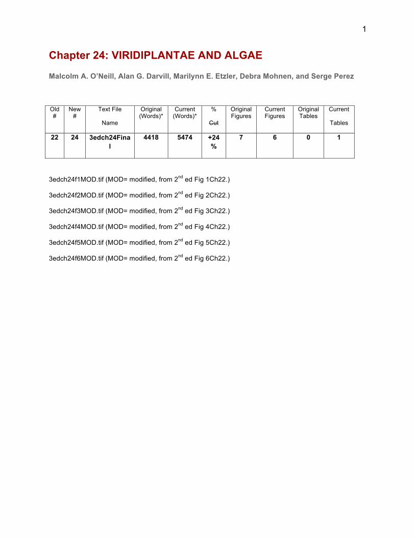
1
Chapter 24: VIRIDIPLANTAE AND ALGAE
Malcolm A. O’Neill, Alan G. Darvill, Marilynn E. Etzler, Debra Mohnen, and Serge Perez
Old #
New #
Text File
Name
Original (Words)*
Current (Words)*
%
Cut
Original Figures
Current Figures
Original Tables
Current
Tables
22 24 3edch24Final
4418 5474 +24%
7 6 0 1
3edch24f1MOD.tif (MOD= modified, from 2nd ed Fig 1Ch22.)
3edch24f2MOD.tif (MOD= modified, from 2nd ed Fig 2Ch22.)
3edch24f3MOD.tif (MOD= modified, from 2nd ed Fig 3Ch22.)
3edch24f4MOD.tif (MOD= modified, from 2nd ed Fig 4Ch22.)
3edch24f5MOD.tif (MOD= modified, from 2nd ed Fig 5Ch22.)
3edch24f6MOD.tif (MOD= modified, from 2nd ed Fig 6Ch22.)
2
Recent research on plant glycan structure and function has typically emphasized model plants
such as Arabidopsis and plants of commercial importance. However, there is increasing interest
in studying the glycans produced by plants from all the major orders of the viridiplantae. Such
studies, together with the availability of transcriptomic data for numerous green algae and land
plants, have begun to reveal a rich diversity in glycan structures and insight into how some of
these structures have changed during the evolution of the viridiplantae. In this chapter we
provide an overview of the current knowledge of green plant glycan structures with an emphasis
on the features that are unique to land plants.
INTRODUCTION
Viridiplantae (green plants) are a clade of photosynthetic organisms that contain chlorophylls a
and b, produce and store their photosynthetic products inside a double-membrane-bounded
chloroplast, and have cell walls that typically contain cellulose. The Viridiplantae are comprised
of two clades - the Chlorophyta and the Streptophyta. The Chlorophyta contain most of the
organisms typically referred to as “green algae”. The term "algae" is also used for several other
groups of photosynthetic eukaryotes including Diatoms and the red, brown, golden and yellow-
green algae. The Streptophyta comprise several other lineages that are also referred to as “green
algae" and the land plants. Land plants include the liverworts, mosses, hornworts, lycopods,
ferns, gymnosperms and flowering plants.
PLANT GLYCAN DIVERSITY
Green plants synthesize diverse glycans that vary in their structural complexity and molecular
size. Raffinose oligosaccharides (raffinose, stachyose, and verbascose) are almost ubiquitous in
the plant kingdom and rank second only to sucrose in abundance as soluble carbohydrates. These
oligosaccharides are derivatives of sucrose that contain one or more α-Gal residues. Plants also
synthesize numerous low molecular weight glycoconjugates that contain either aromatic (e.g.
phenolic glycosides) or aliphatic (glycolipids) aglycones.
Plant polysaccharides are linear or branched polymers composed of the same or different
monosaccharides. For example, cellulose is composed of 1-4-linked β-D-Glc residues (Figure
24.1A), whereas the structurally complex plant cell wall pectic polysaccharide referred to as
rhamnogalacturonan II (RG-II) contains 12 different monosaccharides linked together by up to
3
21 different glycosidic linkages (Figure 24.2). Plant proteoglycans are structurally diverse
glycans in which carbohydrate accounts for up to 90% of the molecule and is O-linked to the
protein via hydroxyamino acids (Figure 24.3). Plant glycoproteins typically contain 15% or less
of carbohydrate in the form of N-linked oligomannose, complex, hybrid, and paucimannose
oligosaccharides (Figure 24.4). Land plants also form O-GlcNAc-modified nuclear and cytosolic
proteins (Chapter 19).
NUCLEOTIDE SUGARS - THE BUILDING BLOCKS
Nucleotide sugars are the donors used for synthesis of glycoconjugates and glycosylated
secondary metabolites (Chapter 5). In plants, the majority of these activated monosaccharides
exist as their nucleotide-diphosphates (e.g. UDP-Glc), although at least one monosaccharide,
Kdo, exists as its cytidine monophosphate derivative (CMP-Kdo). Nucleotide sugars are formed
from the carbohydrate generated by photosynthesis, from the monosaccharides released by
hydrolysis of sucrose and storage carbohydrates, and by recycling monosaccharides from glycans
and the cell wall. Nucleotide sugars are also formed by interconverting pre-existing activated
monosaccharides. To date 30 different nucleotide-sugars and at least 100 genes encoding
proteins involved in their formation and interconversion have been identified in plants.
PLANT GLYCOSYLTRANSFERASES AND GLYCAN-MODIFYING ENZYMES
Plant genomes contain numerous genes encoding proteins involved in the synthesis and
modification of glycans. These proteins are spread across many enzyme classes in the
Carbohydrate Active enZymes (CAZy) database (Table 24. 1). Many of these proteins may be
involved in the formation and modification of the polysaccharide-rich cell wall. Indeed, the
unicellular alga Ostreococcus tauri, which is one of the few plants that does not form a cell wall,
has a much smaller number of genes predicted to be involved in glycan metabolism
PLANT CELL WALLS
A substantial portion of the carbohydrate formed by photosynthesis is used to produce the
polysaccharide-rich walls that surround plant cells. Primary and secondary cell walls are
distinguished by their composition, architecture and functions. A primary wall surrounds
growing and dividing plant cells and non-growing cells in the soft tissues of fruits and leaves.
4
These walls are capable of controlled extension to allow the cell to grow and expand yet are
sufficiently strong to resist the cells internal turgor pressure. A much thicker and stronger
secondary wall is often formed once a cell has ceased to grow. This wall is deposited between
the plasma membrane and the primary wall and is composed of layers that differ in the
orientation of their cellulose microfibrils. The secondary walls of vascular tissues involved in the
movement of water and nutrients through the plant are further strengthened by the incorporation
of lignin. The ability to form conducting tissues with lignified and rigid secondary walls was an
indispensable event in the evolution of vascular land plants, as it facilitated the transport of water
and nutrients and allowed extensive upright growth. Secondary cell walls account for most of the
carbohydrate in plant biomass being considered as feedstock for the production of biofuels and
value-added chemicals (Chapter 59).
PRIMARY CELL WALL GLYCANS
Primary cell walls are composites that resemble fiber-reinforced porous gels. The complex
structures and functions of these walls result from the assembly and interactions of a limited
number of structurally-defined polysaccharides and proteoglycans. Wall structure and
organization may change during cell division and development and in response to biotic and
abiotic challenges by the differential synthesis and modification of the non-cellulosic
components or by the addition of new components.
Primary walls of land plants contain cellulose, hemicellulose and pectin, in different proportions.
They also contain structural proteins/proteoglycans, enzymes, phenolics and minerals. Pectin and
hemicellulose are present in approximately equal amounts in the so-called type I primary walls
of gymnosperms, dicots and non-graminaceous monocots, whereas hemicellulose is far more
abundant than pectin in the type II walls of the grasses. Much less is known about the
composition of the walls of avascular plants and green algae, although it is likely that the walls
of these plants contain cellulose, pectin and hemicellulose.
Cellulose
Cellulose, the most abundant biopolymer in nature, is a linear polymer composed of 1-4 linked
β-D-Glc residues (Figure 24.1A). Several of these chains are hydrogen bonded to one another to
form paracrystaline microfibrils. Each microfibril is predicted to contain between 18 and 24
5
glucan chains. The glucan chain is synthesized by a cellulose synthase (CESA) complex that
exists as a hexameric rosette structure on the cells plasma membrane. Three CESAs, encoded by
three different genes, are believed to interact to form a trimeric complex, which in turn
assembles into a hexameric rosette. The catalytic site of each cellulose synthase is located in the
cytosol and transfers glucose from UDP-Glc onto the elongating glucan chain. The mechanisms
involved in the formation of a microfibril from individual glucan chains are not well understood,
although it may involve a self-assembly process that is facilitated by specific proteins. The newly
formed microfibrils are then deposited in the wall of a growing cell with an orientation that is
transverse to the axis of elongation. This orientation may be guided in part by protein-mediated
interactions between CESA proteins and cortical microtubules.
Naturally occurring cellulose is a mixture of two crystalline forms, Iα and Iβ, together with
surface chains and less crystalline material. The 1α and 1β polymorphs differ mainly in the
packing arrangement of their hydrogen-bonded sheets. Many properties of native cellulose
depend on interactions that occur at the surface of the microfibrils. The surface chains are
accessible and reactive whereas the hydroxyl groups of the internal chains in the crystal
participate in extensive intra- and inter-molecular hydrogen bonding. Cellulose is insoluble in
water and somewhat resistant to hydrolysis by endo- and exo-glucanases because of this highly
packed arrangement of the glucan chains.
Several types of enzymes including endoglucanases cellobiohydrolases and β-glucosidases are
required for cellulose depolymerization. Many of these enzymes consist of a catalytic domain
connected to a cellulose-binding module. This module facilitates binding of the enzyme to the
insoluble substrate. Some micro-organisms also produce copper-dependent oxidases that render
crystalline cellulose more susceptible to hydrolysis. Cellulases and other enzymes involved in
cellulose hydrolysis often exist as macromolecular complexes referred to as cellulosomes.
Improving the effectiveness of cellulosomes is an area of active research, to increase the
conversion of plant biomass to fermentable sugar (Chapter 59).
Hemicelluloses
Hemicelluloses are branched polysaccharides with a backbone composed of 1-4-linked β-D-
pyranosyl residues with an equatorial O-4 (Glc, Man, and Xyl). Xyloglucan, glucuronoxylan,
arabinoxylan and glucomannan (Figure 24.1B- E) are included under this chemical definition of
6
hemicelluloses. Hemicelluloses and cellulose have structural and conformational similarities that
allow them to form strong, noncovalent associations with one another in the cell wall, although
the biological significance of these interactions is a subject of debate.
Xyloglucans are distinguished from one another by the number of 1-4-linked β-D-Glcp backbone
residues that are branched. XXXG-type xyloglucans are composed of subunits in which three
consecutive backbone residues bear an α-Xyl substituent at O-6 and a fourth, unbranched
backbone residue (Figure 24.1B). Each Xyl residue (X side chain) may itself be extended by the
addition of one or more monosaccharides including β-D-Gal, α-L-Fuc, α-L-Ara and β-D-GalA.
Eighteen structurally unique sidechains have been identified to date, although only a subset of
these are synthesized by a single plant species. XXXG-type xyloglucan is present in the primary
walls of hornworts, lycopods, ferns, gymnosperms, a diverse range of dicots and all monocots
with the exception of the grasses. XXGG-type xyloglucans, which have only two consecutive
backbone residues bearing an α-D-Xyl substituent at O-6, are present in the primary walls of the
grasses, some Solanaceae, mosses and liverworts. The Xyl may be extended by the addition of
Gal, Ara or GalA, but rarely if ever with Fuc.
Early models of dicot primary walls predicted that xyloglucan acted as tethers between cellulose
microfibrils and that controlled enzymatic cleavage of the xyloglucan facilitated wall expansion
and thus plant cell growth. However, the identification of an Arabidopsis mutant that is unable to
synthesize xyloglucan yet exhibits near normal growth and development challenged this notion
and led to the suggestion that pectin may have a more important role in controlling wall
expansion than previously believed.
Glucuronoarabinoxylan (GAX, Figure 24.1C) is the predominant non-cellulosic polysaccharide
in the type II walls of the grasses. Its backbone is composed of 1-4-linked β-D-Xyl residues
many of which are substituted at O-3 with α-L-Araf residues. These Araf residues may be further
substituted at O-2 with an α-L-Araf or a β-D-Xylp residue. A small number of the backbone
residues are substituted at O-2 with α-D-GlcpA and its 4-O-methylated counterpart (MeGlcpA).
The presence of 1-3, 1-4-linked β-glucans (also referred to as mixed-linkage glucans) in the
walls of grasses was once considered to be a unique feature of these plants. However,
structurally related mixed-linkage β-glucans have also been identified in the walls of Selaginella
7
(lycopod) and Equisetum (horsetails), although the evolutionary relationship between these β-
glucans is not known.
Pectins
Pectins are structurally complex polysaccharides that contain 1-4-linked α-GalA. Three
structurally distinct pectins - homogalacturonan, substituted galacturonan, and
rhamnogalacturonan - have been identified in plant cell walls (Figure 24.2). Homogalacturonan,
which may account for up to 65% of the pectin in a primary wall, is composed of 1-4-linked α-
GalA. The carboxyl group may be methyl-esterified and the glycose itself may be acetylated at
O-2 or O-3. The extent of methyl-esterification is controlled by pectin methyl-esterases present
in the wall and affects the ability of homogalacturonan-containing glycans to form ionic calcium
cross-links with themselves and with other pectic polymers. Such interactions alter the
mechanical properties of the wall and may influence plant growth and development.
Rhamnogalacturonans are polysaccharides with a backbone composed of GalA and rhamnose
(Rha) residues in the repeating disaccharide 4-α-D-GalpA-1-2-α-L-Rhap-1. Many of the GalAs
are acetylated at O-2 and/or O-3. Depending on the plant, between 20 and 80% of the Rha
residues may be substituted at O-4 with linear or branched side chains composed predominantly
of Ara and Gal, together with smaller amounts of Fuc and GlcA (Figure 24.2). Little is known
about the functions of these side chains and their contribution to the properties of the primary
wall.
Substituted galacturonans have a backbone composed of 1-4-linked α-D-GalA acid residues that
are substituted to varying degrees with mono-, di- or oligosaccharides. Xylogalacturonans
contain single β-Xyl residues linked to O-3 of some of the backbone residues (Figure 24.2)
whereas apiogalacturonans have β-D-apiose (Api) and apiobiose linked to O-2 of some of the
backbone residues. Apiogalacturonans have only been identified in the walls of duckweeds and
seagrasses.
Rhamnogalacturonan-II, which accounts for between 2% and 5% of the primary cell wall, is the
most structurally complex polysaccharide yet identified in nature. It is composed of 12 different
linked together by up to 21 different glycosidic linkages (Figure 24.2). Four structurally different
sidechains are attached to the galacturonan backbone. Two structurally conserved disaccharides
8
(sidechains C and D) are linked to O-2 of the backbone. The A and B sidechains, which contain
between 7 and 9 monosaccharides, are linked to O-3 of the backbone. Several of the
monosaccharides in RG-II are O-methylated and/or O-acetylated.
Virtually all of the RG-II exists in the primary wall as a dimer cross-linked by a borate ester. The
ester is formed between the Api residue in side chain A of each RG-II monomer (Figure 24.2).
The dimer forms rapidly in vitro when the RG-II monomer is reacted with boric acid and a
divalent cation. However, the mechanism and site of dimer formation in planta has not been
determined. Borate cross-linking of RG-II is likely to have substantial effects on the properties of
pectin and the primary wall as RG-II is itself linked to homogalacturonan (Figure 24.2). Indeed,
mutations that affect RG-II structure and cross-linking result in plants with abnormal walls and
severe growth defects. Swollen primary walls and abnormal growth together with reduced RG-II
cross-linking are also a characteristic of boron deficient plants.
Pectin is believed to exist in the cell wall as a macromolecular complex comprised of structural
domains - homogalacturonan, rhamnogalacturonan and substituted galacturonan - that are
covalently linked to one another. However, there is only a limited understanding of how these
structural domains are organized (Figure 24.2). Molecular modeling of a pectin (~50 kDa)
containing homogalacturonan (degree of polymerization ~100) and rhamnogalacturonan with
arabinogalactan sidechains together with modeling of RG-II conformation have begun to provide
insights into the conformations and relative dimensions of each pectin structural domain.
The conformation of the homogalacturonan chain is largely unaffected by the conformation at
the glycosidic linkage, by changes in its degree of methyl-esterification or the presence of
counter ions. The homogalacturonan region has a persistence length of ~20 GalA residues, which
is likely to be sufficient to stabilize junction zones formed with Ca++. In vitro studies suggest that
the maximum stability of such junction zones is obtained with oligomers containing ~15 non-
esterified GalA residues. Thus controlling the distribution of methyl-ester groups along the
homogalacturonan backbone provides a mechanism to regulate the physical properties of pectin,
including its ability to form gels. For a gel to form and not to be brittle, other features including
sequences that interrupt inter-chain associations in the pectin macromolecule may be important.
For example the structural diversity and the conformational flexibility of the oligosaccharide
sidechains of the rhamnogalacturonan domain will limit or prevent inter-chain pairing. The
9
presence of 1-2-linked Rha residues does not introduce kinks into the backbone geometry of
rhamnogalacturonan and thereby limit inter-chain associations. Rather, it is the side chains linked
to these residues that are responsible for preventing or limiting inter-chain associations.
The conformations of the four side chains attached to the homogalacturonan backbone may lead
RG-II to adopt a “disk-like” shape. Well-defined tertiary structures are predicted for the
monomer and the dimer. In the dimer, borate-ester cross-linking and Ca++ inter-chain pairing
further stabilizes the two disks. The apparent resistance of RG-II to wall modifying enzymes
together with the formation of a cation-stabilized RG-II dimer likely results in a structure that
resists temporal changes. By contrast, homogalacturonan is continually modified by the action of
wall enzymes and its contribution to wall architecture is therefore time dependent.
Increased knowledge of the physical properties of primary wall polysaccharides and
proteoglycans is required to understand how modulating the amounts and structural features of a
few common polysaccharides and glycan domains lead to primary walls with diverse properties
and functionalities. Further research is also needed to determine if wall structure and function
results from the non-covalent interactions of polysaccharides and proteoglycans or from the
formation of glycan-containing architectural units with specific structural and functional roles.
The later scenario is analogous to the organization of proteoglycans and O-linked mucins in the
extracellular matrix of animal cells (Chapters 10, 16 and 17).
PLANT SECONDARY CELL WALL GLYCANS
The secondary walls of woody tissue and grasses are composed predominantly of cellulose,
hemicellulose, and lignin. The inclusion of lignin results in a hydrophobic composite that is a
major contributor to the structural characteristics of secondary walls.
Heteroxylans are the major hemicellulosic polysaccharide present in the secondary (lignified)
cell walls of flowering plants. These heteroxylans are classified according to the type and
abundance of the substituents on the 1-4-linked β-D-Xylp residues of the polysaccharide
backbone. Glucuronoxylans (GX), which are major components in the secondary walls of woody
and herbaceous eudicots, have a α-D-GlcA or MeGlcA substituent at O-2 (Figure 24.1C).
Gymnosperm secondary walls contain arabinoglucuronoxylans (AGXs), which in addition to
MeGlcA substituents, have Araf residues attached to O-3 of some of the backbone residues. The
10
GAX in the secondary walls of grasses typically contain less Araf residues than their primary
wall counterpart (Figure 24.1D). Ferulic or coumaric acids are often esterified to the Araf
residues of GAX in grass primary and secondary cells walls.
Eudicot and gymnosperm secondary wall GX and AGX have a well-defined glycosyl sequence -
4-β-D-Xylp-1-3-α-L-Rhap-1-2-α-D-GalpA-1-4-D-Xylp at their reducing end (Figure 24.1C). This
sequence is required for normal xylan synthesis during secondary cell wall formation and may
have a role in regulating the polymers chain length. This sequence is present at the reducing end
of heteroxylans of all monocots except the grasses.
HEMICELLULOSE AND PECTIN BIOSYTHESIS
Genes that encode polysaccharide biosynthetic enzymes, including many of those required for
xyloglucan, glucuronoxylan, arabinoxylan and cellulose synthesis and some of those required for
pectin synthesis have been identified. This information, together with improved methods to
generate recombinant plant GTs with high enzymatic activity is providing a framework for an
increased understanding of how plant cell wall polysaccharides are synthesized. There is general
consensus that cellulose is synthesized by GT complexes localized at the plasma membrane and
that most pectins and hemicellulose are synthesized in the Golgi apparatus. Members of the
cellulose synthase-like gene families, CSLF and CSLH are likely involved in 1-3, 1-4-linked β-
glucan biosynthesis. However, there is considerable debate about whether this polysaccharide is
formed in the Golgi apparatus or at the plasma membrane.
Despite advances in understanding how polysaccharides are synthesized we still do not know
how many of the wall polymers are synthesized by Golgi-localized multi-enzyme complexes or
if they are assembled by GTs localized in different regions of the Golgi apparatus. We also do
not understand how the newly synthesized polymers are assembled into a functional cell wall.
PLANTS PRODUCE PROTEOGLYCANS CONTAINING O-LINKED OLIGOSACCHARIDES AND O-LINKED POLYSACCHARIDES
Plants produce glycoproteins and proteoglycans that contain oligo- or polysaccharides that are
linked to hydroxyproline (Hyp) and serine (Ser). Hyp is formed post-translationally by
endoplasmic reticulum-localized prolyl hydroxylases and is O-glycosylated in the ER and in the
Golgi apparatus. The degree and type of Hyp glycosylation is determined to a large extent by the
11
protein's primary sequence and the arrangement of Hyp residues. Hyp glycosylation is initiated
by the addition of an Ara or a Gal residue. Contiguous Hyp residues are arabinosylated, whereas
clustered but noncontiguous Hyp residues are galactosylated. Ser residues and occasionally Thr
residues may also be O-glycosylated in these proteins.
Three structurally distinct plant proteoglycans containing glycosylated Hyp and Ser – the
extensins, proline/hydroxyproline-rich proteoglycans and arabinogalactan proteins – have been
identified. Extensins are hydroxyproline-rich proteoglycans with Ser(Hyp)4 repeat sequences and
contain between 50 and 60% (w/w) glycan. Most of the carbohydrate exists as oligosaccharides
containing one to four Ara residues linked to Hyp together with a small number of single Gal
residues α-linked to Ser. The proline/hydroxyproline-rich proteoglycans, which contain from 3%
to 70% (w/w) carbohydrate, are distinguished from the extensins by amino acid sequence. Both
of these families of HRGPs likely have a structural role in the cell wall. The expression of genes
involved in their synthesis is developmentally regulated and is often induced by wounding and
fungal attack of plant tissues.
The glycopeptide signaling molecules PSY1, CLE2 and CLV3 contain arabinosylated
hydroxyproline and have numerous roles in plant growth and development. A highly
glycosylated Hyp-rich domain in which three or four Ara residues are attached to Hyp and a
single Gal residue is linked to Ser is present in potato, tomato, and thorn-apple lectins.
Arabinogalactan proteins (AGPs) have a glycan content of up to 90% (w/w). Chains of between
30 and 150 monosaccharides are linked to the protein by Gal-O-Ser and Gal-O-Hyp linkages.
These chains have a 1-3-linked β-Gal backbone that is extensively substituted at O-6 with
sidechains of 1-6-linked β-Gal. These sidechains are terminated with Ara, GlcA and Fuc
residues. Some AGPs may contain homogalacturonan, RG-I and xylan covalently linked to the
arabinogalactan (Figure 24.3) thereby forming a protein-hemicellulose-pectin complex referred
to as APAP1. The location of this complex in the plant and its biological function remains to be
determined.
Several AGPs are secreted into the cell wall whereas others are linked to the plasma membrane
by a glycosylphosphatidylinositol (GPI) anchor. Plant GPI anchors contain a phosphoceramide
core. The glycan portion of the GPI anchor of pear cell AGP has the sequence α-D-Man1-2α-D-
Man-1-6-α-D-Man-1-4-GlcN-inositol. At least 50% of the Man attached to the GlcN (see

12
Chapter 12) is itself substituted at O-4 with a β-Gal, a feature that may be unique to plants. Many
functions have been proposed for the AGPs including their participation in signaling,
development, cell expansion, cell proliferation, and somatic embryogenesis.
THE N-LINKED GLYCANS OF PLANT GLYCOPROTEINS HAVE UNIQUE STRUCTURES
Many of the proteins that have passed through the plant secretory system contain N-linked
oligomannose, complex, hybrid, or paucimannose-type glycans (Figure 24.4). The initial stages
of the synthesis of these N-glycans, including the transfer of the oligosaccharide precursor from
its dolichol derivative and the control of protein folding in the endoplasmic reticulum are
comparable in plants and animals (Chapter 9). However, two modifications of N-glycans during
passage through the Golgi are unique to plants.
Oligomannose-type N-glycans are often trimmed in the cis-Golgi and then modified in the
medial-Golgi by N-GlcNAc transferase I (GnT-I) catalyzed addition of GlcNAc to the distal
Man of the core. In reactions that are unique to plants, a β-Xyl is often added to O-2 of the core
Man. In the trans-Golgi, α-Fuc may be added to O-3 of the GlcNAc residue that is itself linked to
asparagine (Figure 24.5). The XylT and FucT that catalyze these reactions act independently of
one another but do require at least one terminal GlcNAc residue for activity. The FucT is related
to the Lewis FucT family, whereas the XylT is unrelated to other known GTs.
The xylosylated and fucosylated N-glycans are often trimmed by α-mannosidase II. A second
GlcNAc may then be added by GnT-II. Some plant N-glycans do not undergo further mannose
trimming and proceed through the Golgi as hybrid-type N-glycans. Complex and hybrid-type N-
glycans may be further modified by the addition of Gal and Fuc in the trans-Golgi. Plant
glycoproteins are either secreted from the cell or transported to the vacuoles. Many of the
glycoproteins present in the vacuoles contain paucimannose type glycans, suggesting that they
are trimmed by vacuolar glycosidases (Figure 24.5).
The presence of sialic acid in the N-glycans of plant glycoproteins was claimed but likely
represented environmental contamination. Plants do have genes that encode proteins containing
sequences similar to sialyltransferase motifs but their functions have not been established.

13
ALGAL GLYCANS
Only a few glycans of green algae have been studied in detail. For example, the cell wall of
Chlamydomonas reinhardii is a crystaline lattice formed from hydroxyproline-rich
glycoproteins. A sulfated polysaccharide composed of Kdo and GalA is the major glycan in the
cell walls of Tetraselmis striata. Some but not all of the glycans present in the cell walls of land
plants may also be present in the cell walls of some green algae. Several of the polysaccharides
produced by red and brown algae are used in the food industry as gelling agents, stabilizers,
thickeners and emulsifiers. They are also used in paints, cosmetics and in paper and as reagents
for scientific research.
Agarose (agar) and carrageenan are sulfated galactans obtained from red seaweeds. The
polysaccharide is comprised of the repeating disaccharide 3-β-D-Galp-1-4-3,6-anhydro-α-L-
Galp-1, unit. Some of the D-Gal and L-Gal units are O-methylated. Pyruvate and sulfate groups
may also present in small quantities. There are three main types of carrageenan - κ-carrageenan
has one sulfate group per disaccharide, ι-carrageenan has two sulfates per disaccharide, and λ-
carrageenan has three sulfates per disaccharide.
Alginic acid, a linear polysaccharide composed of 1-4-linked β-D-ManA and its C-5 epimer 1-4-
linked α-L- GulA, is obtained from various species of brown seaweed. These monosaccharides
are typically arranged in blocks of either ManA or GulA separated by regions comprised of 4-
ManpA-1-4-GulpA-1 sequences.
Brown seaweeds produce polysaccharides that have potential in the treatment of diseases.
Laminaran, a linear storage polysaccharide composed of 1-3 and 1-6-linked β-D-Glc residues.
There are reports that laminaran has anti-apoptotic and anti-tumor activities. Fucoidans are a
group of sulfated polysaccharides isolated from several brown algae that have been reported to
have anticoagulant, anti-tumor, anti-thrombosis, anti-inflammatory and antiviral properties.
Fucoidans have a backbone of 1-3-linked α-Fuc that is substituted at O-2 with fucose and at O-4
with sulfate or fucose. Other fucoidans have backbones of alternating 1-3- and 1-4-linked α-Fuc
residues.
PLANT GLYCOLIPIDS
14
Glycoglycerol lipids are the most abundant glycolipids in plants. Mono- and
digalactosyldiacylglycerol have been identified in all plants, whereas tri- and
tetragalactosyldiacylglycerol have a more restricted taxonomic distribution (Figure 24.6). The
synthesis of these galactolipids is initiated by the formation of diacylglycerol in the endoplasmic
reticulum (ER) membrane and the chloroplast membrane. Galactolipids formed in the ER
membrane contain predominantly C16 fatty acids at the sn2 position and C18 fatty acids at the
sn3 position. The chloroplast pathway produces C18 fatty acids at both positions. Each of these
fatty acids is then desaturated to 16:3 or 18:3 acyl groups. Monogalactosyldiacylglycerol
(MGDG) is synthesized by the transfer of Gal from UDP-Gal to diacylglycerol by an MGDG
synthase. Digalactosyldiacylglycerol (DGDG) is formed from MGDG by the transfer of Gal
from UDP-Gal by a DGDG synthase. These reactions occur primarily in the outer chloroplast
membrane. The products are then transported to the inner membrane and the thylakoid
membranes of the chloroplast. The presence and abundance of MGDG in the chloroplast
thylakoid membrane is important for normal photosynthesis to occur.
Sulfoquinovosyldiacylglycerol, which is formed from diacylglycerol is also abundant in the
thylakoid membrane, and may also have a role in photosynthesis.
Small amounts of MGDG and DGDG are present in the plasma membranes of cells, although the
mechanism of galactolipid exchange among the membranes is not understood.
Inositolphosphoceramides that contain GlcN and GlcA have been identified in plants, although
their function remains to be determined. Glycosphingolipids containing Gal and Fuc (Chapter
11) are present in plant cell plasma membranes, but none have been fully characterized. No
gangliosides have been identified in plants. There is however evidence that Kdo-containing
lipids with homology to bacterial Lipid A are present in plant organelle membranes.
OTHER PLANT GLYCOCONJUGATES
Plants produce numerous phenolics, terpenes, steroids, and alkaloids that are collectively referred
to as secondary metabolites. Many of these compounds are O-glycosylated or contain sugars
linked via N, S or C atoms. Glycosylated secondary metabolites often have important roles in a
plants response to biotic and abiotic challenges and may also have value as pharmaceuticals.
In general, the addition of a single sugar or an oligosaccharide may increase water solubility,
enhance chemical stability, or alter both chemical and biological activity. For example, the
15
activity of several plant hormones may be regulated by converting them to their glucose esters or
their glucosides. Digoxin and oleandrin are potent cardiac glycosides isolated from Foxglove and
Oleander, respectively. Myrosinase-catalyzed cleavage of S-linked Glc from glucosinolates leads
to the formation of pungent mustard oils when mustard and horseradish are damaged. The steviol
glycosides, which are far sweeter than sucrose, are used as natural sugar substitutes. The bitter
taste of citrus fruits is due to naringin, a glycosylated flavanoid.
PLANT MUTANTS PROVIDE CLUES TO GLYCAN FUNCTION
The availability of plant lines carrying mutations in specific genes has yielded considerable
insight into glycan biosynthesis and function. Arabidopsis is widely used as a model dicot as it is
easy to grow, has a short life cycle and its relatively small genome has been sequenced and
extensively annotated. Numerous databases and resources are available for Arabidopsis, most
notably TAIR, which provides access to a large number of plants carrying chemically induced or
T-DNA insertion mutations in genes of both known and unknown function. Collections of other
plant mutant lines including soybean and Brachypodium exist, but are not yet as well developed
as TAIR.
Several Arabidopsis cell wall mutants were identified by screening 5200 chemically
mutagenized Arabidopsis plants for changes in the glycosyl residue compositions of their cell
walls. One of these mutants (mur1) lacks an isoform of GDP-Man-4,6-dehydrase (Chapter 6) and
is deficient in Fuc. The mutant partially compensates for this deficiency by adding L-Gal, a
homolog of Fuc (6-deoxy-L-Gal), to xyloglucan, RG-I, and RG-II. The addition of L-Gal to RG-
II results in incomplete synthesis of the A sidechain and a decrease in borate cross-linking of
RG-II, which is likely responsible for the dwarf stature of mur1 plants. Two additional mutants
(mur2 and mur3) identified in the same screen were found to be defective in xyloglucan
sidechain synthesis. Subsequently, all the other Arabidopsis genes encoding the GTs required for
xyloglucan sidechain formation have been identified. Some of the genes involved in xyloglucan
synthesis in rice and tomato have also been identified and functionally characterized.
Genes involved in the synthesis of the GX backbone (IRX10 and IRX10-L), the addition of GlcA
(GUX), the O-methylation of GlcA (GXMT1), and O-acetylation of the backbone (XOAT) have
been identified and functionally characterized. There has also been progress in identifying grass

16
genes and GTs involved in GAX synthesis. No genes involved in the synthesis of the Rha-
containing reducing end sequence of secondary wall GX have yet been identified.
A family of genes that encode GTs involved in the synthesis of 1-4-linked α-galacturonans have
been identified. Two of the encoded proteins, GAUT1 and GAUT7, exist as a complex that
synthesizes homogalacturonan. Only one GT involved in RG-II synthesis has been identified.
Similarly, few genes have been identified and shown to have a role in the synthesis of the
backbone and side chains of RG-I.
Fucosyltransferases, glucuronosyltransferases galactosyltransferases, and arabinosyltransferases
involved in arabinogalactan and extensin biosynthesis have been identified. Plants carrying loss-
of-function mutations in some of the genes encoding these GTs exhibit growth defects and
reduced fertility.
Plants carrying mutations at different steps along the N-glycosylation pathway have begun to
provide insight into the role of protein glycosylation in plants. Mutants (dgl1) defective in the
oligosaccharyltransferase complex have phenotypes that range from reduced cell elongation to
embryonic lethality. However, a mutant (cgl), which lacks GnT-I activity has no discernible
developmental or growth defects even though it produces glycoproteins enriched in
Man5GlcNAc2 but lacking complex N-glycans.
Plant mutants defective in O-GlcNAc modification of proteins exhibit numerous changes in
growth and development processes (Chapter 19). It is not known how this modification affects
cellular processes as only a small number of O-GlcNAc modified proteins have been identified.
The plant-specific modifications of N-glycans result in glycoproteins that are often highly
immunogenic and cause allergic responses in humans. The demonstration that complex N-
glycans are not essential for plant growth initiated studies to engineer plant N-glycosylation
pathways to produce glycoproteins that that do not activate the mammalian immune system.
Plants lacking the GTs that add Xyl and Fuc to N-linked glycans produce glycoproteins lacking
immunogenic glyco-epitopes. Other glycosylation pathways involved in the addition of sialic
acid and Gal must be introduced to fully "humanize" the glycoproteins if plants are to be used to
produce recombinant therapeutic glycoproteins.
17
Glycolipids are important for chloroplast development and for photosynthesis. The mgd1 mutant,
which contains 50% of the normal amounts of MGDG, is deficient in chlorophyll production and
has altered chloroplast ultrastructure. Additional evidence for the role of MGDG has been
obtained using galvestine-1 (2-oxobenzo[d]imidazol-3-yl) piperidine-1-carboxylate), a chemical
inhibitor of monogalactosyldiacylglycerol synthases, which has been shown to impair
chloroplast development in Arabidopsis.
ACKNOWLEDGEMENTS
The authors appreciate helpful comments and suggestions from Corinna Landig, Robert
Townley, and James Prestegard.
18
FURTHER READING
Painter T. 1983. Algal Polysaccharides. in The Polysaccharides (ed. G Aspinall), pp. 195-285.
Academic Press, New York.
Pérez S, Mazeau K, du Penhoat CH. 2000. The three-dimensional structures of the pectic
polysaccharides. Plant Physiol Biochem 38: 37-55.
Gachon CM, Langlois-Meurinne M, Saindrenan P. 2005. Plant secondary metabolism
glycosyltransferases: the emerging functional analysis. Trends Plant Sci 10: 542-549. .
Hölzl G, Dörmann P. 2007. Structure and function of glycoglycerolipids in plants and bacteria.
Prog Lipid Res 46: 225-243.
Albersheim P, Darvill A, Roberts K, Sederoff R, Staehelin A. 2010. Plant Cell Walls. From
Chemistry to Biology. Garland Science, New York.
Gomord V, Fitchette A-C, Menu-Bouaouiche L, Saint-Jore-Dupas C, Plasson C, Michaud D,
Faye L. 2010. Plant-specific glycosylation patterns in the context of therapeutic protein
production. Plant Biotechnol J 8: 564-587.
Popper ZA, Michel G, Hervé C, Domozych DS, Willats WGT, Tuohy MG, Kloareg B, Stengel
DB. 2011. Evolution and diversity of plant cell walls: from algae to flowering plants.
Annu Rev Plant Biol 62: 567-590.
Bar-Peled M, O'Neill MA. 2011. Plant nucleotide sugar formation, interconversion, and salvage
by sugar recycling. Annu Rev Plant Biol 62: 127-155.
Kieliszewski MJ, Lamport D, Tan L, Cannon M. 2011. Hydroxyproline-rich glycoproteins: form
and function. Annu Plant Rev 41: 321-342.
Atmodjo MA, Hao Z, Mohnen D. 2013. Evolving views of pectin biosynthesis. Annu Rev Plant
Biol 64: 747-779.
Pauly M, Gille S, Liu L, Mansoori N, de Souza A, Schultink A, Xiong G. 2013. Hemicellulose
biosynthesis. Planta 238: 627-642.
Cosgrove DJ. 2014. Re-constructing our models of cellulose and primary cell wall assembly.
Curr Opin Plant Biol 22: 122-131.
Matsubayashi Y. 2014. Posttranslationally modified small-peptide signals in plants. Annu Rev
Plant Biol 65: 385-413.

19
Knoch E, Dilokpimol A, Geshi N. 2014. Arabinogalactan proteins: focus on carbohydrate active
enzymes. Frontiers Plant Sci 5: 198.
McNamara JT, Morgan JL, Zimmer J. 2015. A molecular description of cellulose biosynthesis.
Annu Rev Biochem 84: 895-921.
20
Figure Legends
FIGURE 24.1 Glycosyl sequences of cellulose and selected hemicelluloses present in plant cell
walls.
FIGURE 24.2 Schematic structure of pectin showing the three main pectic polysaccharides:
rhamnogalacturonan I (RG-I) and rhamnogalacturonan II (RG-II), and homogalacturonan (HG).
The borate ester is formed between the apiosyl residue in side chain A of each RG-II monomer.
A region of substituted galacturonan, referred to as xylogalacturonan (XGA), is also shown.
XGA is not present in most cell wall pectins. The relative abundance of each pectin domain (HG,
RG-I and RG-II) is dependent of the plant species.
FIGURE 24.3 Schematic structure of the proteoglycan referred to as Arabinoxylan Pectin
Arabinogalactan Protein1 (APAP1).
FIGURE 24.4 Types of N-glycans identified in plants.
FIGURE 24.5 Processing of N-glycans in the plant secretory system. Only those events that are
unique to plants are shown in detail.
FIGURE 24.6 The most abundant plant galactolipids.
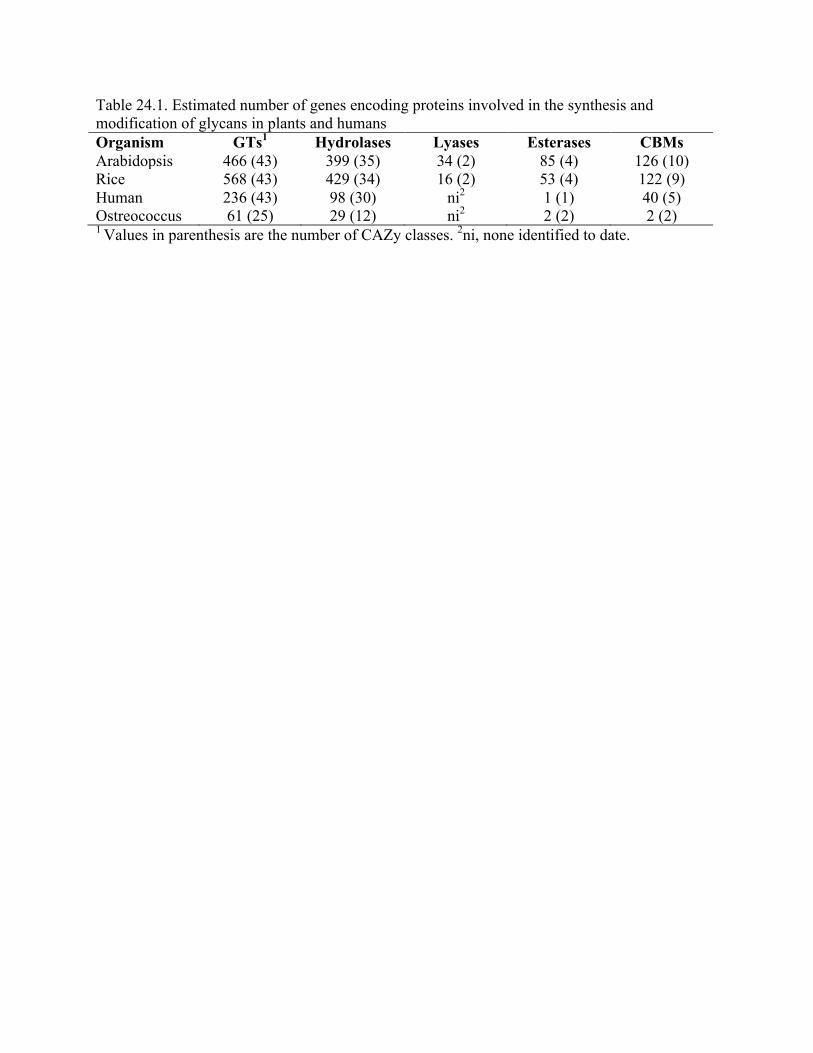
Table 24.1. Estimated number of genes encoding proteins involved in the synthesis and modification of glycans in plants and humans Organism GTs1 Hydrolases Lyases Esterases CBMs Arabidopsis 466 (43) 399 (35) 34 (2) 85 (4) 126 (10) Rice 568 (43) 429 (34) 16 (2) 53 (4) 122 (9) Human 236 (43) 98 (30) ni2 1 (1) 40 (5) Ostreococcus 61 (25) 29 (12) ni2 2 (2) 2 (2) 1 Values in parenthesis are the number of CAZy classes. 2ni, none identified to date.
