associate professor of radiology department of … lifelong learning/meetings/asm2016... · carlos...
TRANSCRIPT

Carlos Torres MD, FRCPC, Associate Professor of Radiology Department of Radiology, University of Ottawa [email protected]

None

1. Simplify the complex imaging anatomy of the BP using clear anatomical landmarks.
2. Outline different MR protocols.
3. Review BP pathologies using case-based approach.

Anatomy

Brachial Plexus Formed by ventral rami of the nerves C5 -T1
- pre fixed - post fixed Responsible for motor and cutaneous innervation of upper extremity, except for:
◦ Motor: Trapezius and levator scapulae ◦ Cutaneous: Axila, suprascapular & scapular regions

Roots
Trunks Divisions
Cords
Branches
Brachial Plexus Segments

Radiologists
Technologists Drink
Cold
Beer
Brachial Plexus Segments

*
^
A
R
*
^
The ventral rami of the spinal nerves C5 to T1 are the roots of the plexus.
Roots
A
A
M
P

T
*
^
*
^
A
Trunks C5 - C6: Upper T C7 : Middle T C8 – T1: Lower T

D
*
^
*
^
A
Each trunk splits in 2 to give an anterior and posterior division
Divisions

Divisions

C
*
^C
B
Lat: Ant divisions of sup & middle trunks Medial: Ant division of lower trunk Post: 3 post divisions
Cords

B
^
Musculocutaneous N.
Axillary N.
Median N.
Radial N.
Ulnar N.
Branches

Branches


Method of choice
Multi planar
Exquisite soft-tissue contrast
Castillo. AJR 2005, 185: S196-204 Todd et al. Top Magn Reson Imaging 2004, 15: 113-125 Saifuddin. Skeletal Radiol 2003, 32: 375-387 Wittenberg et al. Radiographics 2000, 20:1023-1032
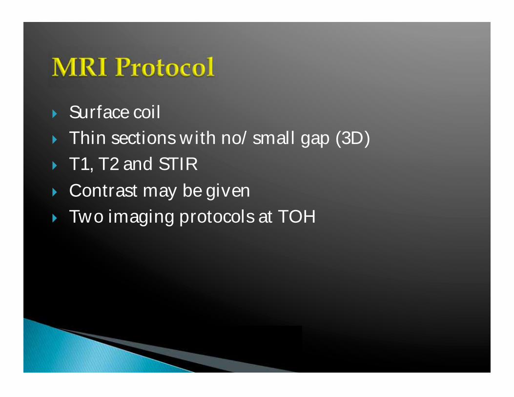
Surface coil Thin sections with no/small gap (3D) T1, T2 and STIR Contrast may be given Two imaging protocols at TOH

Sequence Time ST TR TE
Cor T2 Space 5:02 1 3800 191
Cor T1 2D 4:10 3.5 643 13
Cor T2 STIR 3:27 1.4 3800 195
Sag T1 2D 4:54 4 730 12
Neck coil and body array Localizer in 3 planes

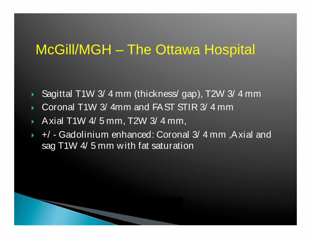
Sagittal T1W 3/4 mm (thickness/gap), T2W 3/4 mm Coronal T1W 3/4mm and FAST STIR 3/4 mm Axial T1W 4/5 mm, T2W 3/4 mm, +/- Gadolinium enhanced: Coronal 3/4 mm ,Axial and
sag T1W 4/5 mm with fat saturation
McGill/MGH – The Ottawa Hospital

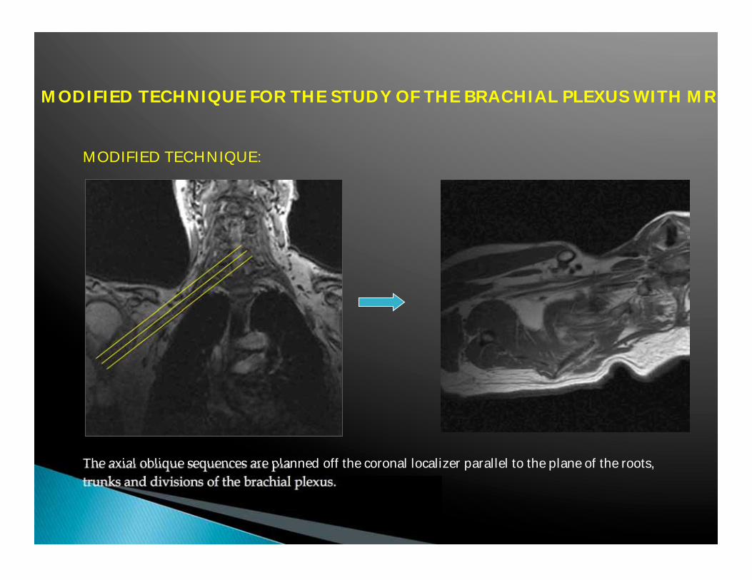
MODIFIED TECHNIQUE FOR THE STUDY OF THE BRACHIAL PLEXUS WITH MR
MODIFIED TECHNIQUE :
• 3 plane LOCALIZER • Increase number of slices in the coronal plane
• Parameters FSE T1 • Parameters FSE T2 Matrix: 448x224 cm

CONVENTIONAL TECHNIQUE MODIFIED TECHNIQUE
LOCALIZER

MODIFIED TECHNIQUE FOR THE STUDY OF THE BRACHIAL PLEXUS WITH MR
MODIFIED TECHNIQUE
MODIFIED TECHNIQUE FOR THE STUDY OF THE BRACHIAL PLEXUS WITH MR
MODIFIED TECHNIQUE:
The axial oblique sequences are planned off the coronal localizer parallel to the plane of the roots, trunks and divisions of the brachial plexus.

MODIFIED TECHNIQUE FOR THE STUDY OF THE BRACHIAL PLEXUS WITH MR
MODIFIED TECHNIQUE:
The coronal sequences are planned off the axial oblique dataset following the plane of the brachial plexus.

MODIFIED TECHNIQUE FOR THE STUDY OF THE BRACHIAL PLEXUS WITH MR
MODIFIED TECHNIQUE:
The sagital sequences are planned off the axial oblique images, perpendicular to the segments of the brachial plexus.

MODIFIED TECHNIQUE FOR THE STUDY OF THE BRACHIAL PLEXUS WITH MR
CONVENTIONAL TECHNIQUE MODIFIED TECHNIQUE

CONVENTIONAL TECHNIQUE MODIFIED TECHNIQUE

CONVENTIONAL TECHNIQUE MODIFIED TECHNIQUE

MODIFIED TECHNIQUE FOR THE STUDY OF THE BRACHIAL PLEXUS WITH MR
CONVENTIONAL TECHNIQUE MODIFIED TECHNIQUE
Axial T1 : 7 min 25 sec 3 min 59 sec Axial T2: 8 min 03 sec 3 min 54 sec Coronal T1: 4 min 22 sec 4 min 36 sec Coronal T2: 4 min 44 sec 3 min 54 sec Sagital T1: 9 min 16 sec 6 min 17 sec Sagital T2: 7 min 32 sec 6 min 08 sec Total scan time: 41 min 22 sec 28 min 48 sec


2005 a b
c
Case: 27 y/o pt with left ulnar neuropathy

2011 d
a b
c


Pathology
Vague and nonspecific symptoms.
Trauma: most common cause of plexopathy
Tumors: 2nd most common
Post radiation
Others : Inflammatory, infectious and hereditary

Imaging studies play an essential role in differentiating preganglionic injuries from postganglionic lesions, a differentiation that is crucial for determining the management of BPI

Trauma may be due to: Traction/Compression Penetrating injuries Local fractures or dislocations.
What to look for? Pseudomeningoceles Clumping, thickening and signal Hematomas

25 y/o pt, assault


Concentric rings of varying signal intensity due to clot that forms the walls of this pseudo aneurysm
Post Traumatic Pseudo aneurysm
c/o Mauricio Castillo, UNC

MVA
Stretch injury
Pseudo meningoceles

A B
C
Stretch injury Pseudo meningocele + n root avulsion

Primary: Schwannoma Neurofibroma Secondary:
Direct extension/compression: tumors in the vicinity of the BP: lung, bones or soft tissues of the neck. Metastasis: Breast, lung.


NEUROFIBROMA


NEUROFIBROMAS NF1

NF1

c/o Manu Shroff, Sickkids Univ of Toronto.
10 y/o pt with neck swelling since he was 18 months

Schwannomatosis
c/o Manu Shroff, Sickkids U of Toronto.
- 3rd major form of NF - Distinct from NF1 and NF2 - Noncutaneous schwannomas - Absence of vest schwannomas


PANCOAST TUMOR


METS MELANOMA

58 y/o pt with pain in the left arm

NEUROLYMPHOMATOSIS: B-cell NHL

B A
54 y/o pt with left brachial plexopathy


B A
NEUROLYMPHOMATOSIS: T-cell Lymphoma

NEUROLYMPHOMATOSIS: T-cell Lymphoma

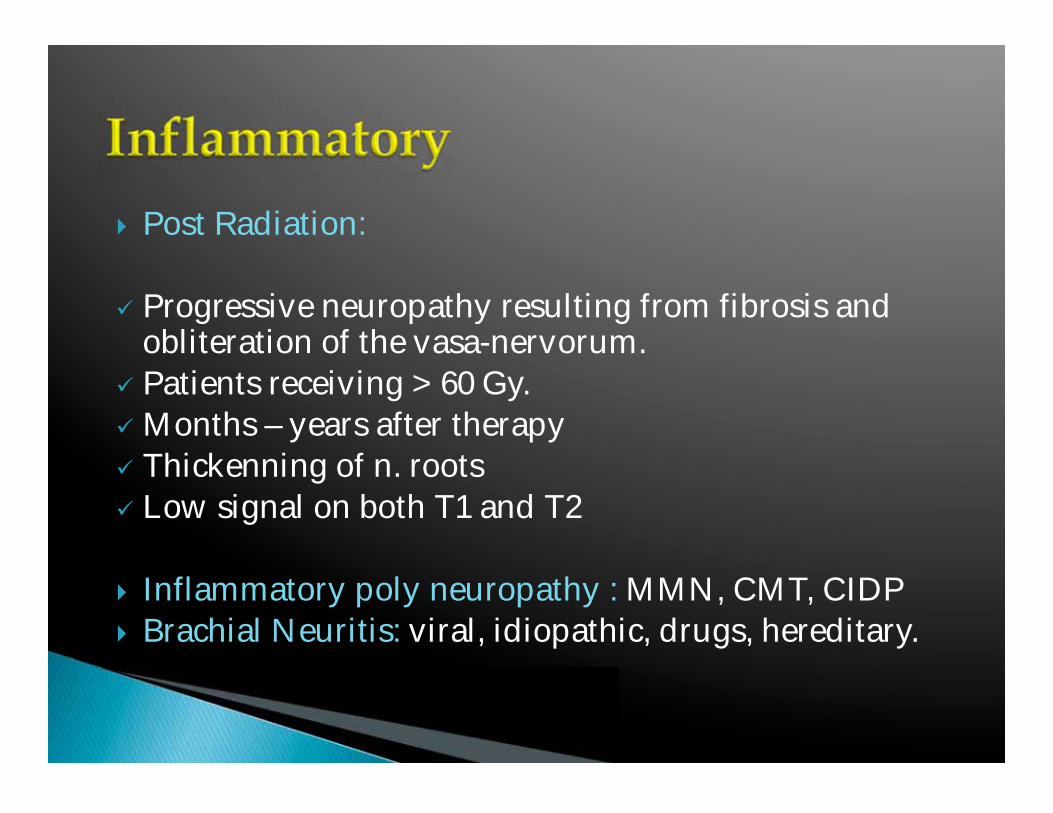
Post Radiation: Progressive neuropathy resulting from fibrosis and obliteration of the vasa-nervorum. Patients receiving > 60 Gy. Months – years after therapy Thickenning of n. roots Low signal on both T1 and T2
Inflammatory poly neuropathy : MMN, CMT, CIDP Brachial Neuritis: viral, idiopathic, drugs, hereditary.
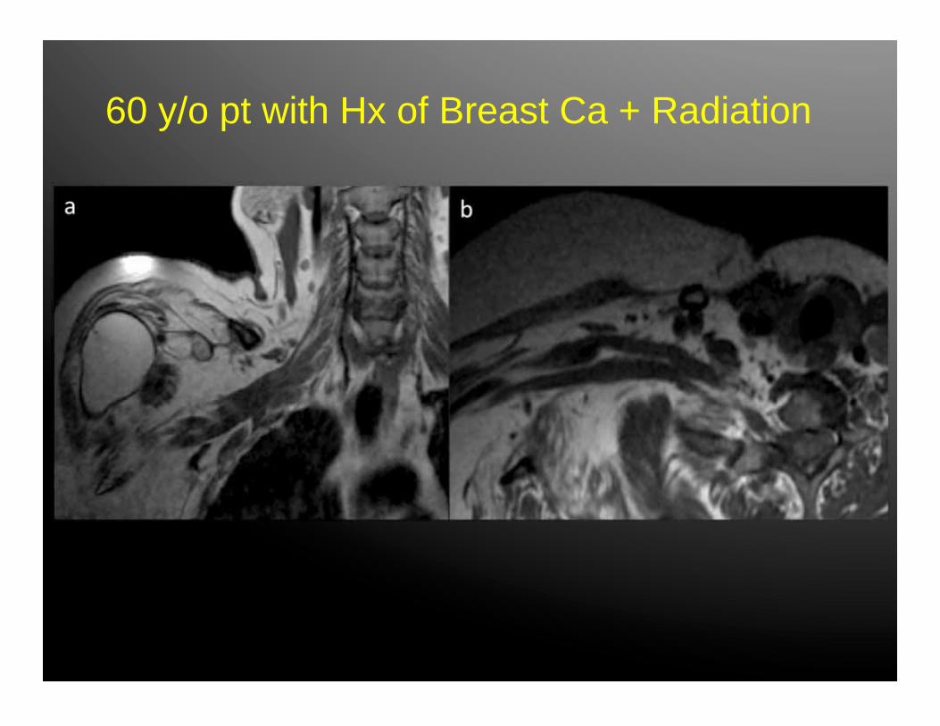
60 y/o pt with Hx of Breast Ca + Radiation


26 y/o pt with bilat weakness and numbness arms/legs

CHRONIC INFLAMMATORY DEMYELINATING POLYNEUROPATHY
CIDP

MR imaging & nerve root thickening:
Seen in ~40% children, ~60% adult CIDP patients

CMT1A patient
CIDP patient
Nerve root thickening also noted in other diseases:
MR imaging & nerve root thickening:


Diffusion tensor imaging (DTI) and tractography of the brachial plexus: Feasibility and initial experience in neoplastic conditions.
Vargas M et al. Neuroradiology (2010) 52:237–245 6 normal volunteers, 12 patients benign & malignant ( 3 Qx, 9 medical) ADC & FA maps 2 Radiologists

No statistically significant difference in FA and ADC values of normal fibers and fibers at the level of pathology. Tractography revealed major differences regarding fiber architecture.
Benign: Malignant: Displacement Disruption/Destruction Encasement Disorganization

Tractography of the brachial plexus in a 37-year-old male volunteer

MIP Coronal reconstruction of the 3D STIR SPACE sequence showing a distal schwannoma of the brachial plexus. The displaced fibers of the posterior cord (white arrows) passing around the schwannoma (asterisk) suggesting an easier surgical enucleation. The findings were confirmed at surgery

Step-by-step reconstruction of the tractography of the brachial plexus in a 42 year-old male patient …fibers within and around the benign neurogenic tumor

Sixty-five-year-old patient with adenocarcinoma of the lung, disorganization and interruption of nerve fibers on the tractography reconstruction image
Rads & Techs Drink Cold Beer MR is the imaging method of choice Different protocols: 3T vs 1.5T Advanced Imaging Techniques

Carlos Torres MD, FRCPC, Associate Professor of Radiology Department of Radiology, University of Ottawa [email protected]