association of a novel human fe65-like protein with the cytoplasmic domain of the $\beta $-amyloid...
TRANSCRIPT
Association of a Novel Human FE65-Like Protein with the Cytoplasmic Domain of the $\beta$-amyloid Precursor ProteinAuthor(s): Suzanne Y. Guénette, Jing Chen, Paul D. Jondro and Rudolph E. TanziSource: Proceedings of the National Academy of Sciences of the United States of America,Vol. 93, No. 20 (Oct. 1, 1996), pp. 10832-10837Published by: National Academy of SciencesStable URL: http://www.jstor.org/stable/40240 .
Accessed: 08/05/2014 11:43
Your use of the JSTOR archive indicates your acceptance of the Terms & Conditions of Use, available at .http://www.jstor.org/page/info/about/policies/terms.jsp
.JSTOR is a not-for-profit service that helps scholars, researchers, and students discover, use, and build upon a wide range ofcontent in a trusted digital archive. We use information technology and tools to increase productivity and facilitate new formsof scholarship. For more information about JSTOR, please contact [email protected].
.
National Academy of Sciences is collaborating with JSTOR to digitize, preserve and extend access toProceedings of the National Academy of Sciences of the United States of America.
http://www.jstor.org
This content downloaded from 169.229.32.137 on Thu, 8 May 2014 11:43:32 AMAll use subject to JSTOR Terms and Conditions
Proc. Natl. Acad. Sci. USA Vol. 93, pp. 10832-10837, October 1996 Cell Biology
Association of a novel human FE65-like protein with the cytoplasmic domain of the ,3-amyloid precursor protein SUZANNE Y. GU1NETTE, JING CHEN, PAUL D. JONDRO, AND RUDOLPH E. TANZI*
Genetics and Aging Unit, and Department of Neurology, Massachusetts General Hospital-East, Harvard Medical School, 149 13th Street, Charlestown, MA 02129
Communicated by Paul Greengard, Rockefeller University, New York, NY July 2, 1996 (received for review May 13, 1996)
ABSTRACT We identified a novel human homologue of the rat FE65 gene, hFE65L, by screening the cytoplasmic domain of f3-amyloid precursor protein (f3PP) with the "in- teraction trap." The cytoplasmic domains of the f3PP homo- logues, APLP1 and APLP2 (amyloid precursor-like proteins), were also tested for interaction with hFE65L. APLP2, but not APLP1, was found to interact with hFE65L. We confirmed these interactions in vivo by successfully coimmunoprecipitat- ing endogenous f3PP and APLP2 from mammalian cells overexpressing a hemagglutinin-tagged fusion of the C- terminal region of hFE65L. We report the existence of a human FE65 gene family and evidence supporting specific interactions between members of the f3PP and FE65 protein families. Sequence analysis of the FE65 human gene family reveals the presence of two phosphotyrosine interaction (PI) domains. Our data show that a single PI domain is sufficient for binding of hFE65L to the cytoplasmic domain of f3PP and APLP2. The PI domain of the protein, Shc, is known to interact with the NPXYp motif'found in the cytoplasmic domain of a number of different growth factor receptors. Thus, it is likely that the PI domains present in the C-terminal moiety of the hFE65L protein bind the NPXY motif located in the cytoplasmic domain of 13PP and APLP2.
Alzheimer disease is characterized by neuronal loss and a relatively high abundance of ,B-amyloid plaques and neurofi- brillary tangles in the brains of affected individuals. ,B-amyloid plaques are primarily composed of an -4-kDa peptide called ,B-amyloid peptide (A,B), which is a normal soluble proteolytic product derived from the ,B-amyloid precursor protein (,BPP) (1, 2). Molecular investigations aimed at determining the pathogenic events leading to neurodegeneration in Alzheimer disease have demonstrated that either an increase in A,B secretion (3, 4) or stoichiometric changes in the ratio of AJ,342/AJ1-40 are brought about by familial Alzheimer dis- ease-associated mutations in the APP and the recently iden- tified presenilin (PS1 and PS2) genes (5, 44). Overexpression of ,BPP or the C-terminal moeity of ,BPP including the A,B domain has been shown to be toxic to neurons (6-8). However, it is not clear in these experiments whether cell death is mediated directly by amyloidogenic A,B or indirectly as a consequence of the overexpression of J3PP/J3PP C terminus. OPP is a member of a family of proteins that include the ,BPP-like proteins, APLP1 and APLP2 (9-11), that resemble type I cell surface receptors (12). Hence, the toxicity associ- ated with overexpression of ,BPP and the ,BPP C terminus may be due to interference with normal ,BPP function.
O,PP is transported to the cell surface following maturation in the Golgi complex and is then either secreted or reinter- nalized (13, 14). Two of the products of ,BPP processing are the secreted ectodomain of ,BPP and the AP. peptide (15, 16). The interdependence of the processing pathways responsible for
The publication costs of this article were defrayed in part by page charge payment. This article must therefore be hereby marked "advertisement" in accordance with 18 U.S.C. ?1734 solely to indicate this fact.
producing these ,BPP products has been illustrated by the observed decrease in the levels of A,B generated when the secreted ectodomain of ,BPP is increased (17). The internal- ization of cell surface ,BPP occurs via the endocytic pathway and leads to the appearance of C-terminal fragments contain- ing intact A,B in lysosomes (18). The relationship between the internalization of ,BPP and the generation of A,B is underscored by the finding that attenuation of A,B release is accompanied by a reduction in ,BPP internalization (19). Internalization of ,BPP by clathrin-coated vesicles is required for entry of cell- surface ,BPP into the endosomal/lysosomal pathway, and this form of internalization of ,BPP uses the NPXY motif in the cytoplasmic domain of ,BPP (20,21). Deletion of the YENPTY sequence of ,BPP not only impairs endocytosis of cell-surface ,BPP, but alters its intracellular trafficking by increasing its targeting into the endocytic pathway without first reaching the cell surface (22).
To identify the cellular factors and mechanisms responsible for mediating the intracellular trafficking and/or function of cell surface ,BPP, we screened for proteins that associate with the cytoplasmic domain of ,BPP. Using the interaction trap, we isolated one of three human homologues of the rat FE65 gene, which we have termed hFE65L. We demonstrate herein the interaction of hFE65L with ,BPP as well as its homologue, APLP2, by coimmunoprecipitation from mammalian cells.
EXPERIMENTAL PROCEDURES
Interaction Trap Screening. Screening for proteins that interact with the cytoplasmic domain of ,BPP was performed as described (23). DNA'fragments coding for the cytoplasmic domains of human OPP (nt 2092-2235), APLP1 (5'- CCGGAATTCAGGAAGAAGCCCTACGGGGCTATC-3' and 5'-CGCGGATCCTCAGGGTCGTTCCTCCAGGAA- GCG-3'), and APLP2 (nt 2222-2365) were amplified using the polymerase chain reaction (PCR) with primers containing EcoRI and BamHI sites on either end. The PCR products were subcloned into the pEG202 backbone and were sequenced to verify that the APP/APLP coding sequences were in-frame with the LexA coding sequence. We also confirmed the expression of these fusion proteins in yeast total protein extracts by Western blot analysis using an anti-LexA antibody (a gift from R. Brent, Massachusetts General Hospital, Bos- ton). We initially transformed the yeast strain EGY48 that contained LexAop-LEU2, LexAop-LacZ, and LexA-,BPP(649- 695) with DNA from a galactose-inducible activation domain- cDNA fusion library constructed from 22-week-old human fetal brain [a gift from D. Krainc (Massachusetts General Hospital, Boston) and R. Brent] using'the LiAc transformation protocol of Schiestl and Gietz (24). Approximately 2 x 10⁶
Abbreviations: A,B, ,B-amyloid peptide; ,BPP, ,B-amyloid precursor protein; APP, ,B-amyloid precursor protein gene; HA, hemagglutinin; PI, phosphotyrosine interaction; CMV, cytomegalovirus. Data deposition: The sequence reported in this paper has been deposited in the GenBank data base (accession no. U62325). *To whom reprint requests should be addressed.
10832
This content downloaded from 169.229.32.137 on Thu, 8 May 2014 11:43:32 AMAll use subject to JSTOR Terms and Conditions

Cell Biology: Guenette et al. Proc. Natl. Acad. Sci. USA 93 (1996) 10833
yeast colonies were scraped, pooled, and frozen, and the library was plated at a multiplicity of 20X on galactose Ura- His- Trp- Leu- media. We isolated 68 Leu+ colonies and streaked these to Ura- His- Trp- glucose plates and retested them on glucose Ura- His- Trp- Leu- glucose, Ura- His- Trp- Leu- galactose, Ura- His- Trp- glucose 5-bromo-4- chloro-3-indolyl ,B-D-galactoside (X-Gal), and Ura- His- Trp- galactose X-Gal media to confirm that induction of the reporter genes was dependent on galactose. Of these 68 Leu+ colonies, 39 showed galactose-dependent leucine prototrophy and blue color production on X-Gal medium. The inserts of rescued plasmids bearing the TRP1 marker were amplified by PCR using the following primers: 5'-CTTGCTGAGTGGA- GATGCC-3' and 5'-GCCGACAACCTTGATTGG-3'; the products were classified by Southern blot analysis using the largest insert as a probe.
cDNA Cloning, Sequencing, and Computer Analyses. The cDNA insert of clone 4-2A capable of producing the highest ,B-galactosidase activity as a result of interaction with the cytoplasmic domain of ,BPP was sequenced using the modified Sequenase T7 DNA polymerase (United States Biochemical) for the dideoxy-chain termination method (25). To obtain a full-length open reading frame for the hFE65L gene, we screened a human kidney cDNA library constructed in AGT10 (CLONTECH) using the 4-2A insert as a probe and isolated the 61-19 clone. The cDNA insert from this clone was sub- cloned into the pSK+ Bluescript sequencing vector (Strat- agene) and both strands were sequenced using' oligonucleotide primers. Sequence comparisons were performed using the BLAST series of programs and protein motifs were identified using the ProfileScan program (ISREC WWW-Server). Sim- ilarities and identities reported are data obtained from BESTFIT
analyses. We assembled hFE65 and hFE65L2 from nucleotide sequences recovered from the expressed sequence tag data base. hFE65 was assembled from GenBank accession nos. T09348, T31017, R67159, F07610, R87225, and D56274. hFE65L2 was assembled from GenBank accession nos. T54909 and R84592.
Northern Blot Analysis. The 32P-labeled 4-2A EcoRI/XhoI cDNA fragment was used as a probe for hybrization to a nitrocellulose filter onto which 2 ,ug of poly(A)+ RNA isolated from human tissues was immobilized (CLONTECH). The hybridization was performed at 42?C in the presence of 50% formamide and washed in 0.1X SSC and 0.1% SDS at 50?C.
Cell Transfections. The hFE65L sequence encoded by the entire 4-2A insert (C-terminal 203 amino acids) was expressed in mammalian cells under the control of the cytomegalovirus (CMV) promoter. To achieve this, we subcloned the Hindill! XhoI fragment of the 4-2A clone into the pCDNA3 vector (Invitrogen) such that the protein expressed is the product of the HA-tag-hFE65L cDNA fusion'initially' created in the construction of the activation domain-cDNA fusion library. As a negative control for coimmunoprecipitation experiments, we used a pCDNA3-derived plasmid, pTPRlHA, constructed with a human cDNA fragment that codes for an HA-tagged fusion protein that does not interact with the ,BPP cytoplasmic domain in, the interaction trap. pTPRlHA [a gift from Anita Murthy (Massachusetts General Hospital, Boston) and Jim Gusella (Massachusetts General Hospital, Boston)] was con- structed by subcloning the EcoRV/XhoI cDNA fragment of the activation domain-cDNA fusion library plasmid into the pCDNA3 backbone. Both of these constructs were transfected into COS7 African green monkey kidney cells, human H4 neuroglioma cells, H4 cells stably transfected with APP695 or APP751 (26), and' human SY5Y neuroblastoma cells. Cells were transfected following the- manufacturer's instructions with 10 ,ug of plasmid DNA using 50 ,u of lipofectamine (GIBCO/BRL) per 100 mm culture dish and were harvested at ~72 hr after transfection. Cells were routinely grown' in DMEM supplemented with 25 -mM Hepes, 4.5 mg/ml D-
glucose, 2 mM glutamine, 20 units/liter penicillin, 100 mg/liter streptomycin, and 10% fetal bovine serum.
Immunoprecipitations and Western Blot Analyses. Cells were washed twice with cold PBS and directly lysed in 10 mM Tris (pH'8.0), 0.14 M NaCl, 0.025% NaN3, 5 mM EDTA, and 1% Triton X-100 buffer containing 1 pg/ml leupeptin, 10 p,g/ml aprotinin, 1 ,ug/ml pepstatin, and 0.2 mM phenylmeth- ylsulfonyl fluoride for 30 min at 4?C. The lysates were centri- fuged at 12,000 rpm for 10 min at 4?C in a microfuge. Total protein was estimated for the recovered supernatants with the BCA protein assay (Pierce) using BSA as a standard. Immu- noprecipitations were performed on 250 ,tg of total protein using the anti-HA epitope tag monoclonal antibody, 12CA5 (Boehringer Mannheim). Immune complexes were collected with protein G-Agarose (Boehringer Mannheim), and then washed three times with lysis buffer. The immunoprecipitates were either heated at 55?C for 15 min or boiled for 5 min and resolved on a 7.5 to 15% gradient gel, transferred to polyvi- nylidene difluoride membrane (Pierce), and immunoblotted with the following antibodies: 22C11 (13), 369W (27), C7 (28), D2-I (29), and 12CA5 (30). The secondary antibody was either peroxidase-conjugated sheep anti-mouse Ig or peroxidase- conjugated donkey anti-rabbit Ig (Amersham). Immunoblot- ted proteins were detected with an Enhanced Chemilumines- cence system (Amersham).
RESULTS
The ,3PP Cytoplasmic Domain Interacts with hFE65L. A homologue of the rat FE65 gene was isolated from a human fetal brain library using the interaction trap screen (23). We isolated 33 independent colonies showing that galactose- dependent transcriptional activation of the two reporter genes contained cDNA inserts of 1.1 to 1.3 kb that cross-hybridize to one another. The longest cDNA insert (clone 4-2A) codes for a 203 amino acid open reading frame that is 56% identical and 73% similar (Fig. 1) to the rat brain transcriptional activator, FE65 (31, 32).
To verify the specificity of this interaction, we transformed cells bearing the activation domain-hFE65L' fusion with five lexA-fusion constructs containing cdc2, fus3, bicoid, the cys- rich region of ,BPP, and the GTPase activating protein-related domain of the neurofibromatosis I protein and found that transcriptional activation of the two reporters requires the expression of the cytoplasmic domain of ,BPP (data not shown).
hFE65L also Interacts with the Cytoplasmic Domain of APLP2. We also tested whether the activation domain-hFE65L fusion was able to activate transcription of both reporter genes in association with the cytoplasmic domains of APLP1 and APLP2. Our results indicate that hFE65L protein interacts with the cytoplasmic domain of APLP2 but not with that of APLP1 (data not shown).
Characterization of the hFE65L Gene and its Expression Pattern. We isolated a cDNA clone, 61-19, that overlaps with the insert of the 4-2A clone. This cDNA clone contains an insert of -2.3 kb. When assembled, these two clones represent 2.9 kb of hFE65L cDNA with a poly(A) tail on the 3' end. There are three putative start methionines upstream of the FE65 homologous sequence'(Fig. 1), with the third represent- ing the best candidate for the translation initiation codon (33). However, we cannot exclude the possibility that the start methionine is positioned 5' of the 61-19 sequence. The pre- dicted open reading frame of clone 61-19 encodes 730 amino acids. Thus, the predicted hFE65L protein contains an'extra 72 amino acids, 55 of these amino acids are N terminal to the start methionine of the rat FE65 protein (Fig. 1).
The predicted amino acid sequence of hFE65L is 51% identical and 70% similar to the overlapping 658 amino acids of the rat FE65 protein (Fig. 1). Searches of the GenBank data base with the open reading frame of the hFE65L revealed the
This content downloaded from 169.229.32.137 on Thu, 8 May 2014 11:43:32 AMAll use subject to JSTOR Terms and Conditions
10834 Cell Biology: Guenette et al. Proc. Natl. Acad. Sci. USA 93 (1996)
50 hPE65L RHTRTHRDTR HTYTHAHTDA HTCTHIIHRDT QMHTHTICRK KYALTNIQAA
*FE 5 S .......... .. hFE65 .......... ..
h 3FE 6 5L 2 .. . 9.... .. . . .@ . @ . . . .. . . . . . . e e . @ @.... . . ............................ . . . . .
ottiL ~~~~~~~100 hFE6SL IUGLSDPAAQP LL LVW RWQOQQDP MKNLSPTAVI
FE65 . M. OMGEGS VPEP WLE I R HRDQN RN....... V hFE65 ....... . . .. . ..........
hFEE 5L2 ........ . .......... ....................
- - ~~~~~~~~~~~150 hFE65L NI rEKLESK PHPQDSSSC E ILPSQPRRT L YAYADL ETSARELEQN
FE65 TJEASQ AETAPLGPK GINHLYSELE 0 LHGSAKIINT hFE65 . X. *....... .......... ....... ;.. .......,
hFE65L:2 e . . 9 . . ...... .......... .......... .I.... @.9 : . . 9...
hFE65L R SHGT' ESQPVQGQAS TIIGNGl LL QKPNRUS iQVAT&SU FE6 5 Q>GPDEq a AGEVEEED EDEEEEEEE EDLSS3L L
hPE6S TI IE TGAUTDCA RIQt&P iS B FE65 *V Wi SLS tL NY WJ
hFE65 .........
hFE6 5L2 ,.. , @. ...,,........., ,, ... . .. .. . ....
299 hFE65L K. I SDHSEQ PIPA SISEARESIP DL
FE65 ...PNA.. hFE65 ........... .
hFE65L2 . .
- - -- - - ~~~~~~349 hFE65L QGSRKGSSLS VTPSPT E K Q N INSN
FE65 . ... GRAAP SQGNSP O S QLr.IH QEEFEEGEPEPSEEAPM h?EE65 ..... ........ ... -IH GEGFEDGE EPSDEAPN
hFE65L2 .... . ........ .... ........... . .........
hFE65L DPS: IA YASLKb NAfIfU)DDS .CSItDbk
hFE6E5L _ E._ FE65 Ep PS - Ai hPEE5 3
hFE 5L N D DR S V W B.U FE65 DEW2 NQ TLLrU(E t
hFE65L2 ... 954
hFE65L YRS SEAl
FE65 DEV GL NOTLSI
hFE6L2 EVSSD.CC.S DI.SP . V *~~~~~~
hPbE6 2_tEzSL;C I)IP
hFE6 5L SSL _E _AtNVNl!
FE65 W hFE65 W LrHSs:.
hFlS65LW VtB i Jl) S Q i nEGTirH TAESRN
hFE65L2 CDS J4ISEK d
FE65 QWPSHVSPSELL.HQ Q r 2 hPE65 Q PSIIVS SEE L.HQ Q TS3JA
hFE65L2 AWIPTSW#HPIQ A .
hPE65L IrQSE V_N P1Q2 6
FE65 A PSN HQ L SQTSTSCL hFE6S A PS _-_N s. L _SQAST.
hFE65L2 .7: A .@@@@.. ..
730 hFE6$L EPTTNSDTS TE
FE65 SWU,S2TSTWSCLOT hFE65 S_S ......SQ
hFE65L2 . . s @. . ...... ......... 9. . ... . . . .. @ ..... . ... .. ..
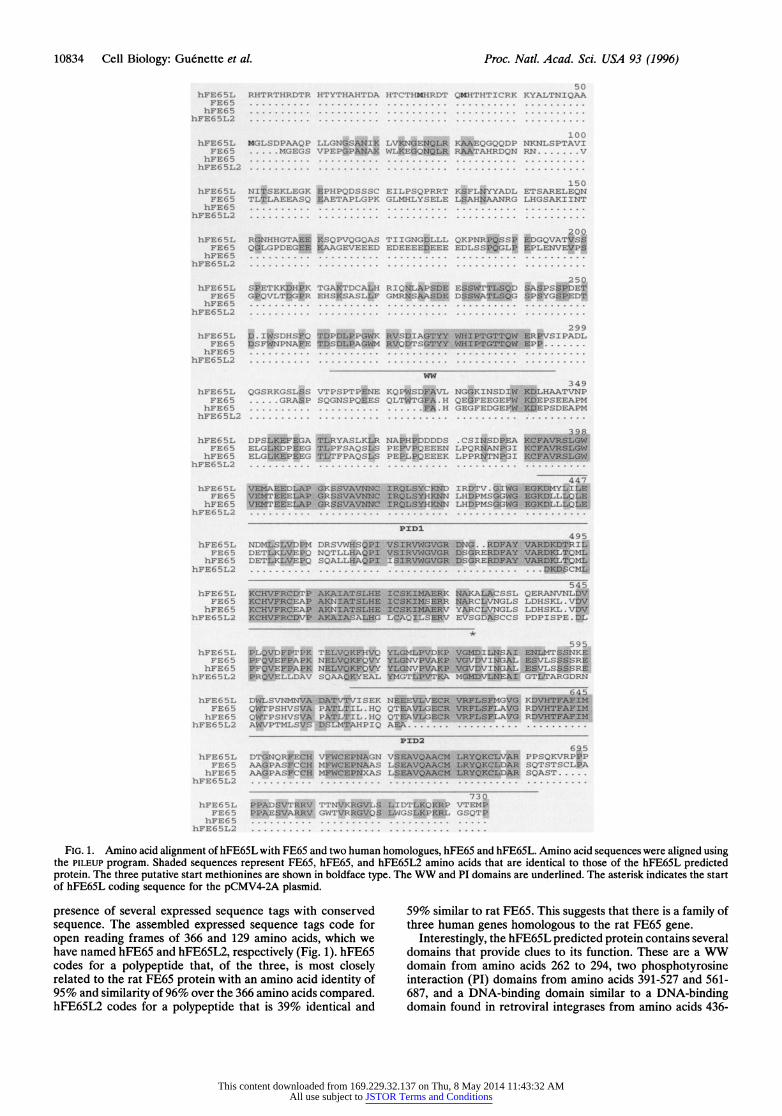
FIG. 1. Amino acid alignment of hFE65L with FE65 and two human homologues, hFE65 and hFE65L. Amino acid sequences were aligned using the PILEUP program. Shaded sequences represent FE65, hFE65, and hFE65L2 amino acids that are identical to those of the hFE65L predicted protein. The three putative start methionines are shown in boldface type. The WW and PI domains are underlined. The asterisk indicates the start of hFE65L coding sequence for the pCMV4-2A plasmid.
presence of several expressed sequence tags with conserved sequence. The assembled expressed sequence tags code for open reading frames of 366 and 129 amino acids, which we have named hFE65 and hFE65L2, respectively (Fig. 1). hFE65 codes for a polypeptide that, of the three, is most closely related to the rat FE65 protein with an amino acid identity of 95% and similarity of 96% over the 366 amino acids compared. hFE65L2 codes for a polypeptide that is 39% identical and
59% similar to rat FE65. This suggests that there is a family of three human genes homologous to the rat FE65 gene.
Interestingly, the hFE65L predicted protein contains several domains that provide clues to its function. These are a WW domain from amino acids 262 to 294, two phosphotyrosine interaction (PI) domains from amino acids 391-527 and 561- 687, and a DNA-binding domain similar to a DNA-binding domain found in retroviral integrases from amino acids 436-
This content downloaded from 169.229.32.137 on Thu, 8 May 2014 11:43:32 AMAll use subject to JSTOR Terms and Conditions

Cell Biology: Guenette et al. Proc. Natl. Acad. Sci. USA 93 (1996) 10835
577. While these domains were initially identified in the rat FE65 protein (34, 35), the consensus sequences of these domains are conserved in the hFE65L predicted protein (Fig. 1).
To determine the expression pattern of the hFE65L gene and the size of the mRNA(s), we hybridized the human fetal brain cDNA insert from clone 4-2A to messenger RNA isolated from human tissues (Fig. 2). In contrast to the expression pattern for the rat FE65 gene for which a single transcript of 2.7 kb is detected (31), our results show that, with the exception of skeletal muscle that contains three additional transcripts sized 1.9, 4.4, and 5.4 kb, there are two major transcripts of -3.7 kb and 7.5 kb for the hFE65L gene in all human tissues tested. The latter pattern of expression was also observed in many different cell lines including COS7 and H4 cells (data not shown). In comparison, the rat FE65 transcript is brain specific (31).
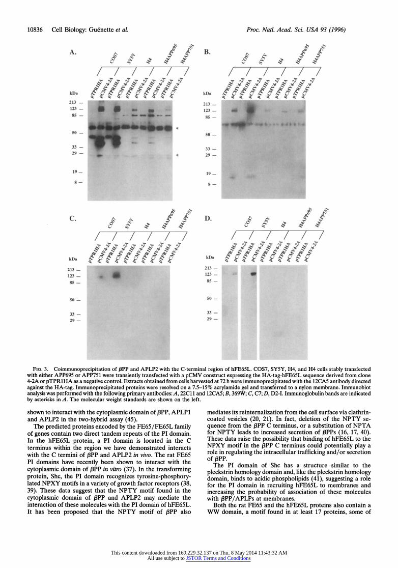
pPP and APLP2 Can Be Coimmunoprecipitated with the C-Terminal 203 Amino Acids of hFE65L from Mammalian Cells. To further characterize the association between the hFE65L and the O3PP family of proteins, we performed im- munoprecipitation experiments on mammalian cells tran- siently transfected with an HA-tagged 4-2A cDNA insert under the control of the CMV promoter. Cell lysates obtained from COS7, H4, and SY5Y cells overexpressing the HA- tagged hFE65L C-terminal fusion protein were immunopre- cipitated with the anti-HA antibody, 12CA5.
Western blot analyses of the immunoprecipitates were per- formed using the 22C11 antibody raised against the N-terminal portion of OPP. The 22C11 staining revealed proteins of approximately 140, 120, and 105 kDa in all three cell types examined (Fig. 3A) corresponding in size to mature ,BPP751, immature f3PP751/mature f3PP695 (unresolved in our gel system), and immature PPP695, respectively. As a negative control for the coimmunoprecipitation experiments, we used
0I u
CX Y
kb
9.5 - 7.5 -
4.4-
2A-
123 -
FIG. 2. Expression of the hFE65L gene in adult human tissues. A multiple tissue Northern blot (CLONThCH) with 2 jig of poly(A)+
RN pe lan wa_yrdzdtote,Plbld-AEoIXo cDNA famn. Th hyrdzto wa pefre at 42Ci,h peecof0%fraieadwsein01 SSC and 01% SDS atJ 50C RN siz makr in kbaesonontergt
an HA-tagged fusion protein (TprlHA) that did not interact with the cytoplasmic domain of I3PP in the interaction trap. Thus, coimmunoprecipitation of ,BPP is dependent on the expression of the HA-tagged hFE65L moiety. We simulta- neously blotted this filter with the 12CA5 antibody to deter- mine the amount of HA-tagged protein expressed and repeat- edly observed that the level of HA-tagged protein produced was cell type-dependent with SYSY > COS7 > H4 cells (Fig. 3A). Two 12CA5 immunoreactive bands of -40 kDa and -34 kDa (Fig. 3A) are produced from the pCMV4-2A plasmid bearing the HA-tagged hFE65L cDNA fusion. The full-length hFE65L fusion protein has a predicted molecular weight of 34 kDa. Thus, it is likely that the two first methionines in the open reading frame are being used since they are separated by -4 kDa. The pTPRlHA control reveals a single polypeptide of -44 kDa (Fig. 3A) and contains only the downstream of the two start methionines described for pCMV4-2A. The low levels of HA-tagged proteins in H4 cell extracts and differ- ences in antibody affinities may explain the absence of endog- enous PP/APLP bands in the H4 lanes of Fig. 3 B-D.
The antibody, 369W, that recognizes the C-terminal frag- ment of I3PP, also recognizes the full-length forms of 13PP present in immunoprecipitates containing the HA-tagged FE65L fusion protein. Interestingly, we also observe the interaction of the 14 to 15 kDa C-terminal fPP fragment with the HA-hFE65L fusion protein (Fig. 3B) by coimmunopre- cipitation.
Since both 22C11 and 369W recognize /3PP and APLP2 (11), we also tested immunoprecipitates with C7, a 3PP-specific (T.-W. Kim, R.E.T., and W. Wasco, unpublished results) rabbit polyclonal antibody directed against the C-terminal region of OPP. Using C7, we were able to confirm that J3PP immunoreactivity is present in immunoprecipitates obtained from COS7 cells and SY5Y cells that contain the HA-tagged FE65L fusion protein (Fig. 3C). In addition, we tested immu- noprecipitates with 6E10, a ,BPP-specific monoclonal antibody directed against the AP3 region of IPP, and similar data were obtained in the COS7 cells (data not shown). To assess whether APLP2 also interacts with the HA-tagged hFE65L fusion protein, we performed a similar experiment using an APLP2- specific antibody, D2-I. We are able to detect the interaction of APLP2 with the HA-FE65L fusion protein in SY5Y and COS7 cells, but not in H4 cells (Fig. 3D). Collectively, these results indicate that the C-terminal region of ,BPP and APLP2 indeed interact with the C-terminal region of hFE65L in mammalian cells.
DISCUSSION
Our results demonstrate that the C-terminal moiety of the hFE65L protein interacts with the cytoplasmic domain of ,BPP and APLP2 in the yeast interaction trap system and in a variety of mammalian cells. We also report the sequence of a novel, ubiquitously expressed human gene, hFE65L, and the exis- tence of two additional human homologues of the rat FE65 protein, indicating the presence of a novel gene family.
The observation that there is more than one human FE65- like protein raises the possibility of specific interactions among members of the ,BPP/APLP family and those of the hFE65L family. Our data obtained with the interaction trap suggest that the hFE65L protein interacts with the C terminus of both 13PP and APLP2, but not with that of APLP1. As with the f3PP and APLP2 gene products (10, 36), the hFE65L gene is ubiqui- tously expressed in all human tissues tested. It is of interest that both the rat FE65 protein and APLP1 are enriched in the brain (9, 31). However, unlike hFE65L, the rat FE65 protein was recently reported to interact with the C terminus of both 13PP and APLP1 in the two-hybrid assay (37). In addition, the human homolog of the rat FE65 protein (hFE65) has also been
This content downloaded from 169.229.32.137 on Thu, 8 May 2014 11:43:32 AMAll use subject to JSTOR Terms and Conditions
10836 Cell Biology: Guenette et al. Proc. Natl. Acad. Sci. USA 93 (1996)
A. B ffB. t ft
kDa W,a D
213- 2 1 3 _ . 12- 123- 85- 85-.
33- l l33- 29- _ 11 * 29-
19_ 19_ - 7 7 7
8- 8- so - ~ ~ ~ ~ ~ ~ ~ ~ ~ 44
WeS 4AN
33- 33- ||_|
| _ |~~~7_ 2,3- 29-
FIG. 3. Coimmunoprecipitation of f3PP and APLP2 with the C-terminal region of hFE65L. COS7, SY5Y, H4, and H4 cells stably transfected with either APP695 or APP751 were transiently transfected with a pCMV construct expressing the HA-tag-hFE65L sequence derived from clone 4-2A or pTPRlHA as a negative control. Extracts obtained from cells harvested at 72 h were immunoprecipitated with the 12CA5 antibody directed against the HA-tag. Immunoprecipitated proteins were resolved on a 7.5-15% acrylamide gel and transferred to a nylon membrane. Immunoblot analysis was performed with the following primary antibodies:A, 22C11 and 12CA5; B, 369W; C, C7; D, D2-I. Immunoglobulin bands are indicated by asterisks in A. The molecular weight standards are shown on the left.
shown to interact with the cytoplasmic domain of P3PP, APLP1 and APLP2 in the two-hybrid assay (45).
The predicted proteins encoded by the FE65/FE65L family of genes contain two direct tandem repeats of the PI domain. In the hFE65L protein, a PI domain is located in the C terminus within the region we have demonstrated interacts with the C termini of ,BPP and APLP2 in vivo. The rat FE65 PI domains have recently been shown to interact with the cytoplasmic domain of OPP in vitro (37). In the transforming protein, Shc, the PI domain recognizes tyrosine-phosphory- lated NPXY motifs in a variety of growth factor receptors (38, 39). These data suggest that the NPTY motif found in the cytoplasmic domain of ,BPP and APLP2 may mediate the interaction of these molecules with the PI domain of hFE65L. It has been proposed that the NPTY motif of 3PP also
mediates its reintemalization from the cell surface via clathrin- coated vesicles (20, 21). In fact, deletion of the NPTY se- quence from the O3PP C terminus, or a substitution of NPTA for NPTY leads to increased secretion of 3PPs (16, 17, 40). These data raise the possibility that binding of hFE65L to the NPXY motif in the ,BPP C terminus could potentially play a role in regulating the intracellular trafficking and/or secretion of P3PP.
The PI domain of Shc has a structure similar to the pleckstrin homology domain and, like the pleckstrin homology domain, binds to acidic phospholipids (41), suggesting a role for the PI domain in recruiting hFE65L to membranes and increasing the probability of association of these molecules with ,BPP/APLPs at membranes.
Both the rat FE65 and the hFE65L proteins also contain a WW domain, a motif found in at least 17 proteins, some of
This content downloaded from 169.229.32.137 on Thu, 8 May 2014 11:43:32 AMAll use subject to JSTOR Terms and Conditions
Cell Biology: Guenette et al. Proc. Natl. Acad. Sci. USA 93 (1996) 10837
which are known to be involved in signaling or regulatory functions (42, 43). Like the SH3 domain, the WW domain interacts with proteins containing a proline-rich motif (43). If, in fact, the interaction of O3PP with the hFE65L protein modulates OPP reinternalization, secretion, and/or function, it will be important to identify proteins that interact with the WW domain of hFE65L.
The PI domains overlap a region in the amino acid sequence of hFE65L that is similar to the retroviral integrase DNA- binding domain (31). FE65 was first described as a transcrip- tional activator based on the observation that the N-terminal half of the protein activates transcription (31). Presently, the function of the hFE65L protein is unclear, but the presence of several motifs believed to play roles in protein-protein inter- action, signal transduction, and transcriptional activation pro- vide a firm foundation for the design of future experiments. The results of such experiments should increase our under- standing of the importance of the interactions among members of the FE65/FE65L and I3PP/APLP families. It will be partic- ularly interesting to determine the consequences of such associ- ations on IPP trafficking, function, and generation of A13.
We thank Roger Brent and his group for providing the interaction trap reagents, Dimitri Krainc and Roger Brent for providing the human fetal brain activation-domain cDNA library, Wilma Wasco for the APLP1 and APLP2 clones, Anita Murthy and Jim Gusella for the pTPRlHA clone and the NFI bait construct, Gopal Thinakaran and Sam Sisodia for the D2-I antiserum, Sam Gandy for the 369W antiserum, and Dennis Selkoe for the C7 antiserum. We thank Steve Schmidt and Mia Liu for technical assistance. This research was supported by a grants from the American Health Assistance Foun- dation and the National Institutes of Health. R.E.T. is a Pew Scholar in the Biomedical Sciences.
1. Seubert, P., Vigo-Pelfrey, C., Esch, F., Lee, M., Dovey, H., Davis, D., Sinha, S., Schlossmacher, M., Whaley, J., Swindleshurst, C., McCormack, R., Wolfert, R., Selkoe, D., Lieberburg, I. & Schenk, D. (1992) Nature (London) 359, 325-327.
2. Haass, C., Koo, E. H., Mellon, A., Hung, A. Y. & Selkoe, D. J. (1992) Nature (London) 357, 500-503.
3. Citron, M., Oltersdorf, T., Haass, C., McConlogue, L., Hung, A. Y., Seubert, P., Vigo-Pelfrey, C., Lieberburg, I. & Selkoe, D. J. (1992) Nature (London) 360, 672-674.
4. Cai, X., Golde, T. E. & Younkin, S. G. (1993) Science 259, 514-516.
5. Suzuki, N., Cheung, T. T., Cai, X. D., Odaka, A., Otvos, L., Eckman, C., Golde, T. E. & Younkin, S. G. (1994) Science 264, 1336-1340.
6. Yoshikawa, K., Aizawa, T. & Hayashi, Y. (1992) Nature (London) 359, 64-67.
7. Fukuchi, K., Kamino, K., Deeb, S. S., Smith, A. C., Dang, T. & Martin, G. M. (1992) Biochem. Biophys. Res. Commun. 182, 165-173.
8. Yankner, B. A., Dawes, L. R., Fisher, S., Villa-Komaroff, Oster- Granite, M. L. & Neve, R. L. (1989) Science 245, 417-420.
9. Wasco, W., Bupp, K., Magendantz, M., Gusella, J. F., Tanzi, R. E. & Solomon, F. (1992) Proc. Natl. Acad. Sci. USA 89, 10758-10762.
10. Wasco, W., Gurubhagavatula, S., Paradis, M. D., Romano, D. M., Sisodia, S. S., Hyman, B. T., Neve, R. L. & Tanzi, R. E. (1993) Nat. Genet. 5, 95-100.
11. Slunt, H. H., Thinakaran, G., Von Koch, C., Lo, A. C. Y., Tanzi, R. E. & Sisodia, S. S. (1994) J. Biol. Chem. 269, 2637-2644.
12. Kang, J., Lemaire, H., Unterbeck, A., Salbaum, J. M., Masters, C. L., Grzeschik, K., Multhaup, G., Beyreuther, K. & Muller-Hill, B. (1987) Nature (London) 325, 733-736.
13. Weidemann, A., Konig, G., Bunke, D., Fischer, P., Salbaum, J. M., Masters, C. L. & Beyreuther, K. (1989) Cell 57, 115-126.
14. Haass, C., Schlossmacher, M. G., Hung, A. Y., Vigo-Pelfrey, C., Mellon, A., Ostaszewski, B. L., Lieberburg, I., Koo, E. H., Schenk, D., Teplow, D. B. & Selkoe, D. J. (1992) Nature (Lon- don) 359, 322-325.
15. Estus, S., Golde, T. E., Kunishita, T., Blades, D., Lowery, D., Eisen, M., Usiak, M., Qu, X., Tabira, T., Greenberg, B. D. & Younkin, S. G. (1992) Science 255, 726-728.
16. Haass, C. & Selkoe, D. J. (1993) Cell 75, 1039-1042. 17. Jacobsen, S. J., Spruy, M. A., Brown, A. M., Sahasrabudhe, S. R.,
Blume, A. J., Vitek, M. P., Muenkel, H. A. & Sonnenberg- Reines, J. (1994) J. Biol. Chem. 269, 8376-8382.
18. Haass, C., Koo, E. H., Mellon, A., Hung, A. Y. & Selkoe, D. J. (1992) Nature (London) 357, 500-503.
19. Koo, E. H. & Squazzo, S. L. (1994) J. Biol. Chem. 269, 17386- 17389.
20. Chen, W., Goldstein, J. L. & Brown, M. S. (1990) J. Biol. Chem. 265, 3116-3123.
21. Nordstedt, C., Caporaso, G. L., Thyberg, J., Gandy, S. E. & Greengard, P. (1993) J. Biol. Chem. 268, 608-612.
22. Lai, A., Sisodia, S. S. & Trowbridge, I. S. (1995) J. Biol. Chem. 270, 3565-3573.
23. Gyuris, J., Golemis, E., Chertkov, H. & Brent, R. (1993) Cell 75, 791-803.
24. Schiestl, R. H. & Gietz, R. D. (1989) Curr. Genet. 16, 339-346. 25. Sanger, F., Nicklen, S. & Coulson, A. R. (1977) Proc. Natl. Acad.
Sci. USA 74, 5463-5467. 26. Felsenstein, K. M., Hunihan, L. W. & Roberts, S. B. (1994) Nat.
Genet. 6, 251-256. 27. Buxbaum, J. D., Gandy, S. E., Cichetti, P., Ehrlich, M. E., Czer-
nik, A. J., Fracasso, R. P., Ramabhadran, T. V., Unterbeck, A. J. & Greengard, P. (1990) Proc. Natl. Acad. Sci. USA 87,6003-6006.
28. Podlisny, M. B., Tolan, B. D. & Selkoe, D. J. (1991)Am. J. Pathol. 138, 1423-1435.
29. Thinakaran, G. & Sisodia, S. S. (1994) J. Biol. Chem. 269, 22099-22104.
30. Niman, H. L., Houghten, R. A., Walker, L. E., Reisfeld, R. A., Wilson, I. A., Hogle, J. M. & Lerner, R. A. (1983) Proc. Natl. Acad. Sci. USA 80, 4949-4953.
31. Duilio, A., Zambrano, N., Mogavero, A. R., Ammendola, R., Cimino, F. & Russo, T. (1991) Nucleic Acids Res. 19, 5269-5274.
32. Faraonio, R., Minopoli, G., Porcellini, A., Costanzo, F., Cimino, F. & Russo, T. (1994) Nucleic Acids Res. 22, 4876-4883.
33. Kozak, M. (1981) Nucleic Acids Res. 9, 5233-5252. 34. Bork, P. & Margolis, B. (1995) Cell 80, 693-694. 35. Bork, P. & Sudol, M. (1994) Trends. Biol. Sci. 19, 531-533. 36. Tanzi, R. E., Gusella, J. F., Watkins, P. C., Bruns, G. A. P., St
George-Hyslop, P., van Keuren, M. L., Patterson, D., Pagan, S., Kurnit, D. M. & Neve, R. L.- (1987) Science 235, 880-884.
37. Fiore, F., Zambrano, N., Minopoli, G., Donini, V., Duilio, A. & Russo, T. (1995) J. Biol. Chem. 270, 30853-30856.
38. Blaikie, P., Immanuel, D., Wu, J., Li, N., Yajnik, V. & Margolis, B. (1994) J. Biol. Chem. 269, 32031-32034.
39. Gustafson, T. A., He, W., Craparo, A., Schaub, C. D. & O'Neill, T. J. (1995) Mol. Cell. Biol. 15, 2500-2508.
40. De Strooper, B., Umans, L., Van Leuven, F. & Van Den Berghe, H. (1993) J. Cell Biol. 121, 295-304.
41. Zhou, M.-M., Ravichandran, K. S., Olejniczak, E. T., Petros, A. M., Meadows, R. P., Sattler, M., Harlan, J. E., Wade, W. S., Burakoff, S. J. & Fesik, S. W. (1995) Nature (London) 378, 584-592.
42. Sudol, M., Bork, P., Einbond, A., Kastury, K., Druck, T., Negrini, M., Huebner, K. & Lehman, D. (1995) J. Biol. Chem. 270, 14733-14741.
43. Chen, H. I. & Sudol, M. (1995) Proc. Natl. Acad. Sci. USA 92, 7819-7823.
44. Scheuner, D., Eckman, C., Jensen, M., Song, X., Citron, M., et al. (1996) Nat. Med. 2, 864-870.
45. Bressler, S. L., Gray, M. D., Sopher, B. L., Hu, Q., Hearn, M. G., Pham, D. G., Dinulos, M. B., Fukuchi, K.-I., Sisodia, S. S., Miller, M. A., Disteche, C. M. & Martin, G. M. (1996) Hum. Mol. Gen., in press.
This content downloaded from 169.229.32.137 on Thu, 8 May 2014 11:43:32 AMAll use subject to JSTOR Terms and Conditions