association of cortactin, fascin-1 and epidermal growth
TRANSCRIPT
Disease Markers 25 (2008) 17–26 17IOS Press
Association of cortactin, fascin-1 andepidermal growth factor receptor (EGFR)expression in ovarian carcinomas: Correlationwith clinicopathological parameters
Chih-kung Lina, Her-Young Sub, Wen-Chiuan Tsaia, Lai-Fa Sheua and Jong-Shiaw Jina,∗aDepartment of Pathology, Tri-Service General Hospital, National Defense Medical Center, Taipei, TaiwanbDepartment of Gynecology & Obstetrics, Tri-Service General Hospital; National Defense Medical Center, Taipei,Taiwan
Abstract. Cortactin, fascin-1 and EGFR are recognized as important factors in tumor progression. We tested the hypothesis thatcortactin, fascin-1 and EGFR expression correlates with clinicopathological parameters of the four most common ovarian surfaceepithelial carcinomas – serous cystadenocarcinoma, mucinous cystadenocarcinoma, endometrioid adenocarcinoma, and clearcell carcinoma. Immunohistochemical analysis of cortactin, fascin-1 and EGFR was performed using tissue microarrays of 172specimens comprising 69 serous cystadenocarcinomas, 44 mucinous cystadenocarcinomas, 45 endometrioid adenocarcinomasand 14 clear cell carcinomas. All ovarian carcinomas showed significant expression of cortactin, fascin-1 and EGFR in stainingintensity, tumor percentages and immunostaining scores. In addition, higher immunostaining scores of fascin-1 correlated withmore advanced cancer stages (TNM), poorer histological differentiation and poorer survival rate of mucinous cystadenocarci-noma. Similarly, higher immunostaining scores of cortactin correlated with T stages and histological differentiation of serouscystadenocarcinoma. The immunostaining scores of EGFR did not correlate with TNM stages, tumor differentiation or prognosisin the four ovarian surface epithelial carcinomas. Our findings suggest that cortactin and fascin-1 may serve as good biomarkers inevaluating aggressiveness of ovarian serous and mucinous cystadenocarcinoma. And the pharmacological inhibitors of fascin-1activity may slow down tumor progression and prolong survival time in patients with mucinous cystadenocarcinoma.
Keywords: Cortactin, fascin-1, EGFR, immunostaining score, ovarian carcinomas
1. Introduction
Cancer of the ovary represents about 30% of all can-cers of the female genital organs. In developed coun-tries ovarian cancer is about as common as cancersof the corpus uteri (35%) and invasive cancer of thecervix (27%). The age-adjusted incidence rates vary
∗Corresponding author: Jong-Shiaw Jin, M.D., Ph.D., Depart-ment of Pathology, Tri-Service General Hospital, National DefenseMedical Center, No. 325, Sec. 2, Cheng-Gong Road, Taipei, Taiwan.Tel.: +886 2 87923311, Ext. 16731; Fax: +886 2 87927159; E-mail:[email protected].
from fewer than two new cases per 100,000 women inmost of Southeast Asia and Africa to over 15 cases per100,000 in Northern and Eastern Europe. Migration oftumor cells outside the ovarian capsule accounts for asignificant percentage of treatment failures in patientswith ovarian malignancies [7,18,27] and degradationof basement membrane by matrix metalloproteinases(MMPs) is one of the most critical steps in variousstages of tumor disease progression, including tumorangiogenesis, tumor growth, as well as local invasionand subsequent distant metastasis. In metastatic ovar-ian carcinoma, cancer cells produced more MMP-2,and MMP-9 and had the poorest prognosis [10]. In
ISSN 0278-0240/08/$17.00 2008 – IOS Press and the authors. All rights reserved
18 C.-k. Lin et al. / Cortactin, fascin-1, and EGFR in ovarian carcinomas
one study, the effect of fascin on cell invasion alsodepended on activation of MMP-2 and MMP-9 [42].Although the detailed pathway is not established, wehypothesized that some relationships existed betweencortactin, fascin-1 and EGFR expression and ovariancarcinomas.
Fascin-1 and cortactin are two important componentsamong actin cross-linking proteins [14,22]. Fascin, anactin-binding protein, is involved in the rearrangementof the cytoskeleton and promotes cellular motility [28].Fascin-actin interactions are regulated by extracellularmatrix, peptide factors, and other actin-binding pro-teins [1]. In the human body, the genome encodes threesubtypes of the fascin family, including fascin-1, fascin-2 and fascin-3 [14]. Expression of fascin-1 in normalepithelia is either absent or low [21,43]. And fascin-1is markedly up-regulated in several different types oftumors, including breast [13], skin [11], lung [25,26],colon [19], brain [28], ovary [17], urinary bladder [34]and gastric malignancies [33].
Cortactin is an actin-binding protein that activatesthe Arp2/3 complex to regulate the actin cytoskele-ton [9,36] and inhibit de-branching of dendritic actinnetworks [39]. The gene responsible for cortactin ex-pression is in the chromosome 11q13 region and isfrequently amplified in some human cancers, such asbreast, head/neck carcinomas and gastric adenocarci-noma [23,31,33].
The epidermal growth factor receptor (EGFR; ErbB-1; HER1 in humans) is a tyrosine kinase receptor,a cell-surface receptor for members of the epidermal growthfactor family (EGF-family) of extracellular protein lig-ands. The EGFR is a member of the ErbB family ofreceptors, a subfamily of four closely related receptortyrosine kinases: EGFR (ErbB-1), HER2/c-neu (ErbB-2), Her 3 (ErbB-3) and Her 4 (ErbB-4). EGFR existson the cell surface and is activated by binding of itsspecific ligands, including epidermal growth factor andtransforming growth factorα (TGFα). EGF receptoractivation induces a variety of changes in intracellu-lar physiology, including activation of Na+/H+ trans-porter [30], oncogene expression [12], stimulation ofDNA synthesis and cell proliferation [8], among otherchanges.
Mutations involving EGFR could lead to its constantactivation which could result in uncontrolled cell divi-sion – a predisposition for cancer. Consequently, mu-tations of EGFR have been identified in several typesof cancer, and the identification of EGFR as an onco-gene has led to the development of anticancer thera-peutics directed against EGFR, including gefitinib and
erlotinib for lung cancer, and cetuximab for colon can-cer. Previous studies have suggested that high levelsof EGFR expression are a marker for bad prognosis inovarian cancer patients [4–6]. In contrast, a publishedstudy by Henzen-Logmans et al. showed that EGFRover-expression only occurs in about 12% of ovariancarcinomas [16]. Schilder and Andrew et al. detectedmutations in the TK domain region in 2 of 56 (3.6%) ofovarian adenocarcinomas and observed that a patient onthe clinical trial with a mutation in the catalytic domainof EGFR responded to gefitinib, suggesting a methodto pre-select a subset of patients whose tumors may bemore responsive to this EGF receptor-targeted therapy.While this mutation is a relatively rare event, but thisfinding could have a dramatic impact on clinical care,and have a profound effect for some ovarian cancerpatients. However, the relationship between cortactin,fascin-1 and EGFR expression and clinicopathologicalparameters of the four most common ovarian carcino-mas remains vague.
In this study, we tested the hypothesis that higherexpression of cortactin, fascin-1 and EGFR in those pa-tients with the most common ovarian carcinomas corre-late with clinicopathologicalparameters and are associ-ated with advanced cancer stages. We set out to test thehypothesis that increased cortactin, fascin-1 and EGFRimmunostaining scores correlate with advanced histo-logical grades, advanced clinical stages and a poorersurvival rate for ovarian carcinoma patients
2. Materials and methods
Paraffin-embedded tumor tissues were obtained andtissue microarray slides were constructed. Tissue mi-croarrays consisted of samples from 172 patients withprimary ovarian tumors, including 69 serous cystade-nocarcinomas, 44 mucinous cystadenocarcinomas, 45endometrioid adenocarcinomas and 14 clear cell carci-nomas.
The histopathological differentiation or clinical stagewas determined according to TNM (WHO criteria) andFIGO staging systems. Stage T1 was defined as a tu-mor limited to ovaries. Stage T2 was defined as a tu-mor involving one or both ovaries with pelvic exten-sion. Stage T3 was defined as a tumor involving oneor both ovaries with microscopically confirmed peri-toneal metastasis outside the pelvis. One core tissuesample was taken from a selected area of each paraffin-embedded tumor tissue, and tissue microarray slideswere constructed. Each representative core sample in

C.-k. Lin et al. / Cortactin, fascin-1, and EGFR in ovarian carcinomas 19
the tissue microarray slide was 2 mm in diameter. Thepathological diagnosis in each case was reviewed bytwo experienced pathologists. None of the patients hadreceived chemotherapy before surgery.
2.1. Immunohistochemistry
Tissue microarray sections were dewaxed in xylene,rehydrated in alcohol, and immersed in 3% hydrogenperoxide for 10 minutes to suppress endogenous per-oxidase activity. Antigen retrieval was performed byheating (100◦C) each section for 30 minutes in 0.01mol/L sodium citrate buffer (pH 6.0). After 3 rinsesin phosphate buffered saline (PBS) for 5 minutes, eachsection was incubated for 1 hour at room temperaturewith a mouse monoclonal anti-human fascin-1 antibody(NeoMarkers, Freemont, CA, USA, 1:100), a polyclon-al mouse anti-human cortactin antibody (1:100; San-ta Cruz Biotechnology, Santa Cruz, CA) and a mousemonoclonalanti-human EGFR antibody (DAKO, cloneE30, 1:25) diluted in PBS. After 3 washes in PBS for 5minutes, each section was incubated with horseradishperoxidase-labeled rabbit anti-mouse immunoglobulin(DAKO, Carpinteria, CA, USA) for 1 hour at room tem-perature. After 3 additional washes, peroxidase activi-ty was visualized with a solution of diaminobenzidine(DAB) at room temperature.
To evaluate immunoreactivity and histological ap-pearance, all tissue microarray experiments were re-peated 2 times and slides were examined and scoredby 2 experienced pathologists. The intensity of cyto-plasmic and membrane immunostaining of tumor cellswas scored on a scale of 0 (no staining) to 3 (strongestintensity), and the percentage of tumor cells with cyto-plasmic or membranous staining at each intensity wasestimated. The percentage of cells (from 0 to 100)at each intensity was multiplied by the correspondingimmunostaining intensity (from 0 to 3) to obtain animmunostaining score ranging from 0 to 300.
2.2. Statistical analysis
All results are expressed as mean± standard error ofthe mean (SEM). Immunostaining scores of cortactin,fascin-1 and EGFR for different types of ovarian car-cinomas were compared to the score for normal ovar-ian epithelia. Statistical analysis was performed usingStudent’st-test and calculated with Pearson ProductMethod Correlation test to analyze the relationships be-tween expression of these three biomarkers and clinico-pathological parameters of the four most common ovar-
ian carcinomas. Statistical significance was defined asa P value of less than 0.05. In addition, survival time ofsubjects was calculated from the date of surgery to thedate of death. Sixty nine of serous cystadenocarcino-ma and forty four of mucinous cystadenocarcinoma pa-tients in this study received 5-year follow up; subjectswere divided into two groups in order to compare sur-vival times with cortactin and fascin-1 immunostainingscores. Statistical analysis of survival time was doneusing the Kaplan-Meier survival test.
3. Results
Immunohistochemical staining patterns of the dif-ferent ovarian carcinomas are presented in Table 1 andrepresentative samples are illustrated in Fig. 1.
3.1. Immunoscores of fascin-1 correlate withhistological grades and clinical stages ofmucinous cystadenocarcinoma
Among 172 ovarian tumors, fascin-1 immunostain-ing scores were significantly higher in the four ovarianepithelial carcinomas (73± 10 for serous cystadeno-carcinoma; 26± 7 for mucinous cystadenocarcinoma;63± 13 for endometrioid adenocarcinoma and 35± 12for clear cell carcinoma; all P values<0.05) (Table 1).Staining of fascin-1 was either very low or absent innormal ovarian epithelial cells. Thirty-seven (84%) of44 mucinous cystadenocarcinomas were well differen-tiated, 4 (9%) were moderately differentiated, and 3(6%) were poorly differentiated. Additional informa-tion, including TNM and AJCC clinical staging distri-bution, is listed in Table 2. Using the Pearson ProductMethod Correlation test, higher immunostainingscoresof fascin-1 showed a positive correlation with histologi-cal grading, AJCC clinical stage, T stages and N stages,but not with M stages (Table 2; Fig. 2). In addition,no significant relationships were seen between fascin-1immunostaining scores and clinicopathological param-eters in the other three ovarian epithelial carcinomas,which included serous, endometrioid and clear cell car-cinoma (data not shown).
3.2. Immunoscores of cortactin correlate withhistological grades and T stages of serouscystadenocarcinoma
The cortactin immunostaining scores were signifi-cantly higher in four ovarian epithelial carcinomas (261

20 C.-k. Lin et al. / Cortactin, fascin-1, and EGFR in ovarian carcinomas
Table 1Immunostaining scores for each marker in different ovarian tumors
Ovarian tumor Intensity Staining (%) Total score
Fascin-1 expressionSerous cystadenoCA (n = 69) 1.5± 0.1∗ 37± 4∗ 73± 10∗Mucinous cystadenoCA (n = 44) 0.6± 0.1∗ 14± 3∗ 26± 7∗Endometrioid adenoCA (n = 45) 1.4± 0.1∗ 30± 5∗ 63± 13∗Clear cell CA (n = 14) 1.5± 0.1∗ 22± 6∗ 35± 12∗normal surface epithelia (n = 10) 0.1± 0.1 1.3± 1 0.6± 0.4
EGFR expressionSerous cystadenoCA (n = 69) 1.5± 0.1∗ 93± 2∗ 144± 9∗Mucinous cystadenoCA (n = 44) 1.3± 0.1∗ 70± 6∗ 104± 13∗Endometrioid adenoCA (n = 45) 1.6± 0.1∗ 96± 2∗ 151± 12∗Clear cell CA (n = 14) 1.4± 0.2∗ 64± 10∗ 106± 21∗normal surface epithelia (n = 10) 0.4± 0.2 3± 2 3± 2
Cortactin expressionSerous cystadenoCA (n = 69) 2.7± 0.1∗ 97± 1∗ 261± 7∗Mucinous cystadenoCA (n = 44) 2.2± 0.1∗ 79± 5∗ 196± 16∗Endometrioid adenoCA (n = 45) 2.7± 0.1∗ 97± 1∗ 261± 7∗Clear cell CA (n = 14) 2.7± 0.1∗ 100± 0∗ 267± 11∗normal surface epithelia (n = 10) 0.3± 0.2 3± 2 2± 2
Abbreviations: serous cystadenocarcinoma, serous cystadeno CA; mucinouscystadenocarcinoma, mucinous cystadeno CA; endometrioid adenocarcinoma,endometrioid adeno CA; clear cell carcinoma, clear cell CA.Data are the means± standard error of the mean (SEM) of immunostainingscores for each marker in ovarian tumors and normal surface epithelia.∗p <0.05 vs. normal surface epithelia.
Fig. 1. Hematoxylin and eosin, fascin-1, EGFR and cortactin staining of serous adenocarcinoma (A0, A1, A2, A3), mucinous adenocarcinoma(B0, B1, B2, B3), endometrioid adenocarcinoma (C0, C1, C1, C2, C3), and clear cell carcinoma (D0, D1, D2, D3). Original magnification X400.
± 7 for serous cystadenocarcinoma; 196± 16 for mu-cinous cystadenocarcinoma; 261± 7 for endometrioidadenocarcinoma and 267± 11 for clear cell carcino-ma; all P values<0.05) (Table 1). Among 69 serouscystadenocarcinomas, 41 (60%) were well differenti-ated, 14 (20%) were moderately differentiated and 14
(20%) were poorly differentiated. Additional informa-tions, including TNM and AJCC clinical staging distri-bution, are listed in Table 3. Using the Pearson ProductMethod Correlation test, higher immunostainingscoresof fascin-1 showed a positive correlation with histolog-ical grading and T stages, but not with AJCC, N and
C.-k. Lin et al. / Cortactin, fascin-1, and EGFR in ovarian carcinomas 21
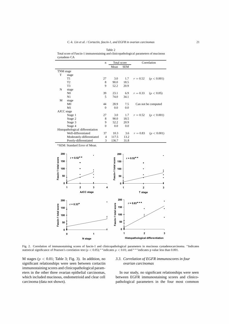
Table 2Total score of Fascin-1 immunostaining and clinicopathological parameters of mucinouscystadeno CA
n Total score CorrelationMean SEM
TNM stageT stage
T1 27 3.0 1.7 r = 0.52 (p < 0.001)T2 8 90.0 18.5T3 9 52.2 20.9
N stageN0 39 23.1 6.9 r = 0.33 (p < 0.05)N1 5 74.0 34.1
M stageM0 44 28.9 7.5 Can not be computedM1 0 0.0 0.0
AJCC stageStage 1 27 3.0 1.7 r = 0.52 (p < 0.001)Stage 2 8 90.0 18.5Stage 3 9 52.2 20.9Stage 4 0 0.0 0.0
Histopathological differentiationWell-differentiated 37 10.3 3.6 r = 0.83 (p < 0.001)Moderately-differentiated 4 117.5 13.2Poorly-differentiated 3 136.7 31.8
aSEM: Standard Error of Mean.
r = 0.33
0
50
100
150
200
0 1
N stage
Fasc
in-1
tota
l sco
re
r = 0.52
0
50
100
150
200
1 2 3 4
AJCC stage
Fasc
in-1
tota
l sco
re r = 0.52
0
50
100
150
200
1 2 3
T stage
Fasc
in-1
tota
l sco
re
r = 0.83
0
50
100
150
200
1 2 3
Histopathological differentiation
Fasc
in-1
tota
l sco
re
Fig. 2. Correlation of immunostaining scores of fascin-1 and clinicopathological parameters in mucinous cystadenocarcinoma.∗Indicatesstatistical significance of Pearson’s correlation test (p < 0.05);∗∗indicatesp < 0.01; and∗∗∗indicates p value less than 0.001.
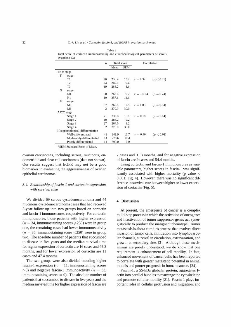
M stages (p < 0.01; Table 3; Fig. 3). In addition, nosignificant relationships were seen between cortactinimmunostaining scores and clinicopathological param-eters in the other three ovarian epithelial carcinomas,which included mucinous, endometrioid and clear cellcarcinoma (data not shown).
3.3. Correlation of EGFR immunoscores in fourovarian carcinomas
In our study, no significant relationships were seenbetween EGFR immunostaining scores and clinico-pathological parameters in the four most common
22 C.-k. Lin et al. / Cortactin, fascin-1, and EGFR in ovarian carcinomas
Table 3Total score of cortactin immunostaining and clinicopathological parameters of serouscystadeno CA
n Total score CorrelationMean SEM
TNM stageT stage
T1 26 236.4 15.2 r = 0.32 (p < 0.01)T2 24 269.6 9.4T3 19 284.2 8.6
N stageN0 50 262.6 9.2 r = −0.04 (p = 0.74)N1 19 257.1 11.1
M stageM0 67 260.8 7.5 r = 0.03 (p = 0.84)M1 2 270.0 30.0
AJCC stageStage 1 21 235.8 18.1 r = 0.18 (p = 0.14)Stage 2 19 283.2 9.2Stage 3 27 264.6 9.2Stage 4 2 270.0 30.0
Histopathological differentiationWell-differentiated 41 241.9 10.7 r = 0.40 (p < 0.01)Moderately-differentiated 14 278.6 11.4Poorly-differentiated 14 300.0 0.0
aSEM:Standard Error of Mean.
ovarian carcinomas, including serous, mucinous, en-dometrioid and clear cell carcinomas (data not shown).Our results suggest that EGFR may not be a goodbiomarker in evaluating the aggressiveness of ovarianepithelial carcinomas.
3.4. Relationship of fascin-1 and cortactin expressionwith survival time
We divided 69 serous cystadenocarcinoma and 44mucinous cystadenocarcinoma cases that had received5-year follow up into two groups based on cortactinand fascin-1 immunoscores, respectively. For cortactinimmunoscores, those patients with higher expression(n = 34, immunostaining scores�250) were in groupone, the remaining cases had lower immunoreactivity(n = 35, immunostaining score<250) were in grouptwo. The absolute number of patients that succumbedto disease in five years and the median survival timefor higher expression of cortactin are 16 cases and 45.3months, and for lower expression of cortactin are 11cases and 47.4 months.
The two groups were also divided incuding higherfascin-1 expression (n = 11, immunostaining scores>0) and negative fascin-1 immunoactivity (n = 33,immunostaining scores= 0). The absolute number ofpatients that succumbed to disease in five years and themedian survival time for higher expression of fascin are
7 cases and 31.3 months, and for negative expressionof fascin are 9 cases and 54.4 months.
Using cortactin and fascin-1 immunoscores as vari-able parameters, higher scores in fascin-1 was signif-icantly associated with higher mortality (p value<0.001; Fig. 4). However, there was no significant dif-ference in survival rate between higher or lower expres-sion of cortactin (Fig. 5).
4. Discussion
At present, the emergence of cancer is a complexmulti-step process in which the activation of oncogenesand inactivation of tumor suppressor genes act syner-gistically to produce the malignant phenotype. Tumormetastasis is also a complex process that involves directinvasion of tumor cells, infiltration into lymphovascu-lar channels, survival in circulation, extravasation, andgrowth at secondary sites [3]. Although these mech-anisms are poorly understood, we do know that onerequirement is enhancement of cell motility. In fact,enhanced movement of cancer cells has been reportedto correlate with greater metastatic potential in animalmodels and poorer prognosis in human cancers [24].
Fascin-1, a 55-kDa globular protein, aggregates F-actin into parallel bundles to rearrange the cytoskeletonand promote cellular motility [21]. Fascin-1 plays im-portant roles in cellular protrusion and migration, and

C.-k. Lin et al. / Cortactin, fascin-1, and EGFR in ovarian carcinomas 23
r =-0.04
0
100
200
300
0 1 2
N stage
Co
rtac
tin
to
tal s
core
r = 0.40
0
100
200
300
1 2 3
Histopathological differentiation
Co
rtac
tin
to
tal s
core
r = 0.18
0
100
200
300
1 2 3 4
AJCC stage
Co
rtac
tin
to
tal s
core
r = 0.32
0
100
200
300
1 2 3
T stage
Co
rtac
tin
to
tal s
core
r = 0.03
0
100
200
300
0 1
M stage
Co
rtac
tin
to
tal s
core
Fig. 3. Correlation of immunostaining scores of cortactin and clinicopathological parameters in serous cystadenocarcinoma.∗∗Indicates statisticalsignificance of Pearson’s correlation test (p < 0.01).
extracellular matrix adhesion [2]. The gene encodingfascin-1 in humans is located at chromosome 7q22 [14].Fascin-1 is either expressed at very low levels or isabsent in normal ovarian epithelia. Over-expressionof fascin-1 is associated with tumor progression andinvasion in skin, lung, breast, ovarian, colon, urinarybladder, brain and gastric malignancies [11,13,17,19,25,26,28,33,34].
Our results suggest that expression of fascin-1 maybe effective in predicting tumor clinicopathological pa-rameters of ovarian mucinous cystadenocarcinoma inChinese women. Average immunostaining scores forfascin-1 have a significant positive correlation with T,N, and AJCC stages, but not with M stage of muci-nous cystadenocarcinoma. However, in our study, noM1-stage case was included, which makes it difficult toshow statistical significance. In addition, higher fascin-1 immunostaining scores are significantly associatedwith poorer survival rate as demonstrated in our study.
Cortactin regulates the actin cytoskeleton through itsinvolvement in several processes, including cell motil-ity, adhesion, polarization, contraction, and others [9,35,40]. The activation of actin-related (Arp) 2/3 pro-tein complex and neuronal Wiscott-Aldrich syndromeprotein (N-Wasp) by cortactin nucleates actin poly-merization and promotes cellular motility. Cortactinis a p80/p85 multidomain actin filament-binding pro-tein [32]; it was first identified as an src kinase substratein chicken fibroblasts [41]. Human cortactin maps tochromosome 11q13 [22]. Amplification of chromo-some 11q13 has been reported in several human car-cinomas as has increased expression of cortactin [37].Over-expression of cortactin induces cell motility andmigration, inhibits cell-cell adhesion, and acceleratestumor spreading [36]. In addition, the effects of cor-tactin may be related to expression of E-cadherin andits effects on intercellular adhesion [15,20,38]. In somein vitro studies, cortactin over-expression induced tu-

24 C.-k. Lin et al. / Cortactin, fascin-1, and EGFR in ovarian carcinomas
Survival Analysis
Time (months)0 10 20 30 40 50 60 70
Su
rvival rate
0.0
0.2
0.4
0.6
0.8
1.0
Fascin score = 0
Fascin score > 0
Fig. 4. Overall survival of 44 patients with mucinous cystadenocarcinoma. Higher fascin-1 immunostaining scores were associated with poorersurvival. Survival rates were analyzed using the Kaplan-Meier survival test (p < 0.001).
mor invasion, and metastasis has been shown to be as-sociated with esophageal and head/neck squamous cellcarcinomas [22,29]. However, direct evidence is stilllacking to establish a relationship between cortactinover-expression and tumor progression and metastasisin ovarian carcinomas. Our current results demonstratethat cortactin is over-expressed in the four most com-mon ovarian carcinomas in Chinese women, and higherimmunostaining scores of cortactin are associated withmore advanced T stage and histological differentiationin serous cystadenocarcinoma (Table 3, Fig. 3).
EGFR is one of a family of receptors that help regu-late cell growth, division, and death. Normal epithelialcells contain two copies of the EGFR gene and producelow levels of EGFR protein on the cell surface. In avariety of cancers, increased amounts of EGFR proteinare present in tumor tissue. This can be due to ampli-fication (too many copies of the gene are produced),over-expression (an increased amount of the protein isproduced), and/or decreased protein destruction. Tu-mors with increased EGFR protein tend to grow moreaggressively, are more likely to metastasize, and aremore resistant to standard chemotherapies. Patientswith these tumors tend to have poorer outcomes.
In our study, EGFR is over-expressed in four ovariancarcinomas in Chinese women, but over-expression ofEGFR was not associated with aggressive clinicopatho-logical parameters in these four ovarian carcinomas.To our knowledge, this is the first report to evaluate theassociation between cortactin, fascin-1 and EGFR ex-pression and tumor progression in the four most com-mon ovarian epithelial carcinomas.
In conclusion, higher fascin-1 immunostainingscores are associated with poorer tumor differentiation,more-advanced TNM stages and shorter survival timein mucinous cystadenocarcinoma. Similarly, we founda correlation between higher immunostaining scoresof cortactin and T stages and histological differentia-tion in serous cystadenocarcinoma. Accordingly, ourresults may support the hypothesis that fascin-1 andcortactin are important factors in migration or invasionof some ovarian epithelial carcinomas. Although un-known mechanisms still exist in tumor progression, wedemonstrated that fascin-1 is a satisfactory biomarkerfor predicting clinical outcome in mucinous cystade-nocarcinoma. Therefore, the development of pharma-cological agents to target the fascin-1 pathway mayprolong survival time and slow tumor progression inpatients with mucinous cystadenocarcinoma.

C.-k. Lin et al. / Cortactin, fascin-1, and EGFR in ovarian carcinomas 25
Survival Analysis
Time (months)0 10 20 30 40 50 60 70
Su
rvival rate
0.0
0.2
0.4
0.6
0.8
1.0
Cortactin score < 250
Cortactin score > 250
Fig. 5. Overall survival of 69 patients with serous cystadenocarcinoma. Higher cortactin immunostaining scores were not associated with poorersurvival rate. Survival rates were analyzed using the Kaplan-Meier survival test (p > 0.05).
Acknowledgments
This study was supported by a grant from Tri-ServiceGeneral Hospital, TSGH-C97-4-S05, Taiwan.
References
[1] J.C. Adams, Roles of fascin in cell adhesion and motility,CurrOpin Cell Biol 16 (2004), 590–596.
[2] J.C. Adams, Fascin protrusions in cell interactions,TrendsCardiovasc Med 14 (2004), 221–226.
[3] S. Anzavoorian, A.N. Murphy, W.G. Steler-Stevenson andL.A. Liotta, Molecular aspects of tumor cell invasion andmetastasis,Cancer 71 (1993), 1368–1383.
[4] T. Bauknecht, I. Janz, M. Kohler and A. Pfleiderer, Humanovarian carcinomas: correlation of malignancy and survivalwith the expression of epidermal growth factor receptors andEGFR-like factors,Med Oncol Tumor Pharmacother 6 (1989),121–127.
[5] T. Bauknecht, Birmelin and F. Kommoss, Clinical significanceof oncogenes and growth factors in ovarian carcinomas,JSteroid Biochem Mol Biol 37 (1990), 855–862.
[6] A. Berchuck, G.C. Rodriguez, A. Kamek, R.K. Dodge, J.T.Soper, D.L. Clarke-Pearson et al., Epidermal growth factorreceptor expression in normal ovarian epithelium and ovariancancer,Am J Obstet Gynecol 164 (1991), 669–674.
[7] E. Brown, M. Stewart, T. Rye, A. Al-Nafussi, A.R. Williams,M. Bradburn et al., Carcinosarcoma of the ovary: 19 years
of prospective data from a single center,Cancer 100 (2004),2148–2153.
[8] G. Carpenter, Epidermal growth factor,Annu Rev Biochem 48(1979), 193–216.
[9] R.J. Daly, Cortactin signaling and dynamic actin networks,Biochem J 382 (2004), 13–25.
[10] B. Davidson, I. Goldberg, A. Berner, G.B. Kristensen andR. Reich, EMMPRIN (extracellular matrix metalloproteinaseinducer) is a novel marker of poor outcome in serous ovariancarcinoma,Clin Exp Metastasis 20 (2003), 161–169.
[11] V.N. Goncharuk, J.S. Ross and J.A. Carlson, Actin-bindingprotein fascin expression in skin neoplasia,J Cutan Pathol 29(2002), 430–438.
[12] M.E. Greenberg and E.B. Ziff, Stimulation of 3T3 cells in-duces transcription of the c-fos proto-oncogene,Nature (Lon-don) 311 (1984), 433–438.
[13] A. Grothey, R. Hashizume, A.A. Sahin and P.D. McCrea,Fascin, an actin-bundling protein associated with cell motility,is upregulated in hormone receptor negative breast cancer,BrJ Cancer 83 (2000), 870–873.
[14] Y. Hashimoto, M. Skacel and J.C. Adams, Roles of fascin inhuman carcinoma motility and signaling: prospects for a novelbiomarker?Int J Biochem Cell Biol 37 (2005), 1787–1804.
[15] F.M. Helwani, E.M. Kovacs, A.D. Paterson, S. Verma, R.G.Ali, A.S. Fanning, S.A. Weed et al., Cortactin is necessary forE-cadherin-mediated contact formation and actin reorganiza-tion, J Cell Biol 164 (2004), 899–910.
[16] S.C. Henzen-Longmans, E.M. Beins, J.G. Klyn, M.E. van denBurg and J.A. Foekens, Epidermal growth factor receptor: cor-relation of immunohistochemistry with ligand binding assay,Br J Cancer 66 (1992), 1015–1021.

26 C.-k. Lin et al. / Cortactin, fascin-1, and EGFR in ovarian carcinomas
[17] W. Hu, P.D. McCrea, M. Deavers, J.J. Kavanagh, A.P. Kudelkaand C.F. Verschraegen, Increased expression of fascin, motilityassociated protein, in cell cultures derived from ovarian cancerand in borderline and carcinomatous ovarian tumors,Clin ExpMetastasis 18 (2000), 83–88.
[18] H. Itamochi, J. Kigawa, T. Sugiyama, Y. Kikuchi, M. Suzukiand N. Terakawa, Low proliferation activity may be associatedwith chemoresistance in clear cell carcinoma of the ovary,Obst & Gynecol 100 (2002), 281–287.
[19] A.U. Jawhari, A.Buda, M. Jenkins, K. Shehzad, C. Sarraf,M. Noda et al., Fascin, an actin-bundling protein, modulatescolonic epithelial cell invasiveness and differentiationin vitro,Am J Pathol 162 (2003), 69–80.
[20] E.M. Kovacs, M. Goodwin, R.G. Ali, A.D. Paterson and A.S.Yap, Cadherin-directed actin assembly: E-cadherin physicallyassociates with the Arp2/3 complex to direct actin assemblyin nascent adhesive contacts,Curr Biol 12 (2002), 379–382.
[21] N. Kureishy, V. Sapountzi, S. Prag, N. Anilkumar and J.C.Adams, Fascins and their roles in cell structure and function,Bio Essays 24 (2002), 350–361.
[22] M.L. Luo, X.M. Shen, Y. Zhang, F. Wei, X. Xu, Y. Cai,X. Zhang et al., Amplification and overexpression of CTTN(EMS1) contribute to the metastasis of esophageal squamouscell carcinoma by promoting cell migration and anoikis resis-tance,Cancer Res 66 (2006), 11690–11699.
[23] C.J. Ormandy, E.A. Musgrove, R. Hui, R.J. Daly and R.L.Sutherland, Cyclin D1, EMS1 and 11q13 amplification inbreast cancer,Breast Cancer Res Treat 78 (2003), 323–335.
[24] A.W. Partin, J.S. Shoeniger, L.L. Mohler and D.S. Coffey,Further analysis of cell motility: correlation of motility withmetastatic potentials,Proc Natl Acad Sci 86 (1989), 1254–1258.
[25] G. Pelosi, F. Pasini, F. Fraggetta, U. Pastorino, A. Iannucci,P. Maisonneuve et al., Independent value of fascin immunore-activity for predicting lymph node metastases in typical andatypical pulmonary carcinoids,Lung Cancer 42 (2003), 203–213.
[26] G. Pelosi, U. Pastorino, F. Pasini, P. Maissoneuve, F. Fragget-ta, A. Iannucci et al., Independent prognostic value of fascinimmunoreactivity in Stage I non-small cell lung cancer,Br JCancer 88 (2003), 537–547.
[27] J.M. Piek, P. Kenemans and R.H. Verheijen, Intraperitonealserous adenocarcinoma: a critical appraisal of three hypothe-ses on its cause,Am J Obst & Gynecol 191 (2004), 718–732.
[28] A.A. Roma and R.A. Prayson, Fascin expression in 90 patientswith glioblastoma multiforme,Ann Diagn Pathol 9 (2005),307–311.
[29] B.L. Rothschild, A.H. Shim, A.G. Ammer, L.C. Kelley, K.B.Irby et al., Cortactin overexpression regulates actin-relatedprotein 2/3 complex activity, motility, and invasion in carci-nomas with chromosome 11q13 amplification,Cancer Res 66(2006), 8017–8025.
[30] E. Rozengurt, Stimulation of Na+ /K+ pump activity and DNA
synthesis in quiescent cultured cells,Adv Enzyme Regul 19(1981), 61–85.
[31] E. Schuuring, The involvement of the chromosome 11q13region in human malignancies: cyclin D1 and EMS1 are twonew candidate oncogenes – a review,Gene 159 (1995), 83–96.
[32] E. Schuuring, E. Verhoeven, S. Litvinov and R.J. Michslides,The product of the EMS1 gene, amplified and overexpressedin human carcinomas, is homologous to a v-src substrate andis located in cell-substratum, contact sites,Mol Cell Biol 13(1993), 2891–2898.
[33] W.C. Tsai, J.S. Jin, W.K. Chang, D.C. Chan, M.K. Yeh, Y.C.Chao et al., Association of cortactin and fascin-1 expression ingastric adenocarcinoma: correlation with clinocopathologicalparameters,Disease Markers 55 (2007), 955–962.
[34] G.X. Tong, H.Yee, L. Chiriboga, O. Hernandes and J. Wais-man, Fascin-1 expression in papillary and invasive urothe-lial carcinomas of the urinary bladder,Human Pathology 36(2005), 741–746.
[35] A.G. van Rossum, J. Gibcus, J. van der Wal and E. Schuur-ing, Cortactin overexpression results in sustained epidermalgrowth factor receptor signaling by preventing ligand-inducedreceptor degradation in human carcinoma cells,Brease CancerRes 7 (2005), 235–237.
[36] A.G. van Rossum, W.H. Moolenaar and E. Schuuring, Cor-tactin affects cell migration by regulating intercellular adhe-sion and cell spreading,Exp Cell Res 312 (2006), 1658–1670.
[37] A.G. van Rossum, E. Schuuring-Scholtes, V. van Buuren-vanSeggelen, P.M. Kluin and E. Schuuring, Comparative genomeanalysis of cortactin and HS1: the significance of the F-actinbinding repeat domain,BMC Genomics 6 (2005), 15.
[38] S. Verma, A.M. Shewan, J.A. Scott, F.M. Helwani, N.R. denElzen et al., Arp2/3 activity is necessary for efficient formationof E-cadherin adhesive contacts,J Biol Chem 279 (2004),34062–34070.
[39] A.M. Weaver, A.V. Karginov, A.W. Kinley, S.A. Weed, Y. Li,J.T. Parsons and J.A. Cooper, Cortactin promotes and stabi-lizes Arp2/3-induced actin filament network formation,CurrBiol 11 (2001), 370–374.
[40] S.A. Weed and J.T. Parsons, Cortactin: coupling membranedynamics to cortical actin assembly,Oncogene 20 (2001),6418–6434.
[41] H. Wu, A.B. Reynolds, S.B. Kanner, R.R. Vines andJ.T. Parsons, Identification and characterization of a novelcytoskeleton-associated pp60src substrate,Mol Cell Biol 11(1991), 5113–5124.
[42] J.J. Xie, L.Y. XU, H.H. Zhang, W.J. Cai and R.Q. Mai, Roleof fascin in the proliferation and invasiveness of esophagealcarcinoma cells,Biochem Biophys Res Commum 337 (2005),355–362.
[43] S. Yamashiro, Y. Yamakita, S. Ono and F. Matsumura, Fascin,an actin-bundling protein, induces membrane protrusions andincreases cell motility of epithelial cells,Mol Biol Cell 9(1998), 993–1006.

Submit your manuscripts athttp://www.hindawi.com
Stem CellsInternational
Hindawi Publishing Corporationhttp://www.hindawi.com Volume 2014
Hindawi Publishing Corporationhttp://www.hindawi.com Volume 2014
MEDIATORSINFLAMMATION
of
Hindawi Publishing Corporationhttp://www.hindawi.com Volume 2014
Behavioural Neurology
EndocrinologyInternational Journal of
Hindawi Publishing Corporationhttp://www.hindawi.com Volume 2014
Hindawi Publishing Corporationhttp://www.hindawi.com Volume 2014
Disease Markers
Hindawi Publishing Corporationhttp://www.hindawi.com Volume 2014
BioMed Research International
OncologyJournal of
Hindawi Publishing Corporationhttp://www.hindawi.com Volume 2014
Hindawi Publishing Corporationhttp://www.hindawi.com Volume 2014
Oxidative Medicine and Cellular Longevity
Hindawi Publishing Corporationhttp://www.hindawi.com Volume 2014
PPAR Research
The Scientific World JournalHindawi Publishing Corporation http://www.hindawi.com Volume 2014
Immunology ResearchHindawi Publishing Corporationhttp://www.hindawi.com Volume 2014
Journal of
ObesityJournal of
Hindawi Publishing Corporationhttp://www.hindawi.com Volume 2014
Hindawi Publishing Corporationhttp://www.hindawi.com Volume 2014
Computational and Mathematical Methods in Medicine
OphthalmologyJournal of
Hindawi Publishing Corporationhttp://www.hindawi.com Volume 2014
Diabetes ResearchJournal of
Hindawi Publishing Corporationhttp://www.hindawi.com Volume 2014
Hindawi Publishing Corporationhttp://www.hindawi.com Volume 2014
Research and TreatmentAIDS
Hindawi Publishing Corporationhttp://www.hindawi.com Volume 2014
Gastroenterology Research and Practice
Hindawi Publishing Corporationhttp://www.hindawi.com Volume 2014
Parkinson’s Disease
Evidence-Based Complementary and Alternative Medicine
Volume 2014Hindawi Publishing Corporationhttp://www.hindawi.com