asymmetric keratoconus study: novel tomographic parameters to enhance sensitivity to detect...
TRANSCRIPT

Asymmetric Keratoconus Study: Asymmetric Keratoconus Study:
Novel Tomographic Parameters to enhance sensitivity Novel Tomographic Parameters to enhance sensitivity
to detect abnormalities in eyes with normal axial to detect abnormalities in eyes with normal axial
curvature maps from patients with obvious curvature maps from patients with obvious
keratoconus in the fellow eye keratoconus in the fellow eye
Marcella Q. Salomão, MD; Marcella Q. Salomão, MD; JoséJosé Salgado-Borges, MD; Eduardo Viteri, MD; Salgado-Borges, MD; Eduardo Viteri, MD;
Canrobert Oliveira, MD; Renato Ambrósio Jr.MD, PhD. Canrobert Oliveira, MD; Renato Ambrósio Jr.MD, PhD.
No financial interest. No financial interest.
Rio de Janeiro - Brazil
PurposePurpose
To introduce new tomographic diagnostic indices, To introduce new tomographic diagnostic indices,
beyond surface topography, to detect beyond surface topography, to detect very early signs of very early signs of
ectasia.ectasia.
To calculate the sensitivity of these new parameters.To calculate the sensitivity of these new parameters.
San Francisco 2009
JustificationJustification Keratoconus is, by definition, a bilateral disease; however, a small Keratoconus is, by definition, a bilateral disease; however, a small
percentage of cases present with a very asymmetric pattern. In percentage of cases present with a very asymmetric pattern. In
some cases, the asymmetry can be so significant some cases, the asymmetry can be so significant that the contra-that the contra-
lateral eye presents with normal anterior curvature maps.lateral eye presents with normal anterior curvature maps.
A bilateral presentation as in these “normalA bilateral presentation as in these “normal”” eyes can occur and eyes can occur and
could even be the explanation for many cases of ectasia after could even be the explanation for many cases of ectasia after
LASIK with no identifiable pre-operative risk factors.LASIK with no identifiable pre-operative risk factors.
Identifying Identifying asymptomatic cases with normal slit lamp exam, good asymptomatic cases with normal slit lamp exam, good
spectacle corrected visual acuity, and normal corneal topography is spectacle corrected visual acuity, and normal corneal topography is
critical; not only for screening of refractive surgery candidates, but to critical; not only for screening of refractive surgery candidates, but to
allow an enhanced treatment choice. allow an enhanced treatment choice.
San Francisco 2009
Patients and MethodsPatients and Methods 53 eyes showing normal anterior curvature maps (based 53 eyes showing normal anterior curvature maps (based on standard Rabinowitz criteria) from patients with on standard Rabinowitz criteria) from patients with advanced keratoconus in the fellow eye were evaluated advanced keratoconus in the fellow eye were evaluated in this multicenter study.in this multicenter study.
All patients had tomographic exams using the Oculus All patients had tomographic exams using the Oculus Pentacam. Pentacam.
Patient demographicsPatient demographics
42 patients ♂ 11 patients ♀42 patients ♂ 11 patients ♀
Average age: 35.34 y (17 to 78)Average age: 35.34 y (17 to 78)
San Francisco 2009
New tomographic parametersNew tomographic parameters
Thickness ProfilesThickness Profiles11: Corneal Thickness Spatial Profile : Corneal Thickness Spatial Profile (CTSP) and Percentage Thickness Increase (PTI)(CTSP) and Percentage Thickness Increase (PTI)
Enhanced anterior and posterior elevation best fit Enhanced anterior and posterior elevation best fit sphere (BFS)sphere (BFS) 2 2
1- Ambrósio R Jr, Alonso RS, Luz A, Coca Velarde LG. 1- Ambrósio R Jr, Alonso RS, Luz A, Coca Velarde LG. Corneal-thickness spatial profile and corneal-volume Corneal-thickness spatial profile and corneal-volume distribution: tomographic indices to detect keratoconus. J Cataract Refract Surg. 2006 Nov;32(11):1851-9.distribution: tomographic indices to detect keratoconus. J Cataract Refract Surg. 2006 Nov;32(11):1851-9.
2- Belin MW, 2- Belin MW, Khachikian SS, Ambrósio R Jr, Salomão MQ. Keratoconus / Ectasia detection with the Oculus Khachikian SS, Ambrósio R Jr, Salomão MQ. Keratoconus / Ectasia detection with the Oculus Pentacam: Belin/Ambrosio enhanced ectasia display. Highlights of Ophthalmology. Volume 35- number 6. Pentacam: Belin/Ambrosio enhanced ectasia display. Highlights of Ophthalmology. Volume 35- number 6.
San Francisco 2009
CTSP and PTICTSP and PTI
TheThe
San Francisco 2009
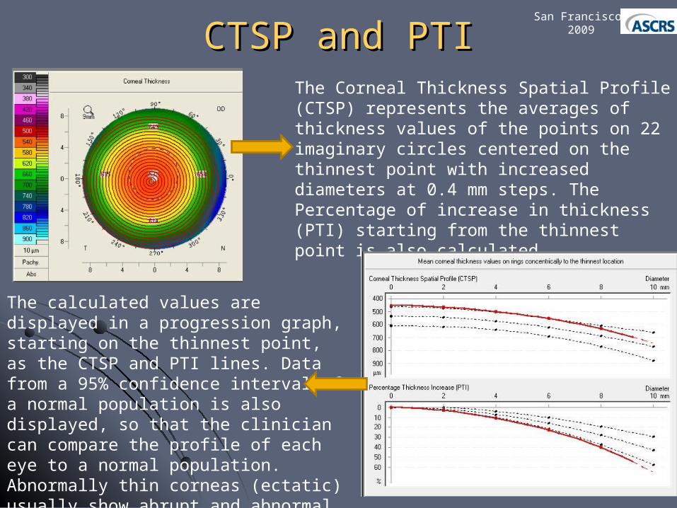
The Corneal Thickness Spatial Profile (CTSP) represents the averages of thickness values of the points on 22 imaginary circles centered on the thinnest point with increased diameters at 0.4 mm steps. The Percentage of increase in thickness (PTI) starting from the thinnest point is also calculated.
The calculated values are displayed in a progression graph, starting on the thinnest point, as the CTSP and PTI lines. Data from a 95% confidence interval of a normal population is also displayed, so that the clinician can compare the profile of each eye to a normal population. Abnormally thin corneas (ectatic) usually show abrupt and abnormal profiles, out of the 95% CI.

Enhanced BFSEnhanced BFSSan Francisco
2009
The Standard BFS is basically an average of high and low elevations of the cornea. It is calculated considering the best reference sphere for the whole corneal surface. Thus, in an abnormal eye, data from an abnormal area might be used in the BFS calculation.
The Enhanced BFS was calculated utilizing all the valid data from within the 9.0 mm central cornea with the exception of a 4mm area centered on the thinnest point. This would better approximate the normal cornea and accentuate the abnormal portion.
Elevation maps using this new “reference shape” were calculated for both anterior and posterior corneal surfaces.
MethodsMethods
Elevation maps considering the standard BFS were Elevation maps considering the standard BFS were subtracted from the elevation maps with the enhanced BFS; subtracted from the elevation maps with the enhanced BFS; and the highest difference between the maps in the central and the highest difference between the maps in the central 4mm area was noted for anterior and posterior corneal 4mm area was noted for anterior and posterior corneal surfaces. surfaces.
The differential map contains only 3 colors, each one The differential map contains only 3 colors, each one corresponding to the amount of elevation change that occurs corresponding to the amount of elevation change that occurs when moving between the standard and enhanced maps. when moving between the standard and enhanced maps.
Anterior - Anterior - greengreen is anything < 6, is anything < 6, yellowyellow is between 6 - 12, is between 6 - 12, redred is > 12 is > 12
Posterior: Posterior: greengreen is anything < 8, is anything < 8, yellowyellow is between 8 - 20, is between 8 - 20, redred is > 20 is > 20
Yellow and red maps were considered abnormal. Yellow and red maps were considered abnormal.
San Francisco 2009

ResultsResultsSan Francisco
2009
Approach KC eyes Contra-lateral eyes
Pachymmetric Profiles 53 (100%) 46 (87%)
Enhanced Anterior Elevation
46 (87%) 8 (15%)
Enhanced Posterior Elevation
53 (100%) 47 (89%)
All eyes but 1 (52/53 = 98%) presented at least one abnormal finding in the new parameters described. The combination of Pachymetric Profiles and Enhanced Elevation add to each other in sensitivity and specificity. A new display (enhanced ectasia detection),combining the two indices, was developed.
Enhanced Ectasia Detection Display Enhanced Ectasia Detection Display San Francisco
2009
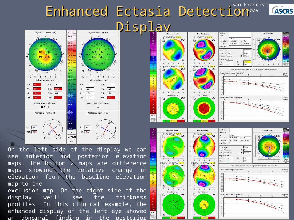
On the left side of the display we can see anterior and posterior elevation maps. The bottom 2 maps are difference maps showing the relative change in elevation from the baseline elevation map to theexclusion map. On the right side of the display we’ll see the thickness profiles. In this clinical example, the enhanced display of the left eye showed an abnormal finding in the posterior elevation approach (yellow) along with abnormal thickness profiles, despite a normal anterior surface map.
Enhanced Ectasia Detection Display Enhanced Ectasia Detection Display San Francisco
2009
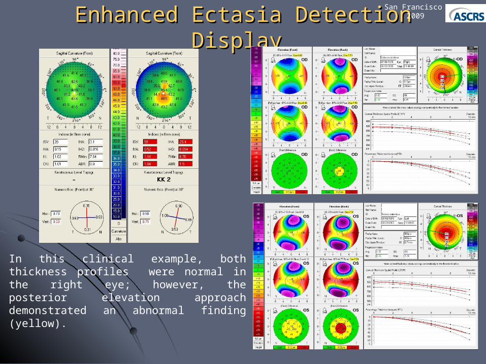
In this clinical example, both thickness profiles were normal in the right eye; however, the posterior elevation approach demonstrated an abnormal finding (yellow).

ConclusionConclusion
The cThe combination of Pachymetric Profiles and Enhanced ombination of Pachymetric Profiles and Enhanced
ElevationElevation was capable of identifying abnormalities in the was capable of identifying abnormalities in the
majority of eyes with normal anterior curvature maps of majority of eyes with normal anterior curvature maps of
patients with very asymmetric keratoconus. patients with very asymmetric keratoconus.
The newThe new parameters increase sensitivity and specificity for parameters increase sensitivity and specificity for
the screening of refractive surgery candidates.the screening of refractive surgery candidates.
San Francisco 2009