atlas food microbiology
TRANSCRIPT
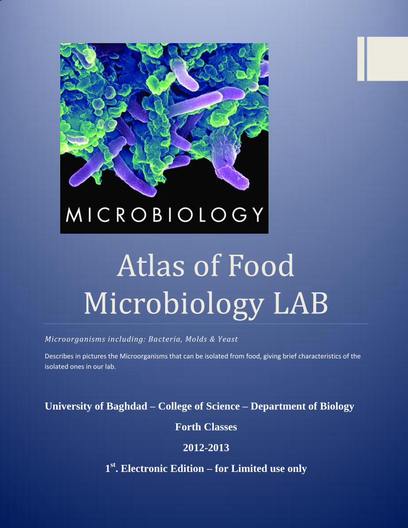
Atlas of Food Microbiology LAB
Microorganisms including: Bacteria, Molds & Yeast
Describes in pictures the Microorganisms that can be isolated from food, giving brief characteristics of the
isolated ones in our lab.
University of Baghdad – College of Science – Department of Biology
Forth Classes
2012-2013
1st. Electronic Edition – for Limited use only

1
Preface “Science knows no country, because knowledge belongs to humanity, and is the torch which illuminates the world. Science is the highest personification of the nation because that nation will remain the first which carries the furthest the works of thought and intelligence”. Louis Pasteur
Science always unites students whatever their specialization was, or their age is… As I was a student once, I’ve been inspired by my Teachers who taught me much & till now, how to learn from the simple things in life, recording notes, keep on trying despite the difficulties, searching the new scientific trends, but the most important thing that I’ve learned is to document the results to put the knowledge in the hands of those who need, to continue the never ending trip of science….
To prevail the benefits, preparing this Atlas was an important thing to work on, that included some of Lab results obtained during the hard work with the undergraduate students, and some of the important results related to the Food Microbiology Lab, collected from scientific references, besides the help of the Web services, in hope that it would be useful to all who read it.
Since no work can ever be done alone….
I would like to Thank ….
The Head of Biology Department Professor Doctor Sabah N. Alwachi for all the support to
encourage such a scientific work that would help our students…
The Advisors of Food Microbiology LAB in Biology Department…
My LAB Colleagues Msc. Teacher Dimah Nazar & Msc. Student Dalia Azhar for their help…
Msc. Teacher Assistant Lina A. Omar-Zahid
2
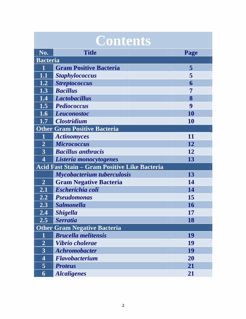
Contents
No. Title Page
Bacteria
1 Gram Positive Bacteria 5
1.1 Staphylococcus 5
1.2 Streptococcus 6
1.3 Bacillus 7
1.4 Lactobacillus 8
1.5 Pediococcus 9
1.6 Leuconostoc 10
1.7 Clostridium 10
Other Gram Positive Bacteria
1 Actinomyces 11
2 Micrococcus 12
3 Bacillus anthracis 12
4 Listeria monocytogenes 13
Acid Fast Stain – Gram Positive Like Bacteria
Mycobacterium tuberculosis 13
2 Gram Negative Bacteria 14
2.1 Escherichia coli 14
2.2 Pseudomonas 15
2.3 Salmonella 16
2.4 Shigella 17
2.5 Serratia 18
Other Gram Negative Bacteria
1 Brucella melitensis 19
2 Vibrio cholerae 19
3 Achromobacter 19
4 Flavobacterium 20
5 Proteus 21
6 Alcaligenes 21

3
Contents
No. Title Page
Fungi
1 Molds 22
1.1 Penicillium 22
1.2 Cladosporium 22
1.3 Aspergillus niger 23
1.4 Aspergillus flavus 23
1.5 Mucor 24
1.6 Rhizopus 24
1.7 Alternaria 25
1.8 Curvilaria 25
1.9 Fusarium 26
1.10 Geotrichum 26
1.11 Botrytis 27
1.12 Sporotrichum 27
2 Yeast 29
2.1 Saccharomyces 29
2.2 Rhodotorula 29
2.3 Endomyces 30
2.4 Candida 30
References 31
4
5
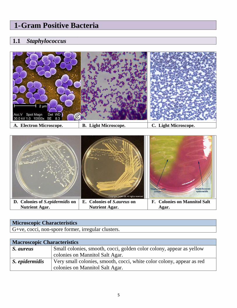
1- Gram Positive Bacteria
1.1 Staphylococcus
A. Electron Microscope. B. Light Microscope. C. Light Microscope.
D. Colonies of S.epidermidis on
Nutrient Agar.
E. Colonies of S.aureus on
Nutrient Agar.
F. Colonies on Mannitol Salt
Agar.
Microscopic Characteristics
G+ve, cocci, non-spore former, irregular clusters.
Macroscopic Characteristics
S. aureus Small colonies, smooth, cocci, golden color colony, appear as yellow
colonies on Mannitol Salt Agar.
S. epidermidis Very small colonies, smooth, cocci, white color colony, appear as red
colonies on Mannitol Salt Agar.
6
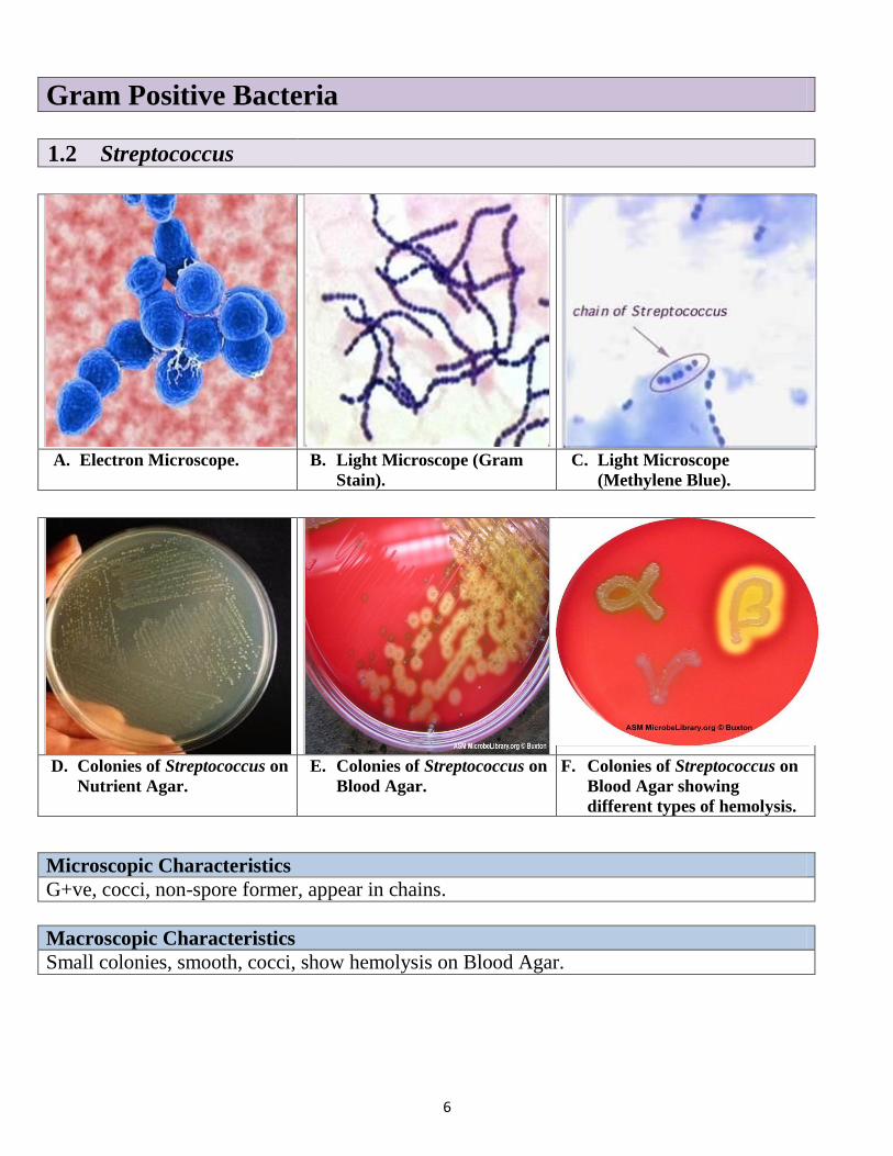
Gram Positive Bacteria
1.2 Streptococcus
A. Electron Microscope. B. Light Microscope (Gram
Stain).
C. Light Microscope
(Methylene Blue).
D. Colonies of Streptococcus on
Nutrient Agar.
E. Colonies of Streptococcus on
Blood Agar.
F. Colonies of Streptococcus on
Blood Agar showing
different types of hemolysis.
Microscopic Characteristics
G+ve, cocci, non-spore former, appear in chains.
Macroscopic Characteristics
Small colonies, smooth, cocci, show hemolysis on Blood Agar.
7
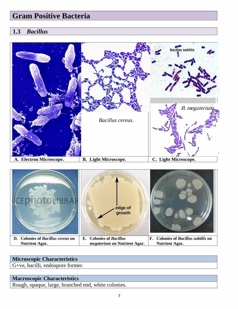
Gram Positive Bacteria
1.3 Bacillus
Bacillus cereus.
A. Electron Microscope. B. Light Microscope. C. Light Microscope.
D. Colonies of Bacillus cereus on
Nutrient Agar.
E. Colonies of Bacillus
megaterium on Nutrient Agar.
F. Colonies of Bacillus subtilis on
Nutrient Agar.
Microscopic Characteristics
G+ve, bacilli, endospore former.
Macroscopic Characteristics
Rough, opaque, large, branched end, white colonies.
B. megaterium
8
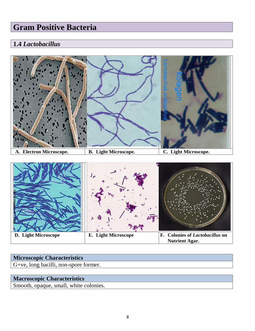
Gram Positive Bacteria
1.4 Lactobacillus
A. Electron Microscope. B. Light Microscope. C. Light Microscope.
D. Light Microscope E. Light Microscope F. Colonies of Lactobacillus on
Nutrient Agar.
Microscopic Characteristics
G+ve, long bacilli, non-spore former.
Macroscopic Characteristics
Smooth, opaque, small, white colonies.
9
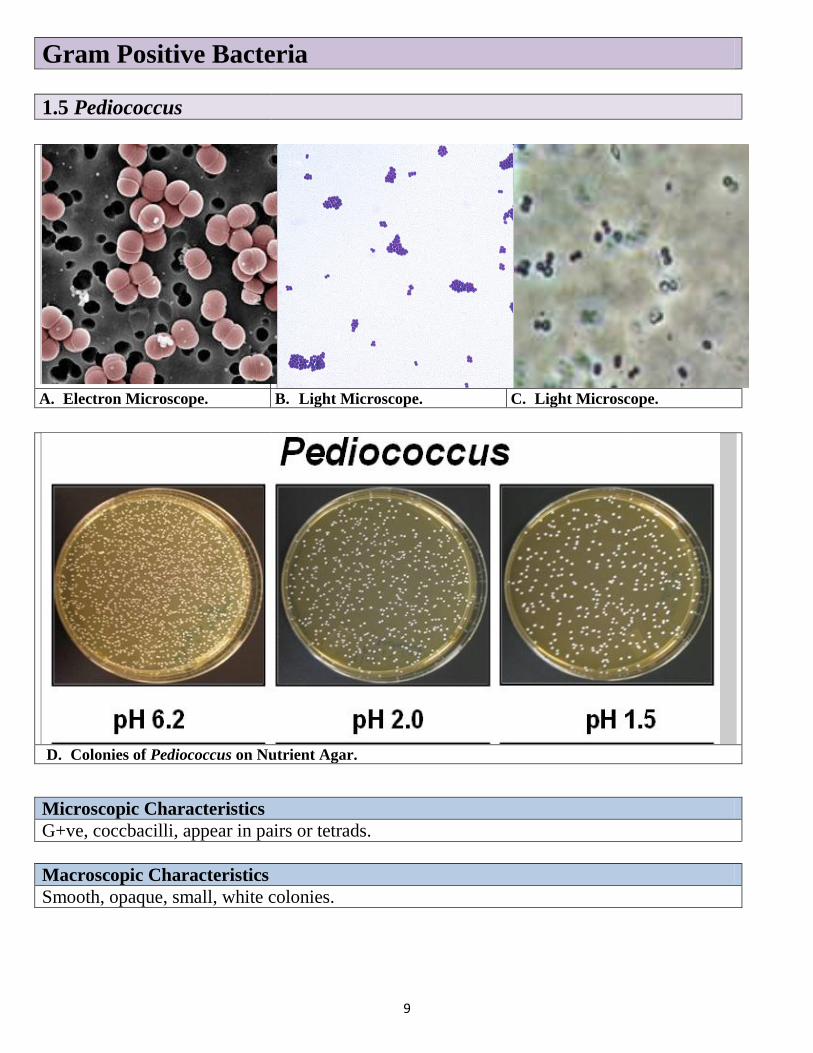
Gram Positive Bacteria
1.5 Pediococcus
A. Electron Microscope. B. Light Microscope. C. Light Microscope.
D. Colonies of Pediococcus on Nutrient Agar.
Microscopic Characteristics
G+ve, coccbacilli, appear in pairs or tetrads.
Macroscopic Characteristics
Smooth, opaque, small, white colonies.
10
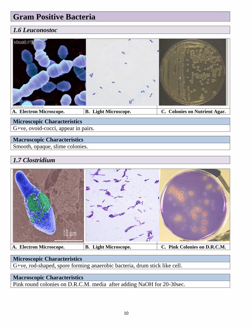
Gram Positive Bacteria
1.6 Leuconostoc
A. Electron Microscope. B. Light Microscope. C. Colonies on Nutrient Agar.
Microscopic Characteristics
G+ve, ovoid-cocci, appear in pairs.
Macroscopic Characteristics
Smooth, opaque, slime colonies.
1.7 Clostridium
A. Electron Microscope. B. Light Microscope. C. Pink Colonies on D.R.C.M.
Microscopic Characteristics
G+ve, rod-shaped, spore forming anaerobic bacteria, drum stick like cell.
Macroscopic Characteristics
Pink round colonies on D.R.C.M. media after adding NaOH for 20-30sec.

11
Other Gram Positive Bacteria
1.Actinomycetes
A. Electron Microscope. B. Light Microscope. C. Light Microscope.
D. Different morphologies of Actinomycetes.
Microscopic Characteristics
G+ve, non-spore former, appear like branched network hyphae.
Macroscopic Characteristics
Colonies small, fragile, compressed, chalky, may be white, yellow or red.
12
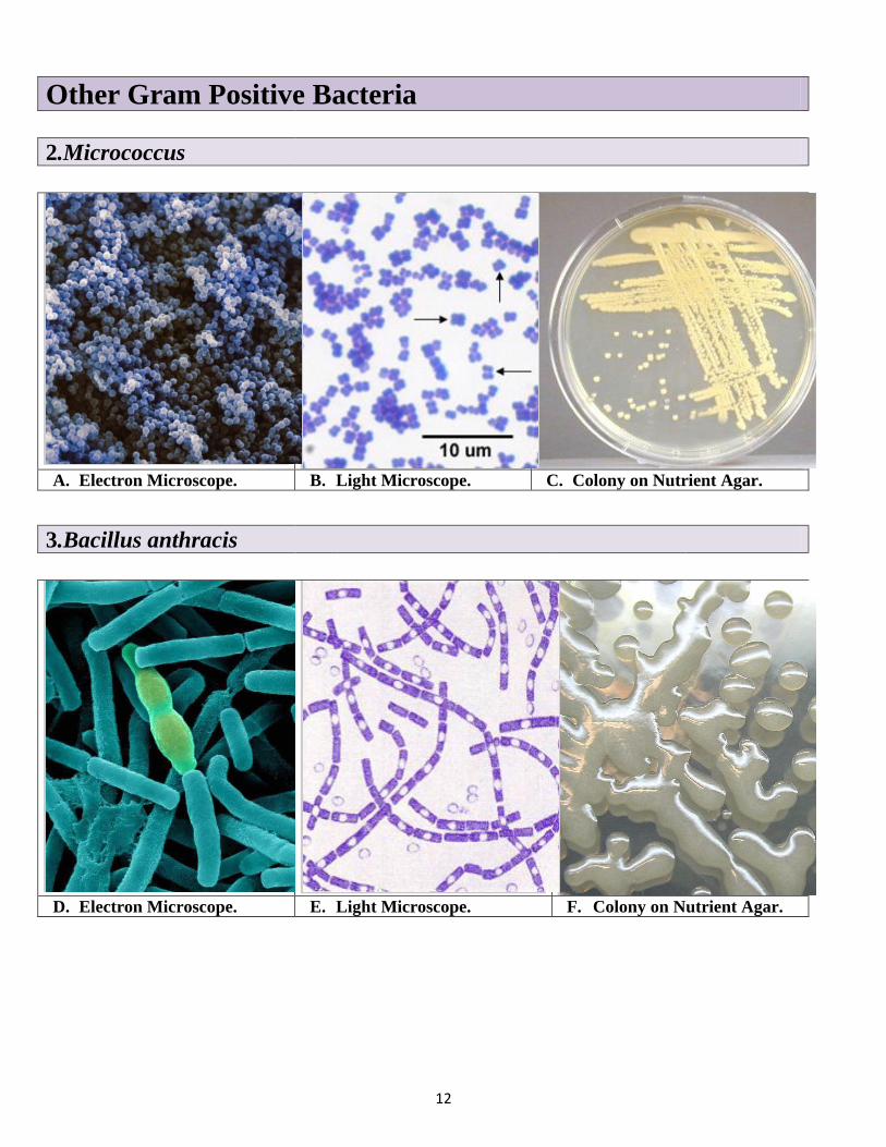
Other Gram Positive Bacteria
2.Micrococcus
A. Electron Microscope. B. Light Microscope. C. Colony on Nutrient Agar.
3.Bacillus anthracis
D. Electron Microscope. E. Light Microscope. F. Colony on Nutrient Agar.

13
4.Listeria monocytogenes
A. Electron Microscope. B. Light Microscope. C. Colony on M.M.A. media.
Acid Fast Stain Gram Positive like Bacteria
Mycobacterium tuberculosis
A. Electron Microscope. B. Light Microscope. C. Colony on Agar media.
14
2- Gram Negative Bacteria
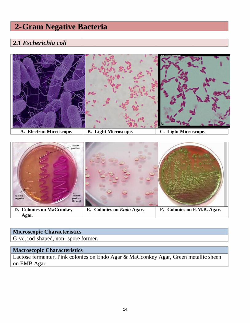
2.1 Escherichia coli
A. Electron Microscope. B. Light Microscope. C. Light Microscope.
D. Colonies on MaCconkey
Agar.
E. Colonies on Endo Agar. F. Colonies on E.M.B. Agar.
Microscopic Characteristics
G-ve, rod-shaped, non- spore former.
Macroscopic Characteristics
Lactose fermenter, Pink colonies on Endo Agar & MaCconkey Agar, Green metallic sheen
on EMB Agar.
15
Gram Negative Bacteria
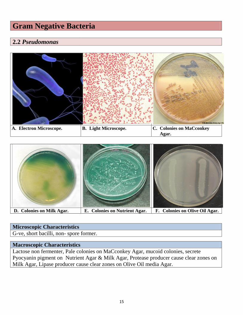
2.2 Pseudomonas
A. Electron Microscope. B. Light Microscope. C. Colonies on MaCconkey
Agar.
D. Colonies on Milk Agar. E. Colonies on Nutrient Agar. F. Colonies on Olive Oil Agar.
Microscopic Characteristics
G-ve, short bacilli, non- spore former.
Macroscopic Characteristics
Lactose non fermenter, Pale colonies on MaCconkey Agar, mucoid colonies, secrete
Pyocyanin pigment on Nutrient Agar & Milk Agar, Protease producer cause clear zones on
Milk Agar, Lipase producer cause clear zones on Olive Oil media Agar.
16
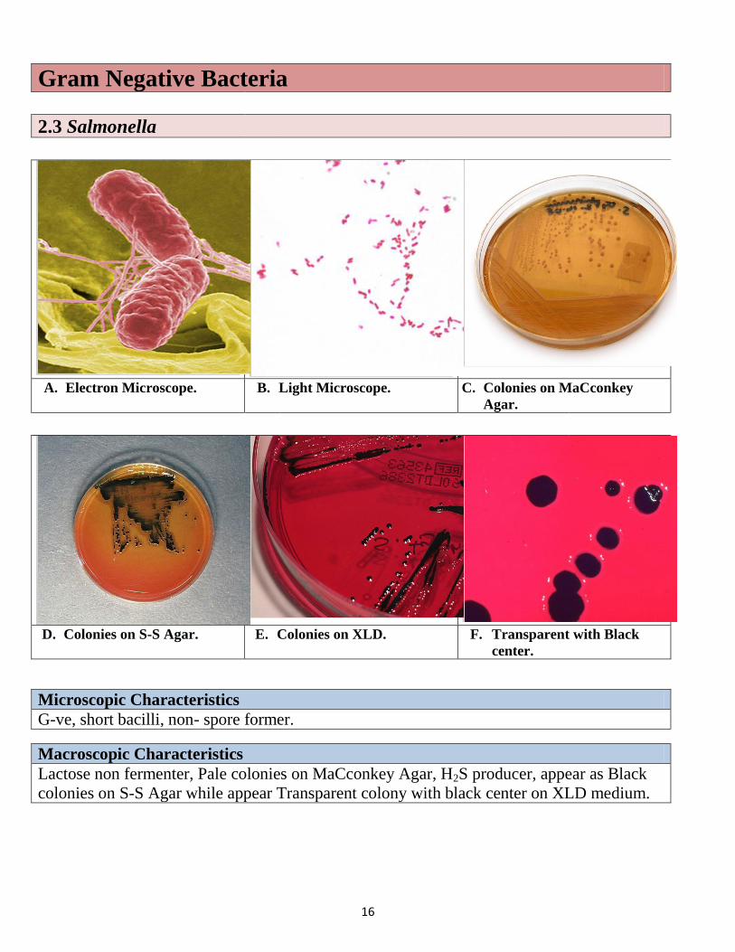
Gram Negative Bacteria
2.3 Salmonella
A. Electron Microscope. B. Light Microscope. C. Colonies on MaCconkey
Agar.
D. Colonies on S-S Agar. E. Colonies on XLD. F. Transparent with Black
center.
Microscopic Characteristics
G-ve, short bacilli, non- spore former.
Macroscopic Characteristics
Lactose non fermenter, Pale colonies on MaCconkey Agar, H2S producer, appear as Black
colonies on S-S Agar while appear Transparent colony with black center on XLD medium.
17
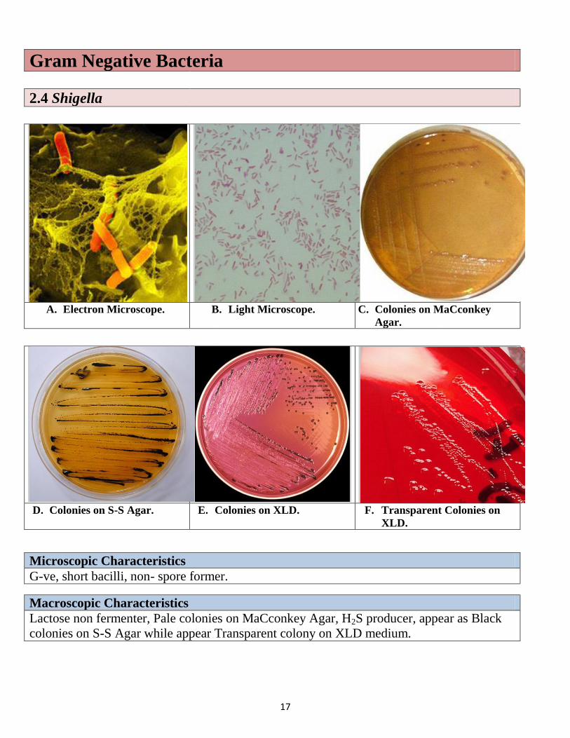
Gram Negative Bacteria
2.4 Shigella
A. Electron Microscope. B. Light Microscope. C. Colonies on MaCconkey
Agar.
D. Colonies on S-S Agar. E. Colonies on XLD. F. Transparent Colonies on
XLD.
Microscopic Characteristics
G-ve, short bacilli, non- spore former.
Macroscopic Characteristics
Lactose non fermenter, Pale colonies on MaCconkey Agar, H2S producer, appear as Black
colonies on S-S Agar while appear Transparent colony on XLD medium.
18
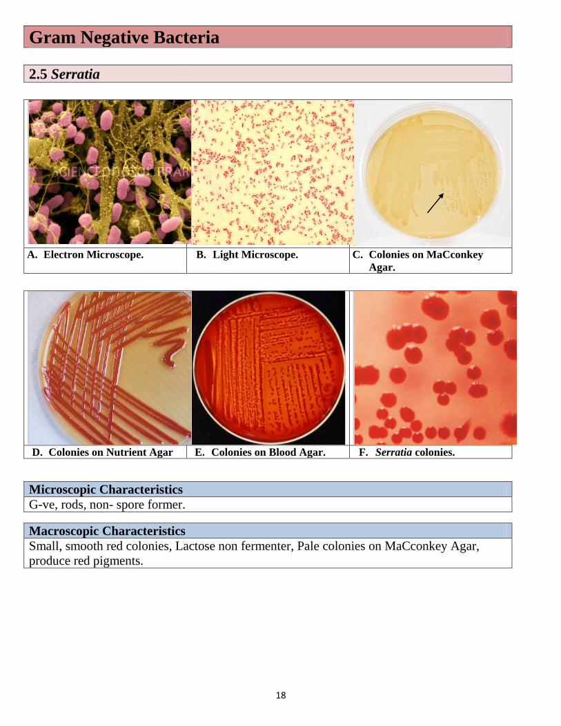
Gram Negative Bacteria
2.5 Serratia
A. Electron Microscope. B. Light Microscope. C. Colonies on MaCconkey
Agar.
D. Colonies on Nutrient Agar E. Colonies on Blood Agar. F. Serratia colonies.
Microscopic Characteristics
G-ve, rods, non- spore former.
Macroscopic Characteristics
Small, smooth red colonies, Lactose non fermenter, Pale colonies on MaCconkey Agar,
produce red pigments.
19
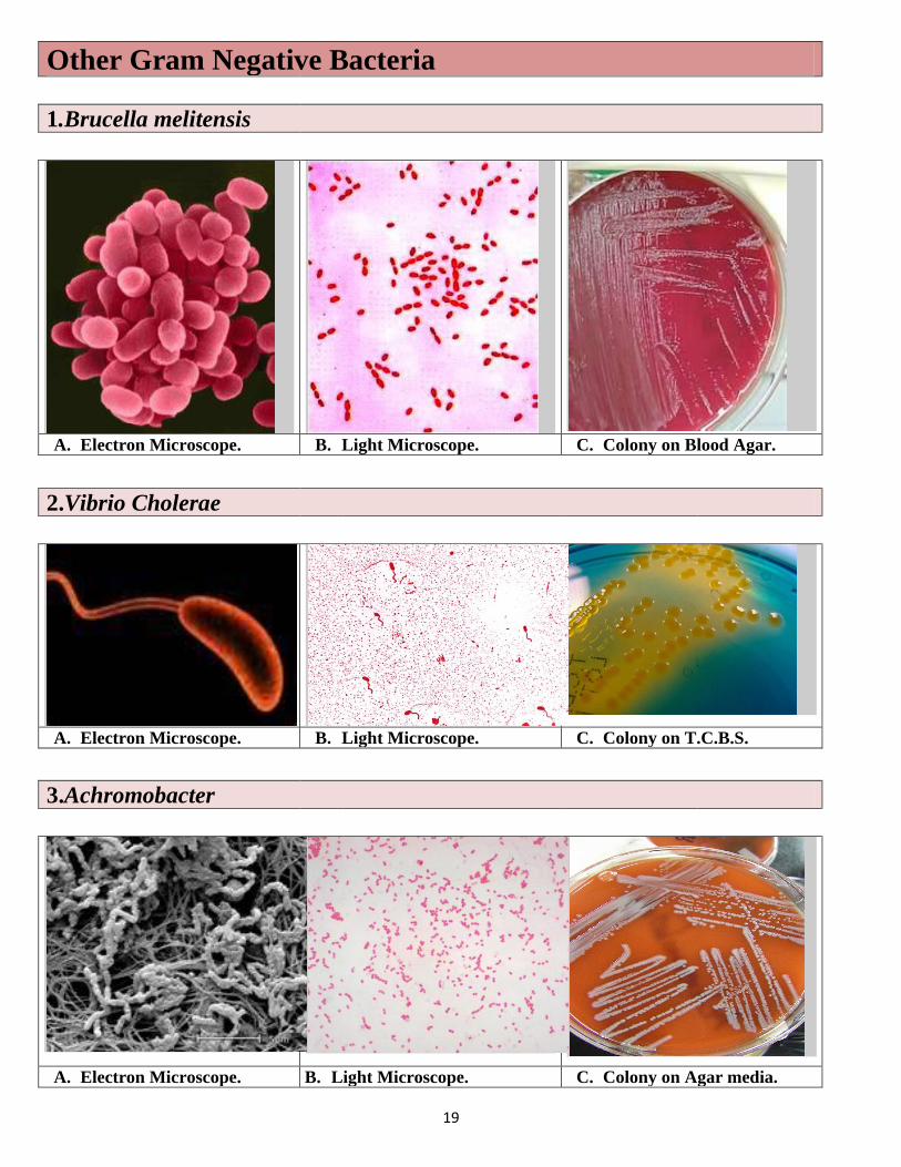
Other Gram Negative Bacteria
1.Brucella melitensis
A. Electron Microscope. B. Light Microscope. C. Colony on Blood Agar.
2.Vibrio Cholerae
A. Electron Microscope. B. Light Microscope. C. Colony on T.C.B.S.
3.Achromobacter
A. Electron Microscope. B. Light Microscope. C. Colony on Agar media.
20
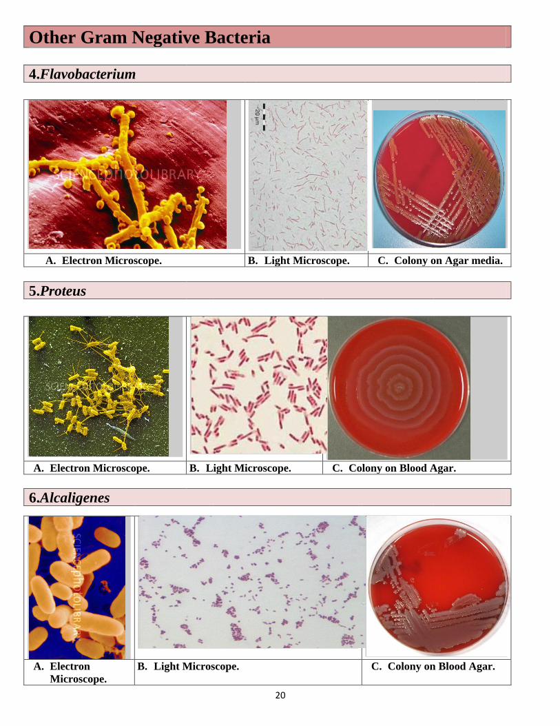
Other Gram Negative Bacteria
4.Flavobacterium
A. Electron Microscope. B. Light Microscope. C. Colony on Agar media.
5.Proteus
A. Electron Microscope. B. Light Microscope. C. Colony on Blood Agar.
6.Alcaligenes
A. Electron
Microscope.
B. Light Microscope. C. Colony on Blood Agar.

21
22
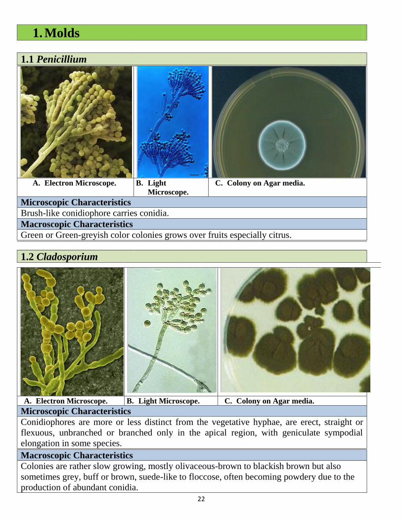
1. Molds
1.1 Penicillium
A. Electron Microscope. B. Light
Microscope.
C. Colony on Agar media.
Microscopic Characteristics
Brush-like conidiophore carries conidia.
Macroscopic Characteristics
Green or Green-greyish color colonies grows over fruits especially citrus.
1.2 Cladosporium
A. Electron Microscope. B. Light Microscope. C. Colony on Agar media.
Microscopic Characteristics
Conidiophores are more or less distinct from the vegetative hyphae, are erect, straight or
flexuous, unbranched or branched only in the apical region, with geniculate sympodial
elongation in some species.
Macroscopic Characteristics
Colonies are rather slow growing, mostly olivaceous-brown to blackish brown but also
sometimes grey, buff or brown, suede-like to floccose, often becoming powdery due to the
production of abundant conidia.

23
Molds
1.3 Asperigillus niger
A. Electron Microscope. B. Light Microscope. C. Colony on Agar media.
Microscopic Characteristics
Non-Branched conidiophore with bulb end carries conidia like sun rays.
Macroscopic Characteristics
Pin like black growth.
1.4 Asperigillus flavus
A. Electron Microscope. B. Light Microscope. C. Colony on Agar media.
Microscopic Characteristics
Non-Branched conidiophore with bulb end carries conidia.
Macroscopic Characteristics
Pin like green growth.
24
Molds
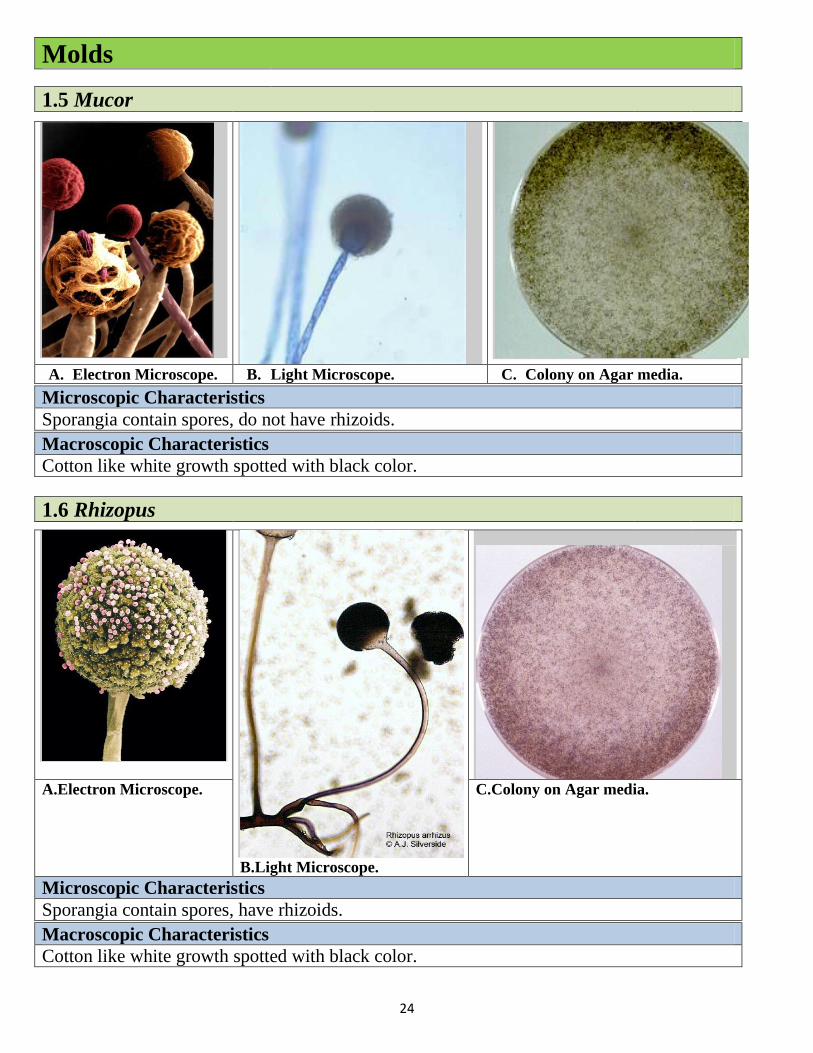
1.5 Mucor
A. Electron Microscope. B. Light Microscope. C. Colony on Agar media.
Microscopic Characteristics
Sporangia contain spores, do not have rhizoids.
Macroscopic Characteristics
Cotton like white growth spotted with black color.
1.6 Rhizopus
B.Light Microscope.
A.Electron Microscope. C.Colony on Agar media.
Microscopic Characteristics
Sporangia contain spores, have rhizoids.
Macroscopic Characteristics
Cotton like white growth spotted with black color.

25
Molds
1.7 Alternaria
A. Electron Microscope. B. Light Microscope. C. Colony on Agar media.
Microscopic Characteristics
Pineapple like conidia multi-cellular, spetated horizontally & vertically, arrange in chains.
Macroscopic Characteristics
Dark green deeply grown colonies, oil-drop like colony when seen upside down the Petri-
dish.
1.8 Curvularia
A. Electron Microscope. B. Light Microscope. C. Colony on Agar media.
Microscopic Characteristics
Swollen conidia, spetated horizontally only, arrange in triple or pentagonal arrangements.
Macroscopic Characteristics
Green or black deeply grown colonies.
26
Molds
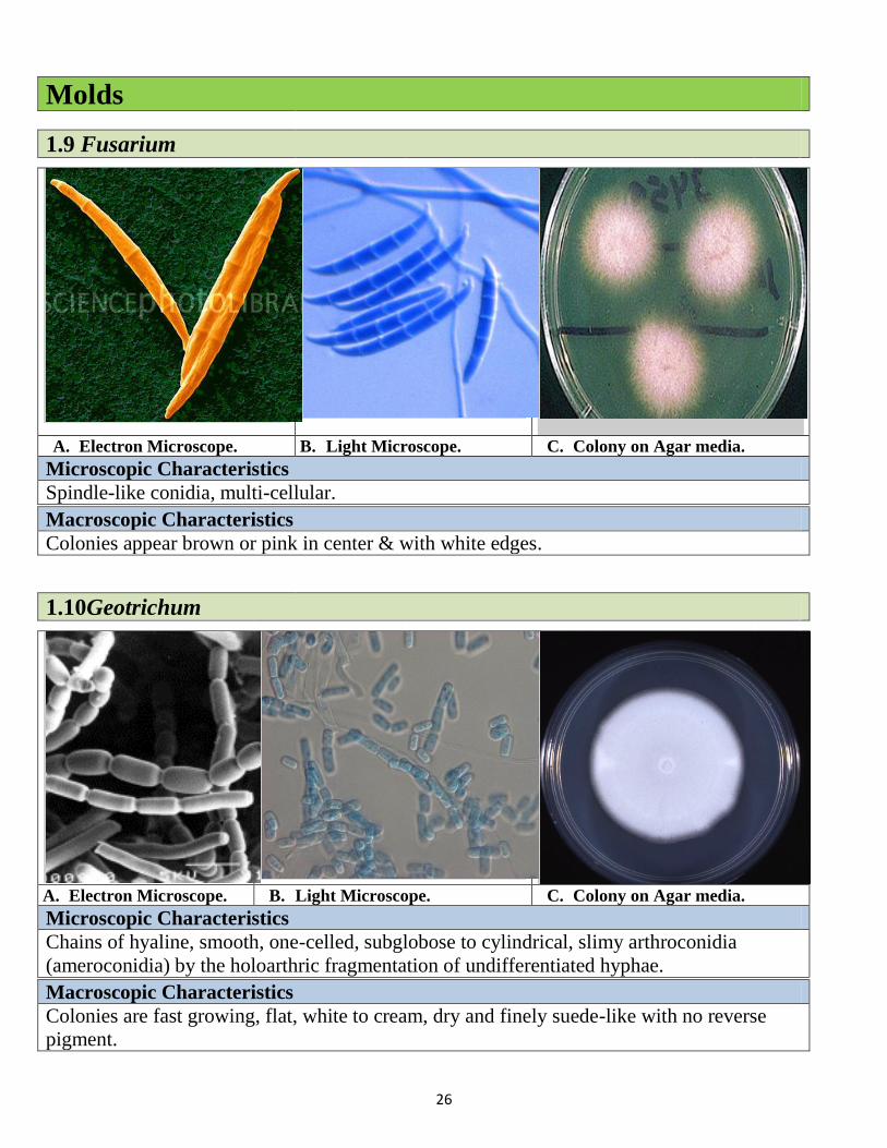
1.9 Fusarium
A. Electron Microscope. B. Light Microscope. C. Colony on Agar media.
Microscopic Characteristics
Spindle-like conidia, multi-cellular.
Macroscopic Characteristics
Colonies appear brown or pink in center & with white edges.
1.10Geotrichum
A. Electron Microscope. B. Light Microscope. C. Colony on Agar media.
Microscopic Characteristics
Chains of hyaline, smooth, one-celled, subglobose to cylindrical, slimy arthroconidia
(ameroconidia) by the holoarthric fragmentation of undifferentiated hyphae.
Macroscopic Characteristics
Colonies are fast growing, flat, white to cream, dry and finely suede-like with no reverse
pigment.
27
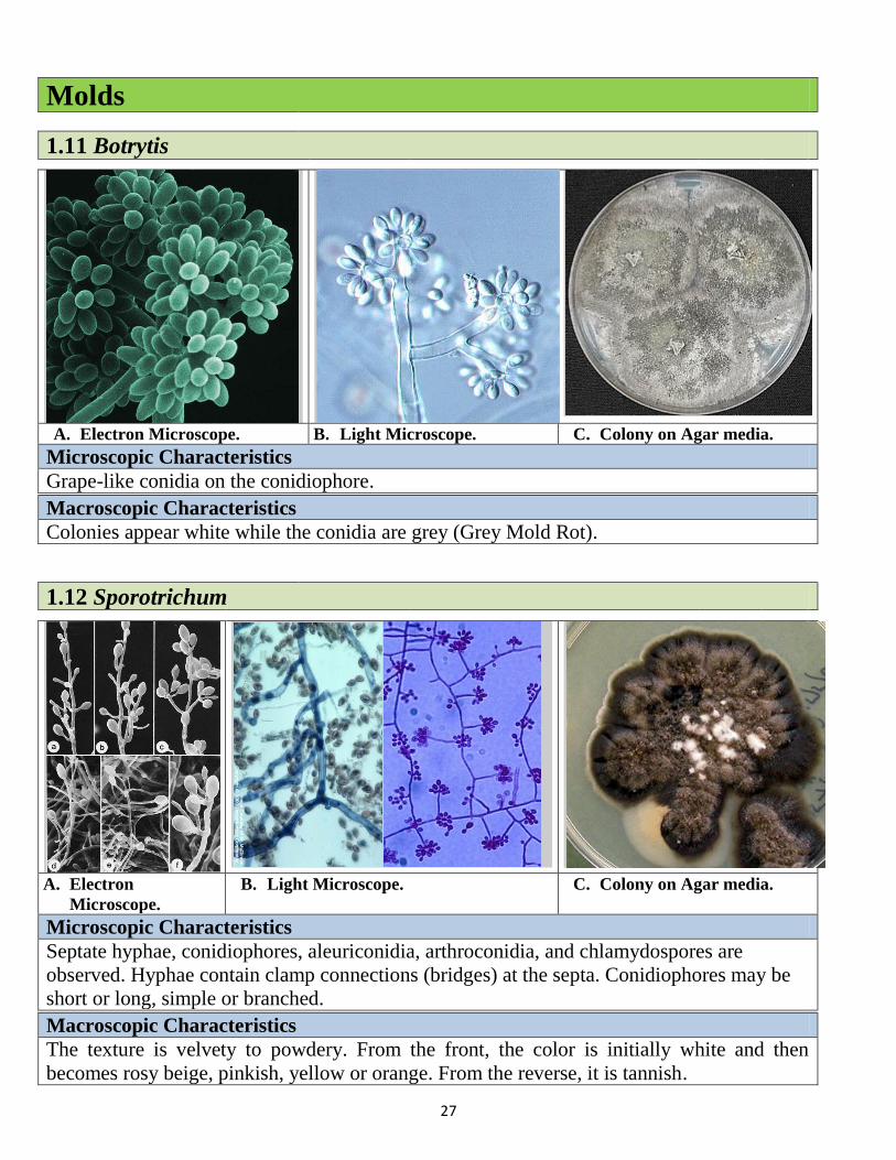
Molds
1.11 Botrytis
A. Electron Microscope. B. Light Microscope. C. Colony on Agar media.
Microscopic Characteristics
Grape-like conidia on the conidiophore.
Macroscopic Characteristics
Colonies appear white while the conidia are grey (Grey Mold Rot).
1.12 Sporotrichum
A. Electron
Microscope.
B. Light Microscope. C. Colony on Agar media.
Microscopic Characteristics
Septate hyphae, conidiophores, aleuriconidia, arthroconidia, and chlamydospores are
observed. Hyphae contain clamp connections (bridges) at the septa. Conidiophores may be
short or long, simple or branched.
Macroscopic Characteristics
The texture is velvety to powdery. From the front, the color is initially white and then
becomes rosy beige, pinkish, yellow or orange. From the reverse, it is tannish.

28
29
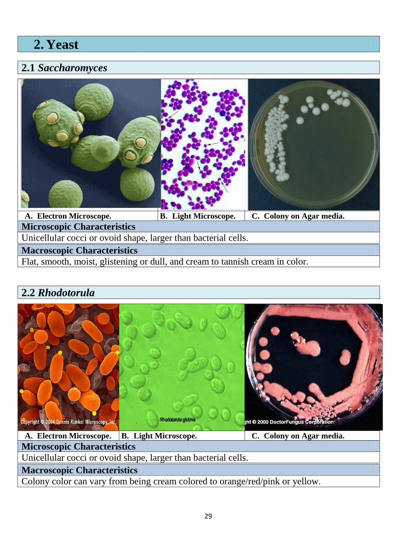
2. Yeast
2.1 Saccharomyces
A. Electron Microscope. B. Light Microscope. C. Colony on Agar media.
Microscopic Characteristics
Unicellular cocci or ovoid shape, larger than bacterial cells.
Macroscopic Characteristics
Flat, smooth, moist, glistening or dull, and cream to tannish cream in color.
2.2 Rhodotorula
A. Electron Microscope. B. Light Microscope. C. Colony on Agar media.
Microscopic Characteristics
Unicellular cocci or ovoid shape, larger than bacterial cells.
Macroscopic Characteristics
Colony color can vary from being cream colored to orange/red/pink or yellow.
30
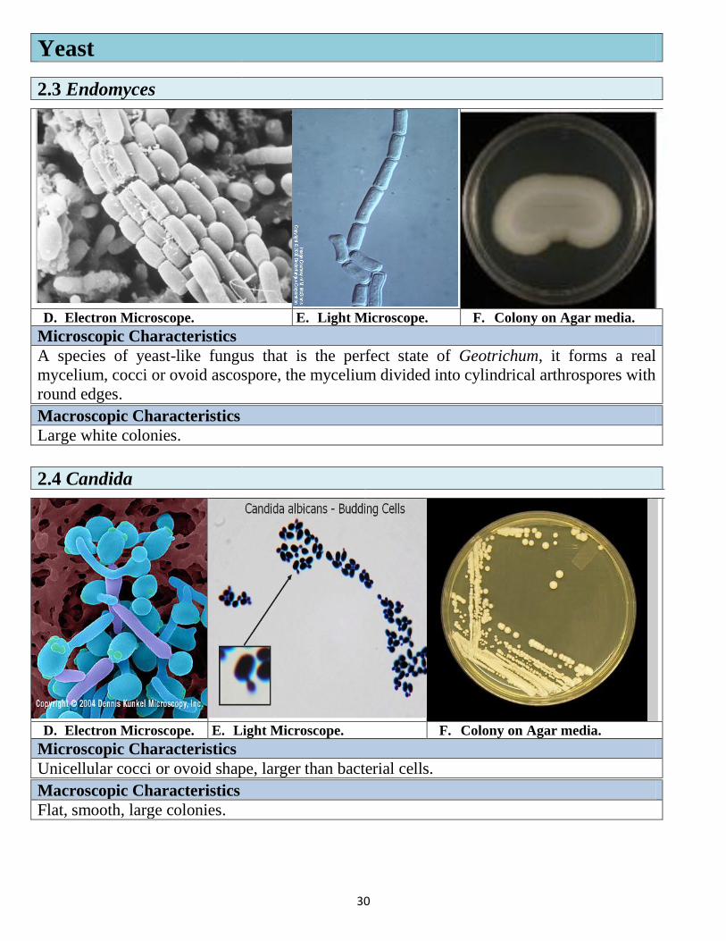
Yeast
2.3 Endomyces
D. Electron Microscope. E. Light Microscope. F. Colony on Agar media.
Microscopic Characteristics
A species of yeast-like fungus that is the perfect state of Geotrichum, it forms a real
mycelium, cocci or ovoid ascospore, the mycelium divided into cylindrical arthrospores with
round edges.
Macroscopic Characteristics
Large white colonies.
2.4 Candida
D. Electron Microscope. E. Light Microscope. F. Colony on Agar media.
Microscopic Characteristics
Unicellular cocci or ovoid shape, larger than bacterial cells.
Macroscopic Characteristics
Flat, smooth, large colonies.
31
References
Dennis Kunkel Microscopy, Inc. Science Stock Photography cited by
http://www.denniskunkel.com/index.php.
Free Encyclopedia online cited by
http://en.wikipedia.org/wiki/Main_Page.
Mycology online reference cited by http://www.doctorfungus.org/.
The University of Adelaide, Australia, Mycology online cited by http://www.mycology.adelaide.edu.au/gallery/.
Tony Hart, Paul Shears (2004).Color Atlas of Medical Microbiology:
Elsevier’s Health Sciences Department.
US National library of Medicine cited by
http://www.ncbi.nlm.nih.gov/pubmed.