atopic urticaria : phenotypes - world allergy … urticaria_msgreaves... · 6/22/2011 · atopic...
TRANSCRIPT
Atopic urticaria : phenotypes
Malcolm W GreavesCutaneous Allergy Clinic,
St John`s Institute of Dermatology, St Thomas` Hospital,
London
Potential conflicts of interest : have received fees
for services from Genentech & Novartis
June 2011
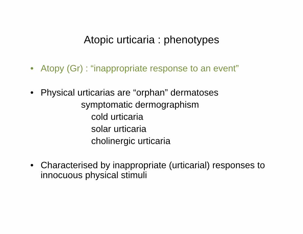
Atopic urticaria : phenotypes
• Atopy (Gr) : “inappropriate response to an event”
• Physical urticarias are “orphan” dermatosessymptomatic dermographism
cold urticariasolar urticariacholinergic urticaria
• Characterised by inappropriate (urticarial) responses to innocuous physical stimuli
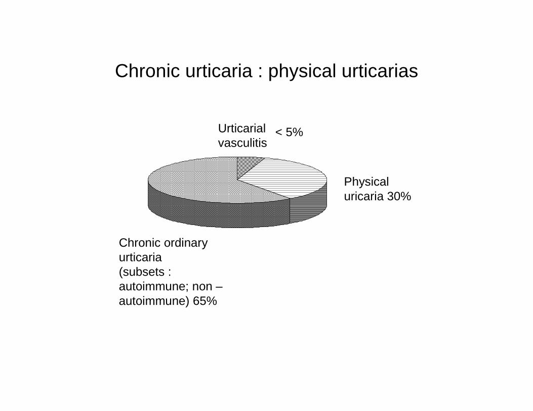
Chronic urticaria : physical urticarias
Physical uricaria 30%
Urticarialvasculitis
Chronic ordinary urticaria (subsets : autoimmune; non –autoimmune) 65%
< 5%
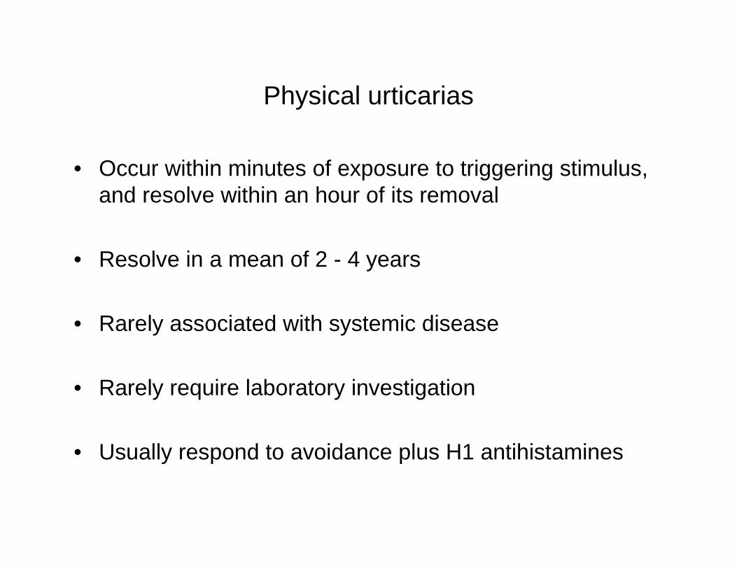
Physical urticarias
• Occur within minutes of exposure to triggering stimulus, and resolve within an hour of its removal
• Resolve in a mean of 2 - 4 years
• Rarely associated with systemic disease
• Rarely require laboratory investigation
• Usually respond to avoidance plus H1 antihistamines
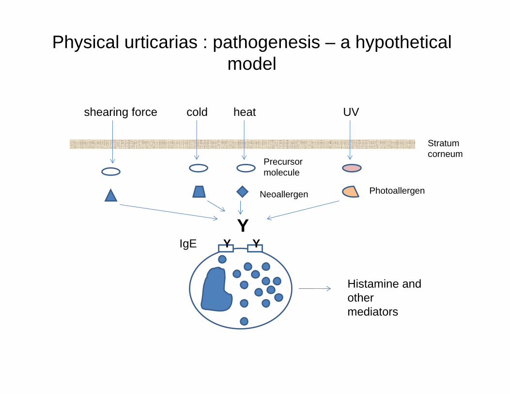
Physical urticarias : pathogenesis – a hypothetical model
shearing force cold heat UV
Stratum corneum
Precursor molecule
Neoallergen Photoallergen
YY YIgE
Histamine and other mediators
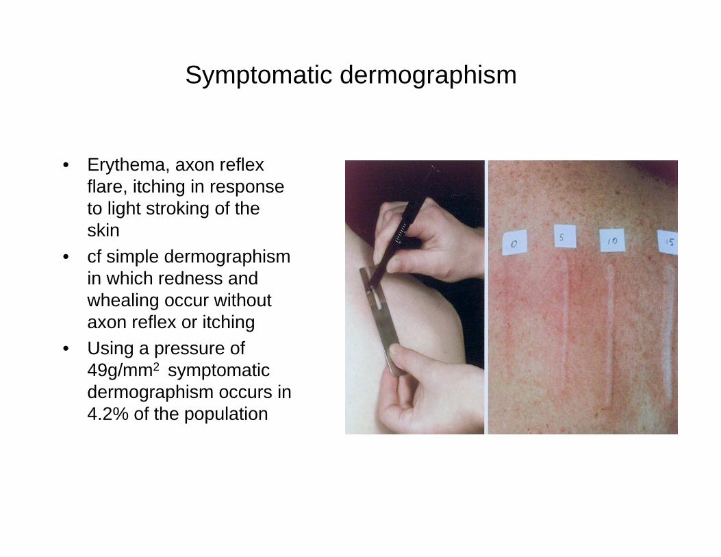
Symptomatic dermographism
• Erythema, axon reflex flare, itching in response to light stroking of the skin
• cf simple dermographismin which redness and whealing occur without axon reflex or itching
• Using a pressure of 49g/mm2 symptomatic dermographism occurs in 4.2% of the population
Symptomatic dermographism : pathophysiology
• Dermal mast cell population is normal, but reduction by application of potent topical steroids under occlusion reduces mast cell population and skin reactivity
• Elevated tissue histamine levels are present in involved skin and recent evidence also implicates Substance P
• Skin reactivity is passively transferable by IgE in a modified PK reaction (Newcombe Am J Med 1973, 54, 174; Aoki Brit J Derm 1984, 114: 545; Murphy, Greaves Br J Derm 1987, 116: 801)
Symptomatic dermographism : treatment
• Treatment is by low sedation H1 antihistamines, but off –label dosage is often necessary.
• H2 antihistamines are often prescribed as well, but several RCT`s have shown that any benefit is statistical rather than clinically useful
• Narrow band UVB phototherapy (311 nm UV) may also be effective, especially for pruritus (Borsova JAAD 2008; 59: 752)

Cold urticaria
Variants include : • Secondary acquired cold urticaria with cryoglobulinaemia
• Cold reflex urticaria : exposure to cold atmosphere causes whealing in covered as well as exposed skin, with a negative local cold challenge test
• Hereditary cold autoinflammatory syndromes (FCAS) with negative cold challenge tests and mutation in CIAS1
• Several other very rare subtypes
Acquired cold urticaria : pathophysiology
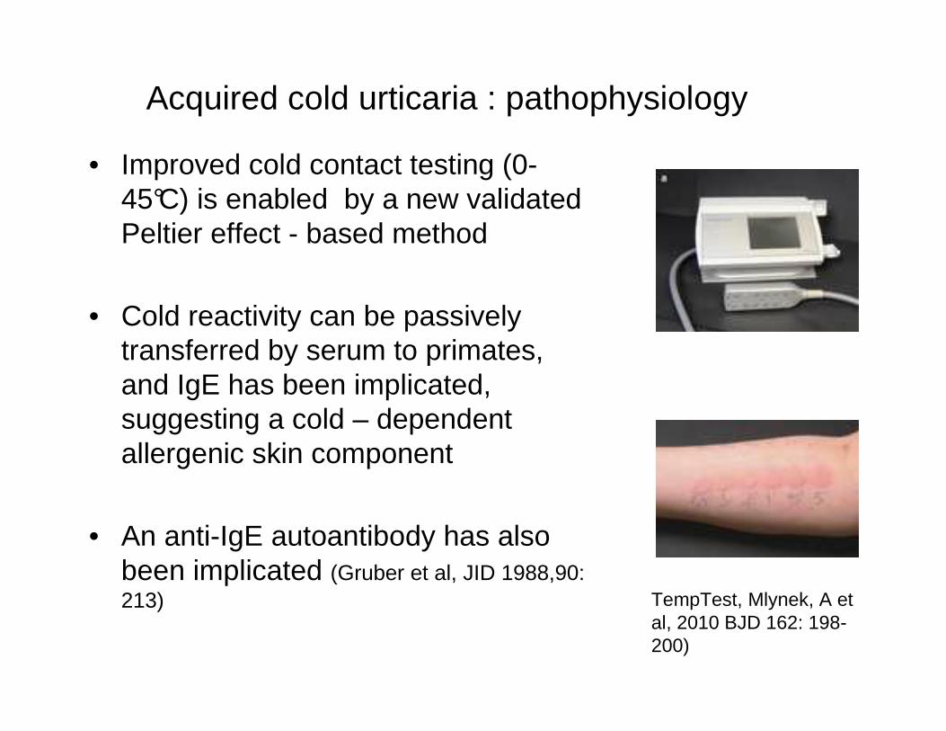
• Improved cold contact testing (0-45°C) is enabled by a new validated Peltier effect - based method
• Cold reactivity can be passively transferred by serum to primates, and IgE has been implicated, suggesting a cold – dependent allergenic skin component
• An anti-IgE autoantibody has also been implicated (Gruber et al, JID 1988,90: 213) TempTest, Mlynek, A et
al, 2010 BJD 162: 198-200)
Acquired cold urticaria : role of virus infections and IgE
Virus infections including EB and HIV have consistently been associated with cold urticaria, and can cause upregulation of IgE
• Low CD4, reduced TH1 and IFN γ cause increased TH2 cells
• HIV – 1 protein gp 120 and Tat induce TH2 cytokines, or interact directly with specific receptors on B cells causing increased IgEmRNA
• In either case some of this IgE binds specifically to a putative cold induced antigenic determinant
Cold urticaria : management
• Off – label dosages of 2nd generation H1 antihistamines are often needed
• Omalizumab (Xolair) has been effective in selected intractable cases (Boyce JACI 2006;117)
• Bathing in cool water risks anaphylactoid reactions which may be fatal
• A recently recognised hazard : anaphylactoid reaction due to systemic hypothermia (4°C) in open heart surgery; avoided by normothermic cardioplegia
Cholinergic urticaria
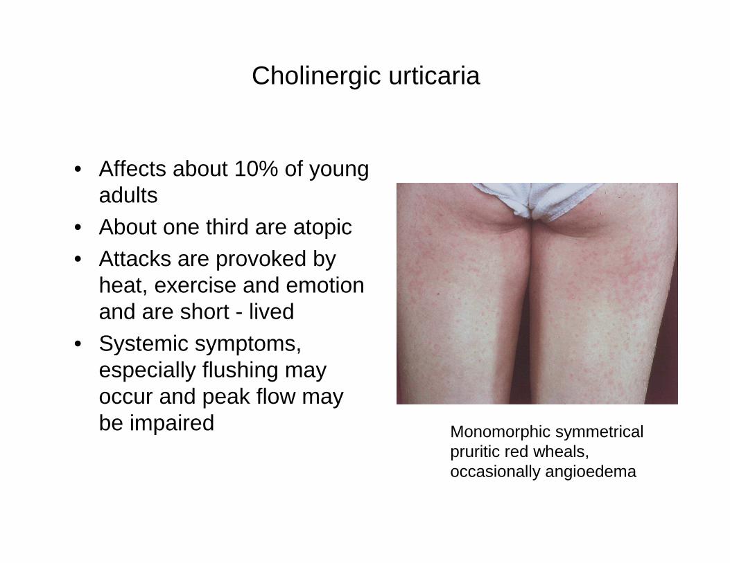
• Affects about 10% of young adults
• About one third are atopic• Attacks are provoked by
heat, exercise and emotion and are short - lived
• Systemic symptoms, especially flushing may occur and peak flow may be impaired Monomorphic symmetrical
pruritic red wheals, occasionally angioedema
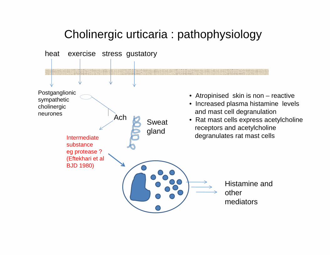
Cholinergic urticaria : pathophysiology
heat exercise stress gustatory
Histamine and other mediators
Ach
Postganglionic sympathetic cholinergic neurones
Sweat gland
• Atropinised skin is non – reactive• Increased plasma histamine levels
and mast cell degranulation• Rat mast cells express acetylcholine
receptors and acetylcholine degranulates rat mast cellsIntermediate
substance eg protease ? (Eftekhari et al BJD 1980)
Cholinergic urticaria : autologous sweat and serum reactions
Recent work (Fukunaga et al JACI 2005; 116: 397-402) has suggested that cholinergic urticaria are reactive to autologous sweat and can be divided into 2 subtypes :
• Type 1 shows strong reactions to autologous sweat, satellite wheals to id acetylcholine and negative reactions to autologous serum
• Type 2 shows a weak reaction to autologous sweat, a positive reaction to autologous serum and no reaction to acetylcholine
Positive autologous sweat tests have recently been reported in atopic eczema (Hide et al 2002) which links cholinergic urticaria and atopy and sweat from cholinergic urticaria patients releases histamine from autologous basophils
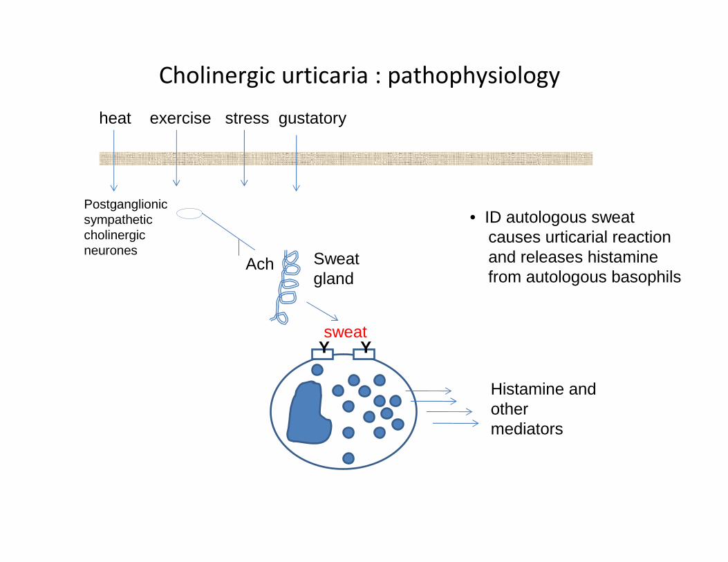
Cholinergic urticaria : pathophysiology
heat exercise stress gustatory
Y Y
Histamine and other mediators
Ach
Postganglionic sympathetic cholinergic neurones
Sweat gland
sweat
• ID autologous sweat causes urticarial reactionand releases histamine from autologous basophils

Cholinergic urticaria : diagnosis
• The diagnosis is clinical and based upon history and challenge testing : usually hot bath, exercise
• Intradermal acetyl choline / methacholine ID testing shows low specificity
• No laboratory investigations are warranted
Cholinergic urticaria : treatment
• H1 antihistamines, often in off-label dosages, are usually effective
• Systemic anticholinergics are usually only effective in toxic dosages
• RCT’s have shown the effectiveness and safety, especially in male patients, of attenuated androgens based upon findings of reduced serum protease inhibitors
• Omalizumab has been used successfully in selected cases
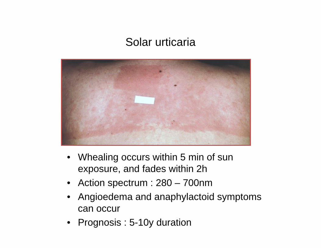
Solar urticaria
• Whealing occurs within 5 min of sun exposure, and fades within 2h
• Action spectrum : 280 – 700nm• Angioedema and anaphylactoid symptoms
can occur• Prognosis : 5-10y duration
Solar urticaria : pathophysiology
• Reactivity can be passively transferred
• Type 1 : abnormal chromophore only occurs in skin and serum of solar urticaria patients and evokes specific IgEantibodies
• Type 2 : a normal chromophore evokes specific IgEantibodies
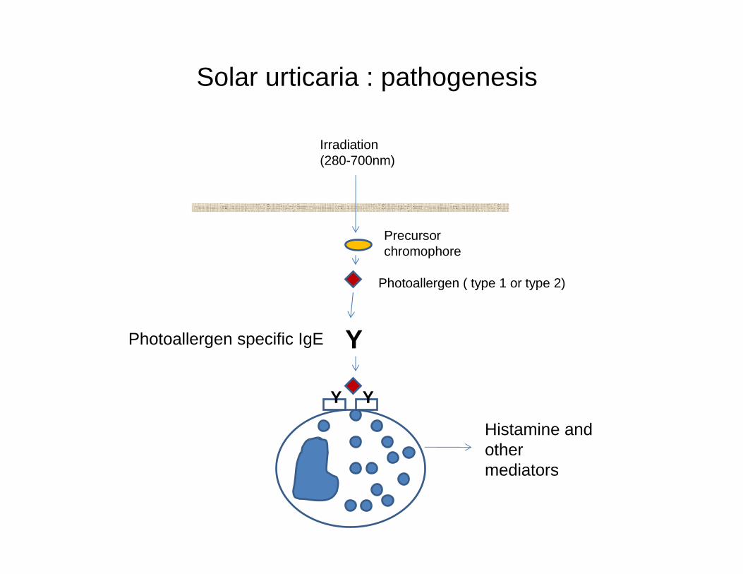
Solar urticaria : pathogenesis
Precursor chromophore
Photoallergen ( type 1 or type 2)
Y
Y Y
Photoallergen specific IgE
Histamine and other mediators
Irradiation (280-700nm)
Solar urticaria : diagnosis
• Most referrals turn out to be PLE (lesions last > 48h, cfsolar urticaria < 2h)
• Can be manifestation of erythropoietic protoporphyria
• Phototesting (natural sunlight, solar simulator lamp) confirms
• Monochromator testing establishes action spectrum, enables focussed photoprotection
Solar urticaria : treatment
• H1 antihistamines + photoprotection
• Inhibition spectra and tolerance treatment : wavelengths greater than the action spectra diminish solar urticaria –probably by inactivating photoallergens
• Tolerance treatment has proved effective though necessitating maintenance
• Plasmapheresis and most recently omalizumab have been used in selected cases