atorvastatin improves plaque stability in diabetic
TRANSCRIPT

1142
Abstract. – OBJECTIVE: To study the improv-ing effect of atorvastatin on plaque stability in diabetes mellitus (DM) mice complicated with atherosclerosis.
MATERIALS AND METHODS: Apolipoprotein E (ApoE)-/- mice were used to establish the DM mouse model. Half of the mice received ator-vastatin after successful modeling. ApoE-/- and C57BL/6J mice were used as controls. Oil red O staining and Masson staining were performed to detect the lipid and collagen components in mice. Immunohistochemical assay was used to observe the expressions of smooth muscle cell (SMC) and Ly-6c. The expressions of receptor for advanced glycation end products (RAGE), monocyte chemoattractant protein-1 (MCP-1) and nuclear factor-κB (NF-κB) in tissues were detected by Western blotting. Finally, the levels of serum soluble RAGE (sRAGE), advanced gly-cation end products (AGEs), malondialdehyde (MDA) and reduced glutathione (GSH) in mice were also detected.
RESULTS: Atorvastatin reduced the area of atherosclerotic plaque and improved the stabili-ty of arterial plaque through reducing lipid depo-sition, the number of macrophages and SMC, in-creasing collagen fibers. In mice in atorvasta-tin group, the levels of serum AGEs and sRAGE were decreased. Moreover, atorvastatin inhibit-ed the downstream pathway of RAGE as well as DM, thus inducing the oxidative stress.
CONCLUSIONS: Atorvastatin improves plaque stability in diabetic atherosclerosis through the RAGE pathway.
Key Words:Atorvastatin, Atherosclerosis, RAGE.
Introduction
Diabetes mellitus (DM) is a kind of systemic disease that affects the quality of life and lon-
gevity of patients, and atherosclerosis is a major complication of type 1 and type 2 DM1. Up to 75% patients diagnosed with DM eventually die of atherosclerosis-related cardiovascular diseas-es, such as myocardial infarction (MI), peripheral arterial disease and stroke2. Both occurrence rate of cardiovascular events and death rate in DM patients complicated with coronary heart disease are significantly higher than those in non-DM patients3. During the occurrence and develop-ment processes of atherosclerosis, the rupture of unstable atherosclerotic plaques can lead to acute coronary syndromes, such as MI. The patholog-ical features of vulnerable plaques include the central lipid core area covered by thinner fibrous tissues and the infiltration of a large number of inflammatory cells4.
During the course of DM, a variety of risk factors, including hyperglycemia, hyperlipidemia and disorders of angiotensin system, are associ-ated with atherosclerosis. In recent years, studies have shown that oxidative stress and advanced glycation end products (AGEs) play important roles in the occurrence and development of vas-cular lesions of DM. AGEs interact with receptor for AGEs (RAGE), a receptor specific for vascu-lar wall cells, to activate oxidative stress5.
RAGE is a member of the cell surface molecule immunoglobulin superfamily and a multi-ligand receptor for vascular cells, which plays a key role in the inflammatory process. In addition to AGEs, other cytokines and inflammatory factors can also up-regulate the RAGE expression. The activation of AGEs-RAGE pathway will increase the level of oxidative stress in vivo, inducing the production of inflammatory factors, leading to vascular endothelial cell injury and apoptosis, and aggravating the occurrence and development of atherosclerosis6.
European Review for Medical and Pharmacological Sciences 2018; 22: 1142-1149
F. ZHOU1, Y. TAN1, X.-H. CHEN1, F.-L. WU2, D.-J. YANG1, X.-W. ZHANG1, X.-M. WU3, Y.-Q. DENG1
1Department of Neurology, The Third Affiliated Hospital of Nanchang University, Nanchang, China2Guangdong Cardiovascular Institute, Guangzhou, China3Department of Neurology, Jiangxi Provincial People’s Hospital, Nanchang, China
Corresponding Author: Xiaomu Wu, MD; e-mail: [email protected]
Atorvastatin improves plaque stability in diabetic atherosclerosis through the RAGE pathway

Atorvastatin improves plaque stability in diabetic atherosclerosis through the RAGE pathway
1143
Atorvastatin, as a 3-hydroxy-3-methyl-glu-taryl coenzyme A (HMG-CoA) reductase inhib-itor, is widely used in the treatment of patients with atherosclerosis. Statins can alleviate the coronary artery inflammatory changes and im-prove the plaque instability, thus reducing the mortality and morbidity rates of cardiovascular disease in DM patients7. In addition, statins have many other effects, including anti-inflammation, anti-oxidation and improvement of vascular en-dothelial function8. Non-antihyperlipidemic ef-fects of these statins have also become research hotspots9.
Materials and Methods
Animals and ReagentsA total of 60 clean-grade adult male apolipo-
protein E (ApoE)-/- mice aged 8 weeks weighing (24.20 ± 0.50) g, and 20 C57BL/6J mice aged 8 weeks weighing (17.20 ± 0.48) g were provided by the Laboratory Animal Center of Nanjing University (Nanjing, China). Nutritional status and mental status of all animals were normal. This investigation was approved by the Animal Ethics Committee of Nanchang University Ani-mal Center. Monocyte chemoattractant protein-1 (MCP-1) and nuclear factor-κB (NF-κB) anti-bodies were purchased from Sigma-Aldrich (St. Louis, MO, USA). Oil red O, hematoxylin-eosin (HE) and Masson staining reagents and triglycer-ide, low-density lipoprotein, high-density lipo-protein, cholesterol, superoxide dismutase (SOD) and glutathione (GSH) enzyme-linked immuno-sorbent assay (ELISA) kits were purchased from Beyotime (Shanghai, China). Ly-6c and smooth muscle cell (SMC) antibodies were bought from CST (Danvers, MA, USA).
Modeling and GroupingAfter 60 ApoE-/- mice were fed for 2 d, 40 mice
were randomly selected to receive intraperitoneal injections of sodium (120 mg/kg) and streptozo-tocin (STZ) (100 mg/kg) to induce the mild DM model. After 1 week, mice showed mild DM symptoms, and they were randomly divided into two groups with 20 mice in each group. Mice in DM + atorvastatin group received atorvastatin (10 mg/kg/d) intragastrically, while those in DM group received no treatment. In addition, 20 ApoE-/- mice were used as ApoE-/- control group, and 20 C57BL/6J mice as blank control group. All mice were fed with high-fat diet supplement-
ed by 0.25% cholesterol and 20% lard oil with relative humidity of (50 ± 15) % at (21 ± 2°C) in 12-h light/dark cycle. Mice were executed after 20 weeks for further data analyses.
HE, Masson and Oil Red O StainingMice were fasted for 12 h and anesthetized,
and the chest was opened to expose the heart, which, with the aortic root and aorta, were dissected, rinsed in phosphate buffered saline (PBS) at 4°C and fixed with 4% paraformalde-hyde. Tissues were embedded into paraffin, and paraffin-embedded tissues were serially sliced into 4 cm-thick sections and placed at 60°C overnight, followed by dewaxing via xylene, dehydration via gradient alcohol, HE staining, Masson staining and oil red O staining. Three cross profiles of each section were detected using the medical image analysis software (Im-age-Pro Plus IPP, Mediaplayer, Silver Springs, MD, USA).
Immumohistochemical StainingAfter paraffin-embedded arterial tissue sec-
tions were dewaxed via xylene, dehydrated via gradient alcohol and incubated with warm de-ionized water containing 0.3% H2O2 for 30 min, the endogenous peroxides were removed, and sections were sealed with serum and added with primary antibody at 4°C overnight. On the next day, immunoglobulin G (IgG) antibody-horserad-ish peroxidase (HRP) were added for incubation. Sections were incubated in the mixed solution prepared using the avidin-biotin complex (ABC) kit, followed by color development reaction via diaminobenzidine (DAB) for 10 min, hematoxy-lin re-staining, washing, dehydration, transparen-cy and observation under an optical microscope (Olympus Optical Co., Ltd, Tokyo, Japan).
General Aorta StainingMice were fasted for 12 h and anesthetized,
and the chest was opened to expose the heart, which, with the aortic root and aorta, were dissected, rinsed in phosphate buffered saline (PBS) at 4°C and fixed with 4% paraformal-dehyde. After oil red O staining, the aorta was paved onto a wax plate under a microscope, and fixed with nails. After photography using a charge-coupled device (CCD) camera, images were integrated using Adobe Photoshop Version 7.0 (Adobe, San Jose, CA, USA), followed by calculation via ImageJ (Rawak Software, Inc., Hamburg, Germany).

F. Zhou, Y. Tan, X.-H. Chen, F.-L. Wu, D.-J. Yang, X.-W. Zhang, X.-M. Wu, Y.-Q. Deng
1144
Western BlottingTissues extracted were ground via liquid ni-
trogen, diluted with normal saline and placed on ice, the supernatant was taken using centrif-ugation at 4°C for 5 min. Then, the supernatant was discarded. The sediment was resuspended by radioimmunoprecipitation assay (RIPA) lysis solution containing phenylmethanesulfonyl flu-oride (PMSF) and centrifuged at 16,000 g and 4°C for 15 min. The supernatant was taken for protein quantification. The protein was added into loading buffer, heated and denatured, fol-lowed by sodium dodecyl sulfate polyacrylamide gel electrophoresis (SDS-PAGE) and membrane transfer. The membrane was sealed with 5% skim milk for 2 h, added with primary antibody for incubation at 4°C overnight and washed with Tris-buffered saline and Tween 20 (TBST) for 3 times (10 min/time). The corresponding second-ary antibody was added for incubation at room temperature for 1 h; the membrane was washed again with TBST for 3 times (10 min/time), and the protein expressions in different samples were detected using the enhanced chemiluminescence (ECL) method.
Statistical AnalysisData were presented as mean ± standard devia-
tion and analyzed using paired or unpaired t-test. One-way analysis of variance was used for the comparisons among groups, and Student-New-man-Keuls (SNK) post-hoc test was used for pairwise comparisons. p < 0.05 suggested that the difference was statistically significant. Statis-tical Product and Service Solutions (SPSS) 20.0 software (IBM, Armonk, NY, USA) was used for data analysis, and GraphPad software (Version X; La Jolla, CA, USA) was used for drawing.
Results
Atorvastatin Improved AtherosclerosisThe aorta specimen in each group was treated
with oil red O staining, and the areas of athero-sclerotic plaque in the four groups were compared to observe the effect of statins on the atheroscle-rotic plaque size in DM mice. Figure 1A shows the oil red O staining of aorta specimens in the four groups of mice. Figure 1B shows the pro-portion of oil red O staining area in artery area. Almost no plaque was observed in the wild-type mice. In the aorta of untreated ApoE-/- mice, atheromatous plaques stained by oil red O were
observed in the aortic arch, thorax and abdom-inal aorta, accounting for about 15%, while ath-eromatous plaques accounting for about 25% in STZ-treated ApoE-/- mice. In addition, the plaque area was reduced to 14% in atorvastatin group, which was similar to that in non-DM ApoE-/- mice. HE staining was performed for aorta sec-tions (Figure 1C-D). There was almost no arterial plaque in mice in control group. In ApoE-/- mice, the area of atherosclerotic plaque accounted for about 25% of the aortic root, which was larger than that in DM group (30%). The fibrous cap of arterial plaque was thinner and the central necrosis area was larger in DM group, which, however, was improved in atorvastatin group, and the proportion of plaque area was also reduced to 21%. Statins reduced the size and composition of atherosclerotic plaques in DM mice. As shown in Table I, the body weight, triglyceride, cholesterol, high-density lipoprotein and low-density lipopro-tein levels of mice in each group were also de-tected. Results showed that low-dose atorvastatin did not significantly reduce the lipid level in mice, and the effect of atorvastatin on atherosclerosis was independent of its lipid-lowering effect.
Atorvastatin Improved the Stability of Arterial Plaque
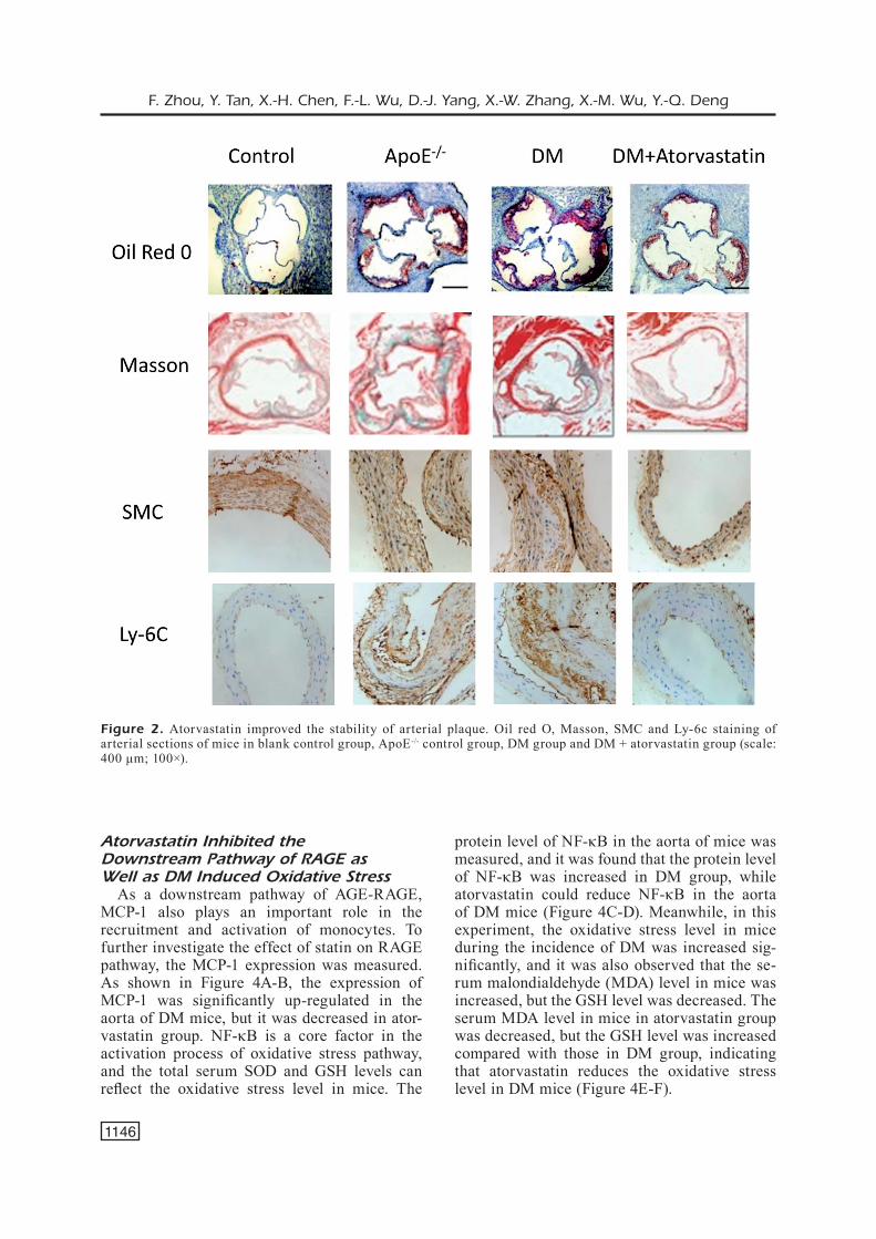
To further study the effect of statin on the sta-bility of aortic plaque, the aortic root tissues of mice were further stained with oil red O (Figure 2). A large amount of lipid deposition could be seen in the artery of mice in DM group, while lipid deposition was improved in atorvastatin group. The production of inflammation is thought to be an important development process of ath-erosclerosis, and Ly-6c in macrophages is used as an index of detecting atherosclerotic inflam-matory response. The number of macrophages in DM group was significantly increased compared with that in control group, while it was reduced by atorvastatin, suggesting that atorvastatin al-leviates the degree of inflammation. In Masson staining, it was observed that the number of collagen fibers in arterial plaque was reduced in DM group, while it was significantly increased in atorvastatin group. The plaque stability mainly depends on the content of lipids, macrophages, SMCs and collagen. Therefore, STZ-induced hy-perglycemia accelerates atherosclerosis, and ath-erosclerotic plaques are more vulnerable. The above phenomena of mice in atorvastatin group were improved, and the stability of atherosclerot-ic plaque was increased.

Atorvastatin improves plaque stability in diabetic atherosclerosis through the RAGE pathway
1145
Atorvastatin Decreased AGEs-RAGE Expression
During the occurrence of DM, oxidative stress existed and AGEs were formed in ad-dition to the increase in blood glucose. AGEs played roles through AGEs-RAGE pathway. The levels of AGEs and soluble RAGE (sRAGE) in mice were measured, and the level of RAGE
in the aorta was also detected (Figure 3). The levels of serum AGEs and sRAGE in DM group were significantly increased, while they were decreased in atorvastatin group. The level of RAGE in the aorta in DM group was increased, but atorvastatin also decreased the expression of RAGE in the aorta, which was consistent with serological results.
Figure 1. Atorvastatin improved atherosclerosis. A, Oil red O staining of aorta specimens of mice in blank control group, ApoE-/- control group, DM group and DM + atorvastatin group (scale: 0.5 cm; magnification: 1×). B, The degree of aortic lesion is presented as the proportion of oil red O staining area in arterial area: *p < 0.05 vs. control group, #p < 0.05 vs. ApoE-/- control group, +p < 0.05 vs. DM group (n=5). C, HE staining of aorta of mice in blank control group, ApoE-/- control group, DM group and DM + atorvastatin group (scale: 400 μm; 100×). D, Arterial plaque area: *p < 0.05 vs. control group, #p < 0.05 vs. ApoE-/- control group, +p < 0.05 vs. DM group (n=5).
F. Zhou, Y. Tan, X.-H. Chen, F.-L. Wu, D.-J. Yang, X.-W. Zhang, X.-M. Wu, Y.-Q. Deng
1146
Atorvastatin Inhibited the Downstream Pathway of RAGE as Well as DM Induced Oxidative Stress
As a downstream pathway of AGE-RAGE, MCP-1 also plays an important role in the recruitment and activation of monocytes. To further investigate the effect of statin on RAGE pathway, the MCP-1 expression was measured. As shown in Figure 4A-B, the expression of MCP-1 was significantly up-regulated in the aorta of DM mice, but it was decreased in ator-vastatin group. NF-κB is a core factor in the activation process of oxidative stress pathway, and the total serum SOD and GSH levels can reflect the oxidative stress level in mice. The
protein level of NF-κB in the aorta of mice was measured, and it was found that the protein level of NF-κB was increased in DM group, while atorvastatin could reduce NF-κB in the aorta of DM mice (Figure 4C-D). Meanwhile, in this experiment, the oxidative stress level in mice during the incidence of DM was increased sig-nificantly, and it was also observed that the se-rum malondialdehyde (MDA) level in mice was increased, but the GSH level was decreased. The serum MDA level in mice in atorvastatin group was decreased, but the GSH level was increased compared with those in DM group, indicating that atorvastatin reduces the oxidative stress level in DM mice (Figure 4E-F).
Figure 2. Atorvastatin improved the stability of arterial plaque. Oil red O, Masson, SMC and Ly-6c staining of arterial sections of mice in blank control group, ApoE-/- control group, DM group and DM + atorvastatin group (scale: 400 μm; 100×).

Atorvastatin improves plaque stability in diabetic atherosclerosis through the RAGE pathway
1147
Discussion
Atorvastatin, as a HMG-CoA reductase in-hibitor, is widely used to lower the serum cho-lesterol level. Atorvastatin reduces total choles-terol and low-density lipoprotein cholesterol, thus reducing the risk and long-term morbidity and mortality rates of coronary heart disease10. Besides, atorvastatin also has other functions than anti-hyperlipidemic effect, such as anti-in-flammation and protection of endothelial cell function11,12. We also found that low-dose ator-vastatin did not significantly improve the blood lipid level in mice, and its effect of stabilizing atherosclerotic plaques was independent of its lipid-lowering effect.
We used immunohistochemical staining to ob-serve the lesion area in atherosclerotic plaques and the expressions of macrophages, SMCs and collagen. It was found that atorvastatin treatment could increase the plaque collagen content. Previ-ous research results have shown that the increase
of collagen plays an important role in reducing the risk of plaque progression, thus improving the outcome of atherosclerotic diseases13. Moreover, it was observed that the number of macrophages in plaques in atorvastatin group was significantly reduced compared with that in DM group, which is also consistent with the decreased MCP-1 con-tent in the aorta. The decrease of macrophages can reduce the formation of foam cells as well as the deposition of cholesterol14. Therefore, atorvas-tatin can improve the stability of atherosclerotic plaques.
AGEs play important roles in the occurrence and development of vascular complications of DM, which bind to the receptor on vascular wall, thereby depositing at the high-risk site of atherosclerosis15. The interaction of AGEs-RAGE can be blocked by sRAGE. These sRAGEs can bind to AGEs and exert the endogenous protec-tion effect to resist atherosclerosis16,17. This work confirmed that in DM group, the levels of serum AGEs, sRAGE and RAGE protein expression in
Figure 3. The levels of serum AGEs (A), sRAGE (B) and RAGE protein expression (C and D) in the artery of mice in blank control group, ApoE-/- control group, DM group and DM + atorvastatin group: *p < 0.05 vs. control group, #p < 0.05 vs. ApoE-/- control group, +p < 0.05 vs. DM group (n=5).

F. Zhou, Y. Tan, X.-H. Chen, F.-L. Wu, D.-J. Yang, X.-W. Zhang, X.-M. Wu, Y.-Q. Deng
1148
vascular wall were increased. However, atorvas-tatin could reduce RAGE protein in the artery and serum sRAGE level.
During the occurrence of vascular inflamma-tion, the recruitment and aggregation of blood mononuclear cells are important initiating links. RAGE can mediate the activation of MCP-1, re-cruit mononuclear macrophages, and initiate the inflammatory response in vivo, thereby increasing the cell apoptosis and accelerating the occurrence and development of atherosclerosis19. Through
measurement of MCP-1, it was found that the anti-inflammatory effect of atorvastatin also re-duced the monocyte aggregation via decreasing MCP-1 expression. The activation of RAGE is also proved to be associated with the activation of NF-κB pathway. The RAGE gene promoter contains three putative NF-κB-like binding sites, thereby stimulating the production of inflamma-tion and increasing the oxidative stress level in vivo19. NF-κB activated by RGAE can further in-crease the expression of RAGE in inflammatory
Figure 4. Levels of MCP-1 (A-B) and NF-κB (C-D) in the artery of mice in blank control group, ApoE-/- control group, DM group and DM + atorvastatin group. Levels of serum SOD and GSH (E-F) in mice in blank control group, ApoE-/- control group, DM group and DM + atorvastatin group: *p < 0.05 vs. control group, #p < 0.05 vs. ApoE-/- control group, +p < 0.05 vs. DM group (n=5).

Atorvastatin improves plaque stability in diabetic atherosclerosis through the RAGE pathway
1149
cells, forming a positive feedback20. In this work, NF-κB in the artery and serum oxidative stress level in mice treated with atorvastatin were sig-nificantly decreased, suggesting that atorvastatin, in addition to the anti-oxidative stress effect, has the potential to break the positive feedback path produced by oxidative stress.
Conclusions
We suggest that atorvastatin improves oxidative stress level and reduces inflammatory response in DM mice through inhibiting AGEs-RAGE path-way and blocking downstream signals of RAGE, such as MCP-1 and NF-κB, thereby improving arterial plaque stability and reducing risks of car-diovascular diseases.
Conflict of InterestThe Authors declare that they have no conflict of interests.
References
1) Bierman eL. George lyman duff memorial lecture. Atherogenesis in diabetes. Arterioscler Thromb 1992; 12: 647-656.
2) Grundy Sm, Benjamin ij, Burke GL, Chait a, eCkeL rh, howard BV, mitCh w, Smith Sj, SowerS jr. Dia-betes and cardiovascular disease: a statement for healthcare professionals from the American Heart Association. Circulation 1999; 100: 1134-1146.
3) Grundy Sm, howard B, Smith Sj, eCkeL r, redBerG r, Bonow ro. Prevention conference VI: diabetes and cardiovascular disease: executive summary: con-ference proceeding for healthcare professionals from a special writing group of the American Heart Association. Circulation 2002; 105: 2231-2239.
4) roSenfeLd me, PoLinSky P, Virmani r, kauSer k, ruBanyi G, SChwartz Sm. Advanced atherosclerot-ic lesions in the innominate artery of the ApoE knockout mouse. Arterioscler Thromb Vasc Biol 2000; 20: 2587-2592.
5) Gao Xj, Qu yy, Liu Xw, zhu m, ma Cy, jiao yL, Cui B, Chen zj, zhao yr. Immune complexes induce TNF-alpha and BAFF production from U937 cells by HMGB1 and RAGE. Eur Rev Med Pharmacol Sci 2017; 21: 1810-1819.
6) mohamed ak, BierhauS a, SChiekofer S, tritSChLer h, zieGLer r, nawroth PP. The role of oxidative stress and NF-kappaB activation in late diabetic compli-cations. Biofactors 1999; 10: 157-167.
7) Liao d, Liu yQ, XionG Ly, zhanG L. Renoprotective effect of atorvastatin on STZ-diabetic rats through inhibiting inflammatory factors expression in dia-
betic rat. Eur Rev Med Pharmacol Sci 2016; 20: 1888-1893.
8) CoLhoun hm, BetteridGe dj, durrinGton Pn, hitman Ga, neiL ha, LiVinGStone Sj, thomaSon mj, maCk-neSS mi, CharLton-menyS V, fuLLer jh. Primary pre-vention of cardiovascular disease with atorvasta-tin in type 2 diabetes in the Collaborative Atorvas-tatin Diabetes Study (CARDS): multicentre ran-domised placebo-controlled trial. Lancet 2004; 364: 685-696.
9) MRC/BHF heart protection study of cholesterol lowering with simvastatin in 20,536 high-risk in-dividuals: a randomised placebo-controlled trial. Lancet 2002; 360: 7-22.
10) takemoto m, Liao jk. Pleiotropic effects of 3-hy-droxy-3-methylglutaryl coenzyme a reductase in-hibitors. Arterioscler Thromb Vasc Biol 2001; 21: 1712-1719.
11) zhou Q, Liao jk. Statins and cardiovascular dis-eases: from cholesterol lowering to pleiotropy. Curr Pharm Des 2009; 15: 467-478.
12) Davignon J. Beneficial cardiovascular pleiotropic effects of statins. Circulation 2004; 109: I39-I43.
13) CaLandra t, BernhaGen j, metz Cn, SPieGeL La, BaCh-er m, donneLLy t, Cerami a, BuCaLa r. MIF as a glu-cocorticoid-induced modulator of cytokine pro-duction. Nature 1995; 377: 68-71.
14) dinG S, zhanG m, zhao y, Chen w, yao G, zhanG C, zhanG P, zhanG y. The role of carotid plaque vul-nerability and inflammation in the pathogenesis of acute ischemic stroke. Am J Med Sci 2008; 336: 27-31.
15) yonekura h, yamamoto y, Sakurai S, PetroVa rG, aBe-din mj, Li h, yaSui k, takeuChi m, makita z, takaSa-wa S, okamoto h, watanaBe t, yamamoto h. Novel splice variants of the receptor for advanced gly-cation end-products expressed in human vascu-lar endothelial cells and pericytes, and their puta-tive roles in diabetes-induced vascular injury. Bio-chem J 2003; 370: 1097-1109.
16) rahBar S, fiGaroLa jL. Novel inhibitors of advanced glycation endproducts. Arch Biochem Biophys 2003; 419: 63-79.
17) fiSher m. Diabetes and atherogenesis. Heart 2004; 90: 336-340.
18) tanimoto a, murata y, wanG ky, tSutSui m, kohno k, SaSaGuri y. Monocyte chemoattractant protein-1 expression is enhanced by granulocyte-macro-phage colony-stimulating factor via Jak2-Stat5 signaling and inhibited by atorvastatin in human monocytic U937 cells. J Biol Chem 2008; 283: 4643-4651.
19) waGner ah, SChwaBe o, heCker m. Atorvastatin in-hibition of cytokine-inducible nitric oxide synthase expression in native endothelial cells in situ. Br J Pharmacol 2002; 136: 143-149.
20) zhao y, Chan my, zhou S, henG Ck. Effects of ath-erogenic diet and atorvastatin treatment on gene expression profiles in the C57BL/6J mouse liver. Gene Expr 2008; 14: 149-158.