attention - masaryk university
TRANSCRIPT

AttentionAttention
DonDon´́t manipulate the light microscopes, t manipulate the light microscopes, please.please.
They are prepared for your work following They are prepared for your work following presentation.presentation.
YouYou´́ll receive an instruction how to use ll receive an instruction how to use the LM and how to study blood smears. the LM and how to study blood smears.

BloodBlood
Plasma Plasma Blood cellsBlood cells

Hematocrit:Hematocrit: the volume of blood cells per unit volume of blood the volume of blood cells per unit volume of blood

Blood cells (formed elements)Blood cells (formed elements)
Red blood cells Red blood cells –– erythrocyteserythrocytes
White blood cells White blood cells –– leukocytesleukocytes
Platelets Platelets –– thrombocytesthrombocytes
Granulocytes Agranulocytes
Neutro- Eosino- Baso- Lymfo- Mono-

REM TEMREM TEM

REMEMBER!REMEMBER!
Erythrocytes: 4 Erythrocytes: 4 –– 6 millions/ 1 6 millions/ 1 l l of bloodof blood
Leukocytes: 5,000 Leukocytes: 5,000 –– 9,000 / 9,000 / 1 1 ll
Thrombocytes: 150,000 Thrombocytes: 150,000 –– 250,000/ 250,000/ 1 1 l l

ERYTHROCYTESERYTHROCYTES 4 4 –– 6 million/6 million/μμll
Shape: biconcave disc,Shape: biconcave disc,
dumbledumble--shapedshaped (cross section)(cross section)
Size: 7.4 Size: 7.4 μμm in diameter m in diameter (= normocyte)(= normocyte)
Structure: plasmalemma, Structure: plasmalemma, cytoplasm + hemoglobin 33 % cytoplasm + hemoglobin 33 % absence of the nucleusabsence of the nucleus and cell and cell organellesorganelles
Lifespan: 120 daysLifespan: 120 days
0.8 μm

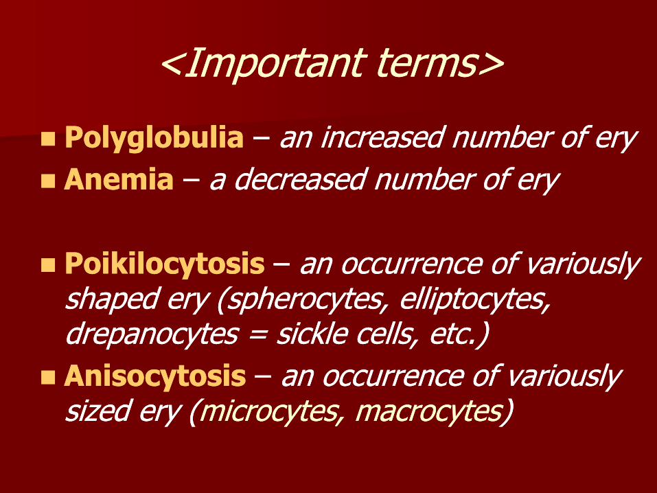
<<Important termsImportant terms>>
PolyglobuliaPolyglobulia –– an increased number of eryan increased number of ery
AnemiaAnemia –– a decreased number of erya decreased number of ery
PoikilocytosisPoikilocytosis –– an occurrence of variously an occurrence of variously shaped ery (spherocytes, elliptocytes, shaped ery (spherocytes, elliptocytes, drepanocytes = sickle cells, etc.)drepanocytes = sickle cells, etc.)
AnisocytosisAnisocytosis –– an occurrence of variously an occurrence of variously sized ery (sized ery (microcytes, macrocytesmicrocytes, macrocytes))

poikilocytosispoikilocytosis drepanocytesdrepanocytes
spherocytes dacryosytesspherocytes dacryosytes
(teardrop)(teardrop)
schystocytes elliptocytesschystocytes elliptocytes
(keratocytes) stomatocytes (keratocytes) stomatocytes

Anisocytosis
Microcytes Microcytes << 6.5 6.5 mm
NormocytesNormocytes
±± 7.47.4 mm
Macrocytes Macrocytes >> 8 8 mm

HemoglobinHemoglobin
a conjugated protein: a conjugated protein: 4 polypeptide chains + heme 4 polypeptide chains + heme groups = protoporphyrin ring groups = protoporphyrin ring with ferrous iron (Fewith ferrous iron (Fe2+ 2+ ))
Hb F (fetal) Hb F (fetal)
Hb A (adult)Hb A (adult)
normochromatic ery: normochromatic ery: 3232±±2 picogramms 2 picogramms ((hyperhyper--, hypo, hypo--))
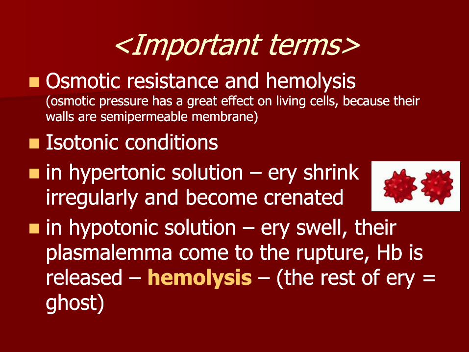
<<Important termsImportant terms>> Osmotic resistance and hemolysis Osmotic resistance and hemolysis
(osmotic pressure has a great effect on living cells, because their (osmotic pressure has a great effect on living cells, because their walls are semipermeable membrane)walls are semipermeable membrane)
Isotonic conditionsIsotonic conditions
in hypertonic solution in hypertonic solution –– ery shrink ery shrink irregularly and become crenatedirregularly and become crenated
in hypotonic solution in hypotonic solution –– ery swell, their ery swell, their plasmalemma come to the rupture, Hb is plasmalemma come to the rupture, Hb is released released –– hemolysishemolysis –– (the rest of ery = (the rest of ery = ghost) ghost)

(a) crenation is caused by water movement out of a cell in a hypertonic solution. (b) hemolysis is caused by water movement into a cell in a hypotonic solution.

ReticulocytesReticulocytes
Immature ery are Immature ery are released from the released from the bone marrow into bone marrow into the peripheral blood the peripheral blood (0.5 (0.5 –– 1.5 %)1.5 %)
the rests of organelles the rests of organelles –– ribosomes, mitochondria ribosomes, mitochondria
maturation into ery maturation into ery during 24 hoursduring 24 hours
brilantcresyl bluebrilantcresyl blue
substantia reticulofilamentosasubstantia reticulofilamentosa

Functions of eryFunctions of ery
transport of oxygen from the lungstransport of oxygen from the lungs
transport of carbon dioxide from the transport of carbon dioxide from the tissuestissues

LEUKOCYTESLEUKOCYTES
Granulocytes:Granulocytes:
-- neutrophilsneutrophils
-- eosinophilseosinophils
-- basophilsbasophils
General characteristic:General characteristic:
Polymorphonuclears withPolymorphonuclears with
acidophilicacidophilic cytoplasm and cytoplasm and
Specific + azurophilicSpecific + azurophilic granulesgranules
AgranulocytesAgranulocytes
-- lymphocyteslymphocytes
-- monocytesmonocytes
General characteristic:General characteristic:
Mononuclears withMononuclears with
basophilicbasophilic cytoplasm and cytoplasm and
azurophilicazurophilic granulesgranules

Erythrocytes (A) Thrombocytes (G) Granulocytes: (pink cytoplasm, specific granules)
neutrophil segment (E,C) neutrophil band (I) eosinophil (D) basophil (J) Agranulocytes: (blue cytoplasm, azurophilic granules)
lymphocyte (H, B) monocyte (F)

GranulocytesGranulocytes
General charcteristicGeneral charcteristic::
-- polymorphonuclears polymorphonuclears –– different shape of nucleidifferent shape of nuclei
bandband segmented with chromatin bridgessegmented with chromatin bridges
-- acidophilicacidophilic cytoplasmcytoplasm –– brightbright--pinkpink
-- specificspecific granulesgranules –– with special enzymeswith special enzymes
-- azurophilic granulesazurophilic granules –– with lysosomal enzymeswith lysosomal enzymes
-- all granulocytes are able to migrate from the vessels all granulocytes are able to migrate from the vessels and by diapedesis invade a site of inflamationand by diapedesis invade a site of inflamation

GranulocytesGranulocytes neutrophils neutrophils –– eosinophils eosinophils -- basophilsbasophils
band band
1212--14 14 mm
1010--12 12 mm
10 10 mm
segmentsegment

Differential white cell count Differential white cell count (DWCC)(DWCC)
Total number of leukocytes:Total number of leukocytes: normal valuesnormal values
Neutrophils Neutrophils -- bandsbands 4 %4 %
-- segmentssegments 68 %68 %
EosinophilsEosinophils 3 %3 %
BasophilsBasophils 1 %1 %
LymphocytesLymphocytes 20 %20 %
MonocytesMonocytes 4 %4 %
∑ = 100 %∑ = 100 %

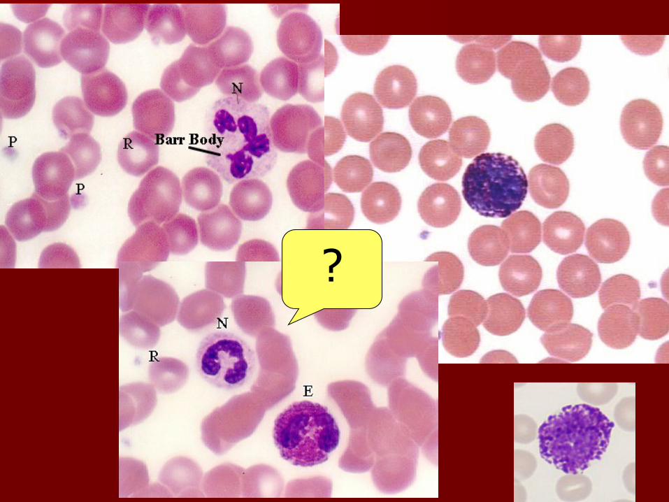
Neutrophil granulocytesNeutrophil granulocytes (neutrophils)(neutrophils)
71 % of all white blood cells (DWCC)71 % of all white blood cells (DWCC)
10 10 –– 12 12 mm
Cytoplasm: bright pinkCytoplasm: bright pink ((eosinophilic = acidophilic eosinophilic = acidophilic ))
Specific granules: neutrophilicSpecific granules: neutrophilic (( 0.3 0.3 mm) ) (enzymes:(enzymes:alcaline phosphatase, kolagenase, lysozymealcaline phosphatase, kolagenase, lysozyme, …), …)
Nucleus: Nucleus: bandband--shaped (4 %) shaped (4 %) or segmented (67 %) or segmented (67 %)
((22--5 segments5 segments))


Functions of NeuFunctions of Neu a central role in inflammatory processes a central role in inflammatory processes –– Neu invade, Neu invade,
by diapedesis from the vessels into sites of infection by diapedesis from the vessels into sites of infection and release some factors (e.g. cytokines) and release some factors (e.g. cytokines)
cell membrane receptors allow Neu to recognise cell membrane receptors allow Neu to recognise foreign bodies (bacteria, tissue debris), which they foreign bodies (bacteria, tissue debris), which they begin to begin to phagocytosephagocytose and destroy. and destroy.
The Neu die once their supply of granules has beenThe Neu die once their supply of granules has been
exhausted. Their lifespan is only about one week. Dead exhausted. Their lifespan is only about one week. Dead neutrophils and tissue debris are the major components of pus. neutrophils and tissue debris are the major components of pus.

Eosinophil granulocytesEosinophil granulocytes (eosinophils)(eosinophils)
11–– 4 % of all white blood cells (DWCC)4 % of all white blood cells (DWCC)
12 12 –– 14 14 mm
Cytoplasm: bright pinkCytoplasm: bright pink ((eosinophilic = acidophilic eosinophilic = acidophilic ))
Specific granules: eosinophilicSpecific granules: eosinophilic (( 0.5 0.5 –– 1 1 mm) ) (enzymes: (enzymes: acid phosphatase, peroxidase,acid phosphatase, peroxidase,histaminase, arylsufatasehistaminase, arylsufatase …)…)
Nucleus: Nucleus: dumbdumb--belt, belt, ((2 segments)2 segments) chromatin bridgechromatin bridge

Functions of EosFunctions of Eos
phagocytosis of antibodyphagocytosis of antibody--antigen antigen complexes and prevention of the immune complexes and prevention of the immune system from "overreacting„ system from "overreacting„
eos are involved in the response of the eos are involved in the response of the body against parasitic infections, which body against parasitic infections, which are accompanied by an increase in the are accompanied by an increase in the number of eosinophils. number of eosinophils.
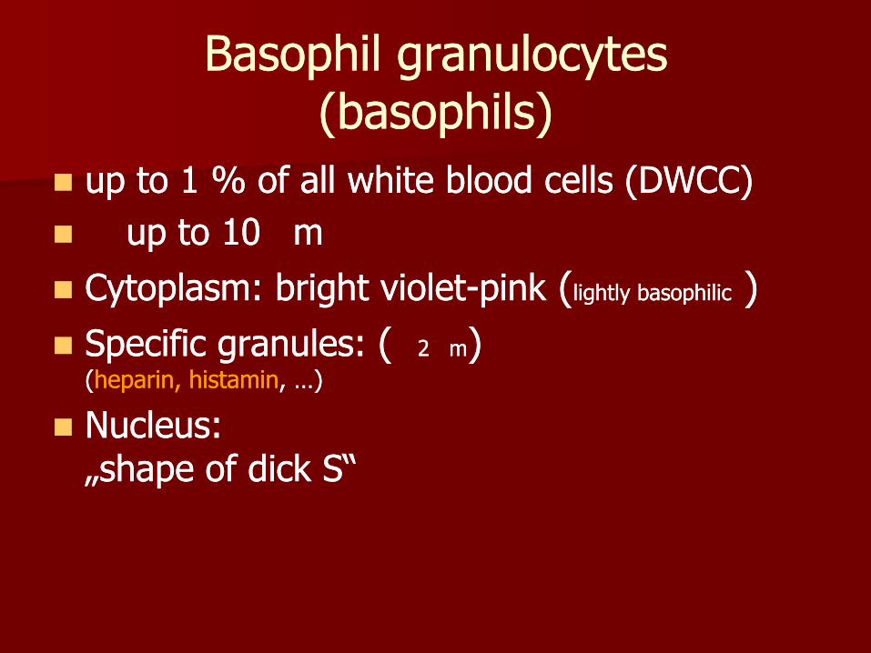
Basophil granulocytes Basophil granulocytes (basophils)(basophils)
up to 1 % of all white blood cells (DWCC)up to 1 % of all white blood cells (DWCC)
up to 10 up to 10 mm
Cytoplasm: bright violetCytoplasm: bright violet--pinkpink ((lightly basophiliclightly basophilic ))
Specific granules: Specific granules: (( 2 2 mm) ) ((heparin, histaminheparin, histamin, …), …)
Nucleus: Nucleus: „shape of dick S“„shape of dick S“


Functions of BasoFunctions of Baso
heparin heparin andand histaminehistamine are vasoactive substances. are vasoactive substances. They dilate the blood vessels, make vessel walls They dilate the blood vessels, make vessel walls more permeable and prevent blood coagulation. more permeable and prevent blood coagulation. They facilitate the access of heparinocyte in a site They facilitate the access of heparinocyte in a site of infection. of infection.
antibodies produced by plasma cells (activated Bantibodies produced by plasma cells (activated B--lymphocytes) bind to the receptors on the plasma lymphocytes) bind to the receptors on the plasma membrane of basophils. If these antibodies come membrane of basophils. If these antibodies come into contact with antigens, they induce the release into contact with antigens, they induce the release of the contents of the basophil granules.of the contents of the basophil granules.

AgranulocytesAgranulocytes
General charcteristicGeneral charcteristic::
-- mononuclears mononuclears –– shape of nuclei is spherical shape of nuclei is spherical ((in Lyin Ly),), oval or beanoval or bean--shaped shaped ((in Monoin Mono))
-- basophilicbasophilic cytoplasm cytoplasm –– blueblue
-- NO specific granules NO specific granules
-- azurophilic granules azurophilic granules –– with lysosomal enzymeswith lysosomal enzymes

LYMPHOCYTES LYMPHOCYTES
Classification:Classification:
-- according to origin according to origin –– TT--LyLy ((thymusthymus), ), BB--LyLy
((bone marrow bone marrow bursa of Fabricius in birdsbursa of Fabricius in birds))
-- according to the size according to the size –– smallsmall (( 8 8 m),m),
mediummedium (( 1010--1212 m), large m), large (( 1616--18 18 m),m),
-- according to the function according to the function –– natural killer cells, natural killer cells,
helper cells, memmory cells, supressor cellshelper cells, memmory cells, supressor cells, ,
-- according to lifeaccording to life--span span (long, short)(long, short)

Lymphocytes Lymphocytes -- structurestructure
20 % 20 % of all white blood cells (DWCC)of all white blood cells (DWCC)
small, mediumsmall, medium--sized, large Lysized, large Ly cytoplasm cytoplasm –– dark blue, contains nondark blue, contains non--specific specific
azurophilic granules with lysosomal enzymes azurophilic granules with lysosomal enzymes (hydrolases)(hydrolases) and numerous ribosomesand numerous ribosomes
nucleus nucleus –– round, hyperchromatic round, hyperchromatic –– coarse grains coarse grains of heterochromatin (of heterochromatin (dark violet colourdark violet colour) )

Functions of LyFunctions of Ly
BB--lymphocyteslymphocytes differentiate into antibody differentiate into antibody producing producing plasma cellsplasma cells and so they and so they represent "humoral immunity" represent "humoral immunity"
TT--lymphocyteslymphocytes represent the "cellular represent the "cellular immunity" and may attack foreign cells, immunity" and may attack foreign cells, cancer cells and cells infected by e.g. cancer cells and cells infected by e.g. a virusa virus

MONOCYTESMONOCYTES
5 % (DWC),5 % (DWC), 15 15 –– 20 20 mm
cytoplasm cytoplasm –– voluminous, bright blue, contains nonvoluminous, bright blue, contains non--specific azurophilic granules with lysosomal specific azurophilic granules with lysosomal enzymes enzymes (hydrolases)(hydrolases) and numerous ribosomesand numerous ribosomes
nucleus nucleus –– oval to beanoval to bean--shaped, finely dispersed shaped, finely dispersed chromatinchromatin


Functions of MonoFunctions of Mono
monocytes enter the connective tissue they monocytes enter the connective tissue they differentiate into differentiate into macrophagesmacrophages. At sites of . At sites of infection macrophages are the dominant cell infection macrophages are the dominant cell type after the death of the invading neutrophils. type after the death of the invading neutrophils.
macrophages phagocyte microorganisms, tissue macrophages phagocyte microorganisms, tissue debris and the dead neutrophils.debris and the dead neutrophils.
mono also give rise to mono also give rise to osteoclastsosteoclasts, which are able to , which are able to distroy bone. They are of importance in bone distroy bone. They are of importance in bone remodelling.remodelling.

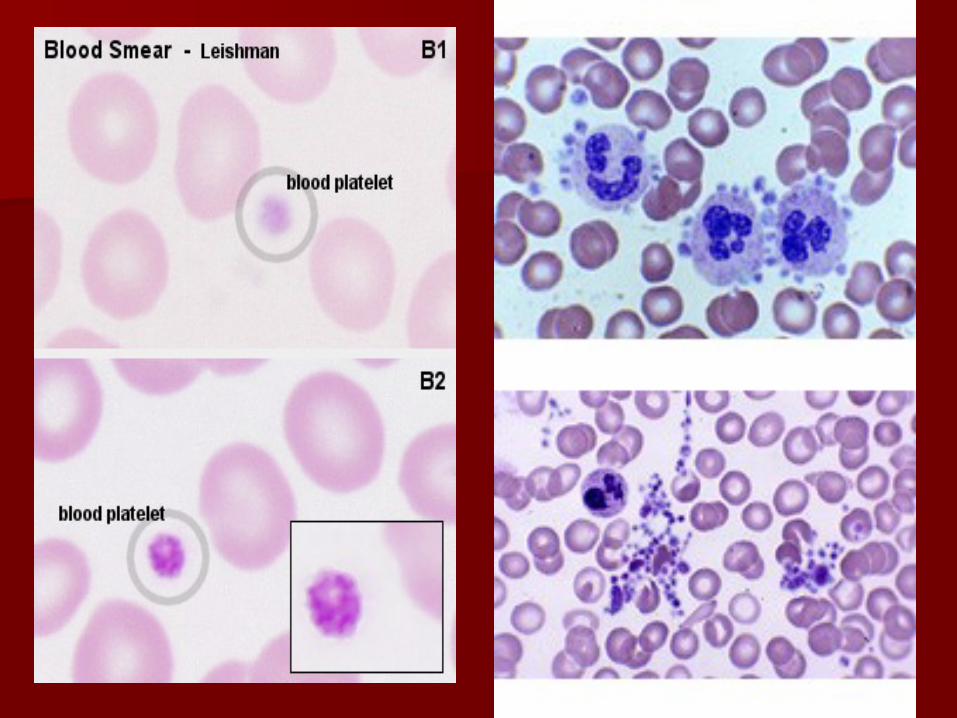
THROMBOCYTES THROMBOCYTES (blood platelets)(blood platelets)
150,000 – 300,000 / 1 / 1 l of blood
thrombocytosis X thrombocytopenia
- are not cells, but cytoplasmic fragments of large cell (megakaryocyte) in bone marrow
shape: spindle-shaped discoid plate
size: 2 – 4 m
cytoplasm – basophilic (bright violet-blue), contains microtubules and , and granules:
alpha granules – fibrinogen, …
delta granules – serotonin, Ca ions, ATP and ADP,…
lambda granules – are small lysosomes

Platelet structurePlatelet structure
HyaloplasmHyaloplasm contains contains microtubules (on the microtubules (on the periphery of platelet)periphery of platelet)
GranuloplasmGranuloplasm contains contains granulesgranules


Functions of thrombocytesFunctions of thrombocytes Platelets assist in Platelets assist in haemostasishaemostasis, the arrest of bleeding, the arrest of bleeding. .
SerotoninSerotonin is a vasoconstrictor. Its release from thrombocytes, is a vasoconstrictor. Its release from thrombocytes, adhering to the walls of a damaged vessels, is sufficient to adhering to the walls of a damaged vessels, is sufficient to close even small arteries. Platelets, which come into contact close even small arteries. Platelets, which come into contact with collagenous fibers in the walls of the vessel, swell, become with collagenous fibers in the walls of the vessel, swell, become "sticky" and activate other platelets to undergo the same "sticky" and activate other platelets to undergo the same transformation. This cascade of events results in the transformation. This cascade of events results in the formation formation of a of a platelet plug (or platelet thrombus)platelet plug (or platelet thrombus).. Finally, activating Finally, activating substances are released from the damaged vessel walls and substances are released from the damaged vessel walls and from the platelets. These substances mediate the from the platelets. These substances mediate the conversion of conversion of the plasma protein the plasma protein prothrombinprothrombin into into thrombinthrombin. . Thrombin Thrombin catalyzes the conversion of catalyzes the conversion of fibrinogen into fibrinfibrinogen into fibrin, which , which polymerizes into fibrils and forms a fibrous net in the arising polymerizes into fibrils and forms a fibrous net in the arising blood clot. Platelets captured in the fibrin net contract leading blood clot. Platelets captured in the fibrin net contract leading to to clot retractionclot retraction, which further assists in haemostasis., which further assists in haemostasis.

Please, note the following instructions

How to prepare blood smear?How to prepare blood smear?

How to prepare blood smear How to prepare blood smear -- II
Smears of peripheral blood must be made Smears of peripheral blood must be made immediately. immediately.
Step 1: Place drop of blood about 1cm Step 1: Place drop of blood about 1cm from the frosted end of a clean slide. from the frosted end of a clean slide.

How to prepare blood smear How to prepare blood smear -- IIII
Step 2: hold the end of a second slide Step 2: hold the end of a second slide (“spreader”) against the surface of the (“spreader”) against the surface of the first slide at an angle of 30first slide at an angle of 30--45 degrees. 45 degrees.

How to prepare blood smear How to prepare blood smear -- IIIIII
Step 3: draw it back to contact the drop of Step 3: draw it back to contact the drop of blood. Allow the blood to spread and fill blood. Allow the blood to spread and fill the angle between the two slides.the angle between the two slides.

How to prepare blood smear How to prepare blood smear -- IVIV
Step 4: Push the “spreader” slide at a Step 4: Push the “spreader” slide at a moderate speed forward until all of the moderate speed forward until all of the blood has been spread into a moderately blood has been spread into a moderately thin film. thin film.

How to prepare blood smear How to prepare blood smear -- VV
Smear is prepared for fixation (methyl Smear is prepared for fixation (methyl alcohol, 3alcohol, 3--5 minutes) and staining (special 5 minutes) and staining (special panoptic method according to Pappenheim panoptic method according to Pappenheim can be used) can be used)

How to study blood smear How to study blood smear in the light microscope (LM)?in the light microscope (LM)?
the lense of immersion objective (magnif. 100x) the lense of immersion objective (magnif. 100x) is immersed into a drop of oil and blood smear is is immersed into a drop of oil and blood smear is prepared for study in the LM;prepared for study in the LM;
switch onswitch on your LM and see into the eypice: blood your LM and see into the eypice: blood cells should be visible in the light fieldcells should be visible in the light field
if not, if not, try to focuse picturetry to focuse picture –– you may use you may use ONLYONLY the fine adjustment knob!the fine adjustment knob!
if you are not succesfull, if you are not succesfull, ask for help your ask for help your teacherteacher, , do NOT use the coarse adjustment do NOT use the coarse adjustment knob! knob!

Light microscope manipulationLight microscope manipulation
Course adjustment knobCourse adjustment knob don„t use todaydon„t use today Fine adjustment knobFine adjustment knob only that can be usedonly that can be used to focuse the imageto focuse the image
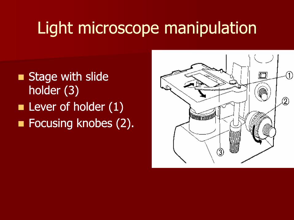
Stage with slide holder (3)Stage with slide holder (3) Lever of holder (1) Lever of holder (1) Focusing knobes (2).Focusing knobes (2).

Monocyte
Ø 10-12 μm
lymphocyte
Ø 8-10 μm
neutrophil – band
Ø 10-12 μm
neutrophil – segment Ø 10-12 μm
eosinophil
Ø 12-14 μm
basophil
Ø 10-12 μm
thrombocyte
Ø 2-4 μm
erytrocytes
Ø 7.4 μm

Erythrocytes (Ø 7,4 μm)
Thrombocytes (2-4 μm)
Granulocytes:
neutrophilic eosinophilic basophilic
10-12 μm 12-14 μm 10 μm
Agranulocytes:
Lymfocytes (8-10 μm)
Monocyte (15-20 μm)
P
R
O
T
O
C
O
L

How to count leukocytes in blood smear?How to count leukocytes in blood smear?
differential white cell count (DWCC) is an differential white cell count (DWCC) is an important hematologic screening which helps important hematologic screening which helps to diagnose to diagnose
leukocytes percentage is the result of this leukocytes percentage is the result of this investigationinvestigation
100 white cells must be count and registered 100 white cells must be count and registered in the table prepared for all types of in the table prepared for all types of leukocytes (Neuleukocytes (Neu--bands, Neubands, Neu--segments, Eos, segments, Eos, Baso, Ly, Mono)Baso, Ly, Mono)
arithmetic sum of each type of leukocytes arithmetic sum of each type of leukocytes represents their percentage (%)represents their percentage (%)

How to count leukocytes in blood smear?How to count leukocytes in blood smear?
blood smear have to be systematicaly blood smear have to be systematicaly viewed (it avoids repeatedly count the viewed (it avoids repeatedly count the same cells)same cells)
oror
vertical browsing horizontal browsing

DWCC

Table Table
11 22
Neu bandsNeu bands //
Neu Neu segmentssegments
//// ////// // //////
EosEos //
BasoBaso
LyLy //// ////////
MonoMono ////
10 cells10 cells 10 cells10 cells
99 1010 resultsresults normnorm
//// 4 %4 %
//// ///// / ////// 68 %68 %
// //// 3 %3 %
// 1 %1 %
// //////// 20 %20 %
4 %4 %
10 cells10 cells 10 cells10 cells 100% 100% 100 %100 %

Differential white cell count Differential white cell count (DWCC)(DWCC)
Total number of leukocytes:Total number of leukocytes: normal valuesnormal values
Neutrophils Neutrophils -- bandsbands 4 %4 %
-- segmentssegments 68 %68 %
EosinophilsEosinophils 3 %3 %
BasophilsBasophils 1 %1 %
LymphocytesLymphocytes 20 %20 %
MonocytesMonocytes 4 %4 %
∑ = 100 %∑ = 100 %

Anomalies of DWCCAnomalies of DWCC
* sum total of bands and segments has to be compared with norm; * sum total of bands and segments has to be compared with norm;
normal value is 71 % (4 % bans + 68 % segments)normal value is 71 % (4 % bans + 68 % segments)
Increased numberIncreased number Decreased numberDecreased number
NeutrophilsNeutrophils** neutrophilic neutrophilic granulogranulocytosiscytosis
neutrophilic neutrophilic granulogranulocytopeniacytopenia
EosinophilsEosinophils eosinophilic eosinophilic granulogranulocytosiscytosis
eosinophilic eosinophilic granulogranulocytopeniacytopenia
BasophilsBasophils basoophilic basoophilic granulogranulocytosiscytosis
basoophilic basoophilic granulogranulocytopeniacytopenia
LymphocytesLymphocytes lympholymphocytosiscytosis lympholymphocytopeniacytopenia
MonocytesMonocytes monomonocytosiscytosis monomonocytopeniacytopenia

Normal ratio Normal ratio of neutrophil bands and segments of neutrophil bands and segments
Turn to the leftTurn to the left –– bands are increased bands are increased
Turn to the rightTurn to the right –– segments are increased in peripheral segments are increased in peripheral
bloodblood
5
35
41
17
2
0
5
10
15
2025
30
35
40
45
band 2 segm. 3 segm. 4 segm. 5 segm.
neutrophils (nuclei)
%
Bands : Segments ratio is 4 % : 68 % = 1 : 17Bands : Segments ratio is 4 % : 68 % = 1 : 17
Turn to the right
Turn to the left

... thank you for attention ...


?

?

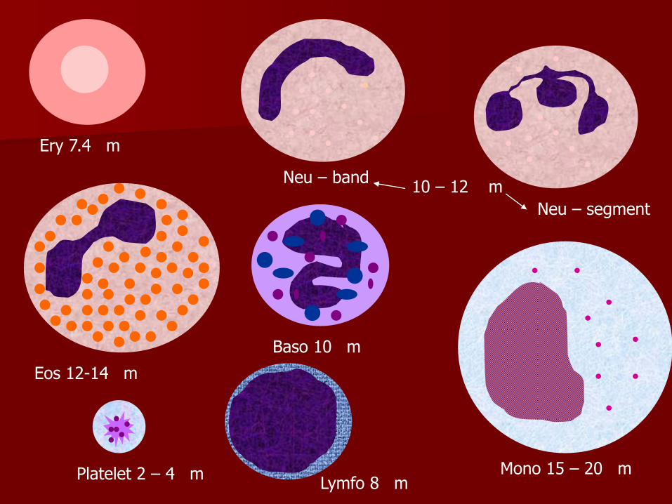
Ery 7.4 m
Neu – band
Neu – segment
Eos 12-14 m
Baso 10 m
10 – 12 m
Platelet 2 – 4 m Lymfo 8 m
Mono 15 – 20 m

Blood investigation
Blood smear

Light microscopeLight microscope
EyepiecesEyepieces
Objective lenseObjective lense
Stage with slide holderStage with slide holder
CondenserCondenser
On/off switchOn/off switch
Light intensity controlLight intensity control
Source of light beamSource of light beam
Course adjustment knobCourse adjustment knob
Fine adjustment knobFine adjustment knob

Light microscope manipulationLight microscope manipulation
on/off switch knob (1) on/off switch knob (1) (rocker or wheel SW) (rocker or wheel SW)
pivoted potenciometer (2) pivoted potenciometer (2) regulate intensity of regulate intensity of emited light emited light
Light microscope manipulationLight microscope manipulation
Stage with slide Stage with slide holder (3)holder (3)
Lever of holder (1) Lever of holder (1)
Focusing knobes (2). Focusing knobes (2).

Light microscope manipulationLight microscope manipulation
Use mechanism of cross Use mechanism of cross shift for shift of the slide shift for shift of the slide on the stageon the stage

Light microscope manipulation Light microscope manipulation
focuse a picture in LM focuse a picture in LM and look at it with both and look at it with both eyes eyes
regulate a distance regulate a distance between the eyepices between the eyepices so, you can see one so, you can see one focused circular fieldfocused circular field
bad correct

Light microscope manipulationLight microscope manipulation
Look at the slide only Look at the slide only through the right through the right eyepiece and focuse eyepiece and focuse some point in the some point in the picture.picture.
Without refocusing, look Without refocusing, look at the left eyepiece.at the left eyepiece.
In doing so, screw the In doing so, screw the ring below the left ring below the left eyepiece to focuse the eyepiece to focuse the same point. same point.
So, the dioptric So, the dioptric correction is set up. correction is set up.
Now, you can start to study blood Now, you can start to study blood
smear in your LM smear in your LM


