attenuation of botulinum toxin mediated blockade of...
TRANSCRIPT

Attenuation of botulinum toxin mediated blockade of exocytosis at
neuromuscular junctions by viral vector mediated gene transfer into
spinal motor neurons
by
Arvind Raghunath
International Centre for Neurotherapeutics,
Dublin City University,
Dublin – 9,
Ireland.
December, 2008
A dissertation submitted under the supervision of Prof. Oliver
Dolly to fulfil the requirements for award of the degree of Ph.D. from
Dublin City University.

1
Declaration
I hereby certify that this material, which I now submit for assessment on the programme
of study leading to the award of Ph.D is entirely my own work, that I have exercised
reasonable care to ensure that the work is original, and does not to the best of my
knowledge breach any law of copyright, and has not been taken from the work of others
save and to the extent that such work has been cited and acknowledged within the text
of my work.
Signed: ____________
(Candidate) ID No.: 53155173
Date: 18th December 2008

2
Abstract
Botulinum neurotoxin type A (BoNT/A) is implicated in a majority of
human cases of botulism and causes long-lasting neuromuscular paralysis due
to the persistence in nerve endings of its light chain protease that cleaves
synaptosomal-associated protein of 25 kDa (S25). This highly debilitating and,
often fatal, neuroparalytic condition cannot be adequately treated by traditional
pharmacological methods due to difficulties in efficiently accessing either the
pre-synaptic terminals or the cell body of the supplying spinal motor neurons
and because of the lack of efficient drugs that can block or reverse the
paralysis.
A viral vector mediated gene delivery technique was therefore utilised to
confer protection against toxin-induced blockade of exocytosis by the
expression of cleavage-resistant S25 (S25-R198T) in susceptible cells.
Recombinant adeno-associated viral (AAV) vectors expressing a His6-tagged
S25-R198T (AAV-S25-R198T) were initially used to obtain proof-of-principle of
this approach in chromaffin cell cultures, neuroendocrine cells that mimic the
exocytotic pathway found in neurons. Stereotaxic injection of AAV-S25-R198T
into anterior horns of rat spinal cords resulted in substantial protection against a
subsequent challenge by BoNT/A injected into the soleus muscle. Interestingly,
intra-muscular injection of this virus also resulted in protection against
neuroparalytic action of a subsequently injected toxin.
In order to obtain more efficient neuronally-targeted, peripherally-
deliverable viral vectors – which would also confer great utility in the treatment

3
of genetic diseases affecting the spinal cord – the more versatile lentiviral
vectors (LV) were used. Biotinylated LVs were attached to targeting agents,
different core-streptavidin (CS) tagged protease-inactive BoNT/E (BoTIM)
mutants, and applied to cerebellar granule neuron cultures. These targeting
molecules substantially reduced the viral infection of non-neuronal cultures with
only minimal effects on the transfection of neurons.
The results obtained in this study demonstrate the applicability of gene
therapy to ameliorate the neuroparalytic effects of botulism. In addition, the
novel tools and techniques developed, especially in the targeted, peripheral,
delivery of viral vectors, could also be adapted to treat genetic disorders of
motor neurons.

4
Acknoledgements
It is my pleasure to thank the many people who made this thesis
possible.
First of all, I would like to thank my supervisor, Prof. J. O. Dolly. Without
his advice, enthusiasm and encouragement this dissertation would not have
reached fruition. I particularly value the detailed scientific discussions we had
over the course of this project and, especially, during the grant and paper
writing process. The two most important attributes that I hope to have gained
from my stay here are his atitudes on positive thinking and of always moving
forward (be it in science or rugby!).
I would like to sincerely thank all current, and past, members of ICNT for
their help and assistance, especially Francesc, Gary, Valerie, Macdara, Maxim
and Roman. Without their advice and support this study would have have never
have been feasible. I would like to acknowledge and thank the endless support
extended by Rita, Carolyn and Frank throughout my stay here.
I would also like to express my gratitude to USAMRIID and DTRA for
providing me with a research assistantship during my PhD.
Finally, I would like to thank my family back home in India for all their
love and understanding. Without them I would not have been here and certainly
would not have the means or courage to undertake this adventure in science!

5
Table of contents
Abstract ............................................................................................................... 1
Acknowledgements ............................................................................................ 4
Table of contents ................................................................................................ 5
Abbreviations used........................................................................................... 11
List of figures and tables ................................................................................. 14
1. Introduction.......................................................................................... 18
1.1. Brief overview of neuromuscular transmission in man........................... 19
1.2. The microscopic architecture of a NMJ ................................................. 20
1.2.1. The pre-synaptic exocytotic apparatus in motor nerves ........................ 23
1.2.2. Organisation of the post-synaptic NMJ.................................................. 28
1.2.3. Other important actors involved in nerve-muscle conduction and its
control – Schwann cells, muscle spindles and spinal inter-neurons. ..... 31
1.3. The molecular machinery for fusion of synaptic vesicles with the
plasmalemma ........................................................................................ 33
1.3.1. Molecular mechanisms of SNARE-dependent exocytosis..................... 35
1.3.1.1. Recycling of SNAREs for further rounds of exocytosis.......................... 41
1.3.1.2. SNAP-25 ............................................................................................... 43
1.3.1.3. Syntaxin................................................................................................. 45
1.3.1.4. VAMP .................................................................................................... 46
1.3.2. Other important molecular actors in the synaptic vesicle cycle –
synaptotagmin, Rab3, Munc18, SV2. .................................................... 47
1.4. Etiology and clinical symptoms of human botulism................................ 50

6
1.5. Viral vectors for gene therapy................................................................ 55
1.6. Model systems used to study botulism and to assess the efficacy of
various therapies ................................................................................... 60
1.7. Principal goals of this study ................................................................... 61
2. Attenuation of BoNT-induced blockade of exocytosis at
peripheral motor terminals by viral vector mediated gene
transfer into spinal motor neurons .................................................... 67
2.1.1. Trafficking of Clostridial neurotoxins to their site(s) of action................. 68
2.1.2. Proteolytic cleavage of specific SNAREs by the LC of clostridial
neurotoxins............................................................................................ 77
2.1.2.1. Unique trafficking properties of tetanus toxin......................................... 80
2.1.3. The structure of purified CNTs............................................................... 81
2.1.4. Recovery of cholinergic transmission at the NMJs after Clostridial
toxin poisoning....................................................................................... 82
2.1.5. Engineering SNAREs resistant to cleavage by CNTs............................ 85
2.1.6. Adeno-associated viruses (AAV) ........................................................... 86
2.2. Experimental strategies employed .................................................... 91
2.2.1. Animal husbandry, LD50 determination and DAS scoring ...................... 91
2.2.2. Construction, production and purification of recombinant AAVs
expressing BoNT/A-resistant S25 (S25RT), wild-type S25, markers
hrGFP or hrGFP with S25RT................................................................. 97
2.2.2.1. Titration of produced AAVs – PAGE, RT-QPCR, Western blotting
and FACS-based assays....................................................................... 99
2.2.2.2. Subcellular localisation of produced S25 by confocal microscopy....... 104

7
2.2.3. Measurement of the relative proteolytic activities of BoNT /A, /E and
/C1 against mutant and wild-type S25 ................................................. 105
2.2.4. Culture of chromaffin cells, infection with recombinant AAVs,
intoxication with BoNT/A and assay of catecholamine release............ 106
2.2.5. Stereotaxic intra-spinal administration of AAVs for transfection of
anterior horn neurons supplying the soleus muscle in rats.................. 108
2.2.6. In situ electrophysiological recording of the efficiency of
neuromuscular transmission as a measure of synaptic exocytosis
remaining after BoNT/A administration................................................ 110
2.2.7. Immuno-histochemical detection of expressed S25, His6-S25, His6-
S25-R198T and GFP........................................................................... 111
2.2.8. Determination of the distribution of ACh receptors at NMJs using
fluorophore-conjugated α-BuTx ........................................................... 112
2.3. Results ............................................................................................... 116
2.3.1. Cleavage resistance of different SNAP-25 mutants to BoNT/A, /E or
/C1....................................................................................................... 116
2.3.2. Longevity of neuromuscular paralysis induced by BoNT/A and
BoNT/E in rats models of botulism ...................................................... 117
2.3.3. Expression of BoNT-resistant S25 in mammalian cell lines using
AAVs as gene transfer vectors ............................................................ 122
2.3.4. Demonstration of dose-dependent protection of stimulated
exocytosis from BoNT/A-induced blockade by prior expression in
chromaffin cells of His6-S25R198T but not wild-type S25.................... 123
2.3.5. Efficient expression of cleavage-resistant S25 in peripheral nerve

8
terminals of rats following intra-spinal administration of AAVs
protects against the neuroparalytic effects of BoNT/A......................... 125
2.3.6. BoNT/A-induced synapse remodelling is reduced by protection of
neuromuscular transmission with a spinal injection of AAV-His6-S25-
R198T.................................................................................................. 129
2.3.7. Partial protection against BoNT/A-induced neuroparalysis in rats by
neuronal expression of His6-S25R198T after peripheral injection of
its AAV construct ................................................................................. 131
2.4. Discussion ......................................................................................... 134
2.4.1. AAV vectors are efficient vehicles for neuronal gene delivery when
directly injected into spinal cord........................................................... 134
2.4.2. Delivery of cleavage resistant S25 into motor nerve terminals
ameliorates the transmission blockade induced by a later toxin
challenge ............................................................................................. 136
2.4.3. The retrograde trafficking ability of AAVs in rats – necessity to
develop efficient targeting agents ........................................................ 137
2.4.4. Sprouting induced by BoNT/A is a result of exocytotic blockade and
is reduced by the alleviation of such paralysis..................................... 139
3. Targeting viral vectors to cholinergic nerve terminals .................. 142
3.1.1. Introduction – rationale behind the utilisation of the cholinergic
targeting property of Clostridial neurotoxins to target therapeutic viral
vectors to NMJs................................................................................... 142
3.1.2. Core-Streptavidin................................................................................. 144
3.1.3. Lentiviral vectors (LV).......................................................................... 148

9
3.2. Experimental strategies employed....................................................... 152
3.2.1. Development of LVs carrying marker EGFP and/or S25 mutants
resistant to /A or /A, /E and /C1 ........................................................... 152
3.2.1.1. Titration of produced LVs by p24 ELISA.............................................. 153
3.2.1.2. Biotinylation of purified LVs ................................................................. 154
3.2.2. Isolation of cerebellar granule neurons (CGNs) from neonatal rat
cerebellum........................................................................................... 157
3.2.2.1. Radioisotopic measurement of K+-evoked, and Ca2+-stimulated,
14C-glutamate release from CGNs ..................................................... 158
3.2.3. In vitro and in vivo assays of LV function............................................. 160
3.2.4. Development of core-streptavidin tagged protease-inactive and
active BoNT/E targeting chimeras ....................................................... 161
3.2.5. Testing the targeting abilities of different CS-tagged BoTIMs.............. 163
3.2.5.1. Immuno-histochemical determination of binding of CS-tagged
BoTIMs to CGNs ................................................................................. 164
3.2.6. Testing the ability of CS-tagged BoTIMs to bind various biotinylated
cargo ................................................................................................... 164
3.2.7. Chromatographic analysis of CS-tagged BoTIMs................................ 165
3.2.8. Assessment of the ability of CS-tagged BoTIMs to bind larger cargo
– biotinylated microspheres and LVs................................................... 165
3.2.9. Effect of bound CS-BoTIM on biot-LV infectivity.................................. 167
3.3. Results ............................................................................................... 171
3.3.1. LVs are an efficient tool to deliver genes into neurons – in vivo and
in culture.............................................................................................. 171

10
3.3.2. CS-tagged BoTIMs retain the properties of both their parent
molecules ............................................................................................ 178
3.4. Discussion ......................................................................................... 195
4. General Discussion ........................................................................... 198
5. Bibliography....................................................................................... 205
Appendix 1 – Buffers and solutions.............................................................. 225
Appendix 2 – Presentations and published material based on the
current study...................................................................................... 227

11
Abbreviations used
A-488/546/568 alexa-fluor 488/546/568 nm AA amino acid ANS autonomic nervous system ACh acetylcholine AAV adeno-associated virus BoNT botulinum neurotoxin BoTIM BoNT protease inactive mutant BSA bovine serum albumin α-BuTx alpha bungarotoxin CMV cytomegalovirus CNS central nervous system CNT clostridial neuro-toxins CS core-streptavidin ChAT choline acetyltransferase dNTP deoxynucleotide triphosphate DAS digit abduction score DC di-chain DβH dopamine β-hydroxylase DMEM Dulbecco’s modified Eagle’s medium DNA deoxyribonucleic acid EF-1α elongation factor 1α ECL enhanced chemiluminescence EDTA ethylenediamine tetra-acetic acid ENT efficiency of neuromuscular transmission EP end-plate FCS foetal calf serum GFP green fluorescent protein HEK-293 human embryonic kidney cell line hrGFP humanised Renilla green fluorescent protein His6 six-histidine tagged HBS hepes-buffered saline

12
HBSS Hank’s buffered saline solution HC heavy chain HRP horseradish peroxidase HIV human immunodeficiency virus IRES internal ribosome entry site IMAC immobilised metal affinity chromatography ITR inverted terminal repeats kDa kilo Dalton LC light chain LDCV large dense core vesicle LISM low ionic strength medium LV Lentivirus LTR long terminal repeats MOI multiplicity of infection NMJ neuromuscular Junction NS normal saline NSF N-ethylmaleimide sensitive factor PAGE poly-acrylamide gel electrophoresis PBS phosphate buffered saline PCR polymerase chain reaction PNS peripheral nervous system RIMs Rab3-interacting molecules RNA ribonucleic acid SC single chain VAMP synaptobrevin SDS sodium dodecyl sulphate S25 synaptosomal-associated protein of 25 kDa S25A BoNT/A-truncated SNAP-25 S25E BoNT/E-truncated SNAP-25 SNAP soluble N-ethylmaleimide-sensitive factor
attachment protein SNARE SNAP receptor STX Syntaxin SYT Synaptotagmin
13
SV synaptic vesicle TBS Tris-buffered saline TeNT tetanus toxin t-SNARE target SNARE v-SNARE vesicular SNARE VAMP vesicle-associated membrane protein VSV-G vesicular stomatitis virus glycoprotein WPRE woodchuck post-transcriptional regulatory element
14
List of figures and tables
Fig. 1: The microscopic architecture of a NMJ................................................. 22
Fig. 2: Spinal control of muscle contraction ..................................................... 24
Fig. 3: The synaptic vesicle cycle..................................................................... 26
Fig. 4: The Acetylcholine cycle......................................................................... 30
Fig. 5: Synaptic vesicles .................................................................................. 34
Fig. 6: SNAREs and synaptic exocytosis ......................................................... 38
Fig. 7: CNTs inhibit exocytosis in neurons by site-specific cleavage of
SNAREs ................................................................................................ 54
Fig. 8: AAV and LV structures.......................................................................... 59
Fig. 9: Overview of gene therapy against botulism ......................................... 66
Fig. 10: Mechanism of action of BoNT ............................................................. 74
Fig. 11: Crystal structure of BoNT/A ................................................................ 75
Fig. 12: The AAV vector production process.................................................... 94
Fig. 13: Cloning of pAAV-His6-S25 and pAAV- His6-S25MTR ......................... 95
Fig. 14: Efficient production, purification and concentration of AAV-hrGFP
virus..................................................................................................... 101
Fig. 15: Purified and concentrated AAVs show high infectivity without
causing any deleterious effects ........................................................... 102
Fig. 16: The introduced His6-tagged S25 gene is efficiently expressed in
eukaryotic cell lines and partially localized to the membrane .............. 103
Fig. 17: Cleavage resistance of different SNAP-25 mutants to BoNT/A , /E
and /C1................................................................................................ 114

15
Fig. 18: Quantitation of ENT in the soleus muscle of live rats gives an
accurate and reproducible measure of the blockade of ACh release
by different BoNTs. ............................................................................. 115
Fig. 19: Longevity of BoNT/A-mediated neuromuscular paralysis is age-
dependent in rats ................................................................................ 119
Fig. 20: AAV-mediated expression in chromaffin cells of cleavage-resistant
S25, but not wild-type, antagonises inhibition of stimulated
catecholamine release by BoNT/A ...................................................... 120
Fig. 21: Stereotaxic intra-spinal injection of therapeutic AAVs results in
strong local expression and subsequent delivery of cleavage-
resistant S25 and hrGFP proteins to the peripheral NMJs .................. 121
Fig. 22: Prophylactic, intra-spinal, administration of AAV-His6-S25R198T
protects against neuromuscular paralysis induced by BoNT/A ........... 124
Fig. 23: Synaptic remodeling in adult soleus muscles exposed to BoNT/A.... 127
Fig. 24: Virally-expressed His6-S25R198T attenuates BoNT/A-induced
motor endplate remodeling in vivo ...................................................... 128
Fig. 25: Peripheral administration of therapeutic AAVs results in their
retrograde transport and partial protection against subsequently
administered BoNT/A .......................................................................... 133
Fig. 26: Constituents of the LV production system......................................... 146
Fig. 27: Creating LVs expressing wt and mutant SNAP-25............................ 147
Fig. 28: p24 ELISA to determine LV titers...................................................... 156
Fig. 29: A typical glutaminergic synapse........................................................ 159
Fig. 30: Evaluation of LV-mediated wt, RT and MTR SNAP-25 expression in

16
CGNs .................................................................................................. 168
Fig. 31: Effect of His6-SNAP-25-197 and -180 expression on K+-evoked
Ca2+ mediated exocytosis in CGNs..................................................... 169
Fig. 32: Injected LVs efficiently infect and rapidly produce the introduced
transgene in spinal neurons ................................................................ 170
Fig. 33: Schematic of the creation of CS-BoTIM and BoTIM-CS ................... 174
Fig. 34: Expression and purification of CS-His6 and BoTIM-His6 ................... 175
Fig. 35: Expression and purification of CS-BoTIM-His6 and BoTIM-CS-His6 . 176
Fig. 36: Nicking of CS-BoTIM-His6 and CS-BoNT/E-His6 to generate His-tag
free dichains........................................................................................ 177
Fig. 37: CS-tagged BoNT/E variants show similar activity to their un-
modified counterparts in neuronal/cholinergic model systems ............ 180
Fig. 38: The various CS-tagged BoTIMs show only a small competitive
antagonism of the un-modified toxin’s activity in CGNs ...................... 181
Fig. 39: CS-tagged BoTIMs strongly and specifically bind to CGNs .............. 183
Fig. 40: The CGN binding of the CS-tagged BoTIMs is detectable even at
single-digit nM concentrations............................................................. 184
Fig. 41: CS and CS-tagged BoTIMs efficiently bind to biot-HRP.................... 186
Fig. 42: Characterisation of the properties of a Superose 6 column .............. 187
Fig. 43: Chromatographic determination of the molecular weights and
conformation of CS, BoTIM and CS-BoTIM in solution ....................... 188
Fig. 44: CS-BoTIM binds efficiently to large biotinylated cargo...................... 190
Fig. 45: The effect of bound cargo on the cleavage of SNAP-25 in CGNs by
CS-tagged BoNT/E ............................................................................. 190

17
Fig. 46: Partial ablation of the promiscuous infectivity of LVs by attachment
of CS-tagged BoTIM ........................................................................... 193
Fig. 47: LV infectivity towards neurons is not affected by attachment of CS-
tagged BoTIM...................................................................................... 194
Table 1: Comparison of AAV and LV gene delivery systems........................... 57
Table 2: Sequences of the different primers used ........................................... 95

18
1. Introduction
19
1.1. Brief overview of neuromuscular transmission in
man
Signals from neurons in the motor cortex (Brodmann’s area 4 – pre-
central gyrus) travel down through the lateral cortico-spinal tract and set up
excitatory post-synaptic potentials (EPSPs) in dendrites of motor neurons in
anterior horns of the spinal cord. These EPSPs summate and induce action
potentials in axons of these neurons which travel down through the anterior
roots, and via various nerves, to terminate at the pre-synaptic terminals over the
muscle fibers. Arrival of an action potential to these motor nerve endings opens
voltage-sensitive Ca2+ channels and the resulting influx of Ca2+ activates the
putative Ca2+ sensor, synaptotagmin, triggering into action a complex
machinery of proteins. These include the 3 SNAREs (SNAP receptor) – SNAP-
25, syntaxin and synaptobrevin, inducing fusion of synaptic vesicles containing
the neurotransmitter, acetylcholine (ACh). The released ACh traverses the
synaptic cleft and binds to nicotinic ACh receptors (AChR) on the muscle.
These ligand-gated ion channels open upon binding ACh and let in cations,
predominantly an influx of Na+, thereby, setting up an end-plate (EP) potential in
the underlying muscle fibre. This then causes the rapid activation of the
surrounding voltage-gated Na+ channels, causing an action potential to be
setup and propagated through the T-tubule network in the muscle fibre.
Subsequent release of Ca2+ from stores in the sarcoplasmic reticulum elicits the
contraction of the muscle fibres through a sliding filament model of actin-
myosin, with troponin-tropomysin acting as the Ca2+ sensors.
20
Perturbation of this process at any of the above steps, especially in vital
respiratory muscles, i.e. the diaphragmatic or intercostal muscles leads to
serious morbidity and mortality. This can be caused by agents such as the
botulinum neurotoxin (BoNT – which inhibits the release of ACh) or curare
(which blocks the AChR), in genetic disorders like Amyotrophic Lateral
Sclerosis (ALS – caused by the degeneration of motor neurons) and Duchenne
muscular dystrophy (causing degeneration of muscle fibres) or due to
immunological conditions such as myasthenia gravis (caused by production of
auto-antibodies against AChR) and Lambert-Eaton syndrome (auto-antibodies
against pre-synaptic Ca2+ channels) .
1.2. The microscopic architecture of a NMJ
The NMJ is an area where part of the motor nerve specialized for ACh
release (pre-synaptic terminal) comes into contact with the part of the muscle
fibre, that is most sensitive to ACh (post-synaptic membrane). Upon contact
with the skeletal muscle fibre, the supplying α-motor neuron (MN) axon divides
into multiple terminal swellings, called ‘boutons’, devoid of any myelin sheath
and which project into corresponding depressions in the thickened muscle
membrane (Sanes and Lichtman, 1999). These circumscribed regions of the
muscle EP form 1µm deep clefts called ‘junctional folds’, rich in ACh receptors,
and are mirrored by specialised regions called ‘active zones’ in the pre-synaptic
nerve terminal, rich in ACh containing vesicles (see fig. 1). Schwann cells
processes cover and protect the nerve terminal and, also, form myelin sheaths
covering the pre-terminal axons. Large numbers of mitochondria are found in

21
nerve termini and fulfil the energy requirements inherent in ACh production,
loading and release. The active zones are specialised for the tethering, docking
and, most importantly, in regulated Ca2+-dependent fusion of loaded vesicles
with the plasma membrane (Sanes and Lichtman, 1999). They contain electron-
dense material consisting of proteins such as Rab3-interacting molecules (RIM),
Munc13, Piccolo, Bassoon, liprins, tyrosine phosphatases and other interacting
molecules which help to maintain their anatomical and physiological differences
from the rest of the pre-synaptic membrane (Lonart, 2002; Schoch and
Gundelfinger, 2006).
The crests and sides of the junctional folds are richly endowed with
nicotinic AChRs (>10,000/µm2 – compared to an extra-synaptic density of less
than 10/µm2), with an average human EP estimated to contain about 15-40
million receptors (Salpeter et al., 1988; Shyng and Salpeter, 1989; Salpeter et
al., 1993; Salpeter, 1999). These receptors are predominantly found in the tops
of the junctional folds with neural cell adhesion molecules (NCAMs) and
voltage-gated Na+ channels found at the bottom – an architecture fashioned by
the agrin–MuSK–rapsyn pathway and which has been hypothesized to result in
increased efficiency of transmission (MuSK - muscle specific kinase)
(McConville and Vincent, 2002).

22
Fig. 1: The microscopic architecture of a NMJ: A: Schematic showing the main components of a mammalian NMJ, with the pre-synaptic terminal on top and the post-synaptic in the bottom (Hatched line between the two represents the basal lamina). B: Illustration of the architecture of the post-synaptic elements involved in converting end-plate potentials to muscle contraction. The horizontally organised thick and thin filaments are the actin/myosin/troponin/tropomysin contraction machinery. SR represents the sarcoplasmic reticulum (1: SR Ca2+ channel, 2: ACh, 3: AChR, 4: Neuronal Ca2+ channel, 5: K+ channel, 6: T-tubule Na+ channel, 7: Muscle Cl- channel and 8: Muscle Na+ channel) C & D: Ultrastructure of pre-synaptic frog NMJs. D is the reconstruction of multiple electron microscopic images of freeze-fractured active zones. SVs are the blue spheres, the active zone proteins are in gold and the plasma membrane is in orange. C represents a cartoon of the aspects seen in D, with the plasma membrane shown in white. The orderly structure of the active zone is well represented in these images. A & D are adapted from (Squire et al., 2003) and B & C are adapted from (Harlow et al., 2001).

23
1.2.1. The pre-synaptic apparatus at the NMJ
Mammalian extrafusal skeletal muscle fibres are innervated by axons
from α-MNs of the brainstem and spinal cord. These large lower motor neurons,
are distinguishable by their size and location from gamma motor neurons, which
innervate intrafusal muscle fibers of muscle spindles. The muscle fibres
supplied by α-MN forms a motor unit, comprising of biochemically and
functionally identical fibres, and the α-MNs supplying a single muscle form a
motor neuron pool, whose cell bodies are usually in close proximity to each
other in the spinal cord. In the spinal cord, α-MNs are located within the gray
matter of the anterior horn and their axons exit in the anterior root to form the
motor component of the various spinal nerves (See fig.: 2). These neurons are
in turn supplied by upper motor neurons via the corticonuclear, corticospinal,
and rubrospinal tracts. The corticonuclear and corticospinal tracts are involved
in the control of voluntary movement and end in glutaminergic synapses on α-
MNs. In addition, α-MNs receive extensive sensory input from Golgi tendon
organs, muscle spindles and other sensory neurons in the periphery and from
local inter-neurons in the vicinity, e.g., Renshaw cells. These sensory afferents
underlie the several types of reflex circuits of differing complexities, like the
monosynaptic knee-jerk reflex. Some of the inputs from the inter-neurons are
inhibitory in nature and are involved in the maintenance of proper muscle tone.

24
Sensory afferents
Motor efferents
Fig. 2: Spinal control of muscle contraction: Schematic showing the regulatory system controlling muscle contraction. The extra and intra-fusal muscle fibres are shown in orange. The MNs, α and γ, are shown in green, the sensory input in brown and interneurons in blue. Input from the golgi tendon organs and muscle spindles control the excitability of the α MN, while input from the γ MN controls the sensitivity of the muscle spindles. Adapted from (Squire et al., 2003).

25
Arrival of an action potential at the pre-synaptic terminus, from the
supplying α-MN, results in the opening of Ca2+ channels, and the ensuing Ca2+
influx stimulates a fast (<1 ms), regulated (by a complex cohort of proteins) and
spatially accurate (only at the active zones) fusion of synaptic vesicles to the
membrane (Katz and Miledi, 1968; Wojcik and Brose, 2007) (See fig.: 3). The
trafficking of synaptic vesicles from the cytoplasm to the membrane and back
again is termed the “synaptic vesicle cycle” and can be subdivided into
sequential steps of vesicle loading, clustering and tethering at the membrane,
docking at active zones, priming for release and, finally, release of vesicle
contents by fusion with the membrane (Wojcik and Brose, 2007). These
vesicles are then recycled and refilled with ACh for repeated rounds of
exocytosis, through an intermediate process of acidification by a vacuolar
proton pump followed by active transport of the neurotransmitter into the lumen
(Sudhof et al., 1993; Sudhof, 2004; Rizzoli and Jahn, 2007). The final steps of
fusion and recycling differs in various neuron types and can be classified into a
“kiss-and-run” type (Heuser and Reese, 1973), where transient opening and
closure occur through a small fusion pore, and “full fusion”, where the vesicle
completely merges with the plasma membrane and is endocytosed by clathrin-
coated pits (Ceccarelli et al., 1973; Hurlbut and Ceccarelli, 1974). Another
process of vesicle fusion has also been hypothesized, where the vesicles
remain in the readily releasable pool (“kiss-and-stay”) and are refilled with
neurotransmitters without undocking (Koenig and Ikeda, 1996).

26
Fig. 3: The synaptic vesicle cycle. SVs bud off from endosomes and are then filled with the specific neurotransmitter. They are then recruited to the active zone, where they go through many intermediate stages before final fusion. Recycling of fused SVs can proceed through different pathways like, the “kiss and run” and clathrin-mediated endocytosis. Adapted from (Squire et al., 2003).

27
The faster “kiss-and-stay” and “kiss-and-run” methods were thought to be
used preferentially at faster synapses thereby avoiding the time- and energy-
intensive recycling processes, whereas the slower clathrin-dependent pathway
was thought to be more prevalent in slower synapses. However, current
evidence does not show any such preference, especially in light of the fact that
most neurons only ever undergo exocytosis at a moderate rate and all these
three types of recycling are adequate under such circumstances (reviewed in
(Rizzoli and Jahn, 2007)). Upon high frequency nerve stimulation, the initial
burst of neurotransmitter release is followed by a lower, steady, plateau of
vesicle release. The total number of synaptic vesicles at a synapse that
participate in these exo-and-endocytotic processes is referred to as the
“recycling pool”, comprising an initially released burst called the “ready-
releasable pool” (~20% of vesicles in mammalian NMJs) and a “reserve pool”
that helps to maintain the later steady-state release (the remaining 80%)
(Richards et al., 2003; Rizzoli et al., 2003). Some neurons have a third pool of
vesicles termed the “resting pool”, whose function or absolute number are not
yet clear (Sudhof, 2000).
Large dense core vesicles (LDCVs) in neuroendocrine cells such as the
adrenaline/noradrenaline-secreting adrenal chromaffin cells or insulin-secreting
beta cells in the islet of Langerhans undergo exocytosis by similar mechanisms,
differing mainly in the speed of release (up to 20 fold slower) and the lack of
specialised active zones (Livett, 1984; Livett et al., 1987).

28
1.2.2. Organisation of the post-synaptic NMJ
The mature mammalian skeletal NMJ consists of the pre-synaptic nerve
terminal of a spinal motor neuron in close apposition with a striated muscle
fibre, albeit separated by a thin basal lamina, and is cocooned in Schwann cells
(Hughes et al., 2006) (See fig.: 1 and 4). The assembly of this complex
apparatus is initiated by signals from the axon as it approaches an immature
myotube. While there are many candidates for the actual neuro-chemical
signals involved in this process, the heparin-sulphate proteoglycan agrin is most
studied and one whose requirement for this process has been proven in null
and mutant mice (McConville and Vincent, 2002). Agrin, transported down
motor axons and released by the immature pre-synaptic terminal, binds to the
basal lamina and activates a transmembrane receptor tyrosine kinase called
MuSK on immature muscle cells (Ruegg and Bixby, 1998; Lin et al., 2008).
This then activates a series of down-stream steps which leads to the activation
of a membrane associated protein – rapsyn – and finally to clustering of AChRs
(AChRs), associated 1:1 with rapsyn, and other postsynaptic components like
utrophins, ankyrins, Na+ channels and many cell adhesion and signalling
proteins. An immature myotube transcribes α, β, γ and δ AChR genes to form
the functional pentamer – α2βγδ (the adult counterpart has the ε subtype
instead of δ). In immature myotubes, the functional AChR protein is found
distributed throughout the plasma membrane at a concentration of 1000 per
µm2 – upon contact with the motor axon, this density increases to more than
10000 per µm2 at the junctional folds and decreases to less than 10 per µm2
away from them (a process that takes less than 12 h in certain models like
29
chicken and rat NMJs (Bevan and Steinbach, 1977; Froehner, 1991)). The
motor nerve terminal not only controls the clustering of AChR, but also
stimulates their local production through a signalling molecule called neuregulin.
The innervating motor terminals can also reduce the expression of non-local
AChRs by distant myocyte nuclei – by changes in the amount of local and free
intracellular Ca2+ (Sieburth et al., 2005; Hughes et al., 2006).
The nerve supply to the muscle fibres, while stable, has the property of
remodelling in response to alterations in synaptic transmission. This is also
reflected in post synaptic modifications in the size, density and location of the
AChRs on the muscle fibres. While the molecular signalling mechanisms that
govern these changes are not fully known, the defined application of toxins
capable of perturbing neurotransmission at the pre-synaptic (BoNT) or post-
synaptic level (α bungarotoxin - α-BuTx) can help in studying these processes
at morphological and molecular levels. A more in-depth understanding of these
processes will not only help in gaining further knowledge of synaptic biology, but
also in devising treatments for various muscular and neurological disorders.
The arrival of a nerve impulse at this terminus results in the fusion of ~60
synaptic vesicles per active zone; each containing ~10,000 molecules of ACh.
The released ACh then binds to the nicotinic AChRs found in the junctional
folds resulting in an influx of Na+ and depolarisation of the muscle membrane.
There is a wide safety margin in the whole process of neuromuscular
transmission; the amount of ACh released per impulse being far higher than the
number of AChRs and the number of AChRs being higher than that necessary
to produce a fully propagated response at the muscle membrane (Ganong,

30
2003; Squire et al., 2003).
A
B C
Fig. 4: The Acetylcholine cycle: A: Synthesis of ACh. Free choline in the extracellular fluid is taken up and concentrated in the pre-synaptic compartment by a specific choline transporter. The enzyme Choline acetyl-transferase (ChAT), then acetylates this free choline using acetyl-CoA. This is then actively concentrated in the SV by a specific vesicular transporter. Upon release, ACh binds to the post-synaptic AChR, setting up an EPP in the muscle membrane. Excess ACh is destroyed by basal membrane bound Cholinesterase (AChE) or by the plasma pseudo-cholinesterase (pAChE), releasing free choline. B and C: Structure of the nicotinic AChR. B shows a schematic with the putative structure as exists in intact membranes. It resembles other ligand-gated ion-channel morphology, with a central pore, selectivity filter and an ACh binding pocket. C is a three-dimensional computational representation based on electron-density maps. B and C are adapted from (Unwin, 2005)

31
The excess released ACh is destroyed by cholinesterase found on the
basal lamina and by pseudo-cholinesterases in the plasma, priming the
apparatus for another action potential (See fig. 4). Repeated trains of stimuli,
before the onset of relaxation in the muscle, results in the activation of
additional contractile elements and increase in muscle tension in a
phenomenon known as ‘summation’. With increasing frequency, the tension
developed increases proportionally till it reaches a plateau, at tetanic frequency
(Preston and Shapiro, 2005).
1.2.3. Other important actors involved in nerve-muscle conduction and
its control – Schwann cells, muscle spindles and spinal inter-neurons.
Schwann cells are glia-like cells found along the axons of peripheral
motor nerves involved in the creation of a myelin sheath. The myelin sheath of a
motor axon is formed of up to 100 layers of the Schwann cell membrane tightly
wound around the axon interspersed by the nodes of Ranvier – 1 µm
constrictions devoid of any myelin. Terminal Schwann cells, a specialised
subset of Schwann cells found near the NMJs, form a closely-fitting cover over
the pre-synaptic terminals. Schwann cells are important in initial axon guidance
(Mars et al., 2001; Ullian et al., 2004), in the re-establishment of innervation
following injury or dismemberment of axons, maintenance and modulation of
synaptic terminals and neurotransmission (Hughes et al., 2006) and in the
process of nerve sprouting after perturbation of neuromuscular transmission
(Son and Thompson, 1995; Son et al., 1996). The portion of the myelin sheath
which is in tight contact with itself is composed of extracellular domains of a

32
protein called P0 (Protein zero). Mutations of this crucial protein can lead to
various peripheral neuropathies, especially in certain forms of Charcot-Marie-
Tooth disease.
Muscle spindles are an important sensory and calibration system
involved in the fine control of muscle contraction (See fig.: 1). Also called intra-
fusal fibres, they consist of two morphological distinct, specialised, muscle
fibres termed as the nuclear bag and chain fibres. These fibres, innervated
separately from the rest of the muscle fibres, have a motor supply from small
motor neurons in the lateral horns of the spinal cord via γ efferent axons (along
with some innervation from the α motor neurons via β efferent axons). They
have terminations of sensory afferents from the dorsal root ganglion through
type Ia fibres. The central termination of neurons supplying the muscle spindles
end on α motor neurons. Muscle spindles are arranged in parallel to the regular
muscle fibres and in the classical stretch reflex, stretching causes the sensory
endings on the spindle fibres to start firing. This causes action potentials to be
set up in α motor neurons supplying the muscle, thereby, causing its
contraction. The spindles are also important in gauging changes in the rate of
stretch of the muscle, in reducing tremors, controlling muscle tone and, through
inter-neurons in the contralateral side of the spinal cord, in causing relaxation of
muscle antagonists (Ganong, 2003; Squire et al., 2003).
The majority of α motor neurons input is from interneuron in the
spinal cord, many from those in the same spinal segments. These inter-neurons
are in turn connected to muscle spindles (on the same and opposite side), golgi
tendon organs, α motor neurons, sensory neurons and upper motor neurons in
33
the CNS. Inter-neurons act as computational tools that integrate signals from
various sensory and central sources and distributes them to α motor neurons
and other inter-neurons. They are crucial in relaying sensory and central
information to motor neurons that initiate or modulate output and in the
formation of rhythmic patterns of muscle contraction (Squire et al., 2003).
1.3. The molecular machinery for fusion of synaptic
vesicles with the plasmalemma
Small vesicles, with a phospholipid bilayer and a high protein (>30%)
content, predominate at mammalian NMJs. While between 20-50nm in size,
they contain all the proteins necessary for acidification, neurotransmitter
transport, exocytosis and finally endocytotic recycling (See fig. 5). One of the
largest proton on the vesicle is the vacuolar protein pump, most synaptic
vesicles can only contain one of these large transmembrane multi-protein
complexes containing components responsible for the ATPase activity and
proton translocation. The proton gradient setup by this pump is exploited by the
12-transmembrane vesicular ACh transporter to concentrate ACh inside the
synaptic vesicles (SVs) – exchanging 2 protons for every ACh molecule (Weihe
et al., 1996; Ojeda et al., 2004).

34
A
B
Fig. 5: Synaptic vesicles: A: Molecular constituents of an average SV. Computer generated 3D model of all the molecules associated with SVs. The biggest molecule visible is the vesicular ATPase (V-ATPase – in blue) and the most abundant molecule is synaptobrevin (in red). Adapted from (Takamori et al., 2006). B: Ca2+ microdomains near active zones. The top panel shows the Ca2+ concentration upon arrival of an action potential, while the bottom panel shows the dispersal of the ionic gradient a few milliseconds after the end of the action potential (Gradients of pink represent Ca2+ concentration – higher extra-cellular concentrations diffuse inwards through open channels in the top panel). The yellow band represents the lipid bi-layer and docked SVs are shown in blue. Adapted from (Squire et al., 2003).

35
These synaptic vesicles are typically congregated in highly organised
regions of the synapse called active-zones. Another of the most important
constituents of these active zones are voltage-dependent Ca2+ channels –
found at a density of more than 100 channels per active zone. These channels
are crucial in the excitation-secretion coupling in synapses. The normally low,
and strongly buffered, Ca2+ concentration inside the synapses undergoes a
rapid, <100µs, and massive, >1000 fold, increase in the local concentration of
free Ca2+ upon arrival of an action potential at the terminal. This high
concentration in a spatially localised area is the trigger for synaptic vesicle
fusion (Katz and Miledi, 1968; Meinrenken et al., 2003). The high buffering
capacity of the terminals also causes the equally rapid reduction in Ca2+
concentrations at the end of the action potential. Recent studies have indicated
synaptotagmin, with its 5 Ca2+ binding sites and micromolar affinity for Ca2+,
integrates the Ca2+ entry with fast exocytosis (Schneggenburger and Neher,
2000) (See fig. 5).
1.3.1. Molecular mechanisms of SNARE-dependent exocytosis
SNARE proteins found on the vesicles along with their
counterparts in the active zone membranes form multi-protein “nano-machines”
that mediate membrane fusion responsible for neurotransmitter release. This
family of versatile molecules, which despite their widely divergent sequences
form similar coiled-coil helical complexes, perform the basic actions of vesicle
budding and fusion in all eukaryotes. They are also involved in fusion reactions

36
necessary for cell growth, membrane repair and movement. A core complex is
formed when four characteristic and conserved, 60-70 amino-acid long, SNARE
motifs assemble into a parallel four-helical bundle (See fig. 6). The currently
accepted molecular model of SNARE-mediated membrane fusion involves the
initiation of merger opposing membranes by using energy released during the
formation of this bundle. These SNARE motifs are usually, but not always,
flanked by a transmembrane domain at the C-terminal and widely variant
domains at the N-terminal. Some important exceptions are the synaptobrevins
(with no N-terminal domain) and SNAP-25 (with no trans-membrane domain
and two SNARE motifs joined by a palmitoylated linker).
While SNAREs were originally classified as v-SNAREs (vesicle-
membrane SNAREs) or t-SNAREs (target-membrane SNAREs), the propensity
of certain members of this family to act as both v-or-t-SNAREs (Sec22 acting as
both v-and-t-SNARE - (McNew et al., 2000) and (Burri et al., 2003)) has
resulted in the acceptance of a more refined classification into 4 sub-families.
Current classification is based on the detailed analysis of core complexes of
four different classes of SNARE motifs – Qa, Qb, Qc and R – based on the
identity of highly conserved three glutamine (Q) or one arginine (R) in the
central ‘0’ layer of the core complexes.
In vitro SNAREs can interact promiscuously, but in vivo they typically
only form when a hetero-oligomeric four helical bundle is created containing one
SNARE motif from each class (Jahn et al., 2003; Jahn and Scheller, 2006). For
example, exocytosis at mammalian NMJs is mediated by the formation of a core
complex consisting of the R-SNARE motif from synaptobrevin, the Qa-SNARE

37
motif from syntaxin 1 and the Qb & Qc-SNARE motifs from SNAP-25 (Söllner et
al., 1993; Sollner et al., 1993; Fasshauer et al., 1998). These SNAREs were
first identified based on their homology to genes implicated in temperature
sensitive trafficking mutations in yeast (Wilson et al., 1989). Their absolute
importance to synaptic exocytosis was first established when they were shown
to be substrates cleaved by the paralysing botulinum and tetanus neurotoxins
(Schiavo et al., 1992b; Schiavo et al., 1992a; Schiavo et al., 1995). While there
is continuing controversy as to whether the SNARE complexes are the minimal
essential fusion motors or if they are components of a more complex
machinery, evidence is increasing for the latter hypothesis being probably more
descriptive of the majority of mammalian synapses. VAMP knockout mice, with
a severe deficiency in the Ca2+ triggered fast exocytotic pathway, still show
~10% of normal release when triggered by other factors such as hypertonic
sucrose (Schoch et al., 2001). Similarly, SNAP-25 knockout mice also retain
some amount of exocytosis (Washbourne et al., 2002). Only the deletion of
Munc18-1/Nsec-1, a syntaxin binding protein from the Sec-Munc (SM) family,
eliminated release completely ((Verhage et al., 2000) and reviewed in (Jahn et
al., 2003). Munc18 knockout mice died at birth, showed some synapse
formation, albeit with early degeneration and demonstrated an absolute lack of
spontaneous or evoked fusion.

38
A B
C
D
Fig. 6: SNAREs and synaptic exocytosis. A: Schematic of the SNARE core complex showing VAMP II (blue – R motif), Stx (red – Qa motif) and SNAP-25 (green – Qb and Qc motifs) in their four-helix bundle configuration. The left panel shows a side-view of the SNARE complex backbone, with the central ionic layer (red) and 15 hydrophobic layers (black) that mediate the core interactions highlighted. The right panel shows top-down views of side-chain interactions, with important amino acid H-bonds and salt bridges indicated. The central layer shows the position of glutamine (Q) or arginine (R) residues that define Q-SNAREs and R-SNAREs, respectively. B, C and D: A screen-capture from a Cn-3D rendering of a 2.4Ǻ structure of a mature SNARE-complex, showing the top, bottom and side views respectively – this shows the tight helix-of-helices structure of a core SNARE complex. Adapted from (Chen and Scheller, 2001) and (Sutton et al., 1998).

39
Initial clues about the organisation of the central SNARE complex was
found from in vitro studies which showed that SNAP-25, syntaxin 1 and VAMP
can form a stable, detergent- and neurotoxin-resistant complex with a 1:1:1
stoichiometric ratio (Hayashi et al., 1994; Pellegrini et al., 1995). This complex
dissociates upon addition of NSF and α-SNAP, in the presence of ATP, and
renders the components susceptible to cleavage by BoNT and TeNT. The
minimal ternary core complex consists of two domains from SNAP-25, spanning
residues 1-84 and 143-206, and one domain from syntaxin and VAMP,
encompassing residues 199-243 and 27-96, respectively. SNAP-25 domains,
which are separated by a long linker region, are organised in a parallel manner
in the complex. Furthermore, vesicles containing VAMP were able to fuse with
those containing SNAP-25 and syntaxin (Weber et al., 1998), albeit at rates
much slower then those observed in vivo. While important clues were gained by
pull-down assays and cell free systems, the relative abundance of these
SNAREs in neurons and their propensity to interact with many different proteins,
meant that conclusive evidence was not obtainable by such in vitro assays
(Jahn and Scheller, 2006). However, based on all accumulated knowledge from
yeast homology studies, neurotoxin mediated neuroparalysis and in vitro
studies, it does appear that membrane fusion is mediated by these SNAREs, in
association with other essential proteins like complexins, Stx and Munc-18. The
progression of SNARE-mediated fusion can be divided into a step-wise process
involving – a) assembly of free SNAREs into an acceptor complex, a process
probably mediated by the SM proteins, b) association with VAMP to form a
loose trans-SNARE complex and c) creation of tight SNARE complexes,

40
probably catalysed by complexins and Stx and, finally, fusion with formation of a
stable cis-SNARE complex ((Hanson and Jahn, 1997; Lin and Scheller, 1997)
and reviewed in (Jahn and Scheller, 2006)). While all these phases can explain
the processes involved into bringing the two membranes into close proximity,
there is still some ambiguity about the steps involved in actual membrane fusion
and lipid mixing. The currently accepted hypothesis is that close membrane
apposition is brought about by the ‘zippering’ of the coil-coil domains of
SNAREs, from their N-terminal towards their C-terminal, which clamps the
membranes together (Fiebig et al., 1999). The “stalk” hypothesis is the most
favoured one to describe the fusion of lipid bi-layers that then takes place. The
closely apposed bilayers undergo a transition to a “hemi-fusion” state where the
outer membranes are continuous but the inner ones are still separate and there
is no aqueous connection. Later, the inner membranes fuse, followed by the
release of vesicle contents. After the formation of the hemi-fusion state and
before complete fusion, the physical constraints imposed by the close
apposition of these membranes probably imposes strong strains on the linker
regions that connect the SNAREs to their trans-membrane domains, probably
resulting in their C-terminal ends being pulled into the hydrophobic core of the
bilayer . This, and the later complete fusion, results in the formation of a cis-
SNARE complex – being stable even at 80°C, 8M urea or 2% SDS ((Kozlovsky
et al., 2002; Chernomordik and Kozlov, 2003) and reviewed in (Jahn and
Grubmuller, 2002)). As yet, how all these complex processes described above
fit into the previously described fast and slow, “kiss and run” or “kiss and stay”
modes of exo-endocytosis, is as yet unclear.

41
1.3.1.1. Recycling of SNAREs for further rounds of exocytosis
After the formation of the cis-SNARE complex, the individual
SNAREs need to be redeployed as their free components in the plasma and
vesicular membranes in order to catalyse a subsequent round of exocytosis.
Indeed, if the particular muscle is receiving stimulation at tetanic frequencies,
this needs to happen within milliseconds. While the “reserve pool” of synaptic
vesicles helps to maintain the later steady-state release (the remaining 80%)
(Richards et al., 2003; Rizzoli et al., 2003), these will soon be exhausted if the
vesicles are not quickly and constantly recycled. The cis-SNARE complex, the
most energetically stable conformation, needs to be disassembled for them to
regain their fusogenic potential. The study of yeast and Drosophila temperature-
sensitive mutants, that show an accumulation of SNARE cis-complexes at
restrictive temperatures, has yielded valuable clues about this process (Littleton
et al., 1998). NSF, a hexameric protein of the AAA+-protein family (ATPases
associated with various cellular activities – proteins specialised across many
organisms in dismantling protein aggregates), along with co-factors called
SNAP (soluble NSF attachment proteins – with three isoforms α, β and γ) are
involved in this process (Marz et al., 2003). Three SNAP molecules bind to the
cis-SNARE complexes at approximately their 0-layer - one arginine and three
glutamines, followed by the activation of NSF (The binding to and importance of
the 0-layer is controversial with some studies finding no evidence of its

42
importance (Lauer et al., 2006)). The ATPase activity of NSF is utilised to
generate the energy to separate the highly stable cis-SNARE complex. The
exact process of disassembly is still not fully known and might need many
cycles of NSF attachment and ATP hydrolysis to bring to completion. This
necessity of disassembly, and the absolute dependence on active NSF for in
vitro fusion, initially lead many into believing that this was an essential step in
the process of exocytosis - indeed, while cis-SNARE complex disassembly by
NSF is certainly crucial for maintaining fusion competence, fusion can still take
place in Drosophila mutants at restrictive temperature until all free SNAREs are
exhausted (Littleton et al., 1998).
The final part of the recycling story, the recycling of lipids that
went into forming the vesicle, has not yet been fully elucidated with two modes
proposed – “kiss and run” or “kiss and stay” (reviewed in (Rizzoli and Jahn,
2007)). The latter, with a large amount of data from yeast homologs, involves
the fusion and subsequent total collapse of the vesicle into the plasma
membrane. This is then retrieved by a clathrin-and-dynamin-mediated retrieval
pathway, similar to regular vesicle fusion in eukaryotic cells (Perrais and
Merrifield, 2005). Indeed the uptake of neurotoxins and other proteins from the
synaptic terminals shows that this process is taking place to a certain extent.
However, the speed of exo-endocytosis at fast synapses and the discovery that
peptide blockers of endocytosis do not completely inhibit synaptic transmission,
implies that this cannot be the predominant mode in all synapses (Zhang, 2003;
Jockusch et al., 2005; Granseth et al., 2006). On the other hand, the “kiss and
run” mode has the advantages of easy and rapid recycling and conservation of

43
many essential vesicle proteins. Evidence of such recycling has accumulated
from studies examining capacitance flickers at synaptic terminals, from studies
using FM1-43 and other hydrophobic fluorescent dyes and through the use of
pH sensitive VAMP-GFP chimeric proteins called PHlourins – all these
demonstrated recapture and re-acidification of synaptic vesicles with 100-
1000ms, a time-scale probably beyond a clathrin-and-dynamin-mediated
retrieval pathway (Richards et al., 2003; Rizzoli et al., 2003; Rizzoli and Jahn,
2007). On the other hand, classical imaging studies with electron-micrography
have not shown clear visual evidence of such activity and the molecular
machinery that could potentially function in such a rapid process has yet to be
fully elucidated.
1.3.1.2. SNAP-25
The SNAP-25 family of SNAREs consists of a related group of ~23-25
kDa t-SNARE proteins which contribute the Qb/c-SNARE motifs to a mature
SNARE complex (Steegmaier et al., 1998). The two SNARE motifs flank a
central, cysteine-rich, membrane targeting/binding domain which is usually
palmitoylated. The two neuronal/neuroendocrine specific isoforms, SNAP-25aA
and SNAP-25b, are highly homologous (with a difference of ~9 AAs (Bark,
1993; Bark et al., 1995)). The 23 kDa SNAP-23 protein, showing a 60%
homology with SNAP-25, has a more ubiquitous tissue distribution and is
essential for regulated exocytosis in non-neuronal cells (Ravichandran et al.,
1996; Vaidyanathan et al., 2001). The SNAP-25 family also includes the yeast
homologues sec9p (Brennwald et al., 1994), spo20 (Neiman, 1998) and vam7p

44
(Ungermann and Wickner, 1998). SNAP-25 is usually found associated with
syntaxin 1 A in neurons.
SNAP-25 is an essential protein and knockout mice die at birth. These
mice also show an almost complete absence of evoked fusion with a near-
normal quantity of spontaneous fusion events. Palmitoylation, by the palmitoyl-
acyl-transferase enzyme, of the cysteine rich central portion is essential for
membrane targeting and exocytotic activity of SNAP-25 (Hess et al., 1992;
Gonzalo and Linder, 1998). This is a dynamic process with the half-life of acyl
groups being shorter than the half-life of the protein (half-life of SNAP-25 is ~8 h
and the half-life for its palmitate derivative is ~4 h in PC12 cells; reviewed in
(Salaun et al., 2004)). Membrane association is highly dependent on the central
cysteines and removal of any of the four cysteine residues reduces
palmitoylation and subsequent membrane association (Veit et al., 1996; Lane
and Liu, 1997). Presence of a 36-amino-acid sequence containing the cysteine-
rich domain, and the 28 amino acids that follow, is sufficient and essential for
efficient palmitoylation (Gonzalo and Linder, 1998; Gonzalo et al., 1999). While
spontaneous palmitoylation of SNAP-25 has been shown to occur in the
presence of syntaxin 1, the functional importance of such a mechanism is as yet
unknown (Salaun et al., 2004). The N-terminal SNARE domain is probably
involved in the initial formation of a Ca2+-sensitive binary complex with Stx,
before VAMP binding and formation of a final SNARE complex (An and Almers,
2004). BoNT/A cleaves SNAP-25 between Gln197-Arg198, /E between Arg180-
Ile181 and /C1 between Lys253-Ala254 (Blasi et al., 1993b; Montecucco and

45
Schiavo, 1994; Schiavo et al., 2000).
1.3.1.3. Syntaxin
The Syntaxin family consists of a homologous group of ~35 kDa t-
SNARE proteins which contribute the Qa-SNARE motif to a mature SNARE
complex. It is anchored at its C-terminal, through a transmembrane domain, to
the plasma membrane. The cytosolic N-terminus consists of a conserved
SNARE domain and a Munc18 binding domain. Despite differences in their
sequence, the tertiary structures of related syntaxins are very similar and
consist of an anti-parallel bundle of three α-helices (e.g.: Neuronal Stx, the N-
terminal domain of yeast syntaxin homolog Vam3 and the N-terminal domain of
syntaxin 6). On their own, SNARE domains of syntaxin (the H3 domain) form a
homo-tetramers (Misura et al., 2000, , 2001; Misura et al., 2002). Syntaxins are
expressed as multiple isoforms, with numerous splice variants possible for
many of them (15 mammalian Stx gene have been identified, with four of them,
1-4, participating in exocytosis (Teng et al., 2001; Wang and Tang, 2006).
Syntaxin 1 gets proteolysed by Cl (Blasi et al., 1993a); Syntaxin 1A is cleaved
between Lys253 and Ala254, whereas syntaxin 1B is cleaved between Lys252 and
Ala253. Syntaxins 1A and 1B are differentially expressed in the peripheral
nervous system, with 1B localized to nerve terminals of the NMJ, and 1A to
perivascular nerve endings (Aguado et al., 1999; Kalandakanond and Coffield,
2001). BoNT/C1 is unique in that it cleaves both SNAP-25 (Foran et al., 1996;
Osen-Sand et al., 1996) and many members of the syntaxin family, including
the neuronal isoforms 1A/1B, 2 and 3 (Blasi et al., 1993a; Schiavo et al., 1995).
46
1.3.1.4. VAMP
Synaptobrevin or vesicle-associated membrane protein (VAMP) is the
smallest member of the group of neuronal SNAREs. It consists of a family of
~13 kDa transmembrane proteins located on SVs and large dense core vesicles
(LDCV) (Sudhof et al., 1993). Two neuronal isoforms VAMP 1 and 2 and
cellubrevin (Cbr), a non-neuronal homologue, have been identified (McMahon
et al., 1993; Rossetto et al., 1996; Advani et al., 1998). These v-SNAREs
provide the R-SNARE motif for a SNARE complex. VAMP 2 knockout mice die
at birth, form synapses with reduced spontaneous fusion and show no evoked
exocytosis. It consists of very short C-terminal end which protrudes into the
lumen of the vesicle. This is attached to a trans-membrane domain, followed by
a conserved SNARE core and is capped by an unstructured, proline-rich and
isoform-specific N-terminal in neuronal VAMPs (Cornille et al., 1995). Some
non-neuronal synaptobrevin homologs have conserved N-terminal domains
called longin domains (Brunger, 2005). These conserved longin domains
probably play a role in vacuolar and sub-cellular targeting. TeNT and BoNT/B
cleave VAMP 2 and Cbr but not rat Vamp 1 (Volchuk et al., 1994). TeNT and
BoNT/B cleave VAMP2 between Gln76-Phe77 and Cbr between Gln59-Phe60;
BoNT/D cleaves VAMP1/2 between Lys59-Leu60 and Cbr between Ala67-
Asp68 and possibly Lys42-Leu43; and BoNT/F cleaves VAMP1/2 between
Gln58-Lys59 and Cbr between Gln41-Lys42 (Schiavo et al., 2000; Aoki, 2004).
Clostridial neurotoxins require a 15-20 amino acid recognition/buffer sequence
spanning the cleavage site for efficient proteolysis (Foran et al., 1994).

47
1.3.2. Other important molecular actors in the synaptic vesicle cycle –
synaptotagmin, Rab3, Munc18, SV2, etc.
It has been known for a long time that the fast, evoked, release at nerve
terminals is strongly tied to an increase in intra-neuronal Ca2+ concentration
(Katz and Miledi, 1968). Other studies showed that time-scale of exocytotic
fusion of vesicles can best be explained in terms of Ca2+ binding to a sensor
with at least five binding sites of micromolar affinity, thereby, causing
downstream activities that result in fusion (Geppert et al., 1994; Li et al., 1995;
Catterall, 1999; Lawrence and Dolly, 2002). The group of Ca2+-binding proteins
called synaptotagmins (Syt) best fit this role. While more than 14 different
isoforms have been identified, Syt 1 and 2 are found in high abundance in
neuronal tissues and have the Ca2+ binding properties that match the putative
sensor (Ullrich et al., 1994). Syt 1 is a transmembrane protein that also contains
two Ca2+-binding C2 domains (conserved region-2 of protein kinase C domains)
and, when bound to Ca2+ can interact with SNAREs and/or membrane
phospholipids. Through a putative syn-print motif, Ca2+-channels can interact
with Syt 1 (Sheng et al., 1997) and syntaxin1A/B (Keith et al., 2007) – thus
filling another blank in the Ca2+-fast-exocytosis puzzle (Catterall, 1999). Syt 1
knockout mice die within 48 h after birth, form synapses with elevated
spontaneous fusion and reduced evoked fusion (Geppert et al., 1994).
Structurally, Syt 1 contains two cytoplasmic C2 domains - the C2A domain
binds three and the C2B domain binds two Ca2+ ions (Schneggenburger and
Neher, 2000). While the binding affinities of the free C2 domains are very low

48
(0.5–5 mM), this increases >1000-fold upon binding to phospholipids
membranes; this corresponds quite accurately with the hypothesized neuronal
Ca2+-sensor. In addition, Syt 1 knockout mice show a selective loss of fast,
Ca2+-triggered exocytosis in hippocampal synapses and in chromaffin cells with
a small elevation of the rate of spontaneous fusion. Mutations affecting Ca2+
affinity of Syt 1 corresponded with an identical shift in the Ca2+ affinity of fast,
evoked, exocytosis (reviewed in (Stevens and Sullivan, 2003; Meldolesi and
Chieregatti, 2004; Yoshihara and Montana, 2004)).
Munc18 knockouts, lethal at birth, are the only ones that show a
complete absence of fusion activity in synapses – both spontaneous and
evoked fusion is abolished in these animals which synaptic degeneration
(Verhage et al., 2000). Belonging to the group of Sec1/Munc18 (SM) proteins,
they are conserved in many species and play a central role in synaptic and non-
synaptic exo-endocytosis. Munc18-1, the isoform predominantly expressed in
neuronal and neuroendocrine tissues, interacts and co-localises in vivo with
monomeric (closed) Stx and with SNARE complexes. Munc18 has been
hypothesised in almost all aspects of synaptic fusion – the transport, docking,
tethering, priming and fusion of SVs, in the creation of specific and accurate
SNARE complexes by reducing promiscuity and, even in synaptic plasticity
(Toonen and Verhage, 2007). The synaptic function of Munc18 is correlated
with its interaction with Stx1 – initially, it was thought that since Munc18 binds to
the closed form of Stx1, its dissociation from Stx must be necessary for SNARE
complexes to form and, therefore, Munc18 must act as a negative regulator of
exocytosis (Dulubova et al., 1999). But more recent evidence which showed

49
that Munc18 can also bind SNARE complexes, at physiological concentrations
and in vivo, has complicated this picture (Dulubova et al., 2007). Further
studies, and analysis of Munc18 knockout mice, have revealed that it plays
crucial roles in regulating vesicle translocation to active zones, on vesicle
preparation (tethering, docking, priming) for fusion and in increasing the fusion
rate (reviewed in (Toonen and Verhage, 2007)).
Rab3 proteins, especially Rab3A, are the most abundant of the GTP-
binding Rab proteins in the brain. They regulate general intracellular transport
and are found on synaptic vesicles. Initial clues about the importance of Rab3
proteins in exo-endocytosis was found when it was discovered that they
undergo a cycle of vesicle association-dissociation that parallels exo-
endocytosis (Fischer von Mollard et al., 1991). They are attached to synaptic
vesicles in the GTP-bound state via covalently-linked geranylgeranyl moieties.
After Ca2+-triggered fusion, Rab3A binds to a GDP-dissociation-inhibitor that, by
utilising the GTP, dissociates it from the vesicle. This cycling of Rab3A probably
has important implications if the finer aspects of synaptic function like synaptic
plasticity and memory (Thakker-Varia et al., 2001; Ring et al., 2006).
Synaptic vesicle protein 2 (SV2) is a conserved membrane glycoprotein
specifically localized to SVs and neuroendocrine secretory vesicles. The three
homologous SV2 isoforms (SV2A, SV2B, and SV2C – 60-80% homology) found
in mammals contain 12 potential transmembrane domains, with cytoplasmic N-
and C-termini and a large glycosylated loop that protrudes into the vesicle
lumen – this loop gets exposed to the extracellular environment upon vesicular
fusion (Bajjalieh et al., 1994). The N-terminal half of SV2 shows considerable

50
homology to a family of eukaryotic carbohydrate transporter proteins, while the
C-terminal shows homology to neurotransmitter transporters. The antiepileptic
drug, Levetiracetam, has been shown to specifically bind to, and modulate, the
function of SV2A (Lynch et al., 2004; Lambeng et al., 2005; Matveeva et al.,
2008). The major isoform SV2A, and to a lesser extent SV2B, is widely
distributed in the CNS, in peripheral NMJs and in neuroendocrine cells; SV2C
has a more unique and restricted pattern of distribution (Bajjalieh et al., 1994).
Phenotypically, SV2A and SV2A/SV2B knockout mice exhibited severe seizures
and died after birth; the electrophysiological consequence of this deletion was
the accumulation of presynaptic Ca2+ during consecutive action potentials (Janz
et al., 1999). While evidence points to SV2 being a Ca2+ transporter in synaptic
vesicles, this has not yet been fully corroborated (Xu and Bajjalieh, 2001).
Many other proteins play essential roles in synaptic neurotransmission.
Munc13, Rab3-interacting molecule, ELKS/Rab3-interacting molecule, α-liprins,
synapsins, sec6/8 tethering complexes, SNAP-25-interacting proteins such as
SNAPin and SNIP, synapsins and synaptophysin are just a few of them
(Sudhof, 2000, , 2004).
Despite the huge amount of knowledge and experimental data that has
accumulated on all these different molecular actors at the synapse, a complete
and final picture of evoked vesicle exocytosis and recycling has yet to emerge.
1.4. Etiology and clinical symptoms of human botulism
Botulism is a group of related neuroparalytic diseases caused by the
action of neurotoxins produced by the anaerobic bacillus, Clostridium botulinum.

51
This group of gram-positive, spore-forming bacteria produces one of the seven
antigenically distinct neurotoxin serotypes - /A, /B, /E, /F, /C1, /D & /G (Brin,
1997; Dolly and Aoki, 2006). Of these, the first four are implicated in human
disease (Sobel et al., 2004) and types /C1 and /D are known to cause similar
paralytic conditions in other mammals and birds; type /G is not known to cause
any human or animal disease. While rare, some strains of C. butyricum and C.
baratii have also been known to produce type /E and type /F, respectively
(Simpson, 2004).
The clinical manifestation of botulism, derived from the Latin word for
sausage - ‘botulus’, was first described by the German physician and poet,
Justinus Kerner in early 19th century Würrtemberg. The isolation of the
Clostridium bacilli and the discovery of its association with the clinical condition
of botulism was achieved by Emil Ermengem in Belgium in 1895 (see (Erbguth,
2007) for a more detailed history of botulism). Botulism presents in humans as
an acutely developing, symmetric flaccid paralysis that starts usually in cranial
nerves and manifest within 18-36 h as diplopia, dysphagia, slurred speech and
dryness of the oral mucosa (CDC, 1998; Cherington, 1998; Shapiro et al., 1998;
Cherington, 2004; Sobel, 2005). The paralysis then progresses to respiratory
muscles, upper and lower limbs and, if untreated, this ultimately leads to
respiratory paralysis and death (Arnon et al., 2001). Type /A causes the longest
lasting paralysis in humans, mice and rats (Sloop et al., 1997; Dolly, 2003;
Foran et al., 2003; Meunier et al., 2003) and is responsible for more than half of
all reported human cases in the US. The minimal lethal dose of the toxin varies
from less than a microgram in adults to nanograms in infants (extrapolated from
52
primate studies); type /A, which is the most potent serotype, has a LD50 of 0.001
µg/kg (Arnon et al., 2001; Arnon et al., 2006). Botulism can be contracted by
ingestion of neurotoxin contaminated food (food botulism), ingestion of
Clostridial spores by infants (infant botulism) or by direct bacterial infection of
wounds (Shapiro et al., 1998; Sobel et al., 2004). While food-borne botulism is
now seldom found, it is an important public health emergency, especially in
poorer countries, due to the explosive epidemic nature of occurrences. This
form of transmission results from the ingestion of food containing preformed
toxin. With the advent of scientific food processing techniques, the most
common cause of botulism in rich countries is infant botulism. This is caused by
the ingestion of Clostridial spores, which due to the low acidity of gastric
mucosa in infants pass unscathed, colonise the intestines and release the toxin
after a short germination. While this form of botulism is predominantly found in
infants less than 1 year old, it can also rarely be seen in individuals suffering
from a natural or iatrogenic deficiency of gastric acid secretion. In general, while
botulism is now a relatively rare cause of mortality, the toxin’s exquisite potency
and lethality means that it poses a considerable threat as a bio-weapon (WHO,
2002).
The closely related Clostridial toxin, tetanus neurotoxin (TeNT), is
produced by Clostridium tetani and causes the disease tetanus. First identified
by Kitasato, the spores of this strict anaerobe are widely distributed in nature.
Upon entry into wounds, the spores germinate, grow and produce TeNT.
Following lysis of the bacilli, the released TeNT causes spastic paralysis
characteristic of tetanus and which can lead to death, indirectly, from
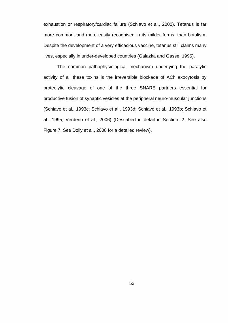
53
exhaustion or respiratory/cardiac failure (Schiavo et al., 2000). Tetanus is far
more common, and more easily recognised in its milder forms, than botulism.
Despite the development of a very efficacious vaccine, tetanus still claims many
lives, especially in under-developed countries (Galazka and Gasse, 1995).
The common pathophysiological mechanism underlying the paralytic
activity of all these toxins is the irreversible blockade of ACh exocytosis by
proteolytic cleavage of one of the three SNARE partners essential for
productive fusion of synaptic vesicles at the peripheral neuro-muscular junctions
(Schiavo et al., 1993c; Schiavo et al., 1993d; Schiavo et al., 1993b; Schiavo et
al., 1995; Verderio et al., 2006) (Described in detail in Section. 2. See also
Figure 7. See Dolly et al., 2008 for a detailed review).

54
C N
Stx 1A
C
N
N
VAMP II
SNAP-25
BoNT /G
BoNT /A
BoNT /DBoNT /F
BoNT /EBoNT /C1
BoNT /B & TeNT
SV
C
Fig. 7: CNTs inhibit exocytosis in neurons by site-specific cleavage of SNAREs. The SNAREs affected by CNTs are shown in this schematic, with the proteolytic cleavage sites for the different toxins indicated by arrows (BoNT /A, /E and /C1 cleave SNAP-25; BoNT /B, /G, /D, /F and TeNT cleave VAMP II and BoNT /C1 cleaves Stx). The SNARE core domains on the different proteins are indicated in red. N and C refer to the terminals of the different proteins, SV is the synaptic vesicle, P is the plasma membrane and the squiggles represent the palmitoyl groups on SNAP-25.

55
1.5. Viral vectors for gene therapy
Mammalian post-mitotic neurons are by their very nature incapable of
replicating to provide substitutes in case of death or injury of existing neurons.
In addition, the extensive macro- and microscopic axo-dendritic network, built
up slowly over the life of the animal and which defines the functional capacity of
every neuron, cannot be reproduced with other advanced therapies like
introduction of stem-cell based substitute neurons. Therefore, a direct approach
to manipulate gene expression in the mammalian neurons can lead to the
development of novel therapies to treat neurodegenerative diseases or to repair
physical, chemical, toxicological, bacterial and even viral injuries in them. This
also offers new approaches to probe the patho-physiological basis of neural
disorder and to identify new molecular targets for intervention.
Two of the most promising candidates for the role of neuronal gene
delivery vectors are the non-enveloped Adeno-Associated Viral vectors (AAVs)
and lipid enveloped Lentiviruses (LVs). These vectors, derived from common
human viral pathogens, have an outstanding safety profile, show relative ease
of production, have been engineered to have a broad tropism and have defined,
minimal, genomes. They differ in their transfection rates, target specificity and
the timescale and efficiency of transgene production (Kay et al., 2001) (See
Table 1 for a comparison of the properties of the two main viral vectors used in
gene therapy).
Viruses have evolved efficient strategies over millions of years of

56
evolution for transferring genes essential to their function and reproduction into
mammalian cells. Harnessing this capability, while eliminating their deleterious
features has yielded viral vectors that can act as agents for gene delivery for the
introduction and expression of genes encoding therapeutic proteins. They can
thus be used to rectify phenotypes induced by known genetic mutations or to
counteract progressive neurological diseases by introduction of known
protective genes. To be useful, viral vectors needs to be replication-deficient (to
limit the gene expression in the area of interest and to eliminate cell death that
is the usual consequence of replication), non-immunogenic (to prevent the host
immune system from mopping up all the introduced viral vectors and to prevent
the immune-mediated culling of transfected cells) and non-toxic (by the
elimination of non-essential and deleterious proteins found in the viral genome),
while still retaining their capability to deliver genetic material to the nucleus of a
target cell (thereby ensuring continuous expression of necessary proteins
without the necessity of repeated administration). But, an ideal vector system is
a difficult task to engineer and all currently used ones involve compromises in
one or the other facet. The early candidate vectors used for gene therapy,
herpes-virus and adenoviral vectors, contained large fragments of their native
genome, expressed non-essential and toxic/immunogenic viral proteins and
suffered from immunogenicity and neuronal toxicity. Also, they were
occasionally found contaminated with native, and replication-competent, viruses
or could recombine with those wild-type viruses that can be often found
naturally infecting mammals (Davidson and Breakefield, 2003).
Viral vectors bind to specific receptors on the target cell surface and

57
undergo internalisation. Non-enveloped, capsid shrouded viruses harness the
host cells receptor-mediated endocytosis pathways. Upon endosomal
acidification, the capsid breaks down and the DNA or RNA released into the
cytoplasm, binds to nucleus-seeking proteins and the genetic material is
delivered into the nucleus through the nuclear pore. Some of the capsidated
viruses might be transported along the cytoskeleton before nuclear delivery. On
the other hand, enveloped virions usually undergo membrane fusion at the cell
surface or in endosomes, depending on the nature of the envelope . While there
are neurotropic viruses, e.g., herpes, rabies and polioviruses, that specifically
bind to pre-synaptic terminals and undergo retrograde transport into neuronal
cell bodies, they have their own drawbacks like enhanced toxicity and
immunogenicity, The natural binding processes can be exploited to redirect viral
tropism by introducing receptor ligands into capsid proteins, by exchanging
capsid/lipid-glycoprotein proteins between serotypes, or by using bi-functional
neurotoxins or neuron specific antibodies that bind both to the virus and
presynaptic targets (e.g., capsid proteins of AAVs and the lipid envelope
glycoproteins of lentiviruses) (Schaffer et al., 2008).

58
Feature AAV Lentivirus
Packaging capacity
Low, ~4.9 kb High, ~8 kb
Host range Very broad, infects neurons and glia
Very broad, infects neurons and glia
Long-term expression
Yes Yes
Genomic integration
Yes, 19q11 Yes, random
Bio-safety Very-high High, esp. later generations Size 20-25nm ~100nm Yield/Titer Very high High Cellular entry HSPG + co-receptor;
receptor-mediated endocytosis
VSV-G mediated fusion of virus followed by entry of genomic content and core
Oncogenesis risk No (very low) Yes (depends on MOI and cell type)
Replication competent virus risk
No Minimal
Immunogenicity Low Very low Envelope Protein Lipid with proteins embedded Retrograde transport
Yes (In CNS and from periphery)
Yes (In CNS, periphery unknown)
Expression onset Slow Faster Purity Very high High Table 1: Comparison of AAV and LV gene delivery systems. (HSPG: Heparan sulphate proteo-glycan)
59
A
B
Fig. 8: AAV and LV structure A: Computer generated surface topology map of AAV-2. The generated view is colored according to the distance from the center of the AAV. Adapted from (Xie et al., 2002). B: Computer generated surface topology map of a model retrovirus. This is a surface rendered tomogram of an individual virion. Proteins on the lipid membrane are represented in magenta and the lipid surface of the virus is shown in purple (Some discontinuities are seen in the lipid membrane exposing the yellow interior). Adapted from (Forster et al., 2005).

60
1.6. Model systems used to study botulism and to
assess the efficacy of various therapies
The gold standard to assess the effects of CNTs is their natural target,
the cholinergic NMJ. In vivo murine model systems accurately mimic the
physiological and anatomic effects of CNT intoxication. But due to the difficulties
inherent in this system i.e., the limited amount of tissue and the need for large
number of animal subjects, necessitated the use of other in vitro and ex vivo
models systems. The in vitro models – protease assays with purified SNAP-25
substrates and permeabalised cell preparations - were used to study enzymatic
activity of natural and modified BoNT/A, /E and /C1 on susceptible and
cleavage-resistant SNAP-25. These systems, enabled and accurate and high-
throughput measurement of the relative cleavage resistance of SNAP-25
mutants and of the innocuousness of toxin mutants. Ex vivo models – using
bovine chromaffin cells and rat cerebellar granule neurons or mouse hemi-
diaphragms – enabled a more physiological measure of the effectiveness of
viral delivery of cleavage resistant SNAP-25 genes or of the binding,
internalisation and SNARE-cleaving properties of native and mutant BoNT
toxins and chimeras. Finally, the benchmark in vivo models for studying the
effects of toxins and efficacy of the counter-acting therapeutic strategies -
mouse LD50, rat/mouse digit abduction scoring and efficiency of neuromuscular
transmission (ENT) in rat soleus – enabled a near-accurate mimicking of clinical
botulism.

61
1.7. Principal goals of this study
A major challenge facing gene therapy for motor neuron disorders __ a
group of rare yet debilitating and ultimately fatal diseases affecting spinal motor
neurons __ is targeted, minimally-traumatic delivery. Due to the limited number
of motor neurons (e.g. rat sciatic nerve has only ~2000 efferents) (Nicolopoulos-
Stournaras and Iles, 1983), insults that are merely sub-lethal in other cells can
be extremely deleterious (e.g. in superoxide-dismutase mutant mice, which
ubiquitously express the damaged protein, motor neurons are the most affected
(Bruijn et al., 2004)). Their anatomical structure, with a relatively inaccessible
cell body in the spinal column and an elongated axon, complicates therapies
including gene transfer by viral vectors. Those that transfect motor neurons
(Adeno-associated, Lenti and Herpes viruses) do so most efficiently when in
direct contact with the cell body (Mannes et al., 1998). As intra-spinal injection
is difficult under clinical conditions, the use of peripherally-deliverable vectors
would be desirable; moreover, such an advance requires a model that allows
accurate measurement of the graded improvements currently achievable.
An ideal system to evaluate such therapies is the blockade of
neuromuscular transmission by botulinum neurotoxin type A (BoNT/A), by
specific cleavage of SNAP-25. This toxin is also implicated in the clinical cases
of botulism, which, while relatively rare, is a serious condition with no known
pharmacological therapies. While some prophylactic vaccines do exist to
combat botulism, they are increasingly out of favour due to the recognised
therapeutic application of BoNT/A and /B for a variety of neuromuscular

62
conditions. Furthermore, they only confer a limited period of protection, are not
licensed for use in the general population and are ineffective after exposure to
the toxin (Shapiro et al., 1998). Anti-toxin serum, especially those generated in
humans against multiple toxin serotypes (human botulism immune globulin), is
a valuable aid if administered within 24 h of exposure in infants preventing
further deterioration of the exposed patient’s condition and shortening the
duration of hospitalisation, esp. the amount of time spent on mechanical
ventilation (Tacket et al., 1984; Arnon et al., 2006). Unfortunately, these anti-
toxins have very limited utility after the initial 24 h due to the intracellular action
of the toxin’s LC and are complicated by factors such as hypersensitivity, for the
equine anti-serum (Black and Gunn, 1980), and the currently limited
applicability, of the human anti-serum, for any outbreaks in adult populations.
The ineffectiveness of current treatments in combating botulism necessitates
the development of a refined therapy with the ability to abrogate the prolonged
convalescence, high mortality and morbidity associated with this condition. The
finding that protection from BoNT/A mediated exocytotic blockade can be
achieved by expression of a toxin-resistant S25 mutant gene was an important
step in realising this goal (O'Sullivan et al., 1999). Thus the twin goals of
developing efficient gene transfer tools to motor neurons and of finding novel
therapies for botulism can be simultaneously considered (See fig. 9).
In this study, neuromuscular paralysis by BoNT/A was attenuated using
Adeno-associated (AAV) vectors that give long-term expression of a cleavage-
resistant S25 mutant (S25-R198T), having all the properties of the wild-type
including ability to substitute for truncated S25 and recover exocytosis in

63
intoxicated chromaffin cells (O'Sullivan et al., 1999). AAV vectors provide an
efficient and relatively safe way to ensure prolonged expression of genes in
neurons (Burger et al., 2005), and botulism in rats provides a much more valid
model for evaluating this gene therapy. Injection of AAVs encoding S25-R198T
into the spinal anterior horn innervating the soleus muscle protected
neuromuscular transmission from subsequent inhibition by BoNT/A. Moreover,
muscles treated in this way showed a decreased amount of nerve sprouting
compared to unprotected controls. Finally, administering the therapeutic AAVs
directly into the soleus muscle resulted in their retrograde transport to motor cell
bodies and a consequential reduction of BoNT/A-mediated blockade of
neurotransmission at peripheral synapses. These findings provide the first
evidence for the potential applicability of gene therapy to the intractable
problems posed by botulism and, possibly in the longer-term, genetic disorders
of motor neurons.
The specificity of BoNTs and TeNT for NMJs makes their non-toxic
derivatives valuable as transporters for the delivery of therapeutic agents into
the motor-neurons (Johnstone et al., 1990). Steps in this direction have been
taken through the production of recombinant toxins in which the protease site
has been inactivated to create variants that are still able to undergo binding,
internalisation and translocation at cholinergic motorneurons (Zhou et al., 1995).
The utilisation of such 'cholinergic transporter' molecules might be useful in
targeting viral vectors for the prevention of conditions affecting motor neurons.
For this purpose, the dichain form of BoTIM was utilized. This has been shown
to be essential for productive uptake into motor nerve endings (Dolly et al.,

64
1994; Daniels-Holgate and Dolly, 1996) while yielding more efficient
internalisation than can be achieved with HC alone (Bade et al., 2004). The
creation of a core-streptavidin-BoTIM/E chimera (CS-BoTIM) with the targeting
properties of BoTIM and the biotin binding properties of CS should yield a
versatile molecule that could be attached to biotinylated cargo in the desired
stochiometry (Pahler et al., 1987; Sano et al., 1995). The CS-BoTIM also offers
the advantage of a smaller conjugate, ease of production, control of positioning
of the attachment site and attachment to a single biotin molecule. BoTIM was
conjugated at the genomic level to a monomeric streptavidin domain – the
minimal streptavidin core (12-16kDa) was cloned into the N-terminus of the
His6BoTIM single chain gene, expressed in E.coli and the resultant CS-BoTIM
purified using a Ni-NTA column. Retention of neuronal targeting ability by CS-
BoTIM was tested after application to rat CGNs and HEK-293s in culture;
immuno-histochemical visualization of stronger binding to the former confirmed
the neuronal preference of binding. Application of active BoNT/E, conjugated as
before to CS at the genomic level, resulted in SNAP-25 cleavage and exocytotic
blockade, confirming the binding, internalisation and release of LC.
The next refinement of the gene replacement methodology involved the
attempted targeting of lentiviral vectors to neurons by availing of the unique
selectivity of CS-BoTIM. This potentially opens up routes of peripheral
administration due to the therapeutic becoming concentrated on to the pre-
synaptic membrane at the NMJ. Such a targeted virus would have a huge
number of applications not just in the treatment of botulism but, also, in
hereditary motor neuron diseases (e.g. spinal motor atrophy, amyotrophic

65
lateral sclerosis) and those caused by viruses, physical and biochemical
trauma. A second-generation LV system was exploited to create high titer
viruses expressing GFP, which were later biotinylated with a reducible linker
interface. Proof of principle for this exciting new strategy was obtained by the
successful coupling of the CS-BoTIM protein to the viral vectors, followed by
their later application onto CGN and HEK-293 cells. To determine optimal
coating conditions, CS-BoTIM was mixed with different ratios of the test and
control viruses in order to generate viral particles coated with the targeting
agent. The optimally coated viruses, preferentially binding to neuronal terminals,
showed a reduction in their promiscuous infectivity to HEK-293 cells, with only a
small attenuation of their natural neuronal infectivity.
These devised tools and therapies, targeted initially at botulism, should
provide a strong foundation for the later development of sustained motor neuron
gene therapies applicable centrally or peripherally.

66
Fig 9: Overview of gene therapy against botulism. A motor neuron, with its nucleus, dendrites, axons and cholinergic terminals is represented here. The hexagonal structures represent viral vectors, clear J-shaped structures - native SNAP-25, shaded J-shaped structures – multi-toxin-resistant SNAP-25 (MTR) and labelled boxes attached to viral particles – targeting agents and the green genomes – specific promoter. The dotted red line represents the path of the virus to the MN nucleus and the solid blue line the path of the SNAP-25 protein. The virus can be delivered spinally or peripherally (in which case it has to undergo retrograde transport). Once in the host nucleus, the viruses start producing cleavage-resistant SNAP-25 mRNA, and thus the protein. Once the MTR SNAP-25 protein is transported down to the cholinergic terminals, they can no longer be inactivated by the introduced CNTs (BoNT A/ E /C1). Thus the treated terminals are able to retain exocytotic activity, in the face of a later challenge with these CNTs, or can retain activity quicker, in the case of a prior treatment with these toxins. Further refinements can be introduced by the addition of targeting molecules (BoTIM/E or TeTIM) and/or by the addition of a cholinergic/neuron-specific promoter.

67
2. Attenuation of BoNT-induced blockade of
exocytosis at peripheral NMJs by viral vector
mediated gene transfer into spinal motor neurons
The characteristic symptoms of botulism are caused by the BoNT-
mediated blockade of neurotransmission in cholinergic nerve terminals,
especially NMJs. The processes involved in blockade of neurotransmitter
release by Clostridial neurotoxins involve – targeting to the NMJ, binding,
internalisation, translocation of light chain to the cytoplasm and, finally,
proteolytic cleavage of the requisite SNAREs. The Clostridial neurotoxins share
many properties and, upon entry into the bloodstream, target to pre-synaptic
nerve terminals. The HCs mediate selectivity by targeting to high-affinity
membrane ecto-acceptors located exclusively on cholinergic (motor/autonomic)
nerve endings (Dolly et al., 1984a; Black and Dolly, 1986b; Black and Dolly,
1987; Daniels-Holgate and Dolly, 1996). Upon uptake into the target vesicles,
the heavy chain helps form a transmembrane pore which allows the light chain
to escape into the cytoplasm. Upon intracellular entry, the light chains cleave a
defined member of the SNARE family and cause an inhibition of evoked
exocytosis (Schiavo et al., 1993a; Schiavo et al., 2000) (The targets and the
cleavage sites are described in detail in Section. 1.3). When the rate of
synthesis of the new SNARE molecules and/or turnover of the handicapped

68
SNAREs overwhelms the enzymatic activity of remaining BoNT, normal
neuromuscular transmission returns (de Paiva et al., 1999).
2.1.1. Trafficking of Clostridial neurotoxins (CNTs) to their site of
action
The Clostridial bacilli producing the different BoNT toxins are quite
fastidious anerobes; the growth conditions vary between different serotypes and
are sensitive to changes in temperature, pH and oxygenation. Therefore, the
usual method of contact with the toxin is through consumption of contaminated
food, which already contains previously produced toxin. As the BoNTs are
sensitive to the harsh proteolytic conditions found in the gastric lumen, they are
packaged with other non-toxic proteins, which protect a proportion of toxin until
it reaches the small intestine. Those that protect and contain BoNT/A can be
purified as 19 S, 16 S, or 12 S stable complexes. The non-neurotoxic
components consist of non-hemagglutinins, with Mr ~120 kDa, and
hemagglutinins, consisting of a combination of five different proteins of 52, 35,
20, 19, and 15 kDa (Inoue et al., 1996). In the small intestine, the slightly
alkaline pH causes dissociation of the toxin complexes and the free neurotoxin
undergoes transcytosis across intestinal epithelial cells and reaches the
bloodstream.
The process of migration of CNTs to the extra-cellular space surrounding
the NMJ has not yet been completely characterised – a journey that takes the
toxin molecule across capillary endothelial and basal laminar barriers while,
presumably, evading the body’s immune and scavenging systems and

69
simultaneously preserving its activity for the final target. The clinical importance
of this migratory stage, especially in relation to prophylactic treatments such as
vaccination or injection of antibodies, is discussed later. Such is the exquisite
specificity of the toxin to NMJs that, at physiological doses, the only effect of the
toxin is the perturbation of neuromuscular transmission – despite the ready
presence of potential substrates in non-neuronal secretory cells. Indeed, in
cultured neuroendocrine cells which share the same SNARE targets as the
NMJ, permeabalisation or treatment with non-physiological buffers is necessary
for the uptake of the toxin. Once near the pre-synaptic motor terminals the toxin
undergoes a series of steps that culminates in the delivery of the LC protease to
near the SNARE targets. For BoNTs, this process can be divided into a three
step model – binding and internalisation of the holo-toxin, pore formation by the
HC and, finally, translocation of the LC into the cytoplasm (Dolly et al., 1992;
Verderio et al., 2006; Brunger et al., 2007; Fischer and Montal, 2007a, 2007b)
(See fig. 10).
The evidence accumulated points to a “dual-receptor”, “dual-affinity”
mode of entry of BoNTs. The “double specificity” conferred by such an ecto-
acceptors can help in explaining the CNT’s remarkable specificity for NMJs.
Gangliosides, especially the sialylated glycosphingolipids containing N- and O
substituted neuraminic acid residues, are hypothesized to act as an intial,
accumulatory, ecto-acceptor component for CNTs. The accumulated CNTs then
bind to specific protein receptors on the pre-synaptic terminal surface and
undergo endocytosis. These two ecto-acceptor families are the most probable
candidates for the physico-chemically determined, low–affinity (e.g.:

70
synaptosomes: Kd ~26 nM, Bmax ~7.5 fmol/mg protein; presynaptic plasma
membrane: Kd ~30 nM, Bmax ~52 fmol/mg protein for labelled BoNT/A in electric
organ of Torpedo) and high-affinity (e.g.: synaptosomes: Kd = 0.11 +/- 0.03 nM,
Bmax = 50 +/- 10 fmol/mg protein; presynaptic plasma membrane: Kd = 0.2 +/-
0.05 nM, Bmax = 150 +/- 15 fmol/mg protein for labelled BoNT/A in electric organ
of Torpedo) receptors (Dolly et al., 1984b; Blasi et al., 1992; Daniels-Holgate
and Dolly, 1996).
The ganglioside-binding part of the theory is now well established (See
(Simpson and Rapport, 1971; Nishiki et al., 1996; Bullens et al., 2002; Dong et
al., 2003; Rummel et al., 2004a; Mahrhold et al., 2006; Rummel et al., 2007)
and is reviewed in (Verderio et al., 2006)). The importance of gangliosides on
acceptor membranes for the uptake of BoNT and TeNT was confirmed when it
was shown that pre-incubation with ganglioside GT1 inhibited their uptake in
chromaffin cells (Stoeckel et al., 1977), while pre-treatment of cells with the
polysialogangliosides GD1a, GT1b and GD1b enhanced the toxin-mediated
blockade of exocytosis (Marxen et al., 1991). Also, ganglioside depletion in
neuroblastoma cells impairs BoNT/A internalization, treatment with
neuraminidase decreases BoNT binding and knockout mice defective in a-
series and b-series ganglioside production showed highly reduced sensitivity to
BoNT/A and BoNT/B (lacking the complex gangliosides GM1, GD1a, GD1b, GT1b
and GQ1b because of disrupted β1,4-N-acetylgalactosaminyltransferase (GM2/GD2
synthase; EC 2.4.1.92) gene) (Bullens et al., 2002, , 2003; Kitamura et al.,
2005). The current understanding is that polysialogangliosides, richly distributed
on the presynaptic motor nerve terminal, bind BoNTs HCC, a sub-domain which
71
shows a high degree of homology to the carbohydrate-binding domain found in
plant lectins; BoNT/A and /B has been shown to bind GT1b through a conserved
H…SXWY…G peptide motif in this region (Poulain et al., 1989; Miller et al., 2008;
Stenmark et al., 2008; Tsukamoto et al., 2008) while binding property of
BoNT/C to GD1b, and GT1b and BoNT/D to phosphatidylethanolamine has also
been shown to reside in the HC region (Tsukamoto et al., 2005).
However, and complicating matters quite substantially, these same
studies showed that ganglioside-binding is not an essential, or the only, pre-
requisite for toxin uptake, since the amount of inhibition obtained was not
complete when antagonising just the ganglioside part of binding (Stoeckel et al.,
1977; Marxen et al., 1991). In addition, neuraminidase treatment of chromaffin
cells did not completely abolish CNT activity and ganglioside knockout mutants
were still capable of being paralysed by BoNT/A and TeNT, albeit at a far
slower rate (Kitamura et al., 1999; Kitamura et al., 2005). So, ganglioside
binding alone cannot completely explain the specificity of BoNTs for the
cholinergic motoneurons. The best studied protein receptor candidates are Syts
and SV2. Studies have shown that binding of BoNT/B to Chinese hamster ovary
and PC12 cells is dependent of expression of Syt2 (Dong et al., 2003). In vitro
pull-down assays have also shown that BoNT/B and /G interact with Syt1 and
Syt2 and pre-treatment with peptide fragments corresponding to the luminal
domains of these proteins can inhibit toxin-mediated blockade in mouse hemi-
diaphragm assays (Rummel et al., 2004b). The binding to BoNT/B was activity
dependent, hinting at the luminal region being involved. Truncation analysis of
Syt2 revealed that residues adjacent to the transmembrane domain, amino

72
acids 40–60, are critical for toxin binding. Interestingly, Syt2 binds to
gangliosides through residues 61-267, thereby linking the initial binding of CNTs
to sialogangliosides and the later binding to Syt2 (there is a high homology
between Syt1 and 2 in these regions (Dong et al., 2003)). This finding also
explains the frequent finding that CNT uptake in neurons is activity-dependent;
the luminal domain of Syt will only be exposed upon fusion of SVs. The dual-
receptor hypothesis for CNTs in general and BoNT/B and /G in particular, has
been further established by mutation studies that showed a strong and,
sometimes, near-complete inactivation of the toxin’s activity in vivo upon
introduced changes in the putative binding regions (Rummel et al., 2007).
The protein receptor for BoNT/A has also been recently identified – the
intra-vesicular luminal domain, L4, of SV2 has been shown to be critical for
BoNT/A binding (Dong et al., 2006). Proof for this specific interaction has been
obtained by pull-down assays with fragments of SV2 (L4- 529-566AA) and by
competition assays. BoNT/A binding to hippocampal neurons cultured from
SV2B-/- mice was reduced by 28%, by 53% in SV2A+/-SV2B-/- mice and binding
was almost completely abolished in SV2A-/-SV2B-/- mice (Dong et al., 2006).
Uptake was restored by the expression of SV2A in SV2A/2B double knock-out
mice. BoNT/A was also shown to bind the least prevalent SV2C isoform as well;
indeed, its reported affinity was higher than that for SV2A or 2B (Mahrhold et
al., 2006).
Once the ganglioside-protein-receptor-toxin complexes form, they
undergo receptor mediated endocytosis, most probably involving clathrin-coated
vesicles (the energy- and temperature-dependent uptake of these neurotoxins

73
is discussed in (Dolly et al., 1984a; Black and Dolly, 1986b, 1986a)). The onset
of neuroparalysis induced by CNTs can be shortened under condition of
increased SV recycling, a process that is intimately connected with SV fusion
[Dolly >>]. While the HC domains of CNTs have been shown to be essential and
sufficient for the internalization into vesicles (Lalli et al., 1999), it is not yet
known if they, or other parts of the CNTs, in any way influence the endocytotic
process, or if they are mere passengers.
Once internalised, the vesicles containing the CNT-receptor
complex are sorted in a TeNT- or BoNT-specific manner, leading to the distinct
symptoms associated with each disease. While the cholinergic NMJ is the final
target of BoNTs, TeNT undergoes retrograde transport back to the spinal cord,
transcytoses across the synaptic junction and targets inhibitory glycinergic inter-
neurons in the spinal cord; BoNT/A is >1000 fold more effective than TeNT in
blocking release from the peripheral cholinergic nerve endings (Habermann et
al., 1980), while the reverse is true at the inter-neurons (Habermann and
Erdmann, 1978; Bigalke et al., 1985). The differential sorting of CNTs in the
endosomes of NMJs could be a property of the protein/ganglioside receptor or
due to a determinant on the HC. The former is supported by studies that show
that TeNT co-segregates with p75NTR while the latter model is supported by
hybrid toxin studies which show that when the HC of BoNT/A is linked to the LC
of TeNT, the hybrid has the binding and sorting features of BoNT/A (Poulain et
al., 1991; Weller et al., 1991). The putative mechanism by which TeNT
undergoes retrograde axonal transport into the spinal cord is described in the
next section.

74
A B
C
D
E
FG
H
BoNT LC
BoNT HCN
BoNT HCC
Endosome
SV
SNAREs
Pre-synaptic membrane
Fig. 10: Mechanism of action of BoNT. The different phases in BoNT action are schematically represented here. After initial binding to specific receptors (A), the holotoxin undergoes internalisation into endosomes (B). Upon endosomal acidification (C), the HC forms a pore through which the LC is extruded into the cytoplasm (D). The LC now targets its specific SNARE substrate, SNAP-25 (F), VAMP (G) or Stx (H).

75
C
N
HCC HCN LCA
B C
Fig. 11: Crystal structure of BoNT/A. A: A screen-shot of a Cn-3D rendering of the determined 3.3Ǻ structure of BoNT/A holotoxin shows the different, biochemically identified, domains. At the C-terminus, comprising mainly of β-sheets, is the HCC which is involved in specific binding to receptors on cholinergic terminals. This is followed by α-helix rich HCN and the N-terminus LC. (β-sheets are in gold and α-helices are in green, with intermediate AAs in blue. Also seen is the Zn2+ ion at the catalytic site). B and C: Structure of BoNT/A LC bound to its SNAP-25 substrate. The complex inter-play between the different α and β exosites on SNAP-25 and the toxin’s LC can been seen in this 2.1 Ǻ structure. The LC is shown in gold (β-sheets) and green (α-helices) and the SNAP-25 substrate is shown in yellow (with one α-helix in green). Panel B shows both the LC and the substrate, while C shows only the substrate for increased clarity. Also seen are the Zn and Cl ions. Based on data from (Lacy et al., 1998) and (Breidenbach and Brunger, 2004).

76
Since the substrates for the CNTs are in the cytoplasm, the process of
transfer of the LC into the cytoplasm is critical to their in vivo activity. Extensive
clues about this process can be gained by studying similar processes involved
in the entry of bacterial and plant toxins, like Shiga, Diptheria and Cholera
toxins (Sandvig and van Deurs, 2005). This translocation requires the HC, is
triggered by low vesicular pH and requires a reducing environment in the
cytoplasm (Boquet and Duflot, 1982). The pH sensitivity of this process has
been demonstrated by studies that showed abolition of the toxin’s activity upon
pre-treatment with drugs such as bafilomycin (inhibitor of the proton pump) or
lysosomotropic drugs like chloroquin or methylamine (Simpson, 1982, , 1983;
Lawrence et al., 2007). The HN terminal of BoNT/A consists of two long
amphipathic α-helical coil-coil domains, analogous to some viral proteins (Lacy
et al., 1998; Lacy and Stevens, 1999). This amphipathic HN domain of CNTs,
exposed under low pH conditions, inserts into the endosomal membrane to form
cation-selective conductance channels (Schmid et al., 1993). Simultaneously,
the LC which is attached to the HC by a di-sulphide bridge, partially unfolds at
low pH and slides into the pore formed by the HC. Upon contact with the
reducing environment in the cytoplasm, the di-sulphide bond is reduced and LC
liberated (Koriazova and Montal, 2003). If non-reducing conditions or
holotoxins, with the LC and HC still attached, are used, the LC does not free
itself upon insertion and continues to block the HC pore. Once in the cytoplasm,
the free LC now refolds back to its native configuration. The process of
translocation has been studied using electrophysiological tools in great detail in
Neuro 2a neuroblastoma cells and reconstituted liposomes, mimicking the

77
natural endosomal environment (Montal et al., 1992; Fischer and Montal,
2007a, 2007b; Fischer et al., 2008). In such conditions, the HC forms channels
which open in bursts interspersed between periods of inactivity. These channels
are open at negative voltages and show a main conductance of 105 +/- 5 pS or
65 +/- 4 pS in 200 mM CsCl or NaCl, respectively (Fisher and Montal, 2006).
2.1.2. Proteolytic cleavage of specific SNAREs by the LC of CNTs
The protease activity of the CNTs resides in the LC and this alone can
mediate the subsequent paralysis of neuromuscular transmission. Direct
introduction of the LC, by permeabilisation (Ahnert-Hilger et al., 1989) or by
liposomes (de Paiva and Dolly, 1990), results in cessation of synaptic
exocytosis identical to that elicited by the whole molecule. The elucidation of the
different CNTs amino-acid sequences and solving of their crystal structure, with
or without their attached substrates, has enabled a deeper understanding of the
mechanisms underlying their enzymatic activity. The LC sequences show a
metalo-protease domain with a HExxH+E consensus Zn2+-binding site
(Jongeneel et al., 1989; Kurazono et al., 1992). Three residues co-ordinate the
catalytic Zn2+ in the active site (H222, H226 and E261) while E223 ensures its
hydration (Lacy et al., 1998; Lacy and Stevens, 1999). The importance of Zn2+
was shown when its removal inactivated the toxin and prevented neuroparalysis
(Schiavo et al., 1992b; de Paiva et al., 1993). The identity of their synaptic
targets was first revealed when VAMP2 was shown to be the target for BoNT/B
and TeNT (Schiavo et al., 1992c) (See section 1.3 for more details about the
cleaved SNAREs and their cleavage sites. See also Figure 7). The identification

78
of critical SNAREs as targets for the different CNTs fits well with the
electrophysiological observation that inhibition of SV fusion is a prime
consequence of toxin mediated neuroparalysis.
The mechanisms underlying the enzymatic activity of CNTs are now
better understood due to recently solved structures of different LCs bound to
their substrates (Hanson and Stevens, 2000; Breidenbach and Brunger, 2004, ,
2005). Unlike classical metallo-proteases, the LCs, though significantly
homologous at their active sites, have no common recognition motif on the
SNAREs. While they are generally promiscuous in their requirement for
particular side-chains flanking the scissile bond, they do require long stretches
of their target SNAREs for optimal efficiency (Foran et al., 1995; Schmidt and
Bostian, 1995, , 1997; Breidenbach and Brunger, 2004, , 2005). Also, point
mutations in regions remote from the scissile bond can reduce proteolytic
efficiency of these LCs. All these clues point to a bi-partite mode of action of the
LCs, with regions spatially separated from the scissile bond, exosites, being
important in binding and recognising the SNAREs and bringing them into a
conformation where the active site can access the scissile bond.
SNAP-25 interacts quite extensively with BoNT/A LC and upon binding
abandons its native helical configuration and wraps round most of the LC’s
circumference (Breidenbach and Brunger, 2004, , 2005). The part of the LC
opposite the active site plays an important role in identifying the correct
substrate and the C terminus of the correctly bound SNAP-25 probably induces
a conformational change in the catalytic motif of the LC, increasing its catalytic
efficiency. Thus, while only nine residues from the C-terminus of SNAP-25 are

79
actually cleaved, 60 residues in the C-terminal are needed for efficient
proteolysis, encompassing the structurally-identified a and b exosites on the LC.
Truncated forms, with only 40 C-terminal residues, and mutations, including
I156E and M167E, substantially decrease the catalytic efficiency by depriving
the a-exosite of its binding partner. The b-exosite on the other hand binds at the
far C-terminal of SNAP-25; versions of S25 truncated below M202 are cleaved
at a very slow rate.
These processes probably occur in a sequential manner – SNAP-25 first
binds to BoNT/A at the a-exosite, fastens around the LC molecule, binds at the
b-exosite and induces conformational changes at the active site potentiating its
catalytic activity. As these disruptions in SNAP-25 structure, caused by the
multiple interactions with LC, also bring the Gln197-Arg198 bond near the
HExxH+E catalytic domain, allowing efficient and rapid proteolytic cleavage of
the terminal 9 residues. This bi-partite multi-site binding strategy can be used to
explain the selectivity of CNTs and the necessity of a large extent of substrate
for efficient proteolysis. This also explains quite elegantly the finding that
proteolysis of SNAP-25 is retarded if it is already incorporated into a ternary
SNARE complex (Breidenbach and Brunger, 2004). The identification of these
non-scissile substrate binding sites on the surface of BoNT/A, such as the a-
and b-exosites, also presents new opportunities for novel peptide inhibitors.
The specific residues flanking the scissile bond influences toxin
susceptibility. Amino acids at either the P1 or P’1 site are important for cleavage
of VAMP II by BoNT/B (Foran et al., 1994; Regazzi et al., 1996) and SNAP-25
by BoNT/A, /C1 and /E (Schmidt and Bostian, 1997; Schmidt et al., 1998); for

80
BoNT/E, other sites also contribute to the efficacy of cleavage (P5 to P’3)
(Vaidyanathan et al., 1999). The multi-site binding strategy employed by
BoNT/A accounts for the extreme selectivity of this enzyme, both in terms of the
particular SNARE that is cleaved as well as the particular cleavage site. Due to
their high homology, most of the CNT LCs probably use similar mechanisms for
substrate recognition (e.g., the similarity between BoNT/A and /B is very high,
the overall root mean square deviation between the holotoxins is 2.1 Å, and
between the LC active sites is 1.4 Å (Swaminathan and Eswaramoorthy, 2000)).
2.1.2.1. Unique trafficking properties of TeNT
After TeNT enters the motor nerve terminal, it is internalised into a
vesicle and transported retrogradely along the axon into the motor neuron cell
body, at 7.5mm/H, and to the dendrites and released into the synaptic cleft
(Burke et al., 1971). The released TeNT is then taken up by inhibitory inter-
neurons where it blocks transmission, tipping the balance between excitatory
and inhibitory control of the motor neuron towards the latter, resulting in
“tetanic” muscle spasms characteristic of the disease. The differential specificity
and targets of TeNT and BoNT is determined by the different vesicles that they
enter at the NMJ. TeNT enters non-acidified vesicles undergoing retrograde
transport while BoNT enters acidified vesicles and thus remains within the
motorneuron presynaptic terminal (Lalli and Schiavo, 2002; Lalli et al., 2003;
Bohnert and Schiavo, 2005). An alternate hypothesis is that TeNT could be
perverting the course of regular endosomal vesicles and causing them to
undergo retrograde axonal transport. An interesting finding is that TeNT uptake

81
is independent of pre-synaptic activity, unlike BoNT uptake – potentially opening
up avenues to target not only active neurons, but also paralysed ones
(Deinhardt et al., 2006). The peripheral effects of TeNT are very minimal, unless
very high doses are injected. TeNT co-localises with the receptor for NGF
(p75NTR), segregates to non-acidified vesicles, requires F-actin, microtubules,
dynein, kinesins, V-ATPase and myosin to successfully undergo retrograde
transport (Lalli and Schiavo, 2002; Lalli et al., 2003).
2.1.3. The structure of purified CNTs
The solved structures of CNT holotoxins reveal a domain organization
consisting of the receptor binding domain (HCC), the translocation domain
(HCN), and the light chain (LC) protease domain (Lacy et al., 1998; Lacy and
Stevens, 1999) (See fig. 11). The sequence homology and functional similarity
between the CNTs is reflected in their structural resemblance as well - the
overall root mean squared deviation between /A and /B holotoxins is 2.1 Å, and
between the LCs is 1.4 Å. These CNTs are synthesised as single proteins with
a molecular weight of 150 kDa and subsequent proteolytic cleavage by
endogenous or exogenous proteases creates a C-terminally located heavy
chain (HC) and a N-terminally located light chain (LC), with molecular weights of
100 kDa and 50 kDa respectively. Despite cleavage, they persist as a very
stable di-chain molecule due to the presence of a disulphide bond and tight
non-covalent protein-protein interactions between the chains (Dolly and Aoki,
2006). The solved crystal structure for BoNT/A, accurate to within 3.2 Å , shows
that it has an extended structure with dimensions of 45 X 105 X 130 Å in which

82
the HCN domain, composed of two 105 Å α-helical domains, separates the L
domain from the HCC domain. The LC domain, a compact structure measuring
55 X 55 X 62 Å, is composed of a mixture of α-helical and β-strands and is
linked to the HCN via a long loop which wraps around the LC (Lacy et al., 1998).
Despite the overall similarity, small differences in surface residues probably
contribute to the significant substrate specificity of different CNTs. The solved
BoNT/A-SNAP-25 complex structure has revealed that the LC depends on an
extensive array of exosites to provide the substrate specificity, to orient the
scissile peptide bond correctly in relation to the enzyme’s catalytic motif and to
boost the catalytic efficiency (reviewed in (Breidenbach and Brunger, 2004).
See fig. 11).
2.1.4. Recovery of cholinergic transmission at the NMJs after
Clostridial toxin poisoning
The speed of recovery from CNT-induced neuromuscular paralysis is
strongly serotype dependent. Muscle can remain paralysed for many months
after BoNT/A and /C1, while patients exposed to BoNT/B, /D, /E or /F recover
from poisoning within a matter of weeks (Cherington, 1998). The response of
the pre-synaptic membrane to blockade of exocytosis is proposed to
significantly affect the speed and extent of recovery – for example: a higher
plasticity and regenerative capacity after injury in younger animals could
contribute to the faster recovery compared to adults (Pestronk et al., 1980;
Verdu et al., 2000; Chen et al., 2002). At murine muscle endplates, the nerve
endings sprout vigorously after exposure to BoNT/A and, in the initial post-

83
paralytic period, only these sprouts are capable of exocytotic function at newly-
formed synapses (Duchen and Strich, 1968; de Paiva et al., 1999). Such
sprouting, which has also been detected in juvenile rats (Pestronk and
Drachman, 1978b), depends on the amount of toxin introduced and the muscle
type; fast muscles show less sprouting and predominantly axonal outgrowths
compared to the terminal sprouts seen in slow muscles (Brown et al., 1980).
The final phase of recovery involves exocytosis returning to the original synapse
and regression of the sprouts. While the stages of this functional recovery have
been characterised, it is not known whether the fine control of sprouting and
regression are related to the toxin’s persistence, its cleavage product (truncated
SNAP-25) or if it is controlled solely by the loss of neural stimulation of the
muscle resulting from disablement of the SNARE function. The relative
importance of the muscle or Schwann cells in either of these complementary
processes remains poorly defined (Tam and Gordon, 2003). The up-regulation
of synaptic protein production has been observed in paralysed NMJs and could
be the key to recovery or merely be a response to an unknown ‘switch’ for
sprouting (Jung et al., 1997). BoNT/A intoxication has also been found to induce
the extension of Schwann cell processes, which play important roles in the
regeneration by providing paths along which the sprouts extend (Son and
Thompson, 1995; Son et al., 1996). However, the matter is complicated by the
finding that BoNT/A can cause sprouting in nerve-muscle co-cultures deficient
in Schwann cell (Bonner et al., 1994). Hormonal and signalling factors, and their
receptors, like insulin-like growth factor (IGF) could also play an important role
in sprouting (Caroni and Grandes, 1990; Caroni et al., 1994; Caroni, 1997).

84
The extended duration of synaptic stasis associated with BoNT/A
poisoning causes prolonged convalescence and considerable morbidity. This
could be due to the persistence of the toxin LC in the pre-synaptic terminal or
the prolonged destabilisation of the SNARE machinery by the cleaved SNAP-25
or due to the time taken to replenish the incapacitated SNAP-25. The latter can
be ruled out as BoNT/E, which cleaves the very same substrate as /A, has a
very short duration of action (Meunier et al., 2003). The inhibitory effect of the
BoNT/A cleavage product of SNAP-25 was demonstrated in chromaffin cells,
where over-expression of SNAP-25-197 caused an inhibition of Ba2+-evoked
hGH secretion (O'Sullivan et al., 1999). Another important clue to explain the
prolonged duration of BoNT/A action was the finding that the BoNT/A-LC is
distributed in a punctate manner in the plasma membrane and co-localizes with
SNAP-25-197, while BoNT/E LC was predominantly cytoplasmic (Fernandez-
Salas et al., 2004a; Fernandez-Salas et al., 2004b). Furthermore, this
localisation of BoNT/A-LC to a slow recycling compartment at the plasma
membrane may make it less susceptible to degradation or ubiquitination while
cytoplasmic BoNT/E-LC could be more accessible to removal, thus, accounting
for their different time scales of paralysis. There is also some evidence for the
first eight amino acids at the N-terminal of LC/A or a di-leucine motif present at
the C-terminal could contain a signal for plasma membrane localization
(Fernandez-Salas et al., 2004a; Fernandez-Salas et al., 2004b).
The microscopic manifestation of synaptic re-organisation following
intoxication with BoNT/A are well known. Blockade of ACh release for 3 days or
more by BoNT serotypes induced nerve sprouting with the formation of

85
functional extra-junctional synapses whilst the original remains inoperative.
Initially, thin amyelinated neuritic processes emanate from their original parent
nerve endings (terminal sprouting) or from the last node of Ranvier (nodal
sprouting) and extend outside the area occupied by the parent nerve terminal.
In time, these sprouts gain the components necessary for a functioning
synapse, and the AChR clusters on the opposing muscle surface that go with it,
and start to exhibit exo- and endocytosis (de Paiva et al., 1999; Meunier et al.,
2003). At their peak, they are much larger than the, currently quiescent, parent
synapse, but after 9 weeks, the growth of the sprouts ceases. Activity starts to
return to the parent nerve ending and, by 3 months, the sprouts undergo
retraction accompanied by complete resumption of the normal level of neuro-
exocytosis to the fully-remodelled endplate that is then virtually indistinguishable
from the original (de Paiva et al., 1999). This is a very striking and extensive
cascade of events which illustrates how such neuronal plasticity can cope with a
toxin insult and highlights the role of the activity mediated by the sprouts in
allowing and, probably, promoting rehabilitation of the synapse.
2.1.5. Engineering SNAREs resistant to cleavage by CNTs
Extensive work has been undertaken to define the important residues in
the SNARE substrates that affect the rate of their cleavage by different CNTs.
As with any other enzymatic proteolytic process, substitutions in and around the
cleavage site strongly affect the Kd and Vmax of the reaction. For e.g., Q76V,
F77W mutations in VAMP2, the P1 and P1’ residues respectively, confer
resistance to TeNT inhibition of hGH release in PC12 cells and insulin secretion

86
in HIT-T15 cells (Regazzi et al., 1996; Quetglas et al., 2002). Similarly, Q76V or
F77V or F77L mutations of VAMP2 confer resistance to BoNT/B (Shone and
Roberts, 1994) and K59R or K59A mutations induce resistance to /F (Schmidt
and Stafford, 2005). Mutated SNAP-25 isoforms have been engineered to resist
cleavage by BoNT/C1 (R198T or R198E (Vaidyanathan et al., 1999)), /A
(R198T (O'Sullivan et al., 1999)) and /E (I181F (Vaidyanathan et al., 1999) or
D179K, M182T (Koticha et al., 2002)).
2.1.6. Adeno-associated viruses (AAV)
AAV is a non-pathogenic single-stranded DNA containing replication-
deficient parvovirus, with a stringent requirement of co-infection with a helper
adenovirus or herpes virus to complete its lytic life-cycle (McLaughlin et al.,
1988; Berns and Linden, 1995; Linden et al., 1996; Samulski, 2000; Xie et al.,
2002; Burger et al., 2005). While these dependo-viruses are widely prevalent as
a latent infection in many mammals, including humans, they rarely cause overt
clinical symptoms. AAVs are protein enveloped viruses with a capsid diameter
of 26nm. Inside this capsid can be found a single-stranded DNA genome
containing all the cis-acting elements needed for AAV replication and packaging
and flanked by inverted terminal repeats (ITR). Of the 11 known serotypes,
AAV1-5 are the best studied and AAV-2 is widely used in clinical gene therapy
(Choi et al., 2005).
The wild-type AAV-2 genome consists of rep and cap genes encoding
genes necessary for replication and capsid protein production respectively.
Utilizing two promoters and alternative splicing, AAVs generate four rep proteins

87
- Rep78, Rep 68, Rep 52 and Rep 40. Transcripts from another promoter
generates the three structural viral capsid proteins, 1, 2 and 3 (VP1, VP2 and
VP3), through alternate splicing and translation start sites (Berns and Linden,
1995; Linden et al., 1996; Linden and Berns, 2000). These differ only in their N-
terminals while sharing 533 amino acids in a common C-terminal – generating
VP1, VP2 and VP3 proteins of molecular weights 87, 73 and 62 kDA
respectively (the former two contain additional N-terminal sequences of 202 and
65 amino acids, respectively). The intact AAV viral particle contains a total of 60
copies of VP1, VP2, and VP3 at a 1:1:20 ratio, arranged in a T=1 icosahedral
symmetry (McLaughlin et al., 1988). Wt-AAVs (wildtype AAVs) exist as
quiescent integrated forms in the host genome and only replicate in the
presence of helper viral infection.
AAVs need to undergo multiple steps prior to synthesizing its genome
contents – the introduced AAV first binds to cellular surface receptors (usually
heparan sulphate proteoglycans) and is later endocytosed. After uncoating, the
viral particle releases the ss-DNA genome which is trafficked to the nucleus.
The conversion of the genome from single-stranded to double-stranded DNA is
followed by transcription of its constituent proteins in the nucleus. In the
absence of the helper virus, wt-AAVs integrate into the host chromosome,
usually 19q11, with the assistance of Rep proteins and through the interaction
of their ITR with the host chromosome (AAVs replicate epi-chromosomally
under replication permissive conditions and integrate into the host genome at a
rate of 5–10% under non-permissive conditions (Linden et al., 1996)). The
efficiency of infection and the rate of protein production is determined by many

88
factors such as – the abundance of cellular surface receptors for viral
attachment and internalization, the effectiveness of release from endosomes
and lysosomes, the efficiency of unpacking and nuclear trafficking, the rate of
conversion of single-stranded to double-stranded DNA template and, finally, for
a sustained long-term expression, the amount of integrated viral genome. All
these factors differ between the serotypes and are strongly affected by the host
cell, but they might be manipulated to facilitate more efficient or more specific
transduction of cells or tissues for gene therapy (Burger et al., 2004; Choi et al.,
2005).
The properties of this versatile virus have been exploited to produce
recombinant AAV (rAAV) gene delivery vectors. A helper-virus free system for
the production of infectious rAAV can be fashioned by removing the two viral
genes (rep and cap) and inserting a transgene expression cassette between the
two ITRs, with Rep and Cap provided in trans. The absence of the constitutively
expressed native viral genes reduces the toxicity, eliminates the reproductive
capability and lessens the possibility of immune recognition.
A commercially available rAAV helper- free system from Stratagene has
been used widely for scientific and pre-clinical applications (Kay et al., 2001)
(see Fig. 12). In this system, the stripped down human AAV-2 genome, bereft of
all except the ITRs and a coding cassette, is co-transfected into HEK-293
variant (AAV-293) cells with two other plasmids, pHelper and pAAV-RC. The
essential helper adenoviral genes, E2A, E4, and VA RNA genes, required for
the production of infective AAV particles are supplied in trans (through a co-
transfection with the plasmid pHelper). The AAV-293 host cells, which stably

89
express the adenovirus E1 gene, and the plasmid pAAV-RC, producing the Rep
and Cap genes are the other essential requirements supplied in trans. The
removal of the AAV rep and cap genes also makes space for insertion of a gene
of interest, upto 3 kb and driven by a cytomegalovirus (CMV) promoter, in the
formed AAVs. In addition, some variants can also be generated to co-express a
marker Green Fluorescent protein (GFP) through the use of a bovine Internal
Ribosome Entry site (IRES) (with a consequent reduction in the maximum insert
size to 1.7kb).
Elimination of live helper virus, removal of all non-cis genes from the
genome and splitting of the trans components, into two plasmids and one cell
line, provides a safer, purer, replication-deficient, recombination-free and more
convenient alternative to Herpes and Adenoviral gene delivery systems (Buning
et al., 2008). The broad host range, high titer virus production capacity, ability
to infect post-mitotic cells and long-term gene transfer potential render them
promising tools for gene delivery and expression in neuronal populations
(Martinov et al., 2002; Ruitenberg et al., 2002; Burger et al., 2005). There is,
however, some evidence to suggest that AAVs have a preference for certain
types of neurons, a preference strongly mediated by the serotype used (Burger
et al., 2004). There is also strong evidence to suggest that they can undergo
retrograde transport from spatially separated pre-synaptic terminals to neuronal
nuclei and even from NMJs (Kaspar et al., 2003).
Thus, the positive results obtained from ability of a toxin-resistant SNAP-
25 isoform to counteract BoNT/A in chromaffin cells, encouraged the
development of gene therapy strategies to achieve a similar goal in neurons, in

90
vitro and in vivo. Incorporation of this proven gene into the available AAV
system was the first goal of this project. After the creation of such a viral vector,
it was tested in chromaffin cells, in CGNs and finally in vivo, in rat models of
botulism.

91
2.2. Experimental strategies employed
BoNT/A, purified to a specific neurotoxicity of 1 mouse LD50 = 5 pgs as
described previously, was used (Shone and Tranter, 1995). The following
reagents were purchased: AAV vector Helper-free plasmids (pAAV-MCS,
pIRES-hrGFP, pHelper and pRC) and cell lines (Stratagene), heparin agarose,
benzonase, mouse anti-NF200 antibody and basic laboratory chemicals
(Sigma-Aldrich), Tissue-tek (Sakura), anti-S25 SMI-81 antibody (Sternberger
Monoclonals), mouse anti-His6 antibody (Amersham), α-bungarotoxin (α-BuTx)
conjugated to Alexa-488 or rhodamine (Invitrogen), West-Dura ECL developer
(Pierce), gel extraction and plasmid purification kits (Qiagen), enzymes for DNA
production and purification (NEB), stereotaxic table with rat spinal immobilizers
(Stoelting), micro-surgical implements (WPI), micro-syringes and needles
(Hamilton). The constituents of the different prepared buffers and solutions can
be found in Appendix 1.
2.2.1. Animal husbandry, LD50 determination and DAS scoring
Female Wistar rats (8 weeks old, ~200g) and female TO mice (20g) were
acquired, under license, from Trinity College Bioresources Unit or Harlan (UK).
Animals were housed in certified facilities and handled by trained animal
technicians. They were given ad libitum access to water and commercial rodent
food. All techniques were approved by the Environmental Health Unit in the
Department of Health and Children in Ireland and conformed to relevant EU
regulations. Experiments were approved by the University Ethics Committee

92
and supervised by a consultant veterinary surgeon. Animals that were treated
with drugs/toxins, or operated on, were segregated from the normal population.
Data sheets were maintained on these animals and details of their weight, feed,
excreta, digit abduction scores (DAS) and general condition noted regularly. All
potentially painful procedures were done under sedation or anaesthesia. Any
animal that seemed to be in pain was given adequate analgesics or, in extreme
conditions, euthanized.
The two main procedures involving live animals were LD50 determination
and viral vector administration (The latter is described in detail in Section 2.2.5).
In both cases, every effort was taken to minimise the number of animals by first
performing in vitro assays to give an approximation of the activity of the toxins
or the dose of virus needed. LD50 in mice is the only method capable of
quantifying the composite activity of the BoNT and TeNT toxins (i.e. extra- and
intra-cellular stability in the animal, binding to neuronal acceptors, acceptor-
mediated toxin uptake, translocation and enzymatic cleavage of their targets in
the conformations existing within neurons). A mouse bioassay is the ultimate,
most reliable and, thus, the only internationally-accepted measure of toxin
activity for human uses (see (Schantz and Kautter, 1978) and (Schantz and
Johnson, 1990)). This entails a single intraperitoneal injection followed by
monitoring of generalised neuromuscular paralysis (Union, 2000). The toxin
paralyses the respiratory muscles leading to death by asphyxiation; however,
the mice were not subjected to prolonged suffering and were euthanised at the
onset of confirmed respiratory paralysis. After the injection, they were observed
every 3 h during the day, every 6 h at night and more often when symptoms

93
become evident, for the length of the procedure. The animals were examined
for signs of lethargy, which indicates early stages of botulism, and when the
animals ceased to “run away” from the handler, the extent of respiratory distress
was carefully assessed. Signs of distress include cyanosis in the tail and
“drawing in” of its stomach (abdominal contraction). When there was clear
evidence that the animal would not survive from respiratory distress, it was
humanely killed. In early stages, mice sometimes demonstrated signs of
dysphagia and difficulty in accessing food and water and, therefore, the animals
were fed with easily accessible water-soaked food pellets. Another checkpoint
monitored was body weight; if loss exceeded 20%, the mice were humanely
killed.
The speed of onset of the symptoms can differ depending on the dose.
The different subtypes of BoNT to be used have distinct patterns and time
courses of paralysis. For example, an animal injected with BoNT/A will show
serious symptoms on day 1-2 whereas those injected with BoNT/E or other
short-acting forms display symptoms on day 2, yet are likely to recover. Four
days after toxin injection the experiment was ended and animals were
euthanized by cervical dislocation, according to the EU guidelines.
The same monitoring procedures were used for experiments involving
the intra-muscular injection of BoNT and viruses.

94
HEK-293
packaging cells
Neurons
pAAV-gene pAAV-RC pHelper
Fig. 12: The AAV vector production process. This flowchart represents the different plasmids and steps involved in the production of a functional virion and the processes implicated in its integration into the target cell. pAAV-gene: the transfer construct containing the expression cassette with the necessary flanking cis-acting promoter and poly-adenylation sequences. pAAV-RC: Containing the Rep and Cap genes of the AAV2 virus (Rep: AAV2 replication genes. CAP: AAV2 capsid genes). pHelper: Containing the E2A, E4 and VA sequences from adenovirus. HEK-293: human embryonic kidney cell line. CMV: cytomegalovirus promoter. hGHpA: human growth hormone polyadenylation sequence. ITR: inverted terminal repeats. E2A, E4 and VA: Adenoviral genes that along with the E1A and E1B genes from HEK-293 enable the successful production of an AAV2 particle.

95
mSNAP-25
S25BamHIHis6-For
S25XhoI-RevPCR & Digestion
Purification
pAAV-His6-SNAP25R198T
PCR
β-globin-For
hGHpA-Rev
PCR & Digestion
Purification
His6-SNAP-25
His6-SNAP-25-MTR
pAAV-MCS
1 2
3
4
Fig. 13: Cloning of pAAV-His6-S25 and pAAV- His6-S25MTR: The top half of the figure shows the PCR amplification of wild-type S25 using an N-terminal primer containing a BamHI site, 6 repeats of CAT (coding for His) & a start codon, and a C-terminal primer containing a XhoI site with a stop codon. After restriction digestion, the PCR product is inserted into a BamHI and XhoI digested pAAV-MCS vector (Middle). The lower panel shows amplification of the S25R198T gene using two sets of primers (containing the necessary D179K and M182T mutations) resulting in the production of two overlapping PCR products. Using them as templates, PCR with the two initially used outer primers yielded the multiple toxin resistant His6-S25MTR gene which, after digestion with BamHI and XhoI, was inserted into the pre-digested pAAVMCS plasmid. Lane 1 & 2 – undigested & Bam H1/Xho1 digested pAAVMCS; 3 – digested His6-S25 PCR product; 4- digested His6-S25 MTR PCR product.

96
Name of Primer Sequences bGlobulin For ATTCTGAGTCCAAGCTAGGC hGH pA Rev TAGAAGGACACCTAGTCAGA T3 For ATTAACCCTCACTAAAGGGA T7 Rev TAATACGACTCACTATAGGG left ITR For CCTCTGACTTGAGCGTCGAT right ITR Rev TACTATGGTTGCTTTGACGT SNAP-25-XhoI-Rev ATCCGCTCGAGTTAACCACTTCCCAGC SNAP-25BamHIHis6For CGGGATCCATGCATCATCATCATCATCATGCCGAGGACGCAGACA
TGCG SNAp-25RTXhoIrev AGATCTCTCGAGTTAACCACTTCCCAGCATCTTTGTTGCAGTTTGG
TTGGCTTCATCAATTC
CMV Taqman 5'FAM-CCGCTATCCACGCCCATTGATG-3'TAM CMV_TFor TGGAAATCCCCGTGAGTCAA CMV_TRev CATGGTGATGCGGTTTTGG EF-1 short for TGCCGCCAGAACACAGGTG WPRE rev TAGTTAAGAATACCAGTCAATCTTTCAC EF1-a for CCATTTCAGGTGTCGTGAGGA IRES Rev CATATAGACAAACGCACAC S25D-K M-T For AAGAGGATCACAGAGAAGGCTGACTC S25D-K M-T Rev TGTGATCCTCTTAATCTGGCGATTCTG S25T-For GGCTGATGAGTCCCTGGAAA S25T-Rev CGCCTTGCTCATCCAACAT S25Taqman 5'FAM-AGCTGGTTGAAGAGAGTAAAGATGCTGGC-3'TAMRA GAPDH For TGCCAAGTATGATGACATCAAGAAG GAPDH Rev TAGCCCAGGATGCCCTTTAGT GAPDH Taqman 5'FAM-TGGTGAAGCAGGCGGCCGAG-3'TAMRA b-actin for AGGGAAATCGTGCGTGACA b-actin rev GTCTAGGGCAACATAGCACAGCTT pQE For GTATCACGAGGCCCTTTCGTCT pQE Rev CATTACTGGATCTATCAACAGGAG CS-For ATGATGAGATCTCTCGAGGGTGGC CS-Rev GCCGCCAGATCTCTCGAGGGATC Botim Clon1 For: ACTGGATCCCCGACCATTAATAGTTTTAATTATAATGATCCTG Botim Clon1 Rev: CAGGGATCCACCGGTACGCGTAGAATCGAGACCG LCE For: ATTTCAAGTTAAATGTAGGCAAAC HCE Rev: CATAATTATTATTTGAAGTTACTGTATC LCE Rev: GTACCAATTACATTTCTCTCTGG HCE For: TGGAAATAATATTGGGTTGTTAGG Table 2: Sequences of the different primers used: All sequences are represented 5’-3’. The taqman probe, CMV_Taqman is labeled with the reporter dye FAM (6-carboxy-fluorescein) at its 5’ end and the quencher TAMRA (6-carboxy-tetramethyl-rhodamine) at its 3’ end.

97
2.2.2. Construction, production and purification of recombinant AAVs
expressing BoNT/A-resistant S25 (S25RT), wild-type S25, markers
hrGFP or hrGFP with S25RT
An AAV vector Helper-free system consisting of three plasmids – transfer
construct (e.g.: pAAV-hrGFP, pAAV-His6SNAP-25RT etc); pAAV-RC,
containing the rep and cap AAV genes and pHelper, expressing the necessary
adenoviral-proteins – was used to produce replication-deficient AAV virions
(See fig. 12 and 13). PCR, using the S25BamH1His6-For and S25XhoI-Rev
primer pair, was undertaken to generate a His6-tagged S25 product from a
pcDNA1.1 plasmid containing the mouse S25 gene His6-tagged (see Table 2 for
primer sequences). After restriction digestion with Bam HI and Xho I, the
product was purified using a Qiagen gel extraction kit and inserted into the
pAAV-MCS plasmid, pre-digested with the same enzymes, to yield pAAV-His6-
S25 plasmid (see fig. 13 for more information on the cloning process).
Confirmation of cloning and correct orientation of the insert was obtained by
sequencing, using the β-globin-For and hGHpA-Rev primers.
A similar technique, with the substitution of S25RTXhoI-Rev for S25XhoI-
Rev, was used to create pAAV-His6-S25-R198T encoding a functional S25
which is resistant to cleavage by BoNT/A. For generating a S25 gene resistant
to cleavage by /A, /C1 and /E splicing by overlap extension PCR was used to
introduce two further mutations into the S25R198T gene – D179K and
M182T(Koticha et al., 2002). Briefly, PCR using the pAAV-S25R198T plasmid
with the two primer pairs, β-globin-For & S25DKMT-Rev and S25DKMT-For &

98
hGHpA-Rev yielded two overlapping fragments of 680 and 200 bps. When
purified and mixed together in a PCR reaction with β-globin-For & hGHpA-Rev
primers, these generated a full-length 860bp S25 MTR (multi-toxin resistant)
containing BamHI and XhoI restriction sites at its 5’ and 3’ ends. Insertion into
the pre-digested pAAV-MCS plasmid yielded the pAAV-His6-S25-MTR plasmid
containing the triply-mutated S25 gene – R198T, D179K and M182T (Figure
13). Using the same strategies and cloning methods, a second set of plasmids
was created which could co-express these previously described genes along
with a hrGFP marker gene, that is driven by an internal ribosome entry site
(IRES) sequence (pAAV-His6-S25-IRES-hrGFP, pAAV-His6-S25-R198T-IRES-
hrGFP and pAAV-His6-S25-MTR-IRES-hrGFP). Isolation of all these plasmids,
along with the pAAV-hrGFP control, to a high purity was then scaled up using a
Qiagen Giga-prep kit. These different plasmids, along with the commercial AAV-
hrGFP, were used to create their respective AAVs as described in Zolutukhin et
al, 1999 (Zolotukhin et al., 1999).
The cis-construct plasmids, along with pAAV-RC and pHelper, were
transfected into HEK-293 cells using calcium phosphate. An aliquot (10 µg) of
each plasmid was mixed together and used to transfect 70% confluent HEK-193
cells. After 12-18 hr, the medium was replaced and the cells were washed with
PBS and harvested on day 3. The collected cells were subjected to 2-3 cycles
of freeze/thaw, digested with benzonase (an endonuclease from Serratia
marcescens) at 37oC for 15 mins and incubated with 0.5% sodium
deoxycholate for a further 30 mins, before clarification by centrifugation at 200
g for 10 mins. The cleared lysate, sequentially filtered through a 5µm and 0.8µm

99
filters, was then loaded onto pre-equilibrated Heparin-Agarose resin (~200µl per
15 cm plate of HEK-293 cells) and, after repeated washing, the bound virus was
eluted using PBS + 0.5M NaCl. The resultant viruses were concentrated, and
buffer exchanged, using a low-protein binding 100kDa cut-off filter, aliquoted,
snap-frozen and stored at -80 oC.
2.2.2.1. Titration of produced AAVs – PAGE, RT-QPCR, Western
blotting and FACS-based assays
The purity of these preparations was assessed on a Coomassie stained
SDS-PAGE gel, and viral (protein) yield measured by comparing the surface
proteins (VP1, 2 &3) to a bovine serum albumin (BSA) standard. Using the CMV
Taqman probe and CMV_TFor & CMV_TRev primers, real-time quantitative
PCR comparison with known quantities of the respective plasmids gave an
accurate quantification of the number of genomic copies (GC) per preparation
(Table 2 shows the sequence of primer and probes used). A dilution series of
these viruses was then used to infect HEK-293 cells and the number of
transducing units (TU) per preparation calculated by extrapolating the two
highest dilutions where infected cells could be identified, by green fluorescence
(for the hrGFP containing viruses) and by an antibody- based detection (for
those expressing S25). By these methodologies, large quantities of highly pure
AAV viral particles (VP) were successfully prepared; for example, AAV-hrGFP
virus was >95% pure and showed a concentration of ~1011 VP/ml and ~ 1x10⁹
TU/ml (See fig. 14). The disparity between these values is due to the presence
of some empty and damaged virions but is within permissible limits.

100
Furthermore, this prepared virus gave strong expression of hrGFP in widely
different cells like bovine adrenal chromaffin cells (See fig. 14 and 15). Cell
viability assays on a Beckman-Coulter FACS apparatus further demonstrated
the innocuous nature of these viruses in HEK-293 cells (See fig. 15B). Using
the CMV Taqman probe and CMV_TFor & CMV_TRev primers (Table 2), real-
time quantitative PCR comparison with known quantities of the respective
plasmids gave an accurate quantification of the number of genomic copies (GC)
per preparation. A dilution series of these viruses was then used to infect HEK-
293 cells and the number of transducing units (TU) per preparation calculated
by extrapolating the two highest dilutions for which infected cells could be
identified, using green fluorescence (for the hrGFP containing viruses) and by
an antibody-based detection (for those expressing His6-S25).

101
Coomassie
Western – anti-AAV
HEK-293 - overlay
A
B
C
M 1 2 3 4 M 5 6
VP1 87kDa
VP2 73kDa
VP3 62kDa
M 1 2 3 4 M 5 6
VP1 87kDa
VP2 73kDa
VP3 62kDa
Fig. 14. Efficient production, purification and concentration of AAV-hrGFP virus: A: Coomassie stained SDS-PAGE gel of aliquots taken at different points in the production process: M-prestained markers, 1-lysate, 2-deoxycholate extract, 3-Heparin agarose column flow-through, 4-eluate, 5- concentrator FT and 6-concentrated AAV-hrGFP. VP1, 2 and 3 are AAV surface proteins. B: Western blot of a duplicate of the gel in A, developed with an ECL reagent following detection with a rabbit anti-AAV and anti-rabbit-HRP. C: Overlay of a phase contrast and flourescent microscopic views of HEK-293 cells infected with AAV-hrGFP at an MOI of 1.

102
3x10⁷ 1x10⁷ 1.1x10⁶ Control
Fig. 15: The purified and concentrated AAVs show high infectivity without causing any deleterious effects. A: The produced viruses can efficiently infect bovine adrenal chromaffin cells and have only a minimal effect on HEK-293 cell viability. B: Cell death was determined by the ratio of propidium iodide (PI) stained to unstained cells on a Beckman-Coulter FACS apparatus, 5 days after treatment with various volumes of AAV-hrGFP, containing 1x10⁹ TU/ml, or PBS. The percentage of PI positive cells was plotted as a ratio of the total cell number. An average of 4 runs, and standard deviation, is plotted in the graph

103
Fig. 16: The introduced His6-tagged S25 is efficiently expressed and partially localized to the membrane. A: HEK-293 cells were infected with AAV-His6-S25 virus for 5 days before lysis by freeze/thawing. Lysate was centrifuged at 18,000g for 1 hr; the supernatant (lane1) and pellet (lane 2) were separated and run on a 15% SDS-PAGE gel. After transfer, the Western blot was probed with mouse anti-His6 antibody, followed by anti-mouse HRP conjugate and developed with an ECL-plus reagent. B: Representative picture showing some membrane-associated protein: HEK-293 cells infected with AAV-His6-S25R198T were formalin fixed and methanol permeabalised before sequential application of mouse anti-His6 and anti-mouse-Alexa 488 secondary antibodies. The photo was taken on an Olympus inverted fluorescent light microscope with a 40X objective. C: Co-expression of His6-S25R198T with hrGFP: HEK-293 cells infected with AAV-His6-S25R198T-IRES-hrGFP were treated as before except for the substitution of an anti-mouse-phycoerythrin secondary. Images corresponding to the His6-S25R198T-red fluorescence (left) and hrGFP-green fluorescence (middle) were obtained and overlaid (right); yellow indicates cells expressing both these proteins.

104
2.2.2.2. Subcellular localisation of produced S25 by confocal
microscopy
For AAV-His6-S25 virus, sufficient expression and membrane association
of the introduced protein was confirmed by Western blots on the membrane and
cytosolic fractions, using a monoclonal anti-His6 antibody followed by detection
with an anti-mouse-HRP conjugate (See fig. 16). Briefly, the cells were lysed
with 1% Triton X-100 in PBS, the cellular and membrane fractions separated,
aliquots denatured in Laemmli gel loading buffer and run on 12% pre-cast
PAGE gels from Invitrogen. The separated proteins were transferred onto PVDF
membranes over-night at 4oC. The blots were blocked for 30 mins with 4% (w/v)
milk-powder in TBS buffer, with 0.05% Tween-20 and probed with the required
primary and secondary antibodies in the same buffer. The blots were washed
well 3-4 times between the steps with TBS buffer + 0.05% Tween-20. Detection
of species-specific secondary antibodies, covalently attached to horseradish
peroxidase (HRP), required the addition of enhanced chemiluminescence
reagents (ECL) for 5 mins and acquisition of phosphorescence images with a
GeneSnap imager.
A large fraction of the expressed His6-S25R198T protein was found
associated with the plasmalemma, as revealed by immunohistochemistry on
formalin-fixed and methanol-permeabalised cells using an anti-His6 antibody
detected by an Alexa-488 labeled anti-mouse antibody (Fig. 16). Co-expression
of the hrGFP and His6-S25R198T in cells infected with the AAV-His6-
S25R198T-IRES-hrGFP virus was confirmed by identification of hrGFP

105
fluorescence and immunodetection of a His6 signal by a phycoerythrin labeled
secondary (Fig. 16).
2.2.3. Determining the relative proteolytic activity of BoNT /A, /E and
/C1 against mutant and wild-type S25
A highly sensitive assay using digitonin-permeabalised HEK-293 cells
expressing wtSNAP-25 was used to determine the in vitro potency of the
different toxins, their EC50 for cleavage of membrane-associated post-
translationally modified SNAP-25 and also to determine the cleavage-resistance
of the different introduced mutations. Briefly, 5µg of plasmid DNA expressing
wtSNAP-25, His6SNAP-25RT and His6SNAP-25-MTR was transfected using
calcium phosphate into 80% confluent HEK-293 cells in a 10cm plate. The cells
were washed the next day, treated with trypsin-EDTA to dissociate them from
the plate surface and were seeded into 48-well plates. After another 24 h, the
cells were treated with the toxin preparations in KGH permeabalisation buffer +
150µM digitonin (the different toxin preparations were supplemented with 50µM
ZnSO4 and reduced with 10mM DTT for 10 mins at 37ºC before addition on to
cells). After 30min incubation, the cells were collected and the toxin’s activity
stopped by addition of 1mM EDTA. They were freeze-thawed thrice and
centrifuged at 18,000g for 30min at 4ºC. The membrane enriched sediment was
denatured in Laemmli gel loading buffer, run on 12% PAGE gels, transferred on
to a PVDF membrane and probed with a rabbit anti-SNAP-25 antibody. The
different bands were quantitated by the densitometry software, GeneSnap, and
analysed (See fig. 17).

106
2.2.4. Culture of chromaffin cells, infection with recombinant AAVs,
intoxication with BoNT/A and assay of catecholamine release
Chromaffin cells were isolated from bovine adrenal glands and
maintained as described previously (Lawrence et al., 1996; O'Sullivan et al.,
1999). Fresh Bovine adrenal glands were cleaned of surrounding fat and
perfused with Locke’s buffer with a syringe inserted into the portal vein. The
gland was washed with this solution 4-5 times to ensure that all the blood was
removed from the gland. Later it was perfused with the same solution containing
0.1% (w/v) pronase and incubated at 37ºC for 15 mins to digest the connective
tissue between the adrenal medulla and cortex. The digested glands were
sectioned and the medulla separated from the cortex using a sterile blade. The
medulla was minced and incubated with sterile 0.1% collagenase in Locke’s
buffer at 37oC for 30 mins with vigorous agitation. The dissociated chromaffin
cells were filtered through two layers of muslin, centrifuged at 500g in a bench
top centrifuge for 10 mins and resuspended in Locke’s buffer. This process was
repeated 2-3 times before a final centrifugation on a layer of Locke’s buffer
containing 4% (w/v) BSA at 300g for 10mins. The essentially RBC-free
chromaffin cells were counted using a haemocytometer, resuspended in
chromaffin growth medium and plated at a density 1 X 10⁶ viable cells per well
on 24-well tissue-culture plates. If needed the different AAVs were added to the
media 24 h after plating the cells.
After a week of growth, the media was changed and the cells were
incubated at 37oC for 24 h in a low ionic strength medium (LISM) in the absence
or presence of the BoNT toxins. Cells were re-equilibrated with the toxin-free

107
growth medium for 24 h before experiments were performed. They were
exposed to 0.3 ml of ‘basal medium’ (Locke’s buffer) at 25ºC for 15 mins
followed by a similar exposure to a ‘stimulation medium’ (2 mM Ba2+ in Locke’s
buffer). The non-released catecholamines were extracted through the addition
of 0.3 ml of ‘remaining medium’ (1% Triton X-100 in Locke’s buffer). The
amount of catecholamines present in various samples was quantified using a
fluorometric procedure (von Euler and Floding, 1959). Briefly, 2 ml of 1M
CH3COONa and 0.1 ml of 0.25% (w/v) K3Fe(CN)6 were added to the collected
fraction, mixed and incubated for 5 mins, thereby converting adrenaline to
adrenochrome. This unstable product was then converted to adrenolutin by the
addition of 1 ml of 0.2% (w/v) ascorbic acid in 5M NaOH. Adrenolutin can be
quantitated fluorometrically with a Perkin-Elmer spectrophotometer as it emits
light between 510 - 530 nm when excited at 410 nm. The catecholamine
content in each of the three fractions was determined and the % of Ba2+-
induced catecholamine release, with respect to the total content, to be
calculated:
The amounts of release obtained after different viral and toxin treatments
were then normalized to that in the presence of the toxin and represented as
mean +/- SD of measurements from 4 different wells for each condition, from 2
or more sets of experiments. T-tests were used to determine the statistical
{(stimulated medium) - (basal medium)}
{(stimulated medium) + (basal medium) + (remaining medium)}X 100

108
significance of all results.
2.2.5. Stereotaxic intra-spinal administration of AAVs for transfection
of anterior horn neurons supplying the soleus muscle in rats
The precise location of the motor neurons innervating the soleus muscle
was first determined by initial retrograde tracing of injected flourogold and from
previously published literature (Greene, 1968; Nicolopoulos-Stournaras and
Iles, 1983). Rats were anesthetized with medetomidine/fentanyl citrate
(Medetomidine/Domitor from a stock solution of 1mg/mL – dose 0.4mg/kg
subcutaneous; Fentanyl/Sublimaze from a stock solution of 0.05mg/mL – dose
0.45mg/kg IP) and injected with amoxicillin (Amoxycillin/Betamox from a stock
solution of 150mg/mL – dose 140mg/kg subcutaneous). The surgical field was
shaved, cleaned using 5% chlorhexidine gluconate (Hibitane) solution or
Betadine surgical scrub. To prevent drying out of the eyes, Lacrilube was
applied periodically.
The toe pinch and the tail base pinch reflexes were tested to confirm
complete anethesisa, before the first incision was made over T10-L2 vertebras
(up to an extra 1 mL of Fentanyl was injected intraperitoneally if the anethesia
was found to be too shallow). The superficial fascia and the paravertebral
muscles were separated to expose the vertebral bodies and spines before
positioning in a stereotaxic frame (Stoelting); rat spinal adapters were used to
firmly clamp two adjacent vertebras. The vertebral laminas over the T13 and L1
spinal levels were penetrated using a microdrill taking care not to damage the
dura or the underlying spinal tissue. Using a 30G beveled needle attached to a

109
Hamilton microsyringe, known quantities of the virus were injected over 5 mins
at the T13 level; the open bevel was positioned caudo-laterally to prevent any
leakage into the posterior or contralateral horns. After a further 5 mins, the
needle was slowly withdrawn and the procedure repeated at L1 and L2 levels;
in most cases, twitching of the lower limb muscles confirmed the accurate
placement of the needle. The paraspinal muscles were sutured back together
with braided, absorbable vicryl sutures (4-0), followed by a similar
approximation of the superficial fascia reinforced with a Prolene mesh (knitted,
non-absorbable polypropylene) to strengthen the wound and prevent soft-tissue
dehiscence. The skin was closed with a non-absorbable prolene (4-0) suture
and atipamazole (Atipamazole/Antisedan from a stock solution of 5mg/mL –
dose 1mg/kg subcutaneous) and nalbuphine (Nalbuphine/Nubain from a stock
solution of 10mg/mL – dose 2mg/kg subcutaneous) administered to accelerate
recovery from the anesthetic. Carprofen (Carprofen/Rimadyl from a stock
solution of 50mg/mL – dose 2mg subcutaneous) and bupivacaine (Bupivacaine
HCL from a stock solution of 0.25% w/v – dose 0.2mL at wound site) were
injected for systemic and local pain relief. Rats were kept under observation till
they had fully recovered from anaesthesia and only then were they returned to
the animal facility.
Animals were carefully monitored post-operatively and adequate
analgesia, antibiotics, food and hydration provided as needed. Depending on
the individual animal, a saline injection to promote hydration was administered.
A series of criteria was used to determine the post-operative condition of the
animal, in conjunction with the experience of the regular animal handler. These

110
include observations of activity, appearance, food and water intake, excretions,
breathing, vocalisation, weight and the condition of the surgical site. In general,
pain is indicated by: "guarding" (protecting) the surgical site; vocalisation
(squeaking) when the animal moves or if the surgical area is touched; limping, a
"tucked up" abdomen, or other abnormal stances or posture; reclusive
behaviour; lack of appetite; increased respiration rate or abnormal breathing
patterns. In case of any undue distress, animals were euthanized by injection of
Sodium Pentobarbitone (Dolethal from a stock solution of 20% w/v – dose
200mg/kg IP). All animals injected with viral vectors were kept in separate
cages and treated as potential sources of biologically hazardous material.
2.2.6. In situ electrophysiological recording of the efficiency of
neuromuscular transmission as a measure of synaptic exocytosis
remaining after BoNT/A administration
Three weeks after the intra-spinal injection, these rats were sedated
using medetomidine, the skin over the posterior part of the lower leg incised and
~1 mouse LD50 of BoNT/A in 10µL of normal saline (NS)/ BSA injected under
visual guidance into the belly of the soleus muscle. The incision was then
sutured, sedation reversed with atipamazole and the animal allowed to recover
for 16-25 days. Later, they were again deeply anesthetized using
medetomidine/fentanyl citrate and the Achilles tendon was exposed at the level
of its insertion. The soleal contribution to this tendon was isolated and ligated to
a sensitive force transducer (FORT10 – AD Instruments); the gastrocnemius
and plantaris tendons were severed. The sciatic nerve was exposed at mid-

111
thigh level, separated from surrounding fascia and placed on a pair of silver
electrodes. Two more electrodes were positioned on the belly of the exposed
soleus muscle followed by stimulation at 1 Hz using 5V for 2 msec with a
PowerLab 4/20T (AD Instruments). The tension in the muscle and the position
of the electrodes were adjusted using a micrometer in order to obtain the
maximal contraction. The nerve was then stimulated at 1Hz using 200 mV for
0.2 msec and the position of these electrodes adjusted to obtain the largest
nerve-induced contraction. The ratio of the force obtained in response to a
single supra-maximal stimulation of the nerve compared to that for a single
supra-maximal stimulation of the muscle provided the efficiency of
neuromuscular transmission (ENT). Stimulation of the nerve and muscle at 60
Hz provided the ENT at tetanic frequencies.
2.2.7. Immuno-histochemical detection of S25, His6-S25, His6-S25-
R198T and GFP expression
The rats were euthanized at the end of each experiment with sodium
pentobarbitone, perfused with 4% formalin, the spinal cord and relevant
muscles excised and cryoprotected with graded solutions of up to 20%
glycerol/2% DMSO in phosphate buffered saline (PBS). The tissues were then
mounted using Tissue-Tek solution and 25 µm sections cut, thawed out just
before use, washed twice with PBS, permeabalised with ice-cold methanol for
30 secs, washed again with PBS-T (PBS with 0.05% Tween-20) and blocked for
up to 30 mins with filter-sterilized PBS-T with 1% BSA/2% goat serum. The

112
sections were then exposed to the primary antibody diluted into the same
solution (1:50 for SMI-81, anti-NF200 and anti-His6 antibodies; 1:100 for anti-
hrGFP antibodies). After repeated washing at 37 °C, the samples were exposed
to the relevant secondary antibodies (1:50 Alexa-488 or 1:100
A546/phycoerythrin labelled goat anti-mouse; 1:50 A564 conjugated goat anti-
rabbit) with or without 1:500 fluorescently-tagged α-BuTx (with Alexa 488 or
rhodamine). After further washing, the specimens were mounted under a
coverslip with PBS/glycerol and visualized with an Olympus IX51 inverted
fluorescent microscope or, when appropriate, a Zeiss confocal microscope
(LSM510).
Protein expression in virally-infected mammalian cells lines or chromaffin
cells was quantified in the membrane and cytosolic fractions of the lysed cells
by separation on 12% pre-cast SDS PAGE gels and Western blotting. The
blots were probed with different dilutions of the primary (1:5000 SMI-81, 1:1000
anti-His6, or 1:2000 anti-hrGFP) and relevant secondary antibodies (1:4000
rabbit anti-mouse or 1:2000 goat anti-rabbit HRP-conjugated), developed with
West-Dura reagents and chemiluminescence was captured using a cooled-CCD
camera.
2.2.8. Determination of the distribution of ACh receptors at NMJs
using fluorophore-conjugated α-BuTx
Random sections of soleus muscle from rats injected with BoNT/A alone,
AAV-His6-S25-R198T then BoNT/A and untreated controls were stained with a
α-Butx-Alexa 488 conjugate, and images captured on an Olympus IX51

113
inverted fluorescent microscope with 10x objective and a fast digital camera
(Evolution QEi monochrome, MediaCybernetics). A calibrated motorised stage
and ‘Stage-Pro’ software allowed the serial acquisition and stacking of image-
frames to obtain a picture of the whole muscle area. Scans of 20-40 sections of
the muscle were acquired per treatment regime and analysed with ‘Image-Pro
Plus 5.1’ software (MediaCybernetics). The automated ‘count’ function, after
adjusting the ranges, permitted quantitation of the total endplate area, mean
endplate area and size of the largest endplate in each section, along with the
total muscle area. Data from more than 2000 endplates, as determined by the
program, were quantified per treatment, normalized to the total muscle area,
averaged over ~20 sections per treatment and compared to untreated and
toxin-only treated muscles; t-test was used to determine statistical significance.
Staining with anti-NF200 and fluorescently-tagged α-BuTx allowed monitoring of
sprouting at NMJs protected by viral-mediated production of BoNT/A-resistant
S25.

114
0
10
20
30
40
50
60
70
80
90
100
nBoNT/A + wtSNAP-25nBoNT/A + SNAP-25-R198TnBoNT/A + SNAP-25-MTR
nBoNT/E + wtSNAP-25nBoNT/E + SNAP-25-MTRnBoNT/C1 + wtSNAP-25nBoNT/C1 + SNAP-25-MTR
100 1000 10000 100000
Conc in pM
% C
leav
ed S
NA
P-25
Fig. 17: Cleavage resistance of different SNAP-25 mutants to BoNT/A, /E and /C1: A sigmoidal dose response curve of SNAP-25 cleavage at different doses of BoNT/A, /E and /C1 is shown here. Blue lines represent cleavage of wtSNAP-25, while the black lines show cleavage of mutant SNAP-25. (Compared to wtSNAP-25, SNAP-25-R198T was more than 4 log orders resistant to BoNT/A mediated cleavage while SNAP-25-MTR was strongly resistant to all three serotypes – showing >5 log orders resistance to /A and /E and >2 log orders resistance to /C1).

115
Fig. 18: Quantitation of ENT in the soleus muscle of live rats gives an accurate and reproducible measure of the blockade of ACh release induced by different BoNTs. A: The force generated by the soleus muscle after single supra-maximal stimulation of the sciatic nerve (0.2 msec, 200mV) is compared to that developed after direct stimulation of the muscle (2 msec, 5V) (Left). Similarly, peak force generated after nerve and muscle stimulation at tetanic frequencies is represented in the right panel. The ratio of the latter, when expressed as a %, defines the efficiency of neuromuscular transmission (ENT) – an accurate measure of peripheral neuromuscular transmission. In this untreated animal, the force developed by nerve and muscle stimulation are virtually identical – ENT is ≈100%. B: The neuroparalytic effects of BoNT/A in rat peripheral neuromuscular junctions lasts for a long time and is unaffected by concurrent or subsequent /E administration. BoNT/A (10 pg in 10 µL normal saline/BSA) or BoNT/E (1125 pg in 10 µl normal saline/BSA) was injected into the right gasto-soleus region of sedated rats on day 0 along with injections of the carrier into the contra-lateral region. ENT measurements were performed as described earlier on day 14, 26 and 60 for /A and on day 2 and 14 for /E injected animals. The /A then /E animals received BoNT/A as before and were injected with 1125 pg BoNT/E 2 days later. /A +/E animals received 10 pg BoNT/A + 112 5pg BoNT/E in 10 µL NS/BSA on day 0. ENT measurements on the latter two sets of animals were performed on day 26. Each bar represents the mean +/- SD of three measurements after stimulation at tetanic frequencies.

116
2.3. Results
Initial testing in HEK-293 and bovine chromaffin cells was performed to
prove concept of botulism gene therapy. Later, the rat soleus muscle was
chosen as an in vivo system to study the effects of viral vector-induced
prophylaxis for subsequent blockade of Ach release by BoNT/A. The efficiency
of neuromuscular transmission (ENT) – the ratio between the force of
contraction elicited by nerve stimulation alone to that by direct muscle
stimulation – provides an accurate and sensitive means to determine the
progress of, and recovery from, toxin-induced neuromuscular paralysis. Direct
spinal injection, which yields the best MOI for any virus was tested first, followed
by peripheral intra-muscular injection. The results of these experiments, and
others, are presented below.
2.3.1. Cleavage resistance of different SNAP-25 mutants to BoNT/A, /E
and /C1
The induced mutations confer substantial resistance from cleavage by
their respective BoNT toxins. Eukaryotically produced SNAP-25, which is post-
translationally modified and membrane bound, is the natural substrate for the
CNTs (O'Sullivan et al., 1999). While protease assays can be done with SNAP-
25 fragments produced in E.coli, the availability of the natural substrate,
produced in HEK-293, with its inherently higher BoNT susceptibility produces
more physiologically relevant data. In addition, the relative absence of any
natural SNAP-25 in these cells enables a clear determination of the different

117
toxin’s enzymatic properties. While BoNTs cannot enter HEK-293 cells in
sufficient quantity naturally, digitonin permeabalisation enables them to easily
access their substrates.
SNAP-25-R198T is more than 4 log orders resistant to BoNT/A cleavage
than the wtSNAP-25. SNAP-25-MTR is strongly resistant to all three serotypes
– showing >5 log orders resistance to /A and /E and >2 log orders resistance to
/C1 (See fig. 17). While only the R198T mutant was used in the following
experiments, the proven ability of the MTR mutant, and the presence of similar
flanking restriction sites, should enable an easy transfer to the latter, if multi-
toxin-resistance becomes necessary.
2.3.2. Longevity of neuromuscular paralysis induced by BoNT/A and
BoNT/E in rats models of botulism
Human cases of botulism, caused by BoNT/A, show a persistant
neuromuscular paralysis that can last more than 6 months. While mouse
models have been shown to suffer from exocytotic blockade for 4-6 weeks, the
situation in rats was not yet fully clarified. There was also some controversy
regarding the effect of BoNT/E administration with or after BoNT/A injection
(See (Meunier et al., 2003) and (Adler et al., 2001)). For this reason a detailed
dose-response study of the effects of BoNT/A, administered without/with/before
BoNT/E was done. DAS provide an easy, non-invasive, repeatable, yet
moderately inaccurate method of quantifying the function of the small muscles
in rat hind limbs. When used in conjunction with an end-point ENT
determination, a complete picture of the effects the different BoNTs, and the

118
utility of their treatments, can be obtained.
In rat models of botulism, BoNT/A shows a prolonged neuromuscular
paralysis that persists for more than 60 days (See fig. 18 and 19). While there
was some recovery by day 60, it did not reach the pre-injection levels. On the
other hand, injection of BoNT/E alone caused a near total blockade of
exocytosis 2 days after injection, with a complete recovery seen by day 14.
Simultaneous or subsequent administration of BoNT/E did not cause any effect
on the BoNT/A-mediated neuromuscular blockade.
Interestingly, the time-course of paralysis was strongly age-dependent
(See fig. 19). Injection of weight-normalised doses of BoNT/A into younger
animals resulted in a far shorter duration of exocytotic blockade at the NMJ.
While not unexpected, this finding can still have exciting implications in the
search for therapeutics against BoNT/A, as it is possible that a juvenile animal
produces factors either enabling it to recuperate faster from the paralysis, by
sprouting quicker or more profusely, or due to intra-cellular proteases or
ubiquitinases that shorten the lifespan of the toxin’s LC in the pre-synaptic
terminals.

119
0
20
40
60
80
100
120
0 3 7 14 25 60
BoNT/A 10pg (8wks)BoNT/E 1125pg (8wks)BoNT/A 4pg (3wks)
B
DAS
Sco
re%
Nor
mal
ised
EN
T
0
0.5
1
1.5
2
2.5
3
3.5
4
0 10 20 30 40 50 60 70
BoNT/A 10pg (8wks)BoNT/A 4pg (3wks)BoNT/E 1125pg (8wks)
A
Days after IM injection
Days after IM injection
Fig. 19: Longevity of BoNT/A-mediated neuromuscular paralysis is age-dependent in rats: A: DAS scores after IM injection of BoNT/A or /E. 10pg of BoNT/A (~2.5 mLD50) was injected into the soleus of 8-week old rats (~180-200gms, wistar female). Similarly, 5pg of BoNT/A (~1 mLD50) was injected into 3-week old rats (~60-70gms, wistar female). The far shorter activity of BoNT/E (1125 pg) was also compared in this study. Younger rats show an accelerated recovery from BoNT/A as compared to older, adult, animals. B: ENT measurements confirm the shorter duration of action of BoNT/A in juvenile rats.

120
-10
0
10
20
30
40
50
60
Ba2+
evo
ked
cate
chol
amin
e re
leas
e
(% c
hang
e co
mpa
red
to 1
0 nM
BoN
T/A
)
3x 1x
Control ResistantSNAP-25
-ve WT SNAP-25 +Marker4x 3x 2x 1x
2x 1x1x
x = 107 GC/ml
C
α-His6 α-S25α-hrGFP
hrGFPA
B(ii) (iii)(i)
Fig. 20. AAV-mediated expression in chromaffin cells of cleavage-resistant S25, but not wild-type, antagonises inhibition of stimulated catecholamine release by BoNT/A. AAVs (107 GCs per well or multiples thereof) expressing either His6-S25WT, His6-S25R198T, His6-S25R198T+ hrGFP or vehicle only were added to chromaffin cells and maintained for 7 days before further manipulations. Fluorescence microscopy demonstrated a high level of infection by AAVs, as reflected by expression of hrGFP (A); Western blotting confirmed co-expression of hrGFP and His6-S25R198T (B). Infected and control cells were exposed to 10 nM BoNT/A for 24h before stimulating exocytosis with 2mM Ba2+ and quantifying evoked catecholamine release over 15 mins (see Methods). BoNT/A reduced the amount of evoked catecholamine release from non-infected cells by 70% compared to non-intoxicated controls [data not shown and (O'Sullivan et al., 1999)]. The average amounts of release from virus-treated cells are plotted (C) relative to those from cells exposed to vehicle alone (S.D.; n = 4). All the cells treated with clevage-resistant S25 containing virus showed a statistically significant increase in catecholamine release (* = P<0.01, as determined by a t-test) compared to untreated controls.

121
L1-L2 L3-L4A B
C
200
Fig. 21. Stereotaxic intra-spinal injection of therapeutic AAVs results in strong local expression and subsequent delivery of cleavage-resistant S25 and hrGFP proteins to the peripheral NMJs. A, Spinal cord sections from rats injected with AAVs expressing hrGFP and His6-S25-R198T (5µL of 1011 GC/ml) were probed with an anti-His6 antibody followed by a goat anti-mouse Alexa-568 conjugate. The green fluorescence of hrGFP and His6-S25-R198T (red) co-localised in a circumscribed area in the right L1-L2 anterior horns. There is some vertical columnar diffusion of the injected virus, as revealed by similar fluorescence and co-localization at L3-L4. (scale bars = 200 µm). B, Delivery of hrGFP (green) to the presynaptic terminals is demonstrated by its co-localization with the post-synaptic ACh receptor, identified with a α-BuTx-rhodamine conjugate (red), in a soleal muscle section from a rat injected with AAV-hrGFP (scale bars = 20 µm). C, Soleal muscle sections from animals injected with AAV-His6-S25-R198T probed with anti-His6/goat anti-mouse-A568 antibodies to identify protected NMJs. The anatomical structure of the supplying nerve and its presynaptic terminal (red – binding to the His6 tag) and the α-BuTx-Alexa488 bound post-synaptic ACh receptors (green) are clearly seen in this 63x fluorescent micrograph (scale bars = 20 µm).

122
2.3.3. Expression of BoNT-resistant S25 in mammalian cell lines using
AAVs as gene transfer vectors
AAV helper-free vector system based on a non-pathogenic, non-
immunogenic Parvovirus was used to construct recombinant type-2 AAVs
incorporating the different genes. Initially, control viruses expressing the hrGFP
marker protein were produced in HEK-293 cells triply-transfected with the
different viral components. Heparin affinity chromatography (Zolotukhin et al.,
1999) of the cell lysate yielded pure viruses with titers of 108-109 GC/µl, as seen
on Coomassie-stained SDS-PAGE gels and corresponding Western blots; only
bands corresponding to the viral surface protein were visualized (See fig. 13
and 14). Their titer and infectivity were determined in HEK-293 cells (See fig. 14
and 15). Later, wild-type or mutated S25, engineered to be non-susceptible to
cleavage by BoNT/A due to a single residue being substituted at the scissile
bond, were introduced into this vector, produced and purified as before; in some
cases, the proteins were co-expressed with a hrGFP marker, using an IRES
segment. These AAV constructs efficiently expressed His6-S25 protein in
mammalian cells; strong membrane association was confirmed by Western
blots of the membrane and cytosolic fractions (See fig. 16). Confocal
microscopy with an anti-S25 antibody revealed association of a large fraction of
the expressed His6-S25R198T protein with the plasmalemma (See fig. 16B).
Co-expression of the hrGFP and His6-S25R198T in cells infected with the AAV-
His6-S25R198T-IRES-hrGFP virus could be demonstrated by visualizing hrGFP
fluorescence and immuno-detecting the His6 signal, using a phycoerythrin

123
labeled secondary antibody (See fig. 16C). Success with viral-mediated
expression of S25 in HEK-293 cells warranted functional testing in an in vitro
chromaffin cell model.
2.3.4. Demonstration of dose-dependent protection of stimulated
exocytosis from BoNT/A-induced blockade by prior expression in
chromaffin cells of His6-S25R198T but not wild-type S25
The protective ability of these viruses was assessed in chromaffin
cultures exposed for 7 days to various concentrations of the recombinant
viruses before challenge with 10 nM BoNT/A and measurement of
catecholamine release. Expression of the introduced genes was established by
fluorescence microscopic detection of viruses co-expressing hrGFP, or Western
blotting of membrane fragments from lysed cells with antibodies specific for
His6, hrGFP or S25 (See fig. 20A and B). AAV-His6-S25R198T viruses gave
~55% protection against BoNT/A-mediated blockade of exocytosis when
applied at a MOI (multiplicity of infection) of 50 (highest concentration tested –
which infected ~90% of the cells); this protective effect showed a clear dose-
dependency (See fig. 20C). The AAV-His6-S25R198T-IRES-hrGFP virus
yielded a lower, yet significant, protection (~23%) while over-expression of wild-
type S25 alone failed to show any effect (See fig. 20C). These strong
prophylactic outcomes obtained, despite using large numbers (>106 cells per
well) of post-mitotic neuroendocrine cells, justified progression from in vitro to in
vivo testing of this gene therapy paradigm for botulism.
124
A
B
(ii)
(i)
Muscle
Muscle
Nerve
Nerve
mV
0
50
100
0
50
100
mV
(iii)
(iv)
Muscle
Muscle
Nerve
Nerve
0
50
100
mV
0
50
100
mV
Effic
iency
of ne
urom
uscu
lar tr
ansm
ission
%
*
Fig. 22. Prophylactic treatment with AAV-His6-S25R198T protects against neuromuscular paralysis induced by BoNT/A. A, The force of contraction elicited by nerve stimulation was compared to that by direct muscle stimulation in animals with and without protection against BoNT/A by prior spinal injection of AAVs expressing cleavage-resistant S25 in the right anterior T12, L1 and L3 spinal segments. Four groups of animals were used – (i), and (ii) & (iv) were injected with AAV-hrGFP and AAV-His6-S25R198T respectively, with a control group (iii) injected with just the vehicle. Group iii and iv were injected with BoNT/A 21 days later. After a further 16 days, force generated by the soleus muscle after single supra-maximal stimulation of the sciatic nerve (0.2 msec, 200 mV) was compared to that after direct stimulation of the muscle (2 msec, 5V) (left). This ratio defines the efficiency of neuromuscular transmission (ENT) – an accurate measure of neuromuscular transmission. Representative contraction profiles for each treatment group, extracted from the recording program “Chart”, are shown in A. The increased ratio between muscle and nerve stimulated contractions in virally pretreated (iv) compared to the un-protected (iii) animals demonstrates the prophylactic ability of the introduced cleavage-resistant S25. B, Summarized data showing the protective effect of viral pre-treatment on neuromuscular transmission from subsequent paralysis by BoNT/A; each bar represents the mean +/- S.D. of values from four animals (* = p < 0.05).

125
2.3.5. Efficient expression of cleavage-resistant S25 in peripheral nerve
terminals of rats following intra-spinal administration of AAVs
protects against the neuroparalytic effects of BoNT/A
Since animal models suffer from large inherent variability, an additional
internal control was used i.e. ENT in the right muscle after BoNT treatment was
compared to that for the vehicle-injected contra-lateral left soleus. Similarly, the
BoNT-attenuating effect of viral pre-treatment of right soleal neurons could be
accurately assessed by comparing ENT in this muscle with that in a similarly
paralyzed but unprotected left muscle.
The stereotaxic co-ordinates to identify the relevant spinal segments,
T12-L2, were determined; their right anterior horns were injected with AAVs
expressing marker hrGFP and/or His6-S25R198T. Localized co-expression of
hrGFP and His6-S25R198T in the right anterior horn, as determined by
fluorescence/immuno- microscopy confirmed the accuracy of the micro-surgical
procedure (See fig. 21); there was some expected vertical diffusion along the
injected anterior motor column. After 6-7 weeks, the expressed proteins were
also seen in the periphery at NMJs, identified by co-localization of α-Butx bound
to ACh receptors (hrGFP & α-Butx in Figure 21B and His6-S25R198T & α-Butx
in Figure 21). Having confirmed the presence of cleavage-resistant S25 in pre-
synaptic nerve terminals after AAV-mediated gene transfer, its functional
effectiveness was established by a subsequent BoNT/A challenge. For such
functional analysis, animals were injected thrice, into the right anterior T12, L1

126
and L3 spinal segments, with 5uL of various AAVs in order to maximize the
chances of infecting a large portion of neurons supplying the right soleus. A
precise bilateral injection of ~1 mouse LD50 of BoNT/A in NS/BSA was made 21
days later into the soleus using a small skin incision; after a further 16 days,
ENT values were determined. Control animals injected with AAV-hrGFP or
AAV-His6-S25R198T alone displayed no significant variation in ENT between
the left and right soleus [See fig. 22A (i) and (ii)]. The expected paralysis of
nerve conduction in animals injected with BoNT/A alone was confirmed by the
decreased signals compared with that for direct muscle stimulation. The
maintenance of near-normal neurotransmission in unchallenged synaptic
junctions over-expressing hrGFP or S25R198T demonstrates the non-
deleterious nature of these introduced genes, purity of the preparations and the
minimal trauma caused by the careful stereotaxic technique. Animals pre-
injected with AAV-His6 S25R198T showed a significantly greater retention of
neuromuscular transmission [See fig. 22A (iv)] compared to the unprotected
(contralateral) side after a BoNT/A challenge [See fig. 22A (iii)] (p < 0.05);
summarized data from 4 animals for each group is depicted in Figure 22B. This
attenuation of BoNT/A-induced neuroparalysis is the first ever demonstration of
a non-immunological protective therapy in whole animals and establishes the
functional effectiveness of the virally-delivered cleavage-resistant S25.

127
A B C D E
F
G
0
50
100
150
200
250
300
350
400
450
500
EP area per unitarea
EP area Max EP area
Nor
mal
ized
dat
a (%
)
**
*
Control
BoNTA
Fig. 23. Synaptic remodeling in adult soleus muscles exposed to BoNT/A. Confocal immuno-fluorescence pictures from control (A) and BoNT/A-treated muscles (B-F) show, respectively, NMJ (A), axonal sprouting (B and magnified in C), terminal sprouting (D and magnified in E) and mixed sprouting [F, axonal sprouting-arrow and terminal sprouting-arrowhead) (scale bars = 20 µm); α-NF200, in red with α-Butx-A488 in green]. An automated system was developed for the quantification of mean and maximum endplate (EP) sizes and total muscle area in randomly chosen 25 µm sections from control and BoNT/A-injected soleus muscles (see Methods). The fraction of the muscle area occupied by the EPs (EP area per unit area), the average EP area (the EP area) and the area of the largest EP (Max EP area) in each section of BoNT/A-treated muscles were expressed relative to the untreated muscles - average and S.E.M data from BoNTA-treated EPs (in grey) and control EP data (in white) are plotted (G). The results shows clear and significant (* = p ≤ 0.005 by unpaired t-test) differences in EP area per unit area, the EP area and max EP area per section.

128
A B
BoNTA BoNTA + S25R198T
0
20
40
60
80
100
120
EP area per unitarea
EP area Max EP area
Nor
mal
ized
dat
a (%
)
*BoNT/ABoNT/A+S25-R198T
C
Fig. 24. Virally-expressed His6-S25R198T attenuates BoNT/A-induced motor endplate remodeling in vivo. AAV-His6-S25R198T was injected into the right anterior horn of rats and four weeks later ~2 x LD50 BoNT/A was injected bilaterally into both soleus muscles (see Methods). After a further 25 days, both soleus muscles were removed, fixed and sectioned (25 µm thick) for immuno-histochemical staining. Representative confocal micrographs (detailed in Methods) show the pre-synaptic immunofluoresence signals observed with antibodies to NF-200 (red) and the distribution of post-synaptic nicotinic ACh receptors revealed with A488-conjugated α-Butx (green), in muscles from control (A) and virus-injected sides (B). Additionally, randomly chosen sections were stained with Alexa-488-conjugated α-Butx and subjected to automated endplate quantification (as described previously) (C). The average and SEM of EP area per unit area, the EP area and max EP area per section for virus pretreated muscles (hatched bars) are expressed as a % of those for muscles exposed to toxin alone (grey bars). The results shows a significant (* = p ≤ 0.005 by unpaired t-test) difference in the EP area in protected muscles.

129
2.3.6. BoNT/A-induced synapse remodelling is reduced by protection
of neuromuscular transmission with a spinal injection of AAV-His6-
S25-R198T
Muscle paralysis induced by BoNT/A is known to cause motor axons to
sprout and form new, functional synapses around the blocked parent terminals;
in mice, the sprouts start to retract after 7 weeks, around the time when activity
returns to the synapse (de Paiva et al., 1999). While it has been hypothesized
that this sprouting is essential for the recovery of the intoxicated muscle (Juzans
et al., 1996; de Paiva et al., 1999), its absolute contribution is unclear. For
these reasons, it was decided to examine the amount of sprouting in ‘protected
(by AAV-His6-S25-R198T) and challenged (by BoNT/A)’ muscles compared to
the situation in ‘unprotected and challenged’ or ‘unprotected and unchallenged’
muscles. The postsynaptic part of the NMJ was chosen for this quantification as
it is known that any change there is tightly linked to alterations in presynaptic
morphology and, as has been noted in published papers, changes in ACh
receptor density and distribution are more sensitive to perturbations of synaptic
transmission than modifications in the pre-synaptic compartment (Pestronk and
Drachman, 1978b). The exquisitely specific binding and the strong fluorescence
of bound α-Butx-A488 conjugate lends itself to automation (Powell et al., 1984;
Askmark et al., 1991; Wang et al., 2005), whereas the heterogeneous
presynaptic changes necessitate a more labour-intensive and subjective
method of quantification. Using low magnification (to avoid artefacts from
unfocused planes) and a software-controlled motorized stage, the amount of
130
sprouting evoked by BoNT/A and the effect of viral protection on this process
could be determined.
The soleal muscles from BoNT/A-treated and control rats were dissected
at various times after injection, sectioned and stained using α-Butx-A488 and an
antibody against neurofilament 200 k isoform (α-NF) to visualize the post-
synaptic and pre-synaptic parts of the NMJ, respectively. Comparison of
muscle sections from control and these treated 16 or 25 days earlier with
BoNT/A revealed a much greater number of sprouts in the latter (See fig. 23 A
vs B-F). Additionally, in agreement with published results (Brown et al., 1980),
it was observed that axonal and endplate sprouting frequently co-existed in the
same muscle and, sometimes, in the same endplate (See fig. 23F).
Exceptionally, some minimal endplate sprouting was also visualised in control
muscles. When compared to untreated muscles, significant increases were
found after BoNT/A treatment in the mean endplate area, the area of the largest
endplates and, probably due to the muscle wasting that accompanies paralysis,
an enlargement in the endplate space occupied per muscle section (See fig.
23G). After characterizing BoNTA-induced sprouting in the soleus muscle, the
effect of protection on this process was determined. Immuno-staining of 16-day
toxin-treated soleal sections with α-Butx-A488 and anti-αNF, to label endplates
and axons respectively, showed less extensive sprouting at NMJs of rats that
received BoNT/A-resistant S25 than those given toxin alone (See fig. 24A and
B). Furthermore, examination of more than 30 random sections per treatment
revealed that the average endplate area was significantly reduced in those
endplates expressing S25 R198T compared to controls (treated with the toxin

131
alone). Lesser, but non-significant, reduction was seen in the maximum
endplate and in the ratio of endplate to muscle areas (See fig. 24C).
2.3.7. Partial protection against BoNT/A-induced neuroparalysis in
rats by neuronal expression of His6-S25R198T after peripheral
injection of its AAV construct
While intra-spinal AAV injection achieved efficient transduction and
protection of motor neurons against BoNT/A with minimal effect on normal
synaptic transmission, a peripherally-deliverable therapy is preferable as it can
be injected into the affected muscles, thereby, decreasing the dose needed and
reducing any potential risks. The control AAV-hrGFP virus, which was first
tested due to the ease of identification of the expressed product, underwent
retrograde transport to the spinal cord, as confirmed by expression of the
marker protein in motor neurons of the anterior horn supplying the soleus
muscle (See fig. 25A). Having confirmed the utility of such an approach, the
ability of AAV-His6-S25R198T to protect the motor neurons from BoNT/A-
mediated paralysis was next evaluated. Injection of this therapeutic into the
area encompassing the soleus muscle was followed a week later by toxin
administration into the same muscle; the contra-lateral muscle was injected with
the toxin alone. When tested 3 weeks after the viral injection, the protected
NMJs in the best responding animals showed a 4-6 fold higher retention of
synaptic transmission [18.5% v 3.2% for single stimulation [See fig. 25C (i)] and
62.5% v 15.3% for tetanic stimulation [See fig. 25C (ii)]. The combined results,

132
from 6 animals for each treatment, showed that the side injected with the virus
gave a >2.5-fold increased retention of muscle strength in response to tetanic
stimulation (27.5% vs 10.5%) (See fig. 25B). These initial investigations provide
an encouraging demonstration of the potential of minimally-invasive paths to a
gene therapy for botulism.

133
Spinal cord (hrGFP)0
10
20
30
40
Virus + BoNT/A BoNT/A
Treatment
Effic
ienc
y of
neu
rom
uscu
lar
tran
smis
sion
0
50
100
150
200
13
48 50
14
2
0
50
100
150
4 56 6 8
1 Hz 60 Hz
Forc
e of
con
tract
ion
in m
V
Virus + BoNT/A Virus + BoNT/ABoNT/A only BoNT/A only
Nerve NerveMuscle Muscle
0
50
00
50
200
250
3
12
39 40 41 4
19
1:00 1:02
40
2:17 2:18 2:19 2:20
34
1:08 1:09 1:10 1:110
50
100
150
200
250
:0
Nerve NerveMuscle Muscle
Forc
e of
con
tract
ion
in m
V
A
B C
(ii)(i)
Fig. 25. Peripheral administration of therapeutic AAVs results in their retrograde transport and partial protection against subsequently administered BoNT/A. A, Florescence microscopic detection of hrGFP in large spinal motor neurons in the right lumbar spinal cord after peripheral administration of AAV-hrGFP. B, Rats were injected with 3x50µl of 1011 GCs/ml of AAV-His6-S25R198T in PBS into the right soleus muscle; after 7 days, ~2 x LD50 of BoNT/A was injected bilaterally. Summarized data from ENT measurements 15 days later shows partial, albeit non-significant, protection induced by peripheral AAV pre-administration (B). Each bar = mean +/- SEM of the ENTs gathered from 6 rats. C, Single stimulus in (i) and tetanic stimulation in (ii) from best responding rats show the improvement due to viral pre-injection.

134
2.4. Discussion
To overcome the prolonged convalescence associated with human
botulism caused by BoNT/A, a clinically-effective therapy needs to act at
multiple stages by preventing the toxin’s binding or uptake, inhibiting its
breakdown of S25 and/or hastening recovery by replenishing truncated S25
with a functional, BoNT-resistant form (O'Sullivan et al., 1999). While progress
towards the first two goals can, and is, being achieved by pharmacological
means using antibodies and peptide or chemical inhibitors of the toxin’s
protease, a novel gene therapy approach is necessary to attain the final stages,
especially in light of the extraordinary persistence of /A protease at nerve
terminals (Adler et al., 1998; Keller et al., 1999; Foran et al., 2003; Meunier et
al., 2003). The applicability of gene therapy was initially revealed by
experiments demonstrating the attenuation of this toxin’s inhibition of secretion
from chromaffin cells following over-expression of a cleavage-resistant form of
S25, which has a point mutation at the scissile bond (arginine at 198 position to
threonine) that does not affect its participation in exocytosis (O'Sullivan et al.,
1999). As the classical gene transfection used in that earlier study gave very
low efficiency (<0.25%), much more effective gene delivery is a prerequisite for
in vivo therapeutic purposes. To this end, the gene for S25-R198T was
transferred into an AAV vector yielding a highly efficient, non-pathogenic means
for transferring genes into non-dividing neurons.
2.4.1. AAV vectors are highly efficient neuronal gene delivery vehicles

135
when directly injected into spinal cord
Initial experiments confirmed that over-expression of a poly-His tagged
version of cleavage-resistant S25 in eukaryotic cells does not overwhelm the
intrinsic post-translation modification processes, especially palmitoylation, as
revealed by the membrane association observed for this protein. This is
important in light of evidence for the failure of free cytoplasmic S25 to take part
in productive exocytosis (Washbourne et al., 2001) and due to the necessity for
toxin-resistant S25 to antagonize disabled S25 at the membrane (O'Sullivan et
al., 1999). The administered viruses infected 90% of ~106 cells and produced
enough BoNT/A-resistant S25 to overcome the inhibitory effects of the toxin.
Another outcome is the discovery that AAV-2 vectors can efficiently infect
adrenal tissue, adding to the list of neuronal and neuro-endocrine types that can
be targeted by this versatile virus (Burger et al., 2005).
While the results obtained in vitro validated the rationale behind the
strategy, it was necessary to establish its applicability to the much more
complex in vivo system. Paralysis and recovery from BoNT/A treatment can be
accurately followed by measuring the force of contraction evoked by stimulation
of the sciatic nerve in the lower limb muscles of rat (Adler et al., 2001);
expressing this as a ratio to the force elicited by direct muscle stimulation
reduces variations between animals. The well documented synaptic remodeling
of rat soleal NMJs following neuroparalysis by BoNT/A (Pestronk and
Drachman, 1978a; Brown et al., 1980; Son and Thompson, 1995) was another
reason for choosing this muscle. Direct injection into the spinal anterior horn
was prioritized for the initial mode of delivery as it results in a more intense

136
infection of motor neurons and, consequently, recombinant protein production
than intra-thecal administration (Mannes et al., 1998). The controversy in the
literature about the exact coordinates of the lumbar spinal levels supplying the
lower limb between (Greene, 1968) and (Nicolopoulos-Stournaras and Iles,
1983) necessitated retrograde tracing using fluorogold injected into the soleus;
the results obtained, T13-L2, agree with the latter. Stereotaxic intra-spinal
injection of cleavage-resistant S25-expressing AAVs resulted in the localized
expression of this protein in the lumbar anterior horns and, subsequently, their
peripheral transport to the NMJs supplied by these motor neurons (Fig. 21).
There was no evidence of any adverse effect on the synapse at the microscopic
or functional levels; binding of fluorescently-labeled α-Butx showed normal
distribution of post-synaptic ACh receptors; accordingly, only a small, non-
significant, difference could be detected in ENT values between muscles
innervated by virally-infected lumbar motor neurons and vehicle-injected control
neurons. While spinal motor neurons were found to be relatively refractory to
uptake of this capsid type-2 pseudotyped AAVs compared to other central
neurons [agreeing with previous reports (Burger et al., 2004)], this was
overcome by using larger volumes and multiple injections of high titers.
2.4.2. Delivery of cleavage resistant S25 to the exocytotic terminals
ameliorates the transmission blockade induced by a later toxin
challenge
Significantly, the expressed and peripherally-localized transgenic protein
successfully substituted for native cleavage-susceptible S25, as clearly

137
demonstrated by enhanced retention of nerve-stimulated muscle contractility in
pre-protected animals challenged with BoNT/A 16 days later (Fig. 22). This was
detected using two different modes of stimulation – discrete single supra-
maximal stimuli or trains of tetanic pulses – with the recorded protection being
more marked when measured using the latter protocol. Such protection against
a later BoNT/A injection was only observed after pre-injecting AAVs expressing
His6-S25R198T whereas administering AAVs that produce hrGFP or wild-type
His6-S25 failed to show any significant difference from vehicle-only controls.
This is the first report on successful antagonism of BoNT/A action in live
animals using viral vector-mediated gene delivery. The protection seen
demonstrates that the produced protein can not only overcome the persistent
protease activity of the toxin at motor neuron terminals (Adler et al., 1998; Keller
et al., 1999; Foran et al., 2003; Meunier et al., 2003) but, also, any probable
inhibition from the truncated native S25 (Dolly and Aoki, 2006). A combination
of such gene delivery with the recently-described inhibitors (Schmidt et al.,
1998; Schmidt and Stafford, 2005; Park et al., 2006), should yield
supplementary benefits in providing immediate and prolonged therapy.
2.4.3. The retrograde trafficking ability of AAVs in rats – necessity to
develop efficient targeting agents
Published data suggested that AAVs undergo successful retrograde
transport from mouse NMJs and can efficiently infect anterior horn motor
neurons supplying the targeted muscles, with efficiencies ~1 in 1000 virions
138
reaching the spinal cell body (Kaspar et al., 2003). While injection of >5x10¹¹
AAVs into soleal muscle proved necessary to successfully detect infected motor
neurons, this mode of administration offers advantages of minimal trauma and
faster recovery from the operating procedure; most notably, in terms of possible
clinical application, it allows easier access to important respiratory muscles
(intercostal and diaphragmatic muscle). The functional value of this peripheral
mode of application is reflected by the promising protection obtained, though
the intrinsic variability in the responses of whole animals prevented a more
convincing outcome.
The large difference in efficacy between published studies in mice and
this study in adult rats poses fundamental questions about the potential of
peripheral delivery for motor neuron gene therapy. An average mouse weighs
20g, compared to 200-400g for an average rat, and has a sciatic nerve that is
less than 2cms in length, compared to more than 8 cms for an average rat
(>100cms in an average human). Any delivered AAV has to negotiate the whole
length of this nerve in order to reach the spinal motor neuron supplying the
injected muscle. If an increase of 6cms strongly reduces the retrograde
transport of AAVs in rats, the possibility of any virus reaching the MN in humans
seems increasingly miniscule. Also, the greater muscle mass in larger animals
will probably reduce the amount of virus reaching the NMJ, further complicating
matters. One way of increasing retrograde transport would be to increase the
number of AAVs delivered to every NMJ, thus increasing chances that a few will
successfully navigate up the length of the sciatic nerve (or phrenic nerve and
intercostals nerves, if the respiratory muscles are to be targeted). Thus

139
targeting with a NMJ specific molecule will be an essential step in graduating
gene therapy from the laboratory to the clinic. Initial attempts at exploiting the
known cholinergic specificity of CNTs to target viral vectors are described in
Section 3.
2.4.4. Sprouting induced by BoNT/A is a result of exocytotic blockade
and is reduced by the alleviation of such paralysis
It is believed that synaptic remodeling after BoNT/A-induced paralysis
plays an important role in the early phases of recovery (Juzans et al., 1996; de
Paiva et al., 1999; Meunier et al., 2003). The quantity of sprouts is thought to
depend strongly on the amount of toxin introduced [for a more detailed
discussion see (Connold and Vrbova, 1991; Tam and Gordon, 2003)], the kind
of muscle [fast muscles have lower amounts of mostly axonal sprouting,
compared to slow muscles which show a higher percentage of terminal sprouts
(Pestronk and Drachman, 1978b; Brown et al., 1980) and the duration of
paralysis (long-acting serotypes like /A induce a more extensive pattern of
sprouting than short-acting /F; the extremely short duration of paralysis induced
by /E results in no observable sprouts (Meunier et al., 2003)). Further
complicating this picture is the effect of factors released from perisynaptic
Schwann cells and muscle (Gomez et al., 1982; Son and Thompson, 1995; De
Winter et al., 2006; Wright and Son, 2007). The data obtained from the present
study points to a reduction in synaptic remodeling when the extent of paralysis
is limited by over-expression of cleavage-resistant S25 in presynaptic nerve
endings, substantiating the hypothesis that the extent of neurotransmission

140
blockade, along with the duration, are strong determinants of the amount of
remodeling. The changes in sprouting seen upon treatment with the same dose
of /A toxin in the presence or absence of viral protection seems to confirm that
the differential effects on sprouting seen previously with the different toxin
serotypes are a property of the duration of paralysis and not of the substrate
cleaved or properties of their proteases. While these results do not reduce the
importance of the contributions made by other factors towards initiating
sprouting at a NMJ, it does hint that these are ultimately dependent on, and
controlled by, the evoked exocytosis from the motor nerve terminals.
This study reports the attainment of an important milestone in the goal of
a peripherally-deliverable gene therapy that can be administered after exposure
to long-acting BoNT serotypes, raising the exciting prospect of a powerful
means of successfully managing the late stages of botulism which presently
involve a prolonged, resource-intensive recovery phase. The proof of principle
obtained provided an encouraging foundation upon which to build more
versatile vectors based on LVs that could allow the simultaneous transfection of
neurons with multiple-toxin resistant S25, VAMP-2 and syntaxin for combating
several toxin serotypes and even novel chimera. Furthermore, this model
circumvents problems associated with using conventional murine models of
human motor neuron diseases. The ability to control the extent/severity of
‘disease’ (i.e. neuroparalysis) when combined with the availability of a sensitive
assay for measuring peripheral neurotransmission and the knowledge of the
gene required to protect against the toxin’s effects, allows prediction of the
response to successful delivery. The elimination of these multiple variables

141
should enable a more focused investigation of the different changes needed to
obtain a peripherally-deliverable viral vector targeted to motor neurons.

142
3. Targeting viral vectors to cholinergic nerve
terminals
3.1.1. Introduction – rationale behind the utilisation of the cholinergic
targeting property of Clostridial neurotoxins to target therapeutic
viral vectors to NMJs
There are two major aspects to peripherally delivered gene therapy for
motor neurons – the first is to get the virus into the pre-synaptic terminal and
second is getting internalised virus retrogradely back up the axon to the motor
neuron nucleus. Indeed, every virus that’s used for gene therapy can, and does
both, of these things to some extent. But the specialised anatomical aspect of
the NMJ-axon-spinal motor neuron complicates matters considerably.
Therefore, peripherally delivered gene therapy for spinal motor neurons is
relatively inefficient – the best case scenario in mice needs a >1000 fold excess
of viruses for efficient transduction compared to direct delivery (Kaspar et al.,
2003). Indeed, the present study in rats showed it is more than three log orders
less efficient than in mice. Therefore there is an urgent need to funnel the
delivered viruses towards NMJs thereby, increasing the delivery of viruses that
can undergo retrograde transport and thus successfully infect the supplying
motor neurons. This also decreases the number of viruses infecting muscle and
connective tissue, reducing substantially the hazard of insertional mutagenesis
and oncogenesis and also any transgene induced toxic effects. Another

143
advantage is the reduced proliferation of the introduced viruses into the blood
and lymphatic systems, thereby avoiding sensitizing the immune system for
viral components.
There are many potential ways of achieving these goals. The easiest is
to use viruses that are naturally motor neuron specific and modify them to
eliminate their pathogenicity (Keir et al., 1995). But such a procedure is
laborious and discards the huge amount of work that has already gone into
making safe and efficient general viral vectors. Another potential method is
pseudo-typing an existing virus with the components of a motor neuron specific
virus – e.g. lentivirus with a rabies virus glycoprotein (Mazarakis et al., 2001).
Pseudo-typing is a well-established strategy to modfy viral, expecially lentiviral,
specificity (Cronin et al., 2005). Indeed the current generation of lentiviruses use
the envelope protein from the Vesicular stomatitis virus (VSV-G) (Zufferey et al.,
1998). But, rabies virus glycoprotein pseudo-typing for NMJ targeting suffers
from some disadvantages – this can only be used in lipid-enveloped viruses,
like retrovirally-derived vectors, and while it does induce retrograde transport
once inside the nerve terminal, it does not seem to substantially increase the
chances of the virus entering such a terminal. This method of targeting also
complicates assignment of a valid bio-safety profile to these viruses, containing
as they do components of two different, and potentially dangerous, viruses.
Another alternative is attaching immune, or immune-derived, molecules specific
for NMJ surface constituents to the therapeutic viruses (Hedley et al., 2006).
The advantage with this method is that it can be modified relatively easily, with
suitable adapters, for use with enveloped or non-enveloped viruses. While this

144
method does increase the chances of the virus encountering and sticking to a
cholinergic terminal, it does not increase the probability of viral endocytosis or of
retrograde transport.
The natural cholinergic specificity of CNTs is an attractive option to
achieve vector concentration on nerve-terminals. The bi-partite mechanism for
cholinergic binding and internalisation inherent in all CNTs results in their
exquisite specificity (See section 2.1). Exploitation of this specificity to deliver
cargo is an attractive means of achieving motor nerve targeting. The ready
availability of protease-inactive BoNT/E (BoTIM) led to its choice as the first
candidate NMJ targeting molecule. An early decision was made to construct
flexible NMJ-targeting molecules that can, with very little effort, be used to
target various cargo, small molecules, liposomes or viral vectors, to NMJs. For
this reason, BoTIM was conjugated at the genetic level to core-streptavidin
(CS). This hybrid molecule should bind any biotinylated cargo while retaining
the cholinergic specificity of BoTIM, thereby successfully delivering the bound
cargo to NMJs. Some of the preliminary proof of principle studies in this regard
are present in the following sections.
3.1.2. Core-Streptavidin
The high affinity of avidin, derived from egg-white, and streptavidin,
produced by Streptomyces avidinii, for biotin has long been exploited in
histological and biochemical applications (Bayer and Wilchek, 1990). Native
streptavidin is a nonglycosylated, 52Kda tetramer with a near-neutral isoelectric
point, that binds 4 moles of biotin per mole of protein with high selectivity and

145
affinity (Kd~10-15M) (Bayer and Wilchek, 1980). The minimal, biotin binding part
of a streptavidin molecule is called the core-streptavidin. The latter was chosen
over the former, as it is far smaller, ~13Kda, and therefore easier to clone into
the BoTIM gene without greatly perturbing its cholinergic binding abilities.
There are some potential problems with the use of CS. These molecules
retain a strong tendency to tetramerize, and reconstitute the parent streptavidin,
which could cause problems later with, potentially insoluble, aggregates of CS-
BoTIM. In addition, the position of the CS in BoTIM, N- or C-terminal, could
destabilise either the CS or the BoTIM leading on to a loss of biotin-binding or
cholinergic-binding abilities. Also, because a complete biotin binding site in each
CS subunit requires the contribution of tryptophan 120 from a neighboring
subunit, a forcible monomerization, due to the attachment of a large BoTIM
molecule, can reduce affinity towards biotin (Qureshi et al., 2001; Wu and
Wong, 2005). But the high starting affinity results in a lot of leeway in biotin-
binding affinity and indeed, a reduced affinity might be useful in allowing a
targeted cargo to escape from the targeting molecule. While some monomeric,
minimal streptavidins are now available (Qureshi and Wong, 2002), their
performance in large chimeric hybrids are, as yet, unknown.
146
A
C
B
9495bppMD-2G
5824bp
pWPT-GFP
pCMV-d-8.74
11921bp
Fig. 26: Constituents of the LV production system: A: The VSV-G containing plasmid – pMD-2G: An annotated plasmid map, generated using Gene Construction Kit (GCK 2.5) showing the important constituents and unique restriction site. AMP – beta-lactamase gene; CMV – cytomegalovirus promoter; hu-globin pA – human globin poly-adenylation site. B: The transfer construct, pWPT-GFP, containing the essential cis-acting sequences. LTR – Long terminal repeats; SA/SD – splice acceptor and splice donor; RRE – reve responsive element; cPPT – central poly-purine tract; EF1 – elongation factor 1 promoter; SIN – self-inactivating. C: The trans-acting construct, pCMV-d-8.74. GAG – group specific antigen; Pro – protease; POL – polymerase; dVpu. dVpr and dENV – deleted viral protein U, viral protein R and envelope proteins; hInsulin pA – human insulin polyadenylation sequence. For more details about all these different proteins and sequences, please see section 3.1.3.

147
CMV VSV-G hGlob-pA
LTR GFP WPRE LTREF-1RREcPPTψ
GAG hIns-pAProCMV POL TAT/REV RRE
pMD-2G
pCMV-d-8.74
pWPT-GFP
A
B S25BamHIHis6-For
S25XhoI-RevPCR & Digestion
Purification
His6-SNAP-25-MTR
3
4
His6-SNAP-25-MTR
GFP
WPREEF-1
pWPT-GFP
BamH1 Sal1
Colony
PCR
Mini
Prep
(ii)(i)
(iii) (iv) (v)
Fig. 27: Creating LVs expressing wt and mutant SNAP-25: A: A pictorial representation of different plasmids, showing the relative positions of the important constituents and the cloning site for any introduced transgene (See Fig. 26 and section 3.1.3 for more details). B: Cloning His6-tagged multi-toxin resistant SNAP-25 into the cis-transfer construct, pWPT-GFP, by replacing the original GFP gene. PCR, with BamHI and XhoI containing primers, followed by restriction digestion with the relevant enzymes yielded His6-SNAP-25-MTR [(i) and (iii)]. This was then cloned into a pWPT plasmid [(ii) and (iii)], digested with BamHI and SalI, and transformed into Dh5α chemically competent E.coli. Colony PCR with bglob-For and WPRE-Rev primers enabled the selection of only those clones containing the right insert (iv). These were then expanded into giga-preps and purified to yield highly pure plasmids, suitable for transfection of HEK-293 cells in culture (v).

148
3.1.3. Lentiviral vectors (LV)
The LV system is a replication-deficient, hybrid viral vector system made
from the core proteins and enzymes of human immunodeficiency virus type I
(HIV-1) and the envelope of a vesicular stomatitis virus glycoprotein (VSV-G)
(reviewed in (Zufferey et al., 1998; Rohll et al., 2002)). These gene transfer
vectors can mediate efficient integration of the transgene into the host genome,
do not express any viral genes, are not affected by pre-existing immunity in the
recipient and demonstrate the ability to infect non-dividing cells. Their biosafety
profile has been improved by the separation of cis-acting sequences required
for the transfer of the transgene to target cells, from the trans-acting sequences
encoding the essential viral proteins, thus rendering them replication-deficient.
Furthermore, the complete deletion of the native lentiviral envelope proteins and
their replacement by a heterologous, VSV-G, envelope protein (pseudotyping),
increases their infectivity towards a wide spectrum non-immune target cells and
increases their safety, by reducing the chances of recombination with any native
HIV viruses. While other retroviruses, like the feline, simian, bovine or equine
immunodeficiency viruses, have been modified to suit gene delivery, the HIV
based one is the most widely used and studied (reviewed by (Delenda, 2004)).
The most widely used Lentiviral system utilises the simultaneous transfection of
HEK-293 cells by three different plasmids to produce lentiviral particles (See
Fig. 26). The essential cis-acting elements are found in the pWPT or pWPI
plasmids, with the latter containing an IRES sequence for the co-expression of
GFP with the gene of interest. It contains the two long terminal repeats (LTR),

149
encapsidation signal (psi), major splice donor (SD), rev responsive element
(RRE), major splice acceptor (SA) and a central polypurine tract (cPPT). LVs
have a large packaging capacity of approximately 8kb (see Fig. 26-27). The
gene of interest in this plasmid is driven by a very efficient eukayotic promoter
(elongation factor 1a or its minimal domain - EF-1a or short EF-1) and is flanked
at its C-terminal by a woodchuck post-translational regulatory element (WPRE),
which promotes the efficiency of polyadenylation of the nascent transcript and
increases its total amount in the cells. These constructs are self-inactivating,
with a major deletion in the U3 region of the LTR, containing the viral enhancer
and promoter sequences. When infected into target cells, they duplicate this
deletion, resulting in the transcriptional inactivation of the LTR. Such a self-
inactivating vector diminishes the risk of oncogene activation, by promoter
insertion and further reduces the chances of vector mobilization and
recombination with wild-type virus. Also, self-inactivation of the LTRs eliminates
the possibility of their interference in transgene expression, enabling the
insertion of desirable promoters for tissue specific or regulatable activity.
The second plasmid is pCMV-d8.74 containing the GAG, pro, POL and
Tat/Rev coding sequences. The complete deletion of the ENV sequence,
coding for the surface glycoprotein, and Vif/Vpu/Nef/Vpr sequences, coding for
additional non-essential deleterious proteins, further reduces the viral
pathogenicity. The Tat protein acts as a transcriptional activator and Rev is
required for expression of unspliced, packageable transcripts from the transfer
vector construct. The GAG gene, which is codon optimized for optimum
efficiency, provides the viral matrix proteins, RNA genome binding proteins and
150
the major proteins comprising the nucleoprotein core particle. The codon
optimised POL gene encodes the reverse transcriptase, integrase and RNase H
activity. Addition of the third plasmid, pMD2G, coding for an envelope G-
glycoprotein from vesicular stomatitis virus increases the vectors’ tropism (but
could be limiting intracellular transport to relatively short distances). The use of
this heterologous envelope protein strongly limits the theoretical possiblilty that
multiple recombination events can join the viral cis-acting sequences in the
transfer vector to trans-acting viral genes in packaging constructs to generate
replication competent recombinants (RCR). While further gains in biosafety can
be achieved by removing the Tat gene and separating the Rev gene into a
separate plasmid, the compromised expression resulting from this is not
considered a viable payoff for the concomitant increase in safety.
The LV system has a higher gene delivery capacity than AAV; with a
capacity of ~8kb, it can accommodate most genes and tissue specific
promoters. An additional advantage is the relative ease of swapping out the
VSV-G surface glycoprotein for another - e.g., rabies virus (for retrograde
transport (Mazarakis et al., 2001; Wong et al., 2004)), Ross River (more
efficient on glial cells than neurons (Kang et al., 2002)), lymphocytic
choriomeningitis virus (preferentially transduce murine neural stem cells and
human glioma cell lines (Steffens et al., 2004; Stein et al., 2005)). One can also
modulate its activity by attachment of molecules providing specificity for certain
cell types, e.g., by IgG fragments or by clostridial toxins. However, since LVs
are derived from pathogenic viruses, i.e, HIV-1, it is crucial that sufficient
precautions be maintained to ensure adequate biosafety both during production

151
and administration; the AAV virus, a naturally non-pathogenic strain does not
suffer from this drawback. The un-modified viruses also differ in their propensity
to infect neurons over glia, with the ratio changing depending on the site of
administration. But in general, transduction of most neurons has been
demonstrated in vivo in rodent and primate models, using both the systems
(Galimi and Verma, 2002; Azzouz et al., 2004). Neuronal gene transfer could be
compromised by innate or induced immunity against viral proteins or the
transgene products. This is more problematic for the AAV system, as AAVs can
exist as a widespread and quiescent infection in certain populations (Peden et
al., 2004). Innate immune responses are not problematic for LVs as only
minimal and asymptomatic inflammation has been observed in animal models
after their administration (Kordower et al., 2000). The problem of transgene
induced immunity, while theoretically present, can be resolved by the judicious
choice of proper proteins from the same, or highly homologous, species. A
greater problem is that of transgene induced toxicity – the large gene capacity
of the LV system, with their predictable copy numbers and low silencing effects,
allows the introduction of regulated gene expression derived from bacterial
operons such as the Tet repressor or engineered eukaryotic transcription
factors (Vigna and Naldini, 2000; Pluta et al., 2005).
Gene delivery for neurological disorders often requires intricate and
highly invasive surgery; therefore, long-term, stable expression at therapeutic
levels is essential. Expression of transgenes after in vivo LV administration lasts
for up to 16 months and these animals usually maintain expression without
silencing after the first month (Kordower et al., 2000; Bienemann et al., 2003).

152
The biology of LV infection strongly indicates that there would be a reduction in
transgene expression over time – initially there is a high level of mRNA
production from all the LVs that manage to enter the nucleus, but later, only the
integrated forms (in permissive environments) are capable of producing
transgene mRNA while the untegrated ones are culled through natural cellular
processes. The level, timescale and duration of this slump are not fully studied.
3.2. Experimental strategies employed
Many of the tools and techniques below are described in greater detail in
section 2.2. The constituents of all the buffers and solutions can be found in
Appendix 1. Animal surgery protocols are described in sections 2.2.1 and 2.2.5.
3.2.1. Development of LVs carrying marker EGFP and/or S25 mutants
resistant to /A or /A, /E and /C1
SNAP-25 mutants described in section 2.2 were cloned into the pWPT-
GFP plasmid, after the excision of GFP (See Fig. 27). In addition, SNAP-25-197
and SNAP-25-180, mimicing the residual cleavage products after BoNT/A and
/E proteolysis) were cloned into pWPT-GFP. These purified plasmids, in
addition to control plasmid pWPT-GFP and pWPT-SNAP-25 wt, were triple
transfected into HEK-293 cells (20µg per 10cm plate) along with pMD2G (5µg
per 10cm plate) and pCMV-d-8.74 (10µg per 10cm plate). After 12-24 h, the
cells were washed 2-3 times and the media replaced. The LVs, secreted into
the cellular supernatant, were concentrated using protocols modified from
(Zufferey et al., 1998). The supernatant was harvested 24 h after media

153
change, spun down at ~500-1000G and filtered through a 45µm filter, before
storage at 4ºC. Another batch of the supernatant was harvested after a further
24 h and treated as before. The two supernatant were mixed together, layered
on 20% sucrose in PBS solution and centrifuged at 45,000 G for 2 h. The visible
pellet was resupended in more PBS, pooled and centrifuged once again at
45,000G. The final pellet was resuspended in either PBS/5% BSA or DMEM
medium, aliquoted and frozen at -80ºC.
3.2.1.1. Titration of produced LVs by p24 ELISA
LVs were titered using a modified HIV-1-p24 ELISA kit obtained from
Aalto Bio-reagents Ltd. This is a twin-site sandwich ELISA based on the capture
of p24 antigen, from a detergent lysate of virions, onto a polyclonal antibody
adsorbed onto a solid phase. The bound p24 is then detected with an alkaline
phosphatase-conjugated anti-p24 monoclonal antibody and read using the
luminescent detection system (Moore et al., 1990). Briefly, a sheep polyclonal
anti-HIV-1-p24-gag antibody, diluted 1:400 into 100mM NaHCO3 pH 8.5 buffer,
is coated onto ELISA plates. After an overnight incubation, the plates are
washed and blocked with 2% milk in TBS and stored at -20ºC till use. In the
mean time, viral samples are inactivated by adding Empigen zwitterionic
detergent to a final concentration of 1%. These inactivated viruses were then
added to washed plates prepared as described previously and incubated for 3 h
at room temperature. A standard curve, prepared from dilutions of HIV-1-p24
protein obtained from Aalto, is run in parallel with every ELISA reaction. After
washing the plates, a 1:7500 dilution of the secondary antibody, anti-HIV-1-p24

154
mouse monoclonal alkaline phophatase conjugate, is added for a futher 1hr.
The plates are washed again with PBS/0.1% Tween-20, tapped dry and
developed with a Tropix system. The Tropix-ELISA cheimiluminescent detection
system from Applied Biosytems uses CSPD 1, 2-dioxetane substrate to enable
the rapid, sensitive and linear detection of alkaline phosphatase in ELISAs. The
plates are rinsed with a 1x solution of the Tropix wash buffer, before addition of
a CSPD substrate. The luminescence emitted from each well is calculated using
a luminescene plate reader. Plotting the standard curve and extrapolating the
values of the unknown samples onto the curve yields the p24 concentrations of
the viral preparation (See Fig. 28). In addition, estimation of the total protein
content, with a BCA assay, and viral infectivity, by a series of limiting dilutions
on camptothecin treated HEK-293 cells, yields additional data about the titer,
purity and infectivity of the viral preparation (See Fig. 28. The viral particle titers
are obtained by multiplying the p24 values in ng/ml by a factor of 107).
3.2.1.2. Biotinylation of purified LVs
Biotinylation of the surface proteins in LVs is a relatively simple chemical
procedure that can enable the later addition of different targeting moieties, via
CS, to its surface. The EZ-Link Sulfo-NHS-SS-Biotin reagent (sulfosuccinimidyl-
2-[biotinamido]ethyl-1,3-dithiopropionate) from Pierce, a thiol-cleavable amine-
reactive biotinylation reagent, enables the attachment of a biotin that is
separated by an extended spacer arm from the LV surface, thus reducing any
steric hindrances (Altin and Pagler, 1995). This water-soluble, and lipid-
insoluble, reagent only biotinylates the surface proteins, thereby preventing any

155
potential interference in the activity of the important packaged enzymes in the
viral core. The N-hydroxysulfosuccinimide (NHS) ester group on this reagent
reacts with the ε-amine of lysine residues to produce at neutral to basic pH
values. Release of N-hydroxysulfosuccinimide and hydrolysis of the NHS-ester
completes the reaction. A 5-20 fold-molar excess of the reagent was used in
order to adequately biotinylate the surface VSV-G. The exact molar excess of
this reagent needed is difficult to calculate and varies from viral prep to prep; if
the prep is relatively clean with few contaminating proteins or cellular debris, a
1-5 fold excess is chosen in order to prevent over-biotinylation. On the other
hand impure preps, with other competing proteins for the NHS group, need the
higher figure. In any case, the reagent was freshly constituted in PBS before
addition to the viral prep for 15-30 mins at RT. The reaction was then stopped
by the addition of an excess of TBS or serum containing culture media. The
viruses were spun down over a sucrose cushion as before to yield pure LVs,
uncontaminated with any Ez-link reagent. In very case, multiple molar excesses
of the reagent were used and LV viability tested for each batch to determine if
the reaction had severely compromised LV infectivity or titer. A small aliquot of
the final, purified LV was tested with a HABA-avidin kit (Sigma) to determine the
number of biotin molecules per LV (Green, 1965).

156
Curve Formula A B R R²
Log(Y) = A*Log(X)+B 0.701 5.01 0.985 0.97
p24 standard curve
Bla
nk L
um
0.01 0.1 1 10 100 10001000
10000
100000
1000000
10000000
Lum
ines
cenc
e
p24 ng/ml
A
B
Name Amount of p24 No. of Lentiviral particles No. of Infectious Units
Lenti-hisS25 Unconcentrated 100 ng/ml 1.00 x 10⁹ LVs/ml 1.00 x 10⁷ IUs/ml
Lenti-hisS25 Concentrated 2550 ng/ml 2.55 x 10¹⁰ LVs/ml 2.55 x 10⁸ IUs/ml
Lenti-hisRT Unconcentrated 90 ng/ml 9.00 x 10⁸ LVs/ml 9.00 x 10⁶ IUs/ml
Lenti-hisRT Concentrated 2700 ng/ml 2.70 x 10¹⁰ LVs/ml 2.70 x 10⁸ IUs/ml
Lenti-hisMTR Unconcentrated 70 ng/ml 7.00 x 10⁸ LVs/ml 7.00 x 10⁶ IUs/ml
Lenti-hisMTR Concentrated 1800 ng/ml 1.80 x 10¹⁰ LVs/ml 1.80 x 10⁸ IUs/ml
Lenti-GFP H1 Concentrated 24693 ng/ml 2.47 x 10¹¹ LVs/ml 2.47 x 10⁹ IUs/ml
Lenti-GFP H2 Concentrated 31633 ng/ml 3.16 x 10¹¹ LVs/ml 3.16 x 10⁹ IUs/ml
Lenti-hisS25180 Concentrated 1647 ng/ml 1.65 x 10¹⁰ LVs/ml 1.65 x 10⁸ IUs/ml
Lenti-hisS25197 Concentrated 1844 ng/ml 1.84 x 10¹⁰ LVs/ml 1.84 x 10⁸ IUs/ml
Fig. 28: p24 ELISA to determine LV titers: A: p24 standard curve plotted against the luminescence detected. On the X-axis is the concentration of p24 in ng/ml and on the Y-axis is arbitrary luminesnce units acquired. B: Concentration of some important LV preparations used for later experiments. (IU – infectious units; LV – lentiviral particles).

157
3.2.2. Isolation of cerebellar granule neurons (CGNs) from neonatal
rat cerebellum
Cerebellar granule neurons are the most abundant type of homogenous
neurons in mammalian CNS. First described by Ramón y Cajal, these small
neurons are found in the granular layer of the cerebellum. They receive
afferents from mossy fibers while sending out glutaminergic efferent axons
which are in turn organised into a parallel network synapsing with Purkinje cells.
These cells, an ideal neuronal model system, were cultured as per protocols
derived from (Foran et al., 2003).
Briefly, coverslips were inserted into the necessary number of wells and
coated overnight with poly-L-lysine (0.25 mg/ml in dH2O). After removal of the
poly-l-lysine, the wells were rinsed briefly with dH2O and dryed in CO2
incubator. Rat pups, 6 – 10 days old, were killed with sodium pentobarbital and
the cerebelli dissected out into cold basal media eagle (BME). The isolated
cerebelli were finely minced and resuspended into fresh BME. They were then
spun down at 150-200G for 5 mins at 4ºC, resuspended in PBS and dispersed
into a 1:1 trypsin-EDTA (Invitrogen – 1x) and PBS mixture. The cells were
triturated with a 5ml pipette and incubated at 37ºC for 20 mins with frequent
agitation. Benzonase was added to dissociate any clumps and obtain a clear
suspension. The cells were spun at 200G for 5 mins, resuspended in 15ml
partial cerebellar growth medium (PCGM) and filtered through a 100µm filter.
The single cell suspension of predominantly cerebellar granule neurons, free of
contaminating RBCs, was plated at a density of 1.5 X 10⁶ /well for a 24-well

158
plate and grown at 37ºC in a CO2 incubator. The PCGM was replaced with a
complete cerebellar growth media (CCGM), containing the mitotic inhibitor
AraC, after 24-48 h in order to prevent the overgrowth of micro and macro glial
cells. After a further 5-6 days in CCGM, the cells exhibit profuse axo-dendritic
proliferation, express synaptic proteins abundantly and secrete glutamate in
response to an action potential. Cells could be kept alive easily for 10-12 days,
and if needed longer, by replacing half of the existing media every 7-10 days.
3.2.2.1. Radioisotopic measurement of K+-evoked, and Ca2+-
stimulated, 14C-glutamate release from CGNs
To measure the functional exocytosis present in these neurons, a
radioisotopic measurement of K+-evoked, and Ca2+-stimulated, 14C-glutamate
release from CGNs was used. A representation of the processes involved in a
typical glutamatergic synapse is shown in Fig. 29. Cells, cultured for more than
5 days, were exposed to 0.25 µCi/ml of C14-L-Glutamine in 0.5ml of warmed-
oxygenated Hepes basal buffer (HBB). After 60mins incubation at 37ºC, the
supernatant was removed and the cells were washed thrice with HBB. The
basal, unstimulated, glutamate release was measured by the addition of 0.5ml
of HBB for 5mins at 37ºC. A high K+-containing Hepes stimulation buffer (HSB)
was added for a similar period to elicit K+-evoked, and Ca2+-stimulated
exocytosis. Finally, addition of HBB with 1% triton enabled the extraction any
remaining glutamate. The fractions were centrifuged at 14,000G for 15mins at
4ºC and 200µL of supernatant from each was added to 2ml of EcoScint-H
scintillation fluid, mixed and counted using a Beckmann-Coulter counter.
159
Fig. 29: A typical glutamatergic synapse: Free cytoplasmic glutamate, synthesized metabolically or from breakdown of glutamine, is actively transported into synaptic vesicles by VGluT. Upon release, glutamate binds to the post synaptic ionotropic or metabotropic receptors (labelled) or to pre-synaptic autoreceptors. Any free glutamate is rapidly taken up by glial cells or by transporters in the pre-synaptic membrane. Glial glutamate is converted to glutamine by glutamate synthase enzyme and transported back into the pre-synaptic neuron, thus completing the cycle. (Glu – glutamate; Gln – glutamine; PAG – phosphate activated glutaminase; EAA – excitatory amino acid transporter; VGluT – vesicular glutamate transporter). Adopted from (Squire et al., 2003)

160
3.2.3. In vitro and in vivo assays of LV function
As CGNs were found to be relatively refractory to AAV or LV infection
after more than 48 h in culture, only protection studies can be done with these
cells. LVs were added to cultures 2-4 h after plating and were washed out 48 h
later. BoNT/A were added 7 or 11 days after plating and removed before
continuation with the release assay. Some of the cells were not treated with the
toxin and were instead used for immunohistochemical localisation of expressed
SNAP-25 variants (Fig. 30 A-D). The basal release was subtracted from the
evoked C14-glutamate release and expressed as a fraction of the total content of
neuronal glutamate (basal + stimulated + total). In order to quantify the absolute
effect of viral-mediated SNAP-25 variant expression, the evoked component
from toxin-and-LV treated cells was normalised to that from LV-only treated
ones and plotted as a graph (Fig. 30 E).
The effect of SNAP-25-197 and SNAP-25-180 on stimulated exocytosis
was quantified in cells that were exposed to the relevant LVs for 8 or 12 days.
LVs expressing wtSNAP-25 were used as a control (Fig. 31). In addition, the
effect of increasing MOI on the final protein expression was quantified by
western blots of lysed cells probed with anti-His and anti-SNAP-25 antibodies
(Fig. 31).
The in vivo effects of direct LV-His6-SNAP-25 injection into the spinal
cord anterior horn was tested as before in 8-week old Wistar rats. Animals were
euthanized 8, 15 or 21 days after injection, the spinal cords cryo-preserved,
sectioned, immuno-probed and examined under a fluorescent microscope (the

161
best expression level, seen 8 days after injection, is shown in Fig. 32).
3.2.4. Development of core-streptavidin tagged protease-inactive and
active BoNT/E targeting chimeras
BoNT/E with a E12Q mutation in its HEXXH-E protease domain has
been shown to be inactive (Agarwal et al., 2004). This molecule, henceforth
termed BoTIM (Botulinum toxin inactive mutant) has been proposed as a
putative targeting agent to deliver cargo to motor neuron terminals. A version of
this molecule available in our laboratory, with an N-term His6 tag and a C-term
Strep tag, was used to generate core-streptavidin tagged protease-inactive
BoNT/E targeting chimeras (See Fig. 33 for a schematic of the cloning process).
This molecule also had PreScission protease recognition sites between the LC
and HC and between the tags and BoTIM (the enzyme is a genetically
engineered human rhinovirus 3C protease and was acquired from GE
healthcare. It recognises the amino acid sequence LFQ/GP (Cordingley et al.,
1990)).
Briefly, the two existing tags were removed and replaced by a single His6
tag at the C-terminal. In addition, convenient restriction sites, BamHI before the
LC at the N-terminal and XhoI before the His6 tag at the C-term just, were
introduced. The created molecule, BoTIM-His6, was used for subsequent
cloning procedures. A codon-optimised synthetic CS with long, flexible glycine-
serine linker and suitable restriction sites at both ends was designed (pI of 6.7
and Mr of 17.4kDa – the sequence was based on that used in (Schmiedl et al.,
2000) and (Sano et al., 1995)). This was synthesized at Eurofins Medigenomix

162
and cloned into BoTIM as shown in Fig. 33. The orientation of the inserts in the
new plasmids created, pQE-CS-BoTIM-His6 and pQE-BoTIM-CS-His6, was
checked by sequencing. The sequence confirmed plasmids were transformed
into chemically competent M16(pREP4) E.coli strains. These transformed
bacteria were grown in 2TY media (Sigma) overnight and 1 ml of this dense
culture seeded into 120ml of fresh 2TY in a 2L baffle flask. They were allowed
to grow at 37 °C for 3 h before induction with 100µM IPTG. After the addition of
IPTG, the temperature was turned down to 21 °C and the culture grown
overnight with vigorous shaking. The bacteria were then harvested by
centrifugation at 6000G, resuspended in a Tris-NaCl buffer containing
1xProtease inhibitor cocktail type 3 (Calbiochem). The cells were lysed using
lysozyme and several freeze/thaw cycles and the insoluble material was
removed by centrifugation. The different BoTIMs, and CS-His6, were trapped by
immobilized metal affinity chromatography (IMAC) on TALON resin, eluted with
500mM imidazole, and fractions rich in the eluted proteins were pooled into
HEPES-NaCl buffer (pH 8.1) using a Zeba-Spin desalt column (Pierce) (See
Figs. 34 and 35). These were then aliquoted and stored at –80 °C. Some of SC
BoTIM molecules were proteolytically nicked by PreScission protease
(20units/mg of BoTIM) for 1 h at 25 °C to create a di-chain version free of the
His6 tag (See Fig. 36). All long term storage was in HEPES-NaCl buffer (pH
7.4). The expression, purity and nicking of these proteins was monitored on
Coomassie-stained SDS-PAGE gels and by Western blotting with primary
antibodies followed by horseradish peroxidase-conjugated anti-species
secondary antibodies and visualized using an ECL protocol (See Fig. 34, 35 and

163
36 for more details). The protein concentrations were determined using the
Bradford assay and purity assessed based on Coomassie-stained SDS-PAGE
gels.
After the confirmation of the producibility of CS-tagged BoTIMs, versions
with an active LC were created by PCR cloning techniques. These were
expressed and purified as described for their inactive relatives.
3.2.5. Testing the targeting abilities of different CS-tagged BoTIMs
The protease-active CS-tagged BoNT/Es are a valuable proxy to
measure the targeting of CS-tagged BoTIMs (since they differ in only one single
amino acid from the latter). They were applied at different concentration to 8
day old CGNs in culture for 1hr at 37C, washed out and the cells were lysed
and blotted the next day. ECL estimation of the amount of SNAP-25 cleavage at
different concentrations provided an accurate and quantitative assessment of
the perturbation, if any, caused by the attached CS-tag to the various processes
that underlie toxin-mediated SNARE cleavage – binding, internalisation,
extravasation from the lysosome, target finding and finally cleavage. Nicked and
un-nicked versions of the tagged BoNT/Es, along with LCE and BoNT/E DC
provided valuable controls to fully asses this process (See Fig. 37). In addition,
the lethal dose of the toxin in mice was determined by the intra-peritoneal
injection of various concentrations, followed by monitoring for 5 days (See
section 2.2.1 more more details). The amount of toxin that killed 50% of mice
within 4 days was taken as one minimal lethal dose – for some SC versions the
maximum dose injected, which did not kill the animal, is represented in Fig. 37.

164
Another important test was measuring the ability of CS-tagged BoTIMs to
antagonise the activity of BoNT/E in CGN cultures. Various concentrations of
the BoTIMs were added to CGN cultures 1hr before the addition of 500pM
BoNT/E. They were allowed to act for a further hour, before replacement of the
media with fresh CCGM. After an overnight incubation, the cells were tested for
glutamate release and SNAP-25 cleavage (See Fig. 38).
3.2.5.1. Immuno-histochemical determination of binding of CS-
tagged BoTIMs to CGNs
The tagged BoTIMs were applied at various concentrations to 4 day old
CGNs in culture. They were allowed to bind overnight and the cells were
repeatedly washed using PBS the next day to get rid of any unbound proteins.
They were then fixed with ice-cold 4% paraformaldehyde in PBS,
permeabalised with methanol and PBS/0.1%Tween and probed with a rabbit
anti-streptavidin antibody. Probing with a goat anti-rabbit-A546 fluorescent
conjugate releaved the binding pattern of CS-BoTIM and BoTIM-CS (See Fig.
39). To elicit the microscopic details of this binding, some of the slides were
counter-stained with a mouse anti-SNAP-25 antibody and a goat anti-mouse-
A488 fluorescent conjugate (See Fig. 40).
3.2.6. Testing the ability of CS-tagged BoTIMs to bind various
biotinylated cargo
Biotinylated horse radish peroxidase (biot-HRP – Sigma) was extensively
used to not just quantify the binding of the CS-tagged proteins, but also as a

165
screening tool to identify high-producing colonies during the cloning process
(See Fig. 41). In brief, a 1:1500 dilution of biot-HRP (5mg/ml - Pierce) was
diluted into PBS/5% BSA and used to probe aliquots of the different stages of
the BoTIM purification process blotted onto nitrocellulose. After repeated
washing, a commercial ECL kit (Millipore) was used to identify and quantitate
the amount of bound biot-HRP. A similar procedure was used to elucidate the
effect of pH, buffer constituents and detergents on the affinity of the CS tag to
biot-HRP (See Fig. 41).
3.2.7. Chromatographic analysis of CS-tagged BoTIMs
A 50cm chromatographic column filled with Superose 6 resin (GE
Healthcare – useful fractionation range of 5kDa-5000kDa, particle size of 13µm
and flow rate of 0.1-0.5ml/min) was used to measure the molecular weights of
the different CS-tagged BoTIMs in PBS. Initially, the characteristics of the
column were determined by running different protein and non-protein standards
(GE Healthcare; See Fig. 42). The protein concentration in the different
fractions was analysed by Bradfords assay and the peak elution volume for
each standard calculated. A standard curve was generated, showing the
relationship between the Kav and Mr, and used to calculate the molecular
weights of the CS, BoTIM and CS-BoTIM (See Fig. 43).
3.2.8. Assessment of the ability of CS-tagged BoTIMs to bind larger
cargo – biotinylated microspheres and LVs
Two, prototype, large cargos were chosen – biotinylated fluorescent

166
microspheres – bFM (FluoSpheres from Invitrogen - biotin-labeled
microspheres, 0.2µm in diameter, containing yellow-green fluorescent cargo
with 505nm excitation and 515nm emission, comprising a suspension of 1%
solids) and biotinylated LVs described previously. Chromatographic analysis of
the binding of CS-BoTIM to bFM was performed, as before, on a superose 6
column. In addition to protein content estimation of the different fractions, the
fluorescent content was estimated on a plate reader and plotted (See Fig. 44).
Biotinylated LVs, with ~2000 biotins per LV, were the second large cargo
tested. CS-BoTIM, un-nicked and still containing the His-tag, was passed
through a column containing 50ul of TALON resin. Once the resin was saturably
bound with CS-BoTIM, a solution of biot-LVs was passed through the column.
After repeated washing, PBS/1mM EDTA was used to dislodge the bound CS-
BoTIM and a western blot of this probed with antibodies against streptavidin
and p24, identifying the fractions containing CS-BoTIM and biot-LV respectively
(See Fig. 44). A parallel and related experiment was to test the effect of bound
cargo on CS-tagged BoNT/E’s exocytotic blockade activity. CGNs were treated
overnight to a mixture of the CS-tagged BoNT/Es bound to different cargo –
from the biotin (0.4kDa), biot-HRP (60kDa) and bFM (>5000kDa). The cells
were then washed repeatedly, lysed and contents run on a western blot. The
amount of SNAP-25 cleavage was quatified for each mixture and plotted (See
Fig. 45).

167
3.2.9. Effect of bound CS-BoTIM on biot-LV infectivity
HEK-293 and CGNs in culture were treated with a mix of biot-LVs and
CS-BoTIM (or BoTIM-CS). Untargeted biot-LV and non-biotinylated LVs were
used as controls. The mixture was washed off after an overnight incubation and
the cell were allowed to grow for 48 h (HEK-293) – 96 h (CGN) before imaging
the expressed GFP fluorescence. A fluorescence plater reader was also used to
obtain quantitative data, by measuring, and summating, fluorescence readings
from 25 different points in each of the wells (See Fig. 46 and 47).
168
Lenti-hisS25 Lenti-hisS25MTR
A B
C D
-20
0
20
40
60
80
100
120
hS25/250pMBoNTA
hRT/250pMBoNTA
hMTR/250pMBoNTA
-ve/250pMBoNTA
-ve/0pM BoNTA
C14
glut
amat
e re
leas
e as
% u
ntre
ated
con
trol
Evoked release 12d after infectionE
Fig. 30: Evaluation of LV-mediated wt, RT and MTR SNAP-25 expression in CGNs: Immunohistochemical detection of expressed SNAP-25 variants in 5 day old CGN cultures probed with a anti-His6 antibody (in red), anti-VAMP antibody (in green) and the nuclear stain DAPI (in blue). The extensive colocalisation of His6-SNAP-25 (A: 10x and C: 40x) and His6-SNAP-25-MTR (B: 10x and D: 40x) with VAMP is clearly seen. E: Functional evaluation of expressed cleavage-resitant SNAP-25 mutants in CGNs challenged with 250pM of BoNT/A. The purely evoked component of the K+-evoked Ca2+-mediated C14-glutamate exocytosis in CGNs treated with LV+toxin was normalised to that from toxin-free controls, treated with just the virus, and plotted on a graph. (Three replicates each were calculated for each viral and toxin dose. 107 IUs of LV was added to the wells)

169
0
20
40
60
80
100
120
h197 h180 hS25 -ve
0
20
40
60
80
100
120
h197 h180 hS25 -ve
Nor
mal
ised
C14
glut
amat
e re
leas
e
Evoked release 8d after infection
Evoked release 12d after infection
Amount of virus
α-S
25α-
His
107 2x10⁸
h180
h180wtS25
A
B
C
Nor
mal
ised
C14
glut
amat
e re
leas
e
Fig. 31: Effect of His6-SNAP-25-197 and -180 expression on K+-evoked Ca2+ mediated exocytosis in CGNs: A and B: Normalised evoked release 8 or 12 days after infection with the relevant LVs shows a slight, non-significant, decrease in the evoked component of release in cells expressing the 197 or 180 isoforms. In contrast, cells expressing the full-length, wild-type SNAP-25 show no difference in exocytotic release compared to untreated cells. C: The introduced SNAP-25 variant does not overwhelm the native SNAP-25. A significant quantity of wt-SNAP-25 persists in CGNs. Even infection with a high dose dose not lead to a significantly higher protein expression level – a plateau effect that may be mediated at the level of viral infectivity, integration or mRNA expression.

170
SC - Anti-His – 10x composite
SC - Anti-His – 20x
A B
Center Anterior
Thor
acic
Lum
bar
Fig. 32: Spinally-injected LVs efficiently infect and rapidly produce the introduced transgene in spinal neurons. A: Assembled low magnification images show the spread of injected LVs and the relatively wide area of transgene expression. B: A higher magnification enables the identification of the characteristically large anterior horn motor neurons among the infected cells.

171
3.3. Results
3.3.1. LVs are an efficient tool to deliver genes into neurons – in vivo
and in culture
A second generation lentiviral-vector system, obtained from Prof. Trono,
was used to create LVs expressing GFP, wtSNAP-25, SNAP-25-RT, SNAP-25-
MTR, SNAP-25-197 and SNAP-25-180 (See Fig. 26-28). GFP containing
viruses were produced to control for the effects of viral infection, if any, on cell
viability and exocytotic processes. Similarly, wtSNAP-25 is a valuable control to
elicit the effects of SNAP-25 over-production, if any, on the efficiency of
synaptic exocytosis. The BoNT/A resistant SNAP-25-RT and the /A, /E and /C1
resistant SNAP-25-MTR are the therapeutic genes that has been proven to
introduce resistance to the exocytotic blockade by the respective serotypes.
The process of creation of these two has been shown in Fig. 27. A
commericially available p24 ELISA kit, based on a sandwich ELISA utilising a
sheep anti-p24 capturing antibody and a alkaline phosphotase tagged mouse
monoclonal anti-p24 secondary was used to detect the titers of the produced
lentiviruses. Standard curves were generated using known quantities of a
recombinant p24 protein and the estimated yields extrapolated, using published
figures, to obtain the viral particle and infectious unit yields (See Fig. 28). High
titers were obtainable with LVs expressing GFP; the introduction of SNAP-25
into the system resulted in a lower, yet sufficient for most in vitro and in vivo

172
procedures, titer. These viruses were further tested by application of serial
dilutions onto HEK-293 cells, treated with camptothecin to limit overgrowth,
followed by visual/immunohistochemical examination of expressed transgene
products.
The functionality of the RT and MTR SNAP-25 carrying viruses was
tested in CGNs. Application of 10-20 MOIs of the virus onto CGNs resulted in
strong production of the SNAP-25 variants that could be identified
immunohistochemically and functionally (See Fig. 30 and 31). GFP and mutant
SNAP-25 products produced by LV based gene-delivery were identifiable after
2-3 days and showed a strong expression 5 days after infection. Examination of
radio-labelled glutamate release enabled a quantitative, and sensitive,
observation of the effect of the mutant SNAP-25s on BoNT/A mediated
exocytotic blockade (see Fig. 30). There was a strong protective effect, with the
amount of exocytosis increasing to ~60% and ~40% of the normal controls; in
contrast, CGNs treated with wtSNAP-25 showed no perceivable improvement.
CGNs were found to be relatively refractory to AAV or LV infection after more
than 48 h in culture; while this restricts the experiments that can be performed
with these cells to protection studies only, they still offer a valuable model
system to study the perturbation of exocytosis by CNTs.
In a parallel experiment, the total amount of recombinant protein
produced as a percentage of the total natural protein was examined using LVs
expressing the truncated SNAP-25 isoforms. These, due to the difference in
molecular weights, can be easily differentiated from the native SNAP-25, yet
probed with the same antibody. The results obtained show that the introduced

173
protein is produced at levels that rival the natural protein (See Fig 31 C). But
beyond a certain level, increasing the MOI does not lead to an increase in the
produced protein. In addition, the effects of these truncated mutants on normal
exocytosis in CGNs was tested - the application of SNAP-25-197 and -180,
resulted in a reduction, albeit non-significant, in exocytotic activity of CGNs
when measured after 8 or 12 days (See Fig. 31 A and B).
The in vivo activity of these viruses was tested by direct injection into rat
spinal cord anterior horns. When the cords were dissected after 8 days and
observed, the viruses were found to be very active in vivo and had transduced a
large area of the spinal cord. Viral-mediated SNAP-25 and GFP expression
kicked in by day 8 and seemed to strongly infect, the anatomically identifiable,
motor neurons (See Fig 32). Interestingly, the viruses had also diffused
vertically along the spinal cord and had infected muti-verterbral levels spanning
anterior horns.

174
His6
LCE-M HCE
Pre Pre Pre
StrepStart
Designed and synthesized
XhoI
LCE-M HCEPre
Pre
XhoIModify PCRBamHI
CS-Mod
FL FL
BamHI BamHIXhoI XhoI
His6
+
Insert
CS-Mod
FL FLLCE-M
HCE
Pre Pre
His6pQE60-BoTIM-CS-His6
LCE-M HCE
Pre Pre
His6
pQE60-CS-BoTIM-His6
CS-Mod
FL
Fig. 33: Schematic of the creation of CS-BoTIM and BoTIM-CS. PCR based modification of a N-term His6 and C-term Strep tagged BoTIM yielded the single C-term His6 tagged BoTIM-E-His6. Insertion of the designed and synthesized CS in the N- and C-terminals of this molecule yielded CS-BoTIM-His6 and BoTIM-CS-His6 respectively. Long, flexible gycine-serine linkers were introduced at the junctions between CS and BoTIM and between CS and His6. (Pre – PreScission site; LCE-M – light chain of BoNT/E with E212Q mutation; HCE – heavy chain of BoNT/E; FL – flexible linker; CS-mod – designed core streptavidin)

175
AL FT W E1 E2 E3 E4 M
Expression and purification of CS-His6
Coomassie Anti-His
L FT W E1 E2 E3 E4 M
M L FT W E1 E2 E3 E4
Expression and purification of BoTIM-His6
Coomassie Anti-His
M L FT W E1 E2 E3 E4
B
Fig. 34: Expression and purification of CS-His6 and SC BoTIM-His6. A: Coomassie stained SDS-PAGE gels (left) and corresponding Western blot (right) with an anti-His antibody of aliquots from the different stages of purification of CS-His6. The arrow points to the sharp single band visible below 15kDa in both the panels. B: Coomassie stained SDS-PAGE gels (left) and corresponding Western blot (right) with an anti-His antibody of aliquots from the different stages of purification of BoTIM-His6. The arrow points to the sharp single band visible at 150kDa in both the panels. (L – lysate; FT – flow-through from the TALON resin column; E1-4 – elute fractions; M – pre-stained protein molecular weith marker from Pierce)

176
Expression and purification of CS-BoTIM-His6
L FT W E1 E2 E3 M L FT W E1 E2 E3 M
A
Coomassie Anti-His
L P FT W E1 E2 E3 E4 M
Expression and purification of BoTIM-CS-His6
Coomassie Anti-His
M L P FT W E1 E2 E4
B
Fig. 35: Expression and purification of SC CS-BoTIMHis6 and SC BoTIM-CS-His6. A: Coomassie stained SDS-PAGE gels (left) and corresponding Western blot (right) with an anti-His antibody of aliquots from the different stages of purification of CS-BoTIM-His6. The arrow points to the largest and most abundant band visible higher than 150kDa in both the panels. B: Coomassie stained SDS-PAGE gels (left) and corresponding Western blot (right) with an anti-His antibody of aliquots from the different stages of purification of BoTIM-CS-His6. The arrow points to the largest and most abundant band visible higher than 150kDa in both the panels. (L – lysate; P – resuspended cellular pellet; FT – flow-through from the TALON resin column; E1-4 – elute fractions; M – pre-stained protein molecular weith marker from Pierce)

177
Preparation of His-Tag free di-chain CS-BoTIM-His6
Anti-HisAnti-LCE
Red
Pre- + - + - - + +
Red
Pre- + - + - - + +
Preparation of His-Tag free di-chain CS-BoNT/E-His6
Anti-LCECoomassie
Red
Pre- + - + - - + +
Red
Pre- + - + - - + +
A
B
Fig. 36: Nicking of CS-BoTIM-His6 and CS-BoNT-E-His6 to generate His-tag free dichains. A: Coomassie stained SDS-PAGE gels (left) and corresponding anti-LCE Western blot (right) of aliquots, PreScission nicked and un-nicked and reduced and unreduced, of CS-BoNT-E-His6. The arrows on the left panel point to the SC, HC and LC respectively, while the arrows on the right panel point to the SC and LC. The HC and LC are seen clearly in the reduced and nicked fractions. B: Western blots of PreScission nicked and un-nicked, and reduced and unreduced, CS-BoTIM-His6 aliquots probed with anti-LCE (left) and anti-His (right) antibodies. The arrows on the left panel point to the SC and LC forms, while the one on the right points to the SC form. The arrow in the right panel points to the SC form. The absence of free LC in the un-nicked fractions and absence of His-tag in the nicked fractions confirm the totality of nicking (PreScission scission was carried out using 10units/µg protein with 1mM DTT; Red: reduced with 50mM DTT)

178
3.3.2. CS-tagged BoTIMs retain the properties of both their parent
molecules
Having proved the ability of LVs to infect the target cell types, the next
step was to produce the prototype targeting agent. BoTIM, the inactive version
of BoNT/E, was conjugated at the genomic level to a small CS tag. Initially, the
synthetic CS-His6 gene was introduced into the pQE-60 plasmid and its
expression and biotin binding abilities were tested. This minimal-biotin binding
fragment expressed well and bound free biotin and biotin-HRP well (See Fig 34
and 41). Crucially, neither the His tag nor the included long, flexible glycine-
serine linker seemed to interfere with the properties of this protein. Later, this
was cloned into different parts of, the already developed, BoTIM. Attempts were
made to develop four different versions, with the CS tag at the N-terminus, C-
terminus, between the HC and LC and with a tandem linked version with two
CS molecules at the C-terminus (see Fig 33). Of these, only the first two
expressed well enough and showed minimal degradation; production
procedures were then optimised to obtain relatively pure protein (See Fig 33-
36). Utilisation of small culture volumes, reduction of production temperature to
20ºC and rapid shaking enabled production of quantities sufficient for in vitro
and in vivo use (see Fig 35). After IMAC purification, these single-chain
molecules were then treated with PreScission to convert them into di-chain
version, thereby also removing the His6 tag (see Fig 36). In order to test the
effects of the attached CS, active versions of these BoTIMs were then prepared
– a useful and sensitive proxy to track the properties of the inactive-mutant.
PCR-based mutagenesis using primers to restore the essential glutamic acid, at

179
position 212 within the Zn2+-dependent protease active site of the LC yielded
active versions of CS-tagged BoTIMs. These were then produced, purified and
nicked as before to yield CS-BoNT/E (see Fig 36a) and BoNT/E-CS. In total, 13
different proteins were produced and tested – CS-His6, BoTIM-His6 SC, BoTIM
DC, CS-BoTIM-His6 SC, CS-BoTIM DC, BoTIM-CS-His6 SC, BoTIM-CS DC,
BoNT/E-His6 SC, BoNT/E DC, CS-BoNT/E-His6 SC, CS-BoNT/E DC, BoNT/E-
CS-His6 SC and BoNT/E-CS DC.
An initial and essential test was to determine if the addition of the CS-tag
has destabilised the natural neuronal and cholinergic specificity of BoTIM. Two
different approaches were undertaken for this purpose – the first was to use the
active LC in CS-tagged BoNT/E as a reporter (differing in only one single amino
acid from the corresponding BoTIMs) and the second was to test the binding of
CS-tagged BoTIMs themselves. The former yields highly relevant and
quantitative data, while the latter yields a corroborative and qualitative
confirmation. CGNs were used as neuronal model systems for the former test
and the CS-tag seemed to be pretty innocuous and the CS-tagged active
BoNT/E showed properties similar to their unmodified counterpart (See Fig. 37).
While the CS-tagged di-chains plateau early compared to the non-tagged
BoNT/E, it still was found to be many order of magnitude more active than just
the LC on its own or the un-nicked single chains.

180
0
10
20
30
40
50
60
70
80
90
100rBoNT/E DCLC /E-His6CS-BoNT/E SCCS-BoNT/E DCBONT/E-CS SCBoNT/E-CS DC
100 1000 10000 100000Conc in pM
% C
leav
ed S
NA
P-2
5
EC50 S25 cleavage
EC25 S25 cleavage
Mouse lethal Dose
rBoNT/E DC 1nM 200pM 300pg
CS-BonT/E DC 5nM 500pM 4000pg
BoNT/E-CS DC >5nM 400pM 5000pg
CS-BoNT/E SC NA NA >50ng
BoNT/E-CS SC NA NA >50ng
LC-E NA 10nM NA
A
B
Fig. 37: CS-tagged BoNT/E variants show similar activity to their un-modified counterparts in neuronal/cholinergic model systems: A: SNAP-25 cleavage in CGNs exposed to various concentration of the BoNT/E and its CS-tagged variants. The clear demonstration of targeting, binding, internalisation and pathfinding properties of the DC forms shows that the presence of the tag has only a small effect on the toxin’s natural properties. While the SNAP-25 cleavage does plateau earlier compared to the un-modified toxin, the comparitively low cleavage seen with just the LC, or with the SCs, clarifies that the tagged toxins still demonstrate an active, specific and efficient neuronal targeting property. B: Summarised data of the EC50, EC25 and mLD50 values of the different toxins. Due to the very low activity of the SCs and the LC, not all data could be obtainable.

181
0
10
20
30
40
50
60
70
BoNT/E 500pM 38.5 34.3 54.2 45.1 47.7 39.3 47.1
BoNT/E 500pM 39.2 34.7 30.0 32.4 31.4 31.8 28.5
-- CS-BoTIM 10nM
CS-BoTIM 100nM
BoTIM-CS 10nM
BoTIM-CS 100nM
BoTIM/E 10nM
BoTIM/E 100nM
% S
25 c
leav
ed%
nor
mal
ised
C14
-Glu
tam
ate
rele
ase
% Cleavage% Release
Fig. 38: The various CS-tagged BoTIMs show only a small competitive antagonism of the un-modified toxin’s activity in CGNs. 500pM of BoNT/E applied to CGNs in combination with various concentrations of CS-tagged BoTIMs and BoTIM. The amount SNAP-25 cleavage and the amount of evoked C14-Glutamate release was quantitated, normalised and plotted here. While there is some inhibition of cleavage, which corresponds to an increase in glutamate relase, there does not seem to be a significant effect with any of the concentrations tested. (Four replicates were performed per dose and the SD are shown).

182
One test to determine if the addition of the CS-tag has destabilised
BoTIMs properties is to attempt a competitive antagonism of BoNT/E action by
molar excesses of CS-BoTIM and BoTIM-CS. In this test however, these
inactive molecules were not able to significantly antagonise the activity of native
BoNT/E (See Fig. 38). But when BoTIM on its own was tested, it also showed a
similar lack of significant competitive antagonism. One of the reasons for this
could be the previously described dual-receptor dual-affinity mode of uptake in
neurons. The presence of such a large number of binding sites, actively
undergoing recycling, could be the cause of this absence of competitive
antagonism.
When tested on its own, CS-tagged BoTIM bound strongly and
specifically to neurons in culture – a property similar to BoTIM, and BoNT/E,
and missing in CS. This binding was even identifiable at very low protein
concentrations, reflecting the specificity of BoTIMs natural neuronal targeting,
and the sensitivity of the detection method employed (See Fig 39). The
availability of a highly sensitive anti-streptavidin antibody significantly aided in
this analysis. Investigation of the microscopic details of this binding in CGNs,
with co-staining by a SNAP-25 antibody, revealed that the tagged BoTIMs
bounds predomionantly to the neuronal cell bodies, with a large component
bound to the axo-dendritic proceses (See Fig. 40). This is significant as the
native BoNT/E binds and internalises at the active-zones in the axonal
processes of neurons.
The addition of immunohistochemical data to that obtained by the use of
CS-BoNT/E and BoNT/E-CS conclusively proves that most of the properties of

183
BoTIM have been retained by its derivatives.
100nM CS
50nM CS-BoTIM SC
50nM CS-BoTIM DC
50nM BoTIM-CS SC
50nM BoTIM-CS DC
Anti-Strep antibodyA B
C D
E F
Fig. 39: CS-tagged BoTIMs strongly and specifically bind to CGNs: A 12hr incubation of various concentrations of CS (A), CS-BoTIM DC (C), CS-BoTIM SC (E), BoTIM-CS DC (D) and BoTIM-CS SC (F) with 4 day old CGNs in culture was probed with an anti-Streptavidin primary and revealed with an anti-Rabbit-A546 secondary (red). The abundant signals found in cells treated with the CS-tagged BoTIMs and their absence in cells treated with just CS on its own demonstrates the strength and specificity of binding. Untreated cells were probed with just the antibodies as a negative control (B). (Scale bar 100 µm)
184
CS-BoTIM BoTIM-CSA
B
C
D
E
F
Fig. 40: The CGN binding of the CS-tagged BoTIMs is detectable even at single-digit nM concentrations. CGNs were treated, as before, with CS-BoTIM DC (4nM – A & B and 2nM - C) and BoTIM-CS DC (2nM – D & E and 1nM - F). Probing with rabbit anti-streptavidin and mouse anti-SNAP-25 antibodies followed by a goat anti-rabbit-A546 (red) and goat anti-mouse-A488 (green) antibodies revealed the microscopic details of binding. The cells were then counter-stained with DAPI and the images overlaid. Representative fluorescent micrographs at 20x (A & D), 40x (B & E) and 60x (C & F) magnification are shown here. (Scale bar 20µm)
185
A detailed gel-filtration analysis of the effects of the CS-tag on BoTIM
was undertaken to determine the conditions of efficient biotin, and biotinylated
cargo, binding and to examine if there was any polymerisation of the molecules
in solution. The efficacy of biotin-binding was not compromised, with biotin-HRP
being bound strongly to CS and CS-tagged BoTIMs in solution (See Fig. 41).
Most physiological buffers, at physiological pH, were found to be acceptable for
such binding; one interesting finding was that the presence of even small
quantities of detergent in the buffer could destabilise and inhibit biotin binding.
These results prove that the presence of BoTIM does not subvert the properties
of CS.
In order to determine the degree, or presence of polymerisation of the
molecules in solution, a detailed gel-filtration analysis was undertaken. This is
important for later experiments in vivo, as large multi-molecular aggregates
could be inactive in vivo despite being active in vitro. The characteristics of the
home-made column were determined using a variety of protein molecular
weight standards and phenol red/blue dextran (see Fig. 42). The elution peaks
of CS, 37.8 – 39.6ml, corresponds to a tetramer of ~50-60kDa compared to its
native molecular weight of 13.4 kDa. BoTIM elutes from the column between
35.5-36.5ml corresponding to a Mr of ~130-170kDa, while the elution peaks of
CS-BoTIM, at 27, 29, 32 and 33 ml, corresponds to a complex multimer ranging
from >660 to 150kDa (see Fig. 43). Thus there is some degree of
polymerisation of CS-BoTIM – a strong contributor for this effect could be the
propensity of CS on its own to form tetrameric aggregates.

186
L FT W E1 E2 E3 E4
CS
BoTIM
CS-BoTIM
BoTIM-CS
A
0 25 50 75 1000
25
50
75
100
CS-BoTIM uM
pH 6.0pH 6.4pH 6.8pH 7.2pH 7.6pH 8.0
0 25 50 75 1000
25
50
75
100
BoTIM-CS uM0 25 50 75 100
0
25
50
75
100
CS uM
B
0 25 50 75 1000
25
50
75
100
HBSPBSTBS
CS uM
Nor
mal
ised
bin
ding
0 25 50 75 1000
25
50
75
100PBSPBS+0.001% TweenPBS+0.1% Tween
CS uM
Nor
mal
ised
bin
ding
(i) (ii) (iii)
(i) (ii)C
Nor
mal
ised
bin
ding
Fig. 41: CS and CS-tagged BoTIMs efficiently bind to biot-HRP. A: Aliquots from the IMAC purification process were blotted onto a nitrocellulose membrane and probed with biot-HRP. The blots show the strong and efficient binding of CS, and CS-tagged BoTIMs, to biot-HRP and the efficient concentration afforded by IMAC purification. (L – lysate; FT – flow-through from the TALON resin column; E1-4 – elute fractions; M – pre-stained protein molecular weith marker from Pierce). B: CS, and CS-tagged BoTIMs, are capable of binding biot-HRP at physiological pH. Various concentrations of CS and CS-tagged BoTIMs were blotted onto nitrocellulose membranes, blocked with PBS/5% BSA and probed with a 1:1500 dilution of biot-HRP in PBS buffers of pH 6.0 to 8.0. After development with an ECL reagent, the luminescence values were calculated with GeneSnap software, normalised to the maximum for each protein and plotted. C: The effect of different buffers and dertergents on the binding of biot-HRP to CS was determined. While binding is efficient in the physiological buffers tested, PBS, HBS and TBS (pH 7.2), the presence of even small quantities of a mild detergent (Tween 20) is enough to strongly inhibit such binding.

187
00.10.20.30.40.50.60.70.80.9
1
1000 10000 100000 1000000Mr
Kav
Kav= Ve-Vo/Vt-Vo
ml20VoVoid volumeml46.6VtGeometric column Volumecm1.1dDiametercm49lLength
ml20VoVoid volumeml46.6VtGeometric column Volumecm1.1dDiametercm49lLength
Protein Mr in kDa Ve in ml Kav
Aprotinin 6500 44.3 0.92
Ribonuclease A 13700 40.8 0.78
Lysozyme 14300 40.2 0.76
Ovalbumin 43000 37.5 0.66
Aldolase 158000 35.3 0.58
Ferritin 440000 31.8 0.44
Thyroglobulin 669000 29.6 0.36
Streptavidin 60000 36 0.60
0.00
10
20
30
40
50
60
70
80
90
100 Blue Dextran
Phenol red
Aldolase
AprotininOvalbumin
Ferritin
18 22 26 30 34 38 42 46 50 54 58ml
Nor
m A
bs
A
B
C
Fig. 42: Characterisation of the properties of a Superose 6 column: A: Elution profiles of proteins of different Mr superimposed on that of Blue dextran and phenol red. B and C: The relationship between protein Mr, Ve and, the calculated, Kav. A table with the individual values and a semi-log graph of the same is represented here.

188
0.00
10
20
30
40
50
60
70
80
90
100
CSBoTIMCS-BoTIM
22 26 30 34 38 42 46 50ml
Nor
mal
ised
Abs
Fig. 43: Chromatographic determination of the molecular weights and conformation of CS, BoTIM and CS-BoTIM in solution. The elution peaks of CS, 37.8 – 39.6ml, corresponds to a tetramer of ~50-60kDa. BoTIM elutes from the column between 35.5-36.5ml corresponding to a Mr of ~130-170kDa, while the elution peaks of CS-BoTIM, at 27, 29, 32 and 33 ml, corresponds to a complex multimer ranging from >660 to 150kDa.

189
Another query that needs to be clarified if the relative efficiency of
binding to larger biotinylated cargo is different that from smaller ones – an
aspect that is important in determining if there is an in-built limit to CS-tagged
BoTIM’s targeting capacity. The prototype cargo chosen was commercially
available biotinylated fluoromicrospheres. This large, multi-molecular, cargo
was efficiently bound and, most importantly, not dislodged by repeated washing
with physiological buffers (see Fig. 44A). In addition, the actual cargo that will
be used, biotinylated LVs was also tested – due to the hazards involved in using
large amounts of LVs on the bench, a modification of the gel-filtration
experiment was used. Un-nicked CS-BoTIM was bound to an IMAC column and
biotinylated LVs were passed through this. As expected, these LVs bound
strongly to the immobilised CS-BoTIM and could be eluted using EDTA (see
Fig. 44B).
Another test to measure the effect of bound cargo on the native targeting
ability of BoNT, was to measure SNAP-25 cleavage in CGNs after attaching
various biotinylated molecules, of increasing sizes, to CS-BoNT/E and BoNT/E-
CS. There was a small effect on LC entry, and thus SNAP-25 proteolysis, which
seemed to be directly proportional to the size of the attached cargo –
interestingly, activity was retained even in molecules handicapped with bFM
(see Fig. 45). The position of CS on BoTIM did not strongly affect the amount of
SNAP-25 proteolysis, probably due to the fact that most of the cargo is
detached in the acidic lysosomal environment.

190
1 2 M 3 4 5 6 7 1 2 M 3 4 5 6 7
Anti-CS Anti-p24
B(i) (ii)
0.00
25
50
75
100
Fluo bFMProt bFM
15 20 25 30 35 40 45 50ml
Nor
mal
ised
Flu
ores
cenc
eN
orm
alis
ed P
rote
in c
onte
nt
0.00
25
50
75
100 Fluo CS-BoTIM+bFMProt CS-BoTIM+bFM
15 20 25 30 35 40 45 50ml
A (i) (ii)
Fig. 44: CS-BoTIM binds efficiently to large biotinylated cargo. A: The elution profiles of biotinylated fluoromicrospheres (bFM) – bound to CS-BoTIM (ii) or free (i). Panel (ii) shows the strong and specific association between the biotin on bFM and CS on CS-BoTIM. (The normalised fluorescence readings are plotted in red and protein concentrations, measure by a Bradford’s assay, are shown in green. B: CS-BoTIM can bind specifically to Biotinylated decorated. A crude mixture of an excess of CS-BoTIM (30ug) and biotinylated lentiviruses (~2x10¹¹ VPs) was loaded onto a mini-clumn containing 20µl TALON resin (1). Samples from the flow-through (2), wash with 1 ml PBS pH7 (3), FT after adding more CS-BoTIM (30ug) (4) wash with 1 ml PBS pH7 (5), first elute (10mM EDTA) (6) and 2nd elute (50mM EDTA) (7) were then run on an SDS-PAGE gel and blotted with an anti-streptavidin and anti-p24 antibodies. The presence of viral and protein bands in the 2nd elute clearly shows the specific binding of CS-BoTIM to biot-LVs.[Biot-LV concentrations: starting p24 ELISA – 2x10¹¹; 2nd Elute p24 ELISA - ~1011)].

191
0
5
10
15
20
25
30
35
E-CS E-CS+Biot
100
E-CS+Biot
50
E-CS+Biot-HRP 100
E-CS+Biot-HRP 50
E-CS+Biot-micro 50
E-CS+Biot-micro 25
B
A
0
5
10
15
20
25
30
35
CS-E CS-E+Biot
100
CS-E+Biot 50
CS-E+Biot-
HRP 100
CS-E+Biot-HRP 50
CS-E+Biot-
micro 50
CS-E+Biot-
micro 25
% S
25 c
leav
age
% S
25 c
leav
age
500pM1nM
500pM1nM
Fig. 45: The effect of bound cargo on the cleavage of SNAP-25 in CGNs by CS-tagged BoNT/Es. A: The effect of saturably bound biotin (biot – 50 and 100µM), biot-HRP (50 and 100µM) and biotinylated microspheres (biot-micro – 50 and 100µM equivalent) on the cleavage of SNAP-25 in CGNs by N-terminally CS-tagged BoNT/E (CS-E). B: The effect of previously described cargo binding on the cleavage of SNAP-25 in CGNs by C-terminally CS-tagged BoNT/E (E-CS).

192
Preliminary tests were then done on targeting LVs – the prototype large
cargo chosen. HEK-293 cells were the chosen non-neuronal cells and CGNs
were the chosen neurons. While a more rigorous test would have been that
using muscle cells and cholinergic spinal motor neurons in the same dish, there
were difficulties in creating such a co-culture system. Attachment of CS-BoTIM
and BoTIM-CS to the surface of biot-LVs strongly reduced its promiscuity to
non-neuronal cells (see Fig 46), while having a minimal effect of the viruses
natural neuronal affinity (see Fig.47). The effect of targeting on viral infectivity to
HEK-293s was very evident, at least at higher concentrations of the targeting
molecule, while there was no obvious improvement in their neuronal infectivity.
While the reasons for this are yet to be determined, it must be noted that LVs
strongly and readily infect neurons in culture and increasing this pronounced,
pre-existing, neuronal affinity might not be easily achieved. The achieved
decrease in non-neuronal promiscuity does realize the same result as
increasing neuronal infectivity and is an integral part of the goal of cholinergic
targeting.

193
LentiGFP – Untargeted Biot-LentiGFP – Untargeted
Biot-LentiGFP + CS-BoTIM Biot-LentiGFP + BoTIM-CS
B(i) (ii)
A(i) (ii)
Fig. 46: Partial ablation of the promiscuous infectivity of LVs by attachment of CS-tagged BoTIMs: HEK293 cells used as a prototype non-neuronal cells. GFP expression after infection with un-targeted LVs [A(i) shows unmodified and A(ii) biotinylated] is compared to that after targeting with bound CS-BoTIM [B(i)] and BoTIM-CS [B(ii)]. Pre-incubation with a 10-fold molar excess of CS-BoTIM and BoTIM-CS reduces the number of cells expressing GFP after 48 h.

194
Biot-LentiGFP – Untargeted Biot-LentiGFP + CS-BoTIM Biot-LentiGFP + BoTIM-CS
0
20
40
60
80
100
120
140
160
180
Biot-LV + CS-BoTIM + BoTIM-CS
Num
ber o
f +ve
cel
ls
B
A
Fig. 47: LV infectivity towards neurons is not affected by attachment of CS-tagged BoTIM. The cell counts after treatment with biot-LV (targeted or untargeted – as described previously) are shown in A, while representative photomicrographs are shown in B. The cumultive counts from 4 different wells, obtained with a scanning plate readers are plotted along with their SDs.

195
3.4. Discussion
The extra-ordinary specificity of CNTs to NMJs creates formidable
problems that need to be overcome if a successful therapy against botulism is
to be devised. On the other hand, if this very same specificity can be exploited
to deliver therapeutic molecules to NMJs, and their supplying motor neurons,
novel, highly effective, peripherally deliverable and minimally-invasive
treatments can be developed.
The deliverable therapeutic could be a small peptide molecule, a
medium-sized protein/antibody, a virus or even a liposome. Having targeting
molecules that are flexible enough to target all, or atleast many, of these varied
therapeutics, would substantially advance and expand, the currently limited,
area of spinal cord treatments. Attachment of biotin, a small 0.4kDa molecule
should not strongly affect the therapeutic potential of the delivered cargo –
indeed this biotin could have a long, cleavable linker, between itself and the
attached cargo, further minimising deleterious effects. Attaching such a
modified cargo via a streptavidin linker to the CNT based targeting molecule will
bring to fruition the proposed versatile targeting strategy. Thus the currently
study uusing a CS-tagged BoTIM based targeting molecule delivering a
biotinylated LV to neuronal/cholibergic terminals, while limiting its promiscuous
infectivity to non-neuronal cells, was formulated.
Of course, during these involved processes, a large number of things
could go wrong – the cargo could be inactivated by biotinylation, the targeting
properties of BoTIM could be destroyed by the attachment of CS or the biotin

196
binding property of CS destabilised by the attachment of BoTIM. In addition, the
final, assembled, multi-molecular conglomerate might not work as proposed,
despite the individual components being perfectly functional. This study was
primarily aimed at solving the first three set of problems and in only obtaining
some proof-of-principle for the final, and ultimate, goal.
A LV system was chosen for this part of the project, in preference to an
AAV, as is has a larger cloning capacity, easier production of high titers and
since the functional protein core is separate from the protein-studded lipid
envelope. The large amount of work done in re-targeting LVs, albeit by pseudo-
typing rather than targeting, was another reason for its choice. The obtained
LVs performed as described by others and was able to replicate important
results obtained with AAVs – protection from a later application of BoNT/A in
CGNs. In addition, the devised biotinylation protocols worked efficiently while
not perturbing the LV’s infectivity or titer.
The CS-tagging of BoTIMs was another success, with a total of 13
different molecules generated. The molecules not only bound small biotinylated
proteins like biot-HRP, but also larger cargo like Biotinylated LVs and
microspheres. One, expected, result from the chromatographic analysis of CS-
BoTIM was the finding that it exists as a di-tri-tetrameric mixture in physiological
buffers. But this seemed to not have any strong, or identifiable, effects in the in
vitro tests conducted. A crucial finding was the retention of exocytotic blockade
activity by CS-tagged BoNT/Es – validating all the processes that lie behind this
blocklade i.e., specific binding, internalisation, extra-vasation into the cytoplasm,
path-finding to membrane bound SNAP-25 and its disablement by cleavage of a

197
27 AA fragment. The difference of only one single a between the active and the
inactive versions, connotes that all the CS-tagged BoTIMs also undergo the
same steps and therefore are valid as targeting agents.
The final series of tests was the attempted targeting of biot-LVs – this
can be achieved by either increasing their affinity to neurons or by decreasing
their affinity to non-neuronal cells. An ideal targeting molecule should
accomplish both the aspects. But since the LVs seem to naturally prefer
neurons, it was always doubtful if any significant increase in neuronal infectivity
could be conferred by targeting, or indeed if this could be identified by the end-
point assays available. The results obtained demonstrate a clear reduction in
viral infection of HEK-293 cells, with no identifiable effects on their infectivity
towards CGNs in culture.
Targeting peripherally delivered viruses into spinal motor neurons is an
exceptionally difficult proposition (for reasons discussed in the introduction).
The targeting molecule has to overcome the virus’s natural properties till it
reaches the motor neuron nucleus and then has to immediately give way for the
viral properties to come to the fore, to enable successful infection. This inherent
conflict might be difficult to resolve using the currently available tools and
techniques. However, the procedures developed during this study demonstrate
that some aspects of this strategy can be successfully attained, at least in in
vitro model systems. Futher refinements of this protocol, with different targeting
molecules and/or viruses, might soon enable the acquisition of a complete,
clinically-applicable, therapy.

198
4. General Discussion and future prospects
Counter-measures for botulism must contend with several difficulties
posed by the toxins’ intracellular action, the persistence of the protease activity
of some serotypes (e.g. /A, /C1) for months inside neurons and the extended
time required to restore normal neuromuscular transmission (including
replacement of the truncated SNARE and completion of the endplate
remodeling that follows paralysis by most serotypes). To cope with these
challenges, a multi-pronged strategy utilising conjunctive therapies must be
used – an immediate active/passive immunisation based mopping up of the
toxin from systemic circulation, direct interventions at the NMJ including delivery
of protease inhibitors to limit the exocytotic blockade and, finally, delivery of
resistant SNARE genes to obtain sustained rescue from neuro-paralysis. The
exquisite specificity and extreme activity of the BoNTs will probably necessitate
that all these three modes be developed in order to obtain a clinically relevant
treatment for botulism.
The present study, which investigates the most optimal way of delivering
resistant SNAP-25 genes to motor neurons, is an attempt to accomplish the
final facet in this holistic BoNT therapy strategy. The use of viral vectors to
deliver and expressed resistant SNAREs has many advantages – once
infected, there is long-term expression, sustained production and delivery to
presynaptic terminals. The multiple serotypes of BoNTs found in nature, each
highly potent, complicates any potential therapy – especially considering that

199
the main application would be to counter malicious and deliberate dispersal of
such toxins. But, when used in conjunction with neutralizing antibodies that
sequester BoNTs in the extracellular environment preventing further uptake and
inhibitors delivered to cholinergic terminals that counteract the internalized
toxin, viral vector based delivery of multi-toxin resistant SNAREs, and even
multiple SNAREs, could result yield such a versatile treatment. The biggest
problem with viral vector mediated delivery is the amount of time taken by the
virus to infect the neurons, integrate into the chromosome and to start
producing the resistant SNAREs. Indeed in rats and mice, the true value of such
therapy cannot be measured as the toxin’s natural half-life only slightly overlaps
with the viral-vector’s optimal expression time period. It is for these reasons that
protection based assays were chosen to prove the efficacy and utility of gene
delivery as a mode of treatment. Indeed, it is possible that the real value to
gene delivery can only be gauged in larger animals, with their longer paralytic
time periods. But the effect of size on the efficiency of viral delivery and
infection is a factor that could confound such effectiveness. Another potential
roadblock is the known side-effect of insertional mutagenesis seen in many
gene therapy trials. Therefore, limiting viral entry to only those neurons that are
affected will prove to be a critical aspect of a successful therapy. This will
necessitate the development of a virus specific for motor neurons.
Direct spinal delivery is currently the most effective mode of ensuring
viral infection of anterior horn motor neurons. But this is a highly invasive and
time consuming procedure that might not find much use outside a lab
environment. Peripheral delivery, by intravenous/intramuscular injection, will be
200
far easier and, therefore, more clinically applicable – especially in treatments of
mass epidemics of botulism. But since the viruses, AAV and LV, will infect all
cells, the previously described insertional mutagenesis will be a bigger problem
for peripheral delivery. In addition, not enough virus will enter the nerve
terminals and therefore not provide adequate resistant SNARE proteins to
enable measurable rescue. Targeting viruses to the pre-synaptic cholinergic
terminals will materially advance prospects of peripherally delivered gene
therapies for botulism. In addition, such a method, will have great potential in
the treatment of other biological threats that often affect peripheral nerve
endings.
The specificity of CNTs, especially BoNTs, for nerve terminals, which till
now has been the prime stumbling block, has been harnessed here to
therapeutic effect. The proof of principle already obtained for the prophylaxis of
BoNT/A-induced neuromuscular paralysis in rodents by Adeno-associated viral
(AAV) delivery of a gene encoding SNAP-25 mutant resistant to BoNT/A
(Raghunath et al. 2007) when combined with the work that has been started on
targeting viruses has the potential to yield such a therapy. The ultimate goal is a
therapy that utilises the properties of LVs and BoNTs to create a hybrid virus
and also contains larger gene inserts necessary for achieving rescue from all
relevant BoNT serotypes, incorporates a cholinergic promoter to give absolute
specificity and uses regulatory elements for controlling expression.
The next phase leverages the novel tools and techniques developed in
this project to further modify the existing and proven delivery vehicles and

201
assess the effects of such changes on their targeting efficiency. Building on the
initial success with AAV-mediated expression of cleavage-resistant SNAP-25,
the newly-developed second-generation LV gene delivery system adopted has
proven more flexible – with its larger insert capacity, facility for bi-cistronic
production of two genes from the same virus, potential for inserting specific and
regulatable promoters and ease of modifying viral specificity by
introducing/modifying/pseudotyping the surface glycoproteins. Three sequential
experimental strands will be investigated to promote LV mediated gene delivery
after peripheral injection. Firstly, the ability of these LVs to seek and
preferentially bind to nerve terminals will be enhanced by the addition of
cholinergic targeting molecules, thereby, increasing motor neuron expression of
introduced genes whilst decreasing the deleterious effects of non-neuronal viral
integration. The second strand will be the incorporation of promoters that, in
case the LVs enter non-neuronal cells, limit gene expression; another track in
this endeavour will be the incorporation of regulatable promoters that can be
switched off once the toxin’s effects have been effectively countered. Finally, in
order to leverage the maximum scope for treating different outbreaks of
botulism or exposure to the commonly encountered toxins, LVs will be modified
to co-express in neurons multi-toxin resistant (MTR) SNAP-25 and VAMP2 –
two major BoNT targets. Current investigations of LV have shown that, even
unmodified, they preferentially infect neurons and the introduced genes are
transcribed at a highly accelerated pace. Additionally, their surface
glycoproteins have been successfully biotinylated, with long, reducible, linkers
between biotin and the viral surface; such biotinylated LVs retain infectivity.
202
MTR SNAP-25, proven to be resistant to cleavage by BoNT/A, /E and /C1, has
already been successfully incorporated; LV-induced expression of this protein in
cultured neurons has been shown to confer protection of glutamate exocytosis
against a subsequent challenge by BoNT/A or /E.
The previously developed, CS-tagged, toxin- and immune-based
targeting molecules will be attached to these viruses and their infectivity plus
specificity tested in pure neuronal and mixed neuron/glia/muscle cultures. The
neuronal specificity imparted by the targeting agents will be estimated by
immuno-fluorescent tracking of trans-gene expression. In addition, the
availability of fluorescent Taqman-based real-time quantitative PCR assays will
allow a rapid measurement of the number of viral particles entering the target
neurons. Compartmentalised cultures of neurons will be used to track the
retrograde transport of applied targeted-LVs after application to the terminals.
While targeting should enhance motor neuron infection, there will still be some
LVs that promiscuously bind to non-neuronal cells (which grossly outnumber the
motor terminals). Eliminating their expression of the introduced cleavage-
resistant SNAREs should allow the injection of even higher LV doses, thereby,
increasing chances of motor neuron infection. The best optimised promoter is
the choline acetyl-transferase promoter (CHAT – 2-4kb), which is only active in
cholinergic neurons (though, the smaller and proven human synapsin promoter
will also be tried) – it will be used to replace the native elongation factor-1
promoter. RT-QPCR after LV application to cerebellar granule neurons, motor
neurons and myoblasts in culture (or mouse cholinergic neuronal SN6 line,
mouse non-cholinergic neuronal LAN5 line and rat fibroblast FR3T3 line) will

203
facilitate the assessment of introduced cholinergic specificity.
An alternative/additional sophistication is the use of regulatable
promoters. While multiple choices exist (eg. Ecdysone, RU486 and rapamycin
based regulation), the tetracycline-induced regulatable system (TiRS) offers
advantages of rapid induction and minimal promoter leakage, with up to a
1,000-fold dose-dependent induction of gene expression from the basal state.
The TiRS system uses a rtTA (reverse tetracycline-controlled transactivator
attached to 3 repeats of a minimal VP16 transcription activation domain) and a
TRE (tet-responsive element containing the modified tet-operator in conjunction
with a minimal CMV promoter driving cleavage-resistant SNARE expression).
Only upon binding to the tetracycline derivative, doxycycline, is the TRE
activated and cleavage-resistant SNAREs produced. While the TiRS system
originally required two different viruses to provide rtTA and TRE systems, use of
LV and WPRE (Woodchuck hepatitis virus post-translational regulatory
element) permits their incorporation into a single viral molecule. Once again, the
use of RT-QPCR will facilitate the measurement of gene induction upon
introduction of doxycycline; use of a human synapsin promoter to drive rtTA
expression would add neuron-specificity to this TiRS based regulatable
promoter.
The existence of several toxin serotypes with different SNARE
substrates/cleavage sites and the potential to create chimeric toxins demand
the introduction MTR SNAREs into susceptible neurons. An important
demonstration of the practicability of such an advance is the proven ability of

204
MTR SNAP-25(mutated at D179K, M182T and R197T) to resist cleavage by
BoNT/A, /E and /C1 in protease assays in vitro and by BoNT/A and /E in
neuronal cultures. To complement this, MTR forms of VAMP2 will be created –
with mutations at Q76V conferring resistance to BoNT/B (and TeNT) and K59A
to /F (and possibly /D). This MTR-VAMP2 will be tested in vitro and in vivo
before incorporation into the LV system. Its capacity to participate in productive
synaptic exocytosis and to resist BoNTs will be determined in neurons infected
with the virus, and treated with or without BoNTs. If successful, MTR-VAMP2
will be integrated into the existing LV-MTR-SNAP25 viral system. Co-
expression of MTR-SNAP-25 and MTR-VAMP2 from the same promoter
(neuron-specific and/or regulatable) can be ensured by addition of an IRES
sequence (internal ribosome entry site) before the second gene. The bi-cistronic
MTR-SNARE expressing LVs will be tested, as before, in neuronal model
systems.
While not all of the modifications will succeed, the multi-pronged
approach described should lead to improvements that could later be adapted in
clinically-applicable pre- or post-exposure therapies for botulism.

205
Bibliography
Adler M, Nicholson JD, Cornille F, Hackley BE, Jr. (1998) Efficacy of a novel metalloprotease inhibitor on botulinum neurotoxin B activity. FEBS Lett 429:234-238.
Adler M, Keller JE, Sheridan RE, Deshpande SS (2001) Persistence of botulinum neurotoxin A demonstrated by sequential administration of serotypes A and E in rat EDL muscle. Toxicon 39:233-243.
Advani RJ, Bae HR, Bock JB, Chao DS, Doung YC, Prekeris R, Yoo JS, Scheller RH (1998) Seven novel mammalian SNARE proteins localize to distinct membrane compartments. Journal of Biological Chemistry 273:10317-10324.
Agarwal R, Eswaramoorthy S, Kumaran D, Binz T, Swaminathan S (2004) Structural analysis of botulinum neurotoxin type E catalytic domain and its mutant Glu212-->Gln reveals the pivotal role of the Glu212 carboxylate in the catalytic pathway. Biochemistry 43:6637-6644.
Aguado F, Majo G, Ruiz-Montasell B, Llorens J, Marsal J, Blasi J (1999) Syntaxin 1A and 1B display distinct distribution patterns in the rat peripheral nervous system. Neuroscience 88:437-446.
Ahnert-Hilger G, Bader M-F, Bhakdi S, Gratzl M (1989) Introduction of macromolecules into bovine adrenal medullary chromaffin cells and rat pheochromocytoma cells (PC-12) by permeabilization with streptolysin-O: inhibitory effect of tetanus toxin on catecholamine secretion. Journal of Neurochemistry 52:1751-1758.
Altin JG, Pagler EB (1995) A one-step procedure for biotinylation and chemical cross-linking of lymphocyte surface and intracellular membrane-associated molecules. Anal Biochem 224:382-389.
An SJ, Almers W (2004) Tracking SNARE complex formation in live endocrine cells. Science 306:1042-1046.
Aoki KR (2004) Botulinum toxin: a successful therapeutic protein. Curr Med Chem 11:3085-3092.
Arnon SS, Schechter R, Maslanka SE, Jewell NP, Hatheway CL (2006) Human botulism immune globulin for the treatment of infant botulism. N Engl J Med 354:462-471.
Arnon SS, Schechter R, Inglesby TV, Henderson DA, Bartlett JG, Ascher MS, Eitzen E, Fine AD, Hauer J, Layton M, Lillibridge S, Osterholm MT, O'Toole T, Parker G, Perl TM, Russell PK, Swerdlow DL, Tonat K, for the Working Group on Civilian B (2001) Botulinum Toxin as a Biological Weapon: Medical and Public Health Management. JAMA 285:1059-1070.
Askmark H, Backman E, Gillberg PG, Henriksson KG (1991) Quantification of extrajunctional acetylcholine receptors in human muscle biopsies. Acta Neurol Scand 83:259-261.
Azzouz M, Kingsman SM, Mazarakis ND (2004) Lentiviral vectors for treating and modeling human CNS disorders. J Gene Med 6:951-962.
Bade S, Rummel A, Reisinger C, Karnath T, Ahnert-Hilger G, Bigalke H, Binz T

206
(2004) Botulinum neurotoxin type D enables cytosolic delivery of enzymatically active cargo proteins to neurones via unfolded translocation intermediates. J Neurochem 91:1461-1472.
Bajjalieh SM, Frantz GD, Weimann JM, McConnell SK, Scheller RH (1994) Differential expression of synaptic vesicle protein 2 (SV2) isoforms. J Neurosci 14:5223-5235.
Bark IC (1993) Structure of the chicken gene for SNAP-25 reveals duplicated exons encoding distinct isoforms of the protein. Journal of Molecular Biology 233:67-76.
Bark IC, Hahn KM, Ryabinin AE, Wilson MC (1995) Differential expression of SNAP-25 protein isoforms during divergent vesicle fusion events of neural development. Proceedings of the National Academy of Sciences of the United States of America 92:1510-1514.
Bayer EA, Wilchek M (1980) The use of the avidin-biotin complex as a tool in molecular biology. Methods Biochem Anal 26:1-45.
Bayer EA, Wilchek M (1990) Biotin-binding proteins: overview and prospects. Methods Enzymol 184:49-51.
Berns KI, Linden RM (1995) The cryptic life style of adeno-associated virus. Bioessays 17:237-245.
Bevan S, Steinbach JH (1977) The distribution of alpha-bungarotoxin binding sites of mammalian skeletal muscle developing in vivo. J Physiol 267:195-213.
Bienemann AS, Martin-Rendon E, Cosgrave AS, Glover CP, Wong LF, Kingsman SM, Mitrophanous KA, Mazarakis ND, Uney JB (2003) Long-term replacement of a mutated nonfunctional CNS gene: reversal of hypothalamic diabetes insipidus using an EIAV-based lentiviral vector expressing arginine vasopressin. Mol Ther 7:588-596.
Bigalke H, Dreyer F, Bergey G (1985) Botulinum A neurotoxin inhibits non-cholinergic synaptic transmission in mouse spinal cord neurons in culture. Brain Research 360:318-324.
Black JD, Dolly JO (1986a) Interaction of 125I-labeled botulinum neurotoxins with nerve terminals. I. Ultrastructural autoradiographic localization and quantitation of distinct membrane acceptors for types A and B on motor nerves. J Cell Biol 103:521-534.
Black JD, Dolly JO (1986b) Interaction of 125I-labeled botulinum neurotoxins with nerve terminals. II. Autoradiographic evidence for its uptake into motor nerves by acceptor-mediated endocytosis. J Cell Biol 103:535-544.
Black JD, Dolly JO (1987) Selective location of acceptors for botulinum neurotoxin A on central and peripheral nerves. Neuroscience 23:767-779.
Black RE, Gunn RA (1980) Hypersensitivity reactions associated with botulinal antitoxin. Am J Med 69:567-570.
Blasi J, Chapman ER, Yamasaki S, Binz T, Niemann H, Jahn R (1993a) Botulinum neurotoxin C1 blocks neurotransmitter release by means of cleaving HPC-1/syntaxin. Embo J 12:4821-4828.
Blasi J, Egea G, Castiella MJ, Arribas M, Solsona C, Richardson PJ, Marsal J (1992) Binding of botulinum neurotoxin to pure cholinergic nerve

207
terminals isolated from the electric organ of Torpedo. J Neural Transm Gen Sect 90:87-102.
Blasi J, Chapman ER, Link E, Binz T, Yamasaki S, De Camilli P, Südhof TC, Niemann H, Jahn R (1993b) Botulinum neurotoxin A selectively cleaves the synaptic protein SNAP-25. Nature 365:160-163.
Bohnert S, Schiavo G (2005) Tetanus toxin is transported in a novel neuronal compartment characterized by a specialized pH regulation. J Biol Chem 280:42336-42344.
Bonner PH, Friedli AF, Baker RS (1994) Botulinum A toxin stimulates neurite branching in nerve-muscle co-cultures. Developmental Brain Research 79:39-46.
Boquet P, Duflot E (1982) Tetanus toxin fragment forms channels in lipid vesicles at low pH. Proceedings of the National Academy of Sciences of the United States of America 79:7614-7618.
Breidenbach MA, Brunger AT (2004) Substrate recognition strategy for botulinum neurotoxin serotype A. Nature 432:925-929.
Breidenbach MA, Brunger AT (2005) New insights into clostridial neurotoxin-SNARE interactions. Trends Mol Med 11:377-381.
Brennwald P, Kearns B, Champion K, Keranen S, Bankaitis V, Novick P (1994) Sec9 is a SNAP-25-like component of a yeast SNARE complex that may be the effector of Sec4 function in exocytosis. Cell 79:245-258.
Brin MF (1997) Botulinum toxin: chemistry, pharmacology, toxicity, and immunology. Muscle Nerve Suppl 6:S146-168.
Brown MC, Holland RL, Ironton R (1980) Nodal and terminal sprouting from motor nerves in fast and slow muscles of the mouse. J Physiol 306:493-510.
Bruijn LI, Miller TM, Cleveland DW (2004) Unraveling the mechanisms involved in motor neuron degeneration in ALS. Annu Rev Neurosci 27:723-749.
Brunger AT (2005) Structure and function of SNARE and SNARE-interacting proteins. Q Rev Biophys 38:1-47.
Brunger AT, Breidenbach MA, Jin R, Fischer A, Santos JS, Montal M (2007) Botulinum neurotoxin heavy chain belt as an intramolecular chaperone for the light chain. PLoS Pathog 3:1191-1194.
Bullens RW, O'Hanlon GM, Wagner E, Molenaar PC, Furukawa K, Furukawa K, Plomp JJ, Willison HJ (2002) Complex gangliosides at the neuromuscular junction are membrane receptors for autoantibodies and botulinum neurotoxin but redundant for normal synaptic function. J Neurosci 22:6876-6884.
Bullens RW, O'Hanlon GM, Wagner E, Molenaar PC, Furukawa K, Furukawa K, Plomp JJ, Willison HJ (2003) Roles of complex gangliosides at the neuromuscular junction. Ann N Y Acad Sci 998:401-403.
Buning H, Perabo L, Coutelle O, Quadt-Humme S, Hallek M (2008) Recent developments in adeno-associated virus vector technology. J Gene Med 10:717-733.
Burger C, Nash K, Mandel RJ (2005) Recombinant adeno-associated viral vectors in the nervous system. Hum Gene Ther 16:781-791.
Burger C, Gorbatyuk OS, Velardo MJ, Peden CS, Williams P, Zolotukhin S, Reier PJ, Mandel RJ, Muzyczka N (2004) Recombinant AAV viral vectors

208
pseudotyped with viral capsids from serotypes 1, 2, and 5 display differential efficiency and cell tropism after delivery to different regions of the central nervous system. Mol Ther 10:302-317.
Burke RE, Fedina L, Lundberg A (1971) Spacial synaptic distribution of recurrent and group Ia inhibitory systems in cat motoneurons. Journal of Physiology 214:305-326.
Burri L, Varlamov O, Doege CA, Hofmann K, Beilharz T, Rothman JE, Sollner TH, Lithgow T (2003) A SNARE required for retrograde transport to the endoplasmic reticulum. Proc Natl Acad Sci U S A 100:9873-9877.
Caroni P (1997) Intrinsic neuronal determinants that promote axonal sprouting and elongation. Bioessays 19:767-775.
Caroni P, Grandes P (1990) Nerve sprouting in innervated adult skeletal muscle induced by exposure to elevated levels of insulin-like growth factors. J Cell Biol 110:1307-1317.
Caroni P, Schneider C, Kiefer MC, Zapf J (1994) Role of muscle insulin-like growth factors in nerve sprouting: suppression of terminal sprouting in paralyzed muscle by IGF-binding protein 4. J Cell Biol 125:893-902.
Catterall WA (1999) Interactions of presynaptic Ca2+ channels and SNARE proteins in neurotransmitter release. Annals of the New York Academy of Sciences 868:144-159.
CDC (1998) Botulism in the United States, 1899-1996. In: (Handbook for Epidemiologists C, and Laboratory Workers, Atlanta, GA, ed): Center for disease control and prevention.
Ceccarelli B, Hurlbut WP, Mauro A (1973) Turnover of transmitter and synaptic vesicles at the frog neuromuscular junction. J Cell Biol 57:499-524.
Chen R, Cohen LG, Hallett M (2002) Nervous system reorganization following injury. Neuroscience 111:761-773.
Chen YA, Scheller RH (2001) SNARE-mediated membrane fusion. Nat Rev Mol Cell Biol 2:98-106.
Cherington M (1998) Clinical spectrum of botulism. Muscle Nerve 21:701-710. Cherington M (2004) Botulism: update and review. Semin Neurol 24:155-163. Chernomordik LV, Kozlov MM (2003) Protein-lipid interplay in fusion and fission
of biological membranes. Annu Rev Biochem 72:175-207. Choi VW, McCarty DM, Samulski RJ (2005) AAV hybrid serotypes: improved
vectors for gene delivery. Curr Gene Ther 5:299-310. Connold AL, Vrbova G (1991) Temporary loss of activity prevents the increase
of motor unit size in partially denervated rat soleus muscles. J Physiol 434:107-119.
Cordingley MG, Callahan PL, Sardana VV, Garsky VM, Colonno RJ (1990) Substrate requirements of human rhinovirus 3C protease for peptide cleavage in vitro. J Biol Chem 265:9062-9065.
Cornille F, De Loye F, Fournie-Zaluski MC, Roques BP, Poulain B (1995) Inhibition of neurotransmitter release by synthetic proline-rich peptides shows that the N-terminal domain of vesicle-associated membrane protein/synaptobrevin is critical for neuro-exocytosis. Journal of Biological Chemistry 270:16826-16832.
Cronin J, Zhang XY, Reiser J (2005) Altering the tropism of lentiviral vectors through pseudotyping. Curr Gene Ther 5:387-398.

209
Daniels-Holgate PU, Dolly JO (1996) Productive and non-productive binding of botulinum neurotoxin A to motor nerve endings are distinguished by its heavy chain. J Neurosci Res 44:263-271.
Davidson BL, Breakefield XO (2003) Viral vectors for gene delivery to the nervous system. Nat Rev Neurosci 4:353-364.
de Paiva A, Dolly JO (1990) Light chain of botulinum neurotoxin is active in mammalian motor nerve terminals when delivered via liposomes. FEBS Letters 277:171-174.
de Paiva A, Meunier FA, Molgo J, Aoki KR, Dolly JO (1999) Functional repair of motor endplates after botulinum neurotoxin type A poisoning: biphasic switch of synaptic activity between nerve sprouts and their parent terminals. Proc Natl Acad Sci U S A 96:3200-3205.
de Paiva A, Ashton AC, Foran P, Schiavo G, Montecucco C, Dolly JO (1993) Botulinum A like type B and tetanus toxins fulfils criteria for being a zinc-dependent protease. J Neurochem 61:2338-2341.
De Winter F, Vo T, Stam FJ, Wisman LA, Bar PR, Niclou SP, van Muiswinkel FL, Verhaagen J (2006) The expression of the chemorepellent Semaphorin 3A is selectively induced in terminal Schwann cells of a subset of neuromuscular synapses that display limited anatomical plasticity and enhanced vulnerability in motor neuron disease. Mol Cell Neurosci 32:102-117.
Deinhardt K, Berninghausen O, Willison HJ, Hopkins CR, Schiavo G (2006) Tetanus toxin is internalized by a sequential clathrin-dependent mechanism initiated within lipid microdomains and independent of epsin1. J Cell Biol 174:459-471.
Delenda C (2004) Lentiviral vectors: optimization of packaging, transduction and gene expression. J Gene Med 6 Suppl 1:S125-138.
Dolly JO et al., (2009) Mutiple steps in the blockade of exocytosis by Botulinum neurotoxins. In : Botulinum toxin: Therapeutic clinical practice and Science
Dolly JO, Aoki KR (2006) The structure and mode of action of different botulinum toxins. Eur J Neurol 13 Suppl 4:1-9.
Dolly JO, Black J, Williams RS, Melling J (1984a) Acceptors for botulinum neurotoxin reside on motor nerve terminals and mediate its internalization. Nature 307:457-460.
Dolly JO, de Paiva A, Poulain B, Foran P, Ashton A, Tauc L (1992) Insights into the neuronal binding, uptake and intracellular activity of botulinum neurotoxins. In: Proceedings of the 5th European Workshop on Bacterial Protein Toxins (Witholt, ed), pp 31-45. Stuttgart: Gustav Fischer.
Dolly JO, de Paiva A, Foran P, Lawrence G, Daniels-Holgate P, Ashton AC (1994) Probing the process of transmitter release with botulinum and tetanus neurotoxins. Sem Neurosci 6:149-158.
Dolly JO, Halliwell JV, Black JD, Williams RS, Pelchen-Matthews A, Breeze AL, Mehraban F, Othman IB, Black AR (1984b) Botulinum neurotoxin and dendrotoxin as probes for studies on transmitter release. J Physiol (Paris) 79:280-303.
Dolly O (2003) Synaptic transmission: inhibition of neurotransmitter release by botulinum toxins. Headache 43 Suppl 1:S16-24.

210
Dong M, Richards DA, Goodnough MC, Tepp WH, Johnson EA, Chapman ER (2003) Synaptotagmins I and II mediate entry of botulinum neurotoxin B into cells. J Cell Biol 162:1293-1303.
Dong M, Yeh F, Tepp WH, Dean C, Johnson EA, Janz R, Chapman ER (2006) SV2 is the protein receptor for botulinum neurotoxin A. Science 312:592-596.
Duchen LW, Strich SJ (1968) The effects of botulinum toxin on the pattern of innervation of skeletal muscle in the mouse. Q J Exp Physiol Cogn Med Sci 53:84-89.
Dulubova I, Khvotchev M, Liu S, Huryeva I, Sudhof TC, Rizo J (2007) Munc18-1 binds directly to the neuronal SNARE complex. Proc Natl Acad Sci U S A 104:2697-2702.
Dulubova I, Sugita S, Hill S, Hosaka M, Fernandez I, Sudhof TC, Rizo J (1999) A conformational switch in syntaxin during exocytosis: role of munc18. Embo J 18:4372-4382.
Erbguth FJ (2007) From poison to remedy: the chequered history of botulinum toxin. J Neural Transm.
Fasshauer D, Sutton RB, Brunger AT, Jahn R (1998) Conserved structural features of the synaptic fusion complex: SNARE proteins reclassified as Q- and R-SNAREs. Proceedings of the National Academy of Sciences of the United States of America 95:15781-15786.
Fernandez-Salas E, Ho H, Garay P, Steward LE, Aoki KR (2004a) Is the light chain subcellular localization an important factor in botulinum toxin duration of action? Mov Disord 19 Suppl 8:S23-34.
Fernandez-Salas E, Steward LE, Ho H, Garay PE, Sun SW, Gilmore MA, Ordas JV, Wang J, Francis J, Aoki KR (2004b) Plasma membrane localization signals in the light chain of botulinum neurotoxin. Proc Natl Acad Sci U S A 101:3208-3213.
Fiebig KM, Rice LM, Pollock E, Brunger AT (1999) Folding intermediates of SNARE complex assembly. Nature Structural Biology 6:117-123.
Fischer A, Montal M (2007a) Single molecule detection of intermediates during botulinum neurotoxin translocation across membranes. Proc Natl Acad Sci U S A 104:10447-10452.
Fischer A, Montal M (2007b) Crucial Role of the Disulfide Bridge between Botulinum Neurotoxin Light and Heavy Chains in Protease Translocation across Membranes. J Biol Chem 282:29604-29611.
Fischer A, Garcia-Rodriguez C, Geren I, Lou J, Marks JD, Nakagawa T, Montal M (2008) Molecular architecture of botulinum neurotoxin E revealed by single particle electron microscopy. J Biol Chem 283:3997-4003.
Fischer von Mollard G, Sudhof TC, Jahn R (1991) A small GTP-binding protein dissociates from synaptic vesicles during exocytosis. Nature 349:79-81.
Fisher A, Montal M (2006) Characterization of Clostridial botulinum neurotoxin channels in neuroblastoma cells. Neurotox Res 9:93-100.
Foran P, Shone CC, Dolly JO (1994) Differences in the protease activities of tetanus and botulinum B toxins revealed by the cleavage of vesicle-associated membrane protein and various sized fragments. Biochemistry 33:15365-15374.
Foran P, Lawrence G, Dolly JO (1995) Blockade by botulinum neurotoxin B of
211
catecholamine release from adrenochromaffin cells correlates with its cleavage of synaptobrevin and a homologue present on the granules. Biochemistry 34:5494-5503.
Foran P, Lawrence GW, Shone CC, Foster KA, Dolly JO (1996) Botulinum Neurotoxin C1 Cleaves Both Syntaxin and SNAP-25 In Intact and Permeabilized Chromaffin Cells - Correlation With Its Blockade of Catecholamine Release. Biochemistry 35:2630-2636.
Foran PG, Mohammed N, Lisk GO, Nagwaney S, Lawrence GW, Johnson E, Smith L, Aoki KR, Dolly JO (2003) Evaluation of the therapeutic usefulness of botulinum neurotoxin B, C1, E, and F compared with the long lasting type A. Basis for distinct durations of inhibition of exocytosis in central neurons. J Biol Chem 278:1363-1371.
Forster F, Medalia O, Zauberman N, Baumeister W, Fass D (2005) Retrovirus envelope protein complex structure in situ studied by cryo-electron tomography. Proc Natl Acad Sci U S A 102:4729-4734.
Froehner SC (1991) The submembrane machinery for nicotinic acetylcholine receptor clustering. J Cell Biol 114:1-7.
Galazka A, Gasse F (1995) The present status of tetanus and tetanus vaccination. Curr Top Microbiol Immunol 195:31-53.
Galimi F, Verma IM (2002) Opportunities for the use of lentiviral vectors in human gene therapy. Curr Top Microbiol Immunol 261:245-254.
Ganong (2003) Review of Medical Physiology, 21st Edition. Geppert M, Goda Y, Hammer RE, Li C, Rosahl TW, Stevens CF, Sudhof TC
(1994) Synaptotagmin I: a major Ca2+ sensor for transmitter release at a central synapse. Cell 79:717-727.
Gomez S, Duchen LW, Hornsey S (1982) Effects of x-irradiation on axonal sprouting induced by botulinum toxin. Neuroscience 7:1023-1036.
Gonzalo S, Linder ME (1998) SNAP-25 palmitoylation and plasma membrane targeting require a functional secretory pathway. Molecular Biology of the Cell 9:585-597.
Gonzalo S, Greentree WK, Linder ME (1999) SNAP-25 is targeted to the plasma membrane through a novel membrane-binding domain. Journal of Biological Chemistry 274:21313-21318.
Granseth B, Odermatt B, Royle SJ, Lagnado L (2006) Clathrin-mediated endocytosis is the dominant mechanism of vesicle retrieval at hippocampal synapses. Neuron 51:773-786.
Green NM (1965) A Spectrophotometric Assay for Avidin and Biotin Based on Binding of Dyes by Avidin. Biochem J 94:23C-24C.
Greene EC (1968) The Anatomy of the Rat: Hafner Publishing Company, New York and London.
Habermann E, Erdmann G (1978) Pharmacokinetic and histo-autoradiographic evidence for the intra-axonal movement of toxin in the pathogenesis of tetanus. Toxicon 16:611-623.
Habermann E, Dreyer F, Bigalke H (1980) Tetanus toxin blocks the neuromuscular transmission in vitro like botulinum A toxin. Naunyn-Schmiedeberg's Archives of Pharmacology 311:33-40.
Hanson MA, Stevens RC (2000) Cocrystal structure of synaptobrevin-II bound to botulinum neurotoxin type B at 2.0 A resolution. Nat Struct Biol 7:687-

212
692. Hanson PI, Jahn R (1997) Mechanistic analysis of SNARE complex
disassembly by NSF. Molecular Biology of the Cell 8:1729. Harlow ML, Ress D, Stoschek A, Marshall RM, McMahan UJ (2001) The
architecture of active zone material at the frog's neuromuscular junction. Nature 409:479-484.
Hayashi T, McMahon H, Yamasaki S, Binz T, Hata Y, Südhof TC, Niemann H (1994) Synaptic vesicle membrane fusion complex: Action of clostridial neurotoxins on assembly. EMBO Journal 13:5051-5061.
Hedley SJ, Auf der Maur A, Hohn S, Escher D, Barberis A, Glasgow JN, Douglas JT, Korokhov N, Curiel DT (2006) An adenovirus vector with a chimeric fiber incorporating stabilized single chain antibody achieves targeted gene delivery. Gene Ther 13:88-94.
Hess DT, Slater TM, Wilson MC, Skene JHP (1992) The 25 Kda Synaptosomal-Associated Protein SNAP-25 is the major methionine-rich polypeptide in rapid axonal-transport and a major substrate for palmitoylation in adult CNS. Journal of Neuroscience 12:4634-4641.
Heuser JE, Reese TS (1973) Evidence for recycling of synaptic vesicle membrane during transmitter release at the frog neuromuscular junction. J Cell Biol 57:315-344.
Hughes BW, Kusner LL, Kaminski HJ (2006) Molecular architecture of the neuromuscular junction. Muscle Nerve 33:445-461.
Hurlbut WP, Ceccarelli B (1974) Transmitter release and recycling of synaptic vesicle membrane at the neuromuscular junction. Adv Cytopharmacol 2:141-154.
Inoue K, Fujinaga Y, Watanabe T, Ohyama T, Takeshi K, Moriishi K, Nakajima H, Inoue K, Oguma K (1996) Molecular composition of Clostridium botulinum type A progenitor toxins. Infect Immun 64:1589-1594.
Jahn R, Grubmuller H (2002) Membrane fusion. Curr Opin Cell Biol 14:488-495. Jahn R, Scheller RH (2006) SNAREs--engines for membrane fusion. Nat Rev
Mol Cell Biol 7:631-643. Jahn R, Lang T, Sudhof TC (2003) Membrane fusion. Cell 112:519-533. Janz R, Goda Y, Geppert M, Missler M, Sudhof TC (1999) SV2A and SV2B
function as redundant Ca2+ regulators in neurotransmitter release. Neuron 24:1003-1016.
Jockusch WJ, Praefcke GJ, McMahon HT, Lagnado L (2005) Clathrin-dependent and clathrin-independent retrieval of synaptic vesicles in retinal bipolar cells. Neuron 46:869-878.
Johnstone SR, Morrice LM, van Heyningen S (1990) The heavy chain of tetanus toxin can mediate the entry of cytotoxic gelonin into intact cells. FEBS Letters 265:101-103.
Jongeneel CV, Bouvier J, Bairoch A (1989) A unique signature identifies a family of zinc-dependent metallopeptidases. FEBS Letters 242:211-214.
Jung HH, Lauterburg T, Burgunder JM (1997) Expression of neurotransmitter genes in rat spinal motoneurons after chemodenervation with botulinum toxin. Neuroscience 78:469-479.
Juzans P, Comella JX, Molgo J, Faille L, Angaut-Petit D (1996) Nerve terminal sprouting in botulinum type-A treated mouse levator auris longus muscle.

213
Neuromuscul Disord 6:177-185. Kalandakanond S, Coffield JA (2001) Cleavage of intracellular substrates of
botulinum toxins A, C, and D in a mammalian target tissue. J Pharmacol Exp Ther 296:749-755.
Kang Y, Stein CS, Heth JA, Sinn PL, Penisten AK, Staber PD, Ratliff KL, Shen H, Barker CK, Martins I, Sharkey CM, Sanders DA, McCray PB, Jr., Davidson BL (2002) In vivo gene transfer using a nonprimate lentiviral vector pseudotyped with Ross River Virus glycoproteins. J Virol 76:9378-9388.
Kaspar BK, Llado J, Sherkat N, Rothstein JD, Gage FH (2003) Retrograde viral delivery of IGF-1 prolongs survival in a mouse ALS model. Science 301:839-842.
Katz B, Miledi R (1968) The role of calcium in neuromuscular facilitation. J Physiol 195:481-492.
Kay MA, Glorioso JC, Naldini L (2001) Viral vectors for gene therapy: the art of turning infectious agents into vehicles of therapeutics. Nat Med 7:33-40.
Keir SD, Mitchell WJ, Feldman LT, Martin JR (1995) Targeting and gene expression in spinal cord motor neurons following intramuscular inoculation of an HSV-1 vector. J Neurovirol 1:259-267.
Keith RK, Poage RE, Yokoyama CT, Catterall WA, Meriney SD (2007) Bidirectional modulation of transmitter release by calcium channel/syntaxin interactions in vivo. J Neurosci 27:265-269.
Keller JE, Neale EA, Oyler G, Adler M (1999) Persistence of botulinum neurotoxin action in cultured spinal cord cells. FEBS Lett 456:137-142.
Kitamura M, Takamiya K, Aizawa S, Furukawa K (1999) Gangliosides are the binding substances in neural cells for tetanus and botulinum toxins in mice. Biochimica et Biophysica Acta-Molecular and Cell Biology of Lipids 1441:1-3.
Kitamura M, Igimi S, Furukawa K, Furukawa K (2005) Different response of the knockout mice lacking b-series gangliosides against botulinum and tetanus toxins. Biochim Biophys Acta 1741:1-3.
Koenig JH, Ikeda K (1996) Synaptic vesicles have two distinct recycling pathways. J Cell Biol 135:797-808.
Kordower JH, Emborg ME, Bloch J, Ma SY, Chu Y, Leventhal L, McBride J, Chen EY, Palfi S, Roitberg BZ, Brown WD, Holden JE, Pyzalski R, Taylor MD, Carvey P, Ling Z, Trono D, Hantraye P, Deglon N, Aebischer P (2000) Neurodegeneration prevented by lentiviral vector delivery of GDNF in primate models of Parkinson's disease. Science 290:767-773.
Koriazova LK, Montal M (2003) Translocation of botulinum neurotoxin light chain protease through the heavy chain channel. Nat Struct Biol 10:13-18.
Koticha DK, McCarthy EE, Baldini G (2002) Plasma membrane targeting of SNAP-25 increases its local concentration and is necessary for SNARE complex formation and regulated exocytosis. J Cell Sci 115:3341-3351.
Kozlovsky Y, Chernomordik LV, Kozlov MM (2002) Lipid intermediates in membrane fusion: formation, structure, and decay of hemifusion diaphragm. Biophys J 83:2634-2651.
Kurazono H, Mochida S, Binz T, Eisel U, Quanz M, Grebenstein O, Wernars K,

214
Poulain B, Tauc L, Niemann H (1992) Minimal essential domains specifying toxicity of the light chains of tetanus toxin and botulinum neurotoxin type A. J Biol Chem 267:14721-14729.
Lacy DB, Stevens RC (1999) Sequence homology and structural analysis of the clostridial neurotoxins. Journal of Molecular Biology 291:1091-1104.
Lacy DB, Tepp W, Cohen AC, DasGupta BR, Stevens RC (1998) Crystal structure of botulinum neurotoxin type A and implications for toxicity. Nat Struct Biol 5:898-902.
Lalli G, Schiavo G (2002) Analysis of retrograde transport in motor neurons reveals common endocytic carriers for tetanus toxin and neurotrophin receptor p75NTR. J Cell Biol 156:233-239.
Lalli G, Bohnert S, Deinhardt K, Verastegui C, Schiavo G (2003) The journey of tetanus and botulinum neurotoxins in neurons. Trends Microbiol 11:431-437.
Lalli G, Herreros J, Osborne SL, Montecucco C, Rossetto O, Schiavo G (1999) Functional characterisation of tetanus and botulinum neurotoxins binding domains. Journal of Cell Science 112:2715-2724.
Lambeng N, Gillard M, Vertongen P, Fuks B, Chatelain P (2005) Characterization of [(3)H]ucb 30889 binding to synaptic vesicle protein 2A in the rat spinal cord. Eur J Pharmacol 520:70-76.
Lane SR, Liu YC (1997) Characterization of the palmitoylation domain of SNAP-25. Journal of Neurochemistry 69:1864-1869.
Lauer JM, Dalal S, Marz KE, Nonet ML, Hanson PI (2006) SNARE complex zero layer residues are not critical for N-ethylmaleimide-sensitive factor-mediated disassembly. J Biol Chem 281:14823-14832.
Lawrence G, Wang J, Chion CK, Aoki KR, Dolly JO (2007) Two protein trafficking processes at motor nerve endings unveiled by botulinum neurotoxin e. J Pharmacol Exp Ther 320:410-418.
Lawrence GW, Dolly JO (2002) Ca2+-induced changes in SNAREs and synaptotagmin I correlate with triggered exocytosis from chromaffin cells: insights gleaned into the signal transduction using trypsin and botulinum toxins. J Cell Sci 115:2791-2800.
Lawrence GW, Foran P, Dolly JO (1996) Distinct exocytotic responses of intact and permeabilised chromaffin cells after cleavage of the 25-kDa synaptosomal-associated protein (SNAP-25) or synaptobrevin by botulinum toxin A or B. Eur J Biochem 236:877-886.
Li C, Ullrich B, Zhang JZ, Anderson RGW, Brose N, Sudhof TC (1995) Ca2+-dependent and Ca2+-independent activities of neural and nonneural synaptotagmins. Nature 375:594-599.
Lin RC, Scheller RH (1997) Structural organization of the synaptic exocytosis core complex. Neuron 19:1087-1094.
Lin S, Landmann L, Ruegg MA, Brenner HR (2008) The role of nerve- versus muscle-derived factors in mammalian neuromuscular junction formation. J Neurosci 28:3333-3340.
Linden RM, Berns KI (2000) Molecular biology of adeno-associated viruses. Contrib Microbiol 4:68-84.
Linden RM, Ward P, Giraud C, Winocour E, Berns KI (1996) Site-specific integration by adeno-associated virus. Proc Natl Acad Sci U S A

215
93:11288-11294. Littleton JT, Chapman ER, Kreber R, Garment MB, Carlson SD, Ganetzky B
(1998) Temperature-sensitive paralytic mutations demonstrate that synaptic exocytosis requires SNARE complex assembly and disassembly. Neuron 21:401-413.
Livett BG (1984) Adrenal-medullary chromaffin cells-in vitro. Physiological Reviews 64:1103-1161.
Livett BG, Mitchel-Hill KI, Dean DM, eds (1987) Adrenal chromaffin cells their isolation and culture.
Lonart G (2002) RIM1: an edge for presynaptic plasticity. Trends Neurosci 25:329-332.
Lynch BA, Lambeng N, Nocka K, Kensel-Hammes P, Bajjalieh SM, Matagne A, Fuks B (2004) The synaptic vesicle protein SV2A is the binding site for the antiepileptic drug levetiracetam. Proc Natl Acad Sci U S A 101:9861-9866.
Mahrhold S, Rummel A, Bigalke H, Davletov B, Binz T (2006) The synaptic vesicle protein 2C mediates the uptake of botulinum neurotoxin A into phrenic nerves. FEBS Lett 580:2011-2014.
Mannes AJ, Caudle RM, O'Connell BC, Iadarola MJ (1998) Adenoviral gene transfer to spinal-cord neurons: intrathecal vs. intraparenchymal administration. Brain Res 793:1-6.
Mars T, Yu KJ, Tang XM, Miranda AF, Grubic Z, Cambi F, King MP (2001) Differentiation of glial cells and motor neurons during the formation of neuromuscular junctions in cocultures of rat spinal cord explant and human muscle. J Comp Neurol 438:239-251.
Martinov VN, Sefland I, Walaas SI, Lomo T, Nja A, Hoover F (2002) Targeting functional subtypes of spinal motoneurons and skeletal muscle fibers in vivo by intramuscular injection of adenoviral and adeno-associated viral vectors. Anat Embryol (Berl) 205:215-221.
Marxen P, Erdmann G, Bigalke H (1991) The translocation of botulinum A neurotoxin by chromaffin cells is promoted in low ionic strength solution and is insensitive to trypsin. Toxicon 29:181-189.
Marz KE, Lauer JM, Hanson PI (2003) Defining the SNARE complex binding surface of alpha-SNAP: implications for SNARE complex disassembly. J Biol Chem 278:27000-27008.
Matveeva EA, Vanaman TC, Whiteheart SW, Slevin JT (2008) Levetiracetam prevents kindling-induced asymmetric accumulation of hippocampal 7S SNARE complexes. Epilepsia.
Mazarakis ND, Azzouz M, Rohll JB, Ellard FM, Wilkes FJ, Olsen AL, Carter EE, Barber RD, Baban DF, Kingsman SM, Kingsman AJ, O'Malley K, Mitrophanous KA (2001) Rabies virus glycoprotein pseudotyping of lentiviral vectors enables retrograde axonal transport and access to the nervous system after peripheral delivery. Hum Mol Genet 10:2109-2121.
McConville J, Vincent A (2002) Diseases of the neuromuscular junction. Curr Opin Pharmacol 2:296-301.
McLaughlin SK, Collis P, Hermonat PL, Muzyczka N (1988) Adeno-associated virus general transduction vectors: analysis of proviral structures. J Virol 62:1963-1973.
216
McMahon HT, Ushkaryov YA, Edelmann L, Link E, Binz T, Niemann H, Jahn R, Südhof TC (1993) Cellubrevin is a ubiquitous tetanus-toxin substrate homologous to a putative synaptic vesicle fusion protein. Nature 364:346-349.
McNew JA, Parlati F, Fukuda R, Johnston RJ, Paz K, Paumet F, Sollner TH, Rothman JE (2000) Compartmental specificity of cellular membrane fusion encoded in SNARE proteins. Nature 407:153-159.
Meinrenken CJ, Borst JG, Sakmann B (2003) Local routes revisited: the space and time dependence of the Ca2+ signal for phasic transmitter release at the rat calyx of Held. J Physiol 547:665-689.
Meldolesi J, Chieregatti E (2004) Fusion has found its calcium sensor. Nat Cell Biol 6:476-478.
Meunier FA, Lisk G, Sesardic D, Dolly JO (2003) Dynamics of motor nerve terminal remodeling unveiled using SNARE-cleaving botulinum toxins: the extent and duration are dictated by the sites of SNAP-25 truncation. Mol Cell Neurosci 22:454-466.
Miller CE, Busath DD, Strongin B, Majewski J (2008) Integration of Ganglioside GT1b Receptor into DPPE and DPPC Phospholipid Monolayers: An X-ray Reflectivity and Grazing Incidence Diffraction Study. Biophys J.
Misura KM, Scheller RH, Weis WI (2000) Three-dimensional structure of the neuronal-Sec1-syntaxin 1a complex. Nature 404:355-362.
Misura KM, Scheller RH, Weis WI (2001) Self-association of the H3 region of syntaxin 1A. Implications for intermediates in SNARE complex assembly. J Biol Chem 276:13273-13282.
Misura KM, Bock JB, Gonzalez LC, Jr., Scheller RH, Weis WI (2002) Three-dimensional structure of the amino-terminal domain of syntaxin 6, a SNAP-25 C homolog. Proc Natl Acad Sci U S A 99:9184-9189.
Montal MS, Blewitt R, Tomich JM, Montal M (1992) Identification of an ion channel-forming motif in the primary structure of tetanus and botulinum neurotoxins. FEBS Lett 313:12-18.
Montecucco C, Schiavo G (1994) Mechanism of action of tetanus and botulinum neurotoxins. Molecular Microbiology 13:1-8.
Moore JP, McKeating JA, Weiss RA, Sattentau QJ (1990) Dissociation of gp120 from HIV-1 virions induced by soluble CD4. Science 250:1139-1142.
Neiman AM (1998) Prospore membrane formation defines a developmentally regulated branch of the secretory pathway in yeast. Journal of Cell Biology 140:29-37.
Nicolopoulos-Stournaras S, Iles JF (1983) Motor neuron columns in the lumbar spinal cord of the rat. J Comp Neurol 217:75-85.
Nishiki T, Tokuyama Y, Kamata Y, Nemoto Y, Yoshida A, Sato K, Sekiguchi M, Takahashi M, Kozaki S (1996) The high-affinity binding of Clostridium botulinum type B neurotoxin to synaptotagmin II associated with gangliosides G(T1b)/G(D1a). FEBS Letters 378:253-257.
O'Sullivan GA, Mohammed N, Foran PG, Lawrence GW, Oliver Dolly J (1999) Rescue of exocytosis in botulinum toxin A-poisoned chromaffin cells by expression of cleavage-resistant SNAP-25. Identification of the minimal essential C-terminal residues. J Biol Chem 274:36897-36904.
Ojeda AM, Kolmakova NG, Parsons SM (2004) Acetylcholine binding site in the

217
vesicular acetylcholine transporter. Biochemistry 43:11163-11174. Osen-Sand A, Staple JK, Naldi E, Schiavo G, Rossetto O, Petit-Pierre S,
Malgaroli A, Montecucco C, Catsicas S (1996) Common and distinct fusion proteins in axonal growth and transmitter release. Journal of Comparative Neurology 367:222-234.
Pahler A, Hendrickson WA, Kolks MA, Argarana CE, Cantor CR (1987) Characterization and crystallization of core streptavidin. J Biol Chem 262:13933-13937.
Park JG, Sill PC, Makiyi EF, Garcia-Sosa AT, Millard CB, Schmidt JJ, Pang YP (2006) Serotype-selective, small-molecule inhibitors of the zinc endopeptidase of botulinum neurotoxin serotype A. Bioorg Med Chem 14:395-408.
Peden CS, Burger C, Muzyczka N, Mandel RJ (2004) Circulating anti-wild-type adeno-associated virus type 2 (AAV2) antibodies inhibit recombinant AAV2 (rAAV2)-mediated, but not rAAV5-mediated, gene transfer in the brain. J Virol 78:6344-6359.
Pellegrini LL, O'Connor V, Lottspeich F, Betz H (1995) Clostridial neurotoxins compromise the stability of a low energy SNARE complex mediating NSF activation of synaptic vesicle fusion. Embo J 14:4705-4713.
Perrais D, Merrifield CJ (2005) Dynamics of endocytic vesicle creation. Dev Cell 9:581-592.
Pestronk A, Drachman DB (1978a) A new stain for quantitative measurement of sprouting at neuromuscular junctions. Muscle Nerve 1:70-74.
Pestronk A, Drachman DB (1978b) Motor nerve sprouting and acetylcholine receptors. Science 199:1223-1225.
Pestronk A, Drachman DB, Griffin JW (1980) Effects of aging on nerve sprouting and regeneration. Exp Neurol 70:65-82.
Pluta K, Luce MJ, Bao L, Agha-Mohammadi S, Reiser J (2005) Tight control of transgene expression by lentivirus vectors containing second-generation tetracycline-responsive promoters. J Gene Med 7:803-817.
Poulain B, Mochida S, Weller U, Hogy B, Wadsworth JDF, Dolly JO, Tauc L (1991) Heterologous combinations of heavy and light chains from botulinum neurotoxin inhibit neurotransmitter release in Aplysia. Journal of Biological Chemistry 266:9580-9585.
Poulain B, Wadsworth JD, Shone CC, Mochida S, Lande S, Melling J, Dolly JO, Tauc L (1989) Multiple domains of botulinum neurotoxin contribute to its inhibition of transmitter release in Aplysia neurons. J Biol Chem 264:21928-21933.
Powell JA, Rieger F, Blondet B, Dreyfus P, Pincon-Raymond M (1984) Distribution and quantification of ACh receptors and innervation in diaphragm muscle of normal and mdg mouse embryos. Dev Biol 101:168-180.
Preston DC, Shapiro EB (2005) Electromyography and neuromuscular disorders.
Quetglas S, Iborra C, Sasakawa N, De Haro L, Kumakura K, Sato K, Leveque C, Seagar M (2002) Calmodulin and lipid binding to synaptobrevin regulates calcium-dependent exocytosis. Embo J 21:3970-3979.
Qureshi MH, Wong SL (2002) Design, production, and characterization of a

218
monomeric streptavidin and its application for affinity purification of biotinylated proteins. Protein Expr Purif 25:409-415.
Qureshi MH, Yeung JC, Wu SC, Wong SL (2001) Development and characterization of a series of soluble tetrameric and monomeric streptavidin muteins with differential biotin binding affinities. J Biol Chem 276:46422-46428.
Ravichandran V, Chawla A, Roche PA (1996) Identification of a novel syntaxin- and synaptobrevin/VAMP-binding protein, SNAP-23, expressed in non-neuronal tissues. Journal of Biological Chemistry 271:13300-13303.
Regazzi R, Sadoul K, Meda P, Kelly RB, Halban PA, Wollheim CB (1996) Mutational analysis of VAMP domains implicated in Ca2+-induced insulin exocytosis. Embo J 15:6951-6959.
Richards DA, Guatimosim C, Rizzoli SO, Betz WJ (2003) Synaptic vesicle pools at the frog neuromuscular junction. Neuron 39:529-541.
Ring RH, Alder J, Fennell M, Kouranova E, Black IB, Thakker-Varia S (2006) Transcriptional profiling of brain-derived-neurotrophic factor-induced neuronal plasticity: a novel role for nociceptin in hippocampal neurite outgrowth. J Neurobiol 66:361-377.
Rizzoli SO, Jahn R (2007) Kiss-and-run, collapse and 'readily retrievable' vesicles. Traffic 8:1137-1144.
Rizzoli SO, Richards DA, Betz WJ (2003) Monitoring synaptic vesicle recycling in frog motor nerve terminals with FM dyes. J Neurocytol 32:539-549.
Rohll JB, Mitrophanous KA, Martin-Rendon E, Ellard FM, Radcliffe PA, Mazarakis ND, Kingsman SM (2002) Design, production, safety, evaluation, and clinical applications of nonprimate lentiviral vectors. Methods Enzymol 346:466-500.
Rossetto O, Gorza L, Schiavo G, Schiavo N, Scheller RH, Montecucco C (1996) VAMP/synaptobrevin isoforms 1 and 2 are widely and differentially expressed in nonneuronal tissues. Journal of Cell Biology 132:167-179.
Ruegg MA, Bixby JL (1998) Agrin orchestrates synaptic differentiation at the vertebrate neuromuscular junction. Trends Neurosci 21:22-27.
Ruitenberg MJ, Eggers R, Boer GJ, Verhaagen J (2002) Adeno-associated viral vectors as agents for gene delivery: application in disorders and trauma of the central nervous system. Methods 28:182-194.
Rummel A, Mahrhold S, Bigalke H, Binz T (2004a) The HCC-domain of botulinum neurotoxins A and B exhibits a singular ganglioside binding site displaying serotype specific carbohydrate interaction. Mol Microbiol 51:631-643.
Rummel A, Karnath T, Henke T, Bigalke H, Binz T (2004b) Synaptotagmins I and II act as nerve cell receptors for botulinum neurotoxin G. J Biol Chem 279:30865-30870.
Rummel A, Eichner T, Weil T, Karnath T, Gutcaits A, Mahrhold S, Sandhoff K, Proia RL, Acharya KR, Bigalke H, Binz T (2007) Identification of the protein receptor binding site of botulinum neurotoxins B and G proves the double-receptor concept. Proc Natl Acad Sci U S A 104:359-364.
Salaun C, James DJ, Greaves J, Chamberlain LH (2004) Plasma membrane targeting of exocytic SNARE proteins. Biochim Biophys Acta 1693:81-89.
Salpeter MM (1999) Neurobiology. The constant junction. Science 286:424-425.

219
Salpeter MM, Marchaterre M, Harris R (1988) Distribution of extrajunctional acetylcholine receptors on a vertebrate muscle: evaluated by using a scanning electron microscope autoradiographic procedure. J Cell Biol 106:2087-2093.
Salpeter MM, Andreose J, O'Malley JP, Xu R, Fumagalli G, Lomo T (1993) Degradation of acetylcholine receptors at vertebrate neuromuscular junctions. Ann N Y Acad Sci 681:155-164.
Samulski RJ (2000) Expanding the AAV package. Nat Biotechnol 18:497-498. Sandvig K, van Deurs B (2005) Delivery into cells: lessons learned from plant
and bacterial toxins. Gene Ther 12:865-872. Sanes JR, Lichtman JW (1999) Development of the vertebrate neuromuscular
junction. Annu Rev Neurosci 22:389-442. Sano T, Pandori MW, Chen X, Smith CL, Cantor CR (1995) Recombinant core
streptavidins. A minimum-sized core streptavidin has enhanced structural stability and higher accessibility to biotinylated macromolecules. J Biol Chem 270:28204-28209.
Schaffer DV, Koerber JT, Lim KI (2008) Molecular engineering of viral gene delivery vehicles. Annu Rev Biomed Eng 10:169-194.
Schantz EJ, Kautter DA (1978) Microbiological methods: Standardized assay for Clostridium botulinum toxins. AOAC.
Schantz EJ, Johnson EA (1990) Dose standardisation of botulinum toxin. Lancet 335:421.
Schiavo G, Matteoli M, Montecucco C (2000) Neurotoxins affecting neuroexocytosis. Physiological Reviews 80:717-766.
Schiavo G, Rossetto O, Santucci A, DasGupta BR, Montecucco C (1992a) Botulinum neurotoxins are zinc proteins. J Biol Chem 267:23479-23483.
Schiavo G, Poulain B, Benfenati F, DasGupta BR, Montecucco C (1993a) Novel targets and catalytic activities of bacterial protein toxins. Trends Microbiol 1:170-174.
Schiavo G, Shone CC, Rossetto O, Alexander FC, Montecucco C (1993b) Botulinum neurotoxin serotype F is a zinc endopeptidase specific for VAMP/synaptobrevin. J Biol Chem 268:11516-11519.
Schiavo G, Shone CC, Bennett MK, Scheller RH, Montecucco CM (1995) Botulinum neurotoxin type C cleaves a single Lys-Ala bond within the carboxyl-terminal region of syntaxins. Journal of Biological Chemistry 270:10566-10570.
Schiavo G, Poulain B, Rossetto O, Benfenati F, Tauc L, Montecucco C (1992b) Tetanus toxin is a zinc protein and its inhibition of neurotransmitter release and protease activity depend on zinc. Embo J 11:3577-3583.
Schiavo G, Benfenati F, Poulain B, Rossetto O, Polverino de Laureto P, DasGupta BR, Montecucco C (1992c) Tetanus and botulinum-B neurotoxins block neurotransmitter release by proteolytic cleavage of synaptobrevin. Nature 359:832-835.
Schiavo G, Rossetto O, Catsicas S, Polverino de Laureto P, DasGupta BR, Benfenati F, Montecucco C (1993c) Identification of the nerve terminal targets of botulinum neurotoxin serotypes A, D, and E. J Biol Chem 268:23784-23787.
Schiavo G, Santucci A, Dasgupta BR, Mehta PP, Jontes J, Benfenati F, Wilson

220
MC, Montecucco C (1993d) Botulinum neurotoxins serotypes A and E cleave SNAP-25 at distinct COOH-terminal peptide bonds. FEBS Lett 335:99-103.
Schmid MF, Robinson JP, DasGupta BR (1993) Direct visualization of botulinum neurotoxin-induced channels in phospholipid vesicles. Nature 364:827-830.
Schmidt JJ, Bostian KA (1995) Proteolysis of synthetic peptides by type A botulinum neurotoxin. J Protein Chem 14:703-708.
Schmidt JJ, Bostian KA (1997) Endoproteinase activity of type A botulinum neurotoxin: substrate requirements and activation by serum albumin. J Protein Chem 16:19-26.
Schmidt JJ, Stafford RG (2005) Botulinum neurotoxin serotype F: identification of substrate recognition requirements and development of inhibitors with low nanomolar affinity. Biochemistry 44:4067-4073.
Schmidt JJ, Stafford RG, Bostian KA (1998) Type A botulinum neurotoxin proteolytic activity: development of competitive inhibitors and implications for substrate specificity at the S1' binding subsite. FEBS Lett 435:61-64.
Schmiedl A, Breitling F, Winter CH, Queitsch I, Dubel S (2000) Effects of unpaired cysteines on yield, solubility and activity of different recombinant antibody constructs expressed in E. coli. J Immunol Methods 242:101-114.
Schneggenburger R, Neher E (2000) Intracellular calcium dependence of transmitter release rates at a fast central synapse. Nature 406:889-893.
Schoch S, Gundelfinger ED (2006) Molecular organization of the presynaptic active zone. Cell Tissue Res 326:379-391.
Schoch S, Deak F, Konigstorfer A, Mozhayeva M, Sara Y, Sudhof TC, Kavalali ET (2001) SNARE function analyzed in synaptobrevin/VAMP knockout mice. Science 294:1117-1122.
Shapiro RL, Hatheway C, Swerdlow DL (1998) Botulism in the United States: a clinical and epidemiologic review. Ann Intern Med 129:221-228.
Sheng ZH, Yokoyama CT, Catterall WA (1997) Interaction of the synprint site of N-type Ca2+ channels with the C2B domain of synaptotagmin I. Proceedings of the National Academy of Sciences of the United States of America 94:5405-5410.
Shone CC, Roberts AK (1994) Peptide substrate specificity and properties of the zinc-endopeptidase activity of botulinum type B neurotoxin. Eur J Biochem 225:263-270.
Shone CC, Tranter HS (1995) Growth of clostridia and preparation of their neurotoxins. Curr Top Microbiol Immunol 195:143-160.
Shyng SL, Salpeter MM (1989) Degradation rate of acetylcholine receptors inserted into denervated vertebrate neuromuscular junctions. J Cell Biol 108:647-651.
Sieburth D, Ch'ng Q, Dybbs M, Tavazoie M, Kennedy S, Wang D, Dupuy D, Rual JF, Hill DE, Vidal M, Ruvkun G, Kaplan JM (2005) Systematic analysis of genes required for synapse structure and function. Nature 436:510-517.
Simpson LL (1982) The interaction between aminoquinolines and presynaptically acting neurotoxins. J Pharmacol Exp Ther 222:43-48.

221
Simpson LL (1983) Ammonium chloride and methylamine hydrochloride antagonize clostridial neurotoxins. J Pharmacol Exp Ther 225:546-552.
Simpson LL (2004) Identification of the major steps in botulinum toxin action. Annu Rev Pharmacol Toxicol 44:167-193.
Simpson LL, Rapport MM (1971) Ganglioside inactivation of botulinum toxin. J Neurochem 18:1341-1343.
Sloop RR, Cole BA, Escutin RO (1997) Human response to botulinum toxin injection: type B compared with type A. Neurology 49:189-194.
Sobel J (2005) Botulism. Clin Infect Dis 41:1167-1173. Sobel J, Tucker N, Sulka A, McLaughlin J, Maslanka S (2004) Foodborne
botulism in the United States, 1990-2000. Emerg Infect Dis 10:1606-1611.
Sollner T, Whiteheart SW, Brunner M, Erdjument-Bromage H, Geromanos S, Tempst P, Rothman JE (1993) SNAP receptors implicated in vesicle targeting and fusion. Nature 362:318-324.
Söllner T, Bennett MK, Whiteheart SW, Scheller RH, Rothman JE (1993) A protein assembly-disassembly pathway in vitro that may correspond to sequential steps of synaptic vesicle docking, activation, and fusion. Cell 75:409-418.
Son YJ, Thompson WJ (1995) Nerve sprouting in muscle is induced and guided by processes extended by Schwann cells. Neuron 14:133-141.
Son YJ, Trachtenberg JT, Thompson WJ (1996) Schwann-cells induce and guide sprouting and reinnervation of neuromuscular-junctions. Trends in Neurosciences 19:280-285.
Squire, Bloom, McConnell, Roberts, Spitzer, Zigmond (2003) Fundamental Neuroscience.
Steegmaier M, Yang B, Yoo J-S, Huang B, Shen M, Yu S, Luo Y, Scheller RH (1998) Three novel proteins of the syntaxin/SNAP-25 family. Journal of Biological Chemistry 273:34171–34179.
Steffens S, Tebbets J, Kramm CM, Lindemann D, Flake A, Sena-Esteves M (2004) Transduction of human glial and neuronal tumor cells with different lentivirus vector pseudotypes. J Neurooncol 70:281-288.
Stein CS, Martins I, Davidson BL (2005) The lymphocytic choriomeningitis virus envelope glycoprotein targets lentiviral gene transfer vector to neural progenitors in the murine brain. Mol Ther 11:382-389.
Stenmark P, Dupuy J, Imamura A, Kiso M, Stevens RC (2008) Crystal structure of botulinum neurotoxin type A in complex with the cell surface co-receptor GT1b-insight into the toxin-neuron interaction. PLoS Pathog 4:e1000129.
Stevens CF, Sullivan JM (2003) The synaptotagmin C2A domain is part of the calcium sensor controlling fast synaptic transmission. Neuron 39:299-308.
Stoeckel K, Schwab M, Thoenen H (1977) Role of gangliosides in the uptake and retrograde axonal transport of cholera and tetanus toxin as compared to Nerve Growth Factor and wheat germ agglutinin. Brain Research 132:273-285.
Sudhof TC (2000) The synaptic vesicle cycle revisited. Neuron 28:317-320. Sudhof TC (2004) The synaptic vesicle cycle. Annu Rev Neurosci 27:509-547.

222
Sudhof TC, De Camilli P, Niemann H, Jahn R (1993) Membrane-fusion machinery - insights from synaptic proteins. Cell 75:1-4.
Sutton RB, Fasshauer D, Jahn R, Brunger AT (1998) Crystal structure of a SNARE complex involved in synaptic exocytosis at 2.4 A resolution. Nature 395:347-353.
Swaminathan S, Eswaramoorthy S (2000) Structural analysis of the catalytic and binding sites of Clostridium botulinum neurotoxin B. Nat Struct Biol 7:693-699.
Tacket CO, Shandera WX, Mann JM, Hargrett NT, Blake PA (1984) Equine antitoxin use and other factors that predict outcome in type A foodborne botulism. Am J Med 76:794-798.
Takamori S, Holt M, Stenius K, Lemke EA, Gronborg M, Riedel D, Urlaub H, Schenck S, Brugger B, Ringler P, Muller SA, Rammner B, Grater F, Hub JS, De Groot BL, Mieskes G, Moriyama Y, Klingauf J, Grubmuller H, Heuser J, Wieland F, Jahn R (2006) Molecular anatomy of a trafficking organelle. Cell 127:831-846.
Tam SL, Gordon T (2003) Mechanisms controlling axonal sprouting at the neuromuscular junction. J Neurocytol 32:961-974.
Teng FY, Wang Y, Tang BL (2001) The syntaxins. Genome Biol 2:REVIEWS3012.
Thakker-Varia S, Alder J, Crozier RA, Plummer MR, Black IB (2001) Rab3A is required for brain-derived neurotrophic factor-induced synaptic plasticity: transcriptional analysis at the population and single-cell levels. J Neurosci 21:6782-6790.
Toonen RF, Verhage M (2007) Munc18-1 in secretion: lonely Munc joins SNARE team and takes control. Trends Neurosci 30:564-572.
Tsukamoto K, Kohda T, Mukamoto M, Takeuchi K, Ihara H, Saito M, Kozaki S (2005) Binding of Clostridium botulinum type C and D neurotoxins to ganglioside and phospholipid. Novel insights into the receptor for clostridial neurotoxins. J Biol Chem 280:35164-35171.
Tsukamoto K, Kozai Y, Ihara H, Kohda T, Mukamoto M, Tsuji T, Kozaki S (2008) Identification of the receptor-binding sites in the carboxyl-terminal half of the heavy chain of botulinum neurotoxin types C and D. Microb Pathog 44:484-493.
Ullian EM, Harris BT, Wu A, Chan JR, Barres BA (2004) Schwann cells and astrocytes induce synapse formation by spinal motor neurons in culture. Mol Cell Neurosci 25:241-251.
Ullrich B, Li C, Zhang JZ, McMahon H, Anderson RG, Geppert M, Sudhof TC (1994) Functional properties of multiple synaptotagmins in brain. Neuron 13:1281-1291.
Ungermann C, Wickner W (1998) Vam7p, a vacuolar SNAP-25 homolog, is required for SNARE complex integrity and vacuole docking and fusion. EMBO Journal 17:3269-3276.
Union E Monograph on Botulinum toxin preparations. In: The European pharmacopoeia 5th Edition.
Unwin N (2005) Refined structure of the nicotinic acetylcholine receptor at 4A resolution. J Mol Biol 346:967-989.
Vaidyanathan VV, Puri N, Roche PA (2001) The last exon of SNAP-23

223
regulates granule exocytosis from mast cells. J Biol Chem 276:25101-25106.
Vaidyanathan VV, Yoshino K, Jahnz M, Dorries C, Bade S, Nauenburg S, Niemann H, Binz T (1999) Proteolysis of SNAP-25 isoforms by botulinum neurotoxin types A, C, and E: domains and amino acid residues controlling the formation of enzyme-substrate complexes and cleavage. J Neurochem 72:327-337.
Veit M, Sollner TH, Rothman JE (1996) Multiple palmitoylation of synaptotagmin and the t-SNARE SNAP-25. FEBS Letters 385:119-123.
Verderio C, Rossetto O, Grumelli C, Frassoni C, Montecucco C, Matteoli M (2006) Entering neurons: botulinum toxins and synaptic vesicle recycling. EMBO Rep 7:995-999.
Verdu E, Ceballos D, Vilches JJ, Navarro X (2000) Influence of aging on peripheral nerve function and regeneration. J Peripher Nerv Syst 5:191-208.
Verhage M, Maia AS, Plomp JJ, Brussaard AB, Heeroma JH, Vermeer H, Toonen RF, Hammer RE, van den Berg TK, Missler M, Geuze HJ, Sudhof TC (2000) Synaptic assembly of the brain in the absence of neurotransmitter secretion. Science 287:864-869.
Vigna E, Naldini L (2000) Lentiviral vectors: excellent tools for experimental gene transfer and promising candidates for gene therapy. J Gene Med 2:308-316.
Volchuk A, Mitsumoto Y, He LJ, Liu Z, Habermann E, Trimble W, Klip A (1994) Expression of Vesicle Associated Membrane Protein 2 (VAMP-2) synaptobrevin II and cellubrevin in rat skeletal muscle and in a muscle cell line. Biochemical Journal 304:139-145.
von Euler US, Floding I (1959) A fluorometric micromethod for differential estimation of adrenaline and noradrenaline. Acta Physiology Scandinavia Supplement 118:45-56.
Wang P, Yang G, Mosier DR, Chang P, Zaidi T, Gong YD, Zhao NM, Dominguez B, Lee KF, Gan WB, Zheng H (2005) Defective neuromuscular synapses in mice lacking amyloid precursor protein (APP) and APP-Like protein 2. J Neurosci 25:1219-1225.
Wang Y, Tang BL (2006) SNAREs in neurons--beyond synaptic vesicle exocytosis (Review). Mol Membr Biol 23:377-384.
Washbourne P, Cansino V, Mathews JR, Graham M, Burgoyne RD, Wilson MC (2001) Cysteine residues of SNAP-25 are required for SNARE disassembly and exocytosis, but not for membrane targeting. Biochem J 357:625-634.
Washbourne P, Thompson PM, Carta M, Costa ET, Mathews JR, Lopez-Bendito G, Molnar Z, Becher MW, Valenzuela CF, Partridge LD, Wilson MC (2002) Genetic ablation of the t-SNARE SNAP-25 distinguishes mechanisms of neuroexocytosis. Nat Neurosci 5:19-26.
Weber T, Zemelman BV, McNew JA, Westermann B, Gmachl M, Parlati F, Söllner T, Rothman JE (1998) SNAREpins: minimal machinery for membrane fusion. Cell 92:759-772.
Weihe E, Tao-Cheng JH, Schafer MK, Erickson JD, Eiden LE (1996) Visualization of the vesicular acetylcholine transporter in cholinergic

224
nerve terminals and its targeting to a specific population of small synaptic vesicles. Proc Natl Acad Sci U S A 93:3547-3552.
Weller U, Dauzenroth ME, Gansel M, Dreyer F (1991) Cooperative action of the light chain of tetanus toxin and the heavy chain of botulinum toxin type A on the transmitter release of mammalian motor endplates. Neuroscience Letters 122:132-134.
WHO (2002) Botulism. In: Fact sheet N°270 (centre WM, ed), p Fact sheet N°270: World Health Organisation.
Wilson DW, Wilcox CA, Flynn GC, Chen E, Kuang WJ, Henzel WJ, Block MR, Ullrich A, Rothman JE (1989) A fusion protein required for vesicle-mediated transport in both mammalian cells and yeast. Nature 339:355-359.
Wojcik SM, Brose N (2007) Regulation of membrane fusion in synaptic excitation-secretion coupling: speed and accuracy matter. Neuron 55:11-24.
Wong LF, Azzouz M, Walmsley LE, Askham Z, Wilkes FJ, Mitrophanous KA, Kingsman SM, Mazarakis ND (2004) Transduction patterns of pseudotyped lentiviral vectors in the nervous system. Mol Ther 9:101-111.
Wright MC, Son YJ (2007) Ciliary neurotrophic factor is not required for terminal sprouting and compensatory reinnervation of neuromuscular synapses: re-evaluation of CNTF null mice. Exp Neurol 205:437-448.
Wu SC, Wong SL (2005) Engineering soluble monomeric streptavidin with reversible biotin binding capability. J Biol Chem 280:23225-23231.
Xie Q, Bu W, Bhatia S, Hare J, Somasundaram T, Azzi A, Chapman MS (2002) The atomic structure of adeno-associated virus (AAV-2), a vector for human gene therapy. Proc Natl Acad Sci U S A 99:10405-10410.
Xu T, Bajjalieh SM (2001) SV2 modulates the size of the readily releasable pool of secretory vesicles. Nat Cell Biol 3:691-698.
Yoshihara M, Montana ES (2004) The synaptotagmins: calcium sensors for vesicular trafficking. Neuroscientist 10:566-574.
Zhang B (2003) Genetic and molecular analysis of synaptic vesicle recycling in Drosophila. J Neurocytol 32:567-589.
Zhou LQ, de Paiva A, Liu D, Aoki R, Dolly JO (1995) Expression and purification of the light chain of botulinum neurotoxin A: A single mutation abolishes its cleavage of SNAP-25 and neurotoxicity after reconstitution with the heavy chain. Biochemistry 34:15175-15181.
Zolotukhin S, Byrne BJ, Mason E, Zolotukhin I, Potter M, Chesnut K, Summerford C, Samulski RJ, Muzyczka N (1999) Recombinant adeno-associated virus purification using novel methods improves infectious titer and yield. Gene Ther 6:973-985.
Zufferey R, Dull T, Mandel RJ, Bukovsky A, Quiroz D, Naldini L, Trono D (1998) Self-inactivating lentivirus vector for safe and efficient in vivo gene delivery. J Virol 72:9873-9880.

225
Appendix 1
Buffers and Solutions:
Locke’s buffer: 154 mM NaCl; 2.55 mM KCl; 2.13 mM K2HPO4.2H2O; 0.88 mM KH2PO4.2H2O; 10 mM glucose; 15 mM HEPES pH 7.4; 1x Antibiotic/Antimycotic mix from Invitrogen Phosphate buffered Saline (PBS): 0.17 mM NaH2PO4.2H2O, 1.76 mM Na2HPO4, 150 mM NaCl, 2.6 mM KCl Chromaffin growth medium: DMEM with 2 mM L-glutamine; 2 mM Na pyruvate; 25 mM HEPES pH7.4; 1x Antibiotic/Antimycotic mix from Invitrogen; 10 µM cytosine arabinofurinoside; 8 µM fluorodeoxyuridine; 10 µM dibutyryl cAMP; 5 µM dexamethasone-2-1 phosphate; 50 µg / ml sodium ascorbate; 10% (v/v) heat-inactivated FCS Low ionic strength media (LISM): 5 mM NaCl; 4.8 mM KCl; 2.2 mM CaCl2.2H2O; 1.2 mM KH2PO4; 1.2 mM MgSO4; 5.6 mM glucose; 220 mM sucrose; 0.5% (w/v) BSA; 20 mM HEPES pH 7.4; 1x Antibiotic/Antimycotic mix from Invitrogen Laemlli gel loading buffer: 8% SDS; 20% glycerol; 0.012% (w/v) bromophenol blue; 4% 2-mercaptoethanol; 0.5 M Tris HCl; pH 6.8 Western transfer buffer: 25 mM Tris HCl; 192 mM glycine Tris buffered saline (TBS): 150 mM NaCl; 25 mM Tris-HCl pH 7.0 KGH media for permeabilisation: 140mM Potassium glutamate; 1mM MgCl2; 6mM Glucose; 20mM HEPES; pH 7.2 Pre-Scission buffer: 50mM Tris-HCl; 150mM NaCl; 1mM EDTA; 1mM DTT; pH 7.0 Cerebellar mix: 40mM HEPES-NaOH; 20mM NaCl; 80mM KCl; 3.7mM d-Glucose; 2.8mM CaCl2.2H2O; 1.6mM MgSO4; 1mM NaH2PO4.2H20; pH 7.4 HEPES basal buffer for glutamate release (HBB): 40mM HEPES; 240mM NaCl; 10mM KCl; 4mM MgCL2.6H2O; 2.5mM CaCl2.2H2O; 10mM d-Glucose; pH 7.4; 300-310mOSM HEPES stimulation buffer for glutamate release (HSB): 40mM HEPES; 110mM NaCl; 140mM KCl; 4mM MgCL2.6H2O; 2.5mM CaCl2.2H2O; 10mM d-Glucose; pH

226
7.4; 300-310mOSM Partial cerebellar growth media (PCGM): 375 ml Basal Media Eagle (Sigma); 125ml cerebellar mix; 5ml N2 supplement (Invitrogen); 5ml 100xAntibiotic/Antimycotic solution (Invitrogen); 10ml dialysed Horse serum; 5ml Glutamax (Invitrogen) Complete cerebellar growth media (CCGM): PCGM with 40 µM Ara-C (Cytosine β-D-arabinofuranoside) Western stripping buffer: 4.9g Tris-HCl (pH 6.7); 3.52ml 2-β-mercaptoethanol; 10g SDS; Make upto 500ml with dH2O TBS/Tween: 50mM Tris; 138mM NaCl; 2.7mM KCl; 0.05% Tween-20 Protease buffer: 20mM Hepes; 100mM NaCl; 10µM ZnSO4 PCR master mix 2x: 10µl NEB Taq polymerase; 10µl 25mM dNTP mix; 100µl NEB PCR buffer; 50µl 50mM MgCl2; 330µl dH2O SDS for colony blot: 100g SDS in 1000ml dH2O Denaturing buffer for colony blot: 500mM NaOH; 1.5M NaCl Neutralisation buffer for colony blot: 1.5M NaCl; 500mM Tris-HCl; pH 7.4 20xSSC for colony blot: 87.65g NaCl; 50.25g TriSodium.Citrate.2H2O; 500ml dH2O 2xHBS for transfection: 50mM HEPES; 280mM NaCl; 1.5mM Na2HPO4 CaCl2 for transfection: 300mM CaCl2.2H2O 1X PBS for transfection: NaCl 8.05g; KCl 0.195g; Na2HPO4 1.42g; KH2PO4 0.245g; 1000ml dH2O; pH 7.4 10xTE: 100 mM Tris pH 7.5; 10 mM EDTA pH 8.0 10x TAE: 400 mM Tris-acetate; 10 mM EDTA HEPES-NaCl buffer: 50mM HEPES; 150mM NaCl

227
Appendix 2 – Published material

Brief Communications
Adeno-Associated Virus Transfer of a Gene EncodingSNAP-25 Resistant to Botulinum Toxin A AttenuatesNeuromuscular Paralysis Associated with Botulism
Arvind Raghunath,1 Francesc Perez-Branguli,1 Leonard Smith,2 and J. Oliver Dolly1
1International Centre for Neurotherapeutics, Dublin City University, Dublin 9, Ireland, and 2Integrated Toxicology Division, United States Army MedicalResearch Institute of Infectious Diseases, Fort Detrick, Maryland 21702-5011
Advances in viral gene therapy have opened new possibilities for treating a range of motor neuron diseases, but these have not yet beentranslated into clinically applicable therapies because of difficulties in delivery to susceptible/damaged neurons, ambiguities in theidentity of gene(s) implicated, and a paucity of means to quantify any physiological improvement. Most of these hurdles can be overcomeby using the neuromuscular paralysis induced by botulinum neurotoxin type A (BoNT/A) as a prototype disease. Furthermore, becausehuman botulism, occasionally fatal, causes prolonged muscle disablement as a result of the intraneuronal persistence of the toxin’sSNAP-25 (S25)-cleaving protease, development of a genetic approach could lead to a potential treatment for this debilitating disease.Adeno-associated viral delivery of a cleavage-resistant S25 gene (S25-R198T) to chromaffin cells in vitro yielded exocytotically activeS25-R198T that diminished subsequent blockade by BoNT/A of evoked catecholamine release. Evaluation in vivo, by administering thisvirus into rat spinal cord before injecting BoNT/A, showed a decreased inhibition of acetylcholine release as reflected in elevated retentionof neuromuscular transmission. A similar, although smaller, protection of synaptic transmission from the toxin was seen after periph-erally injecting the therapeutic virus. Such therapy also curtailed nerve sprouting normally induced by BoNT/A. This first demonstrationof the utility of a DNA-based therapy for botulism paves the way for further advances in its treatment and for application to geneticdisorders of motor neurons.
Key words: botulinum; gene transfer; neuromuscular junction; motor neuron; SNARE proteins; sprouting
IntroductionA major challenge facing gene therapy for motor neuron disor-ders, a group of rare yet debilitating and ultimately fatal diseasesaffecting spinal motor neurons, is targeted, minimally traumaticdelivery. Because of the limited number of motor neurons (e.g.,rat sciatic nerve has only �2000 efferents) (Nicolopoulos-Stournaras and Iles, 1983), insults that are merely sublethal inother cells can be extremely deleterious [e.g., in superoxide-dismutase mutant mice, which ubiquitously express the damagedprotein, motor neurons are the most affected (Bruijn et al.,2004)]. Their anatomical structure, with a relatively inaccessiblecell body in the spinal column and an elongated axon, compli-cates therapies including gene transfer by viral vectors. Those thattransfect motor neurons [adeno-associated virus (AAV), lentivi-rus, and herpes virus] do so most efficiently when in direct con-tact with the cell body (Mannes et al., 1998). Because intraspinalinjection is difficult under clinical conditions, the use of periph-
erally deliverable vectors would be desirable; moreover, such anadvance requires a model that allows accurate measurement ofthe graded improvements currently achievable.
An ideal system to evaluate such therapies is the blockade ofneuromuscular transmission by botulinum neurotoxin type A(BoNT/A). Its light chain Zn 2�-dependent endoprotease causesirreversible blockade of acetylcholine (ACh) exocytosis by spe-cific cleavage of SNAP-25 (S25), a protein essential for this fun-damental process (Blasi et al., 1994). Importantly, protectionfrom inhibition by BoNT/A can be achieved by expression of atoxin-resistant S25 mutant gene (O’Sullivan et al., 1999). Fur-thermore, because current treatments are ineffective in combat-ing botulism, it is imperative that a refined therapy is designedwith the ability to abrogate the prolonged convalescence and highmortality and morbidity associated with this condition.
Herein, sustained neuromuscular paralysis by BoNT/A wasattenuated using AAV vectors that give long-term expression of acleavage-resistant S25 mutant (S25-R198T), having all the prop-erties of the wild type, including ability to substitute for truncatedS25 and recover exocytosis in intoxicated chromaffin cells(O’Sullivan et al., 1999). AAV vectors provide an efficient andrelatively safe way to ensure prolonged expression of genes inneurons (Burger et al., 2005), and botulism in rats provides amuch more valid model for evaluating this gene therapy. Injec-tion of AAVs encoding S25-R198T into the spinal anterior horn
Received Nov. 15, 2007; revised Feb. 7, 2008; accepted Feb. 8, 2008.This work was supported by United States Army Medical Research Institute of Infectious Diseases Contract
DAMD17-03-C-0094 and Defense Threat Reduction Agency–Joint Science and Technology Office, Medical Scienceand Technology Division Contract HDTRA-1-07-C-0034.
Correspondence should be addressed to Prof. J. Oliver Dolly, X230, International Centre for Neurotherapeutics,Dublin City University, Glasnevin, Dublin 9, Ireland. E-mail: [email protected].
DOI:10.1523/JNEUROSCI.5690-07.2008Copyright © 2008 Society for Neuroscience 0270-6474/08/283683-06$15.00/0
The Journal of Neuroscience, April 2, 2008 • 28(14):3683–3688 • 3683

innervating the soleus muscle protected neuromuscular trans-mission from subsequent inhibition by BoNT/A. Moreover,muscles treated in this way showed a decreased amount of nervesprouting compared with unprotected controls. Finally, admin-istering the therapeutic AAVs directly into the soleus muscle re-sulted in their retrograde transport to motor cell bodies and aconsequential reduction of BoNT/A-mediated blockade of neu-rotransmission at peripheral synapses. These findings provide thefirst evidence for the potential applicability of gene therapy to theintractable problems posed by botulism and, possibly in thelonger-term, genetic disorders of motor neurons.
Materials and MethodsConstruction, production, and purification of recombinant AAVs expressingBoNT-resistant S25. PCR, using the S25BamH1His6-For and S25XhoI-Rev primer pair, generated a His6-tagged S25 from a mouse S25 gene; thiswas inserted into the pAAV-MCS plasmid to generate the pAAV-His6-S25 plasmid [�96% homologous to human S25 (for primer sequences,see supplemental material, available at www.jneurosci.org)]. A similartechnique, with the substitution of S25RTXhoI-Rev for S25XhoI-Rev,created pAAV-His6-S25-R198T encoding a BoNT/A-resistant S25. Thesedifferent plasmids, along with AAV-hrGFP, were used to create theirrespective replication-deficient AAVs and purified on heparin-agarose,as described previously (Zolotukhin et al., 1999). Viral (protein) yieldand purity were assessed on Coomassie-stained SDS-PAGE gels by com-paring the surface proteins (VP1, VP2, and VP3) to a bovine serumalbumin (BSA) standard. Real-time quantitative PCR, with appropriateprimers, quantified the number of genomic copies (GCs) per prepara-tion, and a dilution series applied to HEK-293 cells yielded the number oftransducing units.
Culture of chromaffin cells, infection with recombinant AAVs, intoxica-tion with BoNT/A, and assay of catecholamine release. Bovine adrenalchromaffin cells were isolated as described previously (Lawrence et al.,1996), infected with AAVs, and 7 d later exposed for 24 h to 10 nM
BoNT/A under conditions that promote its uptake. Ba 2�-evoked cate-cholamine release was measured after different viral treatments and nor-malized to that in the presence of the toxin alone and presented asmean � SD (n � 4), from two or more sets of experiments. t tests wereused to determine the statistical significance.
Stereotaxic intraspinal administration of AAVs for transfection of ante-rior horn neurons supplying the soleus muscle. Rats were anesthetized withmedetomidine/fentanyl citrate, and the T10-L2 vertebrae were exposedand positioned in a stereotaxic frame using spinal adapters (Stoelting,Wood Dale, IL). After careful penetration of the vertebral lamina over theT13 spinal level using a microdrill, known quantities of the virus wereinjected over 5 min with a Hamilton microsyringe attached to a 30 gabeveled needle. After a further 5 min, the needle was withdrawn andthe procedure repeated at L1 and L2 levels; twitching of the lower limbmuscles confirmed the accurate placement of the needle. The paraspi-nal muscles and the superficial fascia, reinforced with a polypropylenemesh, were sutured with vicryl sutures, and the skin was closed withProlene sutures before injection of atipamazole/nalbuphine andcarprofen/bupivacaine.
In situ recording of neuromuscular transmission after BoNT/A adminis-tration. Three weeks after the intraspinal injection, �1 mouse LD50 ofBoNT/A in 10 �l of normal saline (NS)/BSA was injected into the soleusmuscle. After a 16 –25 d recovery, the rats were anesthetized, and thesoleal contribution to the Achilles tendon was isolated and ligated to asensitive force transducer (FORT10 and PowerLab 4/20T; ADInstru-ments, Castle Hill, New South Wales, Australia). The sciatic nerve, ex-posed at midthigh level, and the belly of the soleus muscle were stimu-lated at 1 Hz/200 mV/0.2 ms and 1 Hz/5 V/2 ms, respectively; 60 Hzstimulation provided the response at tetanic frequencies.
Immunohistochemical detection of S25, His6-S25, His6-S25-R198T, andGFP expression. Serial 25 �m muscle and spinal cord sections were per-meabilized before exposure to the primary antibody (1:50 for anti-NF200and anti-His6) diluted in PBS with 0.05% Tween 20/1% BSA/2% goatserum and, after washing, to the relevant secondary antibodies (1:50
Alexa-488 or 1:100 A546/phycoerythrin-labeled goat anti-mouse) withor without 1:500 fluorescently tagged �-bungarotoxin (�-BuTx; withAlexa 488 or rhodamine). These were visualized with an Olympus (To-kyo, Japan) IX51 inverted fluorescent microscope or, when appropriate,a Zeiss (Thornwood, NY) confocal microscope (LSM510).
Determination of the distribution of ACh receptors at neuromuscularjunctions using fluorophore-conjugated �-BuTx. Random soleal sectionsfrom rats injected with BoNT/A alone, AAV-His6-S25-R198T, and thenBoNT/A and untreated controls were stained with an �-BuTx–Alexa-488conjugate; for determination of muscle parameters, see supplementalmaterial (available at www.jneurosci.org). Staining with anti-NF200 andfluorescently tagged �-BuTx allowed visual monitoring of sprouting atneuromuscular junctions (NMJs) protected by viral-mediated produc-tion of BoNT/A-resistant S25.
ResultsProtection of stimulated exocytosis from BoNT/A-inducedblockade by prior expression of His6-S25-R198T, but notwild-type S25, in chromaffin cellsAAV helper-free vector system based on a nonpathogenic parvo-virus was used to construct recombinant type-2 AAVs incorpo-rating different genes. Initially, control viruses expressing thehrGFP marker protein were produced; heparin affinity chroma-tography yielded pure viruses containing only viral surface pro-teins (Fig. 1A,B). Their activity was established from expressionof hrGFP fluorescence in infected chromaffin cells (Fig. 1C).Later, wild-type or mutated S25, engineered to be nonsusceptibleto cleavage by BoNT/A, were introduced into this vector. In somecases, the proteins were coexpressed with an hrGFP marker usingan IRES segment (AAV-His6-S25-R198T-IRES-hrGFP); coex-pression was demonstrated by immunodetection of His6, S25,and hrGFP in lysed cells (Fig. 1D).
Their protective abilities were demonstrated in chromaffincells exposed for 7 d to various concentrations of the recombi-nant viruses before challenge with 10 nM BoNT/A (the 150 kDaneurotoxin free of accessory proteins was used throughout).AAV-His6-S25-R198T viruses gave a dose-dependent relative in-crease in exocytosis of �55% compared with the level recordedfor cells treated with toxin alone (Fig. 1E). The highest concen-tration of viral particles tested, equivalent to a multiplicity ofinfection � 50, infected �90% of the cells. The AAV-His6-S25-R198T-IRES-hrGFP virus yielded a lower, yet significant, relativeprotection (�23%), whereas overexpression of wild-type S25failed to show any effect (Fig. 1E). This extent of prophylaxis,despite using large numbers (�10 6 cells per well) of postmitoticneuroendocrine cells, justified progression to in vivo testing ofthis gene therapy paradigm. Moreover, infection of chromaffincells with viral particles expressing toxin-resistant, but not wild-type, S25 gave appreciable rescue of evoked catecholamine exo-cytosis that had been inhibited by preexposure to BoNT/A (seesupplemental material, available at www.jneurosci.org).
Efficient expression of cleavage-resistant S25 in peripheralnerve terminals of rats after intraspinal administration ofAAVs protects against the neuroparalytic effects of BoNT/APublished data (Adler et al., 2001) determined the choice of ratsoleus muscle as an in vivo system to study the effects of viralvector-induced BoNT prophylaxis. The efficiency of neuromus-cular transmission (ENT; the ratio of the force of contractionelicited by nerve stimulation to that elicited by direct musclestimulation) provides an accurate and sensitive means to deter-mine the progress of, and recovery from, toxin-induced paralysis.Stereotaxic injection of AAVs expressing marker hrGFP and/orHis6-S25-R198T into the right anterior T12, L1, and L3 spinal
3684 • J. Neurosci., April 2, 2008 • 28(14):3683–3688 Raghunath et al. • Botulism Combated by Gene Transfer of Mutated S25

horns, containing the cell bodies of neurons supplying the soleus,resulted in a localized coexpression of hrGFP and His6-S25-R198T, confirming the accuracy of the microsurgical procedure(Fig. 2A). After 6 –7 weeks, the expressed proteins were also seenin the periphery at NMJs, identified by colocalization with�-BuTx (hrGFP and �-BuTx in Fig. 2B and His6-S25-R198T and�-BuTx in Fig. 2C). The functional effectiveness of this cleavage-resistant S25 was established by bilateral injection of �1 mouseLD50 of BoNT/A in NS/BSA into the soleus 21 d after viralinjection, followed by ENT measurements after a further 16 d.The expected reduction in transmitter release after injectingBoNT/A alone was confirmed, whereas the maintenance ofnear-normal neurotransmission in synaptic junctions overex-pressing hrGFP or S25-R198T demonstrates purity of thepreparations and the minimal trauma caused by the stereo-taxic technique (Fig. 2 Di,Dii). Animals preinjected with AAV-His6 S25-R198T showed a significantly greater retention ofneuromuscular transmission (Fig. 2 Div) compared with theunprotected (contralateral) side after a BoNT/A challenge(Fig. 2 Diii) ( p � 0.05); summarized data from four animalsfor each group are depicted in Figure 2 E. This attenuation ofBoNT/A-induced neuroparalysis is the first ever demonstra-tion of a nonimmunological protective therapy in whole ani-mals and establishes the functional effectiveness of the periph-erally delivered cleavage-resistant S25.
BoNT/A-induced synapse remodeling is reduced byprotection of neuromuscular transmission with a spinalinjection of AAV-His6-S25-R198TSections of soleal muscles from BoNT/A-treated and control ratswere stained with �-BuTx-A488 and the anti-NF200 antibody tovisualize the postsynaptic and presynaptic parts of the NMJ, re-spectively. Comparison of images from control and those treated16 or 25 d earlier with BoNT/A revealed a much greater numberof sprouts in the latter (Fig. 3, compare A with B–F). Addition-ally, in agreement with published results (Brown et al., 1980), itwas observed that axonal and endplate sprouting frequently co-existed in the same muscle and, sometimes, in the same endplate(Fig. 3F). After characterizing BoNT/A-induced sprouting, theeffect of protection on this process was determined. Immuno-staining of soleal sections treated with toxin for 16 d showed lessextensive sprouting at NMJs of rats that received BoNT/A-resistant S25 than those given toxin alone (Fig. 3G,H). To quan-tify these changes, the amount of sprouting in “protected (byAAV-His6-S25-R198T) and challenged (by BoNT/A)” muscleswere compared with “unprotected and challenged” or “unpro-tected and unchallenged” muscles. Because changes in ACh re-ceptor density and distribution are highly sensitive to perturba-tions of synaptic transmission, an automated quantitation of�-BuTx–fluorophore conjugate binding to the postsynaptic partof the NMJ was chosen (Pestronk and Drachman, 1978; Wang et
Figure 1. AAV-mediated expression in chromaffin cells of cleavage-resistant S25, but not wild-type, antagonizes inhibition of catecholamine release by BoNT/A. A, B, Coomassie-stained gel (A)and Western blot (B) of aliquots from stages of the production of AAV-hrGFP. M, Markers; Lys, lysate; De, deoxycholate extract; Ft1, flow-through from heparin-agarose column; E, eluate; Ft2,flow-through from concentrator; Conc, concentrated AAV-hrGFP. Arrows show surface proteins VP1, VP2, and VP3 with Mr � 87,000, 73,000, and 62,000, respectively. C, Fluorescent microscopydemonstrated extensive infection by AAVs. D, Western blotting confirmed coexpression of hrGFP and His6-S25-R198T. E, Chromaffin cells were infected with AAVs (10 7 GCs per well or multiplesexpressing His6-S25WT, His6-S25-R198T, or His6-S25-R198T plus hrGFP) or vehicle and 7 d later exposed to 10 nM BoNT for 24 h before quantifying Ba 2�-evoked catecholamine release. Averagedrelative release from virus-treated and control cells�SD is plotted (n�4). Cells expressing cleavage-resistant S25 showed a significant increase in catecholamine release (*p�0.01, as determinedby a t test) compared with controls.
Raghunath et al. • Botulism Combated by Gene Transfer of Mutated S25 J. Neurosci., April 2, 2008 • 28(14):3683–3688 • 3685

al., 2005). Relative to untreated muscles, significant increaseswere found after BoNT/A treatment in the mean endplate area,the area of the largest endplates and, probably because of themuscle wasting that accompanies paralysis, the endplate spaceoccupied per muscle section (Fig. 3I). However, interestingly, theaverage endplate area was significantly reduced in those end-plates expressing S25 R198T compared with controls [treatedwith the toxin alone (Fig. 3J)]. A lesser, nonsignificant reductionwas seen in the maximum endplate and in the ratio of endplate tomuscle areas (Fig. 3J).
Partial protection against BoNT/A-induced neuroparalysis inrats by neuronal expression of His6-S25-R198T afterperipheral injection of its AAV constructAlthough intraspinal AAV injection achieved efficient transduc-tion and protection of motor neurons against BoNT/A, a periph-
erally deliverable therapy is still preferable. The control AAV-hrGFP virus underwent retrograde transport to the spinal cord,as confirmed by expression of the marker protein in motor neu-rons of the anterior horn supplying the soleus muscle (Fig. 4A).Having confirmed the utility of such an approach, the ability ofAAV-His6-S25-R198T to protect the motor neurons from BoNT/A-mediated paralysis was next evaluated by its injection into thesoleus muscle, followed a week later by toxin administration; thecontralateral muscle was injected with the toxin alone. Whentested 3 weeks after the viral injection, the protected NMJs in thebest responding animals showed a fourfold to sixfold higher re-tention of synaptic transmission [18.5 vs 3.2% for single stimu-lation (Fig. 4Bi) and 62.5 vs 15.3% for tetanic stimulation (Fig.4Bii)]. The combined results, from six animals for each treat-ment, showed that the side injected with the virus gave a �2.5-fold increased retention of muscle strength in response to tetanic
Figure 2. Treatment with AAV-His6-S25-R198T protects against neuromuscular paralysis induced by BoNT/A. A, Spinal cord sections from rats injected with AAVs (5 �l of 10 11 GC/ml) expressinghrGFP (green) and His6-S25-R198T (red) show colocalization in the right L1/L2 and L3/L4 anterior horns. Scale bar, 200 �m. B, C, Delivery of hrGFP (green; B) and His6-S25-R198T (red; C) topresynaptic terminals demonstrated by colocalization with the postsynaptic ACh receptor (�-BuTx, red in B, green in C), in soleal sections from rats injected with AAV-hrGFP (B) or AAV-His6-S25-R198T (C). Scale bars, 20 �m. D, Effect on ENT of spinal injection of AAV-hrGFP (i) and AAV-His6-S25-R198T (ii, iv) or vehicle (iii) into animals, some of which were challenged with BoNT/A 21 d later(iii, iv). Representative contractions after a further 16 d from single supramaximal stimulation of the sciatic nerve (right) and direct stimulation of the muscle (left) are shown. E, Summarized datashowing that viral pretreatment protects ENT from subsequent paralysis by BoNT/A; each bar represents the mean � SD of values from four animals (*p � 0.05).
3686 • J. Neurosci., April 2, 2008 • 28(14):3683–3688 Raghunath et al. • Botulism Combated by Gene Transfer of Mutated S25

stimulation (27.5 vs 10.5%) (Fig. 4B). These investigations pro-vide an encouraging demonstration of the potential of minimallyinvasive paths to a gene therapy for botulism.
DiscussionTo prevent the prolonged convalescence associated with humanbotulism caused by BoNT/A, a clinically effective therapy needsto act at multiple stages by antagonizing the toxin’s binding oruptake, inhibiting its breakdown of S25, and/or hastening recov-ery by replenishing truncated S25 with a functional, BoNT-resistant form (O’Sullivan et al., 1999). Although progress towardthe first two goals is being achieved by pharmacological meansusing inhibitors of the toxin’s protease, a novel gene therapyapproach is necessary to attain the final stages, especially in lightof the extraordinary persistence of BoNT/A protease at nerveterminals (Keller et al., 1999; Foran et al., 2003; Meunier et al., 2003).The applicability of gene therapy was initially revealed by demon-strating the attenuation of this toxin’s inhibition of secretion fromchromaffin cells after overexpression of a cleavage-resistant S25(O’Sullivan et al., 1999). Because the classical gene transfection usedin that earlier study gave very low efficiency, much more effectivegene delivery is a prerequisite for in vivo therapeutic purposes. Tothis end, the gene encoding S25-R198T was inserted into an AAVvector yielding a highly efficient, nonpathogenic virus for transfer-ring genes into nondividing neurons.
Initial experiments confirmed that overexpression of a His6-tagged cleavage-resistant S25 in eukaryotic cells does not over-whelm the posttranslational processes, especially palmitoylation,as revealed by the membrane association of this protein (see sup-plemental results, available at www.jneurosci.org as supplemen-tal material). This is important in light of the failure of free cyto-plasmic S25 to take part in productive exocytosis (Washbourne et
al., 2001) and because of the necessity fortoxin-resistant S25 to antagonize disabledS25 at the membrane (O’Sullivan et al.,1999). The administered viruses infected90% of �10 6 chromaffin cells and pro-duced enough BoNT/A-resistant S25 toovercome the toxin’s inhibitory effects.
Although the in vitro results validatedthis therapeutic strategy, it was necessaryto establish its applicability to the morecomplex in vivo system. BoNT/A-inducedparalysis can be measured by decrease inforce of contraction evoked by stimulationof the sciatic nerve in rat limb muscles(Adler et al., 2001). The synaptic remodel-ing of rat soleal NMJs after neuroparalysis(Pestronk and Drachman, 1978; Brown etal., 1980) was another reason for choosingthis muscle. Direct injection into the spi-nal anterior horn was used initially be-cause it gives a more intense infection ofmotor neurons and, consequently, recom-binant protein production than intrathe-cal administration (Mannes et al., 1998).Intraspinal injection of cleavage-resistantS25-expressing AAVs resulted in a local-ized expression of this protein in the lum-bar anterior horns and, subsequently, pe-ripheral transport to their synapticterminals (Fig. 2). Significantly, the ex-pressed and peripherally transportedtransgenic protein successfully substituted
for native cleavage-susceptible S25, as demonstrated by enhancedretention of nerve-stimulated muscle contractility in prepro-tected animals later challenged with BoNT/A (Fig. 2), detected bysingle supramaximal stimuli or tetanic pulses with protectionbeing more marked with the latter. Only AAVs expressing His6-S25-R198T, and not those that produce hrGFP or wild-type His6-S25, proved effective. This is the first report on successful antag-onism of BoNT/A action in live animals using viral-mediatedgene delivery. The protection seen demonstrates that the recom-binantly produced protein cannot only overcome the persistentprotease activity of the toxin at motor neuron terminals, but alsoany probable inhibition by the truncated native S25 (Dolly andAoki, 2006). A combination of such gene delivery with effectiveinhibitors of the protease should help provide both immediateand prolonged therapy.
Published data suggested that AAVs undergo successful retro-grade transport from mouse NMJs and can efficiently infect thesupplying motor neurons, with �1 in 1000 reaching the cell body(Kaspar et al., 2003). Although injection of �5 � 10 11 AAVs intosoleal muscle proved necessary to detect infected motor neurons,this mode of administration offers advantages of minimal traumaand faster recovery from the operating procedure; most notably,in terms of possible clinical application, it allows easier access toimportant muscles. The potential of peripheral application is re-flected by the promising protection obtained.
It is believed that synaptic remodeling after BoNT/A-inducedparalysis plays an important role in the early recovery (de Paiva etal., 1999; Meunier et al., 2003). The quantity of sprouts is thoughtto depend strongly on the amount of toxin, the kind of muscle[fast muscles have lower amounts of mostly axonal sprouting,compared with slow muscles, which show a higher percentage of
Figure 3. Influence of virally expressed His6-S25-R198T on synaptic remodeling in rat soleus muscles induced by BoNT/A. A–F,Immunofluorescent images from control (A) and muscles injected with �2 LD50 units of BoNT/A (B–F ) showing axonal sprouting(B, magnified in C), terminal sprouting (D, magnified in E), and mixed sprouting [F; axonal sprouting (arrow) and terminalsprouting (arrowhead)]. Scale bars, 20 �m. �-NF200 is shown in red with �-BuTx-A488 in green. G, H, The effect of havingadministered AAV-His6-S25-R198T 21 d before BoNT/A injection (H ) compared with the muscle given the toxin only (G). I, J, Anautomated quantitation of sprouting determined the various parameters plotted (as average � SEM) in I for muscle treated withBoNT/A (shaded) or vehicle (open) and in J those receiving BoNT/A alone (shaded) or BoNT/A plus AAV-His6-S25-R198T (hatched)(*p � 0.005 by unpaired t test).
Raghunath et al. • Botulism Combated by Gene Transfer of Mutated S25 J. Neurosci., April 2, 2008 • 28(14):3683–3688 • 3687

terminal sprouts (Pestronk and Drachman, 1978; Brown et al.,1980)], and the duration of paralysis (Meunier et al., 2003). Thedata herein revealed a reduction in synaptic remodeling whenparalysis is limited by overexpression of cleavage-resistant S25 innerve endings, substantiating the hypothesis that the extent ofneurotransmission blockade, along with the duration, are strongdeterminants of synapse remodeling. The changes in sproutingseen after treatment with the same dose of BoNT/A toxin in thepresence or absence of viral protection seem to confirm that thedifferential effects on sprouting seen previously with the differenttoxin serotypes are a property of the duration of paralysis and notof the substrate cleaved.
This study attained an important milestone in devising a pe-ripherally deliverable gene therapy that can counteract the pro-longed paralysis by BoNT serotypes. The proof of principle ob-tained provides an encouraging foundation on which to buildmore versatile vectors based on lentiviruses that could allow thesimultaneous transfection of neurons with multiple-toxin-resistant S25, VAMP-2, and syntaxin for combating several sero-types and even novel chimera. Furthermore, botulinizationsolves problems associated with using murine models of humanmotor neuron diseases. The ability to control the extent/severityof “disease” combined with the availability of a sensitive assay formeasuring peripheral neurotransmission and knowledge of thegene required to protect against the toxin’s effects, ought to allowmeaningful assessment of successful delivery. Such eliminationof these multiple variables should enable a more focused andsuccessful optimization of a peripherally administered viral vec-tor that can target therapeutic genes into motor neurons.
ReferencesAdler M, Keller JE, Sheridan RE, Deshpande SS (2001) Persistence of botu-
linum neurotoxin A demonstrated by sequential administration of sero-types A and E in rat EDL muscle. Toxicon 39:233–243.
Blasi J, Binz T, Yamasaki S, Link E, Niemann H, Jahn R (1994) Inhibition ofneurotransmitter release by clostridial neurotoxins correlates with spe-cific proteolysis of synaptosomal proteins. J Physiol (Paris) 88:235–241.
Brown MC, Holland RL, Ironton R (1980) Nodal and terminal sproutingfrom motor nerves in fast and slow muscles of the mouse. J Physiol (Lond)306:493–510.
Bruijn LI, Miller TM, Cleveland DW (2004) Unraveling the mechanismsinvolved in motor neuron degeneration in ALS. Annu Rev Neurosci27:723–749.
Burger C, Nash K, Mandel RJ (2005) Recombinant adeno-associated viralvectors in the nervous system. Hum Gene Ther 16:781–791.
de Paiva A, Meunier FA, Molgo J, Aoki KR, Dolly JO (1999) Functionalrepair of motor endplates after botulinum neurotoxin type A poisoning:biphasic switch of synaptic activity between nerve sprouts and their par-ent terminals. Proc Natl Acad Sci USA 96:3200 –3205.
Dolly JO, Aoki KR (2006) The structure and mode of action of differentbotulinum toxins. Eur J Neurol 13 [Suppl 4]:1–9.
Foran PG, Mohammed N, Lisk GO, Nagwaney S, Lawrence GW, Johnson E,Smith L, Aoki KR, Dolly JO (2003) Evaluation of the therapeutic useful-ness of botulinum neurotoxin B, C1, E, and F compared with the longlasting type A. Basis for distinct durations of inhibition of exocytosis incentral neurons. J Biol Chem 278:1363–1371.
Kaspar BK, Llado J, Sherkat N, Rothstein JD, Gage FH (2003) Retrogradeviral delivery of IGF-1 prolongs survival in a mouse ALS model. Science301:839 – 842.
Keller JE, Neale EA, Oyler G, Adler M (1999) Persistence of botulinum neu-rotoxin action in cultured spinal cord cells. FEBS Lett 456:137–142.
Lawrence GW, Foran P, Dolly JO (1996) Distinct exocytotic responses ofintact and permeabilised chromaffin cells after cleavage of the 25-kDasynaptosomal-associated protein (SNAP-25) or synaptobrevin by botuli-num toxin A or B. Eur J Biochem 236:877– 886.
Mannes AJ, Caudle RM, O’Connell BC, Iadarola MJ (1998) Adenoviral genetransfer to spinal-cord neurons: intrathecal vs. intraparenchymal admin-istration. Brain Res 793:1– 6.
Meunier FA, Lisk G, Sesardic D, Dolly JO (2003) Dynamics of motor nerveterminal remodeling unveiled using SNARE-cleaving botulinum toxins:the extent and duration are dictated by the sites of SNAP-25 truncation.Mol Cell Neurosci 22:454 – 466.
Nicolopoulos-Stournaras S, Iles JF (1983) Motor neuron columns in thelumbar spinal cord of the rat. J Comp Neurol 217:75– 85.
O’Sullivan GA, Mohammed N, Foran PG, Lawrence GW, Oliver Dolly J(1999) Rescue of exocytosis in botulinum toxin A-poisoned chromaffincells by expression of cleavage-resistant SNAP-25. Identification of theminimal essential C-terminal residues. J Biol Chem 274:36897–36904.
Pestronk A, Drachman DB (1978) Motor nerve sprouting and acetylcholinereceptors. Science 199:1223–1225.
Wang P, Yang G, Mosier DR, Chang P, Zaidi T, Gong YD, Zhao NM,Dominguez B, Lee KF, Gan WB, Zheng H (2005) Defective neuromus-cular synapses in mice lacking amyloid precursor protein (APP) and APP-like protein 2. J Neurosci 25:1219 –1225.
Washbourne P, Cansino V, Mathews JR, Graham M, Burgoyne RD, WilsonMC (2001) Cysteine residues of SNAP-25 are required for SNARE dis-assembly and exocytosis, but not for membrane targeting. Biochem J357:625– 634.
Zolotukhin S, Byrne BJ, Mason E, Zolotukhin I, Potter M, Chesnut K, Sum-merford C, Samulski RJ, Muzyczka N (1999) Recombinant adeno-associated virus purification using novel methods improves infectioustiter and yield. Gene Ther 6:973–985.
Figure 4. Peripheral administration of therapeutic AAVs results in their retrograde transport and partial protection against subsequently administered BoNT/A. A, Fluorescent microscopicdetection of hrGFP in rat spinal lumbar motor neurons after peripheral administration of AAV-hrGFP. B, Rats were injected three times with 50 �l of 10 11 GCs/ml AAV-His6-S25-R198T into the rightsoleus muscle; after 7 d, �2 � LD50 of BoNT/A was injected bilaterally. Single (i) and tetanic (ii) stimulation from best-responding rats show the improvement resulting from viral preinjection.
3688 • J. Neurosci., April 2, 2008 • 28(14):3683–3688 Raghunath et al. • Botulism Combated by Gene Transfer of Mutated S25