attenuation of the sensing capabilities of phoq in ... · citation: pontes mh, smith kl, de vooght...
TRANSCRIPT

Attenuation of the Sensing Capabilities of PhoQ inTransition to Obligate Insect–Bacterial AssociationMauricio Henriques Pontes1¤, Kari Lyn Smith1, Linda De Vooght2, Jan Van Den Abbeele2, Colin Dale1*
1 Department of Biology, University of Utah, Salt Lake City, Utah, United States of America, 2 Department of Biological Sciences, Institute of Tropical Medicine, Antwerp,
Belgium
Abstract
Sodalis glossinidius, a maternally inherited endosymbiont of the tsetse fly, maintains genes encoding homologues of thePhoP-PhoQ two-component regulatory system. This two-component system has been extensively studied in facultativebacterial pathogens and is known to serve as an environmental magnesium sensor and a regulator of key virulencedeterminants. In the current study, we show that the inactivation of the response regulator, phoP, renders S. glossinidiussensitive to insect derived cationic antimicrobial peptides (AMPs). The resulting mutant strain displays reduced expressionof genes involved in the structural modification of lipid A that facilitates resistance to AMPs. In addition, the inactivation ofphoP alters the expression of type-III secretion system (TTSS) genes encoded within three distinct chromosomal regions,indicating that PhoP-PhoQ also serves as a master regulator of TTSS gene expression. In the absence of phoP, S. glossinidiusis unable to superinfect either its natural tsetse fly host or a closely related hippoboscid louse fly. Furthermore, we show thatthe S. glossinidius PhoQ sensor kinase has undergone functional adaptations that result in a substantially diminished abilityto sense ancestral signals. The loss of PhoQ’s sensory capability is predicted to represent a novel adaptation to the staticsymbiotic lifestyle, allowing S. glossinidius to constitutively express genes that facilitate resistance to host derived AMPs.
Citation: Pontes MH, Smith KL, De Vooght L, Van Den Abbeele J, Dale C (2011) Attenuation of the Sensing Capabilities of PhoQ in Transition to Obligate Insect–Bacterial Association. PLoS Genet 7(11): e1002349. doi:10.1371/journal.pgen.1002349
Editor: Nancy A. Moran, Yale University, United States of America
Received July 7, 2011; Accepted August 30, 2011; Published November 3, 2011
Copyright: � 2011 Pontes et al. This is an open-access article distributed under the terms of the Creative Commons Attribution License, which permitsunrestricted use, distribution, and reproduction in any medium, provided the original author and source are credited.
Funding: This research was supported by National Science Foundation (www.nsf.gov) Grant EF-0523818 (to CD). MHP was supported by a Stringfellow Award atthe University of Utah. The funders had no role in study design, data collection and analysis, decision to publish, or preparation of the manuscript.
Competing Interests: The authors have declared that no competing interests exist.
* E-mail: [email protected]
¤ Current address: Section of Microbial Pathogenesis, Howard Hughes Medical Institute, Yale University School of Medicine, Boyer Center for Molecular Medicine,New Haven, Connecticut, United States of America
Introduction
Many animals have adopted mutualistic associations with
bacteria. These associations are based on an exchange in which
the bacterial symbiont provides functions that enhance host
survival, and the host provides a stable, nutrient-rich home for the
bacterial symbiont. Over the course of macroevolutionary time,
the metabolic and physiological activities of the host and symbiont
become increasingly integrated, leading to an obligate mutualism.
Under these conditions, the host cannot survive without the
ancillary functions provided by the bacterial symbiont and the
bacterium cannot persist outside of the host. The reliance of the
bacterial symbiont on the animal host is elegantly illustrated in a
number of insect-bacterial symbioses. In these associations,
dependency arises as a result of bacterial genome degeneration
and size reduction [1]. In extreme cases, when the symbiotic
partners have co-evolved for long periods of time, bacterial
endosymbionts display pronounced genome streamlining. For
example, bacterial endosymbionts of aphids, Buchnera spp., have
genome sizes ranging from 0.42 to 0.66 Mbp, that are
substantially smaller than those of close, free-living relatives (e.g.
Escherichia coli K-12; 4.6 Mbp) [2].
Although genome streamlining is most conspicuous in ancient
associations, the early stages of this process have been observed in
symbiotic associations that are more recent in origin. For example, the
relationship between tsetse flies and their bacterial endosymbiont
Sodalis glossinidius is predicted to be relatively recent in origin [3], and
although the genome size of S. glossinidius (4.29 Mbp) is similar to that
of close, free-living relatives (e.g. Yersinia enterocolitica; 4.6 Mbp) [4], a
significant portion of the S. glossinidius genome is composed of
pseudogenes that have been inactivated as a consequence of relaxed
selection, because they no longer play a vital role in the symbiosis [5].
However, despite this extensive genome degeneration, several recently
derived endosymbionts (including S. glossinidius) have been shown to
maintain intact copies of genes sharing high levels of sequence identity
with homologs encoding virulence determinants in common plant and
animal pathogens [6–10]. Since these virulence genes are not found in
ancient mutualistic endosymbionts, it has been suggested that they
play a transient role in the establishment of these symbiotic
associations [1]. Notably, like many plant and animal pathogens,
recently derived insect endosymbionts also maintain an extensive
repertoire of regulatory genes [5,11]. However, while pathogens use
these regulators to rapidly coordinate adaptations to diverse
environments (e.g. host vs. non-host), the mutualistic endosymbionts
of insects are permanently host associated and are therefore
entrenched in a more static lifestyle. The ecological changes associated
with a lifestyle switch from opportunism to obligate host association
are therefore predicted to mediate adaptive changes in the functions of
regulatory circuits that serve as environmental sensors.
Many Gram-negative pathogens utilize the PhoP-PhoQ two-
component regulatory system to modulate adaptive responses to
changes in levels of divalent cations, including magnesium, in the
PLoS Genetics | www.plosgenetics.org 1 November 2011 | Volume 7 | Issue 11 | e1002349

environment [12–14]. When magnesium availability is high, the
inner membrane sensor kinase PhoQ dephosphorylates the
cytoplasmic response regulator PhoP, maintaining the system in
a deactivated state. When magnesium availability is low, PhoQ
autophosphorylates and transfers its phosphoryl group to PhoP.
Phosphorylated PhoP then activates the expression of target genes
that are associated with adaptation to the low magnesium
environment [13,14]. In multicellular eukaryotes, intra and
extracellular concentrations of magnesium vary considerably.
While extracellular concentrations are in the millimolar range,
intracellular concentrations tend to be in the micromolar range
[12,13,15]. Notably, in many facultative intracellular pathogens,
PhoP-PhoQ regulates the expression of genes important for
intracellular survival. Low intracellular magnesium levels drive
PhoP-PhoQ-dependent expression of loci involved in magnesium
transport [13,14] and structural modifications of the lipid A
portion of the bacterial lipopolysaccharide (LPS) [16]. Whereas
magnesium transport genes allow bacteria to obtain adequate
amounts of magnesium for survival, LPS modifications protect the
bacteria against stressful conditions found within eukaryotic
phagosomes, such as low pH and high levels of antimicrobial
peptides (AMPs) [13,15]. Notably, in addition to low magnesium,
the PhoP-PhoQ system is also known to detect and respond to
other host derived signals found within phagosomes, such as AMPs
[17] and acidic pH [18]. In contrast to magnesium, which inhibits
PhoP-PhoQ, the binding of AMPs or the exposure of cells to acidic
conditions results in the activation of PhoP-PhoQ [17,18].
At present, very little is known about how mutualistic
endosymbionts evade or overcome the challenges imposed by
the insect immune system. The immune systems of multicellular
organisms utilize a vast array of mechanisms to combat invading
microorganisms. The immune cells of both insects and vertebrates
are known to synthesize various cationic AMPs that kill bacteria by
interacting with lipid A and forming holes into the bacterial lipid
membrane. In insects, these immune peptides combat bacterial
pathogens by functioning as antibiotics that are secreted into the
hemolymph and stored within phagocytic cells, where they are
used to kill engulfed bacteria [19,20]. To date, only two insect
endosymbionts (S. glossinidius and Candidatus Arsenophonus arthro-
podicus) have been cultured in the laboratory and tested for
resistance to cationic AMPs; notably, both of these endosymbionts
were found to display high levels of resistance in vitro [21–23].
In the current study, we show that the mutualistic insect
endosymbiont S. glossinidius utilizes a PhoP-PhoQ two-component
regulatory system to modulate the expression of genes involved in
lipid A modifications that confer bacterial resistance to host
derived AMPs. In the absence of PhoP, S. glossinidius demonstrates
increased sensitivity to host derived AMPs, an aberrant profile of
type-III secretion system (TTSS) gene expression and an inability
to colonize its natural host, the tsetse fly, and a close dipteran
relative, the hippoboscid louse fly. In addition, our results indicate
that the PhoP-PhoQ system of S. glossinidius has undergone sensory
adaptations in the transition to a permanent association with its
insect host.
Results
PhoP Is Required for Resistance to Cationic AMPs In VitroIn previous studies, S. glossinidius was shown to be highly
resistant to the effects of a number of cationic AMPs [22,23]. In
the current study we examined the sensitivity of a S. glossinidius phoP
mutant [24] to polymyxin B and the insect immune peptide,
cecropin A, which is known to be produced by the host of S.
glossinidius [25,26]. Whereas wild type S. glossinidius demonstrated
high levels of resistance as expected, the phoP mutant strain was
found to be extremely sensitive (c. 1000-fold increase in sensitivity)
to both AMPs (Figure 1). Resistance to polymyxin B, but not
cecropin A, was slightly increased in response to low magnesium
availability in the S. glossinidius culture medium (Figure 1).
However, this effect was mediated in both the wild type and phoP
mutant strains, indicating that it is PhoP-independent. These
results show that PhoP-PhoQ plays a vital role in mediating AMP
resistance in S. glossinidius.
PhoP Activates Expression of a Gene Known to MediateResistance to Cationic AMPs
In facultative intracellular pathogens, resistance to AMPs is
mediated by structural modifications of the lipid A portion of the
bacterial LPS [12,13]. Because cationic AMPs kill bacteria by
interacting with unmodified lipid A and interfering with the
permeability of the bacterial lipid membrane [17,19], we reasoned
that the S. glossinidius phoP mutant might lack the ability to carry
out the necessary structural modifications of lipid A. Inspection of
the S. glossinidius genome sequence revealed the presence of three
loci (pagP, locus tag SG1577; pmrE, locus tag SG1368; and pmrH,
the first gene in the pmrHFIJKLM operon, locus tags SG1845 to
SG1839, respectively) encoding proteins known to mediate
modifications that reduce the negative charge of lipid A,
preventing the binding of positively charged AMPs [13]. PagP
mediates the palmytoylation of lipid A, a structural modification
associated with bacterial resistance to alpha-helical AMPs such as
cecropin A. PmrE and the proteins encoded by the pmrH operon
mediate the synthesis and incorporation of 4-aminoarabinose into
the lipid A, a modification associated with bacterial resistance to
the cyclic lipopeptide polymyxin B [27].
Our quantitative PCR results show that pagP and pmrE are not
regulated by PhoP (Figure 2). Instead, both genes were found to
display high levels of expression under all conditions tested (data
not shown), suggesting that they have likely evolved to a state of
constitutive expression. However, the expression of the pmrH
operon was found to be PhoP-dependent in S. glossinidius (Figure 2).
When cells were grown in medium with high levels of magnesium,
wild type S. glossinidius showed 29-fold higher levels of pmrH
mRNA relative to the phoP mutant (Figure 2). Furthermore, when
cells were grown in medium containing low levels of magnesium,
the levels of pmrH mRNA were 63-fold higher in wild type S.
Author Summary
Mutualistic insect endosymbionts are known to undergo aprocess of degenerative evolution that streamlines theirgene inventory in accordance with the obligate nature ofthe host associated lifestyle. Here we show that themutualistic insect endosymbiont Sodalis glossinidius utiliz-es a two-component regulatory system (PhoP-PhoQ) thatevolved to facilitate the regulation of virulence genes infacultative pathogens in accordance with environmentalmagnesium. While the regulatory targets of PhoP-PhoQare conserved among S. glossinidius and a wide range ofbacterial pathogens, the S. glossinidius PhoP-PhoQ systemis unusual because it has a substantially reduced ability tosense magnesium as a repressing signal. This enables S.glossinidius to express genes encoding critical symbiosisdeterminants within host environments that are rich inboth magnesium and antimicrobial peptides. Thesefindings illustrate how a change in the ecology of ahost–symbiont association can influence the dynamicfunctionality of a regulatory system in the symbiont.
PhoP-PhoQ in Sodalis Glossinidius
PLoS Genetics | www.plosgenetics.org 2 November 2011 | Volume 7 | Issue 11 | e1002349

glossinidius relative to the phoP mutant (Figure 2). Together, these
results indicate that S. glossinidius uses the PhoP-PhoQ system to
activate the transcription of the pmrH operon and direct
modifications of lipid A that are important determinants of
resistance to both polymyxin B and cecropin A.
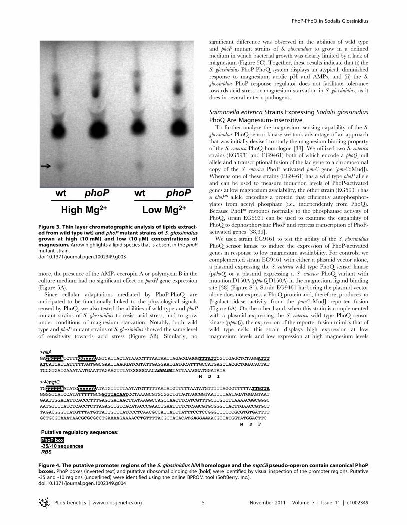
Sodalis glossinidius phoP Mutant Displays an Altered LipidComposition
Because PhoP-PhoQ often govern the expression of genes
involved in the structural modifications of the outer surface of the
bacterial lipid membrane [16,28] we sought to determine if the
genetic inactivation of phoP resulted in changes in the overall lipid
composition of S. glossinidius cells. We carried out a thin layer
chromatography (TLC) analysis of lipids derived from the wild
type and phoP mutant strains of S. glossinidius. Our TLC analysis
shows that the wild type S. glossinidius produces an additional (as yet
uncharacterized) lipid species that is absent in the phoP mutant
strain (Figure 3). This result reinforces the notion that S. glossinidius
also utilizes the PhoP-PhoQ system to regulate expression of genes
involved in lipid metabolism.
PhoP Also Regulates Type-III Secretion System Genes inSodalis glossinidius
Some PhoP regulated promoters have a characteristic direct
repeat sequence, known as a PhoP box, which serves as a binding
site for phosphorylated PhoP [29,30]. Inspection of the S.
glossinidius genome sequence revealed the presence of a consensus
PhoP box upstream of hilA (accession no. AAS66857; Figure 4),
which is known to encode a master regulator of type-III secretion
in Salmonella enterica [31]. Because the S. glossinidius genome has
three distinct symbiosis regions (designated Sodalis Symbiosis
Regions; SSR’s) encoding TTSS genes [5], we elected to measure
the effect of phoP inactivation on the basal expression levels of the
genes found in these three distinct chromosomal locations using
quantitative PCR. The phoP mutant was found to have
significantly lower levels of transcripts encoding YsaE, SycB
(SSR-1), HilA, InvF, SicA (SSR-2), SsrB, SsaB, SseB and SsaH
(SSR-3; Figure 2). These results indicate that the S. glossinidius
PhoP-PhoQ two-component system is also involved in the
activation of TTSS genes that are known to be required for the
invasion of insect cells and for intracellular survival [8].
Sodalis glossinidius phoP Mutant Fails to SuperinfectInsect Hosts
One important component of insect immunity involves the
synthesis and secretion of high quantities of AMPs into the
hemolymph [20]. Since insect endosymbionts, including S.
glossinidius, are often found in the hemolymph of their insect hosts
[32,33], and because S. glossinidius uses TTSSs to invade and
replicate within insect cells [6,8], we assessed the ability of the phoP
mutant strain of S. glossinidius to superinfect their natural tsetse fly
host, Glossina morsitans morsitans, and a closely-related hippoboscid
louse fly, Pseudolynchia canariensis, following intrathoracic microin-
jection. The superinfection approach [34] was selected to avoid
the requirement to treat host insects with antibiotics to remove
native symbionts, because this procedure is known to compromise
the immune system of the fly [35]. The presence of native S.
glossinidius in the tsetse fly therefore mandated the use of a
recombinant (fliM mutant) S. glossinidius strain in lieu of a wild type
control strain so that microinjected bacteria could be differentiated
from native symbionts by PCR. In comparison to the fliM mutant
strain, the phoP mutant strain was found to be completely defective
in its ability to superinfect the host insect (Table 1). At seven days
following microinjection, we were able to detect the fliM mutant
strain in 26 out of 32 tsetse flies sampled. In contrast, we were
unable to detect either the phoP or fliM heat-killed mutant strains
in any of the tsetse flies sampled at seven days post-microinjection
(Table 1). In the hippoboscid louse flies, which are closely related
to tsetse flies [36] but do not maintain a native population of S.
glossinidius, we compared the superinfection abilities of wild type,
fliM and phoP mutant strains of S. glossinidius. In this experiment,
wild type and fliM mutant S. glossinidius were detected in all flies
sampled at seven days following microinjection, and the phoP
mutant and heat-killed strains of S. glossinidius were not detected in
any flies at seven days post injection (Table 1). Based on these
results, we conclude that the phoP mutant strain of S. glossinidius is
killed by the insect immune system following superinfection in
both tsetse flies and hippoboscid louse flies. This indicates that the
PhoP-PhoQ two-component regulatory system is essential for the
Figure 1. Resistance to polymyxin B and cecropin A is PhoP-dependent in S. glossinidius. Whereas magnesium has no effect on S.glossinidius wild type (wt) resistance to cecropin A, it has a small but significant, PhoP-independent effect on resistance to polymyxin B (two-tail t-test:p,0.01). Wild type and phoP mutant cells were cultured at high (10 mM) and low (10 mM) levels of magnesium, exposed to antimicrobial peptidesand enumerated after plating. Error bars represent standard deviations.doi:10.1371/journal.pgen.1002349.g001
PhoP-PhoQ in Sodalis Glossinidius
PLoS Genetics | www.plosgenetics.org 3 November 2011 | Volume 7 | Issue 11 | e1002349

establishment and maintenance of an S. glossinidius infection in an
insect host.
Effects of Acidic pH, Magnesium, and Cationic AMPs onSodalis glossinidius
In many facultative bacterial pathogens, the PhoP-PhoQ system
functions as a magnesium sensor that controls the expression of
genes mediating physiological adaptations to changes in environ-
mental levels of magnesium [13-15]. In addition, the PhoQ sensor
kinase is known to detect and respond to conditions of acidic pH
[18], and the binding of cationic AMPs that displace magnesium
ions [17]. Several lines of evidence indicate that the S. glossinidius
PhoP-PhoQ system has a diminished ability to respond to
environmental magnesium. First, in S. enterica AMP resistance is
controlled by the PhoP-PhoQ two-component system. Salmonella
enterica cells grown under conditions of high magnesium availability
are .1000-fold more susceptible to AMPs [37]. In contrast, our
AMP resistance assay showed that although PhoP-PhoQ controls
AMP resistance in S. glossinidius, this phenotype is only marginally
affected by magnesium availability in the culture medium
(Figure 1). Second, our gene expression analyses show that PhoP
activates the transcription of pmrH and a number of TTSS genes in
S. glossinidius. However, while the expression of some of these genes
is considerably higher in wild type S. glossinidius relative to the phoP
mutant strain, transcriptional changes in response to environmen-
tal magnesium were unexpectedly small. Finally, the results from
our TLC lipid analysis showed that, regardless of magnesium
concentration, wild type S. glossinidius synthesizes a lipid species
that is not found in the phoP mutant strain (Figure 3).
To further explore the role of magnesium, acidic pH and the
presence of AMPs on signaling mediated by the S. glossinidius
PhoQ, we first used quantitative PCR to measure changes in the
numbers of transcripts of pmrH in wild type cells under different
culture conditions. The results show that pmrH expression was only
slightly increased under conditions of low magnesium availability
or low pH (2-fold and 2.6-fold increases, respectively). Further-
Figure 2. Quantitative PCR analysis of transcripts derived from genes involved in lipid A modifications in S. glossinidius. Geneticorganization of lipid A modification genes is depicted: Functional genes are decorated in gray and putative transcriptional units are indicated byarrows. The numbers in boxes indicate the ratios of transcripts detected in the wild type strain relative to the phoP mutant strain of S. glossinidiusgrown under conditions of high (10 mM) and low (10 mM) magnesium availability. Genes displaying significant changes in expression in the wild typerelative to the phoP mutant strain of S. glossinidius are highlighted with asterisks (two-tail t-test: p,0.05). Values in parentheses represent standarddeviations.doi:10.1371/journal.pgen.1002349.g002
PhoP-PhoQ in Sodalis Glossinidius
PLoS Genetics | www.plosgenetics.org 4 November 2011 | Volume 7 | Issue 11 | e1002349
more, the presence of the AMPs cecropin A or polymyxin B in the
culture medium had no significant effect on pmrH gene expression
(Figure 5A).
Since cellular adaptations mediated by PhoP-PhoQ are
anticipated to be functionally linked to the physiological signals
sensed by PhoQ, we also tested the abilities of wild type and phoP
mutant strains of S. glossinidius to resist acid stress, and to grow
under conditions of magnesium starvation. Notably, both wild
type and phoP mutant strains of S. glossinidius showed the same level
of sensitivity towards acid stress (Figure 5B). Similarly, no
significant difference was observed in the abilities of wild type
and phoP mutant strains of S. glossinidius to grow in a defined
medium in which bacterial growth was clearly limited by a lack of
magnesium (Figure 5C). Together, these results indicate that (i) the
S. glossinidius PhoP-PhoQ system displays an atypical, diminished
response to magnesium, acidic pH and AMPs, and (ii) the S.
glossinidius PhoP response regulator does not facilitate tolerance
towards acid stress or magnesium starvation in S. glossinidius, as it
does in several enteric pathogens.
Salmonella enterica Strains Expressing Sodalis glossinidiusPhoQ Are Magnesium-Insensitive
To further analyze the magnesium sensing capability of the S.
glossinidius PhoQ sensor kinase we took advantage of an approach
that was initially devised to study the magnesium binding property
of the S. enterica PhoQ homologue [38]. We utilized two S. enterica
strains (EG5931 and EG9461) both of which encode a phoQ null
allele and a transcriptional fusion of the lac gene to a chromosomal
copy of the S. enterica PhoP activated pmrC gene (pmrC::MudJ).
Whereas one of these strains (EG9461) has a wild type phoP allele
and can be used to measure induction levels of PhoP-activated
genes at low magnesium availability, the other strain (EG5931) has
a phoP* allele encoding a protein that efficiently autophosphor-
ylates from acetyl phosphate (i.e., independently from PhoQ).
Because PhoP* responds normally to the phosphatase activity of
PhoQ, strain EG5931 can be used to examine the capability of
PhoQ to dephosphorylate PhoP and repress transcription of PhoP-
activated genes [38,39].
We used strain EG9461 to test the ability of the S. glossinidius
PhoQ sensor kinase to induce the expression of PhoP-activated
genes in response to low magnesium availability. For controls, we
complemented strain EG9461 with either a plasmid vector alone,
a plasmid expressing the S. enterica wild type PhoQ sensor kinase
(pphoQ) or a plasmid expressing a S. enterica PhoQ variant with
mutation D150A (pphoQ D150A) in the magnesium ligand-binding
site [38] (Figure S1). Strain EG9461 harboring the plasmid vector
alone does not express a PhoQ protein and, therefore, produces no
b-galactosidase activity from the pmrC::MudJ reporter fusion
(Figure 6A). On the other hand, when this strain is complemented
with a plasmid expressing the S. enterica wild type PhoQ sensor
kinase (pphoQ), the expression of the reporter fusion mimics that of
wild type cells; this strain displays high expression at low
magnesium levels and low expression at high magnesium levels
Figure 3. Thin layer chromatographic analysis of lipids extract-ed from wild type (wt) and phoP mutant strains of S. glossinidiusgrown at high (10 mM) and low (10 mM) concentrations ofmagnesium. Arrow highlights a lipid species that is absent in the phoPmutant strain.doi:10.1371/journal.pgen.1002349.g003
Figure 4. The putative promoter regions of the S. glossinidius hilA homologue and the mgtCB pseudo-operon contain canonical PhoPboxes. PhoP boxes (inverted text) and putative ribosomal binding site (bold) were identified by visual inspection of the promoter regions. Putative-35 and -10 regions (underlined) were identified using the online BPROM tool (SoftBerry, Inc.).doi:10.1371/journal.pgen.1002349.g004
PhoP-PhoQ in Sodalis Glossinidius
PLoS Genetics | www.plosgenetics.org 5 November 2011 | Volume 7 | Issue 11 | e1002349

(Figure 6A). When complemented with a plasmid expressing the S.
enterica PhoQ variant with mutation D150A (pphoQ D150A), strain
EG9461 is derepressed under conditions of high magnesium
availability (Figure 6A) [38]. In addition, when strain EG9461 is
complemented with a plasmid expressing the S. glossinidius PhoQ
protein (pphoQ Sg) there is an increase in b-galactosidase activity of
the reporter fusion relative to that of strain EG9461 harboring the
plasmid vector alone (Figure 6A), indicating that S. glossinidius
PhoQ does phosphorylate the S. enterica PhoP response regulator.
However, consistent with our previous observation, the resulting
strain shows only a slight change in reporter gene activity in
response to magnesium (Figure 6A).
Similar results were obtained in an experiment involving S.
enterica strain EG5931, which expresses an autophosphorylating
phoP* allele. In this case strain EG5931, harboring the plasmid
vector alone, displayed high levels of b-galactosidase activity from
the reporter fusion, regardless of magnesium availability
(Figure 6B). Yet, because the phoP* allele responds normally to
the phosphatase activity of PhoQ, complementation of strain
EG5931 with a plasmid expressing the S. enterica wild type PhoQ
(pphoQ) produced a strain that displayed a gene expression profile
mimicking that of wild type cells. As with strain EG9461,
complementation of strain EG5931 with a plasmid expressing
the S. enterica PhoQ variant with mutation D150A (pphoQ D150A)
gave rise to a strain that had a reduced capability to promote
magnesium mediated repression of the pmrC::MudJ reporter fusion
(Figure 6B) [38]. In addition, when strain EG5931 was
complemented with a plasmid expressing the S. glossinidius wild
type PhoQ protein (pphoQ Sg), the resulting strain again
demonstrated little or no response to magnesium. Furthermore,
this strain demonstrated decreased b-galactosidase activity of the
reporter fusion relative to that of strain EG5931 harboring the
plasmid vector alone (Figure 6B), indicating that the S. glossinidius
wild type PhoQ sensor kinase does maintain a phosphatase
activity. Together, these results further support our observation
that the S. glossinidius PhoQ has a substantially diminished ability to
sense magnesium.
Evidence for Magnesium Sensing in an AncestralPrecursor of the S. glossinidius PhoQ
Given that the majority of PhoP-PhoQ homologues studied in
bacteria are known to respond aggressively to changes in acidic
pH, magnesium and cationic AMPs [13,14,17,18], it is striking
that the S. glossinidius PhoQ sensor kinase displays a substantially
reduced ability to respond to these signals. In many facultative
pathogens, PhoP-PhoQ is known to play an important role in
mediating magnesium homeostasis [13,14]. When PhoQ senses
conditions of low magnesium availability, PhoP responds by
increasing expression of specialized, ATP-driven magnesium
transporters (designated MgtA and MgtB in S. enterica)
[14,40,41]. While S. glossinidius maintains intact copies of the
generalized magnesium transporters corA (locus tag SG2341) and
mgtE (locus tags SG0628 and SG1738) that are not regulated by
PhoP-PhoQ in other bacteria, it completely lacks mgtA and
maintains only a disrupted copy of mgtB with multiple frameshifts
(Table 2). However, the disrupted copy of mgtB, encoded within
the mgtCB pseudo-polycistron maintains a canonical PhoP box
(Figure 4), implying that in the recent evolutionary past S.
glossinidius used PhoP-PhoQ to coordinate the expression of genes
involved in magnesium transport. Since it would be unexpected to
have a magnesium transport system controlled by a PhoP-PhoQ
system that is unable to sense magnesium, we conclude that an
ancestral precursor of the S. glossinidius PhoQ protein possessed an
increased magnesium sensing capability. This implies that the
reduction in S. glossinidius PhoQ’s ability to sense magnesium is a
derived characteristic.
Discussion
The transition in lifestyle from opportunism to obligate host
association is often accompanied by the inactivation and loss of
genes that are assumed to have played an important role in a
facultative lifestyle but no longer provide an adaptive benefit in an
obligate host-associated lifestyle [1]. While these degenerative
changes are not expected to negatively impact the function of the
symbiotic relationship, they are anticipated to increase host
dependence as a result of niche specialization on the part of the
symbiont. Furthermore, the transition from a dynamic lifestyle to a
static, obligate host-associated lifestyle is expected to reduce the
requirement for symbionts to engage in regulatory activities that
normally enhance the ability of bacteria to survive in a changing
environment.
In the current study we explored the role of the two-component
regulatory system, PhoP-PhoQ, in the insect endosymbiont, S.
glossinidius, which is an obligate, mutualistic associate of tsetse flies
[42]. PhoP-PhoQ is of particular interest because it is found in a
wide range of facultative pathogens [13,14,16] and plays an
important role in enabling these bacteria to sense their presence in
the host environment and mediate changes in gene expression that
facilitate important adaptations to the host-associated lifestyle. Our
results show that PhoP-PhoQ also plays an essential role in the
maintenance of the mutualistic association between S. glossinidius
and its insect host. In the absence of phoP, S. glossinidius is highly
sensitive to the bactericidal effects of AMPs in vitro and is incapable
of superinfecting either its natural host or a closely related
hippoboscid louse fly.
In S. enterica and other facultative pathogens, the sensor kinase
PhoQ plays an important role in the sensing of magnesium, AMPs
and acidic pH. Thus, the most striking result to emerge from the
current study is that the PhoP-PhoQ system of S. glossinidius differs
from its counterparts in other bacteria by having a substantially
reduced ability to sense these environmental cues. For example,
the S. glossinidius PhoP-PhoQ system elicited only a minor
reduction (c. 2-fold) in the expression of target genes in response
to a 1000-fold increase in the level of magnesium in the culture
medium. Similarly, the S. glossinidius PhoP-PhoQ system was found
to be largely unresponsive to a change in pH; S. glossinidius cells
cultured under acidic conditions showed only a 2.6-fold increase in
the expression of a PhoP-regulated target gene relative to cells
Table 1. PCR detection of Sodalis glossinidius seven daysfollowing microinjection in tsetse flies and louse flies.
S. glossinidius strain
Number of PCR positivesamples/number of total samples
Tsetse flies Louse flies
Wild-type N.D. 19/20
Wild-type heat-killed N.D. 0/20
fliM 26/32 16/16
fliM heat-killed 0/32 N.D.
phoP 0/32 0/20
Differences in colonization patterns between the wild-type, fliM and phoPstrains are statistically significant (Pearson X2 test, p,0.0001). ‘‘N.D.’’ indicatesthat that a particular experiment was not performed.doi:10.1371/journal.pgen.1002349.t001
PhoP-PhoQ in Sodalis Glossinidius
PLoS Genetics | www.plosgenetics.org 6 November 2011 | Volume 7 | Issue 11 | e1002349

cultured at neutral pH. In S. enterica, the constitutive expression of
PhoP-activated genes is known to have a deleterious impact on
bacterial virulence and survival in host tissues [43], due to the fact
that the initial activation of PhoP induces a transcriptional surge
that enables S. enterica to rapidly initiate virulence gene expression
[44]. In the current study, we were unable to identify any
conditions under which the S. glossinidius PhoP-PhoQ system was
effectively repressed. In vitro, S. glossinidius was found to display high
levels of resistance to AMPs under all conditions tested, indicating
that the dynamics of PhoP-based gene regulation in S. glossinidius
are locked in a constitutively active state, at least with respect to
signaling mediated by magnesium, AMPs and pH change.
The presence of a canonical PhoP binding site in the promoter
sequence of an inactivated mgtB allele indicates that in the recent
evolutionary past, S. glossinidius used a magnesium-sensing PhoP-
PhoQ to control the expression of a high affinity magnesium
transporter. The proposed reduction in PhoQ’s ability to sense
magnesium (and other signals) can be rationalized in several ways.
First, if the primary mandate of PhoQ’s magnesium sensing
capability was to control magnesium uptake, then the inactivation
of the gene encoding this magnesium transporter could have
facilitated relaxed selection on the ability of PhoQ to sense
magnesium. Second, since magnesium binding drives the
repression of PhoP-regulated target genes, the loss of a
requirement to repress these genes, perhaps resulting from a
switch to a static host-associated lifestyle, could also have
facilitated relaxed selection on PhoQ’s sensing capabilities. Third,
it is conceivable that the loss of PhoQ’s ability to sense magnesium
and acidic pH provided an adaptive advantage in S. glossinidius’
current lifestyle. Like many recently derived insect endosymbionts,
S. glossinidius inhabits the hemolymph of its insect host [32] where it
is anticipated to be exposed simultaneously to high concentrations
of both AMPs [20,25,26] and magnesium [12,45], at neutral pH
[46]. In this environment, the magnesium- or pH-mediated
repression of PhoP-regulated genes would likely be deleterious due
to the fact that it would yield an AMP sensitive phenotype that is
incompatible with survival in the AMP-rich hemolymph. Con-
versely, a magnesium-insensitive PhoQ would be expected to
facilitate the constitutive activation of PhoP-regulated target genes,
ensuring resistance towards AMPs in magnesium-rich hemo-
lymph.
Since the mechanism of magnesium binding by the S. enterica
PhoQ homologue has been well characterized through structural
and functional studies [38,47], we inspected the sequence of the S.
glossinidius PhoQ protein to determine if there are any obvious
modifications that can explain it’s reduced sensitivity to magne-
sium. Notably, the S. glossinidius PhoQ sequence maintains several
amino acid substitutions that replace key acidic residues in a
location corresponding to a magnesium ligand-binding site in the
PhoQ protein of S. enterica (Figure S1) [38,47]. In S. enterica, mutant
strains harboring PhoQ sequences lacking just one of these key
acidic residues demonstrate .5-fold reduction in their ability to
repress the transcription of PhoP-activated genes in response to
magnesium (pphoQ D150A in Figure S1 and [38]). Thus it is
conceivable that the loss of three key acidic residues in the S.
glossinidius PhoQ substantially reduced the ability of this sensor
kinase to bind magnesium, explaining the reduction in the
magnesium dependent PhoQ transcriptional repression of PhoP-
activated genes observed in S. glossinidius. An alignment of PhoQ
sequences derived from a selected range of gamma Proteobacteria
shows that PhoQ homologues from two insect endosymbionts (S.
glossinidius and Arsenophonus nasoniae) along with the soft-rot plant
pathogen Dickeya dadantii (formerly Erwinia chrysanthemi) each have
putative magnesium-binding patches that lack the requisite acidic
Figure 5. Response of S. glossinidius to antimicrobial peptides,acidic pH, or magnesium. A. Quantitative PCR analysis of pmrHtranscripts derived from S. glossinidius cells grown under variousmedium conditions. Fold increase in pmrH transcripts was calculatedrelative to cells grown in defined medium containing 10 mM ofmagnesium at pH 7. B. Percent survival of S. glossinidius wild type andphoP mutant cells at various pH values. C. Growth dynamics of S.glossinidius wild type (&) and phoP mutant (6) in defined mediumcontaining high (10 mM) and low (10 mM) concentrations of magne-sium. Error bars represent standard deviations.doi:10.1371/journal.pgen.1002349.g005
PhoP-PhoQ in Sodalis Glossinidius
PLoS Genetics | www.plosgenetics.org 7 November 2011 | Volume 7 | Issue 11 | e1002349

residues and are therefore predicted to be compromised in terms
of magnesium-sensing (Figure S1A). While mutations in this
conserved region likely contribute to a decreased magnesium
sensing capability, it is possible that changes in other regions of
PhoQ also contribute to this phenotype. Indeed, we noticed that
the S. glossinidius PhoQ homologue also maintains a hydrophobic
phenylalanine residue located at position 121, instead of a charged
histidine residue that is found in the S. enterica PhoQ (Figure S1A).
In S. enterica, this charged residue has also been shown to be
important for magnesium binding and magnesium mediated
repression [38].
Although magnesium sensing is recognized as the primary
function of PhoQ [14,15], we cannot rule out the possibility that
the S. glossinidius PhoQ has evolved the ability to sense a novel
signal in the insect host. For example, it has recently been shown
that the PhoQ protein of Edwardsiella tarda has evolved to detect
changes in temperature, in addition to magnesium, to coordinate
the expression of protein secretion systems that play an important
role in virulence [48]. However, it is clear that the S. glossinidius
PhoQ has lost the ability to sense key signals that play an
important role in the functionality of PhoP-PhoQ in many
pathogens. The adaptive benefit of PhoP-PhoQ in S. glossinidius
now appears to be solely due to the ability of this regulatory system
to serve as a constitutive driver for the expression of genes that
have critical functions in the symbiosis. To this end, the
modification or loss of sensory functions in a two-component
system is not necessarily paradoxical, as long as an adaptive benefit
is realized as a result of the output of the response regulator. Of
course a similar functional outcome could be achieved by
modulating the promoter sequences of all PhoP-regulated genes
to achieve constitutive expression in the absence of PhoP.
Although more evolutionary steps are needed to achieve this
outcome, it might ultimately be favored by natural selection
because it represents a more frugal solution. Indeed, such a
transition might already be underway in S. glossinidius, evidenced
by the fact that the lipid A modification genes, pagP and pmrE, are
expressed independently of PhoP, whereas PhoP is required for the
expression of these genes in S. enterica [27].
In a wider evolutionary context, it is interesting to note that
intact homologues of phoP-phoQ and the lipid modification genes
are only present in the genome sequences of recently established
insect endosymbionts (Table 2). This suggests that the functions of
PhoP-PhoQ and the lipid A modification genes are required only
as a stopgap in the early stages of symbiotic interactions, to enable
bacteria to resist attack from the host immune system to facilitate
the establishment and maintenance of persistent infections in host
tissues. This is supported by the results of our superinfection
experiments, which show that a phoP mutant strain of S. glossinidius
is unable to establish infection in an insect host. Taken together,
these observations suggest that recently established insect endo-
symbionts have an intrinsic ability to overcome the insect immune
system and establish persistent infections in insects, and this may
help to explain the broad distribution of certain recently
established insect endosymbionts (e.g. relatives of S. glossinidius
and Arsenophonus spp.) in distantly related host insects [10,21,42,49–
51]. It also explains differences observed in patterns of co-
Figure 6. Salmonella enterica strains expressing S. glossinidius PhoQ do not respond to magnesium. A. b-galactosidase activity (top) of theS. enterica PhoP-activated pmrC gene produced by strain EG9461 (phoQ::Tn10 pmrC:: MudJ) harboring the plasmid pUHE21-2 lacIq (vector) or pUHE21-2 lacIq derivatives expressing the S. enterica PhoQ (pphoQ), S. enterica PhoQ variant with mutation D150A (pphoQ D150A) or S. glossinidius PhoQ(pphoQ Sg). Fold induction (bottom) of pmrC::MudJ b-galactosidase activity produced by strain EG9461 harboring various plasmid constructs inresponse to magnesium. B. b-galactosidase activity (top) of the S. enterica PhoP-activated pmrC gene produced by strain EG5931 (phoP* phoQ::Tn10pmrC:: MudJ) harboring various plasmid constructs. EG5931 encodes a phoP allele (phoP*) that efficiently phosphorylates from acetyl phosphate (i.e.,independently of PhoQ) [39]. Fold repression (bottom) of pmrC::MudJ b-galactosidase activity produced by strain EG5931 harboring various plasmidconstructs in response to magnesium. Error bars represent standard deviations.doi:10.1371/journal.pgen.1002349.g006
PhoP-PhoQ in Sodalis Glossinidius
PLoS Genetics | www.plosgenetics.org 8 November 2011 | Volume 7 | Issue 11 | e1002349

evolution between insects and endosymbionts of different inferred
ages. While the phylogenies of insects and their ancient
endosymbionts demonstrate high levels of congruence, implying
little or no ongoing horizontal symbiont transmission or novel
colonization events, the phylogenies of insects and their recently
established endosymbionts are often discordant [1].
At a broad level, the results obtained in the current study
reinforce the notion that the molecular mechanisms facilitating
host-symbiont interactions have a common origin in both
pathogenic and mutualistic associations. More specifically, our
findings illustrate the capability of a complex regulatory circuit to
adapt to a change in lifestyle. Furthermore, the degeneration of the
sensing capability of PhoQ may represent a snapshot of a wider
picture of regulatory simplification that is concomitant with a
transition to a static, mutualistic, host-associated lifestyle that
reduces the requirement for bacteria to engage in environmental
sensing.
Materials and Methods
Bacterial Strains and Culture ConditionsEscherichia coli W25113 was maintained in Luria-Bertani (LB)
medium at 37uC, or 30uC when harboring the repA101 (ts)
plasmid pKD46 [52]. Salmonella enterica strains were maintained at
37uC in either LB medium or N-minimal medium [38] containing
either 10 mM or 10 mM MgCl2. Sodalis glossinidius strains were
maintained at 25uC in the semi-defined liquid Mitsuhashi-
Maramorosch (MM) medium as described previously [42] or in
a defined medium composed of 6 g/l casamino acids, 4 g/l
glucose, 0.2 g/l KCl, 7.0 g/l NaCl, 0.12 g/l NaHCO3, 0.18 g/l
NaH2PO3, 10 mg/ml of thiamine, 10 mM or 10 mM of CaCl2,
and 10 mM or 10 mM MgCl2, pH 7. When rapidly dividing
symbiont cultures were needed, liquid cultures of S. glossinidius
were maintained in an orbital shaker at 200 rpm. For isolation of
symbiont clones, bacteria were plated on 1% MM agar plates,
supplemented with 5% defibrinated horse blood, and incubated at
25uC under microaerophilic conditions (5% Oxygen, 10% CO2,
balanced with N2). Where appropriate, antibiotics were added to
culture media at the following concentrations: 100 mg/ml (E. coli
and S. enterica) or 50 mg/ml (S. glossinidius) of ampicillin, 15 mg/ml
(chromosomal insertions) or 50 mg/ml (high copy number
plasmids) of chloramphenicol, 50 mg/ml of kanamycin, 10 mg/
ml tetracycline. When needed, 39-59-cyclic adenosine monophos-
phate (cAMP; Sigma Aldrich) was added to the culture medium at
a final concentration of 5 mM.
Construction of Replacement AllelesReplacement alleles for S. glossinidius were generated using the
lambda-Red recombineering system in E. coli [52]. The strategy
for construction of replacement alleles for S. glossinidius is
illustrated in Figure S2. Briefly, S. glossinidius genes were amplified
by polymerase chain reaction (PCR) using Phusion High-Fidelity
DNA polymerase (New England Biolabs). PCR products were
ligated into pCR-Blunt II-TOPO (Invitrogen) and transformed
into E. coli by electroporation. Clones were grown and plasmid
DNA was isolated using the QIAprep Spin Miniprep Kit
(Qiagen). Recombinant plasmids were then transformed into E.
coli (pKD46) and replacement alleles were generated by lambda-
Red mediated insertion of a chloramphenicol resistance cassette
(derived from pEpiFOS-5, Epicentre), according to the method
described previously [52]. Following curing of pKD46, plasmids
containing the replacement alleles were extracted, diluted and
introduced into E. coli by electroporation. The replacement alleles
were verified by DNA sequencing and plasmids containing theTa
ble
2.
Dis
trib
uti
on
of
ph
oP
-ph
oQ
,th
em
agn
esi
um
tran
spo
rte
rsm
gtA
and
mg
tB,
and
lipid
Am
od
ific
atio
ng
en
es
amo
ng
the
inse
ctp
ath
og
en
Ph
oto
rha
bd
us
lum
ines
cen
san
dre
cen
tly
de
rive
dan
dan
cie
nt
inse
cte
nd
osy
mb
ion
ts.
Org
an
ism
Cla
ssif
ica
tio
nG
en
om
esi
ze
(Mb
p)
Ag
eo
fA
sso
cia
tio
np
ho
Pp
ho
Qm
gtA
mg
tBL
ipid
Am
od
ific
ati
on
ge
ne
s
pag
Pp
mrE
pm
rFp
mrH
pm
rIp
mrJ
pm
rKp
mrL
pm
rM
Ph
oto
rha
bd
us
lum
ines
cen
sIn
sect
pat
ho
ge
n5
.68
N.
A.
+4
2P
++
++
++
++
+
Ars
eno
ph
on
us
na
son
iae
Inse
ctsy
mb
ion
t.
3.5
6U
nkn
ow
n+
22
2+
++
++
++
++
Sod
alis
glo
ssin
idiu
sIn
sect
sym
bio
nt
4.1
7U
nkn
ow
n+
22
P+
++
++
++
++
Ca
nd
ida
tus
Ham
ilto
ne
llad
efe
nsa
Inse
ctsy
mb
ion
t2
.10
Un
kno
wn
22
+2
2+
+2
++
++
+
Ca
nd
ida
tus
Serr
atia
sym
bio
tica
Inse
ctsy
mb
ion
t2
.57
Un
kno
wn
+1
22
2+
+2
22
++
2
Wig
gle
swo
rth
iag
loss
inid
iaIn
sect
sym
bio
nt
0.6
94
02
80
22
22
2+
22
++
+2
2
Blo
chm
an
nia
spp
.In
sect
sym
bio
nt
0.7
0–
0.7
95
02
22
2-
22
22
22
22
Ca
rso
nel
laru
dd
iiIn
sect
sym
bio
nt
0.1
61
20
22
22
22
22
22
22
2
Bu
chn
era
spp
.In
sect
sym
bio
nt
0.4
2–
0.6
51
50
22
22
22
22
22
22
2
Ba
um
an
nia
cica
del
linic
ola
Inse
ctsy
mb
ion
t0
.68
17
02
22
22
22
22
22
22
Esti
mat
ed
age
of
asso
ciat
ion
(in
mill
ion
of
year
s)is
sho
wn
for
anci
en
tin
sect
sym
bio
nts
[58
].‘‘P
’’in
dic
ate
sth
ata
giv
en
seq
ue
nce
was
de
term
ine
dto
be
ap
seu
do
ge
ne
.’’N
.A
.’’si
gn
ifie
sn
ot
app
licab
le.
Nu
mb
ers
of
acid
icre
sid
ue
sw
ith
inth
eac
idic
pat
char
ed
isp
laye
dfo
rP
ho
Qh
om
olo
gu
es.
No
teal
soth
atC
an
did
atu
sSe
rrat
iasy
mb
ioti
cam
ain
tain
sa
tru
nca
ted
Ph
oQ
ho
mo
log
ue
.d
oi:1
0.1
37
1/j
ou
rnal
.pg
en
.10
02
34
9.t
00
2
PhoP-PhoQ in Sodalis Glossinidius
PLoS Genetics | www.plosgenetics.org 9 November 2011 | Volume 7 | Issue 11 | e1002349

correct allele sequence were used as PCR templates for the
generation of linear DNA substrates for recombineering in S.
glossinidius.
Transformation of Sodalis glossinidiusAll S. glossinidius transformations (using both plasmid and
linear DNA constructs) were conducted using a heat-shock
method [53]. Following transformation, the cells were allowed
to recover overnight at 25 uC in liquid MM medium prior to
plating.
Lambda-Red Mediated Insertions in Sodalis glossinidiusLambda-Red mediated chromosomals insertions were gener-
ated as described previously [24]. Briefly, cultures of S. glossinidius
harboring pKD46 were grown without shaking to an OD600nm of
approximately 0.2. The cultures were then transferred to a
shaking incubator and grown overnight until OD600nm reached
0.5. The cells were pelleted at 5,000 6 g for 10 min at 4uC, and
washed twice with an equal volume of 0.85% (w/v) NaCl and
resuspended in a final volume of 1 ml of 0.85% (w/v) NaCl. The
cell suspensions were inoculated into 200 ml of fresh MM liquid
medium [42] supplemented with 0.5% (w/v) arabinose and
5 mM cAMP, and incubated at 25uC with shaking for 30 min to
facilitate induction of the lambda-Red functions. After induction,
the cells were made chemically competent and transformed with
linear DNA using the heat-shock method [53]. Following
overnight recovery, the cells were resuspended in 150 ml of
MM liquid medium. To assess cell viability following transfor-
mation, 15 ml of cell suspension was plated on MM blood agar
plates alone. To select for recombinant clones, the remaining
135 ml was spread on an MM blood agar plate containing
ampicillin and chloramphenicol. Plates were incubated under
microaerophilic conditions and inspected for growth after 9 days.
Putative recombinant clones were then isolated as single colonies
and the presence of chromosomal insertions was confirmed by
DNA sequencing.
Curing of Lambda-Red Plasmid from Sodalis glossinidiusFollowing lambda-Red recombineering, plasmid pKD46 was
cured from S. glossinidius by maintaining recombinant strains in the
absence of plasmid selection. Cultures were grown under these
conditions with shaking for approximately 50 generations prior to
passage into fresh medium. Cultures were passaged a total of five
times and then plated on MM blood agar plates supplemented
with chloramphenicol alone. After 7 days of growth, colonies were
screened for ampicillin sensitivity by replica plating onto MM
blood agar plates supplemented with chloramphenicol and
ampicillin.
Antimicrobial Peptide Resistance AssayAntimicrobial peptide resistance assays were performed using a
modified version of a previously described method [54]. Wild type
and phoP mutant strains of S. glossinidius were grown to mid-log
phase in MM liquid medium. 20 ml of each culture was
transferred to 50 ml tubes and the cells were harvested by
centrifugation at 5,000 6 g for 10 minutes at 4uC. The cells were
washed twice with an equal volume of 0.85% (w/v) NaCl and
resuspended in 1 ml of 0.85% (w/v) NaCl. 0.5 ml of each cell
suspension was inoculated into 20 ml of defined medium
containing either 10 mM or 10 mM MgCl2 and CaCl2. Following
an 8 h incubation (with shaking), the cells were harvested and
washed, and 1 ml of cell suspension was inoculated into a 50 ml
tube containing 9 ml of either 0.85% (w/v) NaCl alone, or 0.85%
(w/v) NaCl supplemented with polymyxin B or cecropin A at a
final concentration of 50 mg/ml. The cells were incubated at 25uCfor 10 minutes, diluted in 0.85% (w/v) NaCl and approximately
1,000 colony forming units (CFU) were plated on MM blood agar
plates. Plates were incubated for 5 days at 25uC under
microaerophilic conditions and the resulting colonies were
counted. All assays were carried out in triplicate.
Acid Resistance AssayWild type and phoP mutant strains of S. glossinidius were grown to
mid-log phase in MM liquid medium. The cells were harvested by
centrifugation at 5,000 6 g for 10 minutes at 4uC and washed
twice with an equal volume of 0.85% (w/v) NaCl. Approximately
26108 CFU were inoculated in 10 ml of MM medium at various
pH levels (7.0, 5.0, 4.5 and 4.0). Following 1 h incubation, the cells
were diluted in 0.85% (w/v) NaCl solution and approximately
1,000 CFU were plated on MM blood agar plates. Plates were
incubated for 5 days at 25uC under microaerophilic conditions
and the resulting colonies were counted. All assays were carried
out in triplicate.
Growth CurvesWild type and phoP mutant strains of S. glossinidius were grown to
mid-log phase in MM liquid medium. 5 ml of each culture was
harvested by centrifugation at 5,000 6 g for 10 minutes at 4uC.
The cells were then washed twice with an equal volume of defined
medium containing 10 mM MgCl2 and inoculated into 10 ml
aliquots of defined medium containing either 10 mM or 10 mM
MgCl2 and CaCl2. The cultures were maintained at 25uC, and
measurements of turbidity (OD600nm) were obtained at 24 h
intervals.
Salmonella enterica Complementation ExperimentsSalmonella enterica serovar Typhimurium EG9461 [27] and
EG5931 [39] harboring pUHE21-2 lacIq [55] or pUHE21-2 lacIq
derivatives were grown overnight in N-minimal medium contain-
ing 10 mM MgCl2. Overnight cultures were diluted 1:100 in fresh
N-minimal medium containing 10 mM MgCl2 and allowed to
grow for 4 h. The cells were harvested by centrifugation at
8,000 6g for 3 min at 4uC, washed twice with 0.85% (w/v) NaCl,
and resuspended in N-minimal medium containing either 10 mM
or 10 mM MgCl2. Following 3 h of growth, b-galactosidase activity
was determined as described by Miller [56].
Total Lipid Extraction and Thin Layer ChromatographyWild type and phoP mutant strains of S. glossinidius were grown to
mid-log phase in MM liquid medium. 200 ml of each culture was
harvested by centrifugation at 5,000 6 g for 10 minutes at 4uC.
The cells were then washed twice with an equal volume of 0.85%
(w/v) NaCl. After the second wash, the cells were harvested as
described above and resuspended in 5 ml of 0.85% (w/v) NaCl.
2.5 ml of each cell suspension was inoculated into 200 ml of
defined media containing either 10 mM or 10 mM MgCl2 and
CaCl2, and the cultures were incubated for 8 h with shaking.
Following incubation, the cells were harvested by centrifugation at
5,000 6g for 10 minutes at 4uC and washed with an equal volume
of 0.1% (w/v) ammonium acetate. The cell suspensions were
centrifuged as described above and resuspended at a final
concentration of 361011 CFU/ml. 200 ml of each cell suspension
was used for total lipid extraction in accordance to the Folch
method [57]. Lipid extracts were spotted on a 20620 cm C18 thin
layer chromatography plate (Whatman) and developed twice with
chloroform: methanol: water (60:30:5, by volume). After chroma-
PhoP-PhoQ in Sodalis Glossinidius
PLoS Genetics | www.plosgenetics.org 10 November 2011 | Volume 7 | Issue 11 | e1002349

tography, the plate was allowed to dry and lipids were visualized
by iodine staining.
RNA Isolation and Quantitative PCR AnalysesRNA was prepared using the SV Total RNA Isolation System
(Promega). Following RNA purification, sample aliquots were
treated with Turbo DNase (Ambion) to remove contaminating
DNA. RNA samples were then reverse transcribed using the
Maxima First Strand cDNA Sysnthesis kit (Fermentas). Quanti-
tation of cDNA was performed in triplicate using the Maxima
SYBR Green/ROX qPCR Master Mix (Fermentas). Samples
were analyzed using an iCycler iQ Multicolor Real-Time PCR
Detection System (Bio-Rad). Relative transcript levels were
estimated using the standard curve method, with expression levels
normalized against a gene encoding a ribosomal protein (RplB)
that is expressed constitutively in S. glossinidius [8]. A list of the
primer sets and respective target genes is presented in Table S1.
Microinjection of Sodalis glossinidius into Louse andTsetse Flies
Glossina morsitans morsitans Westwood tsetse flies were maintained
at the Institute of Tropical Medicine (Antwerp, Belgium) as
described previously [25]. Pseudolynchia canariensis hippoboscid louse
flies were maintained on a pigeon colony at the University of
Utah. Mid-log phase S. glossinidius cells were collected in a 1.5 ml
tube by centrifugation at 8,000 g for 2 min and resuspended in
PBS. Insects were injected into the ventral thorax with < 26104 CFU of the S. glossinidius phoP mutant strain, fliM mutant strain
[24], serving as a positive control for tsetse flies, or fliM and wild
type strain (serving as a positive controls for louse flies).
Microinjections were also performed with heat-killed wild type
and fliM mutant strains (80uC for 15 min) to serve as negative
controls. Insects were sacrificed for DNA isolation at various times
post-injection and the resulting DNA samples were screened for
the presence of bacteria using PCR primers specific for S.
glossinidius wild type and the chloramphenicol resistance markers of
the mutant strains.
Supporting Information
Figure S1 A. Sequence alignment of PhoQ homologues derived
from S. glossinidius and related Gammaproteobacteria. The box
highlights the PhoQ acidic cluster (acidic residues are shaded in
red) that is known to be involved in magnesium binding
[17,38,47]. The PhoQ homologues of the insect endosymbionts
S. glossinidius and A. nasoniae and the PhoQ homologue of the soft-
rot plant pathogen D. dadantii have accumulated non-acidic amino
acid substitutions within this cluster, suggesting that these proteins
have a reduced ability to bind to magnesium and mediate the
repression of PhoP-activated genes. Notably, the S. glossinidius
PhoQ homologue also has a hydrophobic phenylalanine (shaded
in blue) at a charged position (histidine 120) that is required for
magnesium binding and magnesium-mediated repression in the
PhoQ of S. enterica [38]. The alignment was generated using the
online MAFFT tool [59]. B. Ribbon representation of the
monomeric crystal structure of S. typhimurium PhoQ. Red-colored
side chains represent acidic residues and gray spheres represent
magnesium ions. Missing acidic residues in the S. glossinidius PhoQ
sequence (D149 to D151) are highlighted. The structural diagram
was generated using Pymol software (http://www.pymol.org/).
(TIF)
Figure S2 Schematic illustrating construction of replacement
alleles for S. glossinidius recombineering. The drug resistance
marker (chloramphenicol acetyltransferase, cat) was amplified
using PCR primers with 59-flanking sequences that match 35
base target sequences in the S. glossinidius chromosome. This PCR
product was then integrated into a plasmid borne copy of the
target gene, via lambda-Red mediated homologous recombination
[52]. After selection for integration, the plasmid was used as PCR
template for the synthesis of the replacement construct.
(TIF)
Table S1 Sequences of oligonucleotide primers used in quan-
titative PCR experiments.
(DOC)
Acknowledgments
We thank Serap Aksoy (Yale University), Eduardo Groisman (Yale
University), and Kelly Hughes (University of Utah) for the provision of
bacterial strains. We also thank David Blair (University of Utah) for
assistance with structural analysis of PhoQ and several anonymous
reviewers for useful suggestions.
Author Contributions
Conceived and designed the experiments: MHP KLS JVDA CD.
Performed the experiments: MHP KLS LVD JVDA CD. Analyzed the
data: MHP KLS CD. Contributed reagents/materials/analysis tools:
JVDA CD. Wrote the paper: MHP CD.
References
1. Dale C, Moran NA (2006) Molecular interactions between bacterial symbionts
and their hosts. Cell 126: 453–465.
2. Blattner FR, Plunkett G 3rd, Bloch CA, Perna NT, Burland V, et al. (1997) Thecomplete genome sequence of Escherichia coli K-12. Science 277: 1453–1474.
3. Chen XA, Li S, Aksoy S (1999) Concordant evolution of a symbiont with its hostinsect species: Molecular phylogeny of genus Glossina and its bacteriome-
associated endosymbiont, Wigglesworthia glossinidia. J Mol Evol 48: 49–58.
4. Thomson NR, Howard S, Wren BW, Holden MT, Crossman L, et al. (2006)The complete genome sequence and comparative genome analysis of the high
pathogenicity Yersinia enterocolitica strain 8081. PLoS Genet 2: e206. doi:10.1371/journal.pgen.0020206.
5. Toh H, Weiss BL, Perkin SA, Yamashita A, Oshima K, et al. (2006) Massive
genome erosion and functional adaptations provide insights into the symbioticlifestyle of Sodalis glossinidius in the tsetse host. Genome Res 16: 149–156.
6. Dale C, Young SA, Haydon DT, Welburn SC (2001) The insect endosymbiontSodalis glossinidius utilizes a type III secretion system for cell invasion. Proc Natl
Acad Sci U S A 98: 1883–1888.
7. Dale C, Plague GR, Wang B, Ochman H, Moran NA (2002) Type III secretionsystems and the evolution of mutualistic endosymbiosis. Proc Natl Acad Sci U S A
99: 12397–12402.
8. Dale C, Jones T, Pontes M (2005) Degenerative evolution and functional
diversification of type-III secretion systems in the insect endosymbiont Sodalis
glossinidius. Mol Biol Evol 22: 758–766.
9. Moran NA, Degnan PH, Santos SR, Dunbar HE, Ochman H (2005) The
players in a mutualistic symbiosis: insects, bacteria, viruses, and virulence genes.
Proc Natl Acad Sci U S A 102: 16919–16926.
10. Novakova E, Hypsa V (2007) A new Sodalis lineage from bloodsucking fly
Craterina melbae (Diptera, Hippoboscoidea) originated independently of the tsetseflies symbiont Sodalis glossinidius. FEMS Microbiol Lett 269: 131–135.
11. Degnan PH, Yu Y, Sisneros N, Wing RA, Moran NA (2009) Hamiltonella defensa,
genome evolution of protective bacterial endosymbiont from pathogenicancestors. Proc Natl Acad Sci U S A 106: 9063–9068.
12. Derzelle S, Turlin E, Duchaud E, Pages S, Kunst F, et al. (2004) The PhoP-PhoQ two-component regulatory system of Photorhabdus luminescens is essential for
virulence in insects. J Bacteriol 186: 1270–1279.
13. Groisman EA (2001) The pleiotropic two-component regulatory system PhoP-PhoQ. J Bacteriol 183: 1835–1842.
14. Perez JC, Shin D, Zwir I, Latifi T, Hadley TJ, et al. (2009) Evolution of abacterial regulon controlling virulence and Mg(2+) homeostasis. PLoS Genet 5:
e1000428. doi:10.1371/journal.pgen.1000428.
15. Groisman EA, Mouslim C (2006) Sensing by bacterial regulatory systems in hostand non-host environments. Nat Rev Microbiol 4: 705–709.
16. Mitrophanov AY, Jewett MW, Hadley TJ, Groisman EA (2008) Evolutionand dynamics of regulatory architectures controlling polymyxin B resistance
in enteric bacteria. PLoS Genet 4: e1000233. doi:10.1371/journal.pgen.
1000233.
PhoP-PhoQ in Sodalis Glossinidius
PLoS Genetics | www.plosgenetics.org 11 November 2011 | Volume 7 | Issue 11 | e1002349

17. Bader MW, Sanowar S, Daley ME, Schneider AR, Cho U, et al. (2005)
Recognition of antimicrobial peptides by a bacterial sensor kinase. Cell 122:461–472.
18. Prost LR, Daley ME, Le Sage V, Bader MW, Le Moual H, et al. (2007)
Activation of the bacterial sensor kinase PhoQ by acidic pH. Mol Cell 26:165–174.
19. Guanı-Guerra E, Santos-Mendoza T, Lugo-Reyes SO, Teran LM (2010)Antimicrobial peptides: general overview and clinical implications in human
health and disease. Clin Immunol 135: 1–11.
20. Lemaitre B, Hoffmann J (2007) The host defense of Drosophila melanogaster. AnnuRev Immunol 25: 697–743.
21. Dale C, Beeton M, Harbison C, Jones T, Pontes M (2006) Isolation, pureculture, and characterization of ‘‘Candidatus Arsenophonus arthropodicus,’’ an
intracellular secondary endosymbiont from the hippoboscid louse fly Pseudolyn-
chia canariensis. Appl Environ Microbiol 72: 2997–3004.
22. Haines LR, Hancock RE, Pearson TW (2003) Cationic antimicrobial peptide
killing of African trypanosomes and Sodalis glossinidius, a bacterial symbiont of theinsect vector of sleeping sickness. Vector Borne Zoonotic Dis 3: 175–186.
23. Hao Z, Kasumba I, Lehane MJ, Gibson WC, Kwon J, et al. (2001) Tsetseimmune responses and trypanosome transmission: implications for the
development of tsetse-based strategies to reduce trypanosomiasis. Proc Natl
Acad Sci U S A 98: 12648–12653.
24. Pontes MH, Dale C (2011) Lambda Red-mediated genetic modification of the
insect endosymbiont Sodalis glossinidius. Appl Environ Microbiol 77: 1918–1920.
25. Akoda K, Van den Bossche P, Marcotty T, Kubi C, Coosemans M, et al. (2009)
Nutritional stress affects the tsetse fly’s immune gene expression. Med Vet
Entomol 23: 195–201.
26. Boulanger N, Brun R, Ehret-Sabatier L, Kunz C, Bulet P (2002) Immunepep-
tides in the defense reactions of Glossina morsitans to bacterial and Trypanosoma
brucei brucei infections. Biochem Mol Biol 32: 369–375.
27. Shi Y, Cromie MJ, Hsu FF, Turk J, Groisman EA (2004) PhoP-regulated
Salmonella resistance to the antimicrobial peptides magainin 2 and polymyxinB. Mol Microbiol 53: 229–241.
28. Gunn JS (2008) The Salmonella PmrAB regulon: lipopolysaccharide modifica-tions, antimicrobial peptide resistance and more. Trends Microbiol 16: 284–290.
29. Minagawa S, Ogasawara H, Kato A, Yamamoto K, Eguchi Y, et al. (2003)
Identification and molecular characterization of the Mg2+ stimulon of Escherichia
coli. J Bacteriol 185: 3696–3702.
30. Zwir I, Shin D, Kato A, Nishino K, Latifi T, et al. (2005) Dissecting the PhoPregulatory network of Escherichia coli and Salmonella enterica. Proc Natl Acad Sci
USA 102: 2862–2867.
31. Bajaj V, Lucas RL, Hwang C, Lee CA (1996) Co-ordinate regulation ofSalmonella typhimurium invasion genes by environmental and regulatory factors is
mediated by control of hilA expression. Mol Microbiol 22: 703–714.
32. Cheng Q, Aksoy S (1999) Tissue tropism, transmission and expression of foreign
genes in vivo in midgut symbionts of tsetse flies. Insect Mol Biol 8: 125–132.
33. Pontes MH, Dale C (2006) Culture and manipulation of insect facultative
symbionts. Trends Microbiol 14: 406–412.
34. Weiss BL, Wu Y, Schwank JJ, Tolwinski NS, Aksoy S (2008) An insect symbiosisis influenced by bacterium-specific polymorphisms in outer-membrane protein
A. Proc Natl Acad Sci USA 105: 15088–15093.
35. Weiss BL, Wang J, Aksoy S (2011) Tsetse immune system maturation requires
the presence of obligate symbionts in larvae. PLoS Biol 9: e1000619.
doi:10.1371/journal.pbio.1000619.
36. Petersen FT, Meier R, Kutty SN, Wiegmann BM (2007) The phylogeny and
evolution of host choice in the Hippoboscoidea (Diptera) as reconstructed usingfour molecular markers. Mol Phylogenet Evol 45: 111–122.
37. Garcıa Vescovi E, Soncini FC, Groisman EA (1996) Mg2+ as an extracellular
signal: environmental regulation of Salmonella virulence. Cell 84: 165–174.
38. Chamnongpol S, Cromie M, Groisman EA (2003) Mg2+ sensing by the Mg2+
sensor PhoQ of Salmonella enterica. J Mol Biol 325: 795–807.39. Chamnongpol S, Groisman EA (2000) Acetyl phosphate-dependent activation of
a mutant PhoP response regulator that functions independently of its cognate
sensor kinase. J Mol Biol 300: 291–305.40. Snavely MD, Miller CG, Maguire ME (1991) The mgtB Mg2+ transport locus of
Salmonella typhimurium encodes a P-type ATPase. J Biol Chem 266: 815–823.41. Tao T, Snavely MD, Farr SG, Maguire ME (1995) Magnesium transport in
Salmonella typhimurium: mgtA encodes a P-type ATPase and is regulated by Mg2+
in a manner similar to that of the mgtB P-type ATPase. J Bacteriol 177:2654–2662.
42. Dale C, Maudlin I (1999) Sodalis gen. nov. and Sodalis glossinidius sp. nov., amicroaerophilic secondary endosymbiont of the tsetse fly Glossina morsitans
morsitans. Int J Syst Bacteriol 49: 267–275.43. Miller SI, Mekalanos JJ (1990) Constitutive expression of the phoP regulon
attenuates Salmonella virulence and survival within macrophages. J Bacteriol.
172: 2485–2490.44. Shin D, Lee EJ, Huang H, Groisman EA (2006) A positive feedback loop
promotes transcription surge that jump-starts Salmonella virulence circuit.Science 314: 1607–1609.
45. Natochin YV, Parnova RG (1987) Osmolality and electrolyte concentration of
hemolymph and the problem of ion and volume regulation of cells in higherinsects. Comp Biochem Physiol A 88: 563–570.
46. Petit JP (1968) Hemolymph of Glossina: collection and analysis. Rev Elev MedVet Pays Trop 21: 493–498.
47. Cho US, Bader MW, Amaya MF, Daley ME, Klevit RE, et al. (2006) Metalbridges between the PhoQ sensor domain and the membrane regulate
transmembrane signaling. J Mol Biol 356: 1193–1206.
48. Chakraborty S, Li M, Chatterjee C, Sivaraman J, Leung KY, et al. (2010)Temperature and Mg2+ sensing by a novel PhoP-PhoQ two-component system
for regulation of virulence in Edwardsiella tarda. J Biol Chem 285: 38876–38888.49. Fukatsu T, Koga R, Smith WA, Tanaka K, Nikoh N, et al. (2007) Bacterial
endosymbiont of the slender pigeon louse, Columbicola columbae, allied to
endosymbionts of grain weevils and tsetse flies. Appl Environ Microbiol 73:6660–6668.
50. Kaiwa N, Hosokawa T, Kikuchi Y, Nikoh N, Meng XY, et al. (2010) Primarygut symbiont and secondary, Sodalis-allied symbiont of the Scutellerid stinkbug
Cantao ocellatus. Appl Environ Microbiol 76: 3486–3494.51. Novakova E, Hypsa V, Moran NA (2009) Arsenophonus, an emerging clade of
intracellular symbionts with a broad host distribution. BMC Microbiol 9: 143.
52. Datsenko KA, Wanner BL (2000) One-step inactivation of chromosomal genesin Escherichia coli K-12 using PCR products. Proc Natl Acad Sci U S A 97:
6640–6645.53. Inoue H, Nojima H, Okayama H (1990) High efficiency transformation of
Escherichia coli with plasmids. Gene 96: 23–28.
54. Groisman EA, Heffron F, Solomon F (1992) Molecular genetic analysis of theEscherichia coli phoP locus. J Bacteriol 174: 486–491.
55. Soncini FC, Vescovi EG, Groisman EA (1995) Transcriptional autoregulation ofthe Salmonella typhimurium phoPQ operon. J Bacteriol 177: 4364–4371.
56. Miller JH (1992) A short course in bacterial genetics: a laboratory manual andhandbook for Escherichia coli and related bacteria. New York: Cold Spring
Harbor Laboratory Press. pp 456.
57. Matyash V, Liebisch G, Kurzchalia TV, Shevchenko A, Schwudke D (2008)Lipid extraction by methyl-tert-butyl ether for high-throughput lipidomics.
J Lipid Res 49: 1137–1146.58. Gosalbes MJ, Latorre A, Lamelas A, Moya A (2010) Genomics of intracellular
symbionts in insects. Int J Med Microbiol 300: 271–278.
59. Katoh, K, Misawa, K, Kuma, K, Miyata, T (2002) MAFFT: a novel method forrapid multiple sequence alignment based on fast Fourier transform. Nucleic Acid
Res 30: 3059–3066.
PhoP-PhoQ in Sodalis Glossinidius
PLoS Genetics | www.plosgenetics.org 12 November 2011 | Volume 7 | Issue 11 | e1002349