auditory nerve two parts (1) cochlear portion (also called auditory portion) pars inferior (includes...
TRANSCRIPT

Auditory Nerve
Two parts (1) Cochlear portion (also called auditory portion)
Pars Inferior (includes Saccule)(2) Vestibular portion
Pars Superior
Nerve supply to inner ear - VIIIth
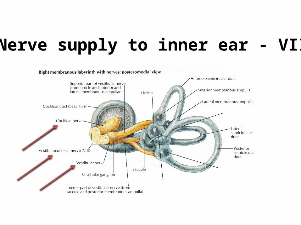
Nerve supply to inner ear - VIIIth
The inner ear: nerve supply
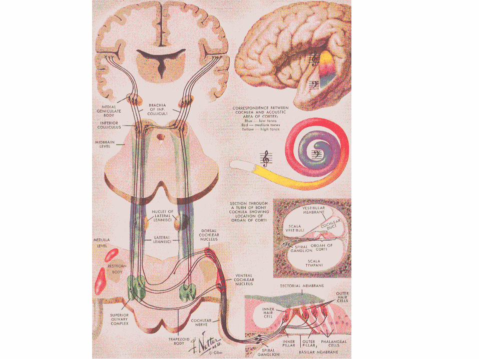
The brain has both ascending (afferent pathway) and descending (efferent pathway) tracts
The inner ear is innervated by the VIIIth cranial nerve (the acoustic nerve)
The VIIIth nerve has two major divisions, one vestibular and the other auditory (both are afferent)
There is also a small efferent bundle
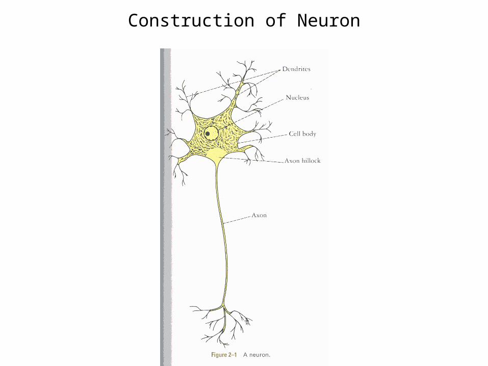
THE NEURON
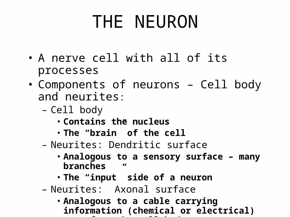
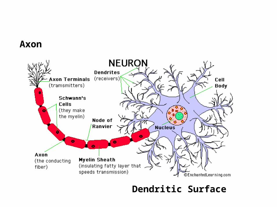
• A nerve cell with all of its processes• Components of neurons – Cell body and neurites:
– Cell body• Contains the nucleus• The “brain” of the cell
– Neurites: Dendritic surface• Analogous to a sensory surface – many branches• The “input” side of a neuron
– Neurites: Axonal surface• Analogous to a cable carrying information (chemical
or electrical) away from the cell body• The “output” side of the neuron
Construction of Neuron
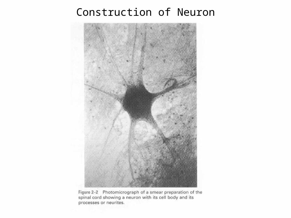
Construction of Neuron
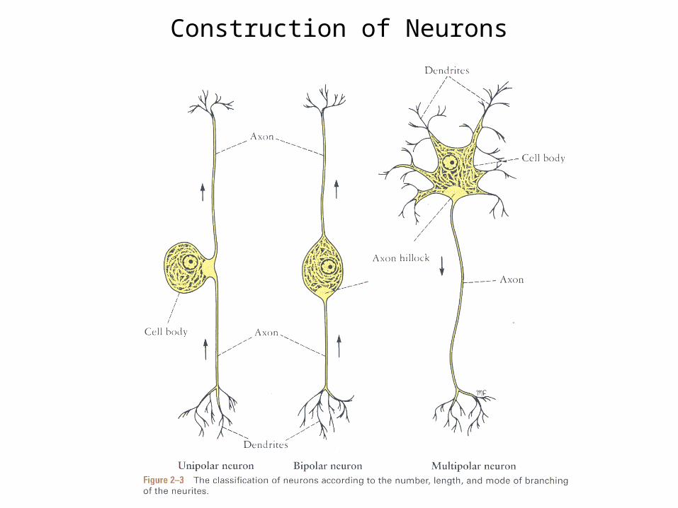
Construction of Neurons
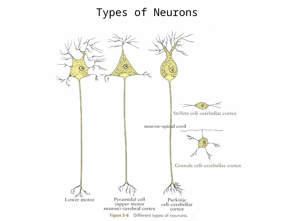
Types of Neurons
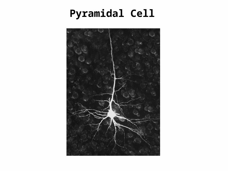
Pyramidal Cell
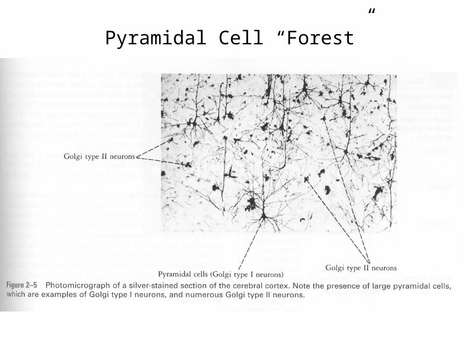
Pyramidal Cell “Forest”
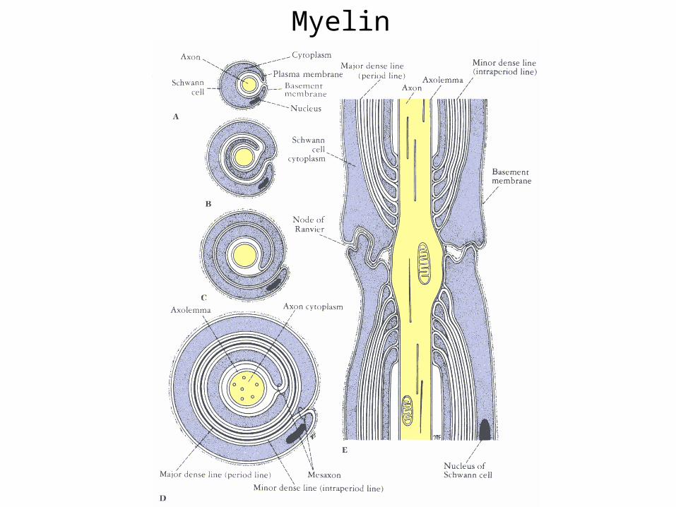
Myelin
Dendritic Surface
Axon

Neurons in Hippocampus
Cell Body

Nerve cellsGlial (support) cells
THE NEURON
• The Cell Body – contents and function– Nucleus: Stores genes (in mature neurons the
chromosomes do not replicate)– Cytoplasm – Contains the axon hillock, an important
component to the generation of action potentials• Golgi complex – maintenance of intracellular
reactions and cell membrane• Mitochondria – possess enzymes for metabolic activity
that generates energy for the cell• Microfilaments and microtubules – transport of
enzymes and nutrients throughout the cell
THE NEURON
• The Cell Membrane (Plasma Membrane)– The cell wall (or external cell boundary)– Maintains separation between intra- and extra-cellular
fluids – Lined with the cell coat, which facilitates ion transport– Semipermeable membrane that allows some ions to pass
while restricting the flow of others– While at rest, membrane allows potassium (K+) pass
through (permeate) the membrane– Restricts the flow of (Na+)– Prepares the neuron for an action potential
THE NEURON
• The Cell Processes (neurites)– Dendrites
• Profuse branching• Short processes• Dendritic spines attached to process• Considered extensions of cell body (increase surface
area• Conduct impulses TOWARD the cell body
THE NEURON
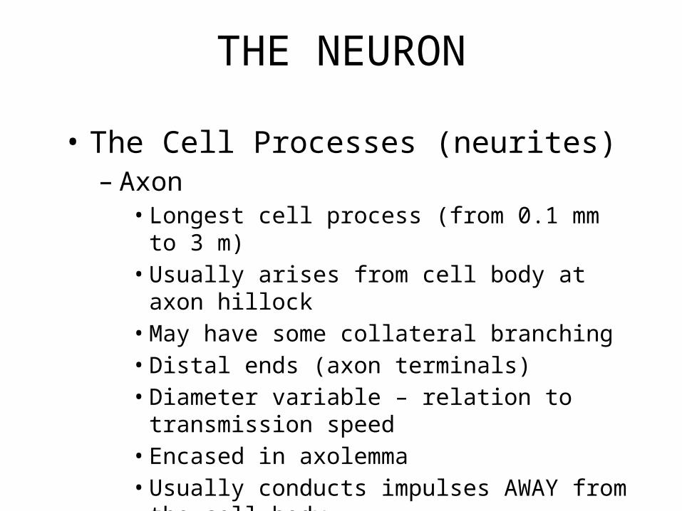
• The Cell Processes (neurites)– Axon
• Longest cell process (from 0.1 mm to 3 m)• Usually arises from cell body at axon hillock• May have some collateral branching• Distal ends (axon terminals)• Diameter variable – relation to transmission speed• Encased in axolemma• Usually conducts impulses AWAY from the cell body
THE NEURON
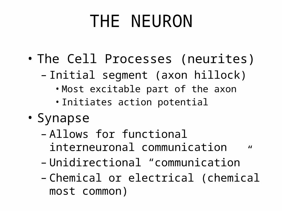
• The Cell Processes (neurites)– Initial segment (axon hillock)
• Most excitable part of the axon• Initiates action potential
• Synapse– Allows for functional interneuronal
communication– Unidirectional “communication”– Chemical or electrical (chemical most common)
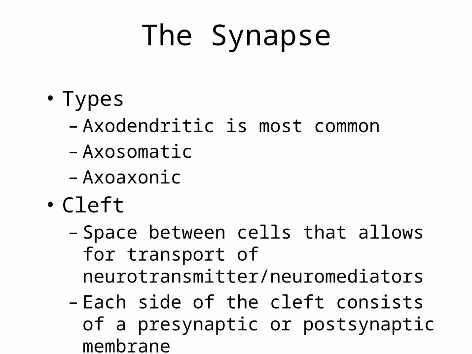
The Synapse
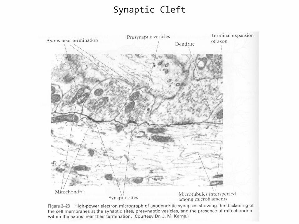
• Types– Axodendritic is most common– Axosomatic– Axoaxonic
• Cleft– Space between cells that allows for transport of
neurotransmitter/neuromediators– Each side of the cleft consists of a presynaptic
or postsynaptic membrane
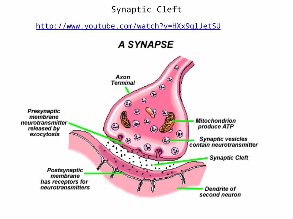
Synaptic Cleft
http://www.youtube.com/watch?v=HXx9qlJetSU
Synaptic Cleft
THE NEURON
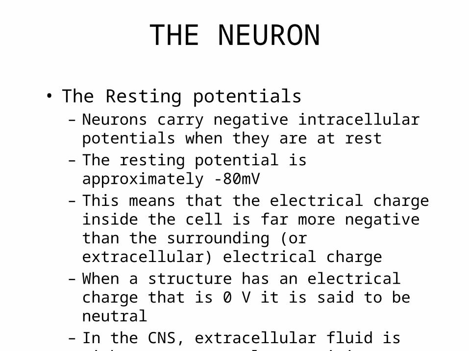
• The Resting potentials– Neurons carry negative intracellular potentials when
they are at rest– The resting potential is approximately -80mV– This means that the electrical charge inside the cell is
far more negative than the surrounding (or extracellular) electrical charge
– When a structure has an electrical charge that is 0 V it is said to be neutral
– In the CNS, extracellular fluid is either near neutral or positive

THE NEURON
• Cellular potentials– A voltage is a potential to do work– The greater the difference between positive and negative
poles, or charges, the more powerful the flow of ions– Think of batteries with pos. and neg. poles– When voltage changes TOWARD zero, it is
depolarization (excitatory)– When voltage changes AWAY FROM zero,
hyperpolarization (inhibitory)– V (extracellular) – V (intracellular) = Battery Voltage

THE NEURON – potentials
• EPSP and IPSP– Excitatory or inhibitory post-synaptic potential– Release of Neurotransmitter across synapse
produces excitatory (depolarizing effect)• Main agent is ACh (Acetylcholine) which facilitates
many muscle movements
– Or inhibitory (hyperpolarizing effect)• Main agent is gamma-amniobutyric acid (GABA)
which is important in sensory systems and for fine muscle control
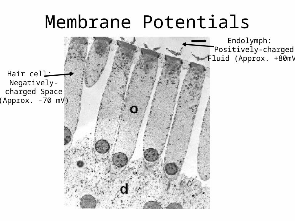
Membrane PotentialsEndolymph:
Positively-chargedFluid (Approx. +80mV)
Hair cell: Negatively-
charged Space(Approx. -70 mV)
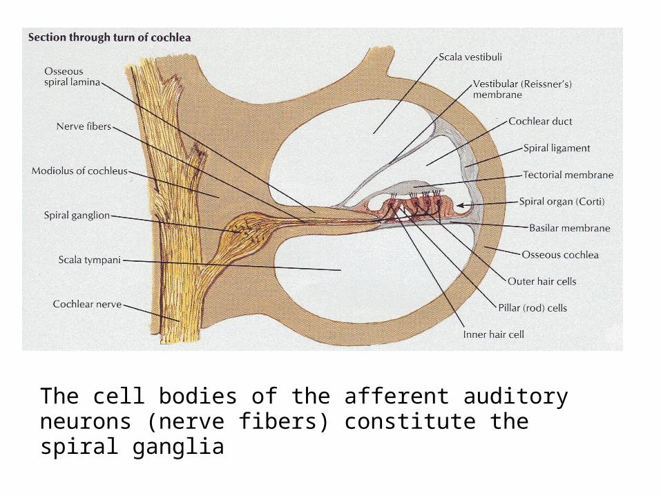
The cell bodies of the afferent auditory neurons (nerve fibers) constitute the spiral ganglia
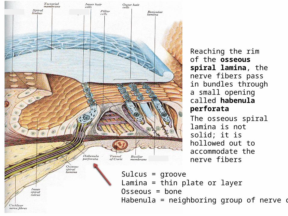
Reaching the rim of the osseous spiral lamina, the nerve fibers pass in bundles through a small opening called habenula perforataThe osseous spiral lamina is not solid; it is hollowed out to accommodate the nerve fibers
Sulcus = grooveLamina = thin plate or layerOsseous = boneHabenula = neighboring group of nerve cells
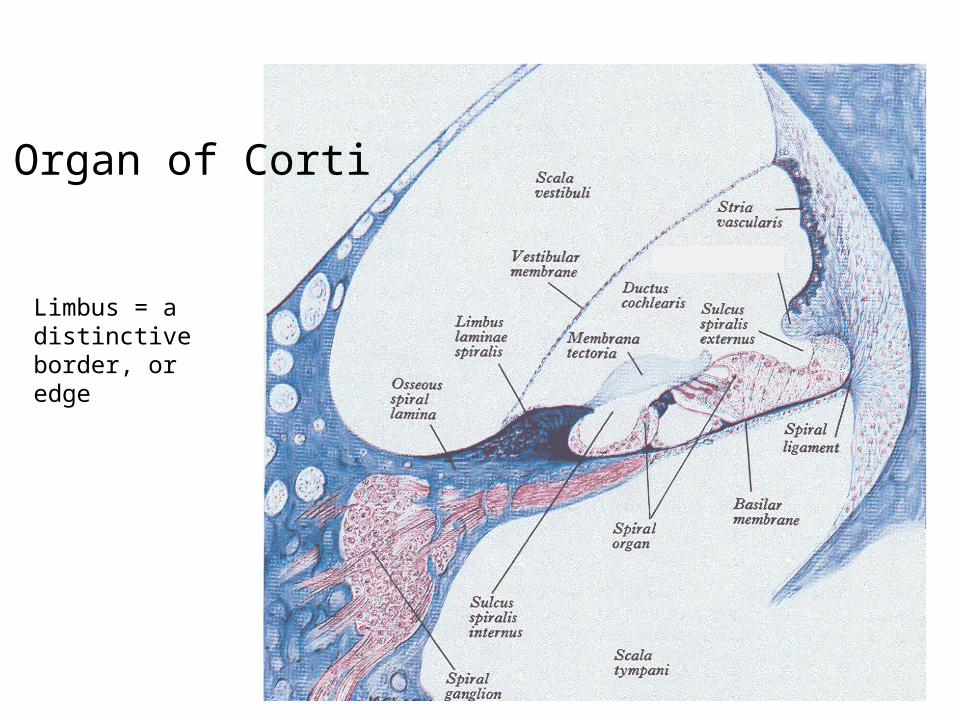
Organ of Corti
Limbus = a distinctive border, or edge
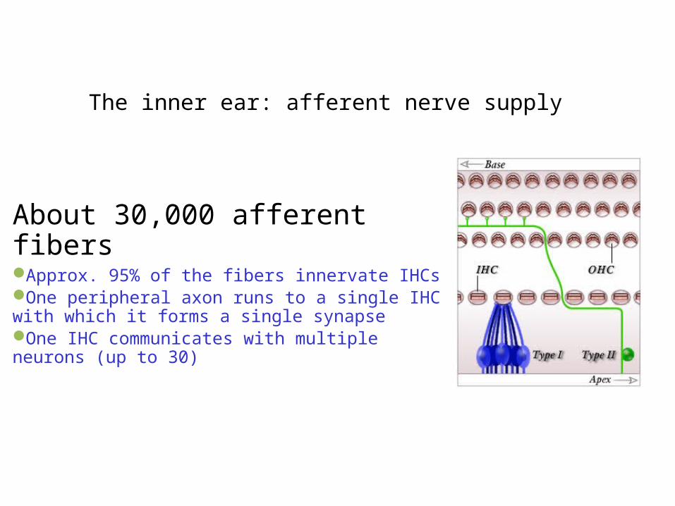
The inner ear: afferent nerve supply
About 30,000 afferent fibers Approx. 95% of the fibers innervate IHCs One peripheral axon runs to a single IHC with which it forms a single synapseOne IHC communicates with multiple neurons (up to 30)
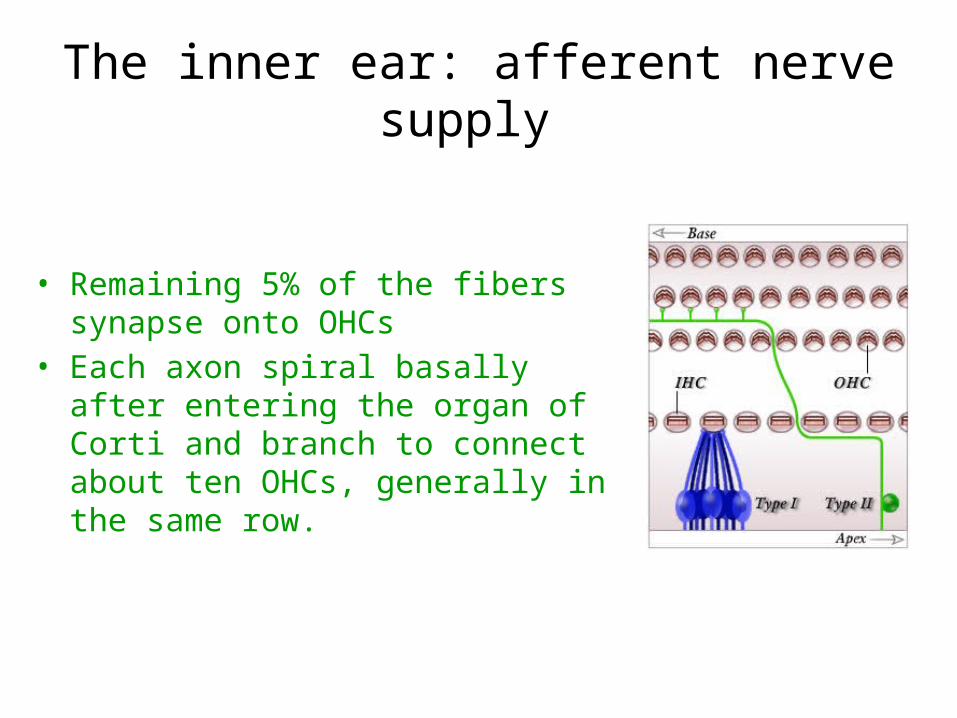
The inner ear: afferent nerve supply
• Remaining 5% of the fibers synapse onto OHCs
• Each axon spiral basally after entering the organ of Corti and branch to connect about ten OHCs, generally in the same row.
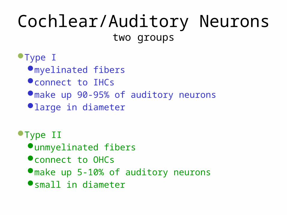
Cochlear/Auditory Neuronstwo groups
Type Imyelinated fibersconnect to IHCsmake up 90-95% of auditory neuronslarge in diameter
Type II unmyelinated fibersconnect to OHCsmake up 5-10% of auditory neuronssmall in diameter
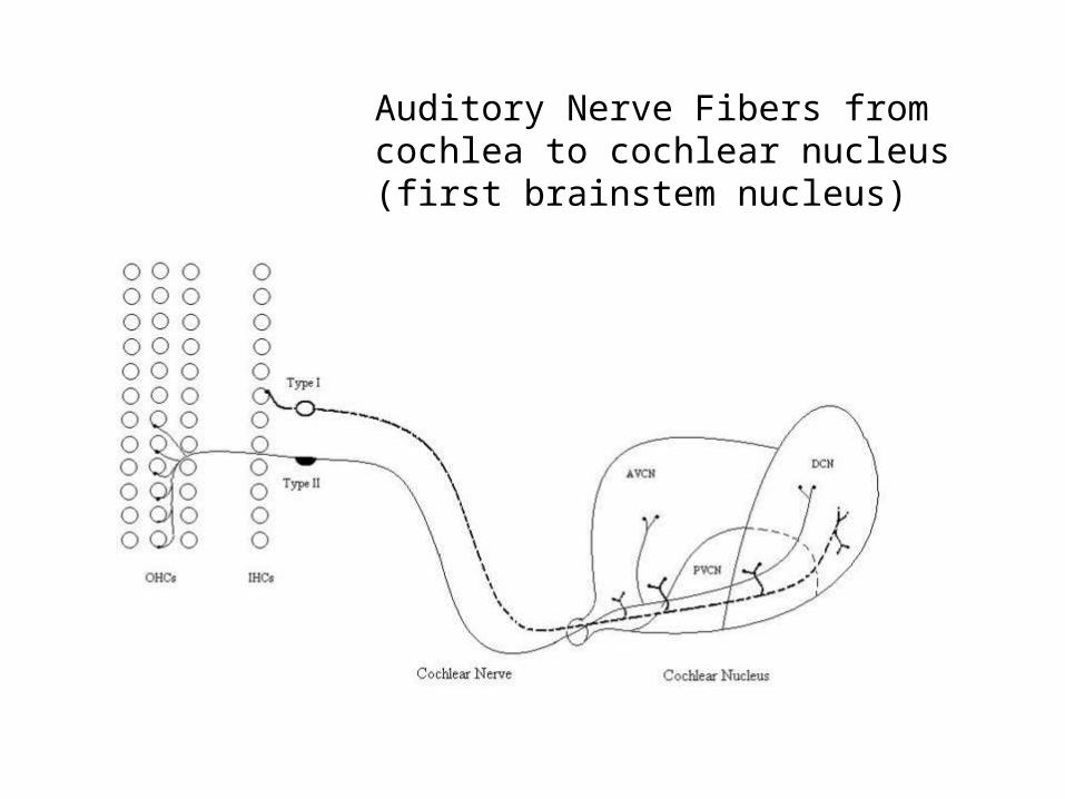
Auditory Nerve Fibers from cochlea to cochlear nucleus (first brainstem nucleus)
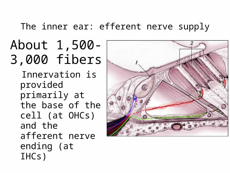
The inner ear: efferent nerve supply
About 1,500-3,000 fibers
Innervation is provided primarily at the base of the cell (at OHCs) and the afferent nerve ending (at IHCs)
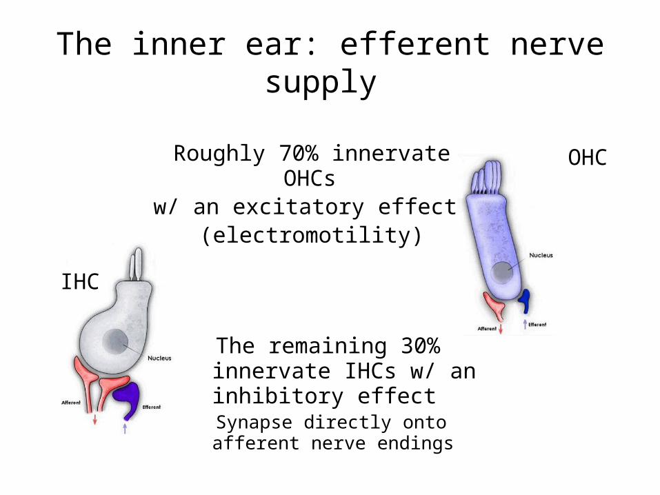
The inner ear: efferent nerve supply
OHC
IHC
Roughly 70% innervate OHCs
w/ an excitatory effect (electromotility)
The remaining 30% innervate IHCs w/ an inhibitory effectSynapse directly onto afferent nerve endings
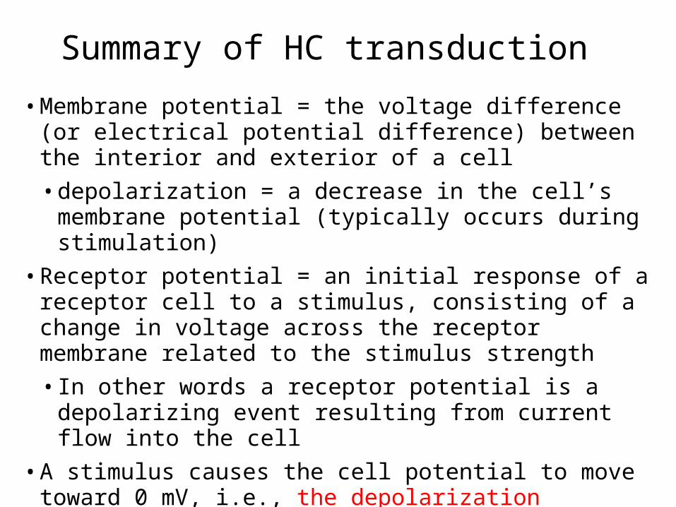
• Membrane potential = the voltage difference (or electrical potential difference) between the interior and exterior of a cell
• depolarization = a decrease in the cell’s membrane potential (typically occurs during stimulation)
• Receptor potential = an initial response of a receptor cell to a stimulus, consisting of a change in voltage across the receptor membrane related to the stimulus strength
• In other words a receptor potential is a depolarizing event resulting from current flow into the cell
• A stimulus causes the cell potential to move toward 0 mV, i.e., the depolarization
• A stimulus of an adequate magnitude is needed for transduction
Summary of HC transduction
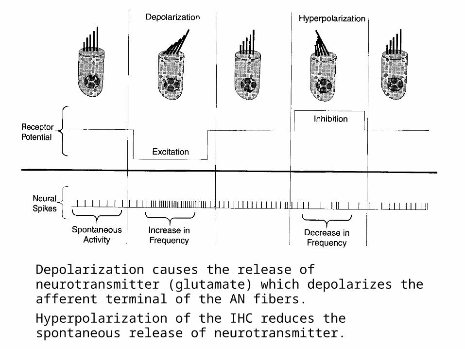
Depolarization causes the release of neurotransmitter (glutamate) which depolarizes the afferent terminal of the AN fibers.
Hyperpolarization of the IHC reduces the spontaneous release of neurotransmitter.
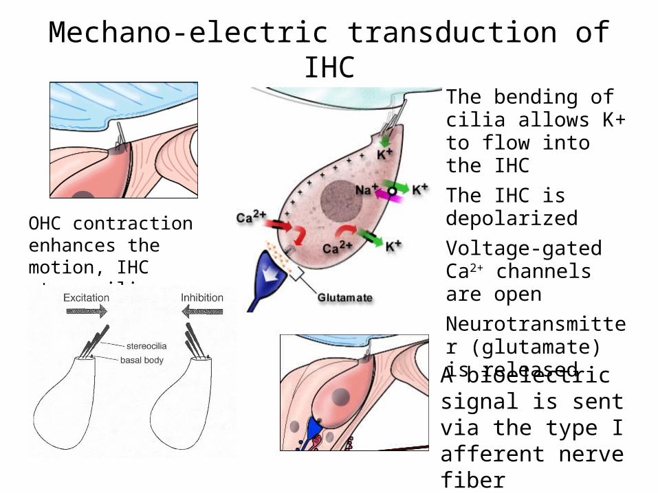
Mechano-electric transduction of IHC
The bending of cilia allows K+ to flow into the IHC
The IHC is depolarized
Voltage-gated Ca2+ channels are open
Neurotransmitter (glutamate) is released
OHC contraction enhances the motion, IHC stereocilia are bent
A bioelectric signal is sent via the type I afferent nerve fiber
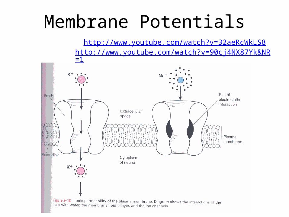
Membrane Potentialshttp://www.youtube.com/watch?v=32aeRcWkLS8
http://www.youtube.com/watch?v=90cj4NX87Yk&NR=1
THE NEURON
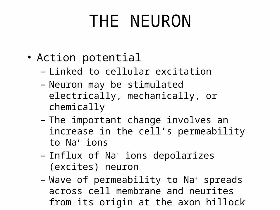
• Action potential– Linked to cellular excitation– Neuron may be stimulated electrically, mechanically, or
chemically– The important change involves an increase in the cell’s
permeability to Na+ ions– Influx of Na+ ions depolarizes (excites) neuron– Wave of permeability to Na+ spreads across cell
membrane and neurites from its origin at the axon hillock
– Quickly passes, followed by reduced permeability
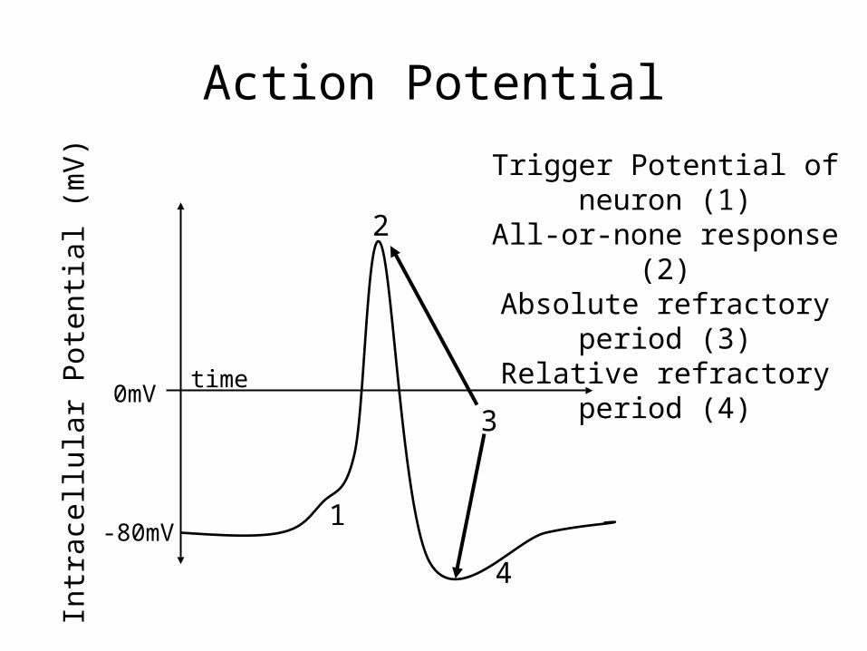
Action Potential
1
2
3
4
time
Intr
acel
lula
r P
oten
tial (
mV
)
-80mV
0mV
Trigger Potential of neuron (1)All-or-none response (2)
Absolute refractory period (3)Relative refractory period (4)
Action Potential
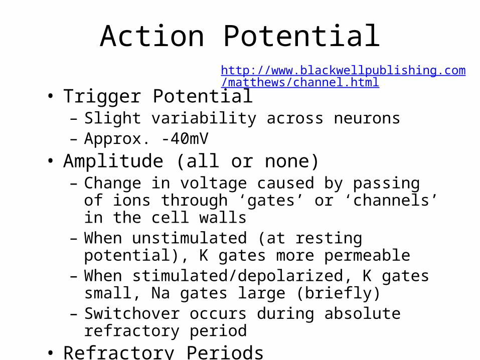
• Trigger Potential– Slight variability across neurons– Approx. -40mV
• Amplitude (all or none)– Change in voltage caused by passing of ions through
‘gates’ or ‘channels’ in the cell walls– When unstimulated (at resting potential), K gates more
permeable– When stimulated/depolarized, K gates small, Na gates
large (briefly)– Switchover occurs during absolute refractory period
• Refractory Periods
http://www.blackwellpublishing.com/matthews/channel.html
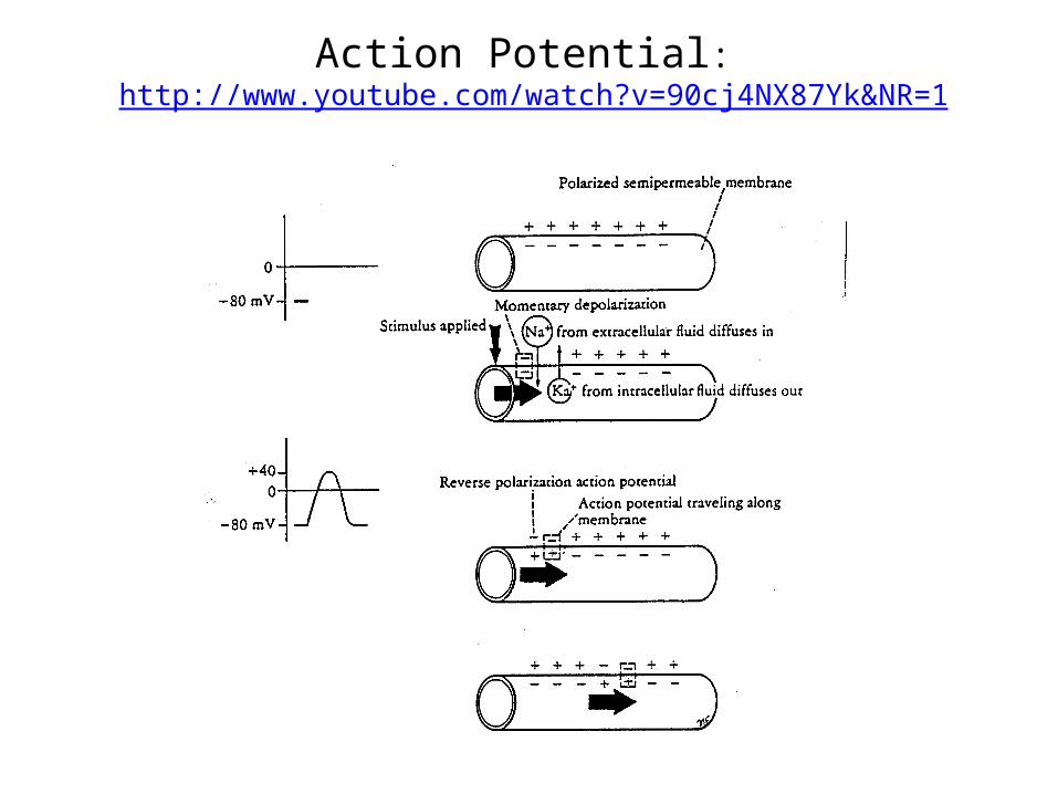
Action Potential: http://www.youtube.com/watch?v=90cj4NX87Yk&NR=1
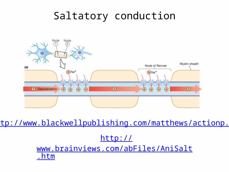
Saltatory conduction
http://www.blackwellpublishing.com/matthews/actionp.html
http://www.brainviews.com/abFiles/AniSalt.htm
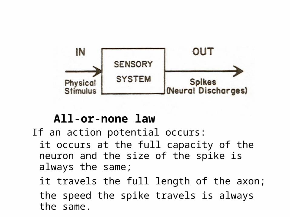
All-or-none lawIf an action potential occurs:
it occurs at the full capacity of the neuron and the size of the spike is always the same;
it travels the full length of the axon;
the speed the spike travels is always the same.
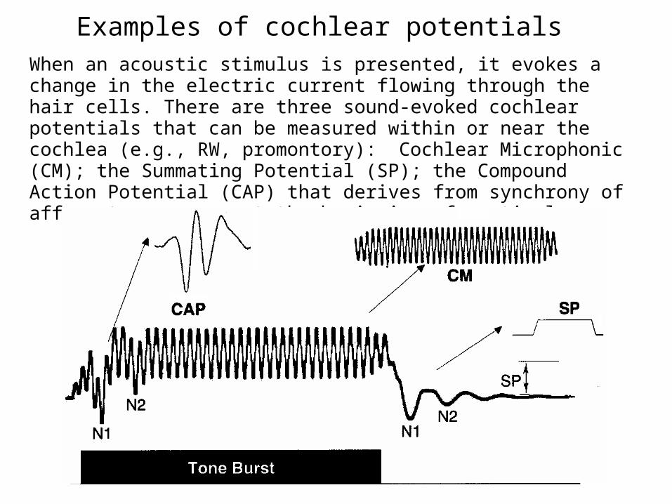
Examples of cochlear potentialsWhen an acoustic stimulus is presented, it evokes a change in the electric current flowing through the hair cells. There are three sound-evoked cochlear potentials that can be measured within or near the cochlea (e.g., RW, promontory):Cochlear Microphonic (CM); the Summating Potential (SP); the Compound Action Potential (CAP) that derives from synchrony of afferent responses at the beginning of a stimulus.

Bar Graph of a Neuron’s Firing Rate (PST Histogram)
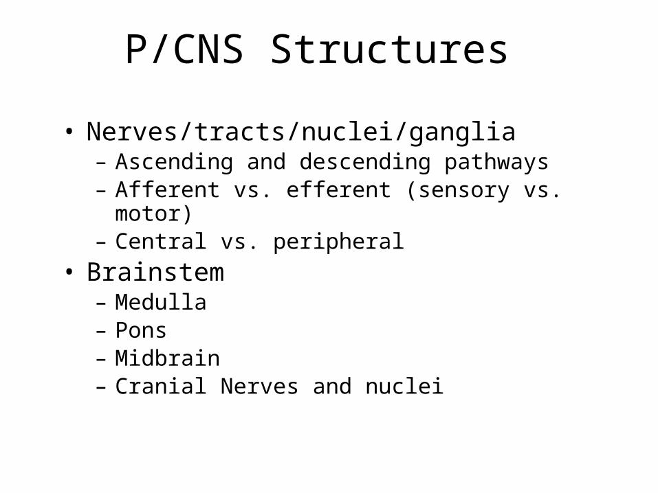
P/CNS Structures
• Nerves/tracts/nuclei/ganglia– Ascending and descending pathways– Afferent vs. efferent (sensory vs. motor)– Central vs. peripheral
• Brainstem– Medulla– Pons– Midbrain– Cranial Nerves and nuclei
CNS Structures
• Cerebellum– Cerebellar hemispheres– Vernix
• Cerebrum – Divisions– Telencephalon (hemispheres)– Diencephalon (“in-between brain”)

PNS Structures
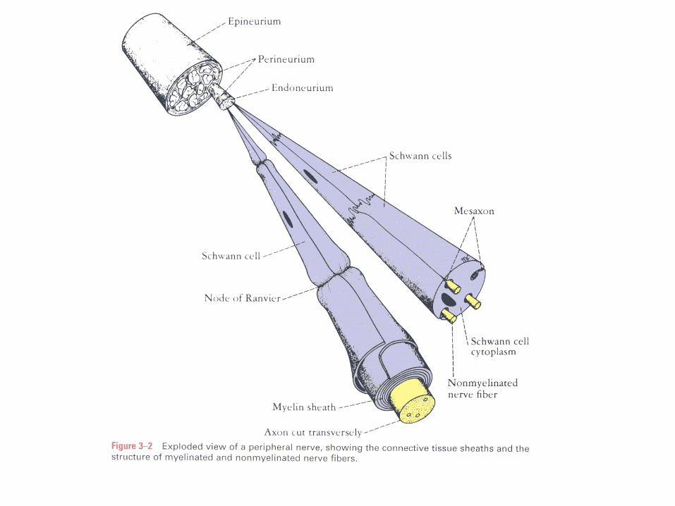
• Nerves – Construction– Fibers Endoneurium Perineurium Epineurium
– Collection of Axons in PNS– Cranial Nerves – 12 pairs that exit brain through
openings in the skull (foramen)• May be sensory, motor, or mixed• Several are of relevance in CDIS (you should know AT LEAST
V, VII, VIII, IX, X, XII)
– Spinal Nerves – 31 pairs that exit the spinal cord through openings in the vertebra
• Mixed nerves with a sensory branch and a motor branch
PNS Structures
• Ganglia– Collection of cell bodies in the PNS– Generally consist of unipolar neurons
surrounded by the epineurium and perineurium of peripheral nerves
– Appear as swellings, on a nerve’s course
CNS Structures
• Nucleus– Collection of cell bodies in the CNS– “Relay stations” or processing centers in
which neural activity is summed, inhibited, etc.
– Many CNS structures we know by name (thalamus, for example) are nuclei
– May also integrate neural activity from across several sources, or modalities
CNS Structures
• Tract– Collection of axons in the CNS– Pipelines, or conduits through with neural
activity migrates from one place to another– Connect different areas of brain that may be
associated with different functions