author's personal copy - lubmc.biomed.lu.lv/site/common/attachments/ecr_2010.pdf ·...
TRANSCRIPT

This article appeared in a journal published by Elsevier. The attachedcopy is furnished to the author for internal non-commercial researchand education use, including for instruction at the authors institution
and sharing with colleagues.
Other uses, including reproduction and distribution, or selling orlicensing copies, or posting to personal, institutional or third party
websites are prohibited.
In most cases authors are permitted to post their version of thearticle (e.g. in Word or Tex form) to their personal website orinstitutional repository. Authors requiring further information
regarding Elsevier’s archiving and manuscript policies areencouraged to visit:
http://www.elsevier.com/copyright

Author's personal copy
Research Article
Up-regulation of the embryonic self-renewal network throughreversible polyploidy in irradiated p53-mutant tumour cells
Kristine Salminaa, Eriks Jankevicsa, Anda Hunaa, Dmitry Perminova, Ilze Radovicaa,Tetyana Klymenkob, Andrey Ivanovc, Elina Jascenkod, Harry Scherthane,Mark Craggf, Jekaterina Erenpreisaa,⁎aLatvian Biomedical Research and Study Centre, Riga, LV-1067, LatviabPaterson Institute of Cancer Research, Manchester University, M20 4BX, UKcBeatson Institute, Glasgow Centre for Cancer Research, Glasgow University, G61 4LG, UKdLatvian Institute of Organic Synthesis, Riga, LV-1006, LatviaeInst. für Radiobiologie der Bundeswehr in Verbindung mit der Univ. Ulm, Munich, GermanyfCancer Sciences Division, Southampton University School of Medicine, General Hospital, Southampton SO16 6YD, UK
A R T I C L E I N F O R M A T I O N A B S T R A C T
Article Chronology:
Received 9 November 2009Revised version received 20 April 2010
Accepted 28 April 2010Available online 10 May 2010
We have previously documented that transient polyploidy is a potential cell survival strategyunderlying the clonogenic re-growth of tumour cells after genotoxic treatment. In an attempt tobetter define this mechanism, we recently documented the key role of meiotic genes in regulating
the DNA repair and return of the endopolyploid tumour cells (ETC) to diploidy through reductiondivisions after irradiation. Here, we studied the role of the pluripotency and self-renewal stem cellgenes NANOG, OCT4 and SOX2 in this polyploidy-dependent survival mechanism. In irradiation-resistant p53-mutated lymphoma cell-lines (Namalwa and WI-L2-NS) but not sensitive p53 wild-type counterparts (TK6), low background expression of OCT4 and NANOG was up-regulated byionising radiation with protein accumulation evident in ETC as detected by OCT4/DNA flowcytometry and immunofluorescence (IF). IF analysis also showed that the ETC generate PML bodiesthat appear to concentrate OCT4, NANOG and SOX2 proteins, which extend into complex nuclearnetworks. These polyploid tumour cells resist apoptosis, overcome cellular senescence andundergo bi- and multi-polar divisions transmitting the up-regulated OCT4, NANOG and SOX2 self-renewal cassette to their descendents. Altogether, our observations indicate that irradiation-
induced ETC up-regulate key components of germ-line cells, which potentially facilitate survivaland propagation of the tumour cell population.
© 2010 Elsevier Inc. All rights reserved.
Keywords:
LymphomaIrradiationPolyploidyOCT4/NANOG/SOX2
Self-renewalCellular senescence
E X P E R I M E N T A L C E L L R E S E A R C H 3 1 6 ( 2 0 1 0 ) 2 0 9 9 – 2 1 1 2
⁎ Corresponding author. LBMC, Ratsupites 1, Riga, LV-1067. Fax: +371 67442407.E-mail addresses: [email protected] (K. Salmina), [email protected] (E. Jankevics), [email protected] (A. Huna), [email protected]
(D. Perminov), [email protected] (I. Radovica), [email protected] (T. Klymenko), [email protected] (A. Ivanov),[email protected] (E. Jascenko), [email protected] (H. Scherthan), [email protected] (M. Cragg), [email protected](J. Erenpreisa).
Abbreviations: ESC, embryonic stem cells; ETC, endopolyploid tumour cells; CSC, cancer stem cells; IF, immunofluorescence; WB, Westernblotting; NT, non-treated cells; PIR, post-irradiation; ARS, accelerated replicative senescence; PBL, peripheral blood lymphocytes
0014-4827/$ – see front matter © 2010 Elsevier Inc. All rights reserved.doi:10.1016/j.yexcr.2010.04.030
ava i l ab l e a t www.sc i enced i r ec t . com
www.e l sev i e r . com/ loca te /yexc r
Author's personal copy
Introduction
Resistance to genotoxic treatments such as chemo- and radio-therapy, causes disease relapse and metastases in up to 50% ofcancer cases. Recently, this property has been attributed to thepresence of cancer stem cells (CSC) in the tumour population,which appear refractive to genotoxic treatment [1–4] or to‘stemloids’ – proliferating tumour cells derived from progenitorswhich can reactivate self-renewal-capability [5]. Targeting thesecells and their implied unique cell survival pathways is thereforean attractive proposition for cancer therapy.
The transcription network which tightly links self-renewal withpluripotency (the capability to produce all three germ layers) isregulated by three master genes Oct4, Sox2 and Nanog [6,7].Pluripotent stem cells derived from preimplantation embryos,primordial germ cells, embryonal carcinoma (teratocarcinoma) orartificially induced from somatic cells are unique in undergoingprolonged symmetrical self-renewal in culture. All of thesepluripotent cell-types are capable of supporting embryonal devel-opment and express key embryonal stem cell (ESC) genes [6,8–10].The pluripotency network has been found expressed in many typesof adult stem cell [11] and inmalignant somatic tumours correlatingwith poor clinical outcome [3,4,12]. However, interpretation of thedata in somatic cells is currently complicated by the knowledge thatOCT4 possesses several alternative splicing forms which can beregulated by multiple post-translational modifications and furtherby the fact that adult stem cells and somatic tumours express manypseudogenes of OCT4 and NANOG [13–16]. For example, thosepseudogenes with similar open reading frames and high homologyto paternal genes may mimic the activity of the homologous genes,or alternatively may act as their recombination templates, func-tional substitutes [17,18], or mRNA stabilisers [19].
In any case, the observation of a shared transcriptional programbetween pluripotent stem cells and tumours [20] provides thestimulus for investigating whether key pluripotency and self-renewal genes engender the basis for the treatment resistance oftumour cells (presumably through CSC or stemloids).
Evidence that endopolyploid cells induced after genotoxictreatment display increased resistance to DNA damaging agentsand are involved in clonogenic survival has been now presented byseveral laboratories [21–24]. We have previously suggested thepotential of this transient polyploidy to reactivate stem cell traits[25]. Here we asked if the core transcriptional cassette expressedby pluripotent stem cells is also expressed in somatic lymphomasand if it is up-regulated in the transient polyploid cells induced bygenotoxic damage in p53-mutant tumour cell lines.
Materials and methods
Cell lines and irradiation procedure
The Burkitt's lymphoma cell line Namalwa was obtained from theAmerican TypeCulture Collection (ATCC) andhas an established p53mutant allele [26]. The human B-lymphoblastoid cell linesWI-L2-NS(p53mutant -mt) and TK6 (p53wild type-wt)were isolated from thesame patient at different times during treatment and were obtainedfromDr. P. Olive (Canada). Lymphoma cell-lines weremaintained inRPMI-1640 containing 10% heat-inactivated fetal calf serum (FCS;
Sigma) at 37 °C in a 5 % CO2 humidified incubator. The EC cell line -human ovarian teratocarcinoma PA1 originated from the ATCC wasobtained from the Vertebrate Cell Culture Collection of the Instituteof Cytology (St-Petersburg, Russia). These cells were grown inDMEM medium containing 10% FCS (Sigma) and penicillin/streptomycin, and were harvested at sub-confluence as a positivecontrol for ESC transcription factors [17,18]. IMR90 cells wereobtained from ATCC (ATCC-CCL-186) and were grown in DMEMwith 10% FCS for 30 passages, for the positive control of replicativecell senescence. For experimental studies, cells were maintained inlog phase of growth for at least 24 hours prior to irradiation. Cellswere irradiatedwith linear accelerator (Clinac 600 C, VarianMedicalSystems) using a 4 MeVphotonbeamat a dose rate of 1 or 2 Gy/min.Correspondingly, a single acute dose of 5 or 10 Gy was delivered inall experiments. After irradiation, cells were maintained byreplenishing culturemedium every 48-72 h. In certain experiments,retinoic acid (Sigma; 1 μM) was added to the culture medium for48 h fromday 3 to day 5 post-irradiation. Smears of peripheral bloodwere used as a negative control for NANOG in immunofluorescencestudies.
RT-PCR
Total RNA was extracted from cells by using TRIZOL (Invitrogen)and treated with DNase I (Fermentas MBI). cDNA was synthesizedusing First Strand cDNA Synthesis Kit (Fermentas MBI) accordingto the protocols of the manufacturer. The absence of contamina-tion with chromosomal DNA was verified by PCR using primersDQA-1 and KIR3DL2. Expression of pluripotent cell markers wasverified in comparison to embryonal stem cells cDNA (Millipore,SCR063). cDNA from peripheral blood lymphocytes (PBL) waskindly provided by Dr. HTC Chan, Southampton University, UK.
Primers for PCR (Table 1) were designed using Primer 3 software[27]; the primers for OCT4 B1 isoform were taken in [28]. GenBankaccession numbers of the templates used for design are as follows:OCT4-A (POU5F1), NM_002701; OCT4-B (POU5F1)_B, NM_203289;OCT4-B1 (POU5F1)_B1, EU518650; POU5F1B (POU5F1P1),NR_002304; OCT-4 ps4 (POU5F1P4), NC_000001; NANOG,NM_024865; SOX2, NM_003106. Amplification was carried out ina total volume of 50 µl with 1 – 4 µl cDNA in standard conditionsusing 0.5 units of Taq DNA polymerase (Fermentas MBI) with aBioCycler TC-S (BioSan). PCR conditions were as follows: 94 °C for5 min (if not specified otherwise) 94 °C for 30 s, 56 – 60 °C for 20 s(for details see Table 1), 72 °C for 1 min; final extension step at 72 °Cfor 7 min. Amplified PCR products were resolved on 1.2 % agarosegels. The resulting PCR products fragments were analyzed bysequencing after ExoI/SAP treatment (Fermentas, MBI) using thefluorescent Big DyeTerminator v. 3.1 Cycle Sequencing protocol on a3130xl Genetic Analyzer (Applied Biosystems).
Western blotting
For protein extraction, ProteoJET™ Cytoplasmic and NuclearProtein Extraction Kit (Fermentas, Lithuania), along with ProteaseInhibitor Cocktail (Sigma P8340) was used, according to themanufacturers’ instructions. The purity of isolated cell nuclei wasassessed by immunofluorescence using the antibodies againstlamin B and α-tubulin (Fig. S1). Equal protein loading in each lanewas checked by Ponceau S staining. As loading and fraction puritycontrols antibodies against GAPDH were used (positive staining
2100 E X P E R I M E N T A L C E L L R E S E A R C H 3 1 6 ( 2 0 1 0 ) 2 0 9 9 – 2 1 1 2
Author's personal copy
was observed in cytoplasmic but not nuclear lysates). Proteinsamples (10 μg) were separated by SDS PAGE on 9% gels and thenblotted onto nitrocellulose membranes (Bio-Rad Labs). Themembranes were blocked with 5% non-fat dry milk in PBS with0.05% Tween 20 (Sigma) and primary antibodies applied over-night. The structure of OCT4 splicing isoforms and location of theepitopes for the two OCT4 antibodies used is presented in Fig. 1.The source and dilutions of antibodies is presented in Table 2 in thesupplementary material. To confirm specificity, a blocking peptidefor the OCT4 rabbit polyclonal (ab20650) was mixed with primaryantibody at 1 μg/ml and incubated at room temperature (RT) for30 min before application. Detection was performed with HRP-conjugated secondary antibodies (Table 2) and ECL (Amersham).
Two channel flow cytometry for OCT4 and DNA content
Cells were harvested at relevant time points, washed in cold TBS,and fixed overnight in cold (–20 °C) 70% ethanol. After two washes
in TBS, cells were permeabilised with TBS/4% BSA/0.1% Triton X-100 for 10 min at RT. After that, samples were incubated with rabbitpolyclonal anti-OCT4 antibody solution (5 µg/ml) (ab19857; Ab2,see on Fig. 1) in TBS/4% BSA/0.1% Triton X100 for 1 h at RT.Following three washes in TBS, cells were incubated with goat anti-rabbit Alexa Fluor 488 solution in TBS/4%BSA/0.1% Triton X100,1:200 for 1 h in the dark. Then, DNA was counterstained with10 µg/ml propidium iodide (PI) solution in PBS, containing 200 µg/ml RNAse (Sigma) and assessed by flow cytometry using a FACScan(BD Biosciences) using Cell Quest Pro Software.
Immunofluorescence (IF)
Standard IF staining was performed according to proceduresdetailed previously [29]. The primary and secondary antibodiesused are listed in Table 2. Detection of apoptosis by FITC Annexin V(BD Pharmingen) in combination with IF was performed asdetailed in [30].
Table 1 – Sequences of primers, length of fragments, and annealing temperatures used for RT-PCR.
Gene/primer Forward primer sequence5’→3’
Reverse primer sequence5’→3’
Length of fragment,bp
Anneal. temp.,°C
β-ACTIN GGACTTCGAGCAAGAGATGG AGCACTGTGTTGGCGTACAG 234/329 58DQA-1 GTGCTGCAGGTGTAAACTTGTACCAG CACGGATCCGGTAGCAGCGGTAGAGTTG 225 56KIR3DL2 CGGTCCCTTGATGCCTGT GACCACACGCAGGGCAG 1941/369 56NANOG CACCTACCTACCCCAGCCTT CTCGCTGATTAGGCTCCAAC 585 60OCT4-A – AF/AR TTCTCGCCCCCTCCAGGT TCAGAGCCTGGCCCAACC 137 56OCT4-B/B1 – BF/BR GAAGTTAGGTGGGCAGCTTG AATAGAACCCCCAGGGTGAG 267 56OCT4-B/B1 – BF1/BR2 AGACTATTCCTTGGGGCCACAC CTCAAAGCGGCAGATGGTCG 267/492 64POU5F1B (OCT4-ps1) AGGCCGATGTGGGGCTCAT CCAGAGTGATGACGGAGACT 567 58OCT4-ps4 – AF1/BR AGGTTGGAGTGGGGCTAGTG AATAGAACCCCCAGGGTGAG 289 56SOX2 ACCTACATGAACGGCTCGC CCGGGGAGATACATGCTGA 197 56
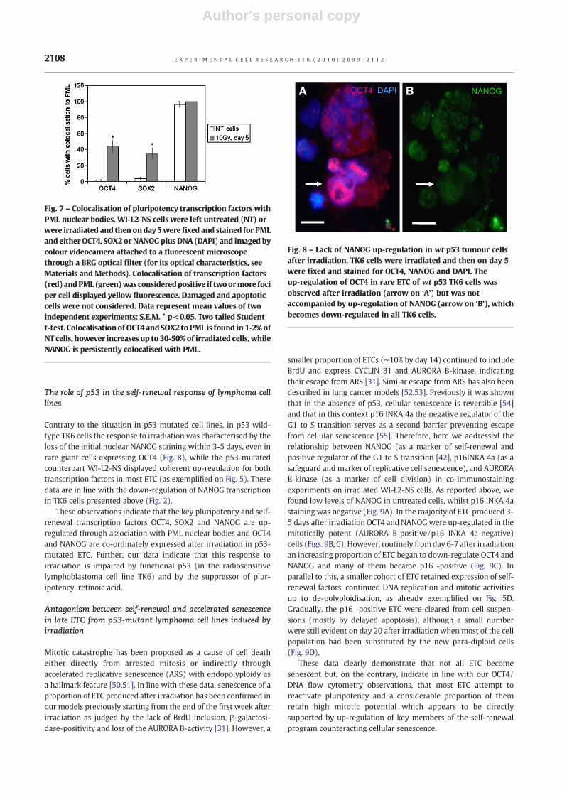
Fig. 1 – Structure of OCT4. Three different OCT4 isoforms are generated by alternative splicing which are schematically shown herewith exons depicted as boxes. The RT-PCR primer binding sites and antibody binding epitope regions used in this study are indicated.POUS – POU-specific domain, POUH – POU homeodomain, NTD – N-transactivation domain, CTD – C-transactivation domain.
2101E X P E R I M E N T A L C E L L R E S E A R C H 3 1 6 ( 2 0 1 0 ) 2 0 9 9 – 2 1 1 2

Author's personal copy
Microscopy
A fluorescence light microscope (Leitz, Ergolux L03-10) incombination with a colour videocamera (Sony DX-S500) wasused to examine cell preparations, record images, and performimage cytometry. For colocalisation studies a confocal microscope(Leica, DM 600), was used with the images scanned in the threedifferent colour channels in sequence. Alternatively, the BRG filtersystem from Leica was used for the colocalisation calculationsusing a fluorescent light microscope (UV excitation band 385-415 nm, emission transmission band 455-475 nm; blue excitationband 487-503 nm, emission transmission band 515-545 nm;green excitation band 560-580 nm, emission transmission band620-660 nm).
DNA image cytometry
DNA image cytometry was performed as detailed previously [31].
Clonogenicity assays
To measure clonogenic survival, a serial dilution assay wasperformed, using a method detailed previously [21]. Experimentswere repeated at least three times.
Results
To assess the expression and induction of self-renewal genes intumour cells before and after genotoxic treatment we performedexperiments on three different lymphoma cell lines; Namalwa,WI-L2-NS and TK6. As we have published previously, the p53mutant cell-line Namalwa is radioresistant, whilst the p53 wild-type TK6 is radiosensitive (Fig. S2A). WI-L2-NS (a p53 mutantcounterpart of TK6 derived from the same patient) is alsoradioresistant and displays 10-fold more polyploidy and two-foldless apoptosis than the wt p53 TK6 cells in response to irradiation(Fig. S2B). Below we will detail the expression of the self-renewalgenes first in untreated cells and then in cells after irradiation.
RT-PCR studies indicate the expression of pluripotency andself-renewal genes in lymphoma cell lines
OCT4 (POU5F) is a member of the POU family of homeodomaintranscription factors required for the maintenance of pluripotencywhich is apparently achieved by establishing a transcriptionallypermissive chromatin structure in the 160 kb NANOG locus [32].Three splicing variants of OCT4 gene have been hitherto described(Fig. 1) with OCT4A the classical form active in transactivation,OCT4 B a cytoplasmic isoform with unknown function [33,34] orpossible function in cell stress of its alternatively translated variant[35] and the recently discovered OCT4 B1 splicing isoform [28] apotential additional marker of stemness [36]. Of seven OCT4pseudogenes, the 1st (POU5FB1) has been recently confirmed as atranscriptional activator similar to POU5F [37], while the 3rd and4th pseudogenes also have considerable open reading frames withtransactivation potential.
Untreated cellsTo assess the expression of OCT4, we performed semi-quantitativeRT-PCR on the three lymphoma cell-lines before and afterirradiation using a carefully designed selection of primers (fordetails seeMaterials andMethods, Fig. 1, and Table 1) andwith theteratocarcinoma PA-1 as a positive control. Of the OCT4 splicingisoforms a transcript for OCT4A was found in all lymphoma celllines and PA1 (Fig. 2A) and its identity was confirmed by sequenceanalysis. Expression of OCT4A was also confirmed in cDNA frombone fide ESC cells (Fig. S3 in Supplementary material). The OCT4B/B1 isoforms were also found in all cell lines (Fig. 2A). The OCT4B1 isoform, when detected separately from the B-isoform wasfound transcribed in PA1 and lymphoma cell lines. The OCT4Bisoform was revealed in WI-L2-NS and TK6 cells at a higher levelthan PA1 cells. In Namalwa cells the expression of OCT4B wasextremely low (Fig. 2A), but was confirmed by sequence analysisof the PCR product. Equivalent expression of the weak transcrip-tion activator POU5F1B (previously known as OCT4-ps1)was foundin the lymphoma and PA1 cell lines (Fig. 2A).
Perhaps surprisingly, our RT-PCR analysis using primerAF1+BR (see on Fig. 1 and in Table 1) also revealed the presence
Table 2 – Antibodies: source and usage.
Primary antibodies: Secondary antibodies (dilution, if not stated otherwise, 1:400)
Mouse monoclonal anti-hOCT4 C-10 (sc-5279, Santa Cruz) IF1:100 WB 1:1000
Goat anti-mouse IgG-Alexa Fluor 488 (A31619, Invitrogen) or 594 (A31623,Invitrogen); Rabbit anti-mouse IgG-HRP (61-6520, Invitrogen) 1:4000
Rabbit polyclonal anti-hOCT4 (ab19857, Abcam) IF 1:75-400;Blocking peptide (ab20650, Abcam) WB 1:400-700
goat anti-rabbit- IgG-Alexa Fluor 488 (A31627, Invitrogen) or 594 (A31631, Invitrogen)Goat anti-rabbit IgG-HRP (32460, Thermo Fisher Scientific) 1:5000
Mouse monoclonal anti-hNANOG (N3038, Sigma) IF 1:50-75;WB 1:1000
Goat anti-mouse IgG-Alexa Fluor 488 or 594 (Invitrogen); Rabbit anti-mouse IgG-HRP(61-6520, Invitrogen) 1:4000
Rabbit polyclonal anti-hNANOG H-155 (sc-33759, Santa Cruz) IF1:50-1:75; WB 1:400
Goat anti-rabbit- IgG-.Alexa Fluor 594 Goat anti-rabbit IgG-HRP (32460, Thermo FisherScientific) 1:5000
Rabbit polyclonal anti-hP16 N-20 (sc-467, Santa Cruz) IF 1:50 Goat anti-rabbit- IgG-.Alexa Fluor 594Mouse monoclonal anti-hPML (sc-966, Santa Cruz) IF 1:150 Goat anti-mouse IgG-Alexa Fluor 488Rabbit polyclonal anti-hAURORA B (ab2254, Abcam) IF 1:150-300
Goat anti-rabbit- IgG-.Alexa Fluor 594
Rabbit polyclonal anti-hSOX2 (sc-20088x, Santa Cruz) IF 1:500 Goat anti-rabbit- IgG-.Alexa Fluor 594Mouse monoclonal anti-h-α-TUBULIN Clone B-512 (T5168,Sigma,) 1:2,000
Chicken anti-mouse IgG-FITC (sc-2989) 1:100
Goat polyclonal anti-hLAMIN B (sc-6216, Santa Cruz), IF 1:100 Rabbit anti-goat IgG-Cy3 (C2821, Sigma) 1:500Mouse monoclonal anti-hGAPDH (MAB374, Millipore) WB1:100
Rabbit anti-mouse IgG-HRP (61-6520, Invitrogen) 1:4000
2102 E X P E R I M E N T A L C E L L R E S E A R C H 3 1 6 ( 2 0 1 0 ) 2 0 9 9 – 2 1 1 2

Author's personal copy
of a transcribed OCT4 pseudogene OCT4-ps4, which is similar instructure to the Oct4A isoform, in Namalwa, WI-L2-NS and TK6cells, but not PA1 cells, where this primer amplified only trueOCT4A as determined by sequence analysis (Fig. 2B). Interestingly,in the wt p53 TK6 cells there was reduced expression of thispseudogene compared with Namalwa and WI-L2-NS cells, whilstPBL did not express OCT4-ps4 at all (Fig. 2B).
Together, these data demonstrate that two OCT4 genes (POU5Fand POU5F1B), all three OCT4 isoforms, and a pseudogene (OCT4-ps4) are transcribed at a low level in the untreated lymphoma celllines and that the proportion of these components varies in eachcell-line, perhaps indicating an aberrant regulation.
Transcriptional regulation of NANOG occurs in the NANOG locusthrough cooperative interaction with the OCT4:SOX2 heterodimer
Fig. 2 – Semi-quantitative RT-PCR analysis. Untreated cells or cells treated with irradiation were assessed by RT-PCR. For primers, seeTable 1. (A)Analysis for differentOCT4 (POU5F) isoforms,POU5F1B,NANOG,and SOX2 showing their expressionanddynamic changes inthepost-irradiation time-course in three lymphomacells lines. Non-treatedPA1 cells (ovarian teratocarcinoma)wereused as a positivecontrol,whileβ-actinwasused as an internal loading control. For locationofprimers in relation toOCT4 structure, see Fig. 1. Expressionof OCT4 A and OCT4 B/B1-isoforms and NANOG increased inmt p53 cell lines, Namalwa and WI-L2-NS, while NANOG expressiondecreased in wt p53 TK6 cells, after irradiation. OCT4-ps1 (POU5FB1) was found equally expressed in all samples. (B) Expression ofOCT4A, OCT4 pseudogene 4 (OCT4-ps4), and OCT4 B/B1 isoforms. In the positive control sample, ovarian teratocarcinoma PA1, OCT4 Aand OCT4 B/B1 isoforms are expressed. In the negative control, peripheral blood leukocytes (PBL), only the OCT4 B/B1 isoform isexpressed. OCT4-ps4 is not expressed in PA1 and PBL but is expressed in lymphoma cell lines, where also OCT4 B/B1 is present.Differentiationbetween theOCT4A isoformandOCT4-ps4pseudogene transcriptsusingprimersAF1+BRwasperformedbysequencing.OCT4 B/B1 and OCT4-ps4 show concordant up-regulation after irradiation in the Namalwa cell line.
2103E X P E R I M E N T A L C E L L R E S E A R C H 3 1 6 ( 2 0 1 0 ) 2 0 9 9 – 2 1 1 2

Author's personal copy
[7,38,39]. In turn, NANOG is a central player in this transcriptionalnetwork, which serves to maintain OCT4 levels, sustains pluripo-tency, facilitates multi-drug resistance, and promotes cytokineindependent self-renewal [40–42]. At least eleven pseudogenes ofNANOG [43] exist although their expression and function in humantumours are little explored. Intriguingly, at least one of them - aretrogene NANOGP8 was previously found expressed in cancers[18]. Expression of NANOG but not retrogene NANOGP8 has beenreported for the ovarian teratocarcinoma PA1 [18], which we usedhere as a positive control.
To avoid competition for the NANOG primer by pseudogenes ofNANOG, the annealing temperature with the primer was elevated(Table 1). Under these conditions, NANOG expression was evidentin PA1 cells but not-detectable in non-treated (NT) Namalwa andWI-L2-NS and weak in TK6 cells (Fig. 2A). As expected, PCR usingthe conditions detailed above confirmed expression of NANOG incDNA from ESC cells (Fig. S3 in Supplementary material).
SOX2, in conjunction with OCT4, stimulates its own transcrip-tion as well as the expression of a growing list of mutually targetedgenes. The essential function of SOX2 is to stabilise ESC into apluripotent state by maintaining the requisite level of OCT4 [7].SOX2 appears to function as a molecular rheostat that controls theexpression of a critical set of embryonic genes, as well as the self-renewal and differentiation of ESC [44]. The expression of SOX2was found here in all cell lines (Fig. 2A).
Expression post-irradiationFollowing irradiation, the OCT4A isoform was up-regulated(Fig. 2A) 5-7 days later. The OCT4B/B1 isoform was also enhancedon days 5-9; with the B-isoform particularly notable for WI-L2-NScells, whilst the B1 isoformwas most prominent for Namalwa cells(Fig. 2A). NANOG was clearly enhanced on days 5-9 in the mt p53cells, Namalwa and WI-L2-NS (verified by sequencing), whilst itbecame down-regulated in the wt p53 TK6 cell line (Fig. 2A). Post-irradiation changes in SOX2 expression were not convincing andthe expression of POU51B also remained unchanged (Fig. 2A).Interestingly, transcription of the OCT4-ps4 pseudogene wasenhanced in Namalwa cells after irradiation similar to that forOCT4B1 (Fig. 2B).
In summary, OCT4 and SOX2 were shown to be expressed inuntreated lymphoma cells and OCT4A and NANOG upregulated byirradiation in cells lacking wild-type p53 function. The transcrip-tion of these genes indicates the potential functionality of thepluripotency and self-renewal transcription network in theinvestigated lymphoma cell lines.
Western blotting reveals OCT4 and NANOG proteins inlymphoma cell lines
Western blotting using a specific monoclonal antibody directed tothe first 134 amino-acids of the N-terminus of OCT4A (Ab1, for itsOCT4 epitope see Fig. 1) revealed strong protein expression in PA1cells (observed as a double band) in both nuclear and cytoplasmicfractions. Weaker expression was observed in similar Namalwacell lysates (albeit as a single upper band), while the expression inWI-L2-NS and TK6 cells was very low (Fig. 3A). However, using apolyclonal antibody for the C-terminal end of OCT4, capable ofbinding both OCT4A and B-isoforms (Ab2), (for its OCT4 epitopesee Fig. 1), OCT4 expression could be demonstrated in the nuclei ofall three lymphoma cell lines (Fig. 3B). In this case, as above, the
positive control PA1 cells displayed two bands in the nuclearfraction, while only the upper band was found in the lymphomacells. It is interesting to note that similar results using the sameantibody (Ab19857) were presented previously [16], with twoOCT4 bands evident for an embryonal carcinoma (P19) and only asingle upper band for neural stem cells, approximately at the samepositions as in our gels. It should be noted that both anti-OCT4antibodies would be able to react with the product of the POU51Bgene whose transcription was detected in PA1 and lymphoma celllines (Fig. 2A).
TheseWestern blotting results support those obtained from theRT-PCR analysis that OCT4 gene products are expressed inlymphoma cells, albeit aberrantly. Importantly, the demonstrationof OCT4 protein products in the nuclear fraction suggests that theymay possess transactivation function. In keeping with thisassumption, the main down-stream target of OCT4, NANOG, wasdetected in the nuclear fraction of PA1 cells and was found up-regulated after irradiation in lymphoma cells (Fig. 3C).
Two channel flow cytometry for OCT4/DNA shows theup-regulation of OCT4 in endopolyploid tumour cells (ETC)
To confirm our findings regarding OCT4 up-regulation and toassess the nature of the cells up-regulating its expression, twochannel flow cytometry for OCT4 (Ab2, see on Fig. 1) and DNAcontent was performed on Namalwa cells before and afterirradiation using PA1 cells as a positive control. In untreatedNamalwa cells, a low level of OCT4, just above background wasobserved, which increased markedly after irradiation, (>50 % ofNamalwa cells had up-regulated OCT4 as compared to the NTcontrol by day 5) (Fig. 4A). Careful analysis of the data showed thatwhilst the level of OCT4 was equivalent in 2C and 4C fractions in
Fig. 3 –Western blot analysis of OCT4 and NANOG. Expression ofthe OCT4A isoform was assessed in PA1 and lymphoma cell linenuclei using the following antibodies: (A) Ab1 and (B) Ab2 withGAPDH also used as a fractionation control in (B). ‘N’ and ‘C’designate nuclear and cytoplasmic fractions, correspondingly;(C) Expression of NANOG in the nuclear fraction of PA1 andWIL2NS cells. ‘NT’ – non-treated cells and ‘IRR’ – irradiated cells.For antibody sources and dilutions see Table 2.
2104 E X P E R I M E N T A L C E L L R E S E A R C H 3 1 6 ( 2 0 1 0 ) 2 0 9 9 – 2 1 1 2

Author's personal copy
untreated cells (Fig. 4B, upper panel), OCT4 expression was clearlyelevated in higher ploidy (4C and 8C) fractions induced afterirradiation (Fig. 4B, lower panel). The difference in OCT4expression between NT and irradiated cells is further evident inFig. 4C where the level of OCT4 positive cells in the 4C (2n, G2)component of NT cells is compared with the far higher level ofOCT4 positive cells in the 4C (4n, G1) cells produced afterirradiation. Previously we have shown that arrest in metaphaseand mitotic slippage is the main method leading to polyploidisa-tion of Namalwa and WI-L2-NS cells in response to 10 Gyirradiation [30,31,45]. In addition, here we illustrate DNA imagecytometry data inWI-L2-NS cells (Fig. S2C) showing accumulationon day 5 after irradiation of metaphases with 4C, 8C andmore DNAas well as a proportion of aneuploid (4C-8C) metaphases. Similarhistograms were also obtained for irradiated Namalwa cells (datanot shown).
Collectively, these results indicate that irradiated cells haveshifted from the normal mitotic cycle to polyploidising (abortedmitotic) cycles up-regulating OCT4. Interestingly, the high level ofOCT4was also evident in the rare (∼ 1.5%) polyploid cells observedin the untreated cell population detected by flow cytometry(Fig. 4B, upper panel) or IF (Fig. S 4a in Supplementary Material).
Immunofluorescence reveals the up-regulation of thepluripotency network in endopolyploid tumour cells (ETC)and their de-polyploidising descendants
ETC cells (>4C) comprise the major viable cell population on days4-6 post-irradiation of p53 mutant lymphoma cells, whilst mitoticcycling of para-diploid cells is depressed. However, by the end ofthe first week after irradiation and secondary to a wave ofapoptosis the polyploid survivors de-polyploidise and the growthof para-diploid mitotic cells is resumed [21,30]. To assess theexpression of pluripotency and self-renewal transcription factorsin these different cell populations we performed IF experimentsusing antibodies for OCT4, NANOG and SOX2.
OCT4 Ab1 (for epitopes see Fig. 1, Table 2) did not react withlymphoma cells, either NT or irradiated, however Ab2 (for epitopessee Fig. 1, Table 2) provided a weak positive reaction in NTlymphoma cell nuclei, which was enhanced post-irradiation in theETC of WI-L2-NS and Namalwa, in line with the flow cytometryresults. This increase in expression of OCT4was accompanied by theenhanced expression of NANOG. To investigate any potential co-localisations, we subsequently combined staining with OCT4 Ab2 orthe antibody for SOX2 with the monoclonal antibody for human
Fig. 4 – OCT4 protein expression in polyploid cells after irradiation. (A) Namalwa cells were irradiated and then on day 5 postirradiation cells were fixed, labeled with anti-OCT4 antibody (Ab2, see Table 2), and then assessed by flow cytometry. In comparisonto NT cells (black line), an increased amount of OCT4 was observed in irradiated cells (green line). The solid red histogramrepresents background fluorescence omitting primary antibodies. (B) Two channel flow cytometry measuring DNA content (PIfluorescence) and OCT4 expression. In control samples, the level of OCT4 fluorescence was similar within the 2C and 4C fractions(upper graph). OCT4 expression was highly elevated in polyploidy 4C and 8C cells after irradiation (lower graph). (C) Kinetics ofOCT4 fluorescence in 4C cells within seven days post irradiation. Cells were non-treated (NT) or irradiated, labeled with PI andanti-OCT4 antibody, and then assessed by flow cytometry as described. Data represent mean values of two independentexperiments: S.E.M. * p<0.05, **p<0.007. Two tailed Student t-test.
2105E X P E R I M E N T A L C E L L R E S E A R C H 3 1 6 ( 2 0 1 0 ) 2 0 9 9 – 2 1 1 2

Author's personal copy
NANOG. These experiments revealed that bothNamalwa andWI-L2-NS cells express low levels ofOCT4/NANOGpositivity in thenuclei ofuntreated cells (Figs. 5A, 6A). From day 3 post-irradiation ETCdisplay enhanced nuclear staining for OCT4 with partial colocalisa-tion with NANOG (Figs. 5 B, C). Furthermore, ETC positive for OCT4staining were typically equivalently positive for NANOG.
As already reported, following polyploidisation, during the de-polyploidisation phase starting from day 5-6 onwards, thesurviving ETC undergo bi- and multi-polar reduction divisions[29,31]. The nuclei of daughter cells resulting from these divisionsare also OCT4- and NANOG-positive (Fig. 5D). Furthermore, duringthe early post-irradiation recovery period, the ETC are intensivelydividing into para-diploid-sized cells which enter mitotic divisionssymmetrically expressing in daughter cells the NANOG-brightcytoplasmic granules (Fig. 5E).
SOX2 staining of these same cell populations revealed similarenhanced expression in ETC, however it was more heterogeneousthan for OCT4: different subpopulations of ETC showing on days 3-5 post-irradiation high and low intensities for SOX2, respectively(Fig. S 4b). In general, NANOG was up-regulated in the ETC withthe moderately elevated levels of SOX2.
Notably, by 4 days post-irradiation, the enrichment of OCT4 wasevident in the nuclei of almost all viable ETC of WI-L2-NS andNamalwa cells, with expression absent or greatly reduced inapoptotic ETCs as found by co-staining with DAPI or pre-incubationwithAnnexinV- FITC (Figs. S 4 c, d). In addition, bothOCT4and SOX2were present in the centrosomal area of NTWI-L2-NS cells, often inthe invagination into the nucleus (verified by co-staining for tubulin,Fig. S 4e). Moreover, these factors increasingly accumulated in this
region in the treated ETCs (marked by asterix on Figs. 5C and 6A, B,H, I) and at astral poles in ETCundergoingbi- ormulti-polar divisions(Fig. S 4f). OCT4was also found in the centrosomal area of NamalwaETC and was enhanced there by irradiation, although less promi-nently than in WI-L2-NS cells. This difference may be related to themuch lower expression of the OCT4B isoform in Namalwa thanWI-L2-NS cells as detected by RT-PCR.
In the positive control PA1 cells, Ab1 for OCT4 stained cellnuclei diffusely (Fig. S 5a), whilst Ab2 gave particulate and moreheterogenous nuclear staining (Fig. S 5b), which was abolished byincubationwith a specific blocking peptide (Fig. S 5c). The nuclei ofPA1 also positively reacted with both antibodies for NANOG (Figs.S 5d, e). Peripheral blood lymphocytes were negative with bothantibodies for NANOG (Figs. S5 f, g), however the cytoplasm ofneutrophils reacted weakly with the monoclonal and moreintensively, with the polyclonal antibody for NANOG (Figs. S 5h, i), although the reasons for this were unclear.
The IF observations above confirm the up-regulation of keypluripotency and self-renewal transcription factors OCT4, NANOGand SOX2 in the ETC and their de-polyploidising descendants,which corresponds to the flow cytometry data and up-regulatedtranscription of the respective genes (and presumably functionalpseudogenes) shown above.
Nuclear PML bodies integrate the transcription factors OCT4,SOX2, NANOG and initiate an intranuclear network in ETC
We subsequently studied the cytological mechanisms involved in theup-regulation of these self-renewal and pluripotency transcription
Fig. 5 – Expression and sub-cellular localisation of OCT4 and NANOG before or after irradiation. Namalwa or WI-L2-NS cells werenon-treated (NT) or irradiated, pelleted, cytospined, fixed, and stained by the conventional IF procedure (detailed in [29]) forNANOG (monoclonal antibody) in combination with polyclonal antibody either for OCT4 (Ab2) or for AURORA B-kinase, DNA wascounterstainedwith DAPI: (A) NT Namalwa cells showing background fine-speckled nuclear staining for transcription factors; (B, C)Namalwa and WI-L2-NS correspondingly, on day 4 after irradiation showing accumulation and partial colocalisation of bothtrascription factors in the nuclei of endopolyploid tumour cells (ETC); on (C) accumulation of OCT4 in the region of a centrosome ismarked by asterix; (D) both transcription factors are still up-regulated in the nuclei of daughter cells resulting from tripolarmitosisof ETC on day 5 after irradiation of WI-L2-NS cells; (E) during extensive re-growth by bi-polar mitoses on day 7 after irradiation(mitotic spindles and mid-bodies are marked by AURORA B-kinase), the cytoplasm of daughter WI-L2-NS cells is symmetricallyhighlighted by bright NANOG-positive granules. Bars=10 μm.
2106 E X P E R I M E N T A L C E L L R E S E A R C H 3 1 6 ( 2 0 1 0 ) 2 0 9 9 – 2 1 1 2

Author's personal copy
factors in ETC. The expression of these factors in untreated lymphomacellswas low, however small (0.2-0.5 μm)OCT4 andNANOGparticleswere detected, although they had very little association (Fig. 6A).However, within 3 days after irradiation, larger ∼1.0-1.5 μm OCT4granules appeared in the nuclei of emerging ETC, often as doubletsand with increasing colocalisation with NANOG (Fig. 6B).
There is growing evidence that PML bodies represent dynamicsites for the assembly and post-translational modification of multi-protein complexes involved in various cellular responses includingthe response to DNA damage [46]. In particular, sumoylation atPML is necessary for the stabilisation, DNA binding, and transacti-vation potential of OCT4 [47]. Therefore we studied further apossible colocalisation of the induced OCT4 and NANOG nucleargranules with PML bodies. In NT cells significant colocalisation forNANOG with PML was found (Figs. 6C, 7), whilst OCT4 and SOX2particles colocalised with PML bodies rarely in NT cells (Fig. 7).
However, an increase in the number and size of PML bodies andtheir colocalisation with all three transcription factors wasobserved in irradiated cells, reaching saturation on day 4-5 post-irradiation (Fig. 6D, E; see counts on Fig. 7), coincident with thepeak of polyploidisation [30]. At this time, the PML bodiesapparently orchestrated the formation of extended chains oflinked transcription factors promoting NANOG throughout thenucleus of ETC (Figs. 6F-H), with the distribution of SOX2 inrelation to NANOG being very similar to that of OCT4 (Figs. 6G, H).
To assess the role of PML in integrating the pluripotencytranscription factors, we used retinoic acid (RA) to down-regulatethe activity of the OCT4 promoter [48,49] and assessed its effectson the p53 mutant lymphoma cells. Addition of RA from day 3 today 5 post-irradiation resulted in the impairment of the integrityand nuclear localisation of nuclear PML bodies, their loss of OCT4and subsequent loss of NANOG expression (Fig. 6I).
Fig. 6 – Sub-cellular localisation of nuclear PML bodies and pluripotency transcription factors in irradiated WI-L2-NS and Namalwacells. WI-L2-NS (A-D, F, H, I) and Namalwa (E, G) cells were untreated (NT) or irradiated and then the expression and localization ofOCT4, NANOG, SOX2 or PML bodies determined: (A) fine nuclear speckles of OCT4 and NANOG do not colocalise in NT cells; (B)however, begin to colocalise (often occurring as doublets) on day 3-4 after irradiation; (C) PML and NANOG colocalise in the nucleiof NT cells; (D, E) beginning of fusion between OCT4, NANOG, and PML in nuclear bodies, which increase in size, on days 4 afterirradiation; (F) a rarer variant: NANOG partially colocalises with OCT4 positive nuclear bodies and is advanced further in the form ofthe intranuclear network on day 4 after irradiation; (G, H) alternative formation of the intranuclear network with increasedclustering and more pronounced fusion of OCT4, SOX2, and NANOG on days 4-6 after irradiation. Fig. 6 H represents only one ofthree similar cell nuclei in a large ETC; (I) loss of nuclear localisation of the ring-shaped nuclear bodies (PML), full depletion of OCT4and partial loss of NANOG from them, on day 5 after irradiation after 48 h incubation in the presence of retinoic acid (RA). Thereactivity of centrosomes for OCT4 or SOX2 is marked on (A, B, H, I) with an asterix (*). Centrosomal OCT4 staining in the doughnut-like nucleus is retained on Fig. 6i after RA treatment. Antibodies: polyclonal (Ab2) – for OCT4 and monoclonal or polyclonal forNANOG (see Table 2). (C, E-I) represent overlays of confocal sections sequentially scanned in three colour channels through the innerpart of the nucleus. Bars=10 μm.
2107E X P E R I M E N T A L C E L L R E S E A R C H 3 1 6 ( 2 0 1 0 ) 2 0 9 9 – 2 1 1 2
Author's personal copy
The role of p53 in the self-renewal response of lymphoma celllines
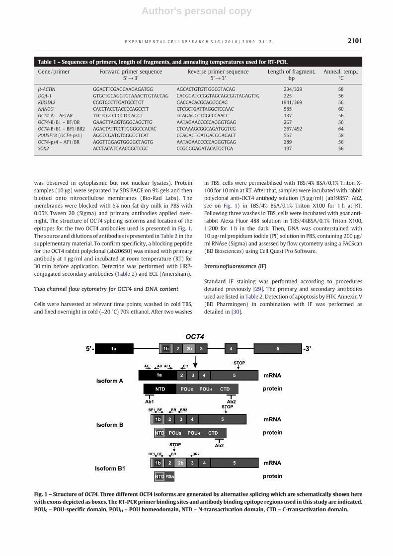
Contrary to the situation in p53 mutated cell lines, in p53 wild-type TK6 cells the response to irradiation was characterised by theloss of the initial nuclear NANOG staining within 3-5 days, even inrare giant cells expressing OCT4 (Fig. 8), while the p53-mutatedcounterpart WI-L2-NS displayed coherent up-regulation for bothtranscription factors in most ETC (as exemplified on Fig. 5). Thesedata are in line with the down-regulation of NANOG transcriptionin TK6 cells presented above (Fig. 2).
These observations indicate that the key pluripotency and self-renewal transcription factors OCT4, SOX2 and NANOG are up-regulated through association with PML nuclear bodies and OCT4and NANOG are co-ordinately expressed after irradiation in p53-mutated ETC. Further, our data indicate that this response toirradiation is impaired by functional p53 (in the radiosensitivelymphoblastoma cell line TK6) and by the suppressor of plur-ipotency, retinoic acid.
Antagonism between self-renewal and accelerated senescencein late ETC from p53-mutant lymphoma cell lines induced byirradiation
Mitotic catastrophe has been proposed as a cause of cell deatheither directly from arrested mitosis or indirectly throughaccelerated replicative senescence (ARS) with endopolyploidy asa hallmark feature [50,51]. In line with these data, senescence of aproportion of ETC produced after irradiation has been confirmed inour models previously starting from the end of the first week afterirradiation as judged by the lack of BrdU inclusion, β-galactosi-dase-positivity and loss of the AURORA B-activity [31]. However, a
smaller proportion of ETCs (∼10% by day 14) continued to includeBrdU and express CYCLIN B1 and AURORA B-kinase, indicatingtheir escape from ARS [31]. Similar escape from ARS has also beendescribed in lung cancer models [52,53]. Previously it was shownthat in the absence of p53, cellular senescence is reversible [54]and that in this context p16 INKA 4a the negative regulator of theG1 to S transition serves as a second barrier preventing escapefrom cellular senescence [55]. Therefore, here we addressed therelationship between NANOG (as a marker of self-renewal andpositive regulator of the G1 to S transition [42], p16INKA 4a (as asafeguard and marker of replicative cell senescence), and AURORAB-kinase (as a marker of cell division) in co-immunostainingexperiments on irradiated WI-L2-NS cells. As reported above, wefound low levels of NANOG in untreated cells, whilst p16 INKA 4astaining was negative (Fig. 9A). In the majority of ETC produced 3-5 days after irradiation OCT4 and NANOGwere up-regulated in themitotically potent (AURORA B-positive/p16 INKA 4a-negative)cells (Figs. 9B, C). However, routinely from day 6-7 after irradiationan increasing proportion of ETC began to down-regulate OCT4 andNANOG and many of them became p16 -positive (Fig. 9C). Inparallel to this, a smaller cohort of ETC retained expression of self-renewal factors, continued DNA replication and mitotic activitiesup to de-polyploidisation, as already exemplified on Fig. 5D.Gradually, the p16 -positive ETC were cleared from cell suspen-sions (mostly by delayed apoptosis), although a small numberwere still evident on day 20 after irradiation when most of the cellpopulation had been substituted by the new para-diploid cells(Fig. 9D).
These data clearly demonstrate that not all ETC becomesenescent but, on the contrary, indicate in line with our OCT4/DNA flow cytometry observations, that most ETC attempt toreactivate pluripotency and a considerable proportion of themretain high mitotic potential which appears to be directlysupported by up-regulation of key members of the self-renewalprogram counteracting cellular senescence.
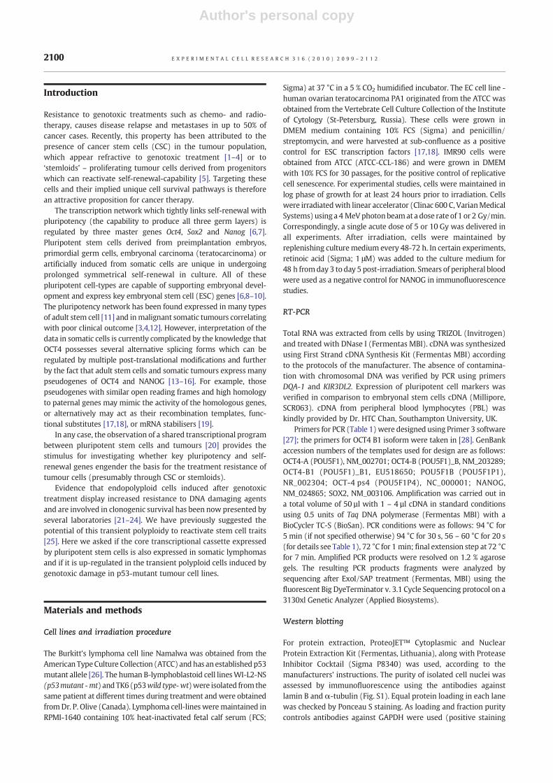
Fig. 7 – Colocalisation of pluripotency transcription factors withPML nuclear bodies. WI-L2-NS cells were left untreated (NT) orwere irradiated and then onday 5were fixed and stained for PMLand eitherOCT4, SOX2 orNANOGplus DNA (DAPI) and imaged bycolour videocamera attached to a fluorescent microscopethrough a BRG optical filter (for its optical characteristics, seeMaterials and Methods). Colocalisation of transcription factors(red)andPML (green)was consideredpositive if twoormore fociper cell displayed yellow fluorescence. Damaged and apoptoticcells were not considered. Data represent mean values of twoindependent experiments: S.E.M. * p<0.05. Two tailed Studentt-test. ColocalisationofOCT4 and SOX2 toPML is found in 1-2%ofNT cells, however increases up to 30-50% of irradiated cells, whileNANOG is persistently colocalised with PML.
Fig. 8 – Lack of NANOG up-regulation in wt p53 tumour cellsafter irradiation. TK6 cells were irradiated and then on day 5were fixed and stained for OCT4, NANOG and DAPI. Theup-regulation of OCT4 in rare ETC of wt p53 TK6 cells wasobserved after irradiation (arrow on ‘A’) but was notaccompanied by up-regulation of NANOG (arrow on ‘B’), whichbecomes down-regulated in all TK6 cells.
2108 E X P E R I M E N T A L C E L L R E S E A R C H 3 1 6 ( 2 0 1 0 ) 2 0 9 9 – 2 1 1 2

Author's personal copy
Discussion
To assess the expression/up-regulation of key pluripotency andself-renewal genes in tumour cells we tested two differentradioresistant p53 mutant B lymphoma cell lines before andafter irradiation. Our detailed RT-PCR analysis provided dataindicating that not only a recognised transactivator, OCT4A, butalso two functional pseudogenes, an OCT4B cytoplasmic isoformand OCT4B1 may be involved in the response of lymphoma celllines after irradiation. In agreement with recent reports [28], itappears that these varying components may be expressedaberrantly and differentially in each individual tumour cell line.
The current understanding of the interplay and function of thedifferent OCT4 isoforms, including the functional pseudogenes isfar from clarity [14,15,35,56], especially regarding their activitiesin somatic tumour cells [13,17,18,36,57]. Therefore, perhaps themost revealing of our findings concerned NANOG, a down-streamtarget and companion of the key pluripotency and self-renewalgenes OCT4 and SOX2 [32]. Prior to irradiation, the basal level ofNANOG transcription was low or un-detectable in both p53mutantlymphoma cell-lines in keeping with published findings inhaematological tumours [58,59]. However, we found by RT-PCR,IF and WB that NANOG became up-regulated after irradiation,indicating that the up-stream regulator genes OCT4 and SOX2became functional upon irradiation in these somatic tumour cell-lines.
Importantly, our IF and flow cytometry observations revealedthat the pathway involving these pluripotency transcriptionfactors is up-regulated in lymphoma cells by genotoxic stressand for the first time show that this up-regulation occurs in ETCwhich are capable of undergoing de-polyploidisation transferringthis phenotype to descendents. Although our data do not excludethe participation of OCT4 and NANOG pseudogenes in thisresponse, several facts indicate that the induced ESC-like transac-tivation network is indeed regulated and functional: Firstly the co-localisation of NANOGwith OCT4 and SOX2 in interphase nuclei ofETC and secondly their absence from mitotic chromosomes arguein favour of this conclusion. Up-regulation of the NANOG promoteris known to be associated with self-renewal symmetrical mitoticdivisions [40–42]. Therefore, finding NANOG foci symmetricallydistributed within the cytoplasm of ana-telophasic-early-G1daughter cells recovering after DNA damage may be interpretedas a hallmark of such divisions, in continuity with the precedingde-polyploidisation of the ETC.
ETC, which were previously considered as reproductively inert,are becoming increasingly accepted as a possible survivalalternative to cell death by mitotic catastrophe [23,60–62]. Theyemerge by abortive mitotic cycles and retain their proliferativepotential as indicated by CYCLIN B1 and AURORA B-kinasepositivity, the capability to recombine and repair DNA double-strand breaks involving meiotic proteins REC8, SGO1 and DMC1,and the ability to de-polyploidise [29–31,63]. Therefore, it isinteresting to note that a similar reversible reproductive endo-polyploidy induced by mitotic catastrophe and associated withOCT4/NANOG expression was described for mouse and human ESCin vitro [64]. In this respect, it is also noteworthy that mixoploidy[65], genome instability [66], and the transcription of pseudogenes[67], are typical features of very early human embryo developmentand have direct parallels to the induced ETC investigated here.
The next question concerns how the key pluripotency and self-renewal transcriptional factors, which have only a backgroundlevel of transcription in non-treated somatic tumour cells, becomefunctional in ETC after genotoxic stress. In this regard, attentionshould be paid to the nucleation centres induced by irradiation,which appear to be initiated by OCT4 foci, expanding and fusingwith SOX2 and NANOG in PML bodies. The association of thesenucleation centres with the initiation of pluripotency wasindirectly confirmed in experiments with retinoic acid, whichcaused loss of OCT4, NANOG, and nuclear localisation of thesestructures. As such, these OCT4-dependent nuclear foci appear tofunction as ‘seeds’ for the growth and extension of the NANOG-enriched nuclear network observed.
It is known that a certain threshold level of ESC factors isrequired to start the positive feed-back circuit of transcriptionactivation [32,68]. Presumably, exactly these conditions arecreated in ETC emerging from p53 mutant cell lines afterirradiation. In this situation, mitotic slippage leading to tetraploidyoccurs and the G1 to S transition of tetraploid cells is favoured,providing an increased gene dosage of the key factors, which arefurther accumulating in the next polyploidising cycles, as many ofthese cells reach at least octaploidy ( [21] and seen on Fig. S 2C). Inaddition, a cellular mechanism capable of bringing key pluripo-tency transcription factors physically together into nucleatingcentres supported and stabilised by PML bodies is induced. Wefurther suggest that stabilisation of OCT4 along with PML-associated NANOG may favour looping out of the NANOG
Fig. 9 – Antagonistic relationship between self-renewal andsenescence markers in WI-L2-NS cells in the post-irradiationtime-course. WI-L2-NS cells were left untreated or wereirradiated and then at various times fixed and stained forNANOG, p16INKA 4a, AURORA B-kinase and DAPI. Samples in (A,C, D) are co-stained for NANOG and for themarker of replicativesenescence p16INKA 4a. Sample in (B) was stained for NANOGand for the marker of cell division AURORA B-kinase, whichlocalises to the centromeres of metaphase chromosomes of ETCrich with NANOG (arrow). Senescing ETC in (C) and (D) aremarked by an asterix and up-regulate p16INKA 4a in the nucleiand cytoplasm and down-regulate NANOG. Bar=10 μm.
2109E X P E R I M E N T A L C E L L R E S E A R C H 3 1 6 ( 2 0 1 0 ) 2 0 9 9 – 2 1 1 2

Author's personal copy
chromatin domain to bring the NANOG promoter and othertranscription factors together for interaction with Pol II aspreviously described [32]. Theoretically, this ‘seeding’ mechanismmay also involve functional pseudogenes of OCT4 and NANOGtranscribed from different chromosomes. The question of whether(and how) functional pseudogenes of OCT4 and NANOG areinvolved in the reactivation of the pluripotency state of somatictumour cells remains a subject for further study. It would also beinteresting to determine whether these ETC undergoing poly-ploidisation by abortive mitoses and de-polyploidisation byreduction divisions are biological candidates for the so-called‘stemloids’ [5].
The self-renewal and plutipotency response described aboveapparently depends on inactivation of p53 function as in p53 wild-type TK6 cells we observed the down-regulation of NANOG afterirradiation in contrast to its induction in the p53mutant counterpartWI-L2-NS. This behaviour is entirely anticipated as p53 is a wellestablished down-regulator of the NANOG promoter [69] and also ofendopolyploidy [70]. The radiosensitivity of the paired TK6 andWI-L2-NS cellswas alsopreviously shown tobe primarily determinedbythe difference in p53 status of these two cell lines [71].
In our previous work [21,30] we studied the role of transientendopolyploidy as a survival alternative of mitotic catastrophe.Here, we extend these observations and indicate that self-renewalof lymphoma cell lines (probably both before and after irradiation)might occur by reactivation of a transcriptional programme similarto that engaged during early embryogenesis through activation oflargely germ-line specific genes and their functional pseudogenes.This finding is in line with several recent reports in somatictumours of various origins showing a correlation between chemo-radio-resistance and poor clinical outcome with the expression ofOCT4, SOX2 and NANOG [3,4,12] or their down-stream targets ofthe pluripotency network [72].
In addition to the induction of the pluripotency and self-renewal genes, endopolyploidy induced by irradiation also causesactivation of several meiotic genes including MOS, REC8, SGO1,DMC1, SPO11 typically involved in metaphase arrest, geneticrecombination, and reduction divisions [24,29,45,63]. These find-ings enabled us to previously suggest that this complex poly-ploidisation-de-polyploidisation response of tumour cells,extended for a week or more following genotoxic damage,possesses the features of an evolutionary asexual life-cycle,which transfers “stemness” from one generation of tumour cellsto another [25,73]. Furthermore, Tam et al [74] have postulatedthat aged stem cells might be rejuvenated by reprogramming to anESC-like state, while Curran et al [75] has shown that aspects ofsoma-to-germ-line transmission are important in C. eleganslongevity. In line with these expectations, we have now confirmedthat the reversible polyploidy does indeed involve induction ofgerm-line genes in ETC which counteract cellular senescence. Thisdata provides a molecular explanation for the self-renewal andsurvival potential of tumour cells through reversible polyploidy.
Conclusions
In conclusion, our data show the clear up-regulation of thepluripotency and self-renewal transcription network in p53mutant lymphoma cell lines induced by irradiation to undergoreversible endopolyploidy.
Acknowledgements
The authors would like to acknowledge Dr. Dace Pjanova forassistance with confocal microscopy, Galina Boka for irradiation ofcells, and Dr. Liene Nikitina-Zake for donating primers DQA-1 andKIR3DL2. This work was supported by ESF grant 1DP/1.1.1.2.0/09/APIA/VIAA/150 for reagent purchases and by the Royal Society ofLondon enabling exchange visits between Riga, Southampton andManchester.
Appendix A. Supplementary data
Supplementary data associated with this article can be found, inthe online version, at doi:10.1016/j.yexcr.2010.04.030.
R E F E R E N C E S
[1] C.T. Jordan, M.L. Guzman, M. Noble, Cancer stem cells, N. Engl. J.Med. 355 (2006) 1253–1261.
[2] C.T. Jordan, Cancer stem cells: controversial or justmisunderstood? Cell Stem Cell 4 (2009) 203–205.
[3] I. Hideshi, I. Masaaki, I. Keisuke, O. Daisuke, H. Naotsugu, M. Koshi,M. Masaki, Cancer stem cells and chemoradiation resistance,Cancer Sci. 99 (2008) 1871–1877.
[4] C.J. Creighton, X. Li, M. Landis, J.M. Dixon, V.M. Neumeister, A.Sjolund, D.L. Rimm, H.Wong, A. Rodriguez, J.I. Herschkowitz, C. Fan,X. Zhang, X. He, A. Pavlick, M.C. Gutierrez, L. Renshaw, Al.A.Larionov, D. Faratian, S.G. Hilsenbeck, C.M. Perou, M.T. Lewis, J.M.Rosen, J.C. Chang, Residual breast cancers after conventionaltherapy display mesenchymal as well as tumor-initiating features,Proc. Natl. Acad. Sci. U. S. A. 106 (33) (2009) 13820–13825.
[5] M.V. Blagosklonny, Cancer stem cell and cancer stemloids: frombiology to therapy, Cancer Biol. Ther. 11 (2007) 1684–1690.
[6] I. Chambers, A. Smith, Self-renewal of teratocarcinoma andembryonic stem cells, Oncogene 23 (2004) 7150–7160.
[7] L.A. Boyer, T.I. Lee, M.F. Cole, S.E. Johnstone, S.S. Levine, J.P. Zucker,M.G. Guenther, R.M. Kumar, H.L. Murray, R.G. Jenner, D.K. Gifford,D.A. Melton, R. Jaenisch, R.A. Young, Core transcriptionalregulatory circuitry in human embryonic stem cells, Cell 122(2005) 947–956.
[8] K. Okita, T. Ichisaka, S. Yamanaka, Generation of germline-competentinduced pluripotent stem cells, Nature 448 (2007) 313–317.
[9] R. Josephson, C.J. Ording, Y. Liu, S. Shin, U. Lakshmipathy, A.Toumadje, B. Love, J.D. Chesnut, P.W. Andrews, M.S. Rao, J.M.Auerbach, Qualification of embryonal carcinoma 2102Ep as areference for human embryonic stem cell research, Stem Cells 25(2007) 437–446.
[10] M.J. Boland, J.L. Hazen, K.L. Nazor, A.R. Rodriguez, W. Gifford, G.Martin, S. Kupriyanov, K.K. Baldwin, Adult mice generated frominduced pluripotent stem cells, Nature 461 (2009) 91–94.
[11] C.J. Lengner, F.D. Camargo, K. Hochedlinger, G.G. Welstead, S.Zaidi, S. Gokhale, H.R. Schцler, A. Tomilin, R. Jaenisch, Oct4expression is not required for mouse somatic stem cellself-renewal, Cell Stem Cell 1 (2007) 403–415.
[12] S. Saigusa, K. Tanaka, Y. Toiyama, T. Yokoe, Y. Okugawa, Y. Ioue, C.Miki, M. Kusunoki, Correlation of CD133, OCT4, and SOX2 in rectalcancer and their association with distant recurrence afterchemoradiotherapy, Ann. Surg. Oncol. (2009)Doi: 10.1245/s10434-009-0617-z.
[13] T. Cantz, G. Key, M. Bleidissel, L. Gentile, D.W. Han, A. Brenne, H.Schöller, Absence of OCT4 Expression in Somatic Tumor CellLines, Stem Cells 26 (2008) 692–697.
2110 E X P E R I M E N T A L C E L L R E S E A R C H 3 1 6 ( 2 0 1 0 ) 2 0 9 9 – 2 1 1 2

Author's personal copy
[14] S. Liedtke, M. Stephan, G. Kögler, Oct4 expression revisited:Potential pitfalls for data misinterpretation in stem cell research,Biol. Chem. 389 (2008) 845–850.
[15] C.J. Lengner, G.G. Welstead, R. Jaenisch, The pluripotencyregulator Oct4: a role in somatic stem cells? Cell Cycle 7 (2008)725–728.
[16] J.H. Chin, H. Shiwaku, O. Goda, A. Komuro, H. Okazawa, Neuralstem cells express Oct-3/4, Biochem. Biophys. Res. Comm. 388(2009) 247–251.
[17] G. Suo, J. Han, X. Wang, J. Zhang, Y. Zhao, Y. Zhao, J. Dai, Oct4pseudogenes are transcribed in cancers, Biochem. Biophys. Res.Comm. 337 (2005) 1047–1051.
[18] J. Zhang, X. Wang, L. Meixiang, J. Han, B. Chen, B. Wang, J. Dai,NANOGP8 is a retrogene expressed in cancers, FEBS J. 273 (8)(2006) 1723–1730.
[19] Y. Yano, R. Saito, N. Yoshida, A. Yoshiki, A. Wynshaw-Boris, M.Tomita, S. Horotsune, A new role for expressed pseudogenes asncRNA: regulation of mRNA stability of its homologous codinggene, J. Mol. Med. 82 (2004) 414–422.
[20] D.J. Wong, E. Segal, H.Y. Chang, Modal map of stem cell genesguides creation of epithelial cancer stem cells, Cell Stem Cell 2(2008) 333–344.
[21] T. Illidge, M. Cragg, B. Fringe, P. Olive, Je. Erenpreisa, Polyploidgiant cells provide survival mechanism for p53 mutant cells afterDNA damage, Cell. Biol. Int. 24 (2000) 621–633.
[22] M. Sundaram, D.L. Guernsey, M.M. Rajaraman, R. Rajaraman,Neosis: a novel type of cell division in cancer, Cancer Biol. Ther. 3(2) (2004) 207–218.
[23] P.E. Puig, M.N. Guill, A. Bouchot, N. Droin, D. Cathelin, F. Bouyer, L.Favier, F. Ghiringhelli, G. Kroemer, E. Solary, F. Martin, B.Chauffert, Tumor cells can escape DNA-damaging cisplatinthrough DNA endoreduplication and reversible polyploidy, CellBiol. Int. 32 (2008) 1031–1043.
[24] L. Vitale, M. Jemaà Senovilla, M. Michaud, L. Galluzzi, O. Kepp, L.Nanty, A. Criollo, S. Rello-Varona, G. Manic, D. Métivier, S. Vivet, N.Tajeddine, N. Joza, A. Valent, M. Castedo, G. Kroemer, Multipolarmitosis of tetraploid cells: inhibition by p53 and dependency onMos, J. EMBO 29 (2010) 1272–1284.
[25] Je. Erenpreisa, M.S. Cragg, Cancer: A matter of life-cycle, Cell Biol.Int. 31 (2007) 1507–1510.
[26] P.M. O'Connor, J. Jackman, D. Jondle, K. Bhatia, J. Magrath, K.W.Kohn, Role of the p53 tumour suppressor gene in cell cycle arrestand radiosensitivity of Burkitt's lymphoma cell lines, Cancer Res.5 (1993) 4776–4780.
[27] S. Rozen, H.J. Skaletsky, Primer3 on the WWW for general usersand for biologist programmers, in: S. Krawetz, S. Misener (Eds.),Bioinformatics Methods and Protocols: Methods in MolecularBiology, Humana Press, Totowa, NJ, 2000, pp. 365–386.
[28] Y. Atlasi, S.J. Mowla, S.A.M. Ziaee, P.J. Gokhale, P.W. Andrews,OCT4 spliced variants are differentially expressed in humanpluripotent and nonpluripotent cells, Stem Cells 26 (2008)3068–3074.
[29] J. Erenpreisa, M.S. Cragg, K. Salmina, M. Hausmann, H. Scherthan,The role of meiotic cohesin REC8 in chromosome segregation ingamma irradiation-induced endopolyploid tumour cells, Exp. CellRes. 315 (2009) 2593–2603.
[30] A. Ivanov, M.S. Cragg, J. Erenpreisa, D. Emzinsh, H. Lukman, T.M.Illidge, Endopolyploid cells produced after severe genotoxicdamage have the potential to repair DNA double strand breaks, J.Cell Sci. 11 (2003) 4095–4106.
[31] J. Erenpreisa, A. Ivanov, S.P. Wheatley, E.A. Kosmacek, F. Ianzini, A.P. Anisimov, M. Mackey, P.J. Davis, G. Plakhins, T.M. Illidge,Endopolyploidy in irradiated p53-defficient tumour cell lines:Persistence of cell division activity in giant cells expressingAurora-B kinase, Cell Biol. Int. 32 (2008) 1044–1056.
[32] D.N. Levasseur, J. Wang, M.O. Dorschner, J.A.Stamatoyannopoulos, S.H. Orkin, Oct4 dependence of chromatinstructure within the extended Nanog locus in ES cells, Genes Dev.22 (2008) 575–580.
[33] J. Lee, H.K. Kim, J.Y. Rho, Y.M. Han, J. Kim, The human OCT-4isoforms differ in their ability to confer self-renewal, J. Biol. Chem.281 (2006) 33554–33565.
[34] G. Caufman, I. Liebaers, A. Van Steirteghem, H. Van de Velde,POU5F1 Isoforms show different expression patterns in humanEmbryonic Stem Cells and preimplantation embryos, Stem Cells24 (2006) 2685–2691.
[35] X.Wang, Y. Zhao, Z. Xiao, B. Chen, Z. Wei, B. Wang, J. Zhang, J. Han,Y. Gao, L. Li, H. Zha, W. Zhao, H. Lin, W. Dai, Embryonic stemcells/Induced pluripotent stem cells alternative translation ofOCT4 by an internal ribosome entry site and its novel function instress response, Stem Cells 27 (2009) 1265–1275.
[36] S.I. Papamichos, V. Kotoula, B.C. Tarlatzis, T. Agorastos, K.Papazisis, A.F. Lambropoulos, OCT4B1 isoform: the novel OCT4alternative spliced variant as a putative marker of stemness, Mol.Hum. Reprod. 15 (2009) 269–270.
[37] E. Moller Panagopoulos, A. Collin, F. Mertens, The POU5F1P1pseudogene encodes a putative protein similar to POU5F1isoform 1, Oncology Rep. 20 (2008) 1029–1033.
[38] D.J. Rodda, J.-L. Chew, L.-H. Lim, Y.-H. Loh, B. Wang, H.-H. Ng, P.Robson, Transcriptional Regulation of Nanog by OCT4 and SOX2, J.Biol. Chem. 280 (2005) 24731–24737.
[39] J. Li, G. Pan, K. Cui, Y. Liu, S. Xu, D. Pei, A dominant-negative formof mouse SOX2 induces trophectoderm differentiation andprogressive polyploidy in mouse embryonic stem cells, J. Biol.Chem. 282 (2007) 19481–19492.
[40] I. Chambers, J. Silva, D. Colby, J. Nichols, B. Nijmeijer, M.Robertson, J. Vrana, K. Jones, L. Grotewold, A. Smith, Nanogsafeguards pluripotency and mediates germline development,Nature 450 (2007) 1230–1234.
[41] J. Silva, J. Nichols, T.W. Theunissen, G. Guo, A.L. van Oosten, O.Barrandon, J.Wray, S. Yamanaka, I. Chambers, A. Smith,Nanog is thegateway to the pluripotent ground state, Cell 138 (2009) 722–737.
[42] X. Zhang, I. Neganova, S. Przyborski, C. Yang, M. Cooke, S.P.Atkinson, G. Anyfantis, S. Feny, N.W. Keith, S.F. Hoare, O. Hughes,T. Strachan, M. Stojkovic, P.W. Hinds, L. Armstrong, M. Lako, A rolefor NANOG in G1 to S transition in human embryonic stem cellsthrough direct binding of CDK6 and CDC25A, J. Cell Biol. 184(2009) 67–82.
[43] H.A. Booth, P.W. Holland, Eleven daughters of NANOG, Genomics84 (2004) 229–238.
[44] J.L. Kopp, B.D. Ormsbee, M. Desler, A. Rizzino, Small increases inthe level of Sox2 trigger the differentiation of mouse embryonicstem cells, Stem Cells 26 (2008) 903–911.
[45] M. Kalejs, A. Ivanov, G. Plakhins, M.S. Cragg, Dz. Emzins, T.M.Illidge, Je. Erenpreisa, Upregulation of meiosis-specific genes inlymphoma cell lines following genotoxic insult and the inductionof mitotic catastrophe, BMC Cancer 6 (2006) 6doi:10.1186/1471-2407-6-6.
[46] G. Dellaire, D.P. Bazzette-Johns, PML nuclear bodies: dynamicsensors of DNA damage and cellular stress, BioEssays 26 (2004)963–977.
[47] F. Wei, H.R. Scho'ller, M.L. Atchison, Sumoylation of Oct4enhances its stability, DNA binding, and transactivation, J. Biol.Chem. 282 (2007) 21551–21560.
[48] J. Schoorlemmer, A. Van Puijenbroek, M. Van den Eijnden, L. Jonk,C. Pals, W. Kruijer, Characterization of a negative retinoic acidresponse element in the murine Oct4 promoter, Mol. Cell Biol. 14(1994) 1122–1136.
[49] S. Minucci, V. Botquin, Y.I. Yeom, A. Dey, I. Sylvester, D.J. Zand, K.Ohbo, K. Ozato, H.R. Schóller, Retinoic acid-mediateddown-regulation of Oct3/4 coincides with the loss of promoteroccupancy in vivo, EMBO J. 15 (1996) 888–899.
[50] J.W. Shay, I.B. Roninson, Hallmarks of senescence in carcinogenesisand cancer therapy, Oncogene 23 (2004) 2919–2933.
[51] D. Yang, D.J. McCrann, H. Nguyen, C. St Hilaire, R.A. DePinho, M.R.Jones, K. Ravid, Increased polyploidy in aortic vascular smoothmuscle cells during aging is marked by cellular senescence, AgingCell 6 (2007) 257–260.
2111E X P E R I M E N T A L C E L L R E S E A R C H 3 1 6 ( 2 0 1 0 ) 2 0 9 9 – 2 1 1 2
Author's personal copy
[52] R.S. Roberson, S.J. Kussick, E. Vallieres, S.-Y.J. Chen, D.Y. Wu,Escape from therapy-induced accelerated cellular senescence inp53-null lung cancer cells and in human lung cancers, Cancer Res.65 (2005) 2795–2803.
[53] M. Sabisz, A. Skladanowski, Cancer stem cells and escape fromdrug-induced premature senescence in human lung tumor cells:implications for drug resistance and in vitro drug screeningmodels, Cell Cycle 8 (2009) 3208–3217.
[54] J.A. Bond, F.S. Wyllie, D. Wynford-Thomas, Escape fromsenescence in human diploid fibroblasts induced directly bymutant p53, Oncogene 9 (1994) 1885–1889.
[55] C.M. Beausejour, A. Krtolica, F. Galimi, M. Narita, S.W. Lowe, P.Yaswen, J. Campisi, Reversal of human cellular senescence: rolesof p53 and p16 pathways, EMBO J. 22 (2003) 4212–4222.
[56] H. Lin, A. Shabbir, M. Molnar, T. Lee, Stem cell regulatory functionmediated by expression of a novel mouse Oct4 pseudogene,Biochem. Biophys. Res. Com. 355 (2007) 111–116.
[57] N. Mizuno, M. Kosaka, Novel variants of Oct-3/4 gene expressedin mouse somatic cells, J. Biol. Chem. 283 (2008) 30997–31004.
[58] A.H. Hart, L. Hartley, M. Ibrahim, L. Robb, Identification, cloningand expression analysis of the pluripotency promoting Nanoggenes in mouse and human, Dev. Dyn. 230 (2004) 187–198.
[59] S. Santagata, J.L. Hornick, K.L. Ligon, Comparative analysis of germcell transcription factors in CNS germinoma reveals diagnosticutility of NANOG, Am. J. Surg. Pathol. 30 (2006) 1613–1618.
[60] R. Rajaraman, D.L. Guernsey, M.M. Rajaraman, S.R. Rajaraman,Stem cells, senescence, neosis and self-renewal in cancer, CancerCell Int. 6 (25) (2006) 17092342 PMID.
[61] H. Vakifahmetoglu, M. Olsson, B. Zhivotovsky, Death through atragedy:mitotic catastrophe, CellDeathDiffer. 15 (2008)1153–1162.
[62] H.O. Lee, J.M. Davidson, R.J. Duronio, Endoreplication: polyploidywith purpose, Genes Dev. 23 (21) (2009) 2461–2477.
[63] F. Ianzini, E.A. Kosmacek, E.S. Nelson, E. Napoli, J. Erenpreisa, M.Kalejs, M.A. Mackey, Activation of meiosis-specific genes isassociated with depolyploidization of human tumor cellsfollowing radiation-induced mitotic catastrophe, Cancer Res. 69(2009) 2296–2304.
[64] C. Mantel, Y. Guo, M.R. Lee, M.-K. Kim, M.-K. Han, H. Shibayama, S.Fukuda, M.C. Yoder, L.M. Pelus, K.-S. Kim, H.E. Broxmeyer,Checkpoint-apoptosis uncoupling in human and mouse
embryonic stem cells: A source of karyotypic instability, Blood109 (2007) 4518–4527.
[65] M. Benkhalifa, L. Janny, P. Vye, P. Malet, D. Boucher, Y. Menezo,Assessment of polyploidy in human morulae and blastocystsusing co-culture and fluorescent in-situ hybridization, Hum.Reprod. 8 (1993) 895–902.
[66] E. Vanneste, T. Voet, C.Le. Caignec, M. Ampe, P. Konings, C.Melotte, S.D. Cebrock, M. Amyere, M. Vikkula, F. Schuit, J.P. Fryns,G. Verbeke, T. D'Hooghe, Y. Moreau, J.R. Vermeesch, Chromosomeinstability is common in human cleavage-stage embryos, Nat.Med. 15 (2009) 577–583.
[67] A.E. Peaston, B.B. Knowles, K.W. Hutchison, Genome plasticity inthe mouse oocyte and early embryo, Biochem. Soc. Trans. 35(2007) 618–622.
[68] M. Hemberger, W. Dean, W. Reik, Epigenetic dynamics of stemcells and cell lineage commitment: digging Waddington's canal,Nat. Rev. Mol. Cell Biol. 10 (2009) 526–537.
[69] T. Lin, C. Chao, S. Saito, S.J. Mazur, M.E. Murphy, E. Appella, Y. Xu,P53 induces differentiation of mouse embryonic stem cells bysuppressing Nanog expression, Nat. Cell Biol. 7 (2005) 165–171.
[70] J.S. Lanni, T. Jacks, Characterization of the p53-dependantpostmitotic checkpoint following spindle disruption, Mol. CellBiol. 18 (1998) 1055–1064.
[71] W. Zhen, C.M. Denault, K. Loviscek, S. Walter, L. Geng, A.T.M.Vaughan, The relative radiosensitivity of TK6 and WI-L2-NSlymphoblastoid cells derived from a common source is primarilydetermined by their p53 mutational status, Mut. Res. Lett. 346(1995) 85–92.
[72] I. Ben-Porath, M.W. Thomson, V.J. Carey, R. Ge, G.W. Bell, A. Regev,R.A. Weinberg, An embryonic stem cell–like gene expressionsignature in poorly differentiated aggressive human tumors, Nat.Genet. 40 (2008) 499–507.
[73] J. Erenpreisa, M.S. Cragg, Life-cycle features of tumour cells, in: P.Pontarotti (Ed.), Evolutionary Biology from Concept to Application,Springer Verlag, Berlin-Heidelberg, 2008, pp. 61–67.
[74] W.L. Tam, Y.S. Ang, B. Lim, The molecular basis of ageing in stemcells, Mech. Ageing Dev. 128 (2007) 137–148.
[75] S.P. Curran, X. Wu, C.G. Riedel, G. Ruvkun, A soma-to-germlinetransformation in long-lived Caenorhabditis elegans mutants,Nature 459 (2009) 1079–1084.
2112 E X P E R I M E N T A L C E L L R E S E A R C H 3 1 6 ( 2 0 1 0 ) 2 0 9 9 – 2 1 1 2