author's personal copy - tamu.edu
TRANSCRIPT

Author's personal copy
Integrins as receptor targets for neurological disorders
Xin Wu, Doodipala Samba Reddy ⁎Department of Neuroscience and Experimental Therapeutics, Texas A&M Health Science Center College of Medicine, Bryan, TX 77807, United States
a b s t r a c ta r t i c l e i n f o
Keywords:Extracellular matrix proteinIntegrinNeuroplasticityNervous diseasesDrug development
This review focuses on the neurobiology of integrins, pathophysiological roles of integrins in neuroplasticityand nervous system disorders, and therapeutic implications of integrins as potential drug targets and possibledelivery pathways. Neuroplasticity is a central phenomenon inmany neurological conditions such as seizures,trauma, and traumatic brain injury. During the course of many brain diseases, in addition to intracellular com-partment changes, alterations in non-cell compartments such as extracellular matrix (ECM) are recognized asan essential process in forming and reorganizing neural connections. Integrins are heterodimeric transmem-brane receptors that mediate cell–ECM and cell–cell adhesion events. Although themechanisms of neuroplas-ticity remain unclear, it has been suggested that integrins undergo plasticity including clustering throughinteractions with ECM proteins, modulating ion channels, intracellular Ca2+ and protein kinase signaling,and reorganization of cytoskeletal filaments. As cell surface receptors, integrins are central to the pathophys-iology of many brain diseases, such as epilepsy, and are potential targets for the development of new drugs forneurological disorders.
© 2012 Elsevier Inc. All rights reserved.
Contents
1. Introduction . . . . . . . . . . . . . . . . . . . . . . . . . . . . . . . . . . . . . . . . . . . . . . . 682. Structure and function of integrins . . . . . . . . . . . . . . . . . . . . . . . . . . . . . . . . . . . . 693. Physiological and pathological roles of integrins in brain behavior and brain disorders . . . . . . . . . . . . 694. Therapeutic implications of integrins as drug targets . . . . . . . . . . . . . . . . . . . . . . . . . . . . 705. Conclusions and future perspectives . . . . . . . . . . . . . . . . . . . . . . . . . . . . . . . . . . . 70Conflict of interest statement . . . . . . . . . . . . . . . . . . . . . . . . . . . . . . . . . . . . . . . . . 71Acknowledgments . . . . . . . . . . . . . . . . . . . . . . . . . . . . . . . . . . . . . . . . . . . . . . 72References . . . . . . . . . . . . . . . . . . . . . . . . . . . . . . . . . . . . . . . . . . . . . . . . . . 72
1. Introduction
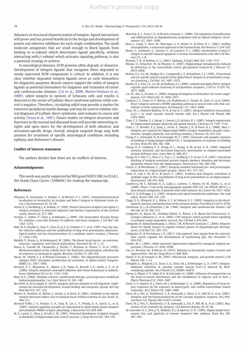
Neuroplasticity is the ability of the nervous system to react withadaptive changes to intrinsic or extrinsic challenges such as epilepsy,trauma, and brain injury. In many cerebrovascular and neuronal dis-eases, in addition to intracellular compartment changes, alterationsin non-cell compartments such as extracellular matrix (ECM) are rec-ognized as an essential process. There has been recent interest in thepossible role of adhesion molecules, particularly integrins as ECM re-ceptors, in neurological disorders because they form an importantlink between the ECM and the intracellular cytoskeleton (CSK) andsignaling molecules (Fig. 1). In the brain, ECM proteins are synthe-sized and secreted into the extracellular space in amesh-like structureby neurons and glial cells. During development and mature nervoussystems, and in wound healing, the interaction between ECM and itsreceptor integrin has a pivotal role in maintaining structural and
Pharmacology & Therapeutics 134 (2012) 68–81
Abbreviations: Ab, antibody; AMPA, α-amino-3-hydroxy-5-methyl-4-isoxazolepro-pionic acid; BBB, blood–brain barrier; CaL, L-type Ca2+ channels; CN, collagen; CNS,central nervous system; CSK, cytoskeleton; DGC, dentate granular cell; ECM, extracel-lular matrix; EGF, epidermal growth factor; EPSP, excitatory postsynaptic potentials;ERK, extracellular signal-regulated protein kinases; FAC, focal adhesion complex;FAK, focal adhesion kinase; FN, fibronectin; GABA, γ-aminobutyric acid-A; LDV,leucine–aspartate–valine; LN, laminin; LTP, long-term potentiation; MEK, mitogen-activated protein kinase; NDMA, N-methyl-D-aspartic acid; NRTK, non-receptor tyro-sine kinase; PNS, peripheral nervous system; RGD, arginine–glycine–aspartate; TLE,temporal lobe epilepsy; VN, vitronectin.⁎ Corresponding author at: Department of Neuroscience and Experimental Thera-
peutics, Texas A&M Health Science Center College of Medicine, 1005 Medical Researchand Education Building, 8447 State Highway 47, Bryan, TX 77807-3260, United States.Tel.: 979 436 0324.
E-mail address: [email protected] (D.S. Reddy).
737677787878
0163-7258/$ – see front matter © 2012 Elsevier Inc. All rights reserved.doi:10.1016/j.pharmthera.2011.12.008
Contents lists available at SciVerse ScienceDirect
Pharmacology & Therapeutics
j ou rna l homepage: www.e lsev ie r .com/ locate /pharmthera

Author's personal copy
functional neuroplasticity. ECM and integrin aberrations are likely tocontribute to imbalanced synaptic function in epilepsy, Alzheimer'sdisease, mental retardation, schizophrenia and other conditions inthe brain. (Dityatev & Schachner, 2003; Gall et al., 2003; Gall &Lynch, 2004; Dityatev et al., 2010).
Studies of integrin-initiated intracellular signaling have shown thatintegrins modulate Ca2+ and K+ ion channels, intracellular Ca2+ con-centrations, cellular contractile properties, protein kinase activity, andgrowth factor receptors (Hynes, 1992; Yip & Marsh, 1997; Porter &Hogg, 1998; Wu et al., 1998; Barouch et al., 2000; Chan et al., 2001;Danen & Yamada, 2001; Davis et al., 2001; Wu et al., 2001; Davis etal., 2002; Waitkus-Edwards et al., 2002; Martinez-Lemus et al., 2003;Rueckschloss & Isenberg, 2004; Gui et al., 2006; Wu et al., 2008a,2008b, 2010b, 2010c; Yang et al., 2010; Wu et al., 2011a). Furthermore,ECM–integrin–CSK interactions play crucial roles in gene expression,cell proliferation, migration and differentiation, and cell survival(Hynes, 1992; Yamada & Miyamoto, 1995; Schwartz, 2001; Kim et al.,2011).
Since the discovery of the first integrin receptor for ECM proteinfibronectin (FN) in 1986 (Tamkun et al., 1986), eighteen α-subunitsand eight β-subunits have been identified (Hynes, 2002). Interesting-ly, there are at least 24 distinct integrins (Reichardt & Prokop, 2011)even though there are twenty-four α-subunit and nine β-subunitgenes in the human genome (Venter et al., 2001). A total of over50,000 papers are published on integrins so far. Fewer than 1000papers are in the field of neuroscience as compared to ~6000 papersin the cardiovascular system, indicating that integrins are not widelyexplored in the brain.
Integrins are necessary for neuronal cells to attach, spread, mi-grate, and extend processes on ECM molecules. Interactions of ECM–
integrin–focal adhesion complex (FAC) including CSK proteins and in-tracellular signaling molecules in neuronal systems play an importantrole in synapse morphology and number, neuron–neuron and neuro-muscular synaptic transmission, and neuroplasticity that modulatesneuronal cell proliferation, migration, and differentiation (Venstrom& Reichardt, 1993). Historically, the role of the ECM and its receptorswas viewed as largely structural molecules. However, there is emerg-ing evidence that these proteins and receptors also perform signalingand regulatory functions in neuronal pathophysiological processessuch as memory, inflammation, wound healing, epileptogenesis, an-giogenesis, and tumor metastasis. Therefore, integrins as cell surfacereceptors are attractive pharmacological targets for designing newtherapies for brain diseases (Horwitz, 1997; Clemetson & Clemetson,1998; Wu & Davis, 1998; Davis et al., 2001; Ross & Borg, 2001; Gall &Lynch, 2004; Jin & Varner, 2004; Lal et al., 2007; Cox et al., 2010;Millard et al., 2011; Reichardt & Prokop, 2011).
This review describes the neurobiology of integrins, pathophysio-logical roles of integrins in neuroplasticity, and the therapeutic impli-cations of integrin targeted drugs for nervous system disorders.
2. Structure and function of integrins
2.1. Integrins in neurons
Integrins are α–β-heterodimeric transmembrane glycoprotein re-ceptors that mediate cell–ECM and cell–cell adhesion events through
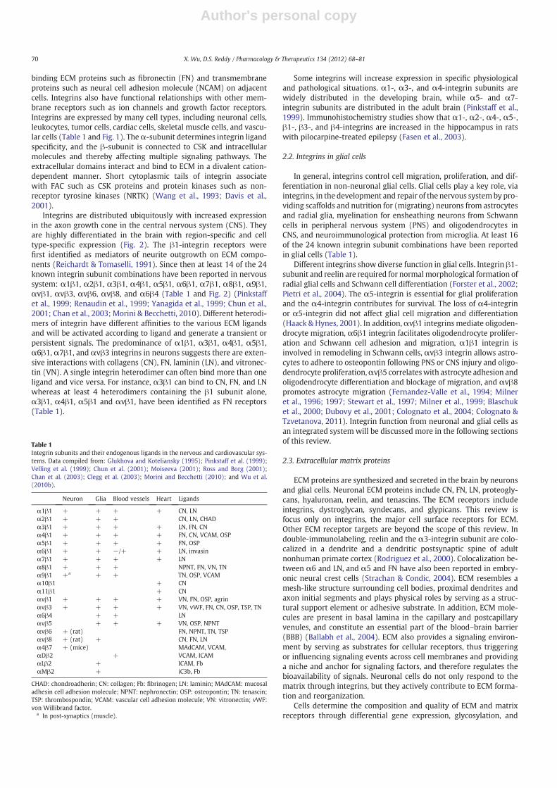
Fig. 1. Schematic diagram for integrin-mediated signaling pathways. Integrins mediate cell–extracellular matrix (ECM) and cell–cell adhesion events through binding ECM proteinssuch as fibronectin and transmembrane proteins such as cell adhesion molecules (CAMs) on adjacent cells. The ECM–integrin system consists of ECM proteins, integrins and focaladhesion complex (FAC) that includes non-receptor tyrosine kinases (NRTK) and cytoskeleton (CSK) proteins. In this model, integrin clustering by multivalent ligands, includingECM proteins, induces recruitment of CSK proteins such as talin, vinculin, actin, tubulin, actinin, paxillin and tensin, and NRTK such as focal adhesion kinase (FAK) and Src tothe focal contact. Integrin-linked kinase (ILK), phospholipase C (PLC), inositol trisphosphate (IP3), diglyceride (DAG), protein kinase A (PKA), PKC, PKG, Raf, GTPase (e.g. Ras,Cdc42, Rac, SOS and C3G), mitogen-activated protein kinase (MEK), extracellular signal-regulated protein kinases (ERK), c-Jun N-terminal kinases (JNK), as well as adaptor proteinssuch as Grb2, Crk, and Sos, are also recruited to the ECM–integrin binding site. Integrins also mediate crosstalk with other cell surface receptors such as growth factor receptor(GFR), cadherins and CAMs. ECM–integrin–FAC interactions play an important role in modulation of ion channels, synaptic transmission, and neuroplasticity that modulates neu-ronal cell proliferation, differentiation, apoptosis and migration. Integrins play an important role in pathological processes such as inflammation, wound healing, epileptogenesis,angiogenesis, and tumor metastasis.
69X. Wu, D.S. Reddy / Pharmacology & Therapeutics 134 (2012) 68–81
Author's personal copy
binding ECM proteins such as fibronectin (FN) and transmembraneproteins such as neural cell adhesion molecule (NCAM) on adjacentcells. Integrins also have functional relationships with other mem-brane receptors such as ion channels and growth factor receptors.Integrins are expressed by many cell types, including neuronal cells,leukocytes, tumor cells, cardiac cells, skeletal muscle cells, and vascu-lar cells (Table 1 and Fig. 1). Theα-subunit determines integrin ligandspecificity, and the β-subunit is connected to CSK and intracellularmolecules and thereby affecting multiple signaling pathways. Theextracellular domains interact and bind to ECM in a divalent cation-dependent manner. Short cytoplasmic tails of integrin associatewith FAC such as CSK proteins and protein kinases such as non-receptor tyrosine kinases (NRTK) (Wang et al., 1993; Davis et al.,2001).
Integrins are distributed ubiquitously with increased expressionin the axon growth cone in the central nervous system (CNS). Theyare highly differentiated in the brain with region-specific and celltype-specific expression (Fig. 2). The β1-integrin receptors werefirst identified as mediators of neurite outgrowth on ECM compo-nents (Reichardt & Tomaselli, 1991). Since then at least 14 of the 24known integrin subunit combinations have been reported in nervoussystem: α1β1, α2β1, α3β1, α4β1, α5β1, α6β1, α7β1, α8β1, α9β1,αvβ1, αvβ3, αvβ6, αvβ8, and α6β4 (Table 1 and Fig. 2) (Pinkstaffet al., 1999; Renaudin et al., 1999; Yanagida et al., 1999; Chun et al.,2001; Chan et al., 2003; Morini & Becchetti, 2010). Different heterodi-mers of integrin have different affinities to the various ECM ligandsand will be activated according to ligand and generate a transient orpersistent signals. The predominance of α1β1, α3β1, α4β1, α5β1,α6β1, α7β1, and αvβ3 integrins in neurons suggests there are exten-sive interactions with collagens (CN), FN, laminin (LN), and vitronec-tin (VN). A single integrin heterodimer can often bind more than oneligand and vice versa. For instance, α3β1 can bind to CN, FN, and LNwhereas at least 4 heterodimers containing the β1 subunit alone,α3β1, α4β1, α5β1 and αvβ1, have been identified as FN receptors(Table 1).
Some integrins will increase expression in specific physiologicaland pathological situations. α1-, α3-, and α4-integrin subunits arewidely distributed in the developing brain, while α5- and α7-integrin subunits are distributed in the adult brain (Pinkstaff et al.,1999). Immunohistochemistry studies show that α1-, α2-, α4-, α5-,β1-, β3-, and β4-integrins are increased in the hippocampus in ratswith pilocarpine-treated epilepsy (Fasen et al., 2003).
2.2. Integrins in glial cells
In general, integrins control cell migration, proliferation, and dif-ferentiation in non-neuronal glial cells. Glial cells play a key role, viaintegrins, in the development and repair of the nervous systemby pro-viding scaffolds and nutrition for (migrating) neurons from astrocytesand radial glia, myelination for ensheathing neurons from Schwanncells in peripheral nervous system (PNS) and oligodendrocytes inCNS, and neuroimmunological protection from microglia. At least 16of the 24 known integrin subunit combinations have been reportedin glial cells (Table 1).
Different integrins show diverse function in glial cells. Integrin β1-subunit and reelin are required for normalmorphological formation ofradial glial cells and Schwann cell differentiation (Forster et al., 2002;Pietri et al., 2004). The α5-integrin is essential for glial proliferationand the α4-integrin contributes for survival. The loss of α4-integrinor α5-integrin did not affect glial cell migration and differentiation(Haack & Hynes, 2001). In addition,αvβ1 integrins mediate oligoden-drocyte migration, α6β1 integrin facilitates oligodendrocyte prolifer-ation and Schwann cell adhesion and migration, α1β1 integrin isinvolved in remodeling in Schwann cells, αvβ3 integrin allows astro-cytes to adhere to osteopontin following PNS or CNS injury and oligo-dendrocyte proliferation,αvβ5 correlateswith astrocyte adhesion andoligodendrocyte differentiation and blockage of migration, and αvβ8promotes astrocyte migration (Fernandez-Valle et al., 1994; Milneret al., 1996; 1997; Stewart et al., 1997; Milner et al., 1999; Blaschuket al., 2000; Dubovy et al., 2001; Colognato et al., 2004; Colognato &Tzvetanova, 2011). Integrin function from neuronal and glial cells asan integrated system will be discussed more in the following sectionsof this review.
2.3. Extracellular matrix proteins
ECM proteins are synthesized and secreted in the brain by neuronsand glial cells. Neuronal ECM proteins include CN, FN, LN, proteogly-cans, hyaluronan, reelin, and tenascins. The ECM receptors includeintegrins, dystroglycan, syndecans, and glypicans. This review isfocus only on integrins, the major cell surface receptors for ECM.Other ECM receptor targets are beyond the scope of this review. Indouble-immunolabeling, reelin and the α3-integrin subunit are colo-calized in a dendrite and a dendritic postsynaptic spine of adultnonhuman primate cortex (Rodriguez et al., 2000). Colocalization be-tween α6 and LN, and α5 and FN have also been reported in embry-onic neural crest cells (Strachan & Condic, 2004). ECM resembles amesh-like structure surrounding cell bodies, proximal dendrites andaxon initial segments and plays physical roles by serving as a struc-tural support element or adhesive substrate. In addition, ECM mole-cules are present in basal lamina in the capillary and postcapillaryvenules, and constitute an essential part of the blood–brain barrier(BBB) (Ballabh et al., 2004). ECM also provides a signaling environ-ment by serving as substrates for cellular receptors, thus triggeringor influencing signaling events across cell membranes and providinga niche and anchor for signaling factors, and therefore regulates thebioavailability of signals. Neuronal cells do not only respond to thematrix through integrins, but they actively contribute to ECM forma-tion and reorganization.
Cells determine the composition and quality of ECM and matrixreceptors through differential gene expression, glycosylation, and
Table 1Integrin subunits and their endogenous ligands in the nervous and cardiovascular sys-tems. Data compiled from: Glukhova and Koteliansky (1995); Pinkstaff et al. (1999);Velling et al. (1999); Chun et al. (2001); Moiseeva (2001); Ross and Borg (2001);Chan et al. (2003); Clegg et al. (2003); Morini and Becchetti (2010); and Wu et al.(2010b).
Neuron Glia Blood vessels Heart Ligands
α1β1 + + + + CN, LNα2β1 + + + CN, LN, CHADα3β1 + + + + LN, FN, CNα4β1 + + + + FN, CN, VCAM, OSPα5β1 + + + + FN, OSPα6β1 + + −/+ + LN, invasinα7β1 + + + + LNα8β1 + + + NPNT, FN, VN, TNα9β1 +a + + TN, OSP, VCAMα10β1 + CNα11β1 + CNαvβ1 + + + + VN, FN, OSP, agrinαvβ3 + + + + VN, vWF, FN, CN, OSP, TSP, TNα6β4 + + LNαvβ5 + + + VN, OSP, NPNTαvβ6 + (rat) FN, NPNT, TN, TSPαvβ8 + (rat) + CN, FN, LNα4β7 + (mice) MAdCAM, VCAM,αDβ2 + VCAM, ICAMαLβ2 + ICAM, FbαMβ2 + iC3b, Fb
CHAD: chondroadherin; CN: collagen; Fb: fibrinogen; LN: laminin; MAdCAM: mucosaladhesin cell adhesion molecule; NPNT: nephronectin; OSP: osteopontin; TN: tenascin;TSP: thrombospondin; VCAM: vascular cell adhesion molecule; VN: vitronectin; vWF:von Willibrand factor.
a In post-synaptics (muscle).
70 X. Wu, D.S. Reddy / Pharmacology & Therapeutics 134 (2012) 68–81

Author's personal copy
endocytic trafficking. Neuronal cells also release extracellular prote-ase enzymes, such as collagenolytic matrix metalloproteinases, a dis-integrin and metalloproteinase with thrombospondin motifs andtissue plasminogen activator, to help in restructuring the matrix andsignificantly influencing the bioavailability of signals in respondingto conditional changes, or regulating the passage of growing or mi-grating cells (Reichardt & Prokop, 2011). LN, VN, reelin and chondroi-tin sulfates are reported to be involved in neuronal development(Heinrich et al., 2006; Dityatev et al., 2007).
Different integrin binding sites even in the same ECM proteinsmight have different functions. Many neuronal matrix proteins, in-cluding FN and CN, contain the arginine–glycine–aspartate (RGD) rec-ognition site which can be recognized by the majority of β1-integrins.Application of RGD peptide interferes with cell–ECM adhesion anddisrupts a wide variety of integrin-mediated intracellular activities.Other than RGD, the IIICS region of FN contains a 25-amino acid se-quence, including the leucine–aspartate–valine (LDV) motif thatbinds the α4β1 integrin (Geiger et al., 2001). RGD and soluble FNcan decrease voltage gated L-type Ca2+ currents (CaL) and dilate thearterioles, while LDV in FN and insoluble FN can increase Ca2+ cur-rents and constrict the arterioles (D'Angelo et al., 1997; Wu et al.,1998; Waitkus-Edwards et al., 2002). These ECM effects may contrib-ute to vascular regulation in stroke and brain trauma. Overall, ECMproteins have pivotal functions at the BBB and contribute to the pro-cesses of neuronal cell migration, axonal growth and myelination,and to the assembly and functional maintenance of the neuromuscu-lar junction. ECM–integrin–FAC axis and itsmechanisms have a crucialimpact on nervous system development, maintenance, degeneration,and regeneration at all levels, acting through the regulation of celladhesion and signaling, endosomal trafficking, CSK dynamics, andgene expression (Reichardt & Prokop, 2011).
2.4. ECM–integrin–FAC axis
Different integrins may have distinct functions according to theirligand binding and type of intracellular signaling that is activated.Integrins can function as signaling receptors that transduce biochem-ical signals both into and out of cells (Clark & Brugge, 1995). Integrin
activation by ECM ligands can be described as three main activationstates of rest, intermediate affinity state and activated high-affinitystate (Cox et al., 2010), or 3 steps as inactive, active and clustering(Clark & Brugge, 1995). Integrins are not constitutively active. In gen-eral, integrins on oncogenic variants could be constitutively activated.Some integrins can bind their ligands in a resting state and then be ac-tivated, whereas others require activation before binding. Other thanECM ligands, extracellular factors such as mechanical stress (e.g.blood pressure), divalent cation concentration (e.g. Mn2+), and inter-leukin signaling also lead to integrin activation. After activation, integ-rin clustering by multivalent ECM proteins induces recruitments ofCSK proteins (ECM–integrin–CSK axis) including actin, vinculin,talin, paxillin and tensin as well as NRTK kinases such as focal adhe-sion kinase (FAK) and the Src kinase family to the focal contact pointforming into large multi-protein aggregates, termed cell-matrix focaladhesion complex (FAC). Mitogen-activated protein kinases (MEK),PLC-γ, and small GTPases rho, rac, and cdc42 as well as adaptor pro-teins such as Grb2 and Sos are also recruited to the ECM–integrinbinding site after integrin activation (Fig. 1). Ligand binding inducesconformational changes in integrins and intracellular signaling, re-ferred to as “outside‐in” signaling. This intracellular signaling cascadesactivated by integrin activation can result in the cytoskeletal changesneeded to modulate growth cone structure, neurite outgrowth andaxonmigration. This signal transduction has been also shown to resultin protein phosphorylation, increased intracellular calcium concentra-tion, activation of glutamate receptor, gene transcription, and the re-lease of neurotransmitters (Clark & Brugge, 1995).
Intracellular signaling molecules (e.g. CSK and protein kinases)converge on the cytoplasmic domain of integrin tails after initialligand occupancy. In turn, ion channel activation, CSK remodeling,and intracellular signaling activities in neuronal cells can controlintegrin expression, activate the high affinity state of integrin (cluster-ing), and manipulate remodeling of the ECM and ECM binding affinityonto integrin in the plasma membrane. Therefore, populations ofintegrins are not static, but change dynamically across periods of acti-vation in response to physiological and pathological events. Thesealterations are referred to as “inside–out” signaling through theECM–integrin–FAC axis (Clark & Brugge, 1995; Wu et al., 2001; Gall
Fig. 2. Integrin subunits distribution in different regions of central nervous system. Data compiled from: Glukhova and Koteliansky (1995); Pinkstaff et al. (1999); Velling et al.(1999); Chun et al. (2001); Moiseeva (2001); Ross and Borg (2001); Chan et al. (2003); Morini and Becchetti (2010) and Wu et al. (2010b). Individual integrin reported in onespecies but perhaps not in others indicated in parentheses.
71X. Wu, D.S. Reddy / Pharmacology & Therapeutics 134 (2012) 68–81

Author's personal copy
et al., 2003; Morini & Becchetti, 2010). The inside–out signaling alsooccurs in regulating synaptic structure, signaling, and various formsof neuronal plasticity. In the brain, integrins are concentrated at sitesof synaptic contact and are critical for the formation, maturation,and maintenance of synaptic structure (Nishimura et al., 1998).Basal synaptic communication requires that the presynaptic and post-synaptic faces of the synapse communicate via adhesive and intracel-lular signaling events (Schachner, 1997; Hoffman et al., 1998a).Cleavage of integrin–ECM focal adhesive connections could be anearly step in the formation of new synaptic configurations frominside–out signaling, followed by rapid formation of new focal con-tacts (Hoffman et al., 1998a).
2.5. Integrin crosstalk with cell surface receptors
As one of the cell surface receptors, integrins mediate crosstalkwith other cell surface receptors such as growth factor receptorsand cell adhesion molecules (CAMs) (Fig. 1). It is highly establishedthat nerve growth factor (NGF) stimulates integrin-dependent axonoutgrowth, but the mechanism connecting the two systems is notwell understood. Integrins and growth factor receptors may activateparallel intracellular signaling pathways including extracellular signalregulated protein kinases (ERK)-type MEK. Co-stimulation of integ-rins and epidermal growth factor (EGF) receptors activates Pyk2and FAK to promote outgrowth of neurite via paxillin induced cyto-skeletal changes (Ivankovic-Dikic et al., 2000). Integrin clusteringtransactivates several receptor tyrosine kinases (RTKs) including plate-let derived growth factor receptor (PDGF) and EGF receptor. Thus,integrins may increase signals generated by growth factor receptorsthrough its FAC.
Transforming growth factor beta-1 (TGF-β1) latency-associatedpeptide is a ligand for the integrinαvβ6, andαvβ6 integrin expressingcells induce spatially restricted activation of TGFβ1.Mice lackingαvβ6integrin develop exaggerated inflammation (Munger et al., 1999). In-teractions between α6-integrin and growth factor enable newlyformed oligodendrocytes to survive in response to limiting concentra-tions of soluble growth factors (Colognato et al., 2002). The αvβ8 hasbeen shown to promote the activation of TGFβ through binding to anRGD sequence in the TGFβ latency-associated peptide (Mu et al.,2002). Baron et al. have demonstrated a physical association betweenPDGF and αvβ3 receptors on oligodendrocytes. PDGF stimulated aprotein kinase C-dependent activation of αvβ3 integrin, which inturn induced oligodendrocyte proliferation via a phosphatidylinositol3-kinase-dependent signaling pathway (Baron et al., 2002).
It has been reported that synaptic remodeling would require alter-ations to interaction between integrin and cell adhesion molecules.Integrin mediate cell–cell interaction through cell adhesionmoleculessuch as neural cell adhesion molecule (NCAM), intercellular adhesionmolecule (ICAM), and vascular-cell adhesion molecule (VCAM). TheNCAM L1 functionally interacts with β1-integrins to potentiateneuronal migration toward ECM proteins such as FN through endocy-tosis and MEK signaling, and that impairment of this function by L1cytoplasmic domain mutations may contribute to neurological defi-cits in the X-linked mental retardation syndrome CRASH (corpus cal-losum agenesis, retardation, aphasia, spasticity, and hydrocephalus)(Thelen et al., 2002). During neuronal injury and cytokine stimula-tion, microglia bind to their targets using αLβ2 and α4β1 through in-teraction with ICAM-1 and VCAM-1 and migrating to the site throughαMβ2 (Clegg et al., 2003).
Cross-talk has also been documented between integrin and cad-herins. N-Cadherins and integrins function in a coordinated mannerto effectively mediate the cellular interactions essential for adhesionand development. A peptide resembling the juxtamembrane (JMP)region of the cytoplasmic domain of N-cadherin results in release ofthe NRTK Fer from the cadherin complex and its accumulation inthe integrin-induced FAC, and results in inhibition of N-cadherin
and β1-integrin function. A peptide that mimics the first coiled-coil domain of Fer prevents Fer accumulation in the integrin cyto-plasmic domain and reverses the inhibitory effect of JMP (Arreguiet al., 2000). These results show that integrin activation is neces-sary to influence the function of other cell surface receptors andvice versa.
2.6. Integrin crosstalk with neurotransmitter receptors
Integrin could also have crosstalk with neurotransmitter recep-tors. It has been reported that α7β1 integrin colocalizes and physical-ly interacts with LN-induced acetylcholine receptors (AchRs) clusters.LN through α7β1 facilitates the clustering of AChRs by a proteoglycanagrin in the post-synaptic membrane of skeletal muscle. Applyingblocking antibodies to αv-, α7-, or β1-integrins inhibit the AchR clus-tering activity of LN and agrin (Martin & Sanes, 1997; Burkin et al.,1998, 2000). RGD-containing peptides or antibodies to the β3-integrin prevent a developmental increase in glutamate release anda concurrent shift in expression of postsynaptic N-methyl-D-asparticacid (NMDA)-receptor subunits (Chavis & Westbrook, 2001). In rathippocampal slices and synaptoneurosomes, a RGD peptide GRGDSP,leads to potentiation of NMDA-gated currents, and increases theslope and amplitude of the fast excitatory postsynaptic potentials(EPSPs) because ofα-amino-3-hydroxy-5-methyl-4-isoxazolepropio-nic acid (AMPA) receptor activation (Lin et al., 2003; Bernard-Trifiloet al., 2005).
Integrin ligands such as RGD peptides and FN via β1-integrin in-crease EPSP in the hippocampus and could pass through two path-ways. One of these pathways is through a Src kinase dependentNMDA and/or AMPA glutamate receptor pathway. Src tyrosine kinaseincreases protein phosphorylation of NMDA receptor subunits NR2Aand NR2B in synaptoneurosomes and acute hippocampal slices. An-other pathway is through a Src tyrosine kinase independent pathway.This pathway is related to activation of NMDA receptor, phosphoryla-tion of Thr286 site on calmodulin-dependent kinase II, and phosphor-ylation of Ser831 site on AMPA receptors. The β1 integrin subunit or amixture of function-blocking antibodies to α3, α5, and αv-integrinsubunits block integrin ligand effects on synaptic responses. The γ-aminobutyric acid-A (GABAA) receptor antagonist picrotoxin doesnot reduce the enhanced effects of EPSP by integrin ligands (Kramaret al., 2003; Lin et al., 2003; Bernard-Trifilo et al., 2005). In β1-integrin knockout (KO) mice, expressions of AMPA and NMDA recep-tor subunits are normal. However, EPSPs at CA3–CA1 synapses aredramatically reduced, probably because of postsynaptic defect inAMPA receptor (Chan et al., 2006). These results show that hippo-campal integrin activation is necessary to control the functionallevel of postsynaptic NMDA and AMPA receptors.
2.7. Integrin and Ca2+ signaling
Integrinsmodulate calcium signaling. The regulation of CaL channelsby integrin–ECM interactionsmay play a role in the regulation of neuro-nal migration, neurite extension and synaptic remodeling. Growth coneextension requires a permissive range of intracellular Ca2+ concentra-tion, whereas the frequency of Ca2+ waves and spikes controls therate of axonal elongation/spreading (Becchetti & Arcangeli, 2010). Neu-ronal CaL channels in fresh isolated neuronal cells and heterologouslyexpressed neuronal CaL channel isoforms in HEK-293 cells are potenti-ated byα5β1 integrin. This potentiation is regulated by an intracellularsignaling pathway involving phosphorylation of calcium channel sub-unit α1C C-terminal residues Ser1901 and Tyr2122 by using truncationand site-directed mutagenesis strategies (Gui et al., 2006; Wu et al.,2010a). These sites are known to be phosphorylated by PKA and c-Src, re-spectively. Soluble RGD containing peptide ligands increase the expres-sion of mRNAs for the neurotrophin brain derived neurotrophic factor,nerve growth factor, neurotrophin-3, the neurotrophin receptors TrkB
72 X. Wu, D.S. Reddy / Pharmacology & Therapeutics 134 (2012) 68–81
Author's personal copy
and TrkC, and cfos at least in part through effects on calcium influx inadult hippocampus (Gall et al., 2003). In this context, integrin-CaL chan-nel interactions appear to be critical for the up-regulation of brain-derived neurotrophic factor mRNA in hippocampal neurons. Work incerebellar neurons, immune cells, and epithelial keratinocytes suggeststhat a stable rear-to-front Ca2+ gradient is formed in cells during che-motaxis/migration, which is thought to be one of the causes of the dif-ferent behavior of the front and rear sides, with retraction anddetachment of the trailing edge during cell movement cycle.
Integrin-CaL channel interactions could also play amajor role in theresponses of neurons and blood vessels to injury and repair (e.g.stroke). A series of observations from our previous studies show thatat least 3 different integrins regulate CaL, Ca2+ activated K+ channelsand myogenic responses in vascular systems, and in heterologouslyexpressed smooth muscle CaL in HEK cells through inside–out andoutside–in signaling between integrin and FAC (Wu et al., 1998,2001; Davis et al., 2002; Waitkus-Edwards et al., 2002; Gui et al.,2006; Wu et al., 2008b; Yang et al., 2010). Both neuronal activity andvascular reactivity are modulated by integrin signaling through thegeneration or exposure of new integrin ligands from limited degrada-tion of ECM and/or turnover of new integrins during wound healing(Davis et al., 2000; Gui et al., 2006). For instance, successful axonal re-generation is highly correlated with the induction of integrins on thesurface of peripheral neurons; therefore, peptides derived from integ-rin ligand have the potential to act as therapeutic agents for neuronalregeneration (Meiners & Mercado, 2003; Cox et al., 2010).
2.8. Integrin in nervous system development
Integrins play a major role in the development of the CNS by stabi-lizing cellular contacts during cell migration, cell proliferation, andneurite extension. Integrin and FAC signaling molecule recruitment(e.g. GTPases Cdc42, Rac1 and CSK actin at leading edge), and integ-rin–ECM detachment/deadhesion (at trailing edge) and efficientattachment/adhesion have been reported to dynamically changetheir position at leading and trailing edges in migrating cells duringdevelopment (Becchetti & Arcangeli, 2010; Huttenlocher & Horwitz,2011). Lacking ECM components such as LN or FN, or lacking the β1-family of integrins, mice die during the early steps of embryonic devel-opment. The loss ofα4,α5, or/and β1-integrins from themice embryoresults in abnormal lamination of the cortex and cerebellum due todisruptions of the basal lamina that separates the brain from the over-lying mesenchyme, and results in severe perturbations of the periph-eral nervous system, including failure of normal nerve arborization,delay in Schwann cell migration, survival, proliferation, and differenti-ation, and defective neuromuscular junction differentiation (Graus-Porta et al., 2001; Haack & Hynes, 2001; Feltri et al., 2002; Pietri etal., 2004). These alterations are likely to reflect the roles of integrin re-ceptors in regulating activation of MEK, Rac, and other signaling path-ways (Campos et al., 2004).
In an in vitro inhibition assays with function-integrin antibodies inavian neural crest cells, cell spreading is essentiallymediated by FN re-ceptorαvβ1 andα8β1, andmigration involvedα4β1,αvβ3 andα8β1(Testaz et al., 1999). In LN receptor α6-integirn KO mice, the corticalbasement membrane is disrupted with alterations of LN deposition,ectopic neuroblastic outgrowths are found on the brain surface andin the vitreous body in the eye, and myelin-forming oligodendrocytesshow increased cell death (Georges-Labouesse et al., 1998). α3-integrin KO mice show a disorganized cortex and defective neuronalmigration (Anton et al., 1999). The α5β1 integrin as FN receptor hasbeen implicated in regulating themorphology of cortical developmentand dendritic spines, formation of synapses in neurons, as well as inmediating neurite outgrowth after injury. Embryos lacking the α5-integrin subunit die at embryonic day 10.5 before cerebral cortex de-velopment (Marchetti et al., 2010). No obvious phenotypes in the pe-ripheral nervous system have been reported inmice lacking theαv- or
β8- integrins. However, expression of αvβ8 is required for normalvascular development in the CNS. Absence of eitherαv or β8 integrinsin the CNS leads to cerebral hemorrhage, seizures, axonal degenera-tion and premature death (McCarty et al., 2005, Proctor et al., 2005).In the future, conditional mutation on more subtle genetic modelsfor integrins, ECM proteins, and FAC molecules will allow a more de-tailed understanding on the highly complicated function of integrin-mediated adhesion in the development of nervous system.
3. Physiological and pathological roles ofintegrins in brain behavior and brain disorders
Very little is known regarding the role of integrins in neurophysio-logical and pathological conditions. Emerging evidence shows thatintegrin and ECM production are altered in physiological and patho-logical conditions such asmemory, tumor, Alzheimer's disease, stroke,and epilepsy (Clegg et al., 2003; Gall & Lynch, 2004; Dityatev & Fellin,2008). Although integrins are known to be involved in various physi-ological conditions and brain disorders, identifying the specific integ-rin involved and their precise role are difficult because many diseasesare multifactorial and integrins are only one of these receptors in-volved. In this section, we will discuss recent reports on integrin rolein the pathophysiology of learning and memory, and selected braindisorders.
3.1. Learning and memory
Integrins are involved in learning and memory through modula-tion of long-term potentiation (LTP). The LTP has long been regardedas a plausible cellular substrate for learning and memory. At least twoessential biochemical phases exist for LTP linked learning and memo-ry: an early phase involving modification of existing synaptic pro-teins, and a late phase conveyed by de novo protein synthesis andformation of new synapses. It is thought that integrins help to formsynapses in learning and memory. Integrins are involved in activity-dependent synaptic plasticity and in spatial memory. Evidence for apotential role in synaptic plasticity has been gathered by attenuatingthe stability of hippocampal LTP by using broad-spectrum peptideinhibitors of integrins or other pharmacological reagents. A directlink of integrin function to memory formation is demonstrated inDrosophila, in which the disruption of Volado, a gene encoding fortwo forms of the α-integrin, impairs short-term olfactory learning.Conditional expression of an integrin subunit rescued the memorydeficits (Grotewiel et al., 1998).
Furthermore, disruption of the integrin-associated protein pro-duces memory deficits in mice (Huang et al., 1998; Chang et al.,1999, 2001). Two vertebrate integrins of α8 and β8 have been local-ized to dendritic spines of pyramidal neuronswhere they are associat-ed with postsynaptic density (Einheber et al., 1996; Nishimura et al.,1998). The α5 integrin has been shown to distribute preferentiallyto apical dendrites of pyramidal cells of the hippocampus and neocor-tex (Bi et al., 2001). Four different integrins of Drosophila have beenlocalized to the presynaptic and/or postsynaptic side of the larval neu-romuscular junction (Prokop, 1999). Heterozygous mutants of theintegrin gene α3-subunit, but not of α5- or α8-subunit, reduce themagnitude of NMDA receptor dependent hippocampal LTP. However,when the expression of the three integrin genes, α3-, α5- and α8-subunits, is reduced simultaneously, a deficiency in spatial memoryis produced but fear conditioning remains normal. These functionalchanges are associated with a fairly rapid but subtle morphologicalchange in the structure of the synapse such that synaptic transmissionis altered (Chan et al., 2003).
Electrophysiological studies demonstrated that mutation or re-moval of β1-integrin have impaired synaptic transmission throughAMPA receptors and diminished NMDA receptor-dependent LTP, andimpaired in some kind of memories (Chan et al., 2006; Huang et al.,
73X. Wu, D.S. Reddy / Pharmacology & Therapeutics 134 (2012) 68–81

Author's personal copy
2006). In addition, the β1-integrin receptors and their ECM ligands(FN, LN and CN) have been implicated in the process of neurite out-growth and LTP (Venstrom & Reichardt, 1993; Chan et al., 2003;Huang et al., 2006). Neurite outgrowth is a process commonly thoughtto contribute to LTP by formation of new synaptic contacts that can bedetected in the process of learning and memory. Activation of β1-integrin or several β1-binding α-integrins, including α3-, α5- andα8, further initiate intracellular signaling, and transcription and trans-lation, and facilitate LTP related learning and memory.
Integrin ligands participate in the process of learning andmemory.ECMmolecule tenascin-C and a fragment of tenascin-C containing thefibronectin type-III repeats 6–8 are involved in hippocampus-dependent contextual memory and synaptic plasticity (Strekalova etal., 2002). The disruption of integrin–ECM interactions using RGDpeptide and other integrin antagonists also interferes with the main-tenance of use-dependent synaptic reorganization and LTP. Hippo-campal synapses are enriched in FN receptors (i.e. α5β1 integrin)that contribute importantly to the stabilization of LTP (Bahr et al.,1997; Staubli et al., 1998; Rohrbough et al., 2000; Chun et al., 2001;Kramar et al., 2002). Overall, integrins are essential for neuroplasti-city in LTP and memory. Thus, future studies that elucidate the signal-ing cascade triggered by integrin engagement at the synapses willprovide new insight into the processes of learning and memory.
3.2. Aging, Alzheimer's disease and Down's syndrome
Integrins may play roles in aging and age-related neurological dis-orders. Integrins are involved in synaptic plasticity in neurodegenera-tive conditions and immune response to the diseases. In humans,hippocampal pyramidal neurons and some neocortical neuronsshowed immunoreactivity with α4-integrin subunit and FN in allaged individuals, but not in younger patients. Antibodies against theα4-integrin subunit and a FN specific antibody also stained the tau-positive plaques (i.e. neuritic-type plaques) in Alzheimer's diseaseand Down's syndrome, while ‘preamyloid’ plaques remained negative(Van Gool et al., 1994). In addition, many senile plaques and neurofi-brillary tangles in Alzheimer brain tissue are prominently stained forhigh level VN and VN receptor β3-integrin (Akiyama et al., 1991).The α4β1 and αLβ2 integrins in activated microglial cells adjacent toamyloidal plaques indicate inflammatory responses in Alzheimer dis-ease (Preciado-Patt et al., 1996). In rat hippocampal neurons and ratcortical astrocytes, cell surface β-amyloid precursor protein is coloca-lized selectively with α1β1 and α5β1 integrins. In transfected humanneuroblastoma cell line, α5β1 integrin appears to mediate the inter-nalization and degradation of exogenous β-amyloid. When depositionof an insoluble amyloid around the α5β1-expressing cells is reduced,the cells show less apoptosis than the control cells (Yamazaki et al.,1997; Matter et al., 1998). Further studies on integrin such as α4β1and α5β1 integrins in human brain tissue may provide more insighton the role of integrins in aging and Alzheimer's disease.
3.3. Injury and stroke
Integrins are believed to play an important role in neuronal injuryand ischemic stroke. FN and α5β1 integrin are expressed at compara-tively high levels in developing nerve, and less prominently expressedduring nerve maturation. Following lesion of mature nerve or hippo-campal hilus lesion by surgery, the up-regulated expression of FNand β1-integrin (e.g. α5β1 integrin) are in the vicinity of the lesion,in the growth cones of regenerating neurons and on Schwann cells(Lefcort et al., 1992; Pinkstaff et al., 1998). During peripheral neuralrepair and regeneration, activated integrins in microglia promotes ad-hesion, endocytosis, phagocytosis, and break down of cellular debris.Increased β1-integrin in neuronal cells relates to neuronal adhesionand neurite outreach and regeneration (Clegg et al., 2003). MatureCNS neurons are believed to lack the capacity to regeneration after
injury except dentate granular cells (DGCs) in the hippocampus. How-ever, the regenerative performance of adult neurons can be restored tothat of young neurons by gene transfer-mediated expression of a sin-gle α-integrin. Transfection of adult neuronal cells with α1-integrinresults in increased neurite outgrowth on LN, while transfectionwith α5-integrin enhances neurite outgrowth on FN in vitro (Condic,2001). Genetic variants with integrin α2-subunit have been reportedto be associated with an increased risk for ischemic stroke (Matarinet al., 2008). After focal ischemic stroke, increased synthesis and re-lease of matrix proteins osteopontin to the normal brain, and the in-creased expression of integrin αvβ3 indicated that osteopontin playsa novel role in glial activation, organization, and repair functions(Ellison et al., 1999). Following stroke, β1-integrin is increased in ce-rebral blood vessel in ipsilateral ischemic cortex, and is involved inmodulating angiogenesis in ischemic stroke (Lathia et al., 2010).
In both rat and mouse stroke models, western blot analysisrevealed elevated levels of ECM fragment of perlecan (domain V).Post-stroke domain V administration increased vascular endothelialgrowth factor levels through brain endothelial cell α5β1 integrin,and the subsequent neuroprotective and angiogenic actions of perle-can domain V were in turn mediated through vascular endothelialgrowth factor receptors (Lee et al., 2011). Becker et al. reportedthat treatment with α4-integrin antibody (Ab) decreased infarctsize, increased lymphocyte/monocyte and lowered neurological defi-cit scores in rat focal cerebral ischemia model (Becker et al., 2001).These results suggest that integrins could represent a promising ap-proach for stroke treatment.
3.4. Epilepsy
There is emerging evidence on the role of integrins in epilepsy. Ep-ilepsy is the second most common neurological disorder that affectsover 3 million people in the US and about 50 million worldwide(Browne & Holmes, 2001). It is a disorder in which the balance be-tween neuronal excitability and inhibition is disrupted, leaningtoward uncontrolled synchronized excitability. In temporal lobeepilepsy (TLE), the hippocampus plays a key role in epileptogenesisand epileptic seizures (Schwartzkroin, 1994; Reddy, 2010). Aberrantexpressions of neurotransmitter receptors, cytoskeletal proteins,synaptic proteins, antioxidant proteins, and MEK have been found inthe hippocampus of patients with TLE (Furtinger et al., 2003;Notenboom et al., 2006; Yang et al., 2006; Perosa et al., 2007). In addi-tion to these intracellular modifications, there is increasing evidencefor a functional correlation between the localization of integrins inthe hippocampus and the role that integrins may play in neuronalepileptiform activities (Chang et al., 1993; Grooms & Jones, 1997).
Integrins are differently distributed in cellular and subcellularcompartments in the hippocampus regions and undergo specific pat-terns of regulation during development of epilepsy. Immunocyto-chemical and co-precipitation studies have suggested 11 potentialintegrin receptors with species difference in the hippocampus includ-ing α1β1, α2β1, α3β1, α4β1, α5β1, α6β1, α8β1, αvβ1, αvβ3, αvβ5and αvβ8 (Fig. 2) (Pinkstaff et al., 1999; Chun et al., 2001; Chan etal., 2003; Fasen et al., 2003). Significant hippocampal immunoreactiv-ity of α1-, α5-, β1-, β3-, β4-, and β5-integrins is observed in the piamater, in vascular endothelia, and in astrocytes at the pial surface; im-munoreactivity of β2-integrin is found exclusively in vascular endo-thelia, and immunoreactivity of α2-, β4-, and β5-integrins is foundin mossy fibers. Pyramidal cells, interneurons of CA1–CA3, and hilarneurons reveal moderated α5-integrin labeling in their cell bodies.After pilocarpine-induced status epilepticus, strong immunoreactivityfor α1-, α2-, α4-, α5-, β1-, β3-, and β4-integrins is observed in reac-tive astrocytes (Fasen et al., 2003). Seizures induce marked increasesin αv- and α1-integrins that are restricted to neuronal cell layersand are particularly striking in hippocampal stratum granulosum,CA1 and CA3.
74 X. Wu, D.S. Reddy / Pharmacology & Therapeutics 134 (2012) 68–81
Author's personal copy
Our immunocytochemistry data show at least 20% increase in α5-integrin expression in hippocampal DGCs in stage 5 kindling mice(unpublished data). In contrast, seizure-induced increases in α6-integrin are more diffusely distributed and are localized in both neu-rons and glia (Gall & Lynch, 2004). The β1-integrin expression isreported broadly increased and have been implicated in the processof neurite outgrowth during the seizure episode (Venstrom &Reichardt, 1993; Gall & Lynch, 2004). Hippocampal neurons andastroglial cells express quite low levels of β1-integrin mRNA innaive rats. However, β1-integrin expression can be dramatically in-creased in response to seizure within hippocampal stratum pyrami-dale and the dentate gyrus hilus. β1-integrin mRNA levels increasedfirst in neurons and peaked with predominant astroglial expressionat 24 h after seizure (Pinkstaff et al., 1998). Laminin β1-subunit andintegrin α2-subunit expression are also elevated in the anterior tem-poral neocortex tissue from patients with intractable epilepsy (Wu etal., 2011b).
Integrins as unique candidates may participate in epileptogenesis.There is increasing evidence for a functional correlation betweenthe localization of integrins in the hippocampus and the role thatintegrins may play in neuronal epileptiform activities (Chang et al.,1993; Grooms & Jones, 1997). Seizure activity has been shown to in-duce a ‘matrix response’. These responses included release of prote-ase, changes in the localization and synthesis of ECM molecules(e.g. FN, reelin, and glycosaminoglycans), and activation of integrin–intracellular signaling pathways following seizure activity in the hip-pocampus (Hoffman et al., 1998b, 1998c; Naffah-Mazzacoratti et al.,1999; Perosa et al., 2002a, 2002b; Gong et al., 2007). In animal epilep-sy and in human TLE, neurons in hippocampus undergo extensiveremodeling, including acute neurodegeneration and intracellular sig-naling adjustments, and chronic processes including reorganizationof mossy fibers, dispersion of the DGC layer and the appearance in ec-topic locations within the dentate gyrus. Integrin recruitment andrelocalization has been observed in migrating cells, and integrin–ECM attachments and focal adhesions have been reported to dynam-ically change their position at leading and rear edges (Becchetti &Arcangeli, 2010; Huttenlocher & Horwitz, 2011). Our data show de-creased FN–integrin binding force/adhesion force and increase cellmembrane stiffness/plasticity in DGCs from stage 5 kindling mice byusing nanoscale atomic force microscopy (unpublished data). Theseresults indicate alternation of ECM–integrin interaction representedas ECM–integrin binding changes and alternation of intracellularCSK remodeling and intracellular molecule activation represented asstiffness/plasticity changes during epilepsy.
As we mentioned above, epilepsy is a disorder in which the bal-ance between neuronal excitability (e.g. NMDA currents) and inhibi-tion (e.g. GABA currents) is leaned toward uncontrolled excitability.Integrin expression is greatest in glutamatergic (NMDA) neuronsand low in GABAergic neurons and glia, and concentrated in specificareas such as synaptic membrane (Gall & Lynch, 2004). There are sev-eral reports regarding β1-integrin family activated excitatory NMDAcurrents and Ca2+ currents in neurons as discussed above (Lin et al.,2003; Bernard-Trifilo et al., 2005; Lin et al., 2005; Gui et al., 2006;Lin et al., 2008). All of these changes will increase glutamate excitabil-ity and burst activities in hippocampal CA1 and DGC, cause hyperex-citation in neurons, and could be primary events leading todevelopment of the epilepsy (Tauck & Nadler, 1985; Sutula et al.,1988; de Lanerolle et al., 1989; Chang et al., 1993; Wuarin & Dudek,2001; Smith & Dudek, 2002; Lin et al., 2003; Bernard-Trifilo et al.,2005; Lin et al., 2005; Gui et al., 2006; Lin et al., 2008). In epilepticconditions, increased neuronal proliferation in the DGCs which areprincipal excitatory glutamatergic phenotype neurons, cause hyper-synchronization (Kokaia, 2011). However, there are still limited re-ports available on whether or how integrin directly or indirectlymodulates GABA channels. Colocalization of α3-integrin subunit andthe GABAA receptor β2/3 subunit has been reported in the Purkinje
neuron. Activating α3β1 integrin impairs the GABAergic miniatureinhibitory postsynaptic currents (Kawaguchi & Hirano, 2006). ECMprotein tenascin-R-deficient mutant mice show impaired LTP, elevat-ed levels of excitatory synaptic transmission, and reduced levels ofperisomatic inhibitory currents mediated by GABAA receptors inCA1 of the hippocampus (Bukalo et al., 2007). These reports revealthe complex interplay between integrins and GABAergic neurotrans-mitter receptors. Because glutamatergic activities are related to LTP asmentioned above, these results indicate that integrin contributions toplasticity are not restricted to ‘good things’ as LTP to memory, but in-cluded ‘bad things’ as long lasting increases in excitatory LTP,strengthen the network and connectivity, and lower the thresholdfor epileptogenesis.
Epileptogenesis involves a local breakdown of normal cell–cell andcell–matrix relationships followed by new synthesis of ECM and integ-rins. During normal neural development, ECM molecules play an im-portant role in neuroplasticity events such as neurogenesis, axonaloutgrowth, and synaptogenesis. Synaptogenesis is associated withthe early stages of epilepsy formation and these plasticity changesare also involved in the development of epilepsy (Chang et al.,1993). ECM–integrin interactions may alter neuronal signalingthrough the RGD binding site or increasing the number of activatedintegrins in epilepsy (Grooms & Jones, 1997; Morini & Becchetti,2010). The RGD-treated brain slices display a significant decrease inthe spontaneous burst rate (epileptic-like activities), but the periodof spontaneous bursting increase dramatically in the hippocampusCA3. This paradoxical effect might be because RGD peptide decreasesthe number of available integrin-RGD containing ECM protein bindingsites, whichmay interferewith neuronal communications required forsynchronized electrical firing rates (Grooms & Jones, 1997). Kainicacid-induced seizure caused a broadly distributed increase of FN asearly as 1 h after recurrent seizure in adult brain hippocampus includ-ing dentate gyrus hilus and cortex as well as a larger focal increase inthe ventral subiculum. FN density is higher in cell hillock within indi-vidual cells in dentate gyrus hilus (Hoffman et al., 1998c). The hippo-campus of TLE patients show increased levels of chondroitin sulfateand hyaluronic acid, another two components of ECM protein glycos-aminoglycans. The increased concentration of hyaluronic acid andchondroitin sulfate in the hippocampus of TLE patients demonstratesthe importance of the matrix compounds during neosynaptogenesis,neurite outgrowth, and the mossy fiber sprouting found in temporallobe epilepsy phenomena (Perosa et al., 2002a).
The expression of ECM molecule tenascin-C, which could bind toα8β1, α9β1, αvβ3 and αvβ6 in nervous system (Varnum-Finney etal., 1995; Nakic et al., 1996; Shapiro et al., 1996; Clegg et al., 2003),reaches peak upregulation at 24 h after kainic acid-induced limbicseizures in the CA1 and granule cell layer of adult rat, coincidentwith activation of granule cells and sprouting of axon terminals. Theremaining tenascin expression 30 days after injection only relates topyramidal cells in CA1 and CA3, coincident with an astroglial response(Nakic et al., 1996). During status epilepticus, overall ECM proteinhevin SC1 levels decrease in DGC. A transient increase in SC1 co-localization with the cellular stress marker Hsp70, the degenerationmarker Fluoro-Jade B, and the neuron activity marker activity-regulated CSK-associated protein are also shown in neurons of thehippocampal CA1, CA3, and hilar regions after status epilepticus. Thelevels of SC1 protein in neurons of the hippocampal seizure-resistantCA2 region do not change throughout the seizure (Lively & Brown,2008).
α-Tubulin, a component of ECM–integrin–FAC (or CSK) axis, ex-hibits increased expression in CA3 and DGC bodies, dendrites andaxons of hippocampus in epilepsy models. Microtubule formationmay contribute to synaptic remodeling, such as mossy fiber sproutingand reorganization of neural networks of kindling-induced epilepto-genesis (Pollard et al., 1994; Hendriksen et al., 2001; Sato & Abe,2001). However, in mesial TLE patients, tubulin α-1 chain, β-tubulin,
75X. Wu, D.S. Reddy / Pharmacology & Therapeutics 134 (2012) 68–81
Author's personal copy
profilin II, and neuronal tropomodulin are significantly reduced,whereas ezrin and vinculin are significantly increased (Yang et al.,2006). Divergent results from animals and patients may be explainedby methodological differences, timing these molecules is examined inanimals and patients, as well as discrepancies between gene and pro-tein levels. Overall, these reports suggest that ECM–integrins–CSK areinvolved in the pathophysiology of epilepsy.
Currently available treatments of epilepsy are only seizuresuppressing ‘antiepileptic’; none are truly disease modifying ‘anti-epileptogenic’ treatment. The major goal of future epilepsy researchis to identify related underlying biological pathways and mechanismsresponsible for epileptogeneis, and drug resistance in certain epilepsypatients. Integrin neurobiology may provide a novel target to developnew drug candidates as ‘anti-epileptogenic’ (Reddy, 2010; Pack et al.,2011).
4. Therapeutic implications of integrins as drug targets
Integrins are being explored for designing drugs for the treatmentof neurological disorders. Because successful axonal regeneration ofattachment, migration, and extension processes are highly correlatedwith the induction of integrins on the surface of neurons, peptides orother types of ligands derived from ECM proteins may have therapeu-tic potential for treatment of brain diseases (Meiners & Mercado,2003).
4.1. Approaches to integrin drugs
Rapid progress has been made in the discovery and developmentof integrin targeted therapeutics in the past 15 years. The firstintegrin-specific drugs target the platelet integrin, αIIbβ3 and havebeen marketed for acute coronary syndromes and prevention of myo-cardial infarction following percutaneous coronary intervention sincethe late 1990s. This drug has been proven to be effective and safe andcontributed to the reduction of mortality and morbidity in acute cor-onary syndromes. There are numerous in vitro, in vivo, preclinical andclinical studies implementing integrins as potential drug candidatesfor human diseases (Table 2). There are several mechanisms to mod-ulate integrin function for drug development. The first mechanism asthe original strategy for currently approved inhibitors is to block the
ligand binding (i.e. block adhesion). Integrin inhibitors targeted onthree integrins of αIIbβ3, α4 and αLβ2 has been approved. Currently,integrin Abs, integrin ligand peptides and small non-peptide inhibi-tors have been clinically targeted. Abs can have longer circulation life-times compared to small peptide-based and non-peptide drugs thusincreasing the duration of therapy. However, the disadvantages asso-ciated with antibody therapeutics are the high cost of production, theneed for intravenous administration, and the propensity for host im-munogenicity and infusion reactions (Cox et al., 2010; Millard et al.,2011).
The second mechanism is to modulate integrin expression and ac-tivation. As recent advances in our understanding of ECM–integrin–intracellular signaling axis, blocking downstream integrin signalingfor both outside–in and inside–out will modulate integrin expressionand activation. For instance, CSK talin binding, known for forming theinitial contacts between integrin β-subunit intracellular tails and theactin cytoskeleton, is a pivotal event and is required specifically forintegrin–ECM–CSK axis signaling and for activation of integrin (Kimet al., 2011; Millard et al., 2011). If talin binding to intracellular β-integrin tail is inhibited, it will prevent inappropriate integrin activa-tion and block subsequent integrin signaling. Similarly, NRTKs such asFAK and Src binding to β-tails and CSK protein paxillin binding to α-subunit intracellular tails represent additional potential key targetsfor development of selective inhibitors in ECM–integrin–FAC path-ways. Some inhibitors such as HYD1 for α6β1 and ATN-161 for FN se-quence to block signaling have been studied in clinical or pre-clinicaltrials (Cianfrocca et al., 2006; Sroka et al., 2006).
4.2. Potential clinical applications
Integrin targeted agentsmay have clinical applications in brain dis-orders. Integrins regulate the transit of lymphocytes, macrophage, andother cells across the blood–brain barrier in response to inflammatorystimuli, anti-integrin reagents are of great interest as therapeuticagents to control demyelinating diseases, such as multiple sclerosisthat involve the immune system and inflammatory responses. A newpotential approach that complements traditional therapy, targetedto specific integrin receptors using integrin ligands, has been reported.The anti-α4-intergin Ab natalizumab, which inhibited attachment ofimmune-competent cells (leukocyte) to inflamed brain endothelium,
Table 2Integrin as target of drugs. Data compiled from: Horwitz (1997); Clemetson and Clemetson (1998); Wu and Davis (1998); Ross and Borg (2001); Staunton et al. (2006); andMillard,et al. (2011).
Integrin Cell types Integrin ligands Example of disorder indication and drugs
αIIbβ3a
(CD41, GPIIbIIIa)Platelets Fibrinogen, von Willebrand
factor, FN, VNAcute coronary syndromea, MI, restenosis after angioplasty,percutaneous coronary interventiona, thrombosis.Drugs: Abciximab, Epifibatide, Abeiximab, etc.
β2 integrinsa
(CD18)Various white blood cells ICAM Reperfusion injury, stroke, shock, MI, multiple sclerosis, psoriasisa,
chronic inflammatory diseases, meningitisDrugs: Erlizumab, Efalizumab (αLβ2)b, etc.
β1 integrins (CD29) Widespread includingnervous system
FN, LN, CN, others Platelet aggregation, angiogenesis, homeostasis,wound healing processing, hypertrophyDrugs: see individual αβ-integrin.
α4β1a (CD49d) Various white blood cells,nervous system
FN, VCAM Multiple sclerosisa, chronic inflammatory diseases,such as Crohn's diseasea, asthma and arthritisDrugs: Tysabri, Natalizumabetc
α5β1 (CD49e) Smooth muscle cells,cardiomyocytes, nervous system
FN (possible biomarkerfor infraction)
Restenosis, muscle dystrophy, tumors, age-related macular degenerationDrugs: Volociximab, etc.
αvβ3 (CD51) Endothelial cells,vascular smooth muscle
FN, VN, others Angiogenesis, glioblastoma, diabetic retinopathy, heart defect,atherosclerosis, prostate cancer, pancreatic cancer,melanoma and postmenopausal osteoporosisDrugs: Cilengitide, Vitaxin, MK0429, etc.
α4β7 Various white blood cells FN, MAdCAM, VCAM Chronic inflammatory diseases, such as ulcerative colitisDrugs: MLN02
FN: fibronectin; VN: vitronectin; CN: collagen; LN: laminin; MAdCAM: mucosal addressin cell adhesion molecule; VCAM: vascular cell adhesion molecule; ICAM: intercellularadhesion molecule; MI: myocardial infarction.
a FDA approved integrins that can be used in diseases are in bold font.b Withdrawn from market in 2009.
76 X. Wu, D.S. Reddy / Pharmacology & Therapeutics 134 (2012) 68–81

Author's personal copy
demonstrated an unequivocal therapeutic effect in preventing re-lapses and slowing down the pace of neurological deterioration in pa-tients with relapsing–remitting multiple sclerosis. Combinationaltreatment with both anti-β7 and α4 integrin subunit Abs led tomore rapid and complete remission than that obtained with anti-α4antibody alone. These reports concluded that α4β1, α4β7, and αEβ7integrins may all play a contributory role in the progression of chronicforms of demyelinating disease, and together with their ligands couldrepresent potential targets for improved treatment of some forms ofmultiple sclerosis (Bartt, 2006; Engelhardt & Kappos, 2008; Kanwaret al., 2000). Treatment with natalizumab (α4-integrin) also led to adramatic reduction of refractory seizures in a patient with multiplesclerosis (Sotgiu et al., 2010). The involvement of integrins in inflam-matory responses has also created interest in the possibility of usinganti-integrin reagents to alleviate several neurodegenerative disor-ders, including Parkinson's and Alzheimer's diseases. Of special inter-est, α1β1, α6β1 integrins and CD47 (integrin-associated protein)appear to interact with amyloid precursor protein and these are pos-tulated tomediate deposition or toxic actions of A and amyloid forma-tion (Bozzo et al., 2004; Koenigsknecht & Landreth, 2004).
Integrin antagonists that target angiogenesis for brain tumors areprogressing through clinical trials. Cilengitide, a selective inhibitor ofαvβ3 and αvβ5 integrins, inhibits cell signaling through focal adhe-sion kinase/Src/Akt and extracellular signal-regulated protein kinasemediated pathways, and attenuates the effect of vascular endothelialgrowth factor stimulation on growth factor signaling in endothelialand glioma tumor cells. Following cilengitide treatment, endothelialand glioma cells show disassembly of cytoskeleton and disruption oftight junction formation (Oliveira-Ferrer et al., 2008). The additionof cilengitide to chemoradiotherapy based treatment regimens hasshown promising preliminary results in ongoing clinical trials innewly diagnosed and progressive glioblastoma multiforme patientsby inhibiting angiogenesis (Millard et al., 2011). The greatest chal-lenge facing the development of anti-angiogenic integrin targetedtherapies is the overall lack of biomarkers by which to measure treat-ment efficacy.
4.3. Drug delivery in nervous system
The major problem in drug delivery to CNS is the presence of theblood–brain barrier (BBB) and blood cerebrospinal fluid barrier,mechanisms that protect CNS against intrusive chemicals but alsoconfound therapeutic interventions. Compared to BBB, blood cerebro-spinal fluid barrier allows the free-movement of small molecules.Many existing drugs including current clinically available integrindrugs are rendered ineffective in the treatment of many CNS diseasesdue to our inability to effectively safely deliver, and sustain themwith-in the CNS. To circumvent the barriers, at least three strategies arepractically possible and have been evaluated. There are manipulatingdrugs, disrupting the BBB and discovering alternative routes for drugdelivery (Misra et al., 2003). Designer drugs are prepared through in-creasing the lipophilicity of therapeutic drugs, and vector (e.g. nano-particles) or receptor and carrier (e.g. levodopa) mediated drugdelivery. Disrupting the BBB is carried out through hypertonic solutioninitiating endothelial cell shrinkage and vasoactive leukotriene in-creasing cell permeability. These two strategies are delivered via thecirculatory system and frequently result in unwanted side effects.The third strategy is to bypass the BBB and deliver the drugs to CNSthrough an intraventricular/intrathecal route, the olfactory pathway,an interstitial/intracranial drug delivery via releasing drugs from bio-logical tissues, nanoparticles and pumps.
Nanoparticles as a drug-carrier system for the antiepileptic val-proic acid have been studied in mice. This study suggested that nano-particles loaded with valproic acid may help to reduce the toxic sideeffects of valproate therapy, not by reducing the therapeutically nec-essary dosage but by inhibition of formation of toxic metabolites
(Darius et al., 2000). Wilson et al. (2008) have used poly-nanoparticles for the targeted delivery of rivastigmine into the ratbrain to treat Alzheimer's disease. Targeted gene therapy with αvβ3integrin-binding nanoparticles as treatment for choroidal neovascu-larization has also been reported (Salehi-Had et al., 2011). Sniffing(intranasal) neuropeptides of melanocortin, vasopressin and insulinachieved direct access to the cerebrospinal fluid within 30 min,bypassing the bloodstream in human trials (Born et al., 2002). RGDpeptides are small peptides that could be applied through this path-way. In addition, application of multifunctional nanocarriers withintegrin ligands across the BBB, allowing for diagnostic and therapeu-tic agents to be selectively targeted on a specific tissue and cell, alsominimizing exposure to the healthy tissue and cell, is one of enor-mous challenges for biological discovery and clinical practice.
BBB dysfunction in the hippocampus could be present in a chronichypertensive state at an early stage, in aged senescence-acceleratedmice, and during inflammatory reaction. BBB structure could be com-promised during stroke, tumor metastasis, and injury (Ueno et al.,2004; Pelegri et al., 2007; Cox et al., 2010). These could provide an op-portunity for applying anti-integrin drugs to CNS.
The road from basic research to clinical application of integrinpharmacology has been very challenging. Approximately 2–4% of pa-tients had anaphylaxis or other hypersensitivity reactions to natalizu-mab. Combined natalizumab and interferon-β1a therapy developedprogressive multifocal leukoencephalopathy in multiple sclerosis be-cause of their immune suppressive properties (Engelhardt & Kappos,2008; Warnke et al., 2010). Thus Natalizumab and related α4-integrin targeting drugs come with a black-box warning on the druglabel and are now limited to patients refractory to standard therapies.In clinical practice, physicians face the tough challenge of accuratelyevaluating both the benefits and the risks of therapy for the patients.The real challenge is that the lack of a full understanding of the func-tion of integrins and the role of integrins in diseases, in particulartheir roles in signaling.
5. Conclusions and future perspectives
In conclusion, ECM–integrin–CSK interactions are involved in rear-rangements of cell body, axonal, dendritic, and astrocytic processes.However, the extent of integrin regulation of the synaptic events duringneuronal diseases remains unclear. ECM–integrin–CSK-intracellularsignaling interactions in the nervous system could play a major role inthe responses of neurons and blood vessels to injury and repair. Bothneuronal activity and vascular reactivity from tissue injury rapidlymod-ulate integrin signaling with consequent modulation of ion channelsand activities, the generation or exposure of new integrin ligandsfrom limited degradation of extracellular matrix through increased ex-tracellular proteases, and/or turnover of new integrins (Banes et al.,1995; Fukai et al., 1995; Jones et al., 1995; Gualandris et al., 1996;Hoffman et al., 1998a, 1998b; Momota et al., 1998; Wu et al., 1998;Endo et al., 1999; Davis et al., 2000; Wu et al., 2000; Davis et al., 2002;Waitkus-Edwards et al., 2002; Kramar et al., 2003; Lin et al., 2003;Martinez-Lemus et al., 2003; Bernard-Trifilo et al., 2005; Gui et al.,2006;Wu et al., 2008b; Yang et al., 2010). Further studies arewarrantedto study how integrins modulate fast dynamic changes of ion channelssuch as GABAergic inhibition.
Integrins as receptors are attractive targets for pharmacologicalmanipulation of integrin-based signaling. Specificity or selectivity isa challenge to develop therapies against integrins. To avoid or lowerrisk of side effects, drugs need to distinguish between integrinsexpressed in healthy and injured tissues, target to activated integrinreceptor not only resting integrins, and compete for single αβ combi-nations, not only inhibiting discrete α or β subunits. For instance,αvβ3 and αIIbβ3 share β3 subunit and both recognize the RGDmotif; drugs that inhibit β3 subunit as αvβ3 antagonist to inhibit an-giogenesis will lead to side effects of bleeding from inhibiting αIIbβ3.
77X. Wu, D.S. Reddy / Pharmacology & Therapeutics 134 (2012) 68–81
Author's personal copy
Advances in structural characterization of integrin–ligand interactionswill prove and has proved beneficial in the design and development ofpotent and selective inhibitors for each single combination. The smallmolecule antagonists that are small enough to block ligands frombinding to α-subunit which determines ligand specificity, withoutinteracting with β-subunit which activates signaling pathway, is alsoa potential strategy to achieve.
In neurological diseases, ECM proteins often degrade or denature.Development of integrin ligands that recognize these degraded ornewly expressed ECM components is critical. In addition, it is notclear whether degraded integrin ligands serve as early biomarkersfor diagnostic purposes. Recent reports support the utility of integrinligands as potential biomarkers for diagnosis and treatment of tumorand cardiovascular diseases (Cai et al., 2008; Martin-Ventura et al.,2009). α6β4 integrin to laminin of Schwann cells and myelin isdetected in the serum of Guillain–Barre syndrome patients while con-trol is negative. Therefore, circulating α6β4 may provide a marker forextensive peripheral myelin damage andmay be used to validate clin-ical diagnosis of Guillain–Barre syndrome, and evaluate its course andactivity (Sessa et al., 1997). Future studies on integrin structures andfunctions in the normal and diseased brain will provide interesting in-sights and open doors for the development of both allosteric andactivation-specific drugs. Overall, integrin targeted drugs may holdpromise for treatment of specific neurological conditions includingepilepsy and Alzheimer's disease.
Conflict of interest statement
The authors declare that there are no conflicts of interest.
Acknowledgments
This workwas partly supported by NIH grant NS051398 (to D.S.R.).We thank Chase Carver (TAMHSC) for reading the manuscript.
References
Akiyama, H., Kawamata, T., Dedhar, S., & McGeer, P. L. (1991). Immunohistochemicallocalization of vitronectin, its receptor and beta-3 integrin in Alzheimer brain tis-sue. J Neuroimmunol 32, 19–28.
Anton, E. S., Kreidberg, J. A., & Rakic, P. (1999). Distinct functions of alpha3 and alpha(v)integrin receptors in neuronal migration and laminar organization of the cerebralcortex. Neuron 22, 277–289.
Arregui, C., Pathre, P., Lilien, J., & Balsamo, J. (2000). The nonreceptor tyrosine kinaseFer mediates cross-talk between N-cadherin and beta1-integrins. J Cell Biol 149,1263–1274.
Bahr, B. A., Staubli, U., Xiao, P., Chun, D., Ji, Z. X., Esteban, E. T., et al. (1997). Arg-Gly-Asp-Ser-selective adhesion and the stabilization of long-term potentiation: pharmaco-logical studies and the characterization of a candidate matrix receptor. J Neurosci17, 1320–1329.
Ballabh, P., Braun, A., & Nedergaard, M. (2004). The blood–brain barrier: an overview:structure, regulation, and clinical implications. Neurobiol Dis 16, 1–13.
Banes, A., Tsuzaki, M., Yamamoto, J., Fischer, T., Brigman, B., Brown, T., et al. (1995).Mechanoreception at the cellular level: the detection, interpretation, and diversityof responses to mechanical signals. Biochem Cell Biol 73, 349–365.
Baron, W., Shattil, S. J., & ffrench-Constant, C. (2002). The oligodendrocyte precursormitogen PDGF stimulates proliferation by activation of alpha(v)beta3 integrins.EMBO J 21, 1957–1966.
Barouch, F. C., Miyamoto, K., Allport, J. R., Fujita, K., Bursell, S. E., Aiello, L. P., et al.(2000). Integrin-mediated neutrophil adhesion and retinal leukostasis in diabetes.Invest Ophthalmol Vis Sci 41, 1153–1158.
Bartt, R. E. (2006). Multiple sclerosis, natalizumab therapy, and progressive multifocalleukoencephalopathy. Curr Opin Neurol 19, 341–349.
Becchetti, A., & Arcangeli, A. (2010). Integrins and ion channels in cell migration: impli-cations for neuronal development, wound healing and metastatic spread. Adv ExpMed Biol 674, 107–123.
Becker, K., Kindrick, D., Relton, J., Harlan, J., & Winn, R. (2001). Antibody to the alpha4integrin decreases infarct size in transient focal cerebral ischemia in rats. Stroke 32,206–211.
Bernard-Trifilo, J. A., Kramar, E. A., Torp, R., Lin, C. Y., Pineda, E. A., Lynch, G., et al.(2005). Integrin signaling cascades are operational in adult hippocampal synapsesand modulate NMDA receptor physiology. J Neurochem 93, 834–849.
Bi, X., Lynch, G., Zhou, J., & Gall, C. M. (2001). Polarized distribution of alpha5 integrinin dendrites of hippocampal and cortical neurons. J Comp Neurol 435, 184–193.
Blaschuk, K. L., Frost, E. E., & ffrench-Constant, C. (2000). The regulation of proliferationand differentiation in oligodendrocyte progenitor cells by alphaV integrins. Devel-opment 127, 1961–1969.
Born, J., Lange, T., Kern, W., McGregor, G. P., Bickel, U., & Fehm, H. L. (2002). Sniffingneuropeptides: a transnasal approach to the human brain. Nat Neurosci 5, 514–516.
Bozzo, C., Lombardi, G., Santoro, C., & Canonico, P. L. (2004). Involvement of beta(1)integrin in betaAP-induced apoptosis in human neuroblastoma cells. Mol Cell Neu-rosci 25, 1–8.
Browne, T. R., & Holmes, G. L. (2001). Epilepsy. N Engl J Med 344, 1145–1151.Bukalo, O., Schachner, M., & Dityatev, A. (2007). Hippocampal metaplasticity induced
by deficiency in the extracellular matrix glycoprotein tenascin-R. J Neurosci 27,6019–6028.
Burkin, D. J., Gu, M., Hodges, B. L., Campanelli, J. T., & Kaufman, S. J. (1998). A functionalrole for specific spliced variants of the alpha7beta1 integrin in acetylcholine recep-tor clustering. J Cell Biol 143, 1067–1075.
Burkin, D. J., Kim, J. E., Gu, M., & Kaufman, S. J. (2000). Laminin and alpha7beta1 integrinregulate agrin-induced clustering of acetylcholine receptors. J Cell Sci 113(Pt 16),2877–2886.
Cai, W., Niu, G., & Chen, X. (2008). Imaging of integrins as biomarkers for tumor angio-genesis. Curr Pharm Des 14, 2943–2973.
Campos, L. S., Leone, D. P., Relvas, J. B., Brakebusch, C., Fassler, R., Suter, U., et al. (2004).Beta1 integrins activate a MAPK signalling pathway in neural stem cells that con-tributes to their maintenance. Development 131, 3433–3444.
Chan, W. L., Holstein-Rathlou, N. H., & Yip, K. P. (2001). Integrin mobilizes intracellularCa(2+) in renal vascular smooth muscle cells. Am J Physiol Cell Physiol 280,C593–C603.
Chan, C. S., Weeber, E. J., Kurup, S., Sweatt, J. D., & Davis, R. L. (2003). Integrin requirementfor hippocampal synaptic plasticity and spatial memory. J Neurosci 23, 7107–7116.
Chan, C. S., Weeber, E. J., Zong, L., Fuchs, E., Sweatt, J. D., & Davis, R. L. (2006). Beta 1-integrins are required for hippocampal AMPA receptor-dependent synaptic trans-mission, synaptic plasticity, and working memory. J Neurosci 26, 223–232.
Chang, F. L., Hawrylak, N., & Greenough, W. T. (1993). Astrocytic and synaptic responseto kindling in hippocampal subfield CA1. I. Synaptogenesis in response to kindlingin vitro. Brain Res 603, 302–308.
Chang, H. P., Lindberg, F. P., Wang, H. L., Huang, A. M., & Lee, E. H. (1999). Impairedmemory retention and decreased long-term potentiation in integrin-associatedprotein-deficient mice. Learn Mem 6, 448–457.
Chang, H. P., Ma, Y. L., Wan, F. J., Tsai, L. Y., Lindberg, F. P., & Lee, E. H. (2001). Functionalblocking of integrin-associated protein impairs memory retention and decreasesglutamate release from the hippocampus. Neuroscience 102, 289–296.
Chavis, P., & Westbrook, G. (2001). Integrins mediate functional pre- and postsynapticmaturation at a hippocampal synapse. Nature 411, 317–321.
Chun, D., Gall, C. M., Bi, X., & Lynch, G. (2001). Evidence that integrins contribute tomultiple stages in the consolidation of long term potentiation in rat hippocampus.Neuroscience 105, 815–829.
Cianfrocca, M. E., Kimmel, K. A., Gallo, J., Cardoso, T., Brown, M. M., Hudes, G., et al.(2006). Phase 1 trial of the antiangiogenic peptide ATN-161 (Ac-PHSCN-NH(2)), abeta integrin antagonist, in patients with solid tumours. Br J Cancer 94, 1621–1626.
Clark, E. A., & Brugge, J. S. (1995). Integrins and signal transduction pathways: the roadtaken. Science 268, 233–239.
Clegg, D. O., Wingerd, K. L., Hikita, S. T., & Tolhurst, E. C. (2003). Integrins in the devel-opment, function and dysfunction of the nervous system. Front Biosci 8, d723–d750.
Clemetson, K. J., & Clemetson, J. M. (1998). Integrins and cardiovascular disease. CellMol Life Sci 54, 502–513.
Colognato, H., Baron, W., Avellana-Adalid, V., Relvas, J. B., Baron-Van Evercooren, A.,Georges-Labouesse, E., et al. (2002). CNS integrins switch growth factor signallingto promote target-dependent survival. Nat Cell Biol 4, 833–841.
Colognato, H., Ramachandrappa, S., Olsen, I. M., & ffrench-Constant, C. (2004). Integrinsdirect Src family kinases to regulate distinct phases of oligodendrocyte develop-ment. J Cell Biol 167, 365–375.
Colognato, H., & Tzvetanova, I. D. (2011). Glia unglued: how signals from the extracel-lular matrix regulate the development of myelinating glia. Dev Neurobiol 71,924–955.
Condic, M. L. (2001). Adult neuronal regeneration induced by transgenic integrin ex-pression. J Neurosci 21, 4782–4788.
Cox, D., Brennan, M., & Moran, N. (2010). Integrins as therapeutic targets: lessons andopportunities. Nat Rev Drug Discov 9, 804–820.
Danen, E. H., & Yamada, K. M. (2001). Fibronectin, integrins, and growth control. J CellPhysiol 189, 1–13.
D'Angelo, G., Mogford, J. E., Davis, G. E., Davis, M. J., & Meininger, G. A. (1997). Integrin-mediated reduction in vascular smooth muscle [Ca2+]i induced by RGD-containing peptide. Am J Physiol 272, H2065–H2070.
Darius, J., Meyer, F. P., Sabel, B. A., & Schroeder, U. (2000). Influence of nanoparticles onthe brain-to-serum distribution and the metabolism of valproic acid in mice. JPharm Pharmacol 52, 1043–1047.
Davis, G. E., Bayless, K. J., Davis, M. J., & Meininger, G. A. (2000). Regulation of tissue in-jury responses by the exposure of matricryptic sites within extracellular matrixmolecules. Am J Pathol 156, 1489–1498.
Davis, M. J., Wu, X., Nurkiewicz, T. R., Kawasaki, J., Davis, G. E., Hill, M. A., et al. (2001).Integrins and mechanotransduction of the vascular myogenic response. Am J Phy-siol Heart Circ Physiol 280, H1427–H1433.
Davis, M. J., Wu, X., Nurkiewicz, T. R., Kawasaki, J., Gui, P., Hill, M. A., et al. (2002). Reg-ulation of ion channels by integrins. Cell Biochem Biophys 36, 41–66.
de Lanerolle, N. C., Kim, J. H., Robbins, R. J., & Spencer, D. D. (1989). Hippocampal inter-neuron loss and plasticity in human temporal lobe epilepsy. Brain Res 495,387–395.
78 X. Wu, D.S. Reddy / Pharmacology & Therapeutics 134 (2012) 68–81

Author's personal copy
Dityatev, A., Bruckner, G., Dityateva, G., Grosche, J., Kleene, R., & Schachner, M. (2007).Activity-dependent formation and functions of chondroitin sulfate-rich extracellu-lar matrix of perineuronal nets. Dev Neurobiol 67, 570–588.
Dityatev, A., & Fellin, T. (2008). Extracellular matrix in plasticity and epileptogenesis.Neuron Glia Biol 4, 235–247.
Dityatev, A., & Schachner, M. (2003). Extracellular matrix molecules and synaptic plas-ticity. Nat Rev Neurosci 4, 456–468.
Dityatev, A., Schachner, M., & Sonderegger, P. (2010). The dual role of the extracellularmatrix in synaptic plasticity and homeostasis. Nat Rev Neurosci 11, 735–746.
Dubovy, P., Svizenska, I., Klusakova, I., Zitkova, A., Houst'Ava, L., & Haninec, P. (2001).Laminin molecules in freeze-treated nerve segments are associated with migratingSchwann cells that display the corresponding alpha6beta1 integrin receptor. Glia33, 36–44.
Einheber, S., Schnapp, L. M., Salzer, J. L., Cappiello, Z. B., & Milner, T. A. (1996). Regionaland ultrastructural distribution of the alpha 8 integrin subunit in developing andadult rat brain suggests a role in synaptic function. J Comp Neurol 370, 105–134.
Ellison, J. A., Barone, F. C., & Feuerstein, G. Z. (1999). Matrix remodeling after stroke. Denovo expression of matrix proteins and integrin receptors. Ann N Y Acad Sci 890,204–222.
Endo, A., Nagai, N., Urano, T., Takada, Y., Hashimoto, K., & Takada, A. (1999). Proteolysisof neuronal cell adhesion molecule by the tissue plasminogen activator-plasminsystem after kainate injection in the mouse hippocampus. Neurosci Res 33, 1–8.
Engelhardt, B., & Kappos, L. (2008). Natalizumab: targeting alpha4-integrins in multi-ple sclerosis. Neurodegener. Dis. 5, 16–22.
Fasen, K., Elger, C. E., & Lie, A. A. (2003). Distribution of alpha and beta integrin subunitsin the adult rat hippocampus after pilocarpine-induced neuronal cell loss, axonalreorganization and reactive astrogliosis. Acta Neuropathol 106, 319–322.
Feltri, M. L., Graus Porta, D., Previtali, S. C., Nodari, A., Migliavacca, B., Cassetti, A., et al.(2002). Conditional disruption of beta 1 integrin in Schwann cells impedes interac-tions with axons. J Cell Biol 156, 199–209.
Fernandez-Valle, C., Gwynn, L., Wood, P. M., Carbonetto, S., & Bunge, M. B. (1994). Anti-beta 1 integrin antibody inhibits Schwann cell myelination. J Neurobiol 25,1207–1226.
Forster, E., Tielsch, A., Saum, B., Weiss, K. H., Johanssen, C., Graus-Porta, D., et al. (2002).Reelin, Disabled 1, and beta 1 integrins are required for the formation of the radialglial scaffold in the hippocampus. Proc Natl Acad Sci U S A 99, 13178–13183.
Fukai, F., Ohtaki, M., Fujii, N., Yajima, H., Ishii, T., Nishizawa, Y., et al. (1995). Release ofbiological activities from quiescent fibronectin by a conformational change andlimited proteolysis by matrix metalloproteinases. Biochemistry 34, 11453–11459.
Furtinger, S., Pirker, S., Czech, T., Baumgartner, C., & Sperk, G. (2003). Increased expres-sion of gamma-aminobutyric acid type B receptors in the hippocampus of patientswith temporal lobe epilepsy. Neurosci Lett 352, 141–145.
Gall, C. M., & Lynch, G. (2004). Integrins, synaptic plasticity and epileptogenesis. AdvExp Med Biol 548, 12–33.
Gall, C. M., Pinkstaff, J. K., Lauterborn, J. C., Xie, Y., & Lynch, G. (2003). Integrins regulateneuronal neurotrophin gene expression through effects on voltage-sensitive calci-um channels. Neuroscience 118, 925–940.
Geiger, B., Bershadsky, A., Pankov, R., & Yamada, K. M. (2001). Transmembrane cross-talk between the extracellular matrix and the cytoskeleton. Nat Rev Mol Cell Biol2, 793–805.
Georges-Labouesse, E., Mark, M., Messaddeq, N., & Gansmuller, A. (1998). Essential roleof alpha 6 integrins in cortical and retinal lamination. Curr Biol 8, 983–986.
Glukhova, M., & Koteliansky, V. (1995). Integrins, cytoskeletal and extracellular matrixproteins in developing smooth muscle cells of human aorta. In S. Schwartz, & R.Mecham (Eds.), The Vascular Smooth Muscle Cell: Molecular and Biological Responsesto the Extracellular Matrix (pp. p37–p79). San Diego, CA: Academic Press.
Gong, C., Wang, T. W., Huang, H. S., & Parent, J. M. (2007). Reelin regulates neuronalprogenitor migration in intact and epileptic hippocampus. J Neurosci 27,1803–1811.
Graus-Porta, D., Blaess, S., Senften, M., Littlewood-Evans, A., Damsky, C., Huang, Z., et al.(2001). Beta1-class integrins regulate the development of laminae and folia in thecerebral and cerebellar cortex. Neuron 31, 367–379.
Grooms, S. Y., & Jones, L. S. (1997). RGDS tetrapeptide and hippocampal in vitro kin-dling in rats: evidence for integrin-mediated physiological stability. Neurosci Lett231, 139–142.
Grotewiel, M. S., Beck, C. D., Wu, K. H., Zhu, X. R., & Davis, R. L. (1998). Integrin-mediatedshort-term memory in Drosophila. Nature 391, 455–460.
Gualandris, A., Jones, T. E., Strickland, S., & Tsirka, S. E. (1996). Membrane depolariza-tion induces calcium-dependent secretion of tissue plasminogen activator. J Neu-rosci 16, 2220–2225.
Gui, P., Wu, X., Ling, S., Stotz, S. C., Winkfein, R. J., Wilson, E., et al. (2006). Integrin re-ceptor activation triggers converging regulation of Cav1.2 calcium channels by c-Src and protein kinase A pathways. J Biol Chem 281, 14015–14025.
Haack, H., & Hynes, R. O. (2001). Integrin receptors are required for cell survival andproliferation during development of the peripheral glial lineage. Dev Biol 233,38–55.
Heinrich, C., Nitta, N., Flubacher, A., Muller, M., Fahrner, A., Kirsch, M., et al. (2006).Reelin deficiency and displacement of mature neurons, but not neurogenesis, un-derlie the formation of granule cell dispersion in the epileptic hippocampus. J Neu-rosci 26, 4701–4713.
Hendriksen, H., Datson, N. A., Ghijsen, W. E., van Vliet, E. A., da Silva, F. H., Gorter, J. A.,et al. (2001). Altered hippocampal gene expression prior to the onset of spontane-ous seizures in the rat post-status epilepticus model. Eur J Neurosci 14, 1475–1484.
Hoffman, K. B., Larson, J., Bahr, B. A., & Lynch, G. (1998a). Activation of NMDA receptorsstimulates extracellular proteolysis of cell adhesion molecules in hippocampus.Brain Res 811, 152–155.
Hoffman, K. B., Martinez, J., & Lynch, G. (1998b). Proteolysis of cell adhesion moleculesby serine proteases: a role in long term potentiation? Brain Res 811, 29–33.
Hoffman, K. B., Pinkstaff, J. K., Gall, C. M., & Lynch, G. (1998c). Seizure induced synthesisof fibronectin is rapid and age dependent: implications for long-term potentiationand sprouting. Brain Res 812, 209–215.
Horwitz, A. F. (1997). Integrins and health. Sci Am 276, 68–75.Huang, Z., Shimazu, K., Woo, N. H., Zang, K., Muller, U., Lu, B., et al. (2006). Distinct roles
of the beta 1-class integrins at the developing and the mature hippocampal excit-atory synapse. J Neurosci 26, 11208–11219.
Huang, A. M., Wang, H. L., Tang, Y. P., & Lee, E. H. (1998). Expression of integrin-associated protein gene associated with memory formation in rats. J Neurosci 18,4305–4313.
Huttenlocher, A., & Horwitz, A. R. (2011). Integrins in cell migration. Cold Spring HarbPerspect Biol 3, a005074.
Hynes, R. O. (1992). Integrins: versatility, modulation, and signaling in cell adhesion.Cell 69, 11–25.
Hynes, R. O. (2002). Integrins: bidirectional, allosteric signaling machines. Cell 110,673–687.
Ivankovic-Dikic, I., Gronroos, E., Blaukat, A., Barth, B. U., & Dikic, I. (2000). Pyk2 and FAKregulate neurite outgrowth induced by growth factors and integrins. Nat Cell Biol 2,574–581.
Jin, H., & Varner, J. (2004). Integrins: roles in cancer development and as treatment tar-gets. Br J Cancer 90, 561–565.
Jones, D., Leivseth, G., & Tenbosch, J. (1995). Mechano-reception in osteoblast-like cells.Biochem Cell Biol 73, 525–534.
Kanwar, J. R., Harrison, J. E., Wang, D., Leung, E., Mueller, W., Wagner, N., et al. (2000).Beta7 integrins contribute to demyelinating disease of the central nervous system.J Neuroimmunol 103, 146–152.
Kawaguchi, S. Y., & Hirano, T. (2006). Integrin alpha3beta1 suppresses long-term po-tentiation at inhibitory synapses on the cerebellar Purkinje neuron. Mol Cell Neu-rosci 31, 416–426.
Kim, C., Ye, F., & Ginsberg, M. H. (2011). Regulation of integrin activation. Annu Rev CellDev Biol.
Koenigsknecht, J., & Landreth, G. (2004). Microglial phagocytosis of fibrillar beta-amyloid through a beta1 integrin-dependent mechanism. J Neurosci 24, 9838–9846.
Kokaia, M. (2011). Seizure-induced neurogenesis in the adult brain. Eur J Neurosci 33,1133–1138.
Kramar, E. A., Bernard, J. A., Gall, C. M., & Lynch, G. (2002). Alpha3 integrin receptorscontribute to the consolidation of long-term potentiation. Neuroscience 110, 29–39.
Kramar, E. A., Bernard, J. A., Gall, C. M., & Lynch, G. (2003). Integrins modulate fast ex-citatory transmission at hippocampal synapses. J Biol Chem 278, 10722–10730.
Lal, H., Guleria, R. S., Foster, D. M., Lu, G., Watson, L. E., Sanghi, S., et al. (2007). Integ-rins: novel therapeutic targets for cardiovascular diseases. Cardiovasc HematolAgents Med Chem 5, 109–132.
Lathia, J. D., Chigurupati, S., Thundyil, J., Selvaraj, P. K., Mughal, M. R., Woodruff, T. M.,et al. (2010). Pivotal role for beta-1 integrin in neurovascular remodelling after is-chemic stroke. Exp Neurol 221, 107–114.
Lee, B., Clarke, D., Al Ahmad, A., Kahle, M., Parham, C., Auckland, L., et al. (2011). Perle-can domain V is neuroprotective and proangiogenic following ischemic stroke inrodents. J Clin Invest 121, 3005–3023.
Lefcort, F., Venstrom, K., McDonald, J. A., & Reichardt, L. F. (1992). Regulation of expres-sion of fibronectin and its receptor, alpha 5 beta 1, during development and regen-eration of peripheral nerve. Development 116, 767–782.
Lin, B., Arai, A. C., Lynch, G., & Gall, C. M. (2003). Integrins regulate NMDA receptor-mediated synaptic currents. J Neurophysiol 89, 2874–2878.
Lin, C. Y., Hilgenberg, L. G., Smith, M. A., Lynch, G., & Gall, C. M. (2008). Integrin regula-tion of cytoplasmic calcium in excitatory neurons depends upon glutamate recep-tors and release from intracellular stores. Mol Cell Neurosci 37, 770–780.
Lin, C. Y., Lynch, G., & Gall, C. M. (2005). AMPA receptor stimulation increases alpha5-beta1 integrin surface expression, adhesive function and signaling. J Neurochem 94,531–546.
Lively, S., & Brown, I. R. (2008). Extracellularmatrix protein SC1/hevin in the hippocampusfollowing pilocarpine-induced status epilepticus. J Neurochem 107, 1335–1346.
Marchetti, G., Escuin, S., van der Flier, A., De Arcangelis, A., Hynes, R. O., & Georges-Labouesse, E. (2010). Integrin alpha5beta1 is necessary for regulation of radial migra-tion of cortical neurons duringmouse brain development. Eur J Neurosci 31, 399–409.
Martin, P. T., & Sanes, J. R. (1997). Integrins mediate adhesion to agrin and modulateagrin signaling. Development 124, 3909–3917.
Martinez-Lemus, L. A., Wu, X., Wilson, E., Hill, M. A., Davis, G. E., Davis, M. J., et al.(2003). Integrins as unique receptors for vascular control. J Vasc Res 40, 211–233.
Martin-Ventura, J. L., Blanco-Colio, L. M., Tunon, J., Munoz-Garcia, B., Madrigal-Matute,J., Moreno, J. A., et al. (2009). Biomarkers in cardiovascular medicine. Rev Esp Car-diol 62, 677–688.
Matarin, M., Brown, W. M., Hardy, J. A., Rich, S. S., Singleton, A. B., Brown, R. D., Jr., et al.(2008). Association of integrin alpha2 gene variants with ischemic stroke. J CerebBlood Flow Metab 28, 81–89.
Matter, M. L., Zhang, Z., Nordstedt, C., & Ruoslahti, E. (1998). The alpha5beta1 integrinmediates elimination of amyloid-beta peptide and protects against apoptosis. J CellBiol 141, 1019–1030.
McCarty, J. H., Lacy-Hulbert, A., Charest, A., Bronson, R. T., Crowley, D., Housman, D.,et al. (2005). Selective ablation of alphav integrins in the central nervous systemleads to cerebral hemorrhage, seizures, axonal degeneration and prematuredeath. Development 132, 165–176.
Meiners, S., & Mercado, M. L. (2003). Functional peptide sequences derived from extra-cellular matrix glycoproteins and their receptors: strategies to improve neuronalregeneration. Mol Neurobiol 27, 177–196.
79X. Wu, D.S. Reddy / Pharmacology & Therapeutics 134 (2012) 68–81

Author's personal copy
Millard, M., Odde, S., & Neamati, N. (2011). Integrin targeted therapeutics. Theranostics1, 154–188.
Milner, R., Edwards, G., Streuli, C., & Ffrench-Constant, C. (1996). A role in migration forthe alpha V beta 1 integrin expressed on oligodendrocyte precursors. J Neurosci 16,7240–7252.
Milner, R., Huang, X., Wu, J., Nishimura, S., Pytela, R., Sheppard, D., et al. (1999). Distinctroles for astrocyte alphavbeta5 and alphavbeta8 integrins in adhesion and migra-tion. J Cell Sci 112(Pt 23), 4271–4279.
Milner, R., Wilby, M., Nishimura, S., Boylen, K., Edwards, G., Fawcett, J., et al. (1997). Di-vision of labor of Schwann cell integrins during migration on peripheral nerve ex-tracellular matrix ligands. Dev Biol 185, 215–228.
Misra, A., Ganesh, S., Shahiwala, A., & Shah, S. P. (2003). Drug delivery to the centralnervous system: a review. J Pharm Pharm Sci 6, 252–273.
Moiseeva, E. P. (2001). Adhesion receptors of vascular smooth muscle cells and theirfunctions. Cardiovasc Res 52, 372–386.
Momota, Y., Yoshida, S., Ito, J., Shibata, M., Kato, K., Sakurai, K., et al. (1998). Blockade ofneuropsin, a serine protease, ameliorates kindling epilepsy. Eur J Neurosci 10,760–764.
Morini, R., & Becchetti, A. (2010). Integrin receptors and ligand-gated channels. Adv ExpMed Biol 674, 95–105.
Mu, D., Cambier, S., Fjellbirkeland, L., Baron, J. L., Munger, J. S., Kawakatsu, H., et al.(2002). The integrin alpha(v)beta8 mediates epithelial homeostasis throughMT1-MMP-dependent activation of TGF-beta1. J Cell Biol 157, 493–507.
Munger, J. S., Huang, X., Kawakatsu, H., Griffiths, M. J., Dalton, S. L., Wu, J., et al. (1999).The integrin alpha v beta 6 binds and activates latent TGF beta 1: a mechanism forregulating pulmonary inflammation and fibrosis. Cell 96, 319–328.
Naffah-Mazzacoratti, M. G., Arganaraz, G. A., Porcionatto, M. A., Scorza, F. A., Amado, D.,Silva, R., et al. (1999). Selective alterations of glycosaminoglycans synthesis andproteoglycan expression in rat cortex and hippocampus in pilocarpine-induced ep-ilepsy. Brain Res Bull 50, 229–239.
Nakic, M., Mitrovic, N., Sperk, G., & Schachner, M. (1996). Kainic acid activates transientexpression of tenascin-C in the adult rat hippocampus. J Neurosci Res 44, 355–362.
Nishimura, S. L., Boylen, K. P., Einheber, S., Milner, T. A., Ramos, D. M., & Pytela, R.(1998). Synaptic and glial localization of the integrin alphavbeta8 in mouse andrat brain. Brain Res 791, 271–282.
Notenboom, R. G., Hampson, D. R., Jansen, G. H., van Rijen, P. C., van Veelen, C. W., vanNieuwenhuizen, O., et al. (2006). Up-regulation of hippocampal metabotropic glu-tamate receptor 5 in temporal lobe epilepsy patients. Brain 129, 96–107.
Oliveira-Ferrer, L., Hauschild, J., Fiedler, W., Bokemeyer, C., Nippgen, J., Celik, I., et al.(2008). Cilengitide induces cellular detachment and apoptosis in endothelial andglioma cells mediated by inhibition of FAK/src/AKT pathway. J Exp Clin Cancer Res27, 86.
Pack, A. M., Reddy, D. S., Duncan, S., & Herzog, A. (2011). Neuroendocrinological aspectsof epilepsy: important issues and trends in future research. Epilepsy Behav.
Pelegri, C., Canudas, A. M., del Valle, J., Casadesus, G., Smith, M. A., Camins, A., et al.(2007). Increased permeability of blood–brain barrier on the hippocampus of amurine model of senescence. Mech Ageing Dev 128, 522–528.
Perosa, S. R., Arganaraz, G. A., Goto, E. M., Costa, L. G., Konno, A. C., Varella, P. P., et al.(2007). Kinin B1 and B2 receptors are overexpressed in the hippocampus ofhumans with temporal lobe epilepsy. Hippocampus 17, 26–33.
Perosa, S. R., Porcionatto, M. A., Cukiert, A., Martins, J. R., Amado, D., Nader, H. B., et al.(2002a). Extracellular matrix components are altered in the hippocampus, cortex,and cerebrospinal fluid of patients with mesial temporal lobe epilepsy. Epilepsia43(Suppl. 5), 159–161.
Perosa, S. R., Porcionatto, M. A., Cukiert, A., Martins, J. R., Passeroti, C. C., Amado, D., et al.(2002b). Glycosaminoglycan levels and proteoglycan expression are altered in thehippocampus of patients with mesial temporal lobe epilepsy. Brain Res Bull 58,509–516.
Pietri, T., Eder, O., Breau, M. A., Topilko, P., Blanche, M., Brakebusch, C., et al. (2004). Con-ditional beta1-integrin gene deletion in neural crest cells causes severe develop-mental alterations of the peripheral nervous system. Development 131, 3871–3883.
Pinkstaff, J. K., Detterich, J., Lynch, G., & Gall, C. (1999). Integrin subunit gene expres-sion is regionally differentiated in adult brain. J Neurosci 19, 1541–1556.
Pinkstaff, J. K., Lynch, G., & Gall, C. M. (1998). Localization and seizure-regulation ofintegrin beta 1 mRNA in adult rat brain. Brain Res Mol Brain Res 55, 265–276.
Pollard, H., Khrestchatisky, M., Moreau, J., Ben-Ari, Y., & Represa, A. (1994). Correlationbetween reactive sprouting and microtubule protein expression in epileptic hippo-campus. Neuroscience 61, 773–787.
Porter, J. C., & Hogg, N. (1998). Integrins take partners: cross-talk between integrinsand other membrane receptors. Trends Cell Biol 8, 390–396.
Preciado-Patt, L., Hershkoviz, R., Fridkin,M., & Lider, O. (1996). Serum amyloid A binds spe-cific extracellular matrix glycoproteins and induces the adhesion of resting CD4+ Tcells. J Immunol 156, 1189–1195.
Proctor, J. M., Zang, K., Wang, D., Wang, R., & Reichardt, L. F. (2005). Vascular develop-ment of the brain requires beta8 integrin expression in the neuroepithelium. J Neu-rosci 25, 9940–9948.
Prokop, A. (1999). Integrating bits and pieces: synapse structure and formation in Dro-sophila embryos. Cell Tissue Res 297, 169–186.
Reddy, D. S. (2010). Neurosteroids: endogenous role in the human brain and therapeu-tic potentials. Prog Brain Res 186, 113–137.
Reichardt, L., & Prokop, A. (2011). Introduction: the role of extracellular matrix in ner-vous system development and maintenance. Dev Neurobiol 71, 883–888.
Reichardt, L. F., & Tomaselli, K. J. (1991). Extracellular matrix molecules and their re-ceptors: functions in neural development. Annu Rev Neurosci 14, 531–570.
Renaudin, A., Lehmann, M., Girault, J., & McKerracher, L. (1999). Organization of pointcontacts in neuronal growth cones. J Neurosci Res 55, 458–471.
Rodriguez, M. A., Pesold, C., Liu, W. S., Kriho, V., Guidotti, A., Pappas, G. D., et al. (2000).Colocalization of integrin receptors and reelin in dendritic spine postsynaptic den-sities of adult nonhuman primate cortex. Proc Natl Acad Sci U S A 97, 3550–3555.
Rohrbough, J., Grotewiel, M. S., Davis, R. L., & Broadie, K. (2000). Integrin-mediated regu-lation of synaptic morphology, transmission, and plasticity. J Neurosci 20, 6868–6878.
Ross, R. S., & Borg, T. K. (2001). Integrins and the myocardium. Circ Res 88, 1112–1119.Rueckschloss, U., & Isenberg, G. (2004). Contraction augments L-type Ca2+ currents in
adherent guinea-pig cardiomyocytes. J Physiol 560, 403–411.Salehi-Had, H., Roh, M. I., Giani, A., Hisatomi, T., Nakao, S., Kim, I. K., et al. (2011). Uti-
lizing targeted gene therapy with nanoparticles binding alpha v beta 3 for imagingand treating choroidal neovascularization. PLoS One 6, e18864.
Sato, K., & Abe, K. (2001). Increases in mRNA levels for Talpha1-tubulin in the rat kin-dling model of epilepsy. Brain Res 904, 157–160.
Schachner, M. (1997). Neural recognition molecules and synaptic plasticity. Curr OpinCell Biol 9, 627–634.
Schwartz, M. A. (2001). Integrin signaling revisited. Trends Cell Biol 11, 466–470.Schwartzkroin, P. A. (1994). Role of the hippocampus in epilepsy. Hippocampus 4,
239–242.Sessa, G., Nemni, R., Canal, N., & Marchisio, P. C. (1997). Circulating fragments of
myelin-associated alpha 6 beta 4 integrin in Guillain–Barre syndrome. J Neuroim-munol 80, 115–120.
Shapiro, M., Zhou, J., & Hille, B. (1996). Selective disruption by protein kinases of g-protein-mediated Ca2+ channel modulation. J Neurophysiol 76.
Smith, B. N., & Dudek, F. E. (2002). Network interactions mediated by new excitatoryconnections between CA1 pyramidal cells in rats with kainate-induced epilepsy. JNeurophysiol 87, 1655–1658.
Sotgiu, S., Murrighile, M. R., & Constantin, G. (2010). Treatment of refractory epilepsywith natalizumab in a patient with multiple sclerosis. Case report. BMC Neurology10. (pp. 84).
Sroka, T. C., Pennington, M. E., & Cress, A. E. (2006). Synthetic D-amino acid peptide in-hibits tumor cell motility on laminin-5. Carcinogenesis 27, 1748–1757.
Staubli, U., Chun, D., & Lynch, G. (1998). Time-dependent reversal of long-term poten-tiation by an integrin antagonist. J Neurosci 18, 3460–3469.
Staunton, D. E., Lupher, M. L., Liddington, R., & Gallatin, W. M. (2006). Targeting integ-rin structure and function in disease. Adv Immunol 91, 111–157.
Stewart, H. J., Turner, D., Jessen, K. R., & Mirsky, R. (1997). Expression and regulation ofalpha1beta1 integrin in Schwann cells. J Neurobiol 33, 914–928.
Strachan, L. R., & Condic, M. L. (2004). Cranial neural crest recycle surface integrins in asubstratum-dependent manner to promote rapid motility. J Cell Biol 167, 545–554.
Strekalova, T., Sun, M., Sibbe, M., Evers, M., Dityatev, A., Gass, P., et al. (2002). Fibronec-tin domains of extracellular matrix molecule tenascin-C modulate hippocampallearning and synaptic plasticity. Mol Cell Neurosci 21, 173–187.
Sutula, T., He, X. X., Cavazos, J., & Scott, G. (1988). Synaptic reorganization in the hippo-campus induced by abnormal functional activity. Science 239, 1147–1150.
Tamkun, J. W., DeSimone, D. W., Fonda, D., Patel, R. S., Buck, C., Horwitz, A. F., et al.(1986). Structure of integrin, a glycoprotein involved in the transmembrane link-age between fibronectin and actin. Cell 46, 271–282.
Tauck, D. L., & Nadler, J. V. (1985). Evidence of functional mossy fiber sprouting in hip-pocampal formation of kainic acid-treated rats. J Neurosci 5, 1016–1022.
Testaz, S., Delannet, M., & Duband, J. (1999). Adhesion and migration of avian neuralcrest cells on fibronectin require the cooperating activities of multiple integrinsof the (beta)1 and (beta)3 families. J Cell Sci 112(Pt 24), 4715–4728.
Thelen, K., Kedar, V., Panicker, A. K., Schmid, R. S., Midkiff, B. R., & Maness, P. F. (2002).The neural cell adhesion molecule L1 potentiates integrin-dependent cell migra-tion to extracellular matrix proteins. J Neurosci 22, 4918–4931.
Ueno, M., Sakamoto, H., Tomimoto, H., Akiguchi, I., Onodera, M., Huang, C. L., et al.(2004). Blood–brain barrier is impaired in the hippocampus of young adult spon-taneously hypertensive rats. Acta Neuropathol 107, 532–538.
Van Gool, D., Carmeliet, G., Triau, E., Cassiman, J. J., & Dom, R. (1994). Appearance of lo-calized immunoreactivity for the alpha 4 integrin subunit and for fibronectin inbrains from Alzheimer's, Lewy body dementia patients and aged controls. NeurosciLett 170, 71–73.
Varnum-Finney, B., Venstrom, K., Muller, U., Kypta, R., Backus, C., Chiquet, M., et al.(1995). The integrin receptor alpha 8 beta 1 mediates interactions of embryonicchick motor and sensory neurons with tenascin-C. Neuron 14, 1213–1222.
Velling, T., Kusche-Gullberg, M., Sejersen, T., & Gullberg, D. (1999). cDNA cloning andchromosomal localization of human alpha(11) integrin. A collagen-binding, Idomain-containing, beta(1)-associated integrin alpha-chain present in muscle tis-sues. J Biol Chem 274, 25735–25742.
Venstrom, K. A., & Reichardt, L. F. (1993). Extracellular matrix. 2: Role of extracellularmatrix molecules and their receptors in the nervous system. FASEB J 7, 996–1003.
Venter, J. C., Adams, M. D., Myers, E. W., Li, P. W., Mural, R. J., Sutton, G. G., et al. (2001).The sequence of the human genome. Science 291, 1304–1351.
Waitkus-Edwards, K. R., Martinez-Lemus, L. A., Wu, X., Trzeciakowski, J. P., Davis, M. J.,Davis, G. E., et al. (2002). alpha(4)beta(1) Integrin activation of L-type calciumchannels in vascular smooth muscle causes arteriole vasoconstriction. Circ Res 90,473–480.
Wang, N., Butler, J. P., & Ingber, D. E. (1993). Mechanotransduction across the cell sur-face and through the cytoskeleton. Science 260, 1124–1127.
Warnke, C., Menge, T., Hartung, H. P., Racke, M. K., Cravens, P. D., Bennett, J. L., et al.(2010). Natalizumab and progressive multifocal leukoencephalopathy: what arethe causal factors and can it be avoided? Arch Neurol 67, 923–930.
Wilson, B., Samanta, M. K., Santhi, K., Kumar, K. P., Paramakrishnan, N., & Suresh, B.(2008). Poly(n-butylcyanoacrylate) nanoparticles coated with polysorbate 80 forthe targeted delivery of rivastigmine into the brain to treat Alzheimer's disease.Brain Res 1200, 159–168.
80 X. Wu, D.S. Reddy / Pharmacology & Therapeutics 134 (2012) 68–81

Author's personal copy
Wu, X., Chakraborty, S., Heaps, C. L., Davis, M. J., Meininger, G. A., & Muthuchamy, M.(2011a). Fibronectin increases the force production of mouse papillary musclesvia alpha5beta1 integrin. J Mol Cell Cardiol 50, 203–213.
Wu, X., & Davis, M. J. (1998). The treatment of acute coronary syndromes. The CirculationFrontier—From Three Corners: Japan, Europe, and USA 2. (pp. 27–38).
Wu, X., Davis, G. E., Meininger, G. A., Wilson, E., & Davis, M. J. (2001). Regulation of theL-type calcium channel by alpha 5beta 1 integrin requires signaling between focaladhesion proteins. J Biol Chem 276, 30285–30292.
Wu, X., Gui, P. C., Li, J., Balasubramanian, B., Braun, A. P., Zamponi, G. W., et al. (2010a).Regulation of cardiac (Cav1.2a), smooth muscle (Cav1.2b) and neuronal (Cav1.2c)L-type calcium channels by alpha (5) beta (1) integrin activation. Texas A&MHealth Science Center Research Symposium. Texas: College Station.
Wu, X., Mogford, J. E., Platts, S. H., Davis, G. E., Meininger, G. A., & Davis, M. J. (1998).Modulation of calcium current in arteriolar smooth muscle by alphav beta3 andalpha5 beta1 integrin ligands. J Cell Biol 143, 241–252.
Wu, Y. P., Siao, C. J., Lu, W., Sung, T. C., Frohman, M. A., Milev, P., et al. (2000). The tissueplasminogen activator (tPA)/plasmin extracellular proteolytic system regulatesseizure-induced hippocampal mossy fiber outgrowth through a proteoglycan sub-strate. J Cell Biol 148, 1295–1304.
Wu, X., Sun, Z., Foskett, A., Trzeciakowski, J. P., Meininger, G. A., & Muthuchamy, M.(2010b). Cardiomyocyte contractile status is associated with differences in fibro-nectin and integrin interactions. Am J Physiol Heart Circ Physiol 298, H2071–H2081.
Wu, X., Sun, Z., Ross, R. S., Meininger, G. A., & Muthuchamy, M. (2010c). Fibronectin-in-duced mechanotransduction mediated by alpha (5) beta (1) integrin requires cal-cium and vinculin in mouse cardiomyocytes. Circulation 122, A19792.
Wu, X., Trzeciakowski, J. P., Meininger, G. A., & Muthuchamy, M. (2008a). Mechanicalproperties of the interaction between fibronectin and integrins on diabetic cardio-myocytes. Circulation 118, S318.
Wu, Y., Wang, X. F., Mo, X. A., Li, J. M., Yuan, J., Zheng, J. O., et al. (2011). Expression oflaminin beta1 and integrin alpha2 in the anterior temporal neocortex tissue of pa-tients with intractable epilepsy. Int J Neurosci 121, 323–328.
Wu, X., Yang, Y., Gui, P., Sohma, Y., Meininger, G. A., Davis, G. E., et al. (2008b). Poten-tiation of large conductance, Ca2+-activated K+ (BK) channels by alpha5beta1integrin activation in arteriolar smooth muscle. J Physiol 586, 1699–1713.
Wuarin, J. P., & Dudek, F. E. (2001). Excitatory synaptic input to granule cells increaseswith time after kainate treatment. J Neurophysiol 85, 1067–1077.
Yamada, K. M., & Miyamoto, S. (1995). Integrin transmembrane signaling and cytoskel-etal control. Curr Opin Cell Biol 7, 681–689.
Yamazaki, T., Koo, E. H., & Selkoe, D. J. (1997). Cell surface amyloid beta-protein precur-sor colocalizes with beta 1 integrins at substrate contact sites in neural cells. J Neu-rosci 17, 1004–1010.
Yanagida, H., Tanaka, J., & Maruo, S. (1999). Immunocytochemical localization of a celladhesion molecule, integrin alpha5beta1, in nerve growth cones. J Orthop Sci 4,353–360.
Yang, J. W., Czech, T., Felizardo, M., Baumgartner, C., & Lubec, G. (2006). Aberrant ex-pression of cytoskeleton proteins in hippocampus from patients with mesial tem-poral lobe epilepsy. Amino Acids 30, 477–493.
Yang, Y., Wu, X., Gui, P., Wu, J., Sheng, J. Z., Ling, S., et al. (2010). Alpha5beta1 integrinengagement increases large conductance, Ca2+-activated K+ channel current andCa2+ sensitivity through c-src-mediated channel phosphorylation. J Biol Chem285, 131–141.
Yip, K. P., & Marsh, D. J. (1997). An Arg-Gly-Asp peptide stimulates constriction in ratafferent arteriole. Am J Physiol 273, F768–F776.
81X. Wu, D.S. Reddy / Pharmacology & Therapeutics 134 (2012) 68–81