author's personal copy - university of wyoming · author's personal copy ... soft...
TRANSCRIPT
This article appeared in a journal published by Elsevier. The attachedcopy is furnished to the author for internal non-commercial researchand education use, including for instruction at the authors institution
and sharing with colleagues.
Other uses, including reproduction and distribution, or selling orlicensing copies, or posting to personal, institutional or third party
websites are prohibited.
In most cases authors are permitted to post their version of thearticle (e.g. in Word or Tex form) to their personal website orinstitutional repository. Authors requiring further information
regarding Elsevier’s archiving and manuscript policies areencouraged to visit:
http://www.elsevier.com/copyright
Author's personal copy
Challenges in design of translational nanocarriers
Qihang Sun b, Maciej Radosz b, Youqing Shen a,b,⁎a Center for Bionanoengineering and State Key Laboratory of Chemical Engineering, Department of Chemical and Biological Engineering, Zhejiang University, Hangzhou 310027, Chinab Department of Chemical and Petroleum Engineering, Soft Materials Laboratory, University of Wyoming, Laramie, Wyoming 82071, USA
a b s t r a c ta r t i c l e i n f o
Article history:Received 25 January 2012Accepted 26 May 2012Available online 1 June 2012
Keywords:Cancer drug deliveryNanomedicineTranslationalNanocarriers
Cancer drug delivery achieving high therapeutic efficacy and low side effects requires a nanocarrier to tight-ly retain the drug, efficiently reach the tumor, then quickly enter the tumor cells and release the drug. Fur-thermore, the nanocarrier intended for clinical applications should use materials safe as pharmaceuticalexcipients and its formulation (nanomedicine) should have goodmanufacture processes with scale-up abil-ity. Thus, the challenge is to design safe, approvable, and easily scaled-up nanocarriers that simultaneouslymeet the two pairs of requirements of ‘drug retention in circulation versus intracellular release’ and‘stealthy in circulation versus sticky (cell-binding) in tumor’ at the right places in order to deliver a cytosolicdrug dose lethal to cancer cells with minimized side effects. Herein, we briefly review these elements aimedat promoting developments of translational nanocarriers.
© 2012 Elsevier B.V. All rights reserved.
1. The three key elements for translational nanomedicine:nanocarrier's 2R2S capability, material excipientabilityand process scale-up ability
Nanometer-sized drug carriers including polymer–drug conju-gates, dendrimers, liposomes, polymer micelles, and nanoparticleshave been extensively investigated in drug delivery for cancer che-motherapy [1,2]. Cancer drug delivery is a process using nanocarrierswith appropriate sizes (usually between several nanometers and200 nm) and stealth properties to preferentially carry drugs totumor tissues via the enhanced permeability and retention (EPR) ef-fect [2]. However, despite the improved pharmacokinetic propertiesand the reduced adverse effects [1,3], currently cancer drug deliveryhas only achieved modest therapeutic benefits [3–5]. Thus, the designof nanocarriers with more efficient drug delivery and thus highertherapeutic efficacy is still a pressing need.
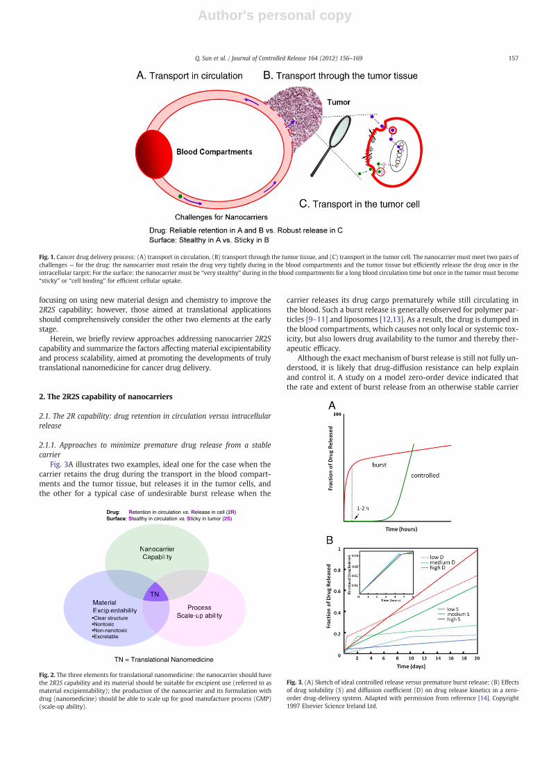
The cancer drug delivery process can be divided into three stages,shown in Fig. 1: Initially, the drug-loaded nanocarriers circulate in theblood compartments, including the liver and the spleen. When pass-ing through tumor blood vessels, some carriers may fall into thepores in the blood vessel wall and diffuse into the tumor tissue (EPReffect) (Fig. 1A) [6,7]. Next, they may further penetrate the tumor tis-sue, which is non-trivial because of the high cell density and high in-terstitial osmotic pressure (Fig. 1B) [8]. Upon sticking to thesurrounding cancer-cell membrane (Fig. 1C), the carrier is expectedto enter the cells via one or several possible pathways, and finally
traverse the crowded intracellular structures and viscous cytosol tothe targeted subcellular sites and release the carried drug cargo.
Thus, to achieve efficient drug delivery from the iv injection site tothe target in the tumor cells, the nanocarrier must simultaneouslymeet two pairs of challenges (Fig. 1): (a) the nanocarrier must retainthe drug very tightly, ideally without any release, during the trans-port in the blood compartments and the tumor tissue, but must beable to efficiently release the drug once reaching the intracellular tar-get to exert its pharmaceutical action; (b) the nanocarrier must be“slippery” or “stealthy”while in the blood compartments to effective-ly evade the reticuloendothelial system (RES) screening, particularlythe capture by liver and spleen for a long blood circulation time. Asthe blood circulation time of the nanocarrier increases so does its op-portunity passing the hyperpermeable tumor blood vessel and ex-travasation into the tumor. But once in the tumor the nanocarriermust become “sticky” or “cell binding” to interact with tumor cellsfor efficient cellular uptake. A nanocarrier capable of simultaneouslysatisfying the opposite 2R2S requirements at the right places, that is,“drug Retention in blood circulation versus Release in tumor cells(2R)” and “Stealthy in blood versus Sticky in tumor (2 S)” will deliverthe drug specifically to the tumor, giving rise to high therapeutic effi-cacy and few side effects.
While the 2R2S capability of a nanocarrier may render its resultingnanomedicine efficacy and safety potential for clinical translation,other two elements, the feasibility of the nanocarrier materials to beproved for use as excipients (referred to as material excipientability)and the ability to establish scaling up production processes for goodmanufacture processes (GMP) for the nanocarrier and its formulationwith drug (nanomedicine) (referred to as process scale-up ability)are also indispensible for the nanomedicine truly translational fromthe benchtop to the bedside (Fig. 2). Most of our current research is
Journal of Controlled Release 164 (2012) 156–169
⁎ Corresponding author at: Center for Bionanoengineering and State Key Laboratoryof Chemical Engineering, Department of Chemical and Biological Engineering, ZhejiangUniversity, Hangzhou 310027, China.
E-mail address: [email protected] (Y. Shen).
0168-3659/$ – see front matter © 2012 Elsevier B.V. All rights reserved.doi:10.1016/j.jconrel.2012.05.042
Contents lists available at SciVerse ScienceDirect
Journal of Controlled Release
j ourna l homepage: www.e lsev ie r .com/ locate / jconre l
Author's personal copy
focusing on using new material design and chemistry to improve the2R2S capability; however, those aimed at translational applicationsshould comprehensively consider the other two elements at the earlystage.
Herein, we briefly review approaches addressing nanocarrier 2R2Scapability and summarize the factors affecting material excipientabilityand process scalability, aimed at promoting the developments of trulytranslational nanomedicine for cancer drug delivery.
2. The 2R2S capability of nanocarriers
2.1. The 2R capability: drug retention in circulation versus intracellularrelease
2.1.1. Approaches to minimize premature drug release from a stablecarrier
Fig. 3A illustrates two examples, ideal one for the case when thecarrier retains the drug during the transport in the blood compart-ments and the tumor tissue, but releases it in the tumor cells, andthe other for a typical case of undesirable burst release when the
carrier releases its drug cargo prematurely while still circulating inthe blood. Such a burst release is generally observed for polymer par-ticles [9–11] and liposomes [12,13]. As a result, the drug is dumped inthe blood compartments, which causes not only local or systemic tox-icity, but also lowers drug availability to the tumor and thereby ther-apeutic efficacy.
Although the exact mechanism of burst release is still not fully un-derstood, it is likely that drug-diffusion resistance can help explainand control it. A study on a model zero-order device indicated thatthe rate and extent of burst release from an otherwise stable carrier
Fig. 1. Cancer drug delivery process: (A) transport in circulation, (B) transport through the tumor tissue, and (C) transport in the tumor cell. The nanocarrier must meet two pairs ofchallenges — for the drug: the nanocarrier must retain the drug very tightly during in the blood compartments and the tumor tissue but efficiently release the drug once in theintracellular target; For the surface: the nanocarrier must be “very stealthy” during in the blood compartments for a long blood circulation time but once in the tumor must become“sticky” or “cell binding” for efficient cellular uptake.
NanocarrierCapability
ProcessScale-up ability
MaterialExcipientability
Drug: Retention in circulation vs. Release in cell (2R)Surface: Stealthy in circulation vs. Sticky in tumor (2S)
MaterialExcipientability Process
Scale-up ability
NanocarrierCapability
TN
TN = Translational Nanomedicine
•Clear structure•Nontoxic•Non-nanotoxic•Excretable
Fig. 2. The three elements for translational nanomedicine: the nanocarrier should havethe 2R2S capability and its material should be suitable for excipient use (referred to asmaterial excipientability); the production of the nanocarrier and its formulation withdrug (nanomedicine) should be able to scale up for good manufacture process (GMP)(scale-up ability).
Fig. 3. (A) Sketch of ideal controlled release versus premature burst release; (B) Effectsof drug solubility (S) and diffusion coefficient (D) on drug release kinetics in a zero-order drug-delivery system. Adapted with permission from reference [14]. Copyright1997 Elsevier Science Ireland Ltd.
157Q. Sun et al. / Journal of Controlled Release 164 (2012) 156–169
Author's personal copy
were affected by drug solubility and drug diffusion in an aqueous me-dium, as shown in Fig. 3B, and by the drug loading content [14]. Suchfindings inspired more recent approaches to prevent burst releaseaimed at enhancing drug loading, inhibiting drug diffusion from thecarrier, or both.
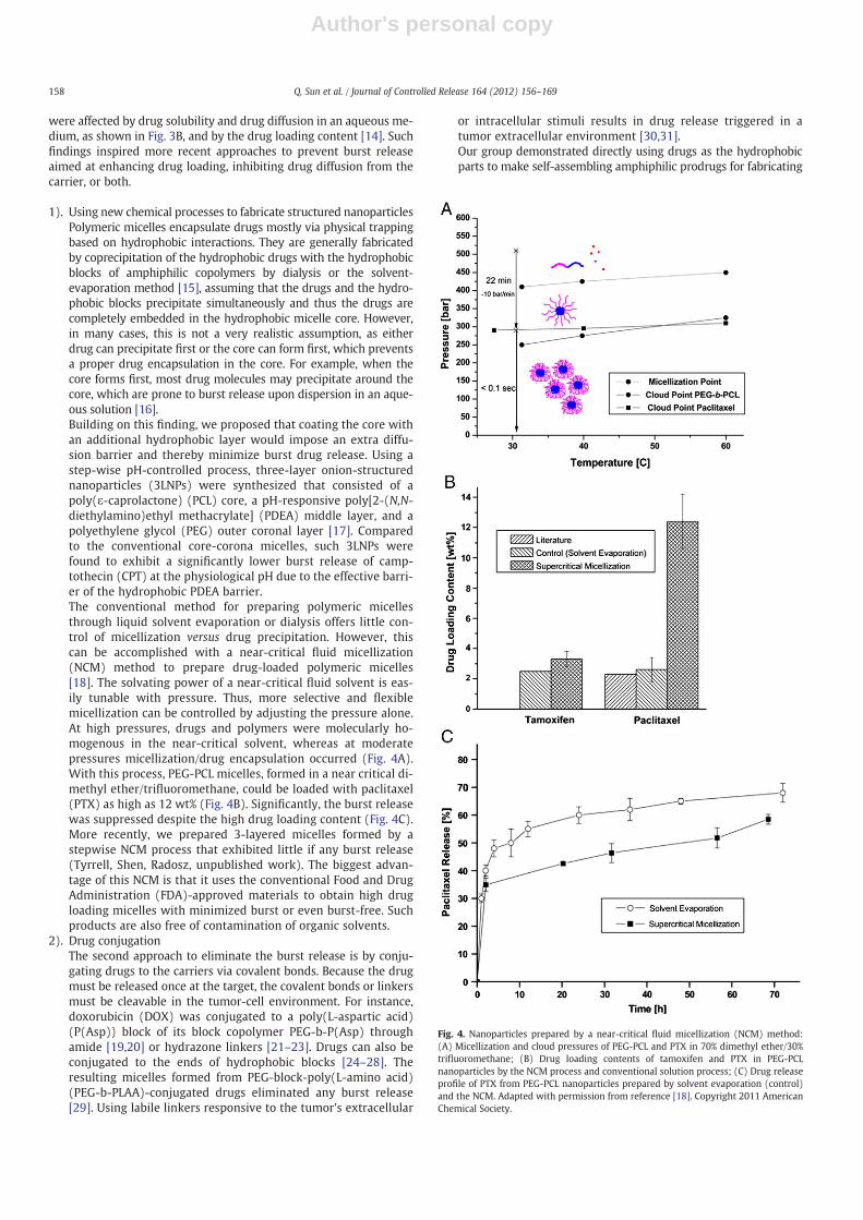
1). Using new chemical processes to fabricate structured nanoparticlesPolymeric micelles encapsulate drugs mostly via physical trappingbased on hydrophobic interactions. They are generally fabricatedby coprecipitation of the hydrophobic drugs with the hydrophobicblocks of amphiphilic copolymers by dialysis or the solvent-evaporation method [15], assuming that the drugs and the hydro-phobic blocks precipitate simultaneously and thus the drugs arecompletely embedded in the hydrophobic micelle core. However,in many cases, this is not a very realistic assumption, as eitherdrug can precipitate first or the core can form first, which preventsa proper drug encapsulation in the core. For example, when thecore forms first, most drug molecules may precipitate around thecore, which are prone to burst release upon dispersion in an aque-ous solution [16].Building on this finding, we proposed that coating the core withan additional hydrophobic layer would impose an extra diffu-sion barrier and thereby minimize burst drug release. Using astep-wise pH-controlled process, three-layer onion-structurednanoparticles (3LNPs) were synthesized that consisted of apoly(ε-caprolactone) (PCL) core, a pH-responsive poly[2-(N,N-diethylamino)ethyl methacrylate] (PDEA) middle layer, and apolyethylene glycol (PEG) outer coronal layer [17]. Comparedto the conventional core-corona micelles, such 3LNPs werefound to exhibit a significantly lower burst release of camp-tothecin (CPT) at the physiological pH due to the effective barri-er of the hydrophobic PDEA barrier.The conventional method for preparing polymeric micellesthrough liquid solvent evaporation or dialysis offers little con-trol of micellization versus drug precipitation. However, thiscan be accomplished with a near-critical fluid micellization(NCM) method to prepare drug-loaded polymeric micelles[18]. The solvating power of a near-critical fluid solvent is eas-ily tunable with pressure. Thus, more selective and flexiblemicellization can be controlled by adjusting the pressure alone.At high pressures, drugs and polymers were molecularly ho-mogenous in the near-critical solvent, whereas at moderatepressures micellization/drug encapsulation occurred (Fig. 4A).With this process, PEG-PCL micelles, formed in a near critical di-methyl ether/trifluoromethane, could be loaded with paclitaxel(PTX) as high as 12 wt% (Fig. 4B). Significantly, the burst releasewas suppressed despite the high drug loading content (Fig. 4C).More recently, we prepared 3-layered micelles formed by astepwise NCM process that exhibited little if any burst release(Tyrrell, Shen, Radosz, unpublished work). The biggest advan-tage of this NCM is that it uses the conventional Food and DrugAdministration (FDA)-approved materials to obtain high drugloading micelles with minimized burst or even burst-free. Suchproducts are also free of contamination of organic solvents.
2). Drug conjugationThe second approach to eliminate the burst release is by conju-gating drugs to the carriers via covalent bonds. Because the drugmust be released once at the target, the covalent bonds or linkersmust be cleavable in the tumor-cell environment. For instance,doxorubicin (DOX) was conjugated to a poly(L-aspartic acid)(P(Asp)) block of its block copolymer PEG-b-P(Asp) throughamide [19,20] or hydrazone linkers [21–23]. Drugs can also beconjugated to the ends of hydrophobic blocks [24–28]. Theresulting micelles formed from PEG-block-poly(L-amino acid)(PEG-b-PLAA)-conjugated drugs eliminated any burst release[29]. Using labile linkers responsive to the tumor's extracellular
or intracellular stimuli results in drug release triggered in atumor extracellular environment [30,31].Our group demonstrated directly using drugs as the hydrophobicparts to make self-assembling amphiphilic prodrugs for fabricating
Fig. 4. Nanoparticles prepared by a near-critical fluid micellization (NCM) method:(A) Micellization and cloud pressures of PEG-PCL and PTX in 70% dimethyl ether/30%trifluoromethane; (B) Drug loading contents of tamoxifen and PTX in PEG-PCLnanoparticles by the NCM process and conventional solution process; (C) Drug releaseprofile of PTX from PEG-PCL nanoparticles prepared by solvent evaporation (control)and the NCM. Adapted with permission from reference [18]. Copyright 2011 AmericanChemical Society.
158 Q. Sun et al. / Journal of Controlled Release 164 (2012) 156–169
Author's personal copy
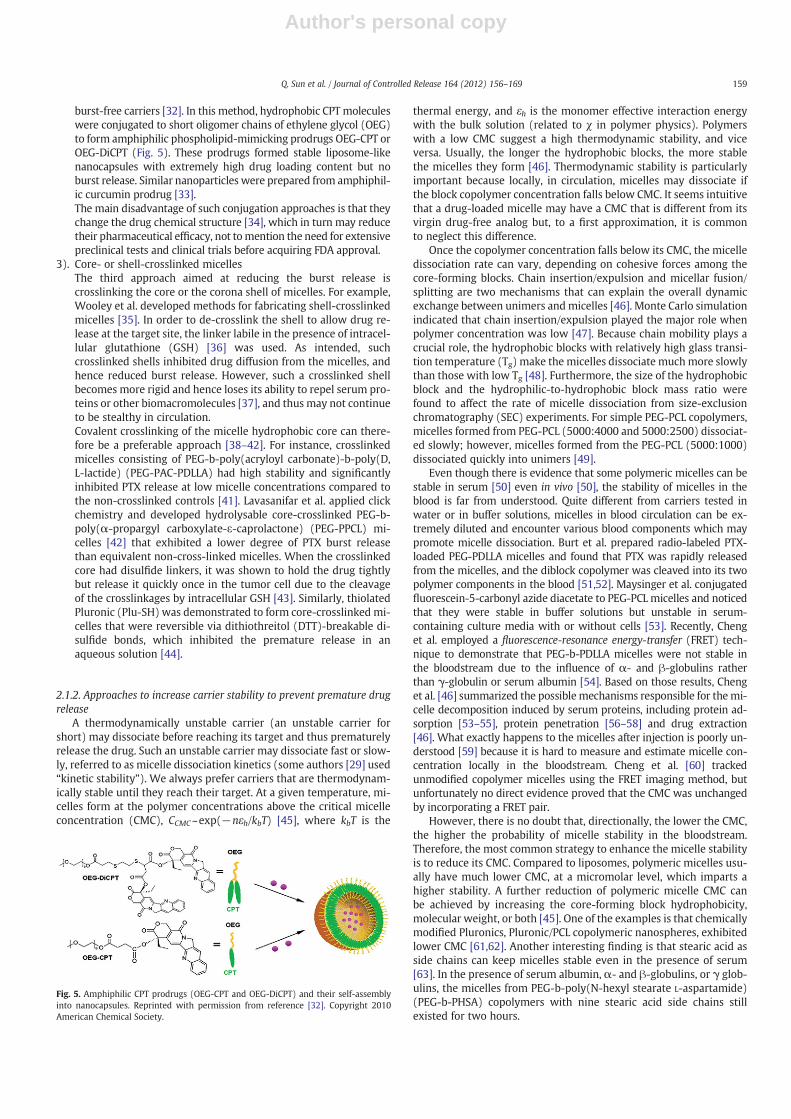
burst-free carriers [32]. In this method, hydrophobic CPT moleculeswere conjugated to short oligomer chains of ethylene glycol (OEG)to form amphiphilic phospholipid-mimicking prodrugs OEG-CPT orOEG-DiCPT (Fig. 5). These prodrugs formed stable liposome-likenanocapsules with extremely high drug loading content but noburst release. Similar nanoparticles were prepared from amphiphil-ic curcumin prodrug [33].The main disadvantage of such conjugation approaches is that theychange the drug chemical structure [34], which in turn may reducetheir pharmaceutical efficacy, not tomention the need for extensivepreclinical tests and clinical trials before acquiring FDA approval.
3). Core- or shell-crosslinked micellesThe third approach aimed at reducing the burst release iscrosslinking the core or the corona shell of micelles. For example,Wooley et al. developed methods for fabricating shell-crosslinkedmicelles [35]. In order to de-crosslink the shell to allow drug re-lease at the target site, the linker labile in the presence of intracel-lular glutathione (GSH) [36] was used. As intended, suchcrosslinked shells inhibited drug diffusion from the micelles, andhence reduced burst release. However, such a crosslinked shellbecomes more rigid and hence loses its ability to repel serum pro-teins or other biomacromolecules [37], and thus may not continueto be stealthy in circulation.Covalent crosslinking of the micelle hydrophobic core can there-fore be a preferable approach [38–42]. For instance, crosslinkedmicelles consisting of PEG-b-poly(acryloyl carbonate)-b-poly(D,L-lactide) (PEG-PAC-PDLLA) had high stability and significantlyinhibited PTX release at low micelle concentrations compared tothe non-crosslinked controls [41]. Lavasanifar et al. applied clickchemistry and developed hydrolysable core-crosslinked PEG-b-poly(α-propargyl carboxylate-ε-caprolactone) (PEG-PPCL) mi-celles [42] that exhibited a lower degree of PTX burst releasethan equivalent non-cross-linked micelles. When the crosslinkedcore had disulfide linkers, it was shown to hold the drug tightlybut release it quickly once in the tumor cell due to the cleavageof the crosslinkages by intracellular GSH [43]. Similarly, thiolatedPluronic (Plu-SH) was demonstrated to form core-crosslinked mi-celles that were reversible via dithiothreitol (DTT)-breakable di-sulfide bonds, which inhibited the premature release in anaqueous solution [44].
2.1.2. Approaches to increase carrier stability to prevent premature drugrelease
A thermodynamically unstable carrier (an unstable carrier forshort) may dissociate before reaching its target and thus prematurelyrelease the drug. Such an unstable carrier may dissociate fast or slow-ly, referred to as micelle dissociation kinetics (some authors [29] used“kinetic stability”). We always prefer carriers that are thermodynam-ically stable until they reach their target. At a given temperature, mi-celles form at the polymer concentrations above the critical micelleconcentration (CMC), CCMC~exp(−nεh/kbT) [45], where kbT is the
thermal energy, and εh is the monomer effective interaction energywith the bulk solution (related to χ in polymer physics). Polymerswith a low CMC suggest a high thermodynamic stability, and viceversa. Usually, the longer the hydrophobic blocks, the more stablethe micelles they form [46]. Thermodynamic stability is particularlyimportant because locally, in circulation, micelles may dissociate ifthe block copolymer concentration falls below CMC. It seems intuitivethat a drug-loaded micelle may have a CMC that is different from itsvirgin drug-free analog but, to a first approximation, it is commonto neglect this difference.
Once the copolymer concentration falls below its CMC, the micelledissociation rate can vary, depending on cohesive forces among thecore-forming blocks. Chain insertion/expulsion and micellar fusion/splitting are two mechanisms that can explain the overall dynamicexchange between unimers andmicelles [46]. Monte Carlo simulationindicated that chain insertion/expulsion played the major role whenpolymer concentration was low [47]. Because chain mobility plays acrucial role, the hydrophobic blocks with relatively high glass transi-tion temperature (Tg) make the micelles dissociate muchmore slowlythan those with low Tg [48]. Furthermore, the size of the hydrophobicblock and the hydrophilic-to-hydrophobic block mass ratio werefound to affect the rate of micelle dissociation from size-exclusionchromatography (SEC) experiments. For simple PEG-PCL copolymers,micelles formed from PEG-PCL (5000:4000 and 5000:2500) dissociat-ed slowly; however, micelles formed from the PEG-PCL (5000:1000)dissociated quickly into unimers [49].
Even though there is evidence that some polymeric micelles can bestable in serum [50] even in vivo [50], the stability of micelles in theblood is far from understood. Quite different from carriers tested inwater or in buffer solutions, micelles in blood circulation can be ex-tremely diluted and encounter various blood components which maypromote micelle dissociation. Burt et al. prepared radio-labeled PTX-loaded PEG-PDLLA micelles and found that PTX was rapidly releasedfrom the micelles, and the diblock copolymer was cleaved into its twopolymer components in the blood [51,52]. Maysinger et al. conjugatedfluorescein-5-carbonyl azide diacetate to PEG-PCL micelles and noticedthat they were stable in buffer solutions but unstable in serum-containing culture media with or without cells [53]. Recently, Chenget al. employed a fluorescence-resonance energy-transfer (FRET) tech-nique to demonstrate that PEG-b-PDLLA micelles were not stable inthe bloodstream due to the influence of α- and β-globulins ratherthan γ-globulin or serum albumin [54]. Based on those results, Chenget al. [46] summarized the possible mechanisms responsible for themi-celle decomposition induced by serum proteins, including protein ad-sorption [53–55], protein penetration [56–58] and drug extraction[46]. What exactly happens to the micelles after injection is poorly un-derstood [59] because it is hard to measure and estimate micelle con-centration locally in the bloodstream. Cheng et al. [60] trackedunmodified copolymer micelles using the FRET imaging method, butunfortunately no direct evidence proved that the CMC was unchangedby incorporating a FRET pair.
However, there is no doubt that, directionally, the lower the CMC,the higher the probability of micelle stability in the bloodstream.Therefore, the most common strategy to enhance the micelle stabilityis to reduce its CMC. Compared to liposomes, polymeric micelles usu-ally have much lower CMC, at a micromolar level, which imparts ahigher stability. A further reduction of polymeric micelle CMC canbe achieved by increasing the core-forming block hydrophobicity,molecular weight, or both [45]. One of the examples is that chemicallymodified Pluronics, Pluronic/PCL copolymeric nanospheres, exhibitedlower CMC [61,62]. Another interesting finding is that stearic acid asside chains can keep micelles stable even in the presence of serum[63]. In the presence of serum albumin, α- and β-globulins, or γ glob-ulins, the micelles from PEG-b-poly(N-hexyl stearate L-aspartamide)(PEG-b-PHSA) copolymers with nine stearic acid side chains stillexisted for two hours.
Fig. 5. Amphiphilic CPT prodrugs (OEG-CPT and OEG-DiCPT) and their self-assemblyinto nanocapsules. Reprinted with permission from reference [32]. Copyright 2010American Chemical Society.
159Q. Sun et al. / Journal of Controlled Release 164 (2012) 156–169
Author's personal copy
Crosslinking is a straightforwardmethod to stabilizemicelles.Whilethe covalent crosslinking of themicelle core or shell can inhibit burst re-lease from a stable micelle, it can also inhibit or prevent micelle dissoci-ation. For instance, PEG-PCL micelles with cores crosslinked by radicalpolymerization of the double bonds introduced to the PCL blocks turnedout to be more stable [64]. Biodegradable thermosensitive micelleswith crosslinked cores formed from PEG-b-(N-(2-hydroxyethyl met-hacrylamide)-oligolactates) (PEG-b-p(HEMAm-Lacn)) kept their integ-rity upon dilution and only degraded after cleavage of the ester bonds inthe crosslinkers [65].
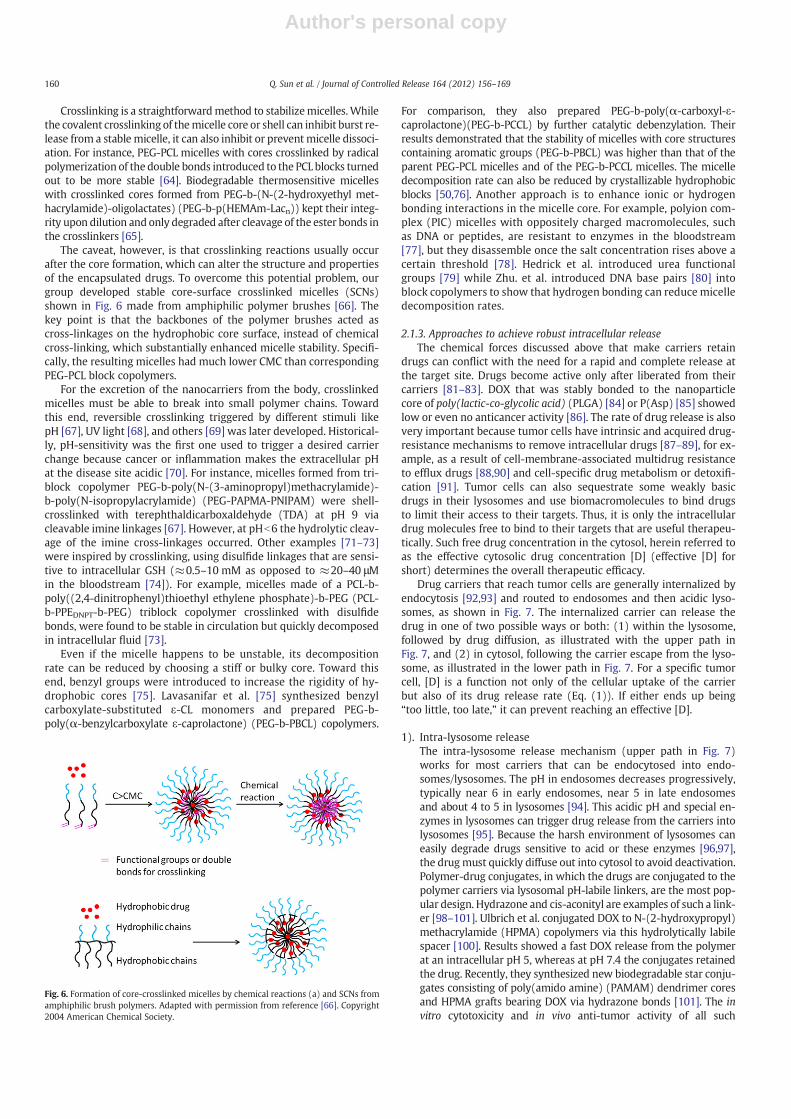
The caveat, however, is that crosslinking reactions usually occurafter the core formation, which can alter the structure and propertiesof the encapsulated drugs. To overcome this potential problem, ourgroup developed stable core-surface crosslinked micelles (SCNs)shown in Fig. 6 made from amphiphilic polymer brushes [66]. Thekey point is that the backbones of the polymer brushes acted ascross-linkages on the hydrophobic core surface, instead of chemicalcross-linking, which substantially enhanced micelle stability. Specifi-cally, the resulting micelles had much lower CMC than correspondingPEG-PCL block copolymers.
For the excretion of the nanocarriers from the body, crosslinkedmicelles must be able to break into small polymer chains. Towardthis end, reversible crosslinking triggered by different stimuli likepH [67], UV light [68], and others [69] was later developed. Historical-ly, pH-sensitivity was the first one used to trigger a desired carrierchange because cancer or inflammation makes the extracellular pHat the disease site acidic [70]. For instance, micelles formed from tri-block copolymer PEG-b-poly(N-(3-aminopropyl)methacrylamide)-b-poly(N-isopropylacrylamide) (PEG-PAPMA-PNIPAM) were shell-crosslinked with terephthaldicarboxaldehyde (TDA) at pH 9 viacleavable imine linkages [67]. However, at pHb6 the hydrolytic cleav-age of the imine cross-linkages occurred. Other examples [71–73]were inspired by crosslinking, using disulfide linkages that are sensi-tive to intracellular GSH (≈0.5–10 mM as opposed to ≈20–40 μMin the bloodstream [74]). For example, micelles made of a PCL-b-poly((2,4-dinitrophenyl)thioethyl ethylene phosphate)-b-PEG (PCL-b-PPEDNPT-b-PEG) triblock copolymer crosslinked with disulfidebonds, were found to be stable in circulation but quickly decomposedin intracellular fluid [73].
Even if the micelle happens to be unstable, its decompositionrate can be reduced by choosing a stiff or bulky core. Toward thisend, benzyl groups were introduced to increase the rigidity of hy-drophobic cores [75]. Lavasanifar et al. [75] synthesized benzylcarboxylate-substituted ε-CL monomers and prepared PEG-b-poly(α-benzylcarboxylate ε-caprolactone) (PEG-b-PBCL) copolymers.
For comparison, they also prepared PEG-b-poly(α-carboxyl-ε-caprolactone)(PEG-b-PCCL) by further catalytic debenzylation. Theirresults demonstrated that the stability of micelles with core structurescontaining aromatic groups (PEG-b-PBCL) was higher than that of theparent PEG-PCL micelles and of the PEG-b-PCCL micelles. The micelledecomposition rate can also be reduced by crystallizable hydrophobicblocks [50,76]. Another approach is to enhance ionic or hydrogenbonding interactions in the micelle core. For example, polyion com-plex (PIC) micelles with oppositely charged macromolecules, suchas DNA or peptides, are resistant to enzymes in the bloodstream[77], but they disassemble once the salt concentration rises above acertain threshold [78]. Hedrick et al. introduced urea functionalgroups [79] while Zhu. et al. introduced DNA base pairs [80] intoblock copolymers to show that hydrogen bonding can reduce micelledecomposition rates.
2.1.3. Approaches to achieve robust intracellular releaseThe chemical forces discussed above that make carriers retain
drugs can conflict with the need for a rapid and complete release atthe target site. Drugs become active only after liberated from theircarriers [81–83]. DOX that was stably bonded to the nanoparticlecore of poly(lactic-co-glycolic acid) (PLGA) [84] or P(Asp) [85] showedlow or even no anticancer activity [86]. The rate of drug release is alsovery important because tumor cells have intrinsic and acquired drug-resistance mechanisms to remove intracellular drugs [87–89], for ex-ample, as a result of cell-membrane-associated multidrug resistanceto efflux drugs [88,90] and cell-specific drug metabolism or detoxifi-cation [91]. Tumor cells can also sequestrate some weakly basicdrugs in their lysosomes and use biomacromolecules to bind drugsto limit their access to their targets. Thus, it is only the intracellulardrug molecules free to bind to their targets that are useful therapeu-tically. Such free drug concentration in the cytosol, herein referred toas the effective cytosolic drug concentration [D] (effective [D] forshort) determines the overall therapeutic efficacy.
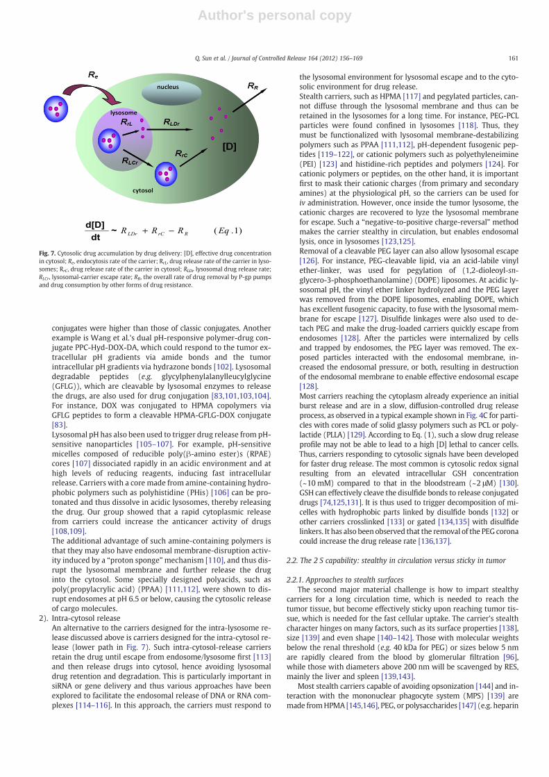
Drug carriers that reach tumor cells are generally internalized byendocytosis [92,93] and routed to endosomes and then acidic lyso-somes, as shown in Fig. 7. The internalized carrier can release thedrug in one of two possible ways or both: (1) within the lysosome,followed by drug diffusion, as illustrated with the upper path inFig. 7, and (2) in cytosol, following the carrier escape from the lyso-some, as illustrated in the lower path in Fig. 7. For a specific tumorcell, [D] is a function not only of the cellular uptake of the carrierbut also of its drug release rate (Eq. (1)). If either ends up being“too little, too late,” it can prevent reaching an effective [D].
1). Intra-lysosome releaseThe intra-lysosome release mechanism (upper path in Fig. 7)works for most carriers that can be endocytosed into endo-somes/lysosomes. The pH in endosomes decreases progressively,typically near 6 in early endosomes, near 5 in late endosomesand about 4 to 5 in lysosomes [94]. This acidic pH and special en-zymes in lysosomes can trigger drug release from the carriers intolysosomes [95]. Because the harsh environment of lysosomes caneasily degrade drugs sensitive to acid or these enzymes [96,97],the drugmust quickly diffuse out into cytosol to avoid deactivation.Polymer-drug conjugates, in which the drugs are conjugated to thepolymer carriers via lysosomal pH-labile linkers, are the most pop-ular design. Hydrazone and cis-aconityl are examples of such a link-er [98–101]. Ulbrich et al. conjugated DOX to N-(2-hydroxypropyl)methacrylamide (HPMA) copolymers via this hydrolytically labilespacer [100]. Results showed a fast DOX release from the polymerat an intracellular pH 5, whereas at pH 7.4 the conjugates retainedthe drug. Recently, they synthesized new biodegradable star conju-gates consisting of poly(amido amine) (PAMAM) dendrimer coresand HPMA grafts bearing DOX via hydrazone bonds [101]. The invitro cytotoxicity and in vivo anti-tumor activity of all such
Fig. 6. Formation of core-crosslinked micelles by chemical reactions (a) and SCNs fromamphiphilic brush polymers. Adapted with permission from reference [66]. Copyright2004 American Chemical Society.
160 Q. Sun et al. / Journal of Controlled Release 164 (2012) 156–169
Author's personal copy
conjugates were higher than those of classic conjugates. Anotherexample is Wang et al.'s dual pH-responsive polymer-drug con-jugate PPC-Hyd-DOX-DA, which could respond to the tumor ex-tracellular pH gradients via amide bonds and the tumorintracellular pH gradients via hydrazone bonds [102]. Lysosomaldegradable peptides (e.g. glycylphenylalanylleucylglycine(GFLG)), which are cleavable by lysosomal enzymes to releasethe drugs, are also used for drug conjugation [83,101,103,104].For instance, DOX was conjugated to HPMA copolymers viaGFLG peptides to form a cleavable HPMA-GFLG-DOX conjugate[83].Lysosomal pH has also been used to trigger drug release from pH-sensitive nanoparticles [105–107]. For example, pH-sensitivemicelles composed of reducible poly(β-amino ester)s (RPAE)cores [107] dissociated rapidly in an acidic environment and athigh levels of reducing reagents, inducing fast intracellularrelease. Carriers with a core made from amine-containing hydro-phobic polymers such as polyhistidine (PHis) [106] can be pro-tonated and thus dissolve in acidic lysosomes, thereby releasingthe drug. Our group showed that a rapid cytoplasmic releasefrom carriers could increase the anticancer activity of drugs[108,109].The additional advantage of such amine-containing polymers isthat they may also have endosomal membrane-disruption activ-ity induced by a “proton sponge”mechanism [110], and thus dis-rupt the lysosomal membrane and further release the druginto the cytosol. Some specially designed polyacids, such aspoly(propylacrylic acid) (PPAA) [111,112], were shown to dis-rupt endosomes at pH 6.5 or below, causing the cytosolic releaseof cargo molecules.
2). Intra-cytosol releaseAn alternative to the carriers designed for the intra-lysosome re-lease discussed above is carriers designed for the intra-cytosol re-lease (lower path in Fig. 7). Such intra-cytosol-release carriersretain the drug until escape from endosome/lysosome first [113]and then release drugs into cytosol, hence avoiding lysosomaldrug retention and degradation. This is particularly important insiRNA or gene delivery and thus various approaches have beenexplored to facilitate the endosomal release of DNA or RNA com-plexes [114–116]. In this approach, the carriers must respond to
the lysosomal environment for lysosomal escape and to the cyto-solic environment for drug release.Stealth carriers, such as HPMA [117] and pegylated particles, can-not diffuse through the lysosomal membrane and thus can beretained in the lysosomes for a long time. For instance, PEG-PCLparticles were found confined in lysosomes [118]. Thus, theymust be functionalized with lysosomal membrane-destabilizingpolymers such as PPAA [111,112], pH-dependent fusogenic pep-tides [119–122], or cationic polymers such as polyethyleneimine(PEI) [123] and histidine-rich peptides and polymers [124]. Forcationic polymers or peptides, on the other hand, it is importantfirst to mask their cationic charges (from primary and secondaryamines) at the physiological pH, so the carriers can be used foriv administration. However, once inside the tumor lysosome, thecationic charges are recovered to lyze the lysosomal membranefor escape. Such a “negative-to-positive charge-reversal” methodmakes the carrier stealthy in circulation, but enables endosomallysis, once in lysosomes [123,125].Removal of a cleavable PEG layer can also allow lysosomal escape[126]. For instance, PEG-cleavable lipid, via an acid-labile vinylether-linker, was used for pegylation of (1,2-dioleoyl-sn-glycero-3-phosphoethanolamine) (DOPE) liposomes. At acidic ly-sosomal pH, the vinyl ether linker hydrolyzed and the PEG layerwas removed from the DOPE liposomes, enabling DOPE, whichhas excellent fusogenic capacity, to fuse with the lysosomal mem-brane for escape [127]. Disulfide linkages were also used to de-tach PEG and make the drug-loaded carriers quickly escape fromendosomes [128]. After the particles were internalized by cellsand trapped by endosomes, the PEG layer was removed. The ex-posed particles interacted with the endosomal membrane, in-creased the endosomal pressure, or both, resulting in destructionof the endosomal membrane to enable effective endosomal escape[128].Most carriers reaching the cytoplasm already experience an initialburst release and are in a slow, diffusion-controlled drug releaseprocess, as observed in a typical example shown in Fig. 4C for parti-cles with cores made of solid glassy polymers such as PCL or poly-lactide (PLLA) [129]. According to Eq. (1), such a slow drug releaseprofile may not be able to lead to a high [D] lethal to cancer cells.Thus, carriers responding to cytosolic signals have been developedfor faster drug release. The most common is cytosolic redox signalresulting from an elevated intracellular GSH concentration(~10 mM) compared to that in the bloodstream (~2 μM) [130].GSH can effectively cleave the disulfide bonds to release conjugateddrugs [74,125,131]. It is thus used to trigger decomposition of mi-celles with hydrophobic parts linked by disulfide bonds [132] orother carriers crosslinked [133] or gated [134,135] with disulfidelinkers. It has also been observed that the removal of the PEG coronacould increase the drug release rate [136,137].
2.2. The 2 S capability: stealthy in circulation versus sticky in tumor
2.2.1. Approaches to stealth surfacesThe second major material challenge is how to impart stealthy
carriers for a long circulation time, which is needed to reach thetumor tissue, but become effectively sticky upon reaching tumor tis-sue, which is needed for the fast cellular uptake. The carrier's stealthcharacter hinges on many factors, such as its surface properties [138],size [139] and even shape [140–142]. Those with molecular weightsbelow the renal threshold (e.g. 40 kDa for PEG) or sizes below 5 nmare rapidly cleared from the blood by glomerular filtration [96],while those with diameters above 200 nm will be scavenged by RES,mainly the liver and spleen [139,143].
Most stealth carriers capable of avoiding opsonization [144] and in-teraction with the mononuclear phagocyte system (MPS) [139] aremade fromHPMA [145,146], PEG, or polysaccharides [147] (e.g. heparin
Fig. 7. Cytosolic drug accumulation by drug delivery: [D], effective drug concentrationin cytosol; Re, endocytosis rate of the carrier; RrL, drug release rate of the carrier in lyso-somes; RrC, drug release rate of the carrier in cytosol; RLDr lysosomal drug release rate;RLCr, lysosomal-carrier escape rate; RR, the overall rate of drug removal by P-gp pumpsand drug consumption by other forms of drug resistance.
161Q. Sun et al. / Journal of Controlled Release 164 (2012) 156–169
Author's personal copy
[148]). Nanoparticles coated with a layer of these polymers becomestealthy by both hydration and steric hindrance [149]. For example,pegylation of particles or liposomes is well-established [144,150–155],and the DOX-loaded stealth liposomes named Doxil® were approvedby the FDA for cancer therapy [156]. Huang et al. reported that, on the100 nm liposomes pegylated with 1,2-distearoyl-sn-glycero-3-pho-sphoethanolamine-PEG2000 (DSPE-PEG2000), PEG chains were arrangedin amushroom configuration at the DSPE-PEG fraction less than 4 mol%but in a brush configuration at the DSPE-PEG content greater than8 mol% (Fig. 8) [157]. The high density of PEG chains on the liposomesurface with the brush configuration was the key to reduce liposomeliver sequestration [157]. Discher et al. incorporated the PEG brushonto polymersomes and obtained polymersomes having a blood circu-lation time two-fold longer than pegylated liposomes [158]. Dai et al.pegylated single-wall carbon nanotubes (SWNT), and found that withthe increase of linear PEG chain length from 2 kDa to 5 kDa, the bloodcirculation time of pegylated SWNTs was significantly extended, butfurther increase of the PEG chain length showed no significant effect[159]. Although pegylation reduces the recognition of the carriers bythe MPS system and thereby extends their blood circulation time, the“accelerated blood clearance (ABC)” phenomenon was observed uponrepeated injection of pegylated liposomes [160–163] due to IgMbound to pegylated liposomes secreted into the bloodstream after thefirst dose [161]. Such immune reaction against the pegylated liposomesoccurred in the spleen at least 2–3 days after the first administration[162,163].
The carrier shape is also recognized as an important parameterthat can substantially affect the blood circulation time. In fact,Mitragotri et al. reported that the particle shape, not size, played adominant role in phagocytosis of polystyrene (PS) particles of varioussizes and shapes; the rod-like particles entered cell much faster [164].Discher et al. found that flexible worm-like micelles efficientlyevaded RES and circulated in the blood for a week [165,166], muchlonger than spherical micelles. Dai et al. found that carbonnanotubes pegylated with long PEG chains exhibited a long bloodcirculation (t1/2=22.1 h) upon intravenous injection into mice[152]. All these studies suggest that particle phagocytosis can beinhibited by minimizing its size-normalized curvature [164,167].Thus, particle shape is an important variable to make it remainstealth in circulation long enough for enhanced tumor accumula-tion [165,166,168–170].
2.2.2. Approaches to becoming sticky in tumor for cellular uptakeHowever, the same properties that impart stealth in circulation
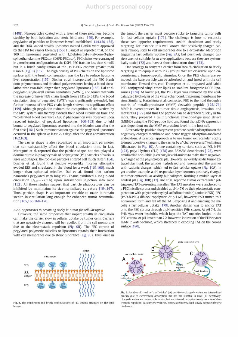
can make the carrier slow in cellular uptake by tumor cells. Carriersthat are negatively charged will be repelled from the cell membranedue to the electrostatic repulsion (Fig. 9B). The PEG corona ofpegylated polymeric micelles or liposomes retards their interactionwith cell membranes due to steric hindrance (Fig. 9C). Thus, once in
the tumor, the carrier must become sticky to targeting tumor cellsfor fast cellular uptake [171]. The challenge is how to reconcilethese two opposite requirements, stealth circulation and stickytargeting. For instance, it is well known that positively charged car-riers reliably stick to cell membranes due to electrostatic adsorptiontriggering fast cellular uptake (Fig. 9A); but positively charged car-riers are not suitable for in vivo applications because they are system-ically toxic [172] and have a short circulation time [173].
One strategy to convert a carrier from stealth circulation to stickytargeting is to equip it with PEG groups that are cleavable upon en-countering a tumor-specific stimulus. Once the PEG chains are re-moved, the bare particle can be adsorbed on and fused with the cellmembrane. Toward this end, Thompson et al. prepared acid-labilePEG conjugated vinyl ether lipids to stabilize fusogenic DOPE lipo-somes [174]. At lower pH, the PEG layer was removed by the acid-catalyzed hydrolysis of the vinyl ether bond, triggering membrane fu-sion. Similarly, Harashima et al. connected PEG to the lipid through amatrix of metalloproteinase (MMP)-cleavable peptide [175,176].MMP is overexpressed in tumor-tissue angiogenesis, invasion, andmetastasis [177] and thus the peptide can be degraded quickly in tu-mors. They prepared a multifunctional envelope-type nano device(MEND) using the PEG-peptide lipid and found that pDNA expressionwas dependent on the MMP expression level in the host cell.
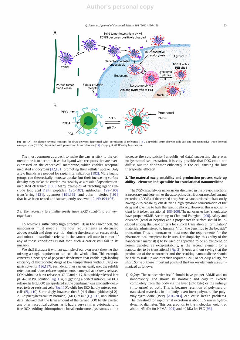
Alternatively, positive charges can promote carrier adsorption on thenegatively charged membrane and hence trigger adsorption-mediatedendocytosis. A practical approach is to use tumor extracellular acidityto impart positive charges to the carrier by a “charge-reversal” technique(illustrated in Fig. 10). Amine-containing carriers, such as PCL-b-PEI[123], poly(L-lysine) (PLL) [178] and PAMAM dendrimers [125], wereamidized to acid-labileβ-carboxylic acid amides tomake themnegative-ly charged at the physiological pH. However, in weakly acidic tumor ex-tracellular fluid, the amides hydrolyzed and regenerated the amineswith cationic charges, which led to fast cellular uptake (Fig. 10A). Inyet another example, a pH-responsive layer becomes positively chargedat tumor extracellular acidity but collapses, forming a middle layer atneutral pH (Fig. 10B) [17]. Bae et al. reported tumor extracellular pH-triggered TAT-presenting micelles. The TAT moieties were anchored toa PEGmicelle corona and shielded at pH>7.0 by their electrostatic com-plexationwith poly(methacryloyl sulfadimethoxine) (anionic PSD)-PEG(PSD-b-PEG) diblock copolymer. At pH 6.6, however, PSD turned to anonionized form and fell off the TAT, exposing it and enabling the mi-celle a fast cellular uptake [179]. Another design was to anchor TATonto the PEG corona through a pH-sensitive PHis spacer. At pH 7.4, thePHis was water-insoluble, which kept the TAT moieties buried in thePEG corona. At pH lower than 7.2, however, ionization of the PHis spacermade it water-soluble, which stretched it, exposing TAT on the coronasurface [180].
Fig. 8. The mushroom and brush configurations of PEG chains arranged on the lipidbilayer.
Fig. 9. Paradox of “stealthy” and “sticky”. (A) positively-charged carriers are internalizedquickly due to electrostatic adsorption, but are not suitable in vivo; (B) negatively-charged carriers are quite stable in vivo, but are internalized quite slowly because of elec-trostatic repulsion; (C) carriers with PEG corona are internalized slowly because of sterichindrance.
162 Q. Sun et al. / Journal of Controlled Release 164 (2012) 156–169
Author's personal copy
The most common approach to make the carrier stick to the cellmembrane is to decorate it with a ligand with receptors that are over-expressed on the cancer-cell membrane, which enables receptor-mediated endocytosis [12,181] promoting their cellular uptake. Onlya few ligands are needed for rapid internalization [182]. More ligandgroups can theoretically increase uptake, but their increasing surfacedensity may make the carrier less stealthy as a result of opsonization-mediated clearance [183]. Many examples of targeting ligands in-clude folic acid [184], peptides [185–187], antibodies [188–190],transferring [121], aptamers [191,192] and other moieties [193],that have been tested and subsequently reviewed [2,149,194,195].
2.3. The necessity to simultaneously have 2R2S capability: our ownexperience
To achieve a sufficiently high effective [D] in the cancer cell, thenanocarrier must meet all the four requirements as discussedabove: stealth and drug retention during the circulation versus stickyand robust intracellular release in the cancer cell once in tumor. Ifany of these conditions is not met, such a carrier will fail in itsmission.
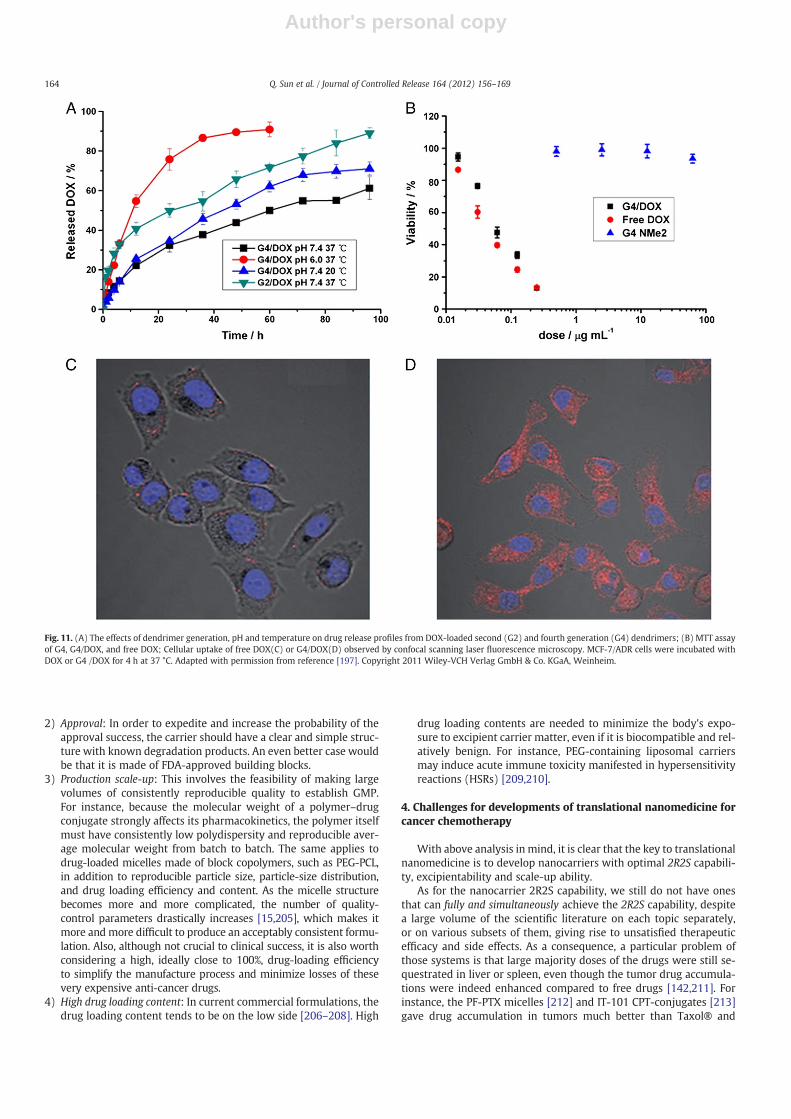
We shall illustrate it with an example of our own work showing thatmissing a single requirement can ruin the whole effort. This exampleconcerns a new type of polyester dendrimers that enable high-loadingefficiency of hydrophobic drugs at low temperatures without using or-ganic solvents [196,197]. Such dendrimer carriers easily met the reliableretention and robust release requirements, namely, that it slowly releasedDOX without a burst release at 37 °C and pH 7, but quickly released it atpH 4–5 in PBS solution (Fig. 11A) suggesting a perfect intracellular DOXrelease. In fact, DOX encapsulated in the dendrimer was efficiently deliv-ered to drug-resistant cells (Fig. 11D),while freeDOXhardly entered suchcells (Fig. 11C). Surprisingly, however, the (3-(4, 5-dimethylthiazolyl-2)-2, 5-diphenyltetrazolium bromide) (MTT) result (Fig. 11B, unpublisheddata) showed that the large amount of the carried DOX barely exertedany pharmaceutical actions, as it had a very similar cytotoxicity tofree DOX. Adding chloroquine to break endosomes/lysosomes didn't
increase the cytotoxicity (unpublished data) suggesting there wasno lysosomal sequestration. It is very possible that DOX could notdiffuse out the dendrimer efficiently in the cell, causing the lowtherapeutic efficacy.
3. The material excipientability and production process scale-upability - elements indispensible for translational nanomedicine
The 2R2S capability for nanocarriers discussed in the previous sectionsis necessary and determines the adsorption, distribution,metabolism andexcretion (ADME) of the carried drug. Such a nanocarrier simultaneouslyhaving 2R2S capability can deliver a high cytosolic concentration of thedrug and give rise to high therapeutic efficacy. However, this is not suffi-cient for it to be translational [198–200]. The nanocarrier itself should alsohave proper ADME. According to Choi and Frangioni [200], safety andclearance (renal or hepatic) and a proper stealth surface should be in-cluded among the basic criteria for clinical translation of formulation/materials administered to humans, “from the benchtop to the bedside”translation. Thus, a nanocarrier must meet the requirements for thepharmaceutical excipient for iv uses. For simplicity, this ability of thenanocarrier material(s) to be used or approved to be an excipient, orherein denoted as excipientability, is the second element for ananocarrier to be translational (Fig. 2). It goes without saying that theproduction of the nanocarrier and the resulting nanomedicine shouldbe able to scale up and establish required GMP, or scale-up ability, forshort. Some of these important points of the two key elements are sum-marized as follows:
1) Safety: The nanocarrier itself should have proper ADME and nonanotoxicity, and should be nontoxic and easy to excretecompletely from the body via the liver (into bile) or the kidneys(into urine) or both. This is because retention of polymers ornanosized materials in the body, even inert polymers like poly-vinylpyrrolidone (PVP) [201–203], can cause health problems.The threshold for rapid renal excretion is about 5.5 nm in hydro-dynamic diameter. This corresponds to the molecular weight ofabout~45 kDa for HPMA [204] and 40 kDa for PEG [96].
Fig. 10. (A) The charge-reversal concept for drug delivery. Reprinted with permission of reference [15]. Copyright 2010 Elsevier Ltd; (B) The pH-responsive three-layerednanoparticles (3LNPs). Reprinted with permission from reference [17]. Copyright 2008 Wiley InterScience.
163Q. Sun et al. / Journal of Controlled Release 164 (2012) 156–169
Author's personal copy
2) Approval: In order to expedite and increase the probability of theapproval success, the carrier should have a clear and simple struc-ture with known degradation products. An even better case wouldbe that it is made of FDA-approved building blocks.
3) Production scale-up: This involves the feasibility of making largevolumes of consistently reproducible quality to establish GMP.For instance, because the molecular weight of a polymer–drugconjugate strongly affects its pharmacokinetics, the polymer itselfmust have consistently low polydispersity and reproducible aver-age molecular weight from batch to batch. The same applies todrug-loaded micelles made of block copolymers, such as PEG-PCL,in addition to reproducible particle size, particle-size distribution,and drug loading efficiency and content. As the micelle structurebecomes more and more complicated, the number of quality-control parameters drastically increases [15,205], which makes itmore and more difficult to produce an acceptably consistent formu-lation. Also, although not crucial to clinical success, it is also worthconsidering a high, ideally close to 100%, drug-loading efficiencyto simplify the manufacture process and minimize losses of thesevery expensive anti-cancer drugs.
4) High drug loading content: In current commercial formulations, thedrug loading content tends to be on the low side [206–208]. High
drug loading contents are needed to minimize the body's expo-sure to excipient carrier matter, even if it is biocompatible and rel-atively benign. For instance, PEG-containing liposomal carriersmay induce acute immune toxicity manifested in hypersensitivityreactions (HSRs) [209,210].
4. Challenges for developments of translational nanomedicine forcancer chemotherapy
With above analysis in mind, it is clear that the key to translationalnanomedicine is to develop nanocarriers with optimal 2R2S capabili-ty, excipientability and scale-up ability.
As for the nanocarrier 2R2S capability, we still do not have onesthat can fully and simultaneously achieve the 2R2S capability, despitea large volume of the scientific literature on each topic separately,or on various subsets of them, giving rise to unsatisfied therapeuticefficacy and side effects. As a consequence, a particular problem ofthose systems is that large majority doses of the drugs were still se-questrated in liver or spleen, even though the tumor drug accumula-tions were indeed enhanced compared to free drugs [142,211]. Forinstance, the PF-PTX micelles [212] and IT-101 CPT-conjugates [213]gave drug accumulation in tumors much better than Taxol® and
Fig. 11. (A) The effects of dendrimer generation, pH and temperature on drug release profiles from DOX-loaded second (G2) and fourth generation (G4) dendrimers; (B) MTT assayof G4, G4/DOX, and free DOX; Cellular uptake of free DOX(C) or G4/DOX(D) observed by confocal scanning laser fluorescence microscopy. MCF-7/ADR cells were incubated withDOX or G4 /DOX for 4 h at 37 °C. Adapted with permission from reference [197]. Copyright 2011 Wiley-VCH Verlag GmbH & Co. KGaA, Weinheim.
164 Q. Sun et al. / Journal of Controlled Release 164 (2012) 156–169
Author's personal copy
CPT, respectively, but the total amounts of drugs accumulated inliver were still about 4.5 and 3.5 times of those in tumors. Inmany cases only several percents of the injected drugs were inthe tumors. Thus, for many nanomedicine systems, liver toxicityis the killer for further developments. Other necessities are howto achieve effective cellular uptake of the nanocarriers once in thetumor and robust intracellular release. Delayed or insufficient in-tracellular release directly leads to lower cytotoxicity than freedrugs [214,215].
The material excipientability of nanocarriers and the productionscale-up ability of the nanocarriers and their nanomedicine systemsare equally important. For instance, a large variety of inorganicnanomaterials and sophisticated polymeric nanostructures havebeen proposed and investigated as nanocarriers for cancer drug deliv-ery. These studies provide useful proof-of-concepts and rich insightsinto various aspects of cancer drug delivery essential to design ofnanocarriers towards the 2R2S capability, but those aimed at clinicalapplications must comprehensively design and characterize their ma-terials, nanosize effects and scale-up ability. Of the three, the materialis the basic concern for a translational nanocarrier. If the materialused for the nanocarrier is not proper for in vivo clinical uses (for in-stance, inherently toxic or non-clearable from the body), the resultingnanocarrier, even with perfect nanosize effects and 2R2S capability,would not be able to, or take an impractically long time, to be trans-lated into clinics. Thus, except for proof of concepts, it's better tolook into these issues early at the bench in order for a successfulnanocarrier to move forward quickly.
NomenclatureABC accelerated blood clearanceADME adsorption, distribution, metabolism, excretionCMC critical micelle concentrationCPT camptothecinDOPE 1,2-dioleoyl-sn-glycero-3-phosphoethanolamineDOX doxorubicinDSPE 1,2-distearoyl-sn-glycero-3-phosphoethanolamineDTT DithiothreitolEPR enhanced permeability and retentionFDA Food and Drug AdministrationFRET fluorescence resonance energy transferGFLG glycylphenylalanylleucylglycineGMP good manufacturing practicesGSH glutathioneHPMA N-(2-hydroxypropyl)methacrylamideHSRs hypersensitivity reactionsMEND multifunctional envelope-type nano deviceMMP matrix metalloproteinaseMPS mononuclear phagocyte systemMTT 3-(4,5-Dimethylthiazol-2-yl)-2,5-diphenyltetrazolium
bromideNCM near-critical fluid micellizationOEG oligomer chain of ethylene glycolP(Asp) poly(L-aspartic acid)p(HEMAm-Lacn) N-(2-hydroxyethyl methacrylamide)-oligolactatesPAC poly(acryloyl carbonate)PAMAM Poly(amido amine)PAPMA poly(N-(3-aminopropyl)methacrylamide)PBCL poly(α-benzylcarboxylate ε-caprolactone)PCCL poly(α-carboxyl-ε-caprolactone)PCL poly(ε-caprolactone)PDEA poly[2-(N,N-diethylamino)ethyl methacrylate]PDLLA poly(D,L-lactide)PEG polyethylene glycolPEI polyethyleneiminePHis polyhistidinePHSA poly(N-hexyl stearate L-aspartamide)
PIC polyion complexPLAA poly(L-amino acid)PLGA poly(lactic-co-glycolic acid)PLL poly(L-lysine)PLLA poly(L-lactide)PNIPAM poly(N-isopropylacrylamide)PPAA poly(propylacrylic acid)PPCL poly(α-propargyl carboxylate-ε-caprolactone)PPEDNPT poly((2,4-dinitrophenyl)thioethyl ethylene phosphate)PS polystyrenePSD poly(methacryloyl sulfadimethoxine)PTX paclitaxelPVP polyvinylpyrrolidoneRES reticuloendothelial systemRPAE reducible poly(β-amino ester)sSCNs core-surface corsslinked micellesSEC size-exclusion chromatographySWNT single-wall carbon nanotubeTDA terephthaldicarboxaldehyde
Take-home message
The challenge to develop truly translational nanocarriers andnanomedicine is to use excipientable materials and processes ofscale-up ability to produce nanocarriers with optimal 2R2S capability.While the research aimed at proof of concepts remains important, itis important to increasingly focus on comprehensive approaches orsystems that include all the three key elements, as early as possiblein the innovation chain to speed up developments of translationalnanomedicine.
Acknowledgements
The authors thank the National Fund for Distinguished YoungScholars (50888001), the Major Program of National Natural ScienceFoundation of China (21090352), the National Natural Science Founda-tion (20974096), the Program for Changjiang Scholars and InnovativeResearch Team in University of China, and the U.S. Department ofDefense (BC090502) for financial support.
References
[1] R. Tong, D.A. Christian, L. Tang, H. Cabral, J.R. Baker Jr., K. Kataoka, D.E. Discher,J. Cheng, Nanopolymeric therapeutics, MRS Bull. 34 (2009) 422–431.
[2] F. Danhier, O. Feron, V. Preat, To exploit the tumor microenvironment: passive andactive tumor targeting of nanocarriers for anti-cancer drug delivery, J. Control. Release148 (2010) 135–146.
[3] M.E.R. O'Brien, N. Wigler, M. Inbar, R. Rosso, E. Grischke, A. Santoro, R. Catane,D.G. Kieback, P. Tomczak, S.P. Ackland, F. Orlandi, L. Mellars, L. Alland, C.Tendler, C.B.C.S. Grp, Reduced cardiotoxicity and comparable efficacy in aphase III trial of pegylated liposomal doxorubicin HCl (CAELYX (TM)/Doxil(R)) versus conventional doxorubicin for first-line treatment of metastaticbreast cancer, Ann. Oncol. 15 (2004) 440–449.
[4] E.P. Winer, D.A. Berry, S. Woolf, W. Duggan, A. Kornblith, L.N. Harris, R.A.Michaelson, J.A. Kirshner, G.F. Fleming, M.C. Perry, M.L. Graham, S.A. Sharp, R.Keresztes, C.I. Henderson, C. Hudis, H. Muss, L. Norton, Failure of higher-dosepaclitaxel to improve outcome in patients with metastatic breast cancer: cancerand leukemia Group B Trial 9342, J. Clin. Oncol. 22 (2004) 2061–2068.
[5] W.J. Gradishar, S. Tjulandin, N. Davidson, H. Shaw, N. Desai, P. Bhar, M.Hawkins, J. O'Shaughnessy, Phase III trial of nanoparticle albumin-bound pac-litaxel compared with polyethylated castor oil-based paclitaxel in womenwith breast cancer, J. Clin. Oncol. 23 (2005) 7794–7803.
[6] V.P. Torchilin, Drug targeting, Eur. J. Pharm. Sci. 11 (2000) S81–S91.[7] H. Maeda, J. Wu, T. Sawa, Y. Matsumura, K. Hori, Tumor vascular permeability
and the EPR effect in macromolecular therapeutics: a review, J. Control. Release65 (2000) 271–284.
[8] C. Wong, T. Stylianopoulos, J. Cui, J. Martin, V.P. Chauhan, W. Jiang, Z. Popovic,R.K. Jain, M.G. Bawendi, D. Fukumura, Multistage nanoparticle delivery systemfor deep penetration into tumor tissue, Proc. Natl. Acad. Sci. U. S. A. 108 (2011)2426–2431.
165Q. Sun et al. / Journal of Controlled Release 164 (2012) 156–169
Author's personal copy
[9] M. Ye, S. Kim, K. Park, Issues in long-term protein delivery using biodegradablemicroparticles, J. Control. Release 146 (2010) 241–260.
[10] S.D. Allison, Effect of structural relaxation on the preparation and drug releasebehavior of poly(lactic-co-glycolic)acid microparticle drug delivery systems,J. Pharm. Sci. 97 (2008) 2022–2035.
[11] F. Mohamed, C.F. van der Walle, Engineering biodegradable polyester particleswith specific drug targeting and drug release properties, J. Pharm. Sci. 97(2008) 71–87.
[12] D. Peer, J.M. Karp, S. Hong, O.C. FaroKhzad, R. Margalit, R. Langer, Nanocarriers asan emerging platform for cancer therapy, Nat. Nanotechnol. 2 (2007) 751–760.
[13] G. Shazly, T. Nawroth, P. Langguth, Comparison of dialysis and dispersionmethods for in vitro release determination of drugs from multimellar liposomes,Dissolut. Technol. 15 (2008) 7–10.
[14] B. Narasimhan, R. Langer, Zero-order release of micro- and macromoleculesfrom polymeric devices: the role of the burst effect, J. Control. Release 47(1997) 13–20.
[15] Z.L. Tyrrell, Y. Shen, M. Radosz, Fabrication of micellar nanoparticles for drug de-livery through the self-assembly of block copolymers, Prog. Polym. Sci. 35(2010) 1128–1143.
[16] C.O. Rangel-Yagui, A. Pessoa, L.C. Tavares, Micellar solubilization of drugs,J. Pharm. Pharm. Sci. 8 (2005) 147–163.
[17] Y. Shen, Y. Zhan, J. Tang, P. Xu, P.A. Johnson, M. Radosz, E.A. Van Kirk, W.J.Murdoch, Multifunctioning pH-responsive nanoparticles from hierarchicalself-assembly of polymer brush for cancer drug delivery, AlChE J. 54 (2008)2979–2989.
[18] Z.L. Tyrrell, Y. Shen, M. Radosz, Near-critical fluid micellization for high and effi-cient drug loading: encapsulation of paclitaxel into PEG-b-PCL micelles, J. Phys.Chem. C 115 (2011) 11951–11956.
[19] M. Yokoyama, G.S. Kwon, T. Okano, Y. Sakurai, T. Seto, K. Kataoka, Preparation ofmicelle-forming polymer drug conjugates, Bioconjug. Chem. 3 (1992) 295–301.
[20] M. Yokoyama, S. Inoue, K. Kataoka, N. Yui, Y. Sakurai, Preparation of adriamycin-conjugated poly(ethylene glycol)-poly(aspartic acid) block Copolymer - a newtype of polymeric anticancer agent, Makromol. Chem., Rapid Commun.8 (1987) 431–435.
[21] M. Harada, I. Bobe, H. Saito, N. Shibata, R. Tanaka, T. Hayashi, Y. Kato, Improvedanti-tumor activity of stabilized anthracycline polymeric micelle formulation,NC-6300, Cancer Sci. 102 (2011) 192–199.
[22] A. Ponta, Y. Bae, PEG-poly(amino acid) block copolymer micelles for tunabledrug release, Pharm. Res. 27 (2010) 2330–2342.
[23] Y. Bae, A.W.G. Alani, N.C. Rockich, T.S.Z.C. Lai, G.S. Kwon, Mixed pH-sensitivepolymeric micelles for combination drug delivery, Pharm. Res. 27 (2010)2421–2432.
[24] C. Jin, N. Qian, W. Zhao, W. Yang, L. Bai, H. Wu, M. Wang, W. Song, K. Dou, Im-proved therapeutic effect of DOX-PLGA-PEG micelles decorated with bivalentfragment HAb18 F(ab ')(2) for hepatocellular carcinoma, Biomacromolecules11 (2010) 2422–2431.
[25] L.S. del Rosario, B. Demirdirek, A. Harmon, D. Orban, K.E. Uhrich, Micellarnanocarriers assembled from doxorubicin-conjugated amphiphilic macromole-cules (DOX-AM), Macromol. Biosci. 10 (2010) 415–423.
[26] S. Aryal, C.-M.J. Hu, L. Zhang, Polymer-cisplatin conjugate nanoparticles foracid-responsive drug delivery, ACS Nano 4 (2010) 251–258.
[27] R. Tong, J. Cheng, Controlled synthesis of camptothecin-polylactide conjugatesand nanoconjugates, Bioconjug. Chem. 21 (2010) 111–121.
[28] R. Tong, J. Cheng, Ring-opening polymerization-mediated controlled formula-tion of polylactide-drug nanoparticles, J. Am. Chem. Soc. 131 (2009) 4744–4754.
[29] A. Lavasanifar, J. Samuel, G.S. Kwon, Poly(ethylene oxide)-block-poly(L-aminoacid) micelles for drug delivery, Adv. Drug Deliv. Rev. 54 (2002) 169–190.
[30] C. Wei, J. Guo, C. Wang, Dual stimuli-responsive polymeric micelles exhibiting"AND" logic gate for controlled release of adriamycin, Macromol. RapidCommun. 32 (2011) 451–455.
[31] L. Wong, M. Kavallaris, V. Bulmus, Doxorubicin conjugated, crosslinked,PEGylated particles prepared via one-pot thiol-ene modification of a homo-polymer scaffold: synthesis and in vitro evaluation, Polym. Chem. 2 (2011)385–393.
[32] Y. Shen, E. Jin, B. Zhang, C.J. Murphy, M. Sui, J. Zhao, J. Wang, J. Tang, M. Fan, E.Van Kirk, W.J. Murdoch, Prodrugs forming high drug loading multifunctionalnanocapsules for intracellular cancer drug delivery, J. Am. Chem. Soc. 132(2010) 4259–4265.
[33] Y. Shen, H.D. Tang, C.J. Murphy, B. Zhang, M.H. Sui, E.A. Van Kirk, X.W. Feng,W.J. Murdoch, Amphiphilic curcumin conjugate-forming nanoparticles as an-ticancer prodrug and drug carriers: in vitro and in vivo effects, Nanomedicine(Lond.) 5 (2010) 855–865.
[34] H.S. Yoo, E.A. Lee, T.G. Park, Doxorubicin-conjugated biodegradable polymericmicelles having acid-cleavable linkages, J. Control. Release 82 (2002) 17–27.
[35] M.J. Joralemon, R.K. O'Reilly, C.J. Hawker, K.L. Wooley, Shell click-crosslinked(SCC) nanoparticles: a new methodology for synthesis and orthogonal func-tionalization, J. Am. Chem. Soc. 127 (2005) 16892–16899.
[36] Y.T. Li, B.S. Lokitz, S.P. Armes, C.L. McCormick, Synthesis of reversible shellcross-linked micelles for controlled release of bioactive agents, Macromolecules39 (2006) 2726–2728.
[37] X.B. Xiong, A. Falamarzian, S.M. Garg, A. Lavasanifar, Engineering of amphiphilicblock copolymers for polymeric micellar drug and gene delivery, J. Control. Re-lease 155 (2011) 248–261.
[38] M. Iijima, Y. Nagasaki, T. Okada, M. Kato, K. Kataoka, Core-polymerized reactivemicelles from heterotelechelic amphiphilic block copolymers, Macromolecules32 (1999) 1140–1146.
[39] J.H. Kim, K. Emoto, M. Iijima, Y. Nagasaki, T. Aoyagi, T. Okano, Y. Sakurai, K.Kataoka, Core-stabilized polymeric micelle as potential drug carrier: in-creased solubilization of taxol, Polym. Adv. Technol. 10 (1999) 647–654.
[40] X. Jiang, J. Zhang, Y. Zhou, J. Xu, S. Liu, Facile preparation of core-crosslinked mi-celles from azide-containing thermoresponsive double hydrophilic diblock co-polymer via click chemistry, J. Polym. Sci. A. Polym. Chem. 46 (2008) 860–871.
[41] J. Xiong, F. Meng, C. Wang, R. Cheng, Z. Liu, Z. Zhong, Folate-conjugatedcrosslinked biodegradable micelles for receptor-mediated delivery of paclitaxel,J. Mater. Chem. 21 (2011) 5786–5794.
[42] S.M. Garg, X.-B. Xiong, C. Lu, A. Lavasanifar, Application of click chemistry in thepreparation of poly(ethylene oxide)-block-poly(epsilon-caprolactone) with hy-drolyzable cross-links in the micellar core, Macromolecules 44 (2011)2058–2066.
[43] F. Meng, W.E. Hennink, Z. Zhong, Reduction-sensitive polymers and bio-conjugates for biomedical applications, Biomaterials 30 (2009) 2180–2198.
[44] N. Abdullah Al, H. Lee, Y.S. Lee, K.D. Lee, S.Y. Park, Development of disulfidecore-crosslinked pluronic nanoparticles as an effective anticancer-drug-delivery system, Macromol. Biosci. 11 (2011) 1264–1271.
[45] D.E. Discher, F. Ahmed, Polymersomes, Annu. Rev. Biomed. Eng. 8 (2006)323–341.
[46] S. Kim, Y. Shi, J.Y. Kim, K. Park, J.-X. Cheng, Overcoming the barriers in micellardrug delivery: loading efficiency, in vivo stability, and micelle-cell interaction,Expert Opin. Drug Deliv. 7 (2010) 49–62.
[47] T. Haliloglu, I. Bahar, B. Erman, W.L. Mattice, Mechanisms of the exchange ofdiblock copolymers between micelles at dynamic equilibrium, Macromolecules29 (1996) 4764–4771.
[48] N. Rapoport, Physical stimuli-responsive polymeric micelles for anti-cancer drugdelivery, Prog. Polym. Sci. 32 (2007) 962–990.
[49] K.K. Jette, D. Law, E.A. Schmitt, G.S. Kwon, Preparation and drug loading ofpoly(ethylene glycol)-block-poly(epsilon-caprolactone) micelles through the evapo-ration of a cosolvent azeotrope, Pharm. Res. 21 (2004) 1184–1191.
[50] J. Liu, F. Zeng, C. Allen, In vivo fate of unimers and micelles of a poly(ethyleneglycol)-block-poly(caprolactone) copolymer in mice following intravenous ad-ministration, Eur. J. Pharm. Biopharm. 65 (2007) 309–319.
[51] X.C. Zhang, J.K. Jackson, H.M. Burt, Development of amphiphilic diblock copoly-mers as micellar carriers of taxol, Int. J. Pharm. 132 (1996) 195–206.
[52] H.M. Burt, X.C. Zhang, P. Toleikis, L. Embree, W.L. Hunter, Development of co-polymers of poly(D, L-lactide) and methoxypolyethylene glycol as micellarcarriers of paclitaxel, Colloids Surf. B 16 (1999) 161–171.
[53] R. Savic, T. Azzam, A. Eisenberg, D. Maysinger, Assessment of the integrity ofpoly(caprolactone)-b-poly(ethylene oxide) micelles under biological condi-tions: a fluorogenic-based approach, Langmuir 22 (2006) 3570–3578.
[54] H. Chen, S. Kim, W. He, H. Wang, P.S. Low, K. Park, J.-X. Cheng, Fast re-lease of lipophilic agents from circulating PEG-PDLLA micelles revealedby in vivo Forster resonance energy transfer imaging, Langmuir 24 (2008)5213–5217.
[55] J.L. Xia, P.L. Dubin, E. Kokufuta, Dynamic and eletrophoretic light-scattering ofa water-soluble comlex formed between pepsin and poly(ethylene glycol), Mac-romolecules 26 (1993) 6688–6690.
[56] S.M. Li, H. Garreau, B. Pauvert, J. McGrath, A. Toniolo, M. Vert, Enzymaticdegradation of block copolymers prepared from epsilon-caprolactone andpoly(ethylene glycol), Biomacromolecules 3 (2002) 525–530.
[57] C. Chen, C.H. Yu, Y.C. Cheng, P.H.F. Yu, M.K. Cheung, Biodegradable nanoparticlesof amphiphilic triblock copolymers based on poly(3-hydroxybutyrate) andpoly(ethylene glycol) as drug carriers, Biomaterials 27 (2006) 4804–4814.
[58] M.G. Carstens, C.F. van Nostrum, R. Verrijk, L.G.J. De Leede, D.J.A. Crommelin,W.E. Hennink, A mechanistic study on the chemical and enzymatic degrada-tion of PEG-Oligo(epsilon-caprolactone) micelles, J. Pharm. Sci. 97 (2008)506–518.
[59] Y.H. Bae, H. Yin, Stability issues of polymeric micelles, J. Control. Release 131(2008) 2–4.
[60] H. Chen, S. Kim, L. Li, S. Wang, K. Park, J.-X. Cheng, Release of hydrophobic mol-ecules from polymer micelles into cell membranes revealed by Forster reso-nance energy transfer imaging, Proc. Natl. Acad. Sci. U. S. A. 105 (2008)6596–6601.
[61] J.C. Ha, S.Y. Kim, Y.M. Lee, Poly(ethylene oxide)-poly(propyleneoxide)-poly(ethylene oxide) (pluronic)/poly(epsilon-caprolactone) (PCL)amphiphilic block copolymeric nanospheres - I. Preparation and characteriza-tion, J. Control. Release 62 (1999) 381–392.
[62] S.Y. Kim, J.C. Ha, Y.M. Lee, Poly(ethylene oxide)-poly(propyleneoxide)-poly(ethylene oxide)/poly(epsilon-caprolactone) (PCL) amphiphilic block co-polymeric nanospheres - II. Thermo-responsive drug release behaviors, J. Control. Re-lease 65 (2000) 345–358.
[63] T.A. Diezi, Y. Bae, G.S. Kwon, Enhanced stability of PEG-block-poly(N-hexyl stea-rate L-aspartamide) micelles in the presence of serum proteins, Mol. Pharm. 7(2010) 1355–1360.
[64] X.T. Shuai, T. Merdan, A.K. Schaper, F. Xi, T. Kissel, Core-cross-linked polymericmicelles as paclitaxel carriers, Bioconjug. Chem. 15 (2004) 441–448.
[65] C.J. Rijcken, C.J. Snel, R.M. Schiffelers, C.F. van Nostrum, W.E. Hennink,Hydrolysable core-crosslinked thermosensitive polymeric micelles: synthesis,characterisation and in vivo studies, Biomaterials 28 (2007) 5581–5593.
[66] P.S. Xu, H.D. Tang, S.Y. Li, J. Ren, E. Van Kirk, W.J. Murdoch, M. Radosz, Y.Q. Shen,Enhanced stability of core-surface cross-linked micelles fabricated from amphi-philic brush copolymers, Biomacromolecules 5 (2004) 1736–1744.
[67] X. Xu, J.D. Flores, C.L. McCormick, Reversible imine shell cross-linked micellesfrom aqueous RAFT-synthesized thermoresponsive triblock copolymers as
166 Q. Sun et al. / Journal of Controlled Release 164 (2012) 156–169
Author's personal copy
potential nanocarriers for "pH-triggered" drug release, Macromolecules 44(2011) 1327–1334.
[68] J. Jiang, B. Qi, M. Lepage, Y. Zhao, Polymer micelles stabilization on demandthrough reversible photo-cross-linking, Macromolecules 40 (2007) 790–792.
[69] V. Torchilin, Multifunctional and stimuli-sensitive pharmaceutical nanocarriers,Eur. J. Pharm. Biopharm. 71 (2009) 431–444.
[70] M. Stubbs, P.M.J. McSheehy, J.R. Griffiths, Causes and consequences of acidic pHin tumors: a magnetic resonance study, Adv. Enzyme Regul. 39 (1999) 13–30.
[71] K. Miyata, Y. Kakizawa, N. Nishiyama, A. Harada, Y. Yamasaki, H. Koyama, K.Kataoka, Block catiomer polyplexes with regulated densities of charge and disul-fide cross-linking directed to enhance gene expression, J. Am. Chem. Soc. 126(2004) 2355–2361.
[72] T. Xing, B. Lai, X. Ye, L. Yan, Disulfide core cross-linked PEGylated polypeptidenanogel prepared by a one-step ring opening copolymerization ofN-Carboxyanhydrides for drug delivery, Macromol. Biosci. 11 (2011) 962–969.
[73] Y.C. Wang, Y. Li, T.M. Sun, M.H. Xiong, J. Wu, Y.Y. Yang, J. Wang, Cor-e-shell-corona micelle stabilized by reversible cross-linkage for intracellulardrug delivery, Macromol. Rapid Commun. 31 (2010) 1201–1206.
[74] G. Saito, J.A. Swanson, K.D. Lee, Drug delivery strategy utilizing conjugation viareversible disulfide linkages: role and site of cellular reducing activities, Adv.Drug Deliv. Rev. 55 (2003) 199–215.
[75] A. Mahmud, X.-B. Xiong, A. Lavasanifar, Novel self-associating poly(ethyleneoxide)-block-poly(epsilon-caprolactone) block copolymers with functionalside groups on the polyester block for drug delivery, Macromolecules 39(2006) 9419–9428.
[76] C. Allen, D. Maysinger, A. Eisenberg, Nano-engineering block copolymer aggre-gates for drug delivery, Colloids Surf. B Biointerfaces 16 (1999) 3–27.
[77] K. Kataoka, A. Harada, Y. Nagasaki, Block copolymer micelles for drug delivery:design, characterization and biological significance, Adv. Drug Deliv. Rev. 47(2001) 113–131.
[78] A.V. Kabanov, T.K. Bronich, V.A. Kabanov, K. Yu, A. Eisenberg, Soluble stoichiomet-ric complexes from poly(N-ethyl-4-vinylpyridinium) cations and poly(ethyleneoxide)-block-polymethacrylate anions, Macromolecules 29 (1996) 6797–6802.
[79] S.H. Kim, J.P.K. Tan, F. Nederberg, K. Fukushima, J. Colson, C. Yang, A. Nelson,Y.-Y. Yang, J.L. Hedrick, Hydrogen bonding-enhanced micelle assemblies fordrug delivery, Biomaterials 31 (2010) 8063–8071.
[80] D. Wang, Y. Su, C. Jin, B. Zhu, Y. Pang, L. Zhu, J. Liu, C. Tu, D. Yan, X. Zhu, Supra-molecular copolymer micelles based on the complementary multiple hydrogenbonds of nucleobases for drug delivery, Biomacromolecules 12 (2011)1370–1379.
[81] D. Putnam, J. Kopecek, Polymer conjugates with anticancer activity, Biopolymers122 (1995) 55–123.
[82] J. Kopecek, P. Kopeckova, T. Minko, Z.R. Lu, C.M. Peterson, Water soluble poly-mers in tumor targeted delivery, J. Control. Release 74 (2001) 147–158.
[83] A. Malugin, P. Kopeckova, J. Kopecek, Liberation of doxorubicin from HPMA co-polymer conjugate is essential for the induction of cell cycle arrest and nu-clear fragmentation in ovarian carcinoma cells, J. Control. Release 124(2007) 6–10.
[84] H.S. Yoo, K.H. Lee, J.E. Oh, T.G. Park, In vitro and in vivo anti-tumor activities ofnanoparticles based on doxorubicin-PLGA conjugates, J. Control. Release 68(2000) 419–431.
[85] M. Yokoyama, S. Fukushima, R. Uehara, K. Okamoto, K. Kataoka, Y. Sakurai, T.Okano, Characterization of physical entrapment and chemical conjugation ofadriamycin in polymeric micelles and their design for in vivo delivery to asolid tumor, J. Control. Release 50 (1998) 79–92.
[86] M. Shahin, A. Lavasanifar, Novel self-associating poly(ethylene oxide)-b-poly(epsilon-caprolactone) based drug conjugates and nano-containers for pacli-taxel delivery, Int. J. Pharm. 389 (2010) 213–222.
[87] R. Agarwal, S.B. Kaye, Ovarian cancer: strategies for overcoming resistance tochemotherapy, Nat. Rev. Cancer 3 (2003) 502–516.
[88] M.M. Gottesman, Mechanisms of cancer drug resistance, Annu. Rev. Med. 53(2002) 615–627.
[89] G.D. Wang, E. Reed, Q.Q. Li, Molecular basis of cellular response to cisplatin che-motherapy in non-small cell lung cancer, Oncol. Rep. 12 (2004) 955–965.
[90] E.V. Batrakova, A.V. Kabanov, Pluronic block copolymers: evolution of drug deliv-ery concept from inert nanocarriers to biological response modifiers, J. Control. Re-lease 130 (2008) 98–106.
[91] M. Michael, M.M. Doherty, Tumoral drug metabolism: overview and its implica-tions for cancer therapy, J. Clin. Oncol. 23 (2005) 205–229.
[92] A.M. Kaufmann, J.P. Krise, Lysosomal sequestration of amine-containing drugs:analysis and therapeutic implications, J. Pharm. Sci. 96 (2007) 729–746.
[93] G. Sahay, D.Y. Alakhova, A.V. Kabanov, Endocytosis of nanomedicines, J. Control.Release 145 (2010) 182–195.
[94] R.M. Steinman, I.S. Mellman, W.A. Muller, Z.A. Cohn, Endocytosis and therecycling of plasma-membrane, J. Cell Biol. 96 (1983) 1–27.
[95] S. Ganta, H. Devalapally, A. Shahiwala, M. Amiji, A review of stimuli-responsivenanocarriers for drug and gene delivery, J. Control. Release 126 (2008) 187–204.
[96] K.D. Jensen, A. Nori, M. Tijerina, P. Kopeckova, J. Kopecek, Cytoplasmic deliveryand nuclear targeting of synthetic macromolecules, J. Control. Release 87(2003) 89–105.
[97] V.P. Torchilin, Recent approaches to intracellular delivery of drugs and DNA andorganelle targeting, Annu. Rev. Biomed. Eng. 8 (2006) 343–375.
[98] B. Rihova, T. Etrych, M. Sirova, L. Kovar, O. Hovorka, M. Kovar, A. Benda, K.Ulbrich, Synergistic action of doxorubicin bound to the polymeric carrierbased on N-(2-Hydroxypropyl)methacrylamide copolymers through an amideor hydrazone bond, Mol. Pharm. 7 (2010) 1027–1040.
[99] K. Ulbrich, T. Etrych, P. Chytil, M. Pechar, M. Jelinkova, B. Rihova, Polymeric an-ticancer drugs with pH-controlled activation, Int. J. Pharm. 277 (2004) 63–72.
[100] K. Ulbrich, T. Etrych, P. Chytil, M. Jelinkova, B. Rihova, HPMA copolymers withpH-controlled release of doxorubicin — in vitro cytotoxicity and in vivo anti-tumor activity, J. Control. Release 87 (2003) 33–47.
[101] T. Etrych, L. Kovar, J. Strohalm, P. Chytil, B. Rihova, K. Ulbrich, Biodegradable starHPMA polymer-drug conjugates: biodegradability, distribution and anti-tumorefficacy, J. Control. Release 154 (2011) 241–248.
[102] J.Z. Du, X.J. Du, C.Q. Mao, J. Wang, Tailor-made dual pH-sensitive polymer-doxorubicin nanoparticles for efficient anticancer drug delivery, J. Am. Chem.Soc. 133 (2011) 17560–17563.
[103] Y. Shiose, H. Kuga, H. Ohki, M. Ikeda, F. Yamashita, M. Hashida, Systematic re-search of peptide spacers controlling drug release from macromolecularprodrug system, carboxymethyldextran polyalcohol-peptide-drug conjugates,Bioconjug. Chem. 20 (2009) 60–70.
[104] V. Subr, J. Strohalm, K. Ulbrich, R. Duncan, I.C. Hume, Polymers containing enzy-matically degradable bonds.12. Effect of spacer structure on the rate of release ofdaunomycin and adriamycin from poly N-(2-hydroxypropyl)-methacrylamide co-polymer drug carriers in vitro and antitumor-activity measured in vivo, J. Control.Release 18 (1992) 123–132.
[105] X. Huang, Y. Xiao, M. Lang, Self-assembly of pH-sensitive mixed micelles basedon linear and star copolymers for drug delivery, J. Colloid Interface Sci. 364(2011) 92–99.
[106] E.S. Lee, K. Na, Y.H. Bae, Doxorubicin loaded pH-sensitive polymeric micelles forreversal of resistant MCF-7 tumor, J. Control. Release 103 (2005) 405–418.
[107] J. Chen, X. Qiu, J. Ouyang, J. Kong, W. Zhong, M.M.Q. Xing, pH and reductiondual-sensitive copolymeric micelles for intracellular doxorubicin delivery, Bio-macromolecules 12 (2011) 3601–3611.
[108] P.S. Xu, E.A. Van Kirk, W.J. Murdoch, Y.H. Zhan, D.D. Isaak, M. Radosz, Y.Q. Shen,Anticancer efficacies of cisplatin-releasing pH-responsive nanoparticles, Bio-macromolecules 7 (2006) 829–835.
[109] P.S. Xu, E.A. Van Kirk, S.Y. Li, W.J. Murdoch, J. Ren, M.D. Hussain, M. Radosz, Y.Q.Shen, Highly stable core-surface-crosslinked nanoparticles as cisplatin carriersfor cancer chemotherapy, Colloids Surf. B 48 (2006) 50–57.
[110] M. Belting, S. Sandgren, A. Wittrup, Nuclear delivery of macromolecules: bar-riers and carriers, Adv. Drug Deliv. Rev. 57 (2005) 505–527.
[111] T.R. Kyriakides, C.Y. Cheung, N. Murthy, P. Bornstein, P.S. Stayton, A.S. Hoffman,pH-sensitive polymers that enhance intracellular drug delivery in vivo, J. Control.Release 78 (2002) 295–303.
[112] R.A. Jones, C.Y. Cheung, F.E. Black, J.K. Zia, P.S. Stayton, A.S. Hoffman, M.R.Wilson, Poly(2-alkylacrylic acid) polymers deliver molecules to the cytosol bypH-sensitive disruption of endosomal vesicles, Biochem. J. 372 (2003) 65–75.
[113] A.K. Varkouhi, M. Scholte, G. Storm, H.J. Haisma, Endosomal escape pathways fordelivery of biologicals, J. Control. Release 151 (2011) 220–228.
[114] S.L. Lo, S. Wang, An endosomolytic Tat peptide produced by incorporation of his-tidine and cysteine residues as a nonviral vector for DNA transfection, Biomate-rials 29 (2008) 2408–2414.
[115] R.F. Minchin, S. Yang, Endosomal disruptors in non-viral gene delivery, ExpertOpin. Drug Deliv. 7 (2010) 331–339.
[116] J.G. Huang, T. Leshuk, F.X. Gu, Emerging nanomaterials for targeting subcellularorganelles, Nano Today 6 (2011) 478–492.
[117] A. Nori, J. Kopecek, Intracellular targeting of polymer-bound drugs for cancerchemotherapy, Adv. Drug Deliv. Rev. 57 (2005) 609–636.
[118] R. Savic, L.B. Luo, A. Eisenberg, D. Maysinger, Micellar nanocontainers distributeto defined cytoplasmic organelles, Science 300 (2003) 615–618.
[119] T. Wang, S. Yang, V.A. Petrenko, V.P. Torchilin, Cytoplasmic delivery of liposomesinto MCF-7 breast cancer cells mediated by cell-specific phage fusion coat pro-tein, Mol. Pharm. 7 (2010) 1149–1158.
[120] H. Hatakeyama, E. Ito, H. Akita, M. Oishi, Y. Nagasaki, S. Futaki, H. Harashima, ApH-sensitive fusogenic peptide facilitates endosomal escape and greatly en-hances the gene silencing of siRNA-containing nanoparticles in vitro and invivo, J. Control. Release 139 (2009) 127–132.
[121] T. Kakudo, S. Chaki, S. Futaki, I. Nakase, K. Akaji, T. Kawakami, K. Maruyama, H.Kamiya, H. Harashima, Transferrin-modified liposomes equipped with apH-sensitive fusogenic peptide: an artificial viral-like delivery system, Biochem-istry 43 (2004) 5618–5628.
[122] E. Wagner, Application of membrane-active peptides for nonviral gene delivery,Adv. Drug Deliv. Rev. 38 (1999) 279–289.
[123] P. Xu, E.A. Van Kirk, Y. Zhan, W.J. Murdoch, M. Radosz, Y. Shen, Targetedcharge-reversal nanoparticles for nuclear drug delivery, Angew. Chem. Int. Ed.46 (2007) 4999–5002.
[124] C. Pichon, C. Goncalves, P. Midoux, Histidine-rich peptides and polymers fornucleic acids delivery, Adv. Drug Deliv. Rev. 53 (2001) 75–94.
[125] Y. Shen, Z. Zhuo, M. Sui, J. Tang, P. Xu, E.A. Van Kirk, W.J. Murdoch, M. Fan, M.Radosz, Charge-reversal polyamidoamine dendrimer for cascade nuclear drugdelivery, Nanomedicine (Lond.) 5 (2010) 1205–1217.
[126] B. Romberg, W.E. Hennink, G. Storm, Sheddable coatings for long-circulatingnanoparticles, Pharm. Res. 25 (2008) 55–71.
[127] J.A. Boomer, M.M. Qualls, H.D. Inerowicz, R.H. Haynes, V.S. Patri, J.-M. Kim, D.H.Thompson, Cytoplasmic delivery of liposomal contents mediated by anacid-labile cholesterol-vinyl ether-PEG conjugate, Bioconjug. Chem. 20 (2009)47–59.
[128] S. Takae, K. Miyata, M. Oba, T. Ishii, N. Nishiyama, K. Itaka, Y. Yamasaki, H.Koyama, K. Kataoka, PEG-detachable polyplex micelles based on disulfide-linked block catiomers as bioresponsive nonviral gene vectors, J. Am. Chem.Soc. 130 (2008) 6001–6009.
167Q. Sun et al. / Journal of Controlled Release 164 (2012) 156–169
Author's personal copy
[129] Z.P. Zhang, S.S. Feng, In vitro investigation on poly(lactide)-tween 80 copolymernanoparticles fabricated by dialysismethod for chemotherapy, Biomacromolecules7 (2006) 1139–1146.
[130] D.P. Jones, J.L. Carlson, P.S. Samiec, P. Sternberg, V.C. Mody, R.L. Reed, L.A.S.Brown, Glutathione measurement in human plasma evaluation of sample collec-tion, storage and derivatization conditions for analysis of dansyl derivatives byHPLC, Clin. Chim. Acta 275 (1998) 175–184.
[131] Y.E. Kurtoglu, R.S. Navath, B. Wang, S. Kannan, R. Romero, R.M. Kannan, Poly(amidoamine) dendrimer-drug conjugates with disulfide linkages for intracel-lular drug delivery, Biomaterials 30 (2009) 2112–2121.
[132] J.H. Ryu, R. Roy, J. Ventura, S. Thayumanavan, Redox-sensitive disassembly ofamphiphilic copolymer based micelles, Langmuir 26 (2010) 7086–7092.
[133] R. Cheng, F. Feng, F. Meng, C. Deng, J. Feijen, Z. Zhong, Glutathione-responsivenano-vehicles as a promising platform for targeted intracellular drug and genedelivery, J. Control. Release 152 (2011) 2–12.
[134] A.M. Sauer, A. Schlossbauer, N. Ruthardt, V. Cauda, T. Bein, C. Braeuchle, Role ofendosomal escape for disulfide-based drug delivery from colloidal mesoporoussilica evaluated by live-cell imaging, Nano Lett. 10 (2010) 3684–3691.
[135] H. Kim, S. Kim, C. Park, H. Lee, H.J. Park, C. Kim, Glutathione-induced intracellularrelease of guests frommesoporous silica nanocontainers with cyclodextrin gate-keepers, Adv. Mater. 22 (2010) 4280–4282.
[136] H.Y. Wen, H.Q. Dong, W.J. Xie, Y.Y. Li, K. Wang, G.M. Pauletti, D.L. Shi, Rapidlydisassembling nanomicelles with disulfide-linked PEG shells for glutathione-mediated intracellular drug delivery, Chem. Commun. 47 (2011) 3550–3552.
[137] H. Sun, B. Guo, R. Cheng, F. Meng, H. Liu, Z. Zhong, Biodegradable micelles withsheddable poly(ethylene glycol) shells for triggered intracellular release ofdoxorubicin, Biomaterials 30 (2009) 6358–6366.
[138] N.T. Huynh, E. Roger, N. Lautram, J.-P. Benoit, C. Passirani, The rise and rise ofstealth nanocarriers for cancer therapy: passive versus active targeting,Nanomedicine 5 (2010) 1415–1433.
[139] S.D. Li, L. Huang, Pharmacokinetics and biodistribution of nanoparticles, Mol.Pharm. 5 (2008) 496–504.
[140] F. Alexis, E. Pridgen, L.K. Molnar, O.C. Farokhzad, Factors affecting the clearanceand biodistribution of polymeric nanoparticles, Mol. Pharm. 5 (2008) 505–515.
[141] J.W. Yoo, E. Chambers, S. Mitragotri, Factors that control the circulation time ofnanoparticles in blood: challenges, solutions and future prospects, Curr.Pharm. Des. 16 (2010) 2298–2307.
[142] P. Decuzzi, B. Godin, T. Tanaka, S.Y. Lee, C. Chiappini, X. Liu, M. Ferrari, Size andshape effects in the biodistribution of intravascularly injected particles, J. Control.Release 141 (2010) 320–327.
[143] D.C. Litzinger, A.M.J. Buiting, N. Vanrooijen, L. Huang, Effect of liposome sizeon the circulation time and intraorgan distribution of amphipathic poly(eth-ylene glycol)-containing liposomes, Biochim. Biophys. Acta 1190 (1994)99–107.
[144] K. Knop, R. Hoogenboom, D. Fischer, U.S. Schubert, Poly(ethylene glycol) in drugdelivery: pros and cons as well as potential alternatives, Angew. Chem. Int. Ed.49 (2010) 6288–6308.
[145] K. Ulbrich, V. Subr, Structural and chemical aspects of HPMA copolymers as drugcarriers, Adv. Drug Deliv. Rev. 62 (2010) 150–166.
[146] M. Talelli, C.J.F. Rijcken, C.F. van Nostrum, G. Storm, W.E. Hennink, Micellesbased on HPMA copolymers, Adv. Drug Deliv. Rev. 62 (2010) 231–239.
[147] M.P. Patel, R.R. Patel, J.K. Patel, Chitosan mediated targeted drug delivery sys-tem: a review, J. Pharm. Pharm. Sci. 13 (2010) 536–557.
[148] Y.I. Chung, J.C. Kim, Y.H. Kim, G. Tae, S.-Y. Lee, K. Kim, I.C. Kwon, The effect of sur-face functionalization of PLGA nanoparticles by heparin- or chitosan-conjugatedPluronic on tumor targeting, J. Control. Release 143 (2010) 374–382.
[149] M. Wang, M. Thanou, Targeting nanoparticles to cancer, Pharmacol. Res. 62(2010) 90–99.
[150] K. Park, To PEGylate or not to PEGylate, that is not the question, J. Control. Re-lease 142 (2010) 147–148.
[151] S. Dhar, N. Kolishetti, S.J. Lippard, O.C. Farokhzad, Targeted delivery of a cisplatinprodrug for safer and more effective prostate cancer therapy in vivo, Proc. Natl.Acad. Sci. U. S. A. 108 (2011) 1850–1855.
[152] G. Prencipe, S.M. Tabakman, K. Welsher, Z. Liu, A.P. Goodwin, L. Zhang, J. Henry,H. Dai, PEG branched polymer for functionalization of nanomaterials with ultra-long blood circulation, J. Am. Chem. Soc. 131 (2009) 4783–4787.
[153] T.M. Allen, A. Chonn, Large unilamellar liposomes with low uptake into the re-ticuloendothelial system, FEBS Lett. 223 (1987) 42–46.
[154] G. Blume, G. Cevc, Liposomes for the sustained drug release in vivo, Biochim.Biophys. Acta 1029 (1990) 91–97.
[155] A.L. Klibanov, K. Maruyama, V.P. Torchilin, L. Huang, Amphipathic poly-ethyleneglycols effectively prolong the circulation time of liposomes, FEBSLett. 268 (1990) 235–237.
[156] J.T. Thigpen, C.A. Aghajanian, D.S. Alberts, S.M. Campos, A.N. Gordon, M.Markman, D.S. McMeekin, B.J. Monk, P.G. Rose, Role of pegylated liposomaldoxorubicin in ovarian cancer, Gynecol. Oncol. 96 (2005) 10–18.
[157] S.D. Li, L. Huang, Stealth nanoparticles: high density but sheddable PEG is a keyfor tumor targeting, J. Control. Release 145 (2010) 178–181.
[158] P.J. Photos, L. Bacakova, B. Discher, F.S. Bates, D.E. Discher, Polymer vesicles invivo: correlations with PEG molecular weight, J. Control. Release 90 (2003)323–334.
[159] Z. Liu, C. Davis, W. Cai, L. He, X. Chen, H. Dai, Circulation and long-term fate offunctionalized, biocompatible single-walled carbon nanotubes in mice probedby Raman spectroscopy, Proc. Natl. Acad. Sci. U. S. A. 105 (2008) 1410–1415.
[160] T. Ishida, H. Kiwada, Accelerated blood clearance (ABC) phenomenon upon re-peated injection of PEGylated liposomes, Int. J. Pharm. 354 (2008) 56–62.
[161] T. Ishida, M. Ichihara, X. Wang, K. Yamamoto, J. Kimura, E. Majima, H. Kiwada,Injection of PEGylated liposomes in rats elicits PEG-specific IgM,which is responsi-ble for rapid elimination of a second dose of PEGylated liposomes, J. Control. Re-lease 112 (2006) 15–25.
[162] T. Ishida, M. Ichihara, X. Wang, H. Kiwada, Spleen plays an important role in theinduction of accelerated blood clearance of PEGylated liposomes, J. Control. Re-lease 115 (2006) 243–250.
[163] T. Tagami, K. Nakamura, T. Shimizu, N. Yamazaki, T. Ishida, H. Kiwada, CpG mo-tifs in pDNA-sequences increase anti-PEG IgM production induced byPEG-coated pDNA-lipoplexes, J. Control. Release 142 (2010) 160–166.
[164] J.A. Champion, S. Mitragotri, Role of target geometry in phagocytosis, Proc. Natl.Acad. Sci. U. S. A. 103 (2006) 4930–4934.
[165] Y. Geng, P. Dalhaimer, S. Cai, R. Tsai, M. Tewari, T. Minko, D.E. Discher, Shape ef-fects of filaments versus spherical particles in flow and drug delivery, Nat.Nanotechnol. 2 (2007) 249–255.
[166] D.A. Christian, S. Cai, O.B. Garbuzenko, T. Harada, A.L. Zajac, T. Minko, D.E.Discher, Flexible filaments for in vivo imaging and delivery: persistent circula-tion of filomicelles opens the dosage window for sustained tumor shrinkage,Mol. Pharm. 6 (2009) 1343–1352.
[167] J.A. Champion, S. Mitragotri, Shape induced inhibition of phagocytosis of poly-mer particles, Pharm. Res. 26 (2009) 244–249.
[168] G. Sharma, D.T. Valenta, Y. Altman, S. Harvey, H. Xie, S. Mitragotri, J.W. Smith,Polymer particle shape independently influences binding and internalizationby macrophages, J. Control. Release 147 (2010) 408–412.
[169] T.J. Merkel, S.W. Jones, K.P. Herlihy, F.R. Kersey, A.R. Shields, M. Napier, J.C. Luft, H.Wu, W.C. Zamboni, A.Z. Wang, J.E. Bear, J.M. DeSimone, Using mechanobiologicalmimicry of red blood cells to extend circulation times of hydrogel microparticles,Proc. Natl. Acad. Sci. U. S. A. 108 (2011) 586–591.
[170] M.E. Fox, F.C. Szoka, J.M.J. Frechet, Solublepolymer carriers for the treatment of cancer:the importance of molecular architecture, Acc. Chem. Res. 42 (2009) 1141–1151.
[171] E. Gullotti, Y. Yeo, Extracellularly activated nanocarriers: a new paradigm oftumor targeted drug delivery, Mol. Pharm. 6 (2009) 1041–1051.
[172] V. Mishra, U. Gupta, N.K. Jain, Surface-engineered dendrimers: a solution fortoxicity Issues, J. Biomater. Sci. Polym. Ed. 20 (2009) 141–166.
[173] N. Malik, R. Wiwattanapatapee, R. Klopsch, K. Lorenz, H. Frey, J.W. Weener, E.W.Meijer, W. Paulus, R. Duncan, Dendrimers: relationship between structure andbiocompatibility in vitro, and preliminary studies on the biodistribution ofI-125-labelled polyamidoamine dendrimers in vivo (vol 65, pg 133, 2000), J. Con-trol. Release 68 (2000) 299–302.
[174] J. Shin, P. Shum, D.H. Thompson, Acid-triggered release via dePEGylation ofDOPE liposomes containing acid-labile vinyl ether PEG-lipids, J. Control. Release91 (2003) 187–200.
[175] H. Hatakeyama, H. Akita, K. Kogure, M. Oishi, Y. Nagasaki, Y. Kihira, M. Ueno, H.Kobayashi, H. Kikuchi, H. Harashima, Development of a novel systemic gene de-livery system for cancer therapy with a tumor-specific cleavable PEG-lipid, GeneTher. 14 (2007) 68–77.
[176] H. Hatakeyama, H. Akita, E. Ito, Y. Hayashi, M. Oishi, Y. Nagasaki, R. Danev, K.Nagayama, N. Kaji, H. Kikuchi, Y. Baba, H. Harashima, Systemic delivery ofsiRNA to tumors using a lipid nanoparticle containing a tumor-specific cleavablePEG-lipid, Biomaterials 32 (2011) 4306–4316.
[177] R. Roy, B. Zhang, M.A. Moses, Making the cut: protease-mediated regulation ofangiogenesis, Exp. Cell Res. 312 (2006) 608–622.
[178] Z.X. Zhou, Y. Shen, J.B. Tang, M.H. Fan, E.A. Van Kirk, W.J. Murdoch, M. Radosz,Charge-reversal drug conjugate for targeted cancer cell nuclear drug delivery,Adv. Funct. Mater. 19 (2009) 3580–3589.
[179] V.A. Sethuraman, Y.H. Bae, TAT peptide-based micelle system for potential ac-tive targeting of anti-cancer agents to acidic solid tumors, J. Control. Release118 (2007) 216–224.
[180] E.S. Lee, Z. Gao, D. Kim, K. Park, I.C. Kwon, Y.H. Bae, Super pH-sensitivemultifunctional polymeric micelle for tumor pH(e) specific TAT exposure andmultidrug resistance, J. Control. Release 129 (2008) 228–236.
[181] N.M. Zaki, N. Tirelli, Gateways for the intracellular access of nanocarriers: a re-view of receptor-mediated endocytosis mechanisms and of strategies in recep-tor targeting, Expert Opin. Drug Deliv. 7 (2010) 895–913.
[182] P. Rai, C. Padala, V. Poon, A. Saraph, S. Basha, S. Kate, K. Tao, J. Mogridge, R.S.Kane, Statistical pattern matching facilitates the design of polyvalent inhibitorsof anthrax and cholera toxins, Nat. Biotechnol. 24 (2006) 582–586.
[183] M. Ferrari, Beyond drug delivery, Nat. Nanotechnol. 3 (2008) 131–132.[184] M.A. Phillips, M.L. Gran, N.A. Peppas, Targeted nanodelivery of drugs and diag-
nostics, Nano Today 5 (2010) 143–159.[185] S. Zhu, L. Qian, M. Hong, L. Zhang, Y. Pei, Y. Jiang, RGD-modified PEG-PAMAM-
DOX conjugate: in vitro and in vivo targeting to both tumor neovascular endo-thelial cells and tumor cells, Adv. Mater. 23 (2011) H84–H89.
[186] X.B. Xiong, H. Uludag, A. Lavasanifar, Virus-mimetic polymeric micelles fortargeted siRNA delivery, Biomaterials 31 (2010) 5886–5893.
[187] C. Zhan, B. Gu, C. Xie, J. Li, Y. Liu, W. Lu, Cyclic RGD conjugated poly(ethyleneglycol)-co-poly(lactic acid) micelle enhances paclitaxel anti-glioblastomaeffect, J. Control. Release 143 (2010) 136–142.
[188] M.V. Pasquetto, L. Vecchia, D. Covini, R. Digilio, C. Scotti, Targeted drug deliveryusing immunoconjugates: principles and applications, J. Immunother. 34 (2011)611–628.
[189] J. Mathew, E.A. Perez, Trastuzumab emtansine in human epidermal growth fac-tor receptor 2-positive breast cancer: a review, Curr. Opin. Oncol. 23 (2011)594–600.
[190] M. Lopus, Antibody-DM1 conjugates as cancer therapeutics, Cancer Lett. 307(2011) 113–118.
168 Q. Sun et al. / Journal of Controlled Release 164 (2012) 156–169
Author's personal copy
[191] W. Tan, H. Wang, Y. Chen, X. Zhang, H. Zhu, C. Yang, R. Yang, C. Liu, Molecularaptamers for drug delivery, Trends Biotechnol. 29 (2011) 634–640.
[192] T. Chen, M.I. Shukoor, Y. Chen, Q. Yuan, Z. Zhu, Z. Zhao, B. Gulbakan, W. Tan,Aptamer-conjugated nanomaterials for bioanalysis and biotechnology applica-tions, Nanoscale 3 (2011) 546–556.
[193] D.J. Yoon, C.T. Liu, D.S. Quinlan, P.M. Nafisi, D.T. Kamei, Intracellular traffickingconsiderations in the development of natural ligand-drug molecular conjugatesfor cancer, Ann. Biomed. Eng. 39 (2011) 1235–1251.
[194] B. Rihova, Receptor-mediated targeted drug or toxin delivery, Adv. Drug Deliv.Rev. 29 (1998) 273–289.
[195] G. Trapani, N. Denora, A. Trapani, V. Laquintana, Recent advances in ligandtargeted therapy, J. Drug Target. 20 (2012) 1–22.
[196] X.P. Ma, J.B. Tang, Y.Q. Shen, M.H. Fan, H.D. Tang, M. Radosz, Facile synthesis ofpolyester dendrimers from sequential click coupling of asymmetrical mono-mers, J. Am. Chem. Soc. 131 (2009) 14795–14803.
[197] Y. Shen, X. Ma, B. Zhang, Z. Zhou, Q. Sun, E. Jin, M. Sui, J. Tang, J. Wang, M. Fan,Degradable dual pH- and temperature-responsive photoluminescent dendri-mers, Chem. Eur. J. 17 (2011) 5319–5326.
[198] E. Lavik, H. von Recum, The role of nanomaterials in translational medicine, ACSNano 5 (2011) 3419–3424.
[199] S.T. Stern, J.B. Hall, L.L. Yu, L.J. Wood, G.F. Paciotti, L. Tamarkin, S.E. Long, S.E.McNeil, Translational considerations for cancer nanomedicine, J. Control. Re-lease 146 (2010) 164–174.
[200] H.S. Choi, J.V. Frangioni, Nanoparticles for biomedical imaging: fundamentals ofclinical translation, Mol. Imaging 9 (2010) 291–310.
[201] P. Dunn, T.T. Kuo, L.Y. Shih, P.N. Wang, C.F. Sun, M.W.J. Chang, Bone marrow fail-ure and myelofibrosis in a case of PVP storage disease, Am. J. Hematol. 57 (1998)68–71.
[202] T.T. Kuo, S. Hu, C.L. Huang, H.L. Chan, M.J.W. Chang, P. Dunn, Y.J. Chen, Cutaneousinvolvement in polyvinylpyrrolidone storage disease: a clinicopathologic studyof five patients, including two patients with severe anemia, Am. J. Surg. Pathol.21 (1997) 1361–1367.
[203] P. Schneider, T.A. Korolenko, U. Busch, A review of drug-induced lysosomal dis-orders of the liver in man and laboratory animals, Microsc. Res. Tech. 36 (1997)253–275.
[204] L.W. Seymour, R. Duncan, J. Strohalm, J. Kopecek, Effect of molecular-weight ofN-(2-hydroxypropyl)methacrylamide copolymers on body distribution and
rate of excretion after subcutaneous, intraperitoneal, and intravenous adminis-tration to rats, J. Biomed. Mater. Res. 21 (1987) 1341–1358.
[205] M. Irfan, M. Seiler, Encapsulation using hyperbranched polymers: from researchand technologies to emerging applications, Ind. Eng. Chem. Res. 49 (2010)1169–1196.
[206] K.M. Huh, S.C. Lee, Y.W. Cho, J.W. Lee, J.H. Jeong, K. Park, Hydrotropic polymermicelle system for delivery of paclitaxel, J. Control. Release 101 (2005) 59–68.
[207] D.M. Vail, L.D. Kravis, A.J. Cooley, R. Chun, E.G. MacEwen, Preclinical trial ofdoxorubicin entrapped in sterically stabilized liposomes in dogs with spontane-ously arising malignant tumors, Cancer Chemother. Pharmacol. 39 (1997)410–416.
[208] S.Y. Kim, Y.M. Lee, Taxol-loaded block copolymer nanospheres composed ofmethoxy poly(ethylene glycol) and poly(epsilon-caprolactone) as novel anti-cancer drug carriers, Biomaterials 22 (2001) 1697–1704.
[209] J. Szebeni, Complement activation-related pseudoallergy: a new class ofdrug-induced acute immune toxicity, Toxicology 216 (2005) 106–121.
[210] J. Szebeni, L. Baranyi, S. Savay, J. Milosevits, R. Bunger, P. Laverman, J.M.Metselaar, G. Storm, A. Chanan-Khan, L. Liebes, F.M. Muggia, R. Cohen, Y.Barenholz, C.R. Alving, Role of complement activation in hypersensitivity reac-tions to doxil and HYNICPEG liposomes: experimental and clinical studies, J. Li-posome Res. 12 (2002) 165–172.
[211] C. Zhang, G. Qu, Y. Sun, X. Wu, Z. Yao, Q. Guo, Q. Ding, S. Yuan, Z. Shen, Q. Ping, H.Zhou, Pharmacokinetics, biodistribution, efficacy and safety of N-octyl-O-sulfatechitosan micelles loaded with paclitaxel, Biomaterials 29 (2008) 1233–1241.
[212] W. Zhang, Y. Shi, Y. Chen, J. Hao, X. Sha, X. Fang, The potential of Pluronic poly-meric micelles encapsulated with paclitaxel for the treatment of melanomausing subcutaneous and pulmonary metastatic mice models, Biomaterials 32(2011) 5934–5944.
[213] T. Schluep, J.J. Cheng, K.T. Khin, M.E. Davis, Pharmacokinetics and biodistributionof the camptothecin-polymer conjugate IT-101 in rats and tumor-bearing mice,Cancer Chemother. Pharmacol. 57 (2006) 654–662.
[214] J. Hu, Y. Su, H. Zhang, T. Xu, Y. Cheng, Design of interior-functionalized fully acet-ylated dendrimers for anticancer drug delivery, Biomaterials 32 (2011)9950–9959.
[215] D. Ding, Z. Zhu, Q. Liu, J. Wang, Y. Hu, X. Jiang, B. Liu, Cisplatin-loaded gelatin-poly(acrylic acid) nanoparticles: synthesis, antitumor efficiency in vivo and pen-etration in tumors, Eur. J. Pharm. Biopharm. 79 (2011) 142–149.
169Q. Sun et al. / Journal of Controlled Release 164 (2012) 156–169