autoantibodies and their role in scleroderma clinical care · autoantibodies and their role in...
TRANSCRIPT
Curr Treat Options in Rheum (2016) 2:239–251DOI 10.1007/s40674-016-0050-y
Autoantibodies and Their Rolein Scleroderma Clinical CareRobyn T. Domsic, MD, MPH*
Thomas A. Medsger Jr, MD
Address*University of Pittsburgh, S724 Biomedical Science Tower, 200 Lothrop St.,Pittsburgh, PA, 15213, USAEmail: [email protected]
Published online: 14 June 2016* Springer International Publishing AG 2016
This article is part of the Topical Collection on Scleroderma
Keywords Systemic sclerosis I Autoantibodies I Anti-nuclear antibodies
Opinion statement
The importance of autoantibodies in systemic sclerosis (SSc) diagnosis and prognosis hasbeen increasingly recognized. This review discusses the current knowledge of autoanti-bodies in systemic sclerosis (SSc) with respect to clinical care. We focus primarily on theSSc-associated serum autoantibodies and their recognized SSc clinical phenotypes orcharacteristics. Geographic regional differences in autoantibody prevalence are discussed.Potential pitfalls in commercially available autoantibody testing are considered. Finally,contemporary literature in regards to antibodies to functional molecules is reviewed.
Introduction
For many decades, experienced clinicians have recog-nized that systemic sclerosis (SSc or scleroderma) is ahighly heterogeneous disease in its clinical presentation,organ system involvement, natural history, and progno-sis. In 1988, an international consensus statement rec-ognized that twomajor disease subsets are present basedon the extent of skin thickening [1]. This publicationdescribed the major features of diffuse cutaneous andlimited cutaneous (previously termed CREST syn-drome) SSc and noted the association of serum anti-topoisomerase I antibody (Scl-70) with diffuse SSc andanti-centromere antibody with limited SSc.
Since that time, there have been additional refine-ments. First, more precise, mutually exclusive defini-tions have been proposed for diffuse and limited SSc.Diffuse SSc patients have skin thickness proximal to the
elbows and knees (upper arms, thighs, anterior chest, orabdomen) at some time during the disease, whereas inlimited SSc patients, skin thickening remains restrictedto the distal extremities (distal to the elbows and knees).Second, it is clear that SSc patients with no skin thicken-ing (SSc sine scleroderma or ssSSc) are part of the limit-ed SSc spectrum [2] and approximately 4 % of the totalSSc population [3]. Finally, both diffuse and limited SSccan coexist with another connective tissue disease (CTD)as an overlap syndrome, most frequently one of theinflammatory myopathies, but also systemic lupus ery-thematosus (SLE) or inflammatory arthritis.
In addition, new SSc-related serum autoantibodieshave been described, along with their clinical associa-tions. Today, over 95 % of SSc patients have a positiveantinuclear antibody (ANA) test, and over 85 % have
Scleroderma (D Khanna, Section Editor)

one or more autoantibodies either restricted to (specificfor) ormost commonly detected in (strongly associated)with SSc. All of these antibodies are directed againstnuclear antigens and are thus antinuclear antibodies(ANAs). Detection of these autoantibodies aids in diag-nosis and prognosis [4]. The importance of SSc-associated autoantibodies has been recently recognizedin the new combined American College of Rheumatol-ogy (ACR)/European League Against Rheumatism(EULAR) clinical classification criteria for SSc [5, 6••],which incorporate the presence of three autoantibodies(anti-Scl-70, anti-RNA polymerase III (RNAP), and anti-centromere) as supporting the classification of SSc.
The combination of clinical classification by extentof skin thickening, augmented by serologic status byautoantibody type, has allowed a clinical-serologic
classification of SSc (Fig. 1) which has proved to bevery helpful for managing physicians, clinical andlaboratory investigators, and patients. In this chapter,we will use this combined clinical-serologic classifica-tion system to focus on the role of the currentlyrecognized ten SSc-associated serum autoantibodiesin patient care and management. We will highlightrecent publications.
In addition, a new group of autoantibodies reac-tive with functional proteins, such as cell surface re-ceptors and extracellular matrix proteins, has beenidentified in SSc patients. They may contribute todisease pathogenesis by activating pathways involvedin damaging vascular endothelial cells or promotingfibrosis. These will be reviewed under the sectionheader, “Antibodies to Functional Molecules.”
SSc-associated ANAs
As of this writing, ten ANA specificities associated with SSc have been reportedand characterized, the most newly recognized autoantibody published in 2014.The original methods of antibody detection have included ANA by indirectimmunofluorescence (IIF), double immunodiffusion (DID), and immuno-precipitation (IP). Today, ELISA and bead-based testing are more commonlyused but have specific and significant limitations in SSc patients (see subhead-ing “Important Clinical Limitations of Commercial Antibody Testing in SScPatients” below).
Fig. 1. Proportional representation of autoantibody frequencies, and their associations with cutaneous subtypes and internal organmanifestations.
240 Scleroderma (D Khanna, Section Editor)
These antibodies are rarely found in patients with other connective tissuediseases (CTDs) unless SSc features are also present, and thus, they are impor-tant diagnostic markers. The antibodies are nearly always present when firsttested, and their presence likely precedes clinical findings bymonths or years, ashas been confirmed in RA and SLE [7, 8]. SSc antibodies typically remaindetectable throughout the course of disease, independent of treatment. SScpatients seldom (2–3 %) have more than one of these SSc-associated antibod-ies, suggesting that they occur in a mutually exclusive fashion. This statementdoes not include coexistence with SSA or Ro antibodies, which can occur insome SSc patients, and supports the possibility of coexistent Sjogren syndrome.In the Pittsburgh experience, anti-U1RNP is the autoantibody seen most fre-quently in combination with other SSc-associated antibodies.
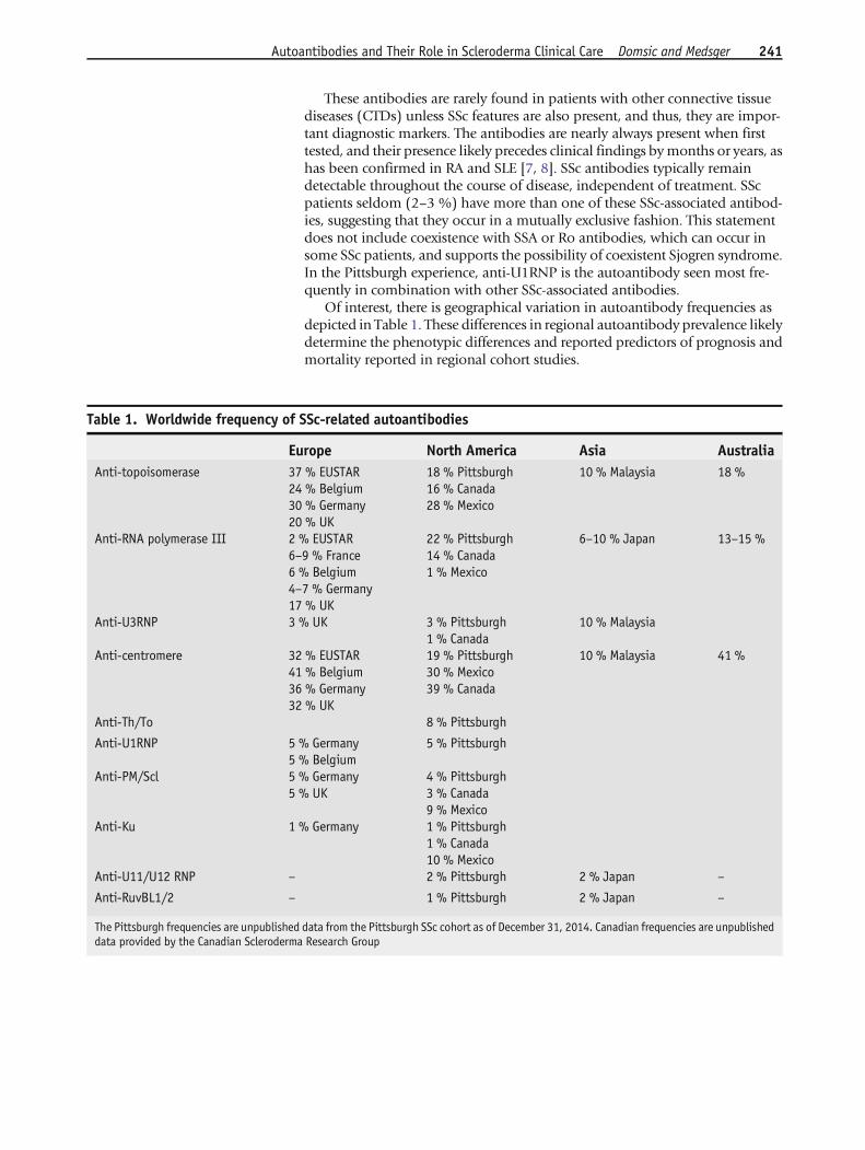
Of interest, there is geographical variation in autoantibody frequencies asdepicted in Table 1. These differences in regional autoantibody prevalence likelydetermine the phenotypic differences and reported predictors of prognosis andmortality reported in regional cohort studies.
Table 1. Worldwide frequency of SSc-related autoantibodies
Europe North America Asia AustraliaAnti-topoisomerase 37 % EUSTAR
24 % Belgium30 % Germany20 % UK
18 % Pittsburgh16 % Canada28 % Mexico
10 % Malaysia 18 %
Anti-RNA polymerase III 2 % EUSTAR6–9 % France6 % Belgium4–7 % Germany17 % UK
22 % Pittsburgh14 % Canada1 % Mexico
6–10 % Japan 13–15 %
Anti-U3RNP 3 % UK 3 % Pittsburgh1 % Canada
10 % Malaysia
Anti-centromere 32 % EUSTAR41 % Belgium36 % Germany32 % UK
19 % Pittsburgh30 % Mexico39 % Canada
10 % Malaysia 41 %
Anti-Th/To 8 % Pittsburgh
Anti-U1RNP 5 % Germany5 % Belgium
5 % Pittsburgh
Anti-PM/Scl 5 % Germany5 % UK
4 % Pittsburgh3 % Canada9 % Mexico
Anti-Ku 1 % Germany 1 % Pittsburgh1 % Canada10 % Mexico
Anti-U11/U12 RNP – 2 % Pittsburgh 2 % Japan –
Anti-RuvBL1/2 – 1 % Pittsburgh 2 % Japan –
The Pittsburgh frequencies are unpublished data from the Pittsburgh SSc cohort as of December 31, 2014. Canadian frequencies are unpublisheddata provided by the Canadian Scleroderma Research Group
Autoantibodies and Their Role in Scleroderma Clinical Care Domsic and Medsger 241

Anti-centromere antibody
Anti-centromere antibody (ACA) is directed against centromere proteins, andthe gold-standard method of detection is by ANA using indirect immunofluo-rescence, and is widely available. Centromere proteins are necessary in theassembly of the kinetochore during cell division and are essential to mitosis.Recent work has confirmed that there are two centromere epitopes recognized,centromere-A and centromere-B. SSc patient antibodies can react to both cen-tromere antigens. Centromere-A has greater specificity in SSc patients [9], butcentromere-B antigen is the antigen included in most commercially availableELISA kits and multiplex assays. Anti-centromere-A antibodies are not part ofnaturally occurring human immunoglobulins [10]. Two novel motifs withinthe immunodominant centromere-A epitope have been identified, one ofwhich is Ap17-30. Interestingly, this motif appears to be within the primarysequence of FOXE-3, which is a transcription factor known to be involved in thedevelopment of lens fiber cells. New work has demonstrated that FOXE-3 isexpressed in monocytes, and that SSc patients with distinct seropositivityresponded differently to cytokine stimulation [11].
ACA occurs in 20–25 % of most SSc populations and is stronglyassociated with limited SSc (Fig. 1) and scleroderma sine scleroderma.The prototypical ACA-positive SSc patient has long-standing (years)Raynaud phenomenon followed by swollen fingers (sclerodactyly) andthen by thickening of the skin of the fingers which evolves over anumber of years or even decades. For patients with Raynaud symptomsand no other clinical clues to an underlying CTD, the presence of ACAand/or nailfold capillary abnormalities is strongly predictive of the sub-sequent development of SSc [12]. Digital ischemia with digital tipulcerations is disproportionately frequent in ACA-positive patients[13••]. Esophageal involvement is common (80 %), but severe intersti-tial lung disease, cardiomyopathy, and scleroderma renal crisis (SRC) arerare [14••, 15]. The most serious long-term complications of ACA-positive SSc are the late occurrence of pulmonary arterial hypertension(15–20 %) or small intestinal involvement with malabsorption [14••,15].
Anti-topoisomerase I (Anti-Scl-70) antibody
This antibody is found in 20–30%of SSc patients inmany ethnic groups, but inEurope, the proportion is higher (40–60 %) [16••]. Three-fourths of patientswith this antibody have dcSSc (Fig. 1). There is a high (60 %) risk of interstitiallung disease (ILD) regardless of the extent of skin thickening (diffuse or limited)[17].
Anti-Scl-70 is associated with myocardial disease and scleroderma renalcrisis (SRC) in patients with rapidly progressive diffuse skin involvement [17].Peripheral vascular complications such as digital ischemia with ulcerations areparticularly common. In the multicenter digital ulcer outcome (DUO) registry,nearly half the patients were anti-Scl70 antibody positive [13••]. This associa-tion was confirmed in a recent EUSTAR multivariate analysis [18] where anti-
242 Scleroderma (D Khanna, Section Editor)

Scl-70 was an independent predictor of developing subsequent digital ulcers.Anti-Scl-70 is a marker for poor prognosis, primarily because of its associationwith ILD and its sequelae including secondary pulmonary hypertension.
The most reliable method for detecting anti-Scl 70, double immunodiffu-sion, has now been replaced inmost commercial laboratories by ELISA or bead-based assays. Unfortunately, these methods result in a number of “low posi-tives” which are, in fact, false positive tests in persons who do not have clinicalevidence of SSc and often have a negative ANA by IIF. Clinicians should becognizant of this when evaluating patients with a positive Scl-70 antibody test.
Anti-RNA polymerase III antibody
Anti-RNA polymerase III (RNAP III) is highly specific to SSc. The gold-standardmethod of detection is by immunoprecipitation, but reliable ELISA-based testsare commercially available. There is a higher frequency in North AmericaCaucasian and UK patients (20–25%) in comparison withmainland Europeanor Japanese patients (5 %) [16••, 19]. Almost all patients with RNA P III haverapidly progressive diffuse skin thickening (Fig. 1). The peak skin thicknessscore is higher in RNAP III than in Scl 70 patients (Pittsburgh unpublisheddata),[20], and recent EUSTAR work has demonstrated that a higher rate ofRNAP III-positive patients develop diffuse SSc within 3 years of the onset ofRaynaud phenomenon than Scl-70-positive patients [18]. In many patients,skin thickness regresses during follow-up, even without treatment.
SSc patients with this antibody have the greatest risk for developing SRC(25%) [21], typically during the early periodwhen skin thickness progression isaccelerated, and if tendon friction rubs are appreciated. Once SRC has occurred,survival is better than in Scl-70 positive SRC patients, perhaps because con-comitant severe ILD and cardiomyopathy are infrequent in RNAP III patients.
In the last few years, two new associations with RNAP have come to light.The first is the occurrence of gastric antral vascular ectasia (GAVE or “water-melon stomach”), confirmed in two cohort studies [22, 23]. The second is aclose temporal relationship between the onset of SSc in RNAP III-positivepatients and the diagnosis of cancer, suggesting that the SSc may be aparaneoplastic process in this patient subset [24••, 25–27].
Anti-U3RNP antibody [28]
This antibody reacts with 34-KD fibrillarin complexed with U3RNA and pro-duces bright nucleolar staining on ANA IIF testing. A reliable ELISA test iscommercially available for detection of this antibody, and a recent publicationsuggested that a line immunoblot assay may be an attractive alternative [29].Some commercial laboratories do have available immunoprecipitation testing,which is the gold standard.
Anti-U3RNP is found in 4–10 % of SSc patients and is more frequentin African Americans [28, 30]. It has been linked to a specific HLA-DRB1*08-04 allele [31] in African Americans. More than half of patientshave diffuse SSc. A non-inflammatory skeletal myopathy is a distinctivefeature, and early severe internal organ involvement including cardio-myopathy, PAH, and small bowel involvement with malabsorption and
Autoantibodies and Their Role in Scleroderma Clinical Care Domsic and Medsger 243

episodes of pseudoobstruction may occur [19]. Prognosis in this subsetof SSc patients is poor [30], similar to those with anti-Scl-70 antibody.
Anti-Th/To antibody
These antibodies are directed against subunits ofmitochondrial RNA processingand ribonuclease P RNP complexes. Most patients with anti-Th/To antibodyhave nucleolar staining on ANA by IIF. One US-based company offers a com-mercially available immunoprecipitation assay for this test. Th/To antibodyoccurs in less than 5 % of SSc patients, almost all of whom have limited SSc(Fig. 1). Patients with this antibody can develop either ILD (45 %) or PAH(25 %), which are most often independent of one another [15]. These pulmo-nary complications result in a reduced survival compared to lcSSc patients withother SSc-associated antibodies. Regular screening for PAH in this patientpopulation is prudent.
Anti-U11/U12RNP antibody
This is a rare antibody found in 1–3 % of SSc patients, equally dividedbetween limited and diffuse subtypes (Fig. 1). A distinctive clinicalfeature is a very high frequency of ILD (80 %) which is often severe andrapidly progressive. As of this writing, there has only been one publi-cation examining the internal organ associations with this autoantibody[32]. To the author’s knowledge, testing for this antibody is not com-mercially available at this time.
Anti-PM/Scl antibody
PM/Scl antibodies target the human exosome complex, of which there areup to 16 proteins. The major B cell targets of the PM/Scl complex have beenidentified as PM100 (also called PM1-alpha) and PM75. SSc patients canhave antibodies to one or both of these antigens. PM/Scl is one of three SSc-associated autoantibodies that have predominantly nucleolar staining onANA by IIF, often with a moderately high ANA titer level. The gold-standarddetection method for PM/Scl has been immunodiffusion and can be ob-tained commercially. The PM100 is available commercially using semi-quantitative immunblot testing.
Two to 4 % of SSc patients have anti-PM/Scl [33–35]. Patients withanti-PM/Scl antibody most often have an overlap with features oflimited SSc and polymyositis (Fig. 1), but some have myositis only[36]. The myositis is usually mild and corticosteroid-responsive. Al-though there is a relatively high percentage of patients with lunginvolvement (40–50 %), serious internal organ involvement is rare,and patients have an excellent prognosis [33, 34]. Calcinosis has beena frequent reported clinical association [37••]. An interesting recentarticle combined Canadian, Australian, and US samples to examine theclinical associations with antibodies to each or both of the PM/Scl
244 Scleroderma (D Khanna, Section Editor)

complexes [34]. The presence of autoantibodies to both complexes wasassociated with an increased rate of inflammatory myositis.
Anti-Ku antibody
The Ku antigen is a heterodimer of 70 kDa (p70) and 80 kDa (p80) subunits.Ku is a multifunctional protein involved in DNA repair, immunoglobulin generecombination, and transcription regulation [38]. Commercial testing for thisantibody is available using immunoprecipitation through specific myositistesting panels. Anti-Ku patients represent less than 2 % of SSc populations, andanti-Ku has most commonly been detected in limited SSc patients with overlapdisorders (myositis or lupus) (Fig. 1) [39, 40••, 41]. In the Pittsburgh experi-ence, 60 % developed diffuse SSc skin changes. A more recent publication haslinked it to a relatively higher rate of ILD [39] and several with arthritis [39, 41].Although there are several case series of anti-Ku-positive SSc patients, overallnumbers reported remain small, and thus, an estimate regarding prognosiscannot be made. The associated myositis is typically corticosteroid-responsive[42].
Anti-U1RNP antibody
This antibody was initially identified by Sharp et al. in the 1970s as beingassociated with features of SSc, PM and SLE and was thus considered todescribe “mixed connective tissue disease” or MCTD [43]. Various combi-nations of these CTDs have been reported, and some patients have few orno scleroderma features. When we encounter anti-U1RNP-positive patientswho have predominantly scleroderma features, we classify them as havingSSc rather than using the term MCTD. High-titer speckled nuclear stainingis most frequently found on ANA by IIF. Disease onset tends to be at arelatively young age, and limited SSc is far more common than diffuse SSc.The presence of arthritis and myositis is included in all proposed criteriafor MCTD [44–47]. Five to 10 % of SSc patients have anti-U1RNP, whichis particularly common in African American and Asian patients [28].Serious complications are relatively uncommon. In our experience, thePAH that occasionally develops in anti-U1RNP-positive SSc patients isresponsive to corticosteroids and immunosuppressive agents, which is nottypically the case in PAH associated with other SSc antibodies.
Anti-RuvBL1/2 antibody
This newly identified antibody is directed against a double hexamer con-taining the proteins pontin and reptin, located in the nucleoplasm (Fig. 1).Detection is by immunoprecipitation less than 1 % of SSc patients have thisantibody, which is more common in males with diffuse skin thickening andcoexistent myositis. Only one publication, combining single cohorts fromJapan and the US have reported [48••]. Information on internal organ andprognosis is limited at this time. To our knowledge, this antibody cannot beidentified in a commercial setting at present.
Autoantibodies and Their Role in Scleroderma Clinical Care Domsic and Medsger 245

Other SSc-associated ANAs
Anti-nucleolar organizing region (NOR) antibodies [49], anti-B23 [50], anti-SSA/R052/TRIMZ1 [51] antibodies to interferon-inducible genes designatedHIN-200 [52], and antibodies to cytoplasmic mitochondrial proteins [53] havebeen reported in SSc patients. Anti-B23 was noted to be associated with PAH,but often coexisted with anti-U3RNP and anti-U1RNP; thus, its independentassociation with PAH is uncertain [51]. Anti-R052TRIM is strongly associatedwith SSc overlap syndromes and ILD [52].
ANA-negative SSc
Fewer than 5 % of patients with documented SSc on clinical grounds are ANAnegative. A large North American SSc registry reported 6 % ANA-negativepatients whoweremostlymales with less vasculopathy and an increase in lowergastrointestinal involvement [54].
Important clinical limitations of commercial antibody testing inSSc patients
In addition to the problem of false positive anti-Scl-70 antibody results,there are other issues with newer commercially available immunoassaysthat have been revealed recently. The majority of these assays require theavailability of highly purified autoantigens, which contain the majorepitopes recognized by virtually all sera positive for given SSc-relatedANAs. However, since most autoantigens recognized by SSc ANAs aremultiprotein complexes, determining the antigenic subunit most com-monly recognized by autoantibodies is a major challenge.
Bead-based multiplex assay testing, which combines multibead ar-rays and flow cytometry, is capable of detecting multiple SSc-associatednuclear antibodies simultaneously. Unfortunately, in the commerciallyavailable multiplex ANA assays, many of the SSc-associated antibodiesare not present. Although the common antibodies of centromere, Scl-70 and U1RNP antigens are included, the other seven SSc-associatedantigens are not included. Thus, when this type of assay was used forANA screening, up to 50 % of SSc patients were reported as a negativeANA in the US population [55] despite the fact that 990 % wouldhave had a positive ANA test by immunofluorescence. Use of bead-based ANA testing for screening holds a potential danger for missingSSc patients, particularly at the primary care level.
More recently, a EUROLINE tests has become available which candetect multiple SSc-associated antibodies (PM/Scl, Ku, Scl-70, centro-mere-B, RNAP III, U3-RNP, Th/To as well as additional specificities).This immunoblot assay produces semi-quantitative results. Although not
246 Scleroderma (D Khanna, Section Editor)

the primary goal of the publication, recent work from the AustralianScleroderma Cohort Study using the EUROLINE test demonstrated thatthe rate of multiple antibody positivity was far higher than in cohortsthat use the gold-standard testing, including immunodiffusion, sug-gesting high rates of false positivity using the EUROLINE test. In theirprincipal component analysis, they demonstrated that the dominantautoantibody (if Scl-70, RNAP, or ACA) accurately reflects diseaseassociations with this testing. We recommend caution in interpretingother results with this assay.
In the SSc literature, authors have commonly grouped and analyzedtogether patients presenting with a nucleolar pattern on ANA testing byimmunofluorescence. The nucleolar pattern generally corresponds toone of three SSc-associated antibodies: anti-PM/Scl, anti-U3RNP, oranti-Th/To. Since the clinical and laboratory associations, internal or-gan involvement, and natural history of disease differ considerably inthese three antibody subsets (see above sections), it is important toidentify each if possible, both for patient care and research. Until veryrecently, the latter two of these antibodies could not be performedcommercially and thus, have not usually been identified in cohortstudies and clinical trials. This is an area in which the SSc communityshould work together in the future to identify and support resourcelaboratories capable of performing these tests in a reliable and vali-dated manner.
Usefulness of SSc-related ANAs in SSc diagnosis
These antibodies are useful in supporting the diagnosis of SSc primarilybecause of their high specificity for the disease. As a consequence, thepresence of ACA, anti-Scl-70, and anti-RNAP III are included in theserologic domain of the 2013 classification criteria for SSc jointly pro-posed by the American College of Rheumatology (ACR) and the Euro-pean League against Rheumatism (EULAR). Their high specificity and theavailability of reliable assay kits worldwide accounts for their inclusionin the criteria set.
Usefulness of SSc-related ANAs regarding disease activity
It has generally been believed that serial measurement of serum ANAtiters is not useful in assessing SSc disease activity. However, few suchstudies have been attempted, and in the past, quantitation of antibodylevels was not possible. Changing levels of anti-Scl 70 by ELISA werefound to be associated with progression of skin thickness [63], andpatients who lost anti-Scl 70 antibody reactivity over time had a morefavorable outcome [55, 64]. Anti-RNAP III antibody levels correlatedpositively with the modified Rodnan total skin thickness score and with
Autoantibodies and Their Role in Scleroderma Clinical Care Domsic and Medsger 247
the onset of scleroderma renal crisis (SRC). Now that semi-quantitativemethods are available, additional studies are likely to be performed toclarify the usefulness of serial autoantibody testing in predicting futureevents in clinical practice and clinical trials.
Antibodies to functional molecules
These autoantibodies are potentially relevant to SSc pathogenesis by modulat-ing disease-related pathways such as platelet-derived growth factor [56],transforming growth factor beta [57], angiotensin II type 1 receptor [58], orendothelin-1 type a receptor [59]. They have been reviewed in a recent textbookchapter [57]. Overall, the potential clinical utility of these autoantibodiesremains uncertain. We will briefly discuss focused recent work examining anti-endothelin 1 type A receptors (ETAR) and anti-angiotensin II type I receptor(AT1R) in SSc which is relevant to patient care.
Serum AT1R and ETAR antibodies were reported to be elevated in autoim-mune diseases [59] and in pre-eclampsia [60]. These antibodies have been foundin human microvascular endothelial cells, but also are expressed in fibroblasts,epithelial, and immune cells. These antibodies have been shown to be biologi-cally active and potentially mediate pathogenic effects of SSc [61]. Riemekastenet al. showed in a cross-sectional cohort study that there was an associationbetween these antibodies and SSc mortality, pulmonary hypertension, renalcrisis, and digital ulcers [59]. In 2015, Avouac et al. examined a small (n = 90),prospective SSc cohort and showed anti-ETAR antibodies to be an independentpredictor of the occurrence of new ischemic DU over 48 months, but not anti-AT1R. A history of DU was also important in their models [62]. This worksuggests a potential promising role of anti-ETAR and perhaps AT1R in futureclinical management or risk stratification in study design of digital ulcers.
Conclusions
Systemic sclerosis-associated serum autoantibodies support the diagnosis ofSSc, and because of their strong clinical associations they compliment thecutaneous-based subgrouping of SSc patients. A combined clinical-serologicclassification system provides an essential framework for predicting patternsand frequency of organ system involvement in SSc patients. This is helpful forpatient management and informing the design and conduct of future clinicaland laboratory research and clinical trials.
Compliance with Ethical Standards
Conflict of InterestRTD reports research support from Biogen-idec and personal fees and research support from Bayer Healthcare,outside the submitted work. TAM declares that he has no conflicts of interest.
248 Scleroderma (D Khanna, Section Editor)

Human and Animal Rights and Informed ConsentWith regard to the authors’ research cited in this paper, all procedures performed in studies involvinghuman participants were in accordance with the ethical standards of the institutional and/or nationalresearch committee and with the 1964 Helsinki declaration and its later amendments or comparableethical standards. In addition, all applicable international, national, and/or institutional guidelines for thecare and use of animals were followed.
References and Recommended ReadingPapers of particular interest, published recently, have beenhighlighted as:•• Of major importance
1. LeRoy EC et al. Scleroderma (systemic sclerosis): clas-sification subsets and pathogenesis. J Rheumatol.1988;15(2):202–5.
2. Poormoghim H, Lucas M, Fertign, Medsger Jr TA. Sys-temic sclerosis sine scleroderma: demographic, clinicaland serologic features and survival in forty-eight pa-tients. Arthritis Rheum 2000;43:444–51.
3. Diab S, Dostrovsky N, Hudson M, Tatibouet S, FritzlerMJ, Baron M, et al. Systemic sclerosis sine scleroderma:a multicenter study of 1417 subjects. J Rheumatol.2014;41(11):790–85.
4. Nihtyanova SID. Autoantibodies as predictive tools insystemic sclerosis. Nat Rev Rheumatol. 2010;5:112–6.
5. van den Hoogen F et al. 2013 classification criteria forsystemic sclerosis: an American College ofRheumatology/European League Against Rheumatismcollaborative initiative. Arthritis Rheum.2013;65(11):2737–47.
6.•• van den Hoogen F et al. 2013 classificationcriteria for systemic sclerosis: an American Col-lege of Rheumatology/European League AgainstRheumatism collaborative initiative. Ann RheumDis. 2013;72D11]:1747–55.
This article presents the new systemic sclerosis classificationcriteria, which now includes SSc-specific antibodies.7. Arbuckle MR, McClain MT, Rubertone MV,
Scofield RH, Dennis GJ, James JA, et al. Devel-opment of autoantibodies before the clinical on-set of systemic lupus erythematosus. N Engl JMed. 2003;349(16):1526–33.
8. Shi J, van de Stadt LA, Levarht EW, Huizinga TW,Hamann D, van Schaardenburg D, et al. Anti-carbamylated protein (anti-CarP) antibodies precedethe onset of rheumatoid arthritis. Ann Rheum Dis.2014;73(4):780–3.
9. Perosa F, Favoino E, Cuomo G, Digiglio L, DammaccoF, Prete M, et al. Clinical correlates of a subset of anti-CENP-A antibodies cross-reacting with FOXE3p53-62in systemic sclerosis. Arthritis Res Ther.2013;15(4):R72.
10. Peroso F, Prete M, DiLernia G, Ostuni C, Favoino E,Valentini G. Anti-centromere protein A antibodies in
systemic sclerosis: significance and origin. AutoimmunRev. 2015;15(1):105–9.
11. Favoino E, Favia I, Valentini G, Perosa F. Expression ofthe transcription factor Forkhead Box E3 (Foxe3) inmonocytes from patients with systemic sclerosis andcorrelation with their serological profile. Ann RheumDis. 2014;73:868.
12. Koenig M, Joyal F, Fritzler MJ, Roussin A,Abrahamowicz M, Boire G, et al. Autoantibodies andmicrovascular damage are independent predictive fac-tors for the progression of Raynaud’s phenomenon tosystemic sclerosis: a twenty-year prospective study of586 patients with validation of proposed criteria forearly systemic sclerosis. Arthritis Rheum.2008;58(12):3902–12.
13.•• Denton CP, Krieg T, Guillevin L, Schwierin B, Rosen-berg D, Silkey M, et al. DUO Registry investigatorsdemographic clinical and antibody characteristics ofpatients with digital ulcers in systemic sclerosis: datafrom the DUO Registry. Ann Rheum Dis.2012;71D5]:718–21.
This article presents antibody associations with the develop-ment of digital ulcerations.14.•• Hudson M, Mahler M, Pope J, You D, Tatibouet S,
Steele R, et al. Clinical correlates of CENP-A and CENP-B antibodies in a large cohort of patients with systemicsclerosis. J Rheumatol. 2012;39D4]:787–94.
This article reports associations with centromere antibodiessubtypes.15. Mitri GM, LucasM, Fertig N, Steen VD,Medsger Jr TA. A
comparison between anti-Th/To- and anticentromereantibody-positive systemic sclerosis patients with lim-ited cutaneous involvement. Arthritis Rheum.2003;48(1):203–9.
16.•• Meier FM, Frommer KW, Dinser R, Walker UA, CzirjakL, Denton CP, et al. Update on the profile of theEUSTAR cohort: an analysis of the EULAR SclerodermaTrials and Research group database. Ann Rheum Dis.2012;71D8]:1355–60.
This article includes information on SSc-associated antibodyfrequencies and clinical characteristics in a large, multicenter,European cohort.
Autoantibodies and Their Role in Scleroderma Clinical Care Domsic and Medsger 249
17. Perera A, Fertig N, Lucas M, Rodriguez-Reyna TS, Hu P,Steen VD, et al. Clinical subsets skin thickness pro-gression rate and serum antibody levels in systemicsclerosis patients with anti-topoisomerase I antibody.Arthritis Rheum. 2007;56(8):2740–6.
18. Wirz EG, Jaeger VK, Allanore Y, Riemekasten G,Hachulla E, Distler O, Airò P, Carreira PE, Tikly M,Vettori S, Balbir Gurman A, Damjanov N, Müller-Ladner U, Distler J, Li M, Häusermann P, Walker UA,EUSTAR coauthors. Incidence and predictors of cuta-neous manifestations during the early course of sys-temic sclerosis: a 10-year longitudinal study from theEUSTAR database. Ann Rheum Dis 2013.
19. Hamaguchi Y, HasegawaM, FujimotoM,Matsushita T,Komura K, Kaji K, et al. The clinical relevance of serumantinuclear antibodies in Japanese patients with sys-temic sclerosis. Br J Dermatol. 2008;158(3):487–95.
20. Nikpour M, Hissaria P, Byron J, Sahhar J, Micallef M,Paspaliaris W, et al. Prevalence correlates and clinicalusefulness of antibodies to RNA polymerase III in sys-temic sclerosis: a cross-sectional analysis of data froman Australian cohort. Arthritis Res Ther.2011;13(6):R211.
21. Hamaguchi Y, Kodera M, Matsushita T, Hasegawa M,Inaba Y, Usuda T, et al. Clinical and immunologicpredictors of scleroderma renal crisis in Japanese sys-temic sclerosis patients with anti-RNA polymerase IIIautoantibodies. Arthritis Rhematol. 2015;67(4):1045–52.
22. Ghrénassia E, Avouac J, Khanna D, Derk CT, Distler O,Suliman YA, et al. Prevalence correlates and outcomesof gastric antral vascular ectasia in systemic sclerosis: aEUSTAR case-control study. J Rheumatol.2014;41(1):99–105.
23. Patterson KA, Roberts-Thomson PJ, Lester S, Tan JA,Hakendorf P, Rischmueller M, et al. Interpretation ofan extended autoantibody profile in a well-characterized Australian systemic sclerosis(scleroderma) cohort using principal componentsanalysis. Arthritis Rheumatol. 2015;67(12):3234–44.
24.•• Shah AA, Rosen A, Hummers L, Wigley F, Casciola-Rosen L. Close temporal relationship between onset ofcancer and scleroderma in patients with RNA poly-merase I/III antibodies. Arthritis Rheum.2010;62D9]:2787–95.
Original article describing the temporal association betweenRNA polymerase III and onset of SSc.25. Airo P, Ceribelli A, Cavazzana I, Taraborelli M,
Zingarelli S, Franceschini F. Malignancies in Italianpatients with systemic sclerosis positive for anti-RNApolymerase III antibodies. J Rheumatol.2011;38(7):1329–34.
26. Moinzadeh P, Fonseca C, Hellmich M, Shah AA,Chighizola C, Denton CP, et al. Association of anti-RNA polymerase III autoantibodies and cancer inscleroderma. Arthritis Res Ther. 2014;16(1):R53.
27. Joseph CG, Darrah E, Shah AA, Skora AD, Casciola-Rosen L,A, Wigley F,M, et al. Association of the
autoimmune disease scleroderma with an immuno-logic response to cancer. Science. 2014;343:152–7.
28. Steen V, Domsic RT, LucasM, Fertig N,Medsger Jr TA. Aclinical and serologic comparison of African AmericanandCaucasian patients with systemic sclerosis. ArthritisRheum. 2012;64(9):2986–94.
29. Petersen WG, Zimmerman R. Limited utility of chestradiograph after thoracentesis. Chest.2000;117(4):1038–42.
30. Aggarwal R, Lucas M, Fertig N, Oddis C,V, Medsger JrT,A. Anti-U3 RNP autoantibodies in systemic sclerosis.Arthritis Rheumatol. 2009;60(4):1112–8.
31. Sharif R, Fritzler MJ, Mayes MD, Gonzalez EB,McNearney TA, Draeger H, et al. Anti-fibrillarinantibody in African American patients with sys-temic sclerosis: immunogenetics clinical featuresand survival analysis. J Rheumatol.2011;60(4):1112–8.
32. Fertig N, Domsic RT, Rodriguez-Reyna T, Kuwana M,Lucas M, Medsger TA, et al. Anti-U11/U12 RNP anti-bodies in systemic sclerosis: a new serologic markerassociated with pulmonary fibrosis. Arthritis Rheum.2009;61(7):958–65.
33. Koschik RW, Fertig N, Lucas MR, Domsic RT, MedsgerJr TA. Anti-PM-Scl antibody in patients with systemicsclerosis. Clin Exp Rheumatol. 2012;30(2 Suppl71):S12–16.
34. Wodkowski M, Hudson M, Proud man S, Walker J,Stevens W, Nikpour M, et al. Clinical correlates ofmonospecific anti-PM75 and anti-PM100 antibodiesin a tri-nation cohort of 1574 systemic sclerosis sub-jects. Autoimmunity. 2015;3:1–10.
35. Marguerie C, Bunn CC, Copier J, Bernstein RM, GilroyJM, Black CM, et al. The clinical and immunogeneticfeatures of patients with autoantibodies to the nucleo-lar antigen PM-Scl. Medicine (Baltimore).1992;71(6):327–36.
36. Oddis CV, Okana Y, Rudert WA, Trucco M, DuquesnoyRJ, Medsger Jr TA. Serum autoantibody to the nucleolarantigen PM-Scl, clinical and immunogenetic associa-tions. Arthritis Rheum. 1992;35(10):1211–7.
37.•• D’Aoust J, Hudson M, Tatibouet S, Wick J, CanadianScleroderma Research Group, Mahler M, et al. Clinicaland serologic correlates of anti-PM/Scl antibodies insystemic sclerosis: a multicenter study of 763 patients.Arthritis Rheumatol. 2014;66D6]:1608–15.
This article provides a descriptive analysis of PM/Sc1 positivepatient characteristics from the Canadian Scleroderma Re-search group.38. Koike M. Dimerization translocation and localization
of Ku70 and Ku80 proteins. J Radiat Res.2002;43(3):223–36.
39. Cavazzana I, Fredi M, Taraborelli M, Quinzanini M,Tincani A, Franceschini F. A subset of systemic sclerosisbut not of systemic lupus erythematosus is defined byisolated anti-Ku autoantibodie. Clin Exp Rheumatol.2013;31(2 Suppl 76):118–21.
40.•• Moinzadeh Aberer E, Ahmadi-Simab K, Blank N,Distler JH, Fierlbeck G, Genth E, et al. All participating
250 Scleroderma (D Khanna, Section Editor)

DNSS centers, disease progression in systemic sclerosis-overlap syndrome is significantly different from limitedand diffuse cutaneous systemic sclerosis. Ann RheumDis. 2015;74D4]:730–7.
This manuscript is a descriptive study of SSc-overlap patients inthe German Network of Systemic Scleroderma. Anti-Ku andanti-PM/Sc1 patients are described.41. Rozman B. Prevalance and clinical associations of anti-
Ku antibodies in patients with systemic sclerosis: aEuropena EUSTAR-initiated multi-centre case-controlstudy. Ann Rheum Dis. 2008;67(9):1282–6.
42. Rigolet AM,DubourgO,Maisonobe T, Grenier CharuelJL, Behin A, Herson S, et al. Inflammatory myopathieswith anti-Ku antibodies: a prognosis dependent onassociated drug disease. Medicine (Baltimore).2012;91(2):95–102.
43. Sharp GC. Current concepts in the classification ofconnective tissue diseases, overlap syndromes andmixed connective tissue disease (MCTD). J Am AcadDermatol. 1980;4:269–79.
44. Alarcon Segovia D, Villareal M, Classification and di-agnostic criteria for mixed connective tissue disease,mixed connective tissue disease and anti-nuclear anti-bodies ed, R, Kasukawa Sharp G, editors. Amsterdam:Elsevier; 1987.
45. Doria A, Ghirardello A, de Zambiasi RA, Gambari PF.Japanese diagnostic criteria for mixed connective tissuedisease in Caucasian patients. J Rheumatol.1992;19(2):259–64.
46. Jonsson J, Norberg R. Symptomatology and diagnosisin connective tissue disease, II, evaluations and follow-up examinations in consequence of a speckled antinu-clear immunofluorescence pattern. Scand J Rheumatol.1978;7(4):229–36.
47. Kahn M, Appelboom T, Syndrom de Shar3rd ed, Lesmaladies systemiques ed, M, Kahn Peltier A, Meyer O,Peiette J, editors. Paris: Flammarion; 1991.
48.•• Kaji K, Fertig N, Medsger Jr TA, Satoh T, Hoshino K,Hamaguchi Y, et al. Autoantibodies to RuvBL1 andRuvBL2: a novel systemic sclerosis-related antibodyassociated with diffuse cutaneous and skeletal muscleinvolvement. Arthritis Care Res DHoboken].2014;66D4]:575–84.
This article presents the identification and clinical associationsof anti-RUVBL1 and RuvBL2 with a SSc-overlap presentation.49. Rodriguez-Sanchez JL et al. A new antibody in sclero-
derma that recognizes a 90-kDa component of thenucleolus-organizing region of chromatin. J Immunol.1987;139(8):2579–84.
50. Dagher JH, Scheer U, Voit R, Grummt I, Lonzetti L,Raymond Y, et al. Autoantibodies to NOR 90/hUBF:longterm clinical and serological followup in a patientwith limited systemic sclerosis suggests an antigendriven immune response. J Rheumatol.2002;29(7):1543–7.
51. Ulanet DB et al. Autoantibodies against B23 a nucleo-lar phosphoprotein occur in scleroderma and are as-sociated with pulmonary hypertension. ArthritisRheum. 2003;49(1):82–92.
52. Hudson M et al. Clinical significance of antibodies toRo52/TRIM21 in systemic sclerosis. Arthritis Res Ther.2012;14(2):R50.
53. Costa S, Mondini M, Caneparo V, Afeltra A, AiroBellisai F, et al. Detection of anti-IF116 antibodies byELISA: clinical and serological associations in systemicsclerosis. Rheumatology (Oxford). 2011;50(4):674–81.
54. Salazar GA, Assassi S, Wigley F, Hummers L, Varga J,Hinchcliff M, et al. Antinuclear antibody-negative sys-temic sclerosis. Semin Arthritis Rheum.2014;44(6):680–6.
55. Shanmugam VK, Swistowski DR, Saddic N, Wang H,Steen VD. Comparison of indirect immunofluores-cence and multiplex antinuclear antibody screening insystemic sclerosis. Clin Rheumatol.2011;30(10):1363–8.
56. Baroni SS et al. Stimulatory autoantibodies to thePDGF receptor in systemic sclerosis. N Engl J Med.2006;354(25):2667–76.
57. Kuwana M, Medsger TA. The clinical aspects of anti-bodies. 2015 In press.
58. Tan FK, Arnett FC, Antohi S, Saito S, Mirarchi A, SpieraH, et al. Autoantibodies to the extracellular matrixmicrofibrillar protein fibrillin-1 in patients withscleroderma and other connective tissue diseases. JImmunol. 1999;163(2):1066–72.
59. Riemekasten G, Philippe A, Näther M, Slowinski T,Müller DN, Heidecke H, et al. Involvement of func-tional autoantibodies against vascular receptors in sys-temic sclerosis. Ann Rheum Dis. 2011;70(3):530–6.
60. Wallukat G, Homuth V, Fischer T, Lindschau C,Horstkamp B, Jüpner A, et al. Patients with pre-eclampsia develop agonistic autoantibodies against theangiotensin AT1 receptor. J Clin Invest.1999;103(7):945–52.
61. Kill A et al. Autoantibodies to angiotensin andendothelin receptors in systemic sclerosis induce cel-lular and systemic events associated with disease path-ogenesis. Arthritis Res Ther. 2014;16(1):r29.
62. Avouac J, Riemekasten G, Meune C. Autoantibodiesagainst endothelin 1 type a receptor are strong predic-tors of digital ulcers in systemic sclerosis. J Rheumatol.2015;42:1801.
63. Hu PQ, Fertig N,Medsger Jr TA,Wright TM. Correlationof serum anti-DNA topoisomerase 1 antibody levelswith disease severity and activity in systemic sclerosis.Arthritis Rheum. 2003;48(5):1363–73.
64. Kuwana M, Kaburaki J, Mimori T, Kawakami Y, Tojo T.Longitudinal analysis of autoantibody response totopoisomerase I in systemic sclerosis. Arthritis Rheum.2000;43(5):1072–84.
Autoantibodies and Their Role in Scleroderma Clinical Care Domsic and Medsger 251