automated solid-phase peptide synthesis to obtain ... · automated solid-phase peptide synthesis to...
TRANSCRIPT
1197
Automated solid-phase peptide synthesisto obtain therapeutic peptides
Veronika Mäde, Sylvia Els-Heindl and Annette G. Beck-Sickinger*
Review Open Access
Address:Institute of Biochemistry, Faculty of Biosciences, Pharmacy andPsychology, Universität Leipzig, Brüderstraße 34, D-04103 Leipzig,Germany
Email:Annette G. Beck-Sickinger* - [email protected]
* Corresponding author
Keywords:automated synthesis; automation; lipidation; PEGylation; peptidedrugs; solid-phase peptide synthesis; therapeutic peptides
Beilstein J. Org. Chem. 2014, 10, 1197–1212.doi:10.3762/bjoc.10.118
Received: 01 February 2014Accepted: 16 April 2014Published: 22 May 2014
Editor-in-Chief: P. H. Seeberger
© 2014 Mäde et al; licensee Beilstein-Institut.License and terms: see end of document.
AbstractThe great versatility and the inherent high affinities of peptides for their respective targets have led to tremendous progress for ther-
apeutic applications in the last years. In order to increase the drugability of these frequently unstable and rapidly cleared molecules,
chemical modifications are of great interest. Automated solid-phase peptide synthesis (SPPS) offers a suitable technology to
produce chemically engineered peptides. This review concentrates on the application of SPPS by Fmoc/t-Bu protecting-group
strategy, which is most commonly used. Critical issues and suggestions for the synthesis are covered. The development of auto-
mated methods from conventional to essentially improved microwave-assisted instruments is discussed. In order to improve phar-
macokinetic properties of peptides, lipidation and PEGylation are described as covalent conjugation methods, which can be applied
by a combination of automated and manual synthesis approaches. The synthesis and application of SPPS is described for neuropep-
tide Y receptor analogs as an example for bioactive hormones. The applied strategies represent innovative and potent methods for
the development of novel peptide drug candidates that can be manufactured with optimized automated synthesis technologies.
1197
IntroductionPeptides and proteins are involved in a large variety of
biochemical processes and physiological functions. Peptides
can consist of up to 50 amino acids and have generally no
tertiary, three-dimensional structure compared to proteins [1].
In nature, the oligomers or polymers are assembled at ribo-
somes by aminoacyl-tRNAs (transfer ribonucleic acid) [2].
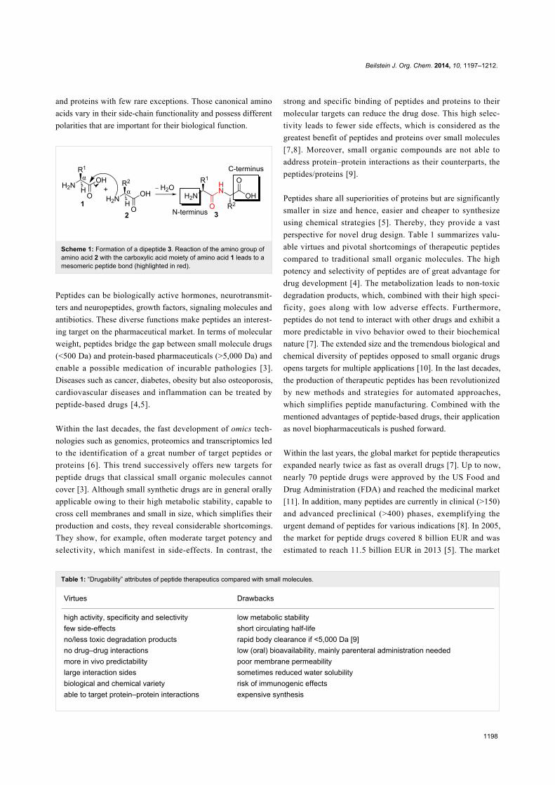
Basically, a condensation reaction of a carboxylic acid moiety
with a functional amine of trifunctional α-amino acids leads to
regioisomeric amide bond (peptide bond) formation
(Scheme 1). The individual building blocks occur as L-enan-
tiomers throughout living organisms in case of ribosomal syn-
thesis and only 20 monomers are generally found in peptides
Beilstein J. Org. Chem. 2014, 10, 1197–1212.
1198
Table 1: “Drugability” attributes of peptide therapeutics compared with small molecules.
Virtues Drawbacks
high activity, specificity and selectivity low metabolic stabilityfew side-effects short circulating half-lifeno/less toxic degradation products rapid body clearance if <5,000 Da [9]no drug–drug interactions low (oral) bioavailability, mainly parenteral administration neededmore in vivo predictability poor membrane permeabilitylarge interaction sides sometimes reduced water solubilitybiological and chemical variety risk of immunogenic effectsable to target protein–protein interactions expensive synthesis
and proteins with few rare exceptions. Those canonical amino
acids vary in their side-chain functionality and possess different
polarities that are important for their biological function.
Scheme 1: Formation of a dipeptide 3. Reaction of the amino group ofamino acid 2 with the carboxylic acid moiety of amino acid 1 leads to amesomeric peptide bond (highlighted in red).
Peptides can be biologically active hormones, neurotransmit-
ters and neuropeptides, growth factors, signaling molecules and
antibiotics. These diverse functions make peptides an interest-
ing target on the pharmaceutical market. In terms of molecular
weight, peptides bridge the gap between small molecule drugs
(<500 Da) and protein-based pharmaceuticals (>5,000 Da) and
enable a possible medication of incurable pathologies [3].
Diseases such as cancer, diabetes, obesity but also osteoporosis,
cardiovascular diseases and inflammation can be treated by
peptide-based drugs [4,5].
Within the last decades, the fast development of omics tech-
nologies such as genomics, proteomics and transcriptomics led
to the identification of a great number of target peptides or
proteins [6]. This trend successively offers new targets for
peptide drugs that classical small organic molecules cannot
cover [3]. Although small synthetic drugs are in general orally
applicable owing to their high metabolic stability, capable to
cross cell membranes and small in size, which simplifies their
production and costs, they reveal considerable shortcomings.
They show, for example, often moderate target potency and
selectivity, which manifest in side-effects. In contrast, the
strong and specific binding of peptides and proteins to their
molecular targets can reduce the drug dose. This high selec-
tivity leads to fewer side effects, which is considered as the
greatest benefit of peptides and proteins over small molecules
[7,8]. Moreover, small organic compounds are not able to
address protein–protein interactions as their counterparts, the
peptides/proteins [9].
Peptides share all superiorities of proteins but are significantly
smaller in size and hence, easier and cheaper to synthesize
using chemical strategies [5]. Thereby, they provide a vast
perspective for novel drug design. Table 1 summarizes valu-
able virtues and pivotal shortcomings of therapeutic peptides
compared to traditional small organic molecules. The high
potency and selectivity of peptides are of great advantage for
drug development [4]. The metabolization leads to non-toxic
degradation products, which, combined with their high speci-
ficity, goes along with low adverse effects. Furthermore,
peptides do not tend to interact with other drugs and exhibit a
more predictable in vivo behavior owed to their biochemical
nature [7]. The extended size and the tremendous biological and
chemical diversity of peptides opposed to small organic drugs
opens targets for multiple applications [10]. In the last decades,
the production of therapeutic peptides has been revolutionized
by new methods and strategies for automated approaches,
which simplifies peptide manufacturing. Combined with the
mentioned advantages of peptide-based drugs, their application
as novel biopharmaceuticals is pushed forward.
Within the last years, the global market for peptide therapeutics
expanded nearly twice as fast as overall drugs [7]. Up to now,
nearly 70 peptide drugs were approved by the US Food and
Drug Administration (FDA) and reached the medicinal market
[11]. In addition, many peptides are currently in clinical (>150)
and advanced preclinical (>400) phases, exemplifying the
urgent demand of peptides for various indications [8]. In 2005,
the market for peptide drugs covered 8 billion EUR and was
estimated to reach 11.5 billion EUR in 2013 [5]. The market
Beilstein J. Org. Chem. 2014, 10, 1197–1212.
1199
growth rate has been projected to be over 10% per year. To
date, 4% of overall approved pharmaceuticals are peptide
hormones or derivatives [12].
Besides this success story, there are limitations restricting the
use of peptides as drugs (Table 1). Notably, their low bioavail-
ability owing to proteolytic degradation by enzymes of the
intestine, blood and cell plasma leads to short circulating half-
lives [13]. Depending on their size, peptides are excreted by
kidneys (renal clearance) or liver (hepatic clearance) within
minutes [5,9]. Nevertheless, their ability to pass through
membranes and the urgent need of alternative, more comfort-
able administration routes as the commonly used parenteral
(subcutaneous, intramuscular and intravenous) application, have
prompted further research in this field [14]. Therefore, methods
to prolong peptide stability are of great interest.
Here, we highlight the importance of automated solid-phase
peptide synthesis (SPPS) in the process of peptide modification.
Principles of chemical synthesis of peptides are covered with a
focus on Fmoc (9-fluorenylmethoxycarbonyl)/t-Bu (tert-butyl)-
based solid-phase peptide synthesis. Recent advances in
automation devices are described, with attention to the compari-
son between conservative SPPS robots and microwave-assisted
automated SPPS. Moreover, strategies for modulating peptide
stability with an emphasis on lipidation and PEGylation are
characterized. Last, the syntheses of selected peptide hormones
are presented exemplarily.
ReviewChemical synthesis of peptides and itsautomationSolid-phase peptide synthesis – the way fromhomogeneous to heterogeneous synthesisIn the past, pioneering of Emil Fischer at the beginning of the
20th century [15] and du Vigneaud in 1953 [16] have made the
synthesis of peptides possible, as at that time, they were rela-
tively unknown biomolecules. Fischer synthesized the first
dipeptide, called glycylglycin, and coined the term “peptide”
[15]. Fifty years later, du Vigneaud developed a strategy for the
production of a polypeptide. For the synthesis of the polypep-
tide hormone oxytocin, organic protecting groups were intro-
duced to trifunctional amino acids [17] in order to ensure
specific amide-bond formation [16]. The principle of peptide
synthesis in homogenous solution is based on the reversible
blocking of the carboxylic acid function of the C-terminal
amino acid and the amino group of the N-terminal amino acid.
In addition, activation of the free carboxy group of the
N-terminal amino acid is necessary to obtain the peptide bond.
For this approach, all peptide intermediates have to be isolated
and purified before they can be used for further reaction steps.
Although this assures a good quality control, it is a very time-
consuming and a technical-demanding process [18]. This mani-
fests especially at larger and more complex peptides, for which
the protected fragments often tend to be rigid and insoluble
[19].
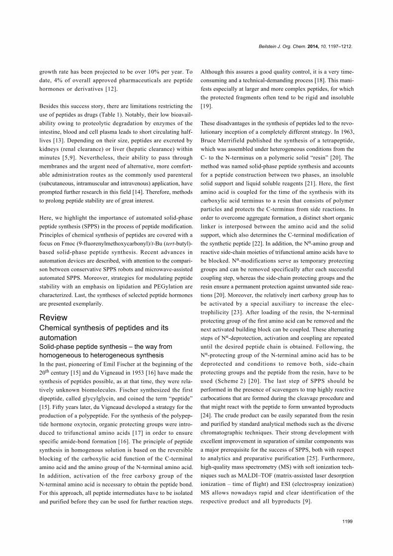
These disadvantages in the synthesis of peptides led to the revo-
lutionary inception of a completely different strategy. In 1963,
Bruce Merrifield published the synthesis of a tetrapeptide,
which was assembled under heterogeneous conditions from the
C- to the N-terminus on a polymeric solid “resin” [20]. The
method was named solid-phase peptide synthesis and accounts
for a peptide construction between two phases, an insoluble
solid support and liquid soluble reagents [21]. Here, the first
amino acid is coupled for the time of the synthesis with its
carboxylic acid terminus to a resin that consists of polymer
particles and protects the C-terminus from side reactions. In
order to overcome aggregate formation, a distinct short organic
linker is interposed between the amino acid and the solid
support, which also determines the C-terminal modification of
the synthetic peptide [22]. In addition, the Nα-amino group and
reactive side-chain moieties of trifunctional amino acids have to
be blocked. Nα-modifications serve as temporary protecting
groups and can be removed specifically after each successful
coupling step, whereas the side-chain protecting groups and the
resin ensure a permanent protection against unwanted side reac-
tions [20]. Moreover, the relatively inert carboxy group has to
be activated by a special auxiliary to increase the elec-
trophilicity [23]. After loading of the resin, the N-terminal
protecting group of the first amino acid can be removed and the
next activated building block can be coupled. These alternating
steps of Nα-deprotection, activation and coupling are repeated
until the desired peptide chain is obtained. Following, the
Nα-protecting group of the N-terminal amino acid has to be
deprotected and conditions to remove both, side-chain
protecting groups and the peptide from the resin, have to be
used (Scheme 2) [20]. The last step of SPPS should be
performed in the presence of scavengers to trap highly reactive
carbocations that are formed during the cleavage procedure and
that might react with the peptide to form unwanted byproducts
[24]. The crude product can be easily separated from the resin
and purified by standard analytical methods such as the diverse
chromatographic techniques. Their strong development with
excellent improvement in separation of similar components was
a major prerequisite for the success of SPPS, both with respect
to analytics and preparative purification [25]. Furthermore,
high-quality mass spectrometry (MS) with soft ionization tech-
niques such as MALDI–TOF (matrix-assisted laser desorption
ionization – time of flight) and ESI (electrospray ionization)
MS allows nowadays rapid and clear identification of the
respective product and all byproducts [9].
Beilstein J. Org. Chem. 2014, 10, 1197–1212.
1200
Scheme 2: Peptide assembly by SPPS, exemplarily shown for atetrapeptide. First, the C-terminal amino acid is coupled to the linker.The peptide chain will be elongated by repeating a cycle of 1) depro-tection of NPG, 2) activation of the carboxy group and 3) coupling. Atthe end of the synthesis, the protecting groups will be cleaved and thedesired peptide obtained. NPG: Nα-protecting group, X: activator,SPG: side-chain protecting group, Aa: amino acid.
This heterogeneous synthesis technique offers great advantages.
Certainly, the most important benefit of SPPS is the feasibility
of carrying out all reactions in a single vessel. Following a
coupling step, unreacted reagents and byproducts can be easily
removed by washing, which makes purification of intermedi-
ates redundant. Based on the use of excess amounts of reactants,
high coupling yields can be obtained and the incorporation of
difficult sequences and modifications to the polymer are
enabled. Moreover, the reaction cycles are very short compared
Figure 1: Five issues that have to be resolved prior to peptide syn-thesis.
to solution synthesis, which allows faster manufacturing [20].
Additionally, the solid-phase concept is not only an elegant way
to build up peptides but also other oligomers such as
polyamides [26], polynucleotides [27] and polysaccharides [28].
This method simplified the chemical synthesis of peptides and
allowed the automation of the process [24], which has led to a
breakthrough of SPPS and the establishment as one major tech-
nique for therapeutic peptide production [8,19].
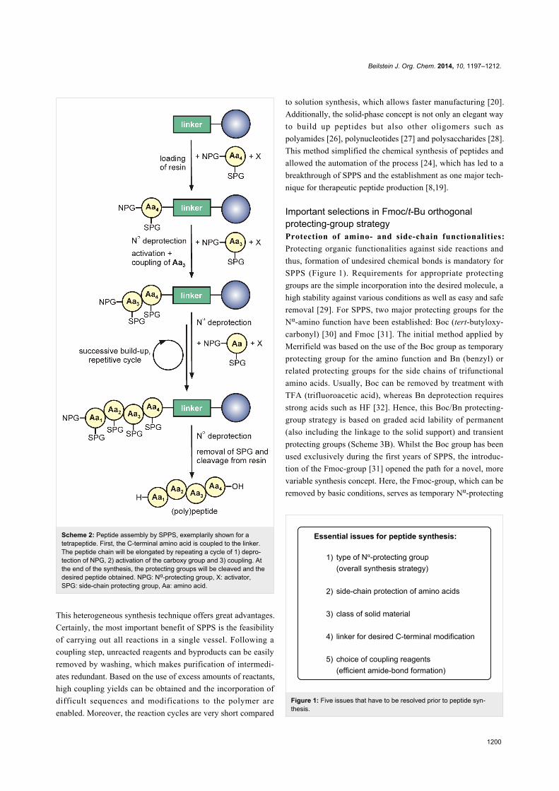
Important selections in Fmoc/t-Bu orthogonalprotecting-group strategyProtection of amino- and side-chain functionalities:
Protecting organic functionalities against side reactions and
thus, formation of undesired chemical bonds is mandatory for
SPPS (Figure 1). Requirements for appropriate protecting
groups are the simple incorporation into the desired molecule, a
high stability against various conditions as well as easy and safe
removal [29]. For SPPS, two major protecting groups for the
Nα-amino function have been established: Boc (tert-butyloxy-
carbonyl) [30] and Fmoc [31]. The initial method applied by
Merrifield was based on the use of the Boc group as temporary
protecting group for the amino function and Bn (benzyl) or
related protecting groups for the side chains of trifunctional
amino acids. Usually, Boc can be removed by treatment with
TFA (trifluoroacetic acid), whereas Bn deprotection requires
strong acids such as HF [32]. Hence, this Boc/Bn protecting-
group strategy is based on graded acid lability of permanent
(also including the linkage to the solid support) and transient
protecting groups (Scheme 3B). Whilst the Boc group has been
used exclusively during the first years of SPPS, the introduc-
tion of the Fmoc-group [31] opened the path for a novel, more
variable synthesis concept. Here, the Fmoc-group, which can be
removed by basic conditions, serves as temporary Nα-protecting
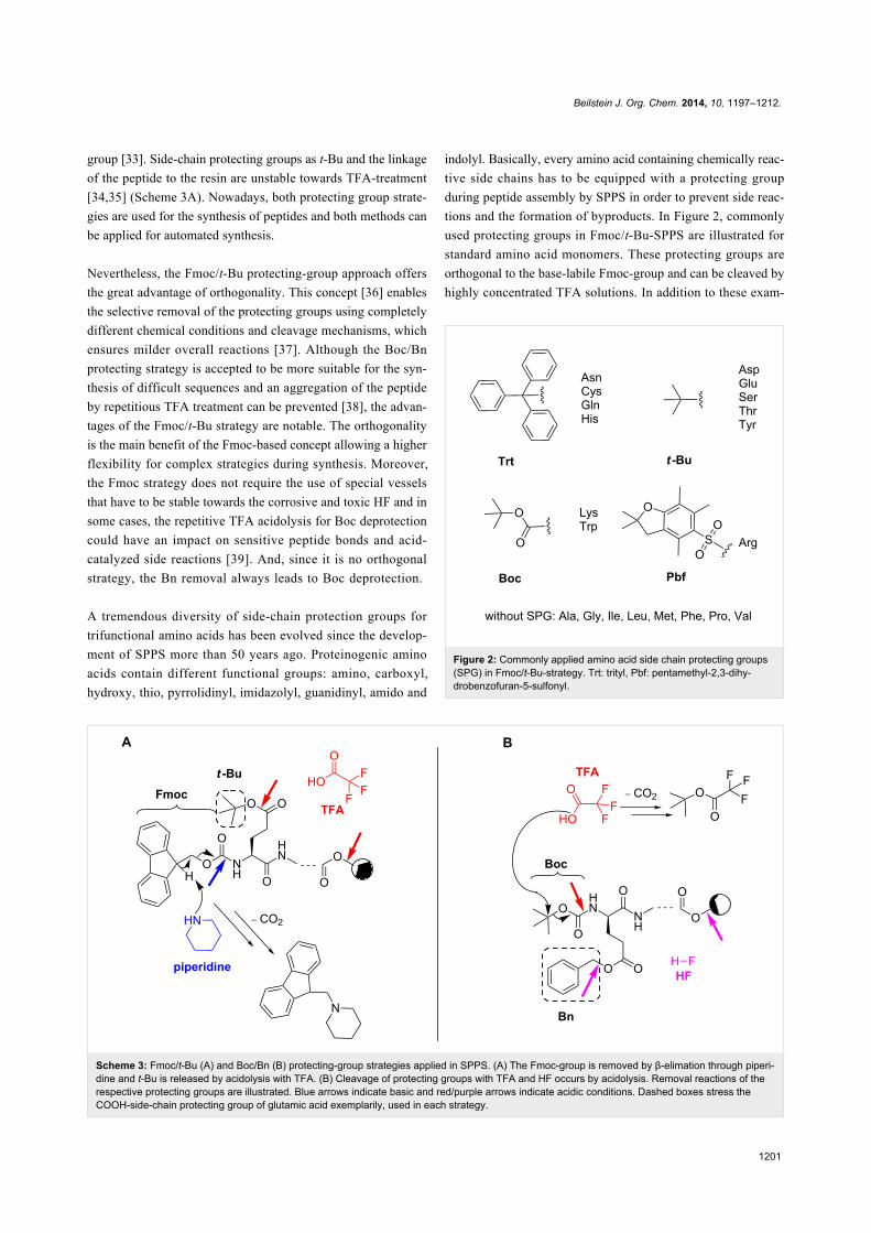
Beilstein J. Org. Chem. 2014, 10, 1197–1212.
1201
Scheme 3: Fmoc/t-Bu (A) and Boc/Bn (B) protecting-group strategies applied in SPPS. (A) The Fmoc-group is removed by β-elimation through piperi-dine and t-Bu is released by acidolysis with TFA. (B) Cleavage of protecting groups with TFA and HF occurs by acidolysis. Removal reactions of therespective protecting groups are illustrated. Blue arrows indicate basic and red/purple arrows indicate acidic conditions. Dashed boxes stress theCOOH-side-chain protecting group of glutamic acid exemplarily, used in each strategy.
group [33]. Side-chain protecting groups as t-Bu and the linkage
of the peptide to the resin are unstable towards TFA-treatment
[34,35] (Scheme 3A). Nowadays, both protecting group strate-
gies are used for the synthesis of peptides and both methods can
be applied for automated synthesis.
Nevertheless, the Fmoc/t-Bu protecting-group approach offers
the great advantage of orthogonality. This concept [36] enables
the selective removal of the protecting groups using completely
different chemical conditions and cleavage mechanisms, which
ensures milder overall reactions [37]. Although the Boc/Bn
protecting strategy is accepted to be more suitable for the syn-
thesis of difficult sequences and an aggregation of the peptide
by repetitious TFA treatment can be prevented [38], the advan-
tages of the Fmoc/t-Bu strategy are notable. The orthogonality
is the main benefit of the Fmoc-based concept allowing a higher
flexibility for complex strategies during synthesis. Moreover,
the Fmoc strategy does not require the use of special vessels
that have to be stable towards the corrosive and toxic HF and in
some cases, the repetitive TFA acidolysis for Boc deprotection
could have an impact on sensitive peptide bonds and acid-
catalyzed side reactions [39]. And, since it is no orthogonal
strategy, the Bn removal always leads to Boc deprotection.
A tremendous diversity of side-chain protection groups for
trifunctional amino acids has been evolved since the develop-
ment of SPPS more than 50 years ago. Proteinogenic amino
acids contain different functional groups: amino, carboxyl,
hydroxy, thio, pyrrolidinyl, imidazolyl, guanidinyl, amido and
Figure 2: Commonly applied amino acid side chain protecting groups(SPG) in Fmoc/t-Bu-strategy. Trt: trityl, Pbf: pentamethyl-2,3-dihy-drobenzofuran-5-sulfonyl.
indolyl. Basically, every amino acid containing chemically reac-
tive side chains has to be equipped with a protecting group
during peptide assembly by SPPS in order to prevent side reac-
tions and the formation of byproducts. In Figure 2, commonly
used protecting groups in Fmoc/t-Bu-SPPS are illustrated for
standard amino acid monomers. These protecting groups are
orthogonal to the base-labile Fmoc-group and can be cleaved by
highly concentrated TFA solutions. In addition to these exam-
Beilstein J. Org. Chem. 2014, 10, 1197–1212.
1202
ples there is a number of diverse orthogonal protecting groups
commercially available. They will have to be used, if peptides
are modified additionally and they are cleaved under specific
conditions as, e.g., hydrazine (Dde (1-(4,4-dimethyl-2,6-dioxo-
cyclohex-1-ylidene)-3-ethyl) group [40]), very low concen-
trated acids (Mmt (monomethoxytrityl) group [41]), palladium-
catalyzed cleaving conditions (Alloc (allyloxycarbonyl) group
[42]) or UV light (Nvoc (6-nitroveratryloxycarbonyl) group
[43]). For a precise overview, the review of Isidro-Llobet 2009
and detailed manuals of major companies are recommended
[44-46].
Optimal resins and linkers for peptide synthesis: The solid
phase has to meet a number of requirements to be suitable for
peptide synthesis. It has to be insoluble in all solvents, chemi-
cally and physically resistant and mechanically stable to allow
filtration. Since peptide synthesis takes place mainly in the inte-
rior of the solid matrix, appropriate solvation, low cross linking
for good accessibility and good swelling properties are very
crucial. The small resin beads can enlarge up to six times of
their original volume in organic solvents. Moreover, the
polymer needs to have a functional group for coupling the
linker [20,21,24]. The first solid polymer for peptide synthesis
was presented by the SPPS founder Merrifield in 1963, a
copolymer consisting of styrene and cross-linked divinylben-
zene [20]. At present, there are mainly three classes of solid
carriers: traditional polystyrene (PS), polyethylene glycol
(PEG)-functionalized PS (such as TentaGel-supports [47]) and
pure PEG-based resins such as PEGA resin [48] and ChemMa-
trix [49]. Shelton et al. recently published a collection of
commonly used resins, together with their individual swelling
and loading (is defined by the equivalents of amino acid in
mmol/g, which can be attached to the resin) properties [50].
With respect to PEG-functionalized linkers, peptide synthesis
yields can be improved by appropriate PEG units, loading and
cross linking leading to elevated solubility and decreased intra-
and intermolecular aggregation of the growing polypeptide [50].
The linker represents the reversible connection between the
solid support and the assembling peptide. It determines the
loading of the resin, the distance between resin and peptide,
chemical conditions for coupling and release and most impor-
tantly, the C-terminal functionality of the synthetic peptide. In
most cases, the peptide is released as acid or amide because
these are naturally occurring C-terminal functionalities. Addi-
tionally, the C-terminus can be modified as hydrazide, alcohol,
aldehyde, thioester and many more [22,50]. Furthermore, there
are linkers that enable the synthesis of partially and fully
protected peptides such as the 2-chlorotrityl resin [51] or the
Sieber amide resin [52]. Consequently, the choice of resin and
linker is based on the complexity of the desired peptide
sequence, and the chemical reaction conditions as well as the
peptide C-terminal modification.
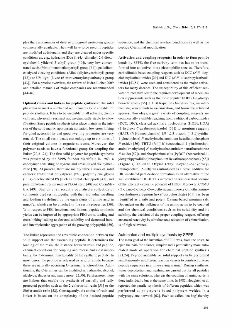
Activation and coupling reagents: In order to form peptide
bonds by SPPS, the free carboxy terminus has to be trans-
formed into an active, more electrophilic species. Therefore,
carbodiimide-based coupling reagents such as DCC (N,N'-dicy-
clohexylcarbodiimide) [20] and DIC (N,N'-diisopropylcarbodi-
imide) [53,54] were used and considered as the major activa-
tors for many decades. The susceptibility of this efficient acti-
vator to racemize led to the required development of racemiza-
tion suppressants such as the most popular HOBt (1-hydroxy-
benzotriazole) [55]. HOBt traps the O-acylisourea, an inter-
mediate, which tends to racemization, and forms the activated
species. Nowadays, a great variety of coupling reagents are
commercially available reaching from traditional carbodiimides
(DCC, DIC), classical auxiliary nucleophiles (HOBt, HOAt
(1-hydroxy-7-azabenzotriazole) [56]) to uronium reagents
(HATU (N-[(dimethylamino)-1H-1,2,3-triazolo-[4,5-b]pyridin-
1-ylmethylene]-N-methylmethanaminium hexafluorophosphate
N-oxide) [56], TBTU (N-[(1H-benzotriazol-1-yl)(dimethyl-
amino)methylene]-N-methylmethanaminium tetrafluoroborate
N-oxide) [57]), and phosphonium salts (PyBOP (benzotriazol-1-
yloxytri(pyrrolidino)phosphonium hexafluorophosphate) [58])
(Figure 3). In 2009, Oxyma (ethyl 2-cyano-2-(hydroxy-
imino)acetate) [59,60] was introduced as a novel additive for
DIC-mediated peptide-bond formation as an alternative to the
well-established HOBt. This introduction was essential because
of the inherent explosive potential of HOBt. Moreover, COMU
((1-cyano-2-ethoxy-2-oxoethylidenaminooxy)dimethylamino-
morpholino-carbenium hexafluorophosphate) [61] has been
identified as a safe and potent Oxyma-based uronium salt.
Dependent on the bulkiness of the amino acids to be coupled
and the chemical conditions such as its solubility and its
stability, the decision of the proper coupling reagent, offering
enhanced reactivity by simultaneous reduction of epimerization,
is of high relevance.
Automated and multiple synthesis by SPPSThe main goal of the invention of SPPS was, from the onset, to
open the path for a faster, simpler and a particularly more auto-
mated mode of operation for chemical peptide synthesis
[21,24]. Peptide assembly on solid support can be performed
simultaneously in different reaction vessels to construct diverse
peptide sequences in a time-saving manner. During synthesis,
Fmoc deprotection and washing are carried out for all peptides
with the same solutions, whereas the coupling of amino acids is
done individually but at the same time. In 1985, Houghten et al.
reported the parallel synthesis of different peptides, which was
performed at polystyrene-based polymers welded in a
polypropylene network [62]. Each so called 'tea bag' thereby
Beilstein J. Org. Chem. 2014, 10, 1197–1212.
1203
Figure 3: Selected coupling reagents for SPPS.
reflects one independent peptide. In this method, collective
deprotection and washing are carried out in a polyethylene (PE)
bottle, whilst for coupling the bags are separated according to
their next amino acid to be attached and reacted in separate
containers. Coding allows the identification and respective
sorting of the tea bags. After synthesis completion, peptides are
individually cleaved from the polymer support [63]. A further
method of multiple parallel SPPS is the synthesis on amino-
functionalized PE rods (pins) [64]. Here, a small amount of
peptides is synthesized in a microtiter plate format. Coupling is
performed in corresponding plates containing individual amino
acid solutions and the collective washing occurs in tanks.
Following the synthesis, the peptides remain on the polymer
carrier enabling a fast and parallel screening for antibody
binding by ELISA (enzyme-linked immunosorbent assay)
(‘PepScan’). Although quality control is not possible using this
strategy, it is suitable for fast epitope mapping. In addition,
SPPS can be carried out on cellulose papers, in which reagents
are spotted onto porous membranes [65]. The typical reactions
proceed only in the volume that has been infused into the solid
pores. Again, coupling and deprotection reactions as well as
washing steps take place simultaneously. The immobilized
peptides can finally be tested for specific enzyme reactions
(peptide arrays [65]). All these methods are variations of the
solid-phase synthesis concept [63].
But Bruce Merrifield had another vision. He wanted peptide
synthesis to be performed in single reaction vessels that are
placed in a special reactor block [20]. The instrument should be
equipped with a plumbing system in order to realize pumping,
mixing and removal of solutions. Moreover, the automated
peptide robot should contain reservoirs for all chemicals (amino
acids, solvents and activators) and ensure adequate delivery of
the solutions. The first liquid-handling apparatus performing
SPPS of this type was described in 1965 [24]. The improve-
ment of chemical reactions, solid supports, linkers and espe-
cially the development of the Fmoc-based SPPS-strategy
[31,37] have contributed to simplification and many advance-
ment of following instruments. Today, peptide synthesizers can
be purchased from more than 15 companies. Pedersen and
Jensen recently reviewed commonly used devices for fully auto-
mated single and parallel Fmoc-SPPS [66]. Peptide synthe-
sizers mainly differ in their type of solution transfer, mixing,
synthesis scale and some special features such as automated
monitoring (e.g., via chromophors [67] or conductivity [68]),
microwave heating, capability for inert atmosphere and auto-
mated peptide cleavage [65]. The systems are mostly based on
filtration by vacuum or pressure application [69], or centrifuga-
tion [70] and can work in a batch-wise or a continuous-flow
modus. The decision for a peptide synthesizer has to be made
according to the intended application and thus, the scale (neces-
sary amounts), the type of chemistry (Fmoc or Boc-strategy)
and the number of reaction tubes (plates or vessels). The deci-
sion should also be influenced by the length and complexity of
the desired peptides.
Difficult and larger peptides often lead to impurities, synthesis
termination and finally low yields owing to inter- and intramol-
ecular aggregation and sterical hindrance. To reduce these
effects, microwave-assisted instruments have been evolved
[71]. In 1992, Yu and coworkers reported for the first time that
microwave irradiation in combination with SPPS leads to
Beilstein J. Org. Chem. 2014, 10, 1197–1212.
1204
enhanced reaction rates and hence, to a higher quality of the
crude peptides [72]. Microwave energy is capable of activating
any molecule containing a dipole moment, which is reflected in
rapid heating on a molecular level [73]. The strength of the
heating is influenced by solvents, reactants, sample volumes
and the mode of mixing. It has to be noted that an optimization
of temperature is mandatory in order to avoid racemization and
side reactions [74].
Application of automated SPPS for drug develop-mentThe described method of chemical synthesis of peptides on the
solid phase and particularly, its outstanding potential for
automation, have led to routine methods in the development of
novel pharmaceuticals. In principle, there are two approaches
for drug design: rational and combinatorial methods. Whilst the
rational procedure is a lead structure-oriented process, there is
typically little knowledge about the evaluated biological system
in combinatorial methods. In order to identify a lead compound
of a relative unknown system, numerous molecules (peptides)
have to be produced in parallel by a combination of building
blocks creating a peptide library [75]. The first parallel syn-
thesis of hundreds of peptides was published by Geysen et al. in
1984 [64]. Here, a series of peptide epitopes was synthesized on
a multipin instrument and used for an enzymatic assay. Peptide
libraries can be created by directed parallel synthesis or com-
plex peptide mixtures and identified by iterative processes or
position screenings [63]. A polymer-bound peptide library can
be produced by the 'one bead one compound' concept [76].
Here, each solid particle serves as a compartment to assemble
an individual peptide, which can be sequenced for identifica-
tion of the lead structure if it has shown an effect (“hit”).
Industrial synthesis of peptides and alternativeproduction methodsThe successful automation of peptide synthesis led to a break-
through on the peptide therapeutics market and vice versa.
Thus, solid-phase synthesis is presently, besides the solution
technique, one of the major procedures for peptide manufac-
turing [10]. The great efforts in improving linkers, protecting
groups, resins etc. provided access for the synthesis of larger
peptides and even proteins. But nevertheless, the sequential and
convergent production for therapeutic applications is often the
only possibility for manufacturing peptides, which are larger
than 50 amino acids [77,78]. This method is based on the inde-
pendent synthesis of fully protected linear peptide fragments
being selectively condensed in solution to obtain the desired
polypeptide. The condensation can occur via chemoselective
ligation techniques such as native chemical ligation (NCL),
expressed protein ligation (EPL), Staudinger ligation or click
reaction [8,79].
Recently, peptide drugs as the pharmaceuticals Enfuvirtide,
Eptifibitide and Bivalirudin have been manufactured in multi-
kilogram scale [10,19]. Enfuvirtide (T-20/ Fuzeon®), for
example, is an efficient membrane fusion inhibitor for HIV
treatment consisting of 36 amino acids [80]. The large-scale
bulk production of Fuzeon® is performed by solution-phase
fragment condensation from three side-chain protected inter-
mediates synthesized at chlorotrityl resin [51]. Despite the
many steps and high costs for this synthesis strategy, it is much
more time-efficient due to the repetitive and semi-automated
processes when compared to classical solution production [81].
Furthermore, the procedure yields in very high purities of the
final peptide, which surely would not have been possible by any
other technique.
In addition to solid and solution-phase synthesis, there are some
other possibilities to produce these important molecules.
Salmon calcitonin, human glucagon and human insulin are
polypeptides being commercially produced by recombinant
expression [10]. In general, the quality of chemically synthe-
sized peptide therapeutics is comparable to recombinantly or
enzymatically produced compounds.
The success story of SPPS, which has been going on for
50 years now, has shown that these molecules can be built up
with a great variety of methods. The appropriate procedure
strongly depends on the application (lead-structure discovery,
biological investigations, potential drug candidate) of the
desired peptide.
Combination of automatic and manual SPPSto obtain therapeutic peptidesThe majority of peptides are hormones being responsible for a
broad scope of physiological functions. Here, we highlight two
successful strategies to modify chemical properties in order to
influence pharmacodynamic and -kinetic profiles – lipidation
and PEGylation. As an example, modern concepts of SPPS-
assisted, selective derivatization is described for neuropeptide Y
(NPY) receptor ligands for therapeutical and analytical applica-
tions.
Modifications of therapeutic peptides to extend theirhalf-lifesAs summarized in Table 1, natural peptides suffer from fast
proteolytic degradation and body clearance. Furthermore,
possible reduced water solubility restricts their drugability. In
the last years, remarkable efforts have been made to modulate
the bioavailability of peptides. Basically, delivery challenges of
peptide drug candidates can be overcome by chemical modifica-
tion or innovative formulation techniques such as the integra-
tion of peptides into particles, gels or liposomes [14,82,83].
Beilstein J. Org. Chem. 2014, 10, 1197–1212.
1205
Recently, the great methodical repertoire for extending the half-
lifes of biological active peptides by covalent chemical
approaches has been reviewed [8]. These methods include
peptide sequence modifications by non-proteinogenic amino
acids such as D- [84] or N-methylated [85,86] monomers or
general truncation or mutation of biologically not relevant posi-
tions creating peptide analogs [87]. Likewise, backbone manip-
ulation by partial or complete cyclization [88] as well as
incorporation of peptide bond mimetics [89] can help to
increase stability towards proteases. Peptide stability can also
be optimized by blocking their respective termini through
N-terminal acylation and C-terminal amidation [5]. Apart from
this, metabolically unstable peptide drugs can be optimized by
the covalent attachment of fatty acids (lipidation) or methoxy
polyethylene glycol (PEG) polymers (PEGylation) [14]. These
two strategies are based on substantially different mechanisms,
which can lead to a remarkable increase of the potential utility
of peptides as pharmaceuticals.
Lipidation of peptides: In general, the half-life extension of
peptides by lipidation is obtained by an increased binding to
albumin, which is the most abundant protein (6 mM in blood
plasma [90]) within the extracellular fluid [91]. Human serum
albumin (HSA) is a fundamental carrier of non-esterified free
fatty acids as well as multiple other endogenous ligands and
drugs in the blood. Early structural studies described a spher-
ical folding of albumin [92] allowing electrostatic interaction
between the carboxylate anion of fatty acids and positively
charged residues of albumin [93]. Furthermore, hydrophobic
interactions were shown to contribute to albumin binding in a
cooperative effect [94], which means that albumin binding
significantly increases until an appropriate fatty acid chain
length is reached. In 1998, seven binding sites of this multifunc-
tional transport protein were identified by Curry et al. using
X-ray crystallographic studies [95]. Later on, they were distin-
guished in high and low affinity binding sites [96]. These prop-
erties were transferred for the first time to an important peptide
hormone with high propensity to degrade in 1995 [91]. Here,
insulin was acylated with saturated fatty acids containing 10 to
12 carbon atoms at the B-chain using the ε-group of the
lysine29. In this study, the authors determined an increased
albumin affinity of lipidated insulin variants depending on the
number of carbon atoms by interaction studies with immobi-
lized HSA. Moreover, they were able to show a sustained
lowering effect of blood glucose, demonstrating a prolonged
action profile of the acylated conjugates. Thus, the extended
action was proposed to be facilitated by serum albumin binding,
which leads to gradual peptide release and an prolonged circula-
tion time [91]. Since then, many biological relevant peptides
and proteins were chemically modified by fatty acid acylation
[97].
The synthesis of these lipidated peptides can be performed by
amidation, S- or O-esterification as well as thioether or -sulfide
formation. Owing to the strength of the covalent bonds, amida-
tion and O-esterfication are preferred over the other strategies
[97]. Chemical synthesis of lipidated peptides is mostly
performed by SPPS using the Fmoc/t-Bu strategy allowing for
selective and efficient modification. Fatty acids can be incorpo-
rated into the peptide sequence at the N-terminus [98], at lysine
[99] or cysteine side chains [100] and by esterification [101]. A
detailed overview of chemical approaches to obtain lipidated
peptides containing examples for each strategy is given by
Zhang et al. [97]. In many cases, the on-resin lipidation is
carried out at the lysine side chain [102,103]. Therefore, the
peptide backbone can be synthesized by automated SPPS and
the Nε-group of the lysine that should be modified, is protected
specifically by a side-chain protecting group that is orthogonal
to the Fmoc group. Acid-labile groups as Mmt [41] and Mtt
(4-methyltrityl) [104] (classical cleavage with 1% TFA in DCM
(dichloromethane)), base-labile groups as ivDde (1-(4,4-
dimethyl-2,6-dioxocyclohex-1-ylidene)-3-methylbutyl) [105]
and Dde [40] (deprotection with 2% hydrazine in DMF (N,N-
dimethylformamide)) or the Alloc group [42] (cleavage with
catalytically amounts of Pd(PPh)3 under inert conditions) are
recommended. A selective removal of the side-chain protecting
group enables specific amide-bond formation by the reaction of
an activated carboxylic group of the fatty acid with the
Nε-group of the lysine. The introduction of a glutamyl spacer
can be helpful in order to increase the solubility of the drug
candidates [99,106]. Lipidation of peptide hormones has led to
great success with the myristoylated insulin analog insulin
determir (Levemir®) [102] and the palmitoylated incretin
mimetic GLP-1 (glucagon-like peptide 1) variant liraglutide
(Victoza®) [106] (Table 2). Both peptide drugs reached market
approval due to their prolonged blood glucose-lowering effects
making them valuable for diabetes treatment. In 2011, a lipi-
dated analog of PP (pancreatic polypeptide), a gut hormone that
is known to mediate satiety, was developed. It showed an im-
proved bioavailability demonstrated in a prolonged action in
decreasing food intake in mice [99]. Apart from albumin inter-
action, there is also the possibility to increase peptide stability
by direct fusion with HSA. One example of this effect is the
GLP-1 analog CJC-1131. It contains a covalently attached
albumin moiety and a D-amino acid at a labile position to
obtain increased metabolic stability [107].
PEGylation of peptides: Another elegant way to modulate
pharmacokinetic and -dynamic properties of peptide drugs is the
formation of drug–polymer conjugates by PEGylation. PEGyla-
tion is the covalent modification of peptides with methoxy poly-
ethyleneglycol polymer units of an averaged molecular weight.
PEG itself is known to be amphiphilic, non-toxic, little
Beilstein J. Org. Chem. 2014, 10, 1197–1212.
1206
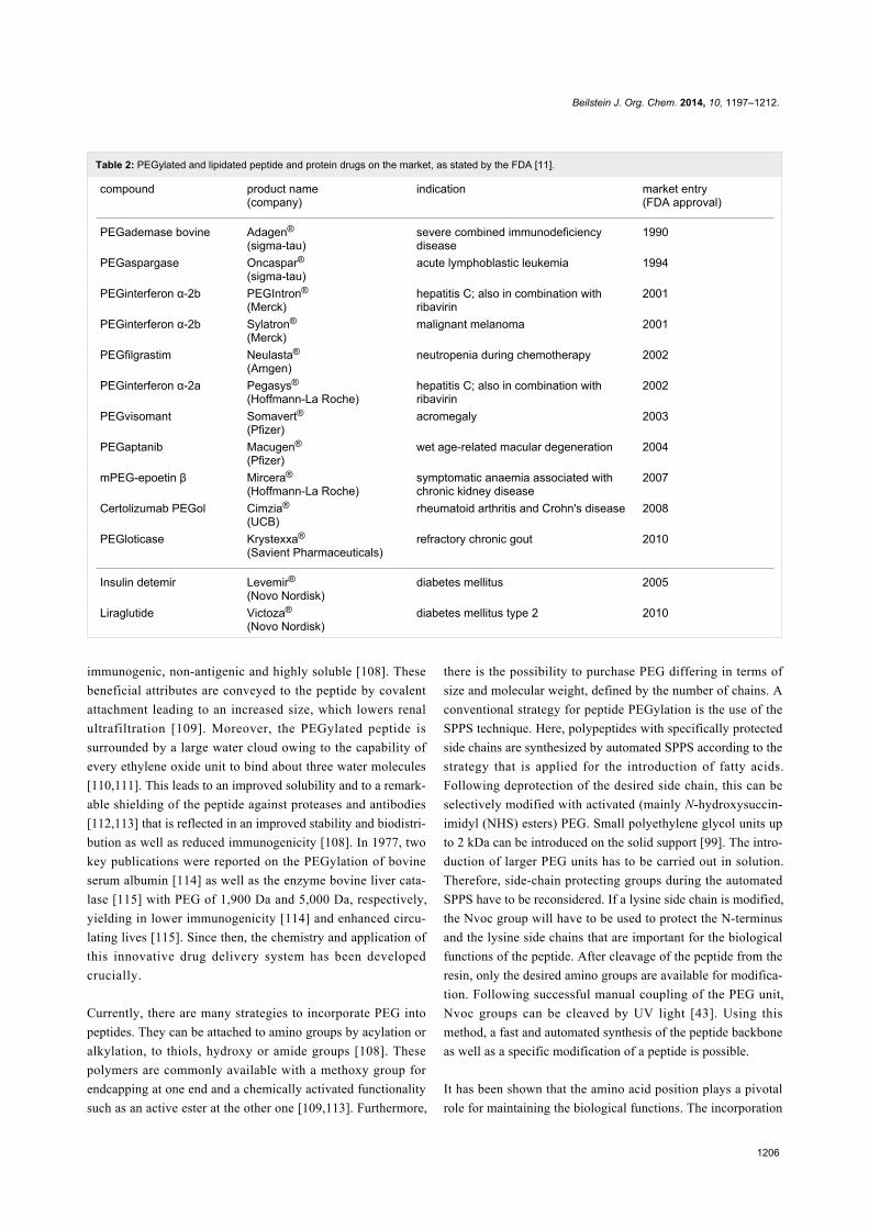
Table 2: PEGylated and lipidated peptide and protein drugs on the market, as stated by the FDA [11].
compound product name(company)
indication market entry(FDA approval)
PEGademase bovine Adagen®
(sigma-tau)severe combined immunodeficiencydisease
1990
PEGaspargase Oncaspar®(sigma-tau)
acute lymphoblastic leukemia 1994
PEGinterferon α-2b PEGIntron®
(Merck)hepatitis C; also in combination withribavirin
2001
PEGinterferon α-2b Sylatron®
(Merck)malignant melanoma 2001
PEGfilgrastim Neulasta®
(Amgen)neutropenia during chemotherapy 2002
PEGinterferon α-2a Pegasys®
(Hoffmann-La Roche)hepatitis C; also in combination withribavirin
2002
PEGvisomant Somavert®(Pfizer)
acromegaly 2003
PEGaptanib Macugen®
(Pfizer)wet age-related macular degeneration 2004
mPEG-epoetin β Mircera®
(Hoffmann-La Roche)symptomatic anaemia associated withchronic kidney disease
2007
Certolizumab PEGol Cimzia®
(UCB)rheumatoid arthritis and Crohn's disease 2008
PEGloticase Krystexxa®
(Savient Pharmaceuticals)refractory chronic gout 2010
Insulin detemir Levemir®(Novo Nordisk)
diabetes mellitus 2005
Liraglutide Victoza®
(Novo Nordisk)diabetes mellitus type 2 2010
immunogenic, non-antigenic and highly soluble [108]. These
beneficial attributes are conveyed to the peptide by covalent
attachment leading to an increased size, which lowers renal
ultrafiltration [109]. Moreover, the PEGylated peptide is
surrounded by a large water cloud owing to the capability of
every ethylene oxide unit to bind about three water molecules
[110,111]. This leads to an improved solubility and to a remark-
able shielding of the peptide against proteases and antibodies
[112,113] that is reflected in an improved stability and biodistri-
bution as well as reduced immunogenicity [108]. In 1977, two
key publications were reported on the PEGylation of bovine
serum albumin [114] as well as the enzyme bovine liver cata-
lase [115] with PEG of 1,900 Da and 5,000 Da, respectively,
yielding in lower immunogenicity [114] and enhanced circu-
lating lives [115]. Since then, the chemistry and application of
this innovative drug delivery system has been developed
crucially.
Currently, there are many strategies to incorporate PEG into
peptides. They can be attached to amino groups by acylation or
alkylation, to thiols, hydroxy or amide groups [108]. These
polymers are commonly available with a methoxy group for
endcapping at one end and a chemically activated functionality
such as an active ester at the other one [109,113]. Furthermore,
there is the possibility to purchase PEG differing in terms of
size and molecular weight, defined by the number of chains. A
conventional strategy for peptide PEGylation is the use of the
SPPS technique. Here, polypeptides with specifically protected
side chains are synthesized by automated SPPS according to the
strategy that is applied for the introduction of fatty acids.
Following deprotection of the desired side chain, this can be
selectively modified with activated (mainly N-hydroxysuccin-
imidyl (NHS) esters) PEG. Small polyethylene glycol units up
to 2 kDa can be introduced on the solid support [99]. The intro-
duction of larger PEG units has to be carried out in solution.
Therefore, side-chain protecting groups during the automated
SPPS have to be reconsidered. If a lysine side chain is modified,
the Nvoc group will have to be used to protect the N-terminus
and the lysine side chains that are important for the biological
functions of the peptide. After cleavage of the peptide from the
resin, only the desired amino groups are available for modifica-
tion. Following successful manual coupling of the PEG unit,
Nvoc groups can be cleaved by UV light [43]. Using this
method, a fast and automated synthesis of the peptide backbone
as well as a specific modification of a peptide is possible.
It has been shown that the amino acid position plays a pivotal
role for maintaining the biological functions. The incorporation
Beilstein J. Org. Chem. 2014, 10, 1197–1212.
1207
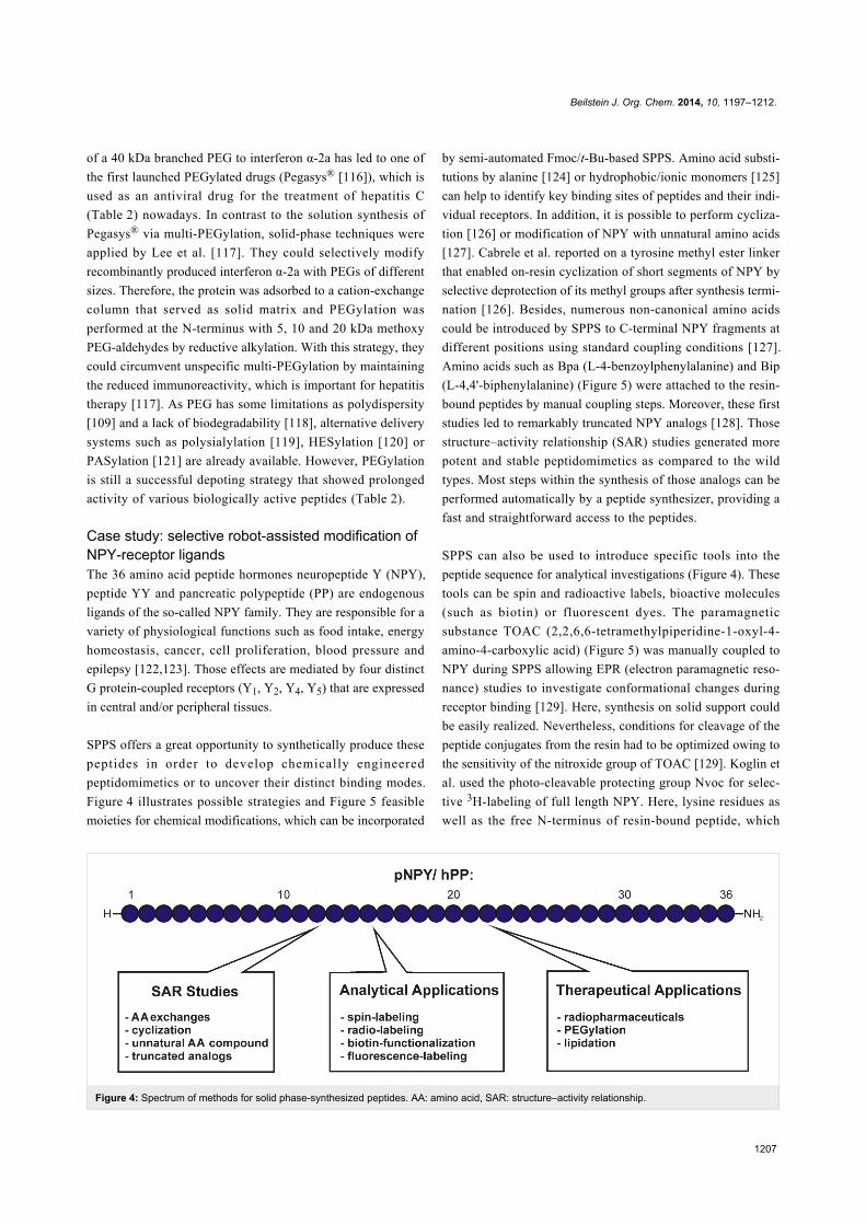
Figure 4: Spectrum of methods for solid phase-synthesized peptides. AA: amino acid, SAR: structure–activity relationship.
of a 40 kDa branched PEG to interferon α-2a has led to one of
the first launched PEGylated drugs (Pegasys® [116]), which is
used as an antiviral drug for the treatment of hepatitis C
(Table 2) nowadays. In contrast to the solution synthesis of
Pegasys® via multi-PEGylation, solid-phase techniques were
applied by Lee et al. [117]. They could selectively modify
recombinantly produced interferon α-2a with PEGs of different
sizes. Therefore, the protein was adsorbed to a cation-exchange
column that served as solid matrix and PEGylation was
performed at the N-terminus with 5, 10 and 20 kDa methoxy
PEG-aldehydes by reductive alkylation. With this strategy, they
could circumvent unspecific multi-PEGylation by maintaining
the reduced immunoreactivity, which is important for hepatitis
therapy [117]. As PEG has some limitations as polydispersity
[109] and a lack of biodegradability [118], alternative delivery
systems such as polysialylation [119], HESylation [120] or
PASylation [121] are already available. However, PEGylation
is still a successful depoting strategy that showed prolonged
activity of various biologically active peptides (Table 2).
Case study: selective robot-assisted modification ofNPY-receptor ligandsThe 36 amino acid peptide hormones neuropeptide Y (NPY),
peptide YY and pancreatic polypeptide (PP) are endogenous
ligands of the so-called NPY family. They are responsible for a
variety of physiological functions such as food intake, energy
homeostasis, cancer, cell proliferation, blood pressure and
epilepsy [122,123]. Those effects are mediated by four distinct
G protein-coupled receptors (Y1, Y2, Y4, Y5) that are expressed
in central and/or peripheral tissues.
SPPS offers a great opportunity to synthetically produce these
peptides in order to develop chemically engineered
peptidomimetics or to uncover their distinct binding modes.
Figure 4 illustrates possible strategies and Figure 5 feasible
moieties for chemical modifications, which can be incorporated
by semi-automated Fmoc/t-Bu-based SPPS. Amino acid substi-
tutions by alanine [124] or hydrophobic/ionic monomers [125]
can help to identify key binding sites of peptides and their indi-
vidual receptors. In addition, it is possible to perform cycliza-
tion [126] or modification of NPY with unnatural amino acids
[127]. Cabrele et al. reported on a tyrosine methyl ester linker
that enabled on-resin cyclization of short segments of NPY by
selective deprotection of its methyl groups after synthesis termi-
nation [126]. Besides, numerous non-canonical amino acids
could be introduced by SPPS to C-terminal NPY fragments at
different positions using standard coupling conditions [127].
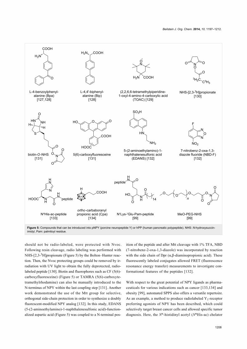
Amino acids such as Bpa (L-4-benzoylphenylalanine) and Bip
(L-4,4'-biphenylalanine) (Figure 5) were attached to the resin-
bound peptides by manual coupling steps. Moreover, these first
studies led to remarkably truncated NPY analogs [128]. Those
structure–activity relationship (SAR) studies generated more
potent and stable peptidomimetics as compared to the wild
types. Most steps within the synthesis of those analogs can be
performed automatically by a peptide synthesizer, providing a
fast and straightforward access to the peptides.
SPPS can also be used to introduce specific tools into the
peptide sequence for analytical investigations (Figure 4). These
tools can be spin and radioactive labels, bioactive molecules
(such as biotin) or fluorescent dyes. The paramagnetic
substance TOAC (2,2,6,6-tetramethylpiperidine-1-oxyl-4-
amino-4-carboxylic acid) (Figure 5) was manually coupled to
NPY during SPPS allowing EPR (electron paramagnetic reso-
nance) studies to investigate conformational changes during
receptor binding [129]. Here, synthesis on solid support could
be easily realized. Nevertheless, conditions for cleavage of the
peptide conjugates from the resin had to be optimized owing to
the sensitivity of the nitroxide group of TOAC [129]. Koglin et
al. used the photo-cleavable protecting group Nvoc for selec-
tive 3H-labeling of full length NPY. Here, lysine residues as
well as the free N-terminus of resin-bound peptide, which
Beilstein J. Org. Chem. 2014, 10, 1197–1212.
1208
Figure 5: Compounds that can be introduced into pNPY (porcine neuropeptide Y) or hPP (human pancreatic polypeptide). NHS: N-hydroxysuccin-imidyl, Pam: palmitoyl residue.
should not be radio-labeled, were protected with Nvoc.
Following resin cleavage, radio labeling was performed with
NHS-[2,3-3H]propionate (Figure 5) by the Bolton–Hunter reac-
tion. Then, the Nvoc protecting groups could be removed by ir-
radiation with UV light to obtain the fully deprotected, radio-
labeled peptide [130]. Biotin and fluorophores such as CF (5(6)-
carboxyfluoresceine) (Figure 5) or TAMRA (5(6)-carboxyte-
tramethylrhodamine) can also be manually introduced to the
N-terminus of NPY within the last coupling step [131]. Another
work demonstrated the use of the Mtt group for selective,
orthogonal side-chain protection in order to synthesize a doubly
fluorescent-modified NPY analog [132]. In this study, EDANS
(5-(2-aminoethylamino)-1-naphthalenesulfonic acid)-function-
alized aspartic acid (Figure 5) was coupled to a N-terminal pos-
ition of the peptide and after Mtt cleavage with 1% TFA, NBD
(7-nitrobenz-2-oxa-1,3-diazole) was incorporated by reaction
with the side chain of Dpr (α,β-diaminopropionic acid). These
fluorescently labeled conjugates allowed FRET (fluorescence
resonance energy transfer) measurements to investigate con-
formational features of the peptides [132].
With respect to the great potential of NPY ligands as pharma-
ceuticals for various indications such as cancer [133,134] and
obesity [99], automated SPPS also offers a versatile repertoire.
As an example, a method to produce radiolabeled Y1-receptor
preferring agonists of NPY has been described, which could
selectively target breast cancer cells and allowed specific tumor
diagnosis. Here, the Nα-histidinyl acetyl (NαHis-ac) chelator
Beilstein J. Org. Chem. 2014, 10, 1197–1212.
1209
(Figure 5) was attached to lysine side chains or the N-terminus
of the peptide by manual coupling, whereas the chelation with99mTc occurred in solution [133]. The incorporation of carbabo-
ranes that can be used for boron neutron capture therapy
demonstrated the potential for tumor therapy as well. Here,
ortho-carbaboranyl propionic acid (Cpa) (Figure 5) was coupled
to lysine side chains on a resin-bound peptide in a manual step.
An alternative Fmoc-cleavage procedure for the following
coupling steps assured the stability of the piperidine-labile
carbaborane moiety [134]. With respect to peptide stabilization
techniques, Bellmann-Sickert et al. described a method for
on-resin lipidation of hPP with palmitic acid and a strategy to
synthesize PEGylated peptides [99]. For lipidation, lysine
residues were protected with Dde during the automated peptide
synthesis. Orthogonal deprotection of Dde enabled coupling of
a glutamate spacer and the desired fatty acid (Figure 5) by
amide-bond formation [99]. Synthesis of PEGylated conjugates
(Figure 5) occurred on solid phase (for 2 kDa PEG) according
to the same procedure. For PEGylation with a much larger
group (20 kDa PEG), the N-terminus of hPP was protected with
Nvoc and after cleavage from resin, selective PEGylation was
performed in solution followed by specific Nvoc removal [99].
ConclusionThe production of peptides by automated synthesis on solid
support provides a great variety of benefits. Chemical reactions
necessary to assemble peptides can be performed simultane-
ously with plenty of reaction tubes allowing parallel and
multiple syntheses. Owing to the simple washing of the resin
following coupling or deprotection steps, no intermediate purifi-
cation is necessary. Furthermore, the possibility to use excess
amounts of reagents facilitates high yields of the synthesized
peptides. This fast, automated and practicable method has
evolved to a major technique to produce chemically synthe-
sized peptide therapeutics, which pushed the market steadily.
Automated SPPS is of great importance, especially for struc-
ture–activity relationship studies and backbone modification of
biologically active peptide hormones. Nevertheless, there are
still some issues that have to be addressed. For instance,
incorporation of N-methylated amino acids in order to improve
their proteolytic stability often is difficult because of steric
hindrance [135]. Moreover, the introduction of fatty acids and
PEG moieties, especially large sizes, is generally performed
manually [99].
Although remarkable progress has been accomplished since the
invention by Merrifield more than 50 years ago [20], novel
technologies in automated peptide synthesis are required.
Microwave-assisted SPPS, for instance, has been shown to not
only enhance reaction rates but also to enable the synthesis of
difficult and rigid peptide sequences [136]. This technique is
progressing but needs more improvements, especially in terms
of practicability. The ongoing need for peptides as biopharma-
ceuticals will surely promote these developments in the future.
AcknowledgementsWe gratefully acknowledge the Graduate School “Leipzig
School of Natural Sciences-Building with Molecules and Nano-
objects” (BuildMoNa) and the financial support from the EU
and the Free State of Saxony, the SFB1052 and the IFB K7-18.
References1. Jakubke, H.-D.; Jeschkeit, H. Aminosäuren, Peptide, Proteine; Verlag
Chemie: Weinheim, 1982.2. Schmeing, T. M.; Ramakrishnan, V. Nature 2009, 461, 1234–1242.
doi:10.1038/nature08403Dfgdf.
3. Craik, D. J.; Fairlie, D. P.; Liras, S.; Price, D. Chem. Biol. Drug Des.2013, 81, 136–147. doi:10.1111/cbdd.12055
4. Bellmann-Sickert, K.; Beck-Sickinger, A. G. Trends Pharmacol. Sci.2010, 31, 434–441. doi:10.1016/j.tips.2010.06.003
5. Vlieghe, P.; Lisowski, V.; Martinez, J.; Khrestchatisky, M.Drug Discovery Today 2010, 15, 40–56.doi:10.1016/j.drudis.2009.10.009
6. Bilello, J. A. Curr. Mol. Med. 2005, 5, 39–52.doi:10.2174/1566524053152898
7. Marx, V. Chem. Eng. News 2005, 83, 17–24.8. Ahrens, V. M.; Bellmann-Sickert, K.; Beck-Sickinger, A. G.
Future Med. Chem. 2012, 4, 1567–1586. doi:10.4155/fmc.12.769. Katsila, T.; Siskos, A. P.; Tamvakopoulos, C. Mass Spectrom. Rev.
2012, 31, 110–133. doi:10.1002/mas.2034010. Lax, R. The Future of Peptide Development in the Pharmaceutical
Industry. PharManufacturing The International Peptide Review; WorldBusiness Journals Ltd.: London, UK , 2010; pp 10–15.
11. U.S. Food and Drug Administration.http://www.accessdata.fda.gov/scripts/cder/drugsatfda/index.cfm(accessed Jan 23, 2014).
12. DrugBank, Open Data Drug & Drug Target Database.http://www.drugbank.ca/ (accessed Jan 22, 2014).
13. McGregor, D. P. Curr. Opin. Pharmacol. 2008, 8, 616–619.doi:10.1016/j.coph.2008.06.002
14. Frokjaer, S.; Otzen, D. E. Nat. Rev. Drug Discovery 2005, 4, 298–306.doi:10.1038/nrd1695
15. Fischer, E. Ber. Dtsch. Chem. Ges. 1901, 34, 433–454.doi:10.1002/cber.19010340173
16. du Vigneaud, V.; Ressler, C.; Swan, C. J. M.; Roberts, C. W.;Katsoyannis, P. G.; Gordon, S. J. Am. Chem. Soc. 1953, 75,4879–4880. doi:10.1021/ja01115a553
17. Bergmann, M.; Zervas, L. Ber. Dtsch. Chem. Ges. 1932, 65,1192–1201. doi:10.1002/cber.19320650722
18. Bruckdorfer, T.; Marder, O.; Albericio, F. Curr. Pharm. Biotechnol.2004, 5, 29–43. doi:10.2174/1389201043489620
19. Zompra, A. A.; Galanis, A. S.; Werbitzky, O.; Albericio, F.Future Med. Chem. 2009, 1, 361–377. doi:10.4155/fmc.09.23
20. Merrifield, R. B. J. Am. Chem. Soc. 1963, 85, 2149–2154.doi:10.1021/ja00897a025
21. Merrifield, B. Angew. Chem., Int. Ed. Engl. 1985, 24, 799–810.doi:10.1002/anie.198507993
Beilstein J. Org. Chem. 2014, 10, 1197–1212.
1210
22. Alsina, J.; Albericio, F. Biopolymers 2003, 71, 454–477.doi:10.1002/bip.10492
23. Montalbetti, C. A. G. N.; Falque, V. Tetrahedron 2005, 61,10827–10852. doi:10.1016/j.tet.2005.08.031
24. Merrifield, R. B. Science 1965, 150, 178–185.doi:10.1126/science.150.3693.178
25. Fekete, S.; Veuthey, J.-L.; Guillarme, D. J. Pharm. Biomed. Anal.2012, 69, 9–27. doi:10.1016/j.jpba.2012.03.024
26. Wurtz, N. R.; Turner, J. M.; Baird, E. E.; Dervan, P. B. Org. Lett. 2001,3, 1201–1203. doi:10.1021/ol0156796
27. Zlatev, I.; Manoharan, M.; Vasseur, J. J.; Morvan, F. UNIT 1.28Solid-Phase Chemical Synthesis of 5′-Triphosphate DNA, RNA, andChemically Modified Oligonucleotides. Current Protocols in NucleicAcid Chemistry; John Wiley and Sons, Inc., 2012; Vol. 50,1.28.1–1.28.16. doi:10.1002/0471142700.nc0128s50
28. Seeberger, P. H. Chem. Soc. Rev. 2008, 37, 19–28.doi:10.1039/b511197h
29. Green, T. W.; Wuts, P. G. M. Protective groups in organic chemistry,3rd ed.; John Wiley & Sons, Inc.: New York, USA, 1999.
30. Carpino, L. A. J. Am. Chem. Soc. 1957, 79, 4427–4431.doi:10.1021/ja01573a050
31. Carpino, L. A.; Han, G. Y. J. Am. Chem. Soc. 1970, 92, 5748–5749.doi:10.1021/ja00722a043
32. Pennington, M. W. HF Cleavage and Deprotection Procedures forPeptides Synthesized Using a Boc/Bzl Strategy. In Peptide SynthesisProtocols; Pennington, M. W.; Dunn, B. M., Eds.; Methods inMolecular Biology, Vol. 35; Humana Press, 1995; pp 41–62.doi:10.1385/0-89603-273-6:41
33. Meienhofer, J.; Waki, M.; Heimer, E. P.; Lambros, T. J.;Makofske, R. C.; Chang, C.-D. Int. J. Pept. Protein Res. 1979, 13,35–42. doi:10.1111/j.1399-3011.1979.tb01847.x
34. Anderson, G. W.; Callahan, F. M. J. Am. Chem. Soc. 1960, 82,3359–3363. doi:10.1021/ja01498a032
35. Chang, C.-D.; Waki, M.; Ahmad, M.; Meienhofer, J.; Lundell, E. O.;Haug, J. D. Int. J. Pept. Protein Res. 1980, 15, 59–66.doi:10.1111/j.1399-3011.1980.tb02550.x
36. Barany, G.; Merrifield, R. B. J. Am. Chem. Soc. 1977, 99, 7363–7365.doi:10.1021/ja00464a050
37. Atherton, E.; Fox, H.; Harkiss, D.; Logan, C. J.; Sheppard, R. C.;Williams, B. J. J. Chem. Soc., Chem. Commun. 1978, 537–539.doi:10.1039/c39780000537
38. Beyermann, M.; Bienert, M. Tetrahedron Lett. 1992, 33, 3745–3748.doi:10.1016/0040-4039(92)80014-B
39. Hsieh, K.-H.; Demaine, M. M.; Gurusidaiah, S.Int. J. Pept. Protein Res. 1996, 48, 292–298.doi:10.1111/j.1399-3011.1996.tb00844.x
40. Bycroft, B. W.; Chan, W. C.; Chhabra, S. R.; Hone, N. D.J. Chem. Soc., Chem. Commun. 1993, 778–779.doi:10.1039/c39930000778
41. Matysiak, S.; Böldicke, T.; Tegge, W.; Frank, R. Tetrahedron Lett.1998, 39, 1733–1734. doi:10.1016/S0040-4039(98)00055-0
42. Loffet, A.; Zhang, H. X. Int. J. Pept. Protein Res. 1993, 42, 346–351.doi:10.1111/j.1399-3011.1993.tb00504.x
43. Patchornik, A.; Amit, B.; Woodward, R. B. J. Am. Chem. Soc. 1970,92, 6333–6335. doi:10.1021/ja00724a041
44. Isidro-Llobet, A.; Álvarez, M.; Albericio, F. Chem. Rev. 2009, 109,2455–2504. doi:10.1021/cr800323s
45. Mergler, M.; Durieux, J. P. The Bachem practice of SPPS: Tips andtricks from the experts at Bachem; Bachem: Bubendorf, Switzerland,2005.
46. Novabiochem. Novabiochem: Peptide Synthesis; Novabiochem:Darmstadt, Germany, 2012.
47. Bayer, E. Angew. Chem., Int. Ed. 1991, 30, 113–129.doi:10.1002/anie.199101133
48. Meldal, M. Tetrahedron Lett. 1992, 33, 3077–3080.doi:10.1016/S0040-4039(00)79604-3
49. García-Martín, F.; Quintanar-Audelo, M.; Garcia-Ramos, Y.;Cruz, L. J.; Gravel, C.; Furic, R.; Côté, S.; Tulla-Puche, J.;Albericio, F. J. Comb. Chem. 2006, 8, 213–220.doi:10.1021/cc0600019
50. Shelton, P. T.; Jensen, K. J. Linkers, Resins, and General Proceduresfor Solid-Phase Peptide Synthesis. In Peptide Synthesis andApplications; Jensen, K. J.; Shelton, P. T.; Pedersen, S. L., Eds.;Methods in Molecular Biology, Vol. 1047; Humana Press, 2013;pp 23–41. doi:10.1007/978-1-62703-544-6_2
51. Barlos, K.; Gatos, D.; Kallitsis, J.; Papaphotiu, G.; Sotiriu, P.;Wenqing, Y.; Schäfer, W. Tetrahedron Lett. 1989, 30, 3943–3946.doi:10.1016/S0040-4039(00)99290-6
52. Sieber, P. Tetrahedron Lett. 1987, 28, 2107–2110.doi:10.1016/S0040-4039(00)96055-6
53. Izdebski, J.; Orlowska, A.; Anulewicz, R.; Witkowska, E.; Fiertek, D.Int. J. Pept. Protein Res. 1994, 43, 184–189.doi:10.1111/j.1399-3011.1994.tb00521.x
54. Els, S.; Beck-Sickinger, A. G.; Chollet, C. Methods Enzymol. 2010,485, 103–121. doi:10.1016/B978-0-12-381296-4.00006-3
55. König, W.; Geiger, R. Chem. Ber. 1970, 103, 788–798.doi:10.1002/cber.19701030319
56. Carpino, L. A. J. Am. Chem. Soc. 1993, 115, 4397–4398.doi:10.1021/ja00063a082
57. Knorr, R.; Trzeciak, A.; Bannwarth, W.; Gillessen, D. Tetrahedron Lett.1989, 30, 1927–1930. doi:10.1016/S0040-4039(00)99616-3
58. Coste, J.; Le-Nguyen, D.; Castro, B. Tetrahedron Lett. 1990, 31,205–208. doi:10.1016/S0040-4039(00)94371-5
59. Itoh, M. Bull. Chem. Soc. Jpn. 1973, 46, 2219–2221.doi:10.1246/bcsj.46.2219
60. Subirós-Funosas, R.; Prohens, R.; Barbas, R.; El-Faham, A.;Albericio, F. Chem.–Eur. J. 2009, 15, 9394–9403.doi:10.1002/chem.200900614
61. El-Faham, A.; Subirós Funosas, R.; Prohens, R.; Albericio, F.Chem.–Eur. J. 2009, 15, 9404–9416. doi:10.1002/chem.200900615
62. Houghten, R. A. Proc. Natl. Acad. Sci. U. S. A. 1985, 82, 5131–5135.doi:10.1073/pnas.82.15.5131
63. Jung, G.; Beck-Sickinger, A. G. Angew. Chem., Int. Ed. Engl. 1992,31, 367–383. doi:10.1002/anie.199203673
64. Geysen, H. M.; Meloen, R. H.; Barteling, S. J.Proc. Natl. Acad. Sci. U. S. A. 1984, 81, 3998–4002.doi:10.1073/pnas.81.13.3998
65. Frank, R. J. Immunol. Methods 2002, 267, 13–26.doi:10.1016/S0022-1759(02)00137-0
66. Pedersen, S. L.; Jensen, K. J. Instruments for Automated PeptideSynthesis. In Peptide Synthesis and Applications; Jensen, K. J.;Shelton, P. T.; Pedersen, S. L., Eds.; Methods Mol. Biol., Vol. 1047;Humana Press, 2013; pp 215–224.doi:10.1007/978-1-62703-544-6_15
67. Salisbury, S. A.; Tremeer, E. J.; Davies, J. W.; Owen, D. E. I. A.J. Chem. Soc., Chem. Commun. 1990, 538–540.doi:10.1039/c39900000538
68. Fox, J. E.; Newton, R.; Stroud, C. H. Int. J. Pept. Protein Res. 1991,38, 62–65. doi:10.1111/j.1399-3011.1991.tb01410.x

Beilstein J. Org. Chem. 2014, 10, 1197–1212.
1211
69. Lebl, M.; Hachmann, J. High-Throughput Peptide Synthesis. InPeptide Synthesis and Applications; Howl, J., Ed.; Methods inMolecular Biology, Vol. 298; Humana Press, 2005; pp 167–194.doi:10.1385/1-59259-877-3:167
70. Lebl, M. J. Lab. Autom. 2003, 8, 30–35.doi:10.1016/S1535-5535(04)00267-9
71. Collins, J. M.; Collins, M. J.; Steorts, R. C., Eds. Novel Method forSolid Phase Peptide Synthesis Using Microwave Energy, Boston, MA,July 19–23, 2003; American Chemical Society: Boston, MA, 2003.
72. Yu, H. M.; Chen, S. T.; Wang, K. T. J. Org. Chem. 1992, 57,4781–4784. doi:10.1021/jo00044a001
73. Vanier, G. S. Microwave-Assisted Solid-Phase Peptide SynthesisBased on the Fmoc Protecting Group Strategy (CEM). In PeptideSynthesis and Applications; Hensen, K. J.; Shelton, P. T.;Pedersen, S. L., Eds.; Methods in Molecular Biology, Vol. 1047;Humana Press, 2013; pp 235–249.doi:10.1007/978-1-62703-544-6_17
74. Palasek, S. A.; Cox, Z. J.; Collins, J. M. J. Pept. Sci. 2007, 13,143–148. doi:10.1002/psc.804
75. Lam, K. S.; Salmon, S. E.; Hersh, E. M.; Hruby, V. J.;Kazmierski, W. M.; Knapp, R. J. Nature 1991, 354, 82–84.doi:10.1038/354082a0
76. Lam, K. S.; Lebl, M.; Krchňák, V. Chem. Rev. 1997, 97, 411–448.doi:10.1021/cr9600114
77. Barlos, K.; Gatos, D.; Schäfer, W. Angew. Chem., Int. Ed. Engl. 1991,30, 590–593. doi:10.1002/anie.199105901
78. Riniker, B.; Flörsheimer, A.; Fretz, H.; Sieber, P.; Kamber, B.Tetrahedron 1993, 49, 9307–9320.doi:10.1016/0040-4020(93)80017-N
79. Chandrudu, S.; Simerska, P.; Toth, I. Molecules 2013, 18, 4373–4388.doi:10.3390/molecules18044373
80. Greenberg, M. L.; Cammack, N. J. Antimicrob. Chemother. 2004, 54,333–340. doi:10.1093/jac/dkh330
81. Bray, B. L. Nat. Rev. Drug Discovery 2003, 2, 587–593.doi:10.1038/nrd1133
82. Shen, W.-C. Drug Discovery Today 2003, 8, 607–608.doi:10.1016/S1359-6446(03)02692-8
83. Veronese, F. M.; Mero, A. BioDrugs 2008, 22, 315–329.doi:10.2165/00063030-200822050-00004
84. Els, S.; Schild, E.; Petersen, P. S.; Kilian, T.-M.; Mokrosinski, J.;Frimurer, T. M.; Chollet, C.; Schwartz, T. W.; Holst, B.;Beck-Sickinger, A. G. J. Med. Chem. 2012, 55, 7437–7449.doi:10.1021/jm300414b
85. Linde, Y.; Ovadia, O.; Safrai, E.; Xiang, Z.; Portillo, F. P.;Shalev, D. E.; Haskell-Luevano, C.; Hoffman, A.; Gilon, C.Biopolymers 2008, 90, 671–682. doi:10.1002/bip.21057
86. Chatterjee, J.; Rechenmacher, F.; Kessler, H. Angew. Chem., Int. Ed.2013, 52, 254–269. doi:10.1002/anie.201205674
87. Miranda, L. P.; Holder, J. R.; Shi, L.; Bennett, B.; Aral, J.; Gegg, C. V.;Wright, M.; Walker, K.; Doellgast, G.; Rogers, R.; Li, H.;Valladares, V.; Salyers, K.; Johnson, E.; Wild, K. J. Med. Chem. 2008,51, 7889–7897. doi:10.1021/jm8009298
88. Green, B. R.; Klein, B. D.; Lee, H.-K.; Smith, M. D.; White, H. S.;Bulaj, G. Bioorg. Med. Chem. 2013, 21, 303–310.doi:10.1016/j.bmc.2012.10.026
89. Gentilucci, L.; De Marco, R.; Cerisoli, L. Curr. Pharm. Des. 2010, 16,3185–3203. doi:10.2174/138161210793292555
90. Peters, T., Jr. Adv. Protein Chem. 1985, 37, 161–245.doi:10.1016/S0065-3233(08)60065-0
91. Kurtzhals, P.; Havelund, S.; Jonassen, I.; Kiehr, B.; Larsen, U. D.;Ribel, U.; Markussen, J. Biochem. J. 1995, 312, 725–731.
92. Bloomfield, V. Biochemistry 1966, 5, 684–689.doi:10.1021/bi00866a039
93. Spector, A. A. J. Lipid Res. 1975, 16, 165–179.94. Ashbrook, J. D.; Spector, A. A.; Santos, E. C.; Fletcher, J. E.
J. Biol. Chem. 1975, 250, 2333–2338.95. Curry, S.; Mandelkow, H.; Brick, P.; Franks, N. Nat. Struct. Biol. 1998,
5, 827–835. doi:10.1038/186996. Simard, J. R.; Zunszain, P. A.; Hamilton, J. A.; Curry, S. J. Mol. Biol.
2006, 361, 336–351. doi:10.1016/j.jmb.2006.06.02897. Zhang, L.; Bulaj, G. Curr. Med. Chem. 2012, 19, 1602–1618.
doi:10.2174/09298671279994500398. Dasgupta, P.; Singh, A. T.; Mukherjee, R. Pharm. Res. 1999, 16,
1047–1053. doi:10.1023/A:101893580005299. Bellmann-Sickert, K.; Elling, C. E.; Madsen, A. N.; Little, P. B.;
Lundgren, K.; Gerlach, L.-O.; Bergmann, R.; Holst, B.;Schwartz, T. W.; Beck-Sickinger, A. G. J. Med. Chem. 2011, 54,2658–2667. doi:10.1021/jm101357e
100.Cheng, W.; Satyanarayanajois, S.; Lim, L.-Y. Pharm. Res. 2007, 24,99–110. doi:10.1007/s11095-006-9128-9
101.Bednarek, M. A.; Feighner, S. D.; Pong, S.-S.; McKee, K. K.;Hreniuk, D. L.; Silva, M. V.; Warren, V. A.; Howard, A. D.;Van der Ploeg, L. H. Y.; Heck, J. V. J. Med. Chem. 2000, 43,4370–4376. doi:10.1021/jm0001727
102.Chapman, T. M.; Perry, C. M. Drugs 2004, 64, 2577–2595.doi:10.2165/00003495-200464220-00008
103.Gallwitz, B. U. Endocrinology 2007, 2, 56–59.104.Aletras, A.; Barlos, K.; Gatos, D.; Koutsogianni, S.; Mamos, P.
Int. J. Pept. Protein Res. 1995, 45, 488–496.doi:10.1111/j.1399-3011.1995.tb01065.x
105.Chhabra, S. R.; Hothi, B.; Evans, D. J.; White, P. D.; Bycroft, B. W.;Chan, W. C. Tetrahedron Lett. 1998, 39, 1603–1606.doi:10.1016/S0040-4039(97)10828-0
106.Vilsboll, T. Drugs Today 2009, 45, 101–113.107.Léger, R.; Thibaudeau, K.; Robitaille, M.; Quraishi, O.; van Wyk, P.;
Bousquet-Gagnon, N.; Carette, J.; Castaigne, J.-P.; Bridon, D. P.Bioorg. Med. Chem. Lett. 2004, 14, 4395–4398.doi:10.1016/j.bmcl.2004.06.066
108.Veronese, F. M.; Pasut, G. Drug Discovery Today 2005, 10,1451–1458. doi:10.1016/S1359-6446(05)03575-0
109.Veronese, F. M. Biomaterials 2001, 22, 405–417.doi:10.1016/S0142-9612(00)00193-9
110.Liu, K.-J.; Parsons, J. L. Macromolecules 1969, 2, 529–533.doi:10.1021/ma60011a015
111.Maxfield, J.; Shepherd, I. W. Polymer 1975, 16, 505–509.doi:10.1016/0032-3861(75)90008-7
112.Israelachvili, J. Proc. Natl. Acad. Sci. U. S. A. 1997, 94, 8378–8379.doi:10.1073/pnas.94.16.8378
113.Harris, J. M.; Chess, R. B. Nat. Rev. Drug Discovery 2003, 2,214–221. doi:10.1038/nrd1033
114.Abuchowski, A.; van Es, T.; Palczuk, N. C.; Davis, F. F. J. Biol. Chem.1977, 252, 3578–3581.
115.Abuchowski, A.; McCoy, J. R.; Palczuk, N. C.; van Es, T.; Davis, F. F.J. Biol. Chem. 1977, 252, 3582–3586.
116.Bailon, P.; Palleroni, A.; Schaffer, C. A.; Spence, C. L.; Fung, W.-J.;Porter, J. E.; Ehrlich, G. K.; Pan, W.; Xu, Z.-X.; Modi, M. W.; Farid, A.;Berthold, W.; Graves, M. Bioconjugate Chem. 2001, 12, 195–202.doi:10.1021/bc000082g

Beilstein J. Org. Chem. 2014, 10, 1197–1212.
1212
117.Lee, B. K.; Kwon, J. S.; Kim, H. J.; Yamamoto, S.; Lee, E. K.Bioconjugate Chem. 2007, 18, 1728–1734. doi:10.1021/bc060245m
118.Knop, K.; Hoogenboom, R.; Fischer, D.; Schubert, U. S.Angew. Chem., Int. Ed. 2010, 49, 6288–6308.doi:10.1002/anie.200902672
119.Gregoriadis, G.; McCormack, B.; Wang, Z.; Lifely, R. FEBS Lett. 1993,315, 271–276. doi:10.1016/0014-5793(93)81177-2
120.Westphal, M.; James, M. F. M.; Kozek-Langenecker, S.; Stocker, R.;Guidet, B.; Van Aken, H. Anesthesiology 2009, 111, 187–202.doi:10.1097/ALN.0b013e3181a7ec82
121.Schlapschy, M.; Binder, U.; Börger, C.; Theobald, I.; Wachinger, K.;Kisling, S.; Haller, D.; Skerra, A. Protein Eng., Des. Sel. 2013, 26,489–501. doi:10.1093/protein/gzt023
122.Babilon, S.; Mörl, K.; Beck-Sickinger, A. G. Biol. Chem. 2013, 394,921–936. doi:10.1515/hsz-2013-0123
123.Pedragosa-Badia, X.; Stichel, J.; Beck-Sickinger, A. G.Front. Endocrinol. 2013, 4, 5. doi:10.3389/fendo.2013.00005
124.Beck-Sickinger, A. G.; Wieland, H. A.; Wittneben, H.; Willim, K.-D.;Rudolf, K.; Jung, G. Eur. J. Biochem. 1994, 225, 947–958.doi:10.1111/j.1432-1033.1994.0947b.x
125.Pedragosa-Badia, X.; Sliwoski, G. R.; Dong Nguyen, E.; Lindner, D.;Stichel, J.; Kaufmann, K. W.; Meiler, J.; Beck-Sickinger, A. G.J. Biol. Chem. 2014, 289, 5846–5859. doi:10.1074/jbc.M113.502021
126.Cabrele, C.; Langer, M.; Beck-Sickinger, A. G. J. Org. Chem. 1999,64, 4353–4361. doi:10.1021/jo982402j
127.Zwanziger, D.; Böhme, I.; Lindner, D.; Beck-Sickinger, A. G.J. Pept. Sci. 2009, 15, 856–866. doi:10.1002/psc.1188
128.Hofmann, S.; Frank, R.; Hey-Hawkins, E.; Beck-Sickinger, A. G.;Schmidt, P. Neuropeptides 2013, 47, 59–66.doi:10.1016/j.npep.2012.12.001
129.Bettio, A.; Gutewort, V.; Pöppl, A.; Dinger, M. C.; Zschörnig, O.;Arnold, K.; Toniolo, C.; Beck-Sickinger, A. G. J. Pept. Sci. 2002, 8,671–682. doi:10.1002/psc.428
130.Koglin, N.; Lang, M.; Rennert, R.; Beck-Sickinger, A. G.J. Med. Chem. 2003, 46, 4369–4372. doi:10.1021/jm0341251
131.Fabry, M.; Cabrele, C.; Höcker, H.; Beck-Sickinger, A. G. Peptides2000, 21, 1885–1893. doi:10.1016/S0196-9781(00)00328-4
132.Haack, M.; Beck-Sickinger, A. G. Chem. Biol. Drug Des. 2009, 73,573–583. doi:10.1111/j.1747-0285.2009.00823.x
133.Khan, I. U.; Zwanziger, D.; Böhme, I.; Javed, M.; Naseer, H.;Hyder, S. W.; Beck-Sickinger, A. G. Angew. Chem., Int. Ed. 2010, 49,1155–1158. doi:10.1002/anie.200905008
134.Ahrens, V. M.; Frank, R.; Stadlbauer, S.; Beck-Sickinger, A. G.;Hey-Hawkins, E. J. Med. Chem. 2011, 54, 2368–2377.doi:10.1021/jm101514m
135.Teixidó, M.; Albericio, F.; Giralt, E. J. Pept. Res. 2005, 65, 153–166.doi:10.1111/j.1399-3011.2004.00213.x
136.Pedersen, S. L.; Tofteng, A. P.; Malik, L.; Jensen, K. J.Chem. Soc. Rev. 2012, 41, 1826–1844. doi:10.1039/c1cs15214a
License and TermsThis is an Open Access article under the terms of the
Creative Commons Attribution License
(http://creativecommons.org/licenses/by/2.0), which
permits unrestricted use, distribution, and reproduction in
any medium, provided the original work is properly cited.
The license is subject to the Beilstein Journal of Organic
Chemistry terms and conditions:
(http://www.beilstein-journals.org/bjoc)
The definitive version of this article is the electronic one
which can be found at:
doi:10.3762/bjoc.10.118