autonomic nervous system anatomy
DESCRIPTION
general understanding of the Autonomic Nervous System AnatomyTRANSCRIPT

Autonomic Nervous System Anatomy The autonomic nervous system (ANS) is a division of the peripheral nervous system that influences the function ofinternal organs.[1] The autonomic nervous system is a control system that acts largely unconsciously and regulates bodily functions such as the heart rate, digestion, respiratory rate, pupillary response, urination, and sexual arousal. This system is the primary mechanism in control of the fight-or-flight response and the freeze-and-dissociate response.
Within the brain, the autonomic nervous system is regulated by the hypothalamus. Autonomic functions include control of respiration, cardiac regulation (the cardiac control center), vasomotor activity (the vasomotor center), and certainreflex actions such as coughing, sneezing, swallowing and vomiting. Those are then subdivided into other areas and are also linked to ANS subsystems and nervous systems external to the brain. The autonomic nervous system (ANS) is a very complex, multifaceted neural network that maintains internal physiologic homeostasis. This network includes cardiovascular, thermoregulatory, gastrointestinal (GI), genitourinary (GU), and ophthalmologic (pupillary) systems (see the following image). Given the complex nature of this system, a stepwise approach to autonomic disorders is required for proper understanding.
Gross Anatomy Central integrationThe central autonomic network is a complex network in the central nervous system (CNS) that integrates and regulates autonomic function. The network involves the cerebral cortex (the insular and medial prefrontal regions), amygdala, stria terminalis, hypothalamus, and brainstem centers (periaqueductal gray, parabrachial pons, nucleus of the tractus solitarius, and intermediate reticular zone of the medulla).[1]
Afferent pathwaysThe afferent pathways have receptors residing in the viscera and are sensitive to mechanical, chemical, or thermal stimuli. They conduct along somatic and autonomic nerves and enter the spinal cord through the dorsal roots or the brainstem through cranial nerves. Impulses initiate local, segmental, or rostral reflexes.
Efferent pathwaysThe autonomic nervous system (ANS) consists of the sympathetic and parasympathetic nervous system. The sympathetic nervous system (SNS) descends to the intermediolateral and intermediomedial cells in the thoracolumbar regions of the spine, extending from TI to L2. Preganglionic axons exiting the spinal cord enter the white rami communicantes to join a network of prevertebral and paravertebral ganglia. These preganglionic axons are relatively short, myelinated, and cholinergic. Postganglionic axons exit the ganglia through the gray rami communicantes and extend with the peripheral nerves and blood vessels to innervate their end organs. These postganglionic axons are long, unmyelinated, and primarily adrenergic, except for the innervation of the sweat glands, which are cholinergic.
Adrenergic receptors are (1) alpha, which cause peripheral vasoconstriction; (2) beta 1, which increase heart rate and contractility; or (3) beta 2, which cause relaxation of smooth muscle located in the peripheral vasculature, bronchi, gastrointestinal (GI) tract, and genitourinary (GU) organs. The parasympathetic nervous system (PNS) exits the central nervous system primarily with cranial nerves III, VII, IX, and X, as well as the sacral spinal roots. Preganglionic axons are generally myelinated and have long peripheral projections before synapsing with postganglionic neurons in ganglia that are located close to the end organs; preganglionic axons are also cholinergic. The postganglionic axons are short and cholinergic; cholinergic receptors are also known as muscarinic receptors because of the pharmacology that defines them. [2]
Nerve fibers contributing to the superior hypogastric plexus and the hypogastric nerves are currently considered to comprise an adrenergic part of the autonomic nervous system located between vertebrae T1 and L2, with cholinergic aspects originating from sacral spinal segments S2-4. The illustrates the nature of the superior hypogastric plexus, which gives a better understanding of the urinary and sexual dysfunctions after surgical injuries. [3]
Causes of ANS dysfunctionThe etiology of autonomic dysfunction can be primary or idiopathic and secondary causes. Autonomic failure is seen in multiple system atrophy, pure or progressive autonomic failure, Parkinson and other neurodegenerative diseases, metabolic diseases such as Wernicke and cobalamin deficiency, diabetes mellitus, hyperlipidemia, trauma, vascular diseases, neoplastic diseases, and multiple sclerosis. In addition, autonomic dysfunction is associated with various medications.
In addition to diabetes, autonomic dysfunction is associated with other neuropathies, including Guillain-Barr é syndrome, Lyme disease, human immunodeficiency virus (HIV) infection, leprosy, acute idiopathic dysautonomia,amyloidosis, porphyria, uremia, and alcoholism. Besides nerve localization in the peripheral nervous system, it occurs in diseases of the presynaptic neuromuscular junction such as botulism and myasthenic syndrome.
In addition to the acquired causes, inherited disorders like hereditary sensory-autonomic neuropathy (HSAN), familial amyloid polyneuropathy (FAP), Tangier disease, and Fabry disease also exist.
Clinical presentationClinically, postural lightheadedness, dry mouth, dry eyes, impotence, loss of sweating or hyperthermia, nocturnal diarrhea, gastroparesis, impaired accommodation, urinary or bowel incontinence, and small fiber neuropathy are some of the presenting symptoms. Most peripheral neuropathies affect all fiber sizes. Few peripheral neuropathies are associated with pure or predominantly small fiber involvement. A large proportion is associated with diabetes. Painful burning feet is caused by a sensory neuropathy with small fiber involvement in more than 90% of cases. Patients with pure small fiber involvement display normal large fiber function. Muscle bulk, strength, muscle stretch reflexes, and large fiber sensory function (ie, vibration, proprioception) are normal.
Myelinated vs unmyelinated small fibersSmall fibers are both myelinated and unmyelinated. Small myelinated fibers transmit preganglionic autonomic efferents (B fibers) and somatic afferents (A delta fibers). Unmyelinated (C) fibers transmit postganglionic autonomic efferents as well as somatic and autonomic afferents. Both A delta and C fibers are widely distributed in skin and deep tissues.
The neurotransmitter for preganglionic sympathetic and parasympathetic nervous system (PNS) as well as postganglionic parasympathetic nervous system is acetylcholine (ACh). The neurotransmitter for the postganglionic sympathetic nervous system (innervating sweat glands) is also acetylcholine, whereas that for the remaining postganglionic sympathetic nervous system is norepinephrine (NE).
ElectromyographyElectromyography (EMG) plays a key role in the evaluation of most peripheral neuropathies and helps in assessing only large myelinated fibers. Thus, pure small fiber neuropathies may be associated with normal findings on routine electrophysiologic studies. Elderly patients who lack sural sensory responses can still be diagnosed with small fiber neuropathy. Patients with symptoms other than neuropathic ones certainly need autonomic function testing for appropriate diagnosis.
Regulation of Autonomic Nervous System Activity
The efferent nervous activity of the ANS is largely regulated by autonomic reflexes. In many of these reflexes, sensory information is transmitted to homeostatic control centers, in particular, those located in the hypothalamus and brainstem. Much of the sensory input from the thoracic and abdominal viscera is transmitted to the brainstem by afferent fibers of cranial nerve X, the vagus nerve. Other cranial nerves also contribute sensory input to the hypothalamus and the brainstem. This input is integrated and a response is carried out by the transmission of nerve signals that modify the activity of preganglionic autonomic neurons. Many important variables in the body are monitored and regulated in the hypothalamus and the brainstem including heart rate, blood pressure, gastrointestinal peristalsis and glandular secretion, body temperature,

hunger, thirst, plasma volume, and plasma osmolarity. An example of this type of autonomic reflex is the baroreceptor reflex. Baroreceptors located in some of the major systemic arteries are sensory receptors that monitor blood pressure. If blood pressure decreases, the number of sensory impulses transmitted from the baroreceptors to the vasomotor center in the brainstem also decreases. As a result of this change in baroreceptor stimulation and sensory input to the brainstem, ANS activity to the heart and blood vessels is adjusted to increase heart rate and vascular resistance so that blood pressure increases to its normal value.
These neural control centers in the hypothalamus and the brainstem may also be influenced by higher brain areas. Specifically, the cerebral cortex and the limbic system influence ANS activities associated with emotional responses by way of hypothalamic-brainstem pathways. For example, blushing during an embarrassing moment, a response most likely originating in the frontal association cortex, involves vasodilation of blood vessels to the face. Other emotional responses influenced by these higher brain areas include fainting, breaking out in a cold sweat, and a racing heart rate.
Some autonomic reflexes may be processed at the level of the spinal cord. These include the micturition reflex (urination) and the defecation reflex. Although these reflexes are subject to influence from higher nervous centers, they may occur without input from the brain.
Efferent Pathways of the Autonomic Nervous System
The efferent pathways of the ANS consist of 2 neurons that transmit impulses from the CNS to the effector tissue. The preganglionic neuron originates in the CNS with its cell body in the lateral horn of the gray matter of the spinal cord or in the brainstem. The axon of this neuron travels to an autonomic ganglion located outside the CNS, where it synapses with a postganglionic neuron. This neuron innervates the effector tissue.
Divisions of the Autonomic Nervous System
The ANS is composed of 2 anatomically and functionally distinct divisions, the sympathetic system and the parasympathetic system. Both systems are tonically active. In other words, they provide some degree of nervous input to a given tissue at all times. Therefore, the frequency of discharge of neurons in both systems can either increase or decrease. As a result, tissue activity may be either enhanced or inhibited. This characteristic of the ANS improves its ability to more precisely regulate a tissue's function. Without tonic activity, nervous input to a tissue could only increase. Several distinguishing features of these 2 divisions of the ANS are summarized in Table

Each system is dominant under certain conditions. The sympathetic system predominates during emergency “fight-or-flight” reactions and during exercise. The overall effect of the sympathetic system under these conditions is to prepare the body for strenuous physical activity. More specifically, sympathetic nervous activity will increase the flow of blood that is well-oxygenated and rich in nutrients to the tissues that need it, in particular, the working skeletal muscles. The parasympathetic system predominates during quiet, resting conditions. The overall effect of the parasympathetic system under these conditions is to conserve and store energy and to regulate basic body functions such as digestion and urination.
Sympathetic Division
The preganglionic neurons of the sympathetic system arise from the thoracic and lumbar regions of the spinal cord (segments T1 through L2). Most of these preganglionic axons are short and synapse with postganglionic neurons within ganglia found in the sympathetic ganglion chains. These ganglion chains, which run parallel immediately along either side of the spinal cord, each consist of 22 ganglia. The preganglionic neuron may exit the spinal cord and synapse with a postganglionic neuron in a ganglion at the same spinal cord level from which it arises. The preganglionic neuron may also travel more rostrally or caudally (upward or downward) in the ganglion chain to synapse with postganglionic neurons in ganglia at other levels. In fact, a single preganglionic neuron may synapse with several postganglionic neurons in many different ganglia. Overall, the ratio of preganglionic fibers to postganglionic fibers is about 1:20. The long postganglionic neurons originating in the ganglion chain then travel outward and terminate on the effector tissues.
This divergence of the preganglionic neuron results in coordinated sympathetic stimulation to tissues throughout the body. The concurrent stimulation of many organs and tissues in the body is referred to as a mass sympathetic discharge.
Other preganglionic neurons exit the spinal cord and pass through the ganglion chain without synapsing with a postganglionic neuron. Instead, the axons of these neurons travel more peripherally and synapse with postganglionic neurons in one of the sympathetic collateral ganglia. These ganglia are located about halfway between the CNS and the effector tissue.
Finally, the preganglionic neuron may travel to the adrenal medulla and synapse directly with this glandular tissue. The cells of the adrenal medulla have the same embryonic origin as neural tissue and, in fact, function as modified postganglionic neurons. Instead of the release of neurotransmitter directly at the synapse with an effector tissue, the secretory products of the adrenal medulla are picked up by the blood and travel throughout the body to all of the effector tissues of the sympathetic system.
An important feature of this system, which is quite distinct from the parasympathetic system, is that the postganglionic neurons of the sympathetic system travel within each of the 31 pairs of spinal nerves. Interestingly, 8% of the fibers that constitute a spinal nerve are sympathetic fibers. This allows for the distribution of sympathetic nerve fibers to the effectors of the skin including blood vessels and sweat glands. In fact, most innervated blood vessels in the entire body, primarily arterioles and veins, receive only sympathetic nerve fibers. Therefore, vascular smooth muscle tone and sweating are regulated by the sympathetic system only. In addition, the sympathetic system innervates structures of the head (eye, salivary glands, mucus membranes of the nasal cavity), thoracic viscera (heart,
lungs) and viscera of the abdominal and pelvic cavities (eg, stomach, intestines, pancreas, spleen, adrenal medulla, urinary bladder).
Parasympathetic Division
The preganglionic neurons of the parasympathetic system arise from several nuclei of the brainstem and from the sacral region of the spinal cord (segments S2-S4). The axons of the preganglionic neurons are quite long compared to those of the sympathetic system and synapse with postganglionic neurons within terminal ganglia which are close to or embedded within the effector tissues. The axons of the postganglionic neurons, which are very short, then provide input to the cells of that effector tissue.
The preganglionic neurons that arise from the brainstem exit the CNS through the cranial nerves. The occulomotor nerve (III) innervates the eyes; the facial nerve (VII) innervates the lacrimal gland, the salivary glands and the mucus membranes of the nasal cavity; the glossopharyngeal nerve (IX) innervates the parotid (salivary) gland; and the vagus nerve (X) innervates the viscera of the thorax and the abdomen (eg, heart, lungs, stomach, pancreas, small intestine, upper half of the large intestine, and liver). The physiological significance of this nerve in terms of the influence of the parasympathetic system is clearly illustrated by its widespread distribution and the fact that 75% of all parasympathetic fibers are in the vagus nerve. The preganglionic neurons that arise from the sacral region of the spinal cord exit the CNS and join together to form the pelvic nerves. These nerves innervate the viscera of the pelvic cavity (eg, lower half of the large intestine and organs of the renal and reproductive systems).
Because the terminal ganglia are located within the innervated tissue, there is typically little divergence in the parasympathetic system compared to the sympathetic system. In many organs, there is a 1:1 ratio of preganglionic fibers to postganglionic fibers. Therefore, the effects of the parasympathetic system tend to be more discrete and localized, with only specific tissues being stimulated at any given moment, compared to the sympathetic system where a more diffuse discharge is possible.
Neurotransmitters of the Autonomic Nervous System
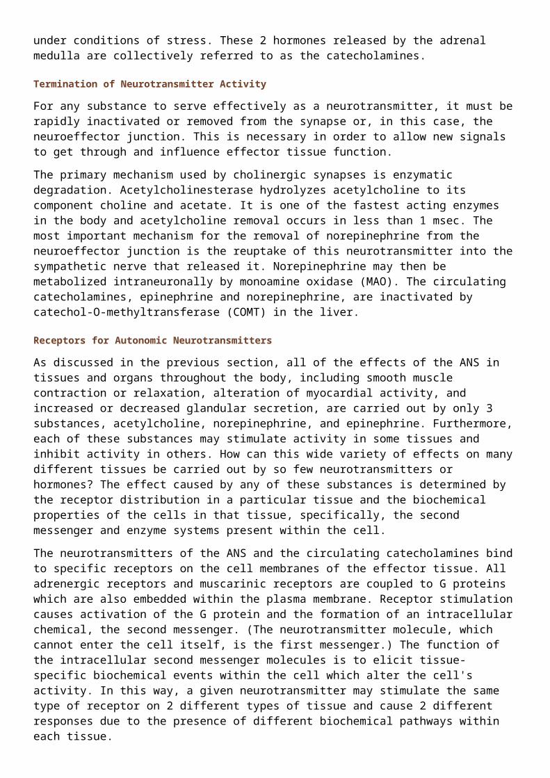
The 2 most common neurotransmitters released by neurons of the ANS are acetylcholine and norepinephrine. Neurotransmitters are synthesized in the axon varicosities and stored in vesicles for subsequent release. Several distinguishing features of these neurotransmitters are summarized in Table Table2.2. Nerve fibers that release acetylcholine are referred to as cholinergic fibers. These include all preganglionic fibers of the ANS, both sympathetic and parasympathetic systems; all postganglionic fibers of the parasympathetic system; and sympathetic postganglionic fibers innervating sweat glands. Nerve fibers that release norepinephrine are referred to as adrenergic fibers. Most sympathetic postganglionic fibers release norepinephrine.

As previously mentioned, the cells of the adrenal medulla are considered modified sympathetic postganglionic neurons. Instead of a neurotransmitter, these cells release hormones into the blood. Approximately 20% of the hormonal output of the adrenal medulla is norepinephrine. The remaining 80% is epinephrine. Unlike true postganglionic neurons in the sympathetic system, the adrenal medulla contains an enzyme that methylates norepinephrine to form epinephrine. The synthesis of epinephrine, also known as adrenaline, is enhanced under conditions of stress. These 2 hormones released by the adrenal medulla are collectively referred to as the catecholamines.
Termination of Neurotransmitter Activity
For any substance to serve effectively as a neurotransmitter, it must be rapidly inactivated or removed from the synapse or, in this case, the neuroeffector junction. This is necessary in order to allow new signals to get through and influence effector tissue function.
The primary mechanism used by cholinergic synapses is enzymatic degradation. Acetylcholinesterase hydrolyzes acetylcholine to its component choline and acetate. It is one of the fastest acting enzymes in the body and acetylcholine removal occurs in less than 1 msec. The most important mechanism for the removal of norepinephrine from the neuroeffector junction is the reuptake of this neurotransmitter into the sympathetic nerve that released it. Norepinephrine may then be metabolized intraneuronally by monoamine oxidase (MAO). The circulating catecholamines, epinephrine and norepinephrine, are inactivated by catechol-O-methyltransferase (COMT) in the liver.
Receptors for Autonomic Neurotransmitters
As discussed in the previous section, all of the effects of the ANS in tissues and organs throughout the body, including smooth muscle contraction or relaxation, alteration of myocardial activity, and increased or decreased glandular secretion, are carried out by only 3 substances, acetylcholine, norepinephrine, and epinephrine. Furthermore, each of these substances may stimulate activity in some tissues and inhibit activity in others. How can this wide variety of effects on many different tissues be carried out by so few neurotransmitters or hormones? The effect caused by any of these substances is determined by the receptor distribution in a particular tissue and the biochemical properties of the cells in that tissue, specifically, the second messenger and enzyme systems present within the cell.
The neurotransmitters of the ANS and the circulating catecholamines bind to specific receptors on the cell membranes of the effector tissue. All adrenergic receptors and muscarinic receptors are coupled to G
proteins which are also embedded within the plasma membrane. Receptor stimulation causes activation of the G protein and the formation of an intracellular chemical, the second messenger. (The neurotransmitter molecule, which cannot enter the cell itself, is the first messenger.) The function of the intracellular second messenger molecules is to elicit tissue-specific biochemical events within the cell which alter the cell's activity. In this way, a given neurotransmitter may stimulate the same type of receptor on 2 different types of tissue and cause 2 different responses due to the presence of different biochemical pathways within each tissue.
Acetylcholine binds to 2 types of cholinergic receptors. Nicotinic receptors are found on the cell bodies of all postganglionic neurons, both sympathetic and parasympathetic, in the ganglia of the ANS. Acetylcholine released from the preganglionic neurons binds to these nicotinic receptors and causes a rapid increase in the cellular permeability to Na+ ions and Ca++ ions. The resulting influx of these 2 cations causes depolarization and excitation of the postganglionic neurons the ANS pathways.
Muscarinic receptors are found on the cell membranes of the effector tissues and are linked to G proteins and second messenger systems which carry out the intracellular effects. Acetylcholine released from all parasympathetic postganglionic neurons and some sympathetic postganglionic neurons traveling to sweat glands binds to these receptors. Muscarinic receptors may be either inhibitory or excitatory, depending on the tissue upon which they are found. For example, muscarinic receptor stimulation in the myocardium is inhibitory and decreases heart rate while stimulation of these receptors in the lungs is excitatory, causing contraction of airway smooth muscle and bronchoconstriction.
There are 2 classes of adrenergic receptors for norepinephrine and epinephrine, alpha (α) and beta (β). Furthermore, there are at least 2 subtypes of receptors in each class: α1, α2, β1 and β2. All of these receptors are linked to G proteins and second messenger systems which carry out the intracellular effects.
Alpha receptors are the more abundant of the adrenergic receptors. Of the 2 subtypes, α1 receptors are more widely distributed on the effector tissues. Alpha one receptor stimulation leads to an increase in intracellular calcium. As a result, these receptors tend to be excitatory. For example, stimulation of α1 receptors causes contraction of vascular smooth muscle resulting in vasoconstriction and increased glandular secretion by way of exocytosis.
Pharmacy Application: Alpha One Adrenergic Receptor Antagonists.
Hypertension, or a chronic elevation in blood pressure, is a major risk factor for coronary artery disease, congestive heart failure, stroke, kidney failure, and retinopathy. An important cause of hypertension is excessive vascular smooth muscle tone or vasoconstriction. Prazosin, an α1-adrenergic receptor antagonist, is very effective in the management of hypertension. Because α1-receptor stimulation causes vasoconstriction, drugs that block these receptors result in vasodilation and a decrease in blood pressure.
Compared to α1 receptors, α2 receptors have only moderate distribution on the effector tissues. Alpha 2 receptor stimulation causes a decrease in cAMP and, therefore, inhibitory effects such as smooth muscle relaxation and decreased glandular secretion. However, α2 receptors have important presynaptic effects. Where α1 receptors are found on the effector tissue cells at the neuroeffector junction, the α2 receptors are found on the varicosities of the postganglionic neuron. Norepinephrine released from this neuron binds to not only the α1 receptors on the effector tissue to cause some physiological effect; it also binds to the α2 receptors on the neuron itself. Alpha 2 receptor stimulation results in “presynaptic inhibition” or in a decrease in the release of norepinephrine. In this way, norepinephrine inhibits its own release from the sympathetic postganglionic neuron and controls its own activity. Both α1 and α2 receptors have equal affinity
for norepinephrine released directly from sympathetic neurons as well as circulating epinephrine released from the adrenal medulla.
Stimulation of each type of β receptor leads to an increase in intracellular cAMP. Whether this results in an excitatory or an inhibitory response depends upon the specific cell type. As with α receptors, β receptors are also unevenly distributed with β2 receptors, the more common subtype on the effector tissues. Beta 2 receptors tend to be inhibitory. For example, β2 receptor stimulation causes relaxation of vascular smooth muscle and airway smooth muscle resulting in vasodilation and bronchodilation, respectively. Beta 2 receptors have a significantly greater affinity for epinephrine than for norepinephrine. Furthermore, terminations of sympathetic pathways are not found near these receptors. Therefore, β2 receptors are stimulated only indirectly by circulating epinephrine instead of by direct sympathetic nervous activity.
Beta 1 receptors are the primary adrenergic receptor on the heart (a small percentage of the adrenergic receptors on the myocardium are β2). Both subtypes of β receptors on the heart are excitatory and stimulation leads to an increase in cardiac activity. Beta 1 receptors are also found on certain cells in the kidney. Epinephrine and norepinephrine have equal affinity for β1 receptors.
Beta three (β3) receptors are found primarily in adipose tissue. Stimulation of these receptors, which have a stronger affinity for norepinephrine, causes lipolysis.
Pharmacy Application: Sympathomimetic Drugs.
Sympathomimetic drugs are those that produce effects in a tissue resembling those caused from stimulation by the sympathetic nervous system. An important use for these drugs is in the treatment of bronchial asthma which is characterized by bronchospasm. As discussed, bronchodilation occurs following β2-adrenergic receptor stimulation. Non-selective β receptor agonists, such as epinephrine and isoproterenol, are capable of causing bronchodilation. However, a potential problem with these drugs is that they stimulate all β-receptors including β1 receptors on the heart. Therefore, in patients with bronchospasm, an undesirable side effect of treatment with these non-selective agents is an increase in heart rate. Instead, β2-selective drugs, such as albuterol, are chosen for this therapy. They are equally effective in causing bronchodilation with a much lower risk of adverse cardiovascular effects.
Functions of the Autonomic Nervous System
The 2 divisions of the ANS are dominant under different conditions. As stated previously, the sympathetic system is activated during emergency “fight-or-flight” reactions and during exercise. The parasympathetic system is predominant during quiet conditions (“rest and digest”). As such, the physiological effects caused by each system are quite predictable. In other words, all of the changes in organ and tissue function induced by the sympathetic system work together to support strenuous physical activity and the changes induced by the parasympathetic system are appropriate for when the body is resting.
The “fight-or-flight” reaction elicited by the sympathetic system is essentially a whole body response. Changes in organ and tissue function throughout the body are coordinated so that there is an increase in the delivery of well-oxygenated, nutrient-rich blood to the working skeletal muscles. Both heart rate and myocardial contractility are increased so that the heart pumps more blood per minute. Sympathetic stimulation of vascular smooth muscle causes widespread vasoconstriction, particularly in the organs of the gastrointestinal system and in the kidneys. This vasoconstriction serves to “redirect” or redistribute the blood away from these metabolically inactive tissues and toward the contracting muscles. Bronchodilation in the lungs facilitates the movement of air in and out of the lungs so that the uptake of oxygen from the atmosphere and the elimination of carbon dioxide from the body are maximized. An enhanced rate of

glycogenolysis (breakdown of glycogen into its component glucose molecules) and gluconeogenesis (formation of new glucose from noncarbohydrate sources) in the liver increases the concentration of glucose molecules in the blood. This is necessary for the brain as glucose is the only nutrient molecule that it can utilize to form metabolic energy. An enhanced rate of lipolysis in adipose tissue increases the concentration of fatty acid molecules in the blood. Skeletal muscles then utilize these fatty acids to form metabolic energy for contraction. Generalized sweating elicited by the sympathetic system enables the individual to thermoregulate during these conditions of increased physical activity and heat production. Finally, the eye is adjusted such that the pupil dilates letting more light in toward the retina (mydriasis) and the lens adapts for distance vision.
The parasympathetic system decreases heart rate which helps to conserve energy under resting conditions. Salivary secretion is enhanced to facilitate the swallowing of food. Gastric motility and secretion are stimulated to begin the processing of ingested food. Intestinal motility and secretion are also stimulated to continue the processing and to facilitate the absorption of these nutrients. Both exocrine and endocrine secretion from the pancreas is promoted. Enzymes released from the exocrine glands of the pancreas contribute to the chemical breakdown of the food in the intestine and insulin released from the pancreatic islets promotes the storage of nutrient molecules within the tissues once they are absorbed into the body. Another bodily maintenance type of function caused by the parasympathetic system is contraction of the urinary bladder which results in urination. Finally, the eye is adjusted such that the pupil contracts (miosis) and the lens adapts for near vision.
Pharmacy application: cholinomimetic drugs.
Cholinomimetic drugs are those that produce effects in a tissue resembling those caused from stimulation by the parasympathetic nervous system. These drugs have many important uses including the treatment of gastrointestinal and urinary tract disorders that involve depressed smooth muscle activity without obstruction. For example, postoperative ileus is characterized by a loss of tone or paralysis of the stomach or bowel following surgical manipulation. Urinary retention may also occur postoperatively or it may be secondary to spinal cord injury or disease (neurogenic bladder). Normally, parasympathetic stimulation of the smooth muscle in each of these organ systems causes contraction to maintain gastrointestinal motility as well as urination. There are 2 different approaches in the pharmacotherapy of these disorders. One type of agent would be a muscarinic receptor agonist which would mimic the effect of the parasympathetic neurotransmitter, acetylcholine, and stimulate smooth muscle contraction. One of the more commonly used agents in this category is bethanechol which can be given subcutaneously. Another approach is to increase the concentration and, therefore, activity of endogenously produced acetylcholine in the neuroeffector junction. Administration of an acetylcholinesterase inhibitor prevents the degradation and removal of neuronally-released acetylcholine. In this case, neostigmine is the most widely used agent. Neostigmine may be given intramuscularly, subcutaneously, or orally.
Pharmacy application: muscarinic receptor antagonists.
Inspection of the retina during an ophthalmoscopic examination is greatly facilitated by mydriasis, or the dilation of the pupil. Parasympathetic stimulation of the circular muscle layer in the iris causes contraction and a decrease in the diameter of the pupil. Administration of a muscarinic receptor antagonist, such as atropine or scopolamine, prevents this smooth muscle contraction. As a result, sympathetic stimulation of the radial muscle layer is unopposed. This causes an increase in the diameter of the pupil. These agents are given in the form of eye drops which act locally and limit the possibility of systemic side effects.
Adrenal Medulla
A mass sympathetic discharge, which typically occurs during the “fight-or-flight” response and during exercise, involves the simultaneous stimulation of organs and tissues throughout the body. Included among these tissues are the adrenal medullae which release epinephrine and norepinephrine into the blood. In large part, the indirect effects of these catecholamines are similar to and, therefore, reinforce those of direct sympathetic stimulation. However, there are some important differences in the effects of the circulating catecholamines and those of norepinephrine released from sympathetic nerves.
The duration of activity of the catecholamines is significantly longer than that of neuronally released norepinephrine. Therefore, the effects on the tissues are more prolonged. This difference has to do with the mechanism of inactivation of these substances. Norepinephrine is immediately removed from the neuroeffector synapse by way of reuptake into the postganglionic neuron. This rapid removal limits the duration of the effect of this neurotransmitter. In contrast, there are no enzymes in the blood to degrade the catecholamines. Instead, the catecholamines are inactivated by COMT in the liver. As one might expect, the hepatic clearance of these hormones from the blood would require several passes through the circulation. Therefore, the catecholamines are available to cause their effects for a comparatively longer period of time (up to 1-2 minutes as opposed to milliseconds).
Because they travel in the blood, organs and tissues throughout the body are exposed to the catecholamines. Therefore, they are capable of stimulating tissues that are not directly innervated by sympathetic nerve fibers: airway smooth muscle, hepatocytes, and adipose tissue, in particular. As a result, the catecholamines have a much wider breadth of activity compared to norepinephrine released from sympathetic nerves.
The third important feature that distinguishes the catecholamines from neuronally released norepinephrine involves epinephrine's affinity for β2 receptors. Norepinephrine has a very limited affinity for these receptors. Therefore, circulating epinephrine causes effects that differ from those of direct sympathetic innervation including a greater stimulatory effect on the heart and relaxation of smooth muscle (vascular, bronchial, gastrointestinal, and genitourinary).
Epinephrine and norepinephrine have equal affinity for β1 receptors, the predominant adrenergic receptor on the heart. However, the human heart also contains a small percentage of β2 receptors which, like β1 receptors are excitatory. Therefore, epinephrine is capable of stimulating a greater number of receptors and of causing a greater stimulatory effect on the myocardium.
Beta two adrenergic receptors are also found on smooth muscle in several organ systems. These receptors tend to be inhibitory and cause relaxation of the smooth muscle. Vascular smooth muscle in skeletal muscle contains both α1 and β2 receptors. Norepinephrine, which stimulates only the excitatory α1 receptors, causes strong vasoconstriction. However, epinephrine, which stimulates both types of receptors, causes only weak vasoconstriction. The vasodilation resulting from β2 receptor stimulation opposes and, therefore, weakens the vasoconstriction resulting from α1 receptor stimulation. Given that skeletal muscle may account for 40% of an adult's body weight, the potential difference in vasoconstriction, blood pressure, and the distribution of blood flow could be quite significant.
Another noteworthy example of the relaxation of smooth muscle by way of β2 receptor stimulation involves the airways. Bronchodilation, or the opening of the airways, facilitates airflow in the lungs. Any direct sympathetic innervation to the lungs is irrelevant in this respect, as only circulating epinephrine is capable of stimulating these receptors on airway smooth muscle.