autophagy facilitates the development of breast cancer
TRANSCRIPT

Autophagy Facilitates the Development of Breast CancerResistance to the Anti-HER2 Monoclonal AntibodyTrastuzumabAlejandro Vazquez-Martin1,2, Cristina Oliveras-Ferraros1,2, Javier A. Menendez1,2*
1 Catalan Institute of Oncology (ICO), Girona, Catalonia, Spain, 2 Girona Biomedical Research Institute (IdIBGi), Dr. Josep Trueta University Hospital of Girona, Girona,
Catalonia, Spain
Abstract
Autophagy has been emerging as a novel cytoprotective mechanism to increase tumor cell survival under conditions ofmetabolic stress and hypoxia as well as to escape chemotherapy-induced cell death. To elucidate whether autophagy mightalso protect cancer cells from the growth inhibitory effects of targeted therapies, we evaluated the autophagic status ofpreclinical breast cancer models exhibiting auto-acquired resistance to the anti-HER2 monoclonal antibody trastuzumab(Tzb). We first examined the basal autophagic levels in Tzb-naive SKBR3 cells and in two pools of Tzb-conditioned SKBR3cells (TzbR), which optimally grow in the presence of Tzb doses as high as 200 mg/ml Tzb. Fluorescence microscopicanalyses revealed that the number of punctate LC3 structures -a hallmark of autophagy- was drastically higher in Tzb-refractory cells than in Tzb-sensitive SKBR3 parental cells. Immunoblotting analyses confirmed that the lipidation product ofthe autophagic conversion of LC3 was accumulated to high levels in TzbR cells. High levels of the LC3 lipidated form in Tzb-refractory cells were accompanied by decreased p62/sequestosome-1 protein expression, a phenomenon characterizing theoccurrence of increased autophagic flux. Moreover, increased autophagy was actively used to survive Tzb therapy as TzbRpools were exquisitely sensitive to chemical inhibitors of autophagosomal formation/function. Knockdown of LC3expression via siRNA similarly resulted in reduced TzbR cell proliferation and supra-additively interacted with Tzb to re-sensitize TzbR cells. Sub-groups of Tzb-naive SKBR3 parental cells accumulated LC3 punctate structures and decreased p62expression after treatment with high-dose Tzb, likely promoting their own resistance. This is the first report showing thatHER2-overexpressing breast cancer cells chronically exposed to Tzb exhibit a bona fide up-regulation of the autophagicactivity that efficiently works to protect breast cancer cells from the growth-inhibitory effects of Tzb. Therapeutic targetingautophagosome formation/function might represent a novel molecular avenue to reduce the emergence of Tzb resistancein HER2-dependent breast carcinomas.
Citation: Vazquez-Martin A, Oliveras-Ferraros C, Menendez JA (2009) Autophagy Facilitates the Development of Breast Cancer Resistance to the Anti-HER2Monoclonal Antibody Trastuzumab. PLoS ONE 4(7): e6251. doi:10.1371/journal.pone.0006251
Editor: Joseph Najbauer, City of Hope Medical Center, United States of America
Received April 11, 2009; Accepted June 17, 2009; Published July 16, 2009
Copyright: � 2009 Vazquez-Martin et al. This is an open-access article distributed under the terms of the Creative Commons Attribution License, which permitsunrestricted use, distribution, and reproduction in any medium, provided the original author and source are credited.
Funding: Alejandro Vazquez-Martin is the recipient of a ‘‘Sara Borrell’’ post-doctoral contract (CD08/00283, Ministerio de Sanidad y Consumo, Fondo deInvestigacion Sanitaria -FIS-, Spain). Javier A. Menendez is the recipient of a Basic, Clinical and Translational Research Award (BCTR0600894) from the Susan G.Komen Breast Cancer Foundation (Texas, USA). This work was supported in part by Instituto de Salud Carlos III (Ministerio de Sanidad y Consumo, Fondo deInvestigacion Sanitaria -FIS-, Spain, Grants CP05-00090, PI06-0778 and RD06-0020-0028 to Javier A. Menendez). Javier A. Menendez was also supported by a Grantfrom the Fundacion Cientıfica de la Asociacion Espanola Contra el Cancer (AECC, Spain). The funders had no role in study design, data collection and analysis,decision to publish, or preparation of the manuscript.
Competing Interests: The authors have declared that no competing interests exist.
* E-mail: [email protected]
Introduction
Significant amount of research has been dedicated to elucidate
molecular mechanisms that could explain de novo and acquired
resistance to the anti-HER2 monoclonal antibody trastuzumab
(Tzb; HerceptinH), the first immunotherapeutic drug for the
successful treatment of breast carcinomas overexpressing the
HER2 (erbB-2) oncogene [1–8]. Proposed mechanisms for innate
or acquired resistance to Tzb include steric inhibition of Tzb
binding to the extracellular domain (ECD) of the HER2 tyrosine
kinase receptor imposed by other extracellular factors such as the
glycoprotein mucin 4 (MUC-4) [9,10], molecular changes in the
target receptor itself (e.g. HER2 mutations [11–13]; accumulation
of a proteolyzed HER2 fragment –p95HER2- lacking the
extracellular Tzb binding epitope but retaining ligand-indepen-
dent TK activity [14–16]), and cross-talk with other transduction
cascades such as the insulin growth factor (IGF)-1, estrogen
receptor (ER) and vascular endothelial growth factor (VEGF)
pathways that could compensate for attenuated HER2 signaling
[8,17–23]. Alterations in the regulation of HER2 downstream
signaling components, including sub-cellular localization of the
cyclin-dependent kinase (CDK)-inhibiting protein p27Kip1 and
independent attenuation of PI-39K/AKT/mTOR-mediated apo-
ptosis through downregulation of the phosphatase and tensin
homolog (PTEN) tumor suppressor have also been implicated as
potential sources of resistance to HER2-targeted therapies [24–
27]. Increased activation of PI-39K and its downstream effector
AKT has also been associated with Tzb resistance in HER2-
dependent breast carcinoma cells [28–30].
To date, the survival pathway of macroautophagy (also referred
as autophagy) has not been implicated in Tzb resistance.
Autophagy –lysosomal degradation, or eating (phagy), of part of
PLoS ONE | www.plosone.org 1 July 2009 | Volume 4 | Issue 7 | e6251

the cell’s self (auto)- is a catabolic process of organelle digestion that
generates ATP during periods on nutrient limitation [31–37].
Autophagy optimizes nutrient utilization in rapidly growing cells
when faced with hypoxic or metabolic stresses and, hence it
contributes to normal and cancer cell survival. During autophagy,
macroautophagosomes (also referred as autophagosomes) are
formed as double membrane-bound vesicles which engulf
cytoplasm and/or cytoplasmic organelles. Then, autophagosomes
fuse with lysosomes to degrade the contents of the autophagic
vesicle and provide essential building blocks, such as amino acids
back to cell. Because this mechanism may be decreased in tumor
cells compared with normal cells, initial studies appreciated
autophagy as a tumor suppressor mechanism [38,39]. Indeed,
cancer cells may undergo autophagic cell death (APCD; also
referred to as active cell death II [ACDII]) following extreme
autophagic degradation associated with exposure to several cancer
therapies [40]. However, the autophagic response can also
function as a protective mechanism allowing the recycling of
proteins and cellular components to survive cell injuries induced
by cytotoxic agents. Although it is well established that autophagy
can protect cancer cells against various stressors, including
chemotherapeutics [41–44], it remains largely unknown whether
‘‘protective autophagy’’ might also defend cancer cells from the
growth inhibitory effects of targeted therapies such as monoclonal
antibodies, tyrosine kinase inhibitors, etc.
In this study, we investigated the autophagic status in preclinical
breast cancer models exhibiting auto-acquired resistance to Tzb that
were obtained by continuously growing Tzb-sensitive HER2-
overexpressing SKBR3 breast cancer cells in the presence of
clinically relevant concentrations of Tzb for more than 10 months.
We followed complementary criteria to accurately monitor autoph-
agy in SKBR3-derived Tzb-refractory cells [45]. First, we monitored
the level of intermediary structures of the autophagic pathway, e.g.
the level of endogenous LC3-II/LC3-I (-microtubule-associated
protein 1 light chain 3 beta- a specific and sensitive autophagosome
marker extensively used to monitor autophagic activity) and the level
of p62 protein (also known as SQSTM1 [sequestosome-1]), which
serves as a link between LC3 and ubiquitinated substrates destined
for autophagic degradation [46–54]. Second, to unambiguously
establish the pro-survival role of an increased catabolic flux through
the autophagic pathway was critical to the development of acquired
Tzb resistance, we assessed how chemical autophagy inhibitors or
siRNA-induced knockdown of LC3 altered cell proliferation in
TzbR cells. Under experimental conditions described here, we
report for the first time that induction of autophagy is closely related
to the cell survival system acquired by HER2-overexpressing breast
cancer cells chronically exposed to Tzb.
Results
We established Tzb-resistant HER2-positive breast cancer cells
by exposing Tzb-naive SKBR3 parental cells to incremental
increases of Tzb. Tzb-resistance selection continued until the
SKBR3 cell population could sustain cell viability and proliferate
when challenged with 200 mg/ml Tzb. Under these experimental
conditions, two pools of Tzb-refractory cells (TzbR POOL1 and
TzbR POOL2) were obtained upon exposure of SKBR3 parental
cells for a minimum of 10 months before starting any experimental
procedure. We confirmed resistance to Tzb by performing MTT-
based cell viability assays. When the concentrations of Tzb needed
to decrease optical density by 50% were calculated from the
percentage of viable cells after exposure to graded concentrations
of Tzb, the Inhibitory Concentration 50 (IC50) value for SKBR3
parental cells was as low as 2 mg/ml Tzb. Treatment with Tzb at
concentrations as high as 200 mg/ml Tzb likewise failed to
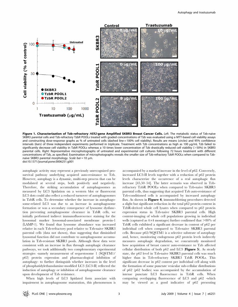
significantly decrease cell viability in TzbR POOLs (Figure 1,left panel). TzbR POOLs exhibited HER2 expression levels
comparable or slightly higher than those naturally occurring in
SKBR3 parental cells (data not shown).
Tzb-refractory Cells Exhibit Increased AutophagosomeFormation
During Tzb selection of the TzbR POOLs, light microscopy
examination consistently showed an increase in the number of
dark, cytosolic granules in surviving, smaller cells (Figure 1, rightpanel). Because these granules were not related to senescent
granules as confirmed by b-galactosidase staining (data not
shown), we sought to determine if they may correspond to
increased cytosolic levels of autophagosomes. Immunoblotting
assessment of LC3 expression is an easy method to predict
autophagic activity of mammalian cells because the amount of
LC3-II –i.e. the product of the autophagic posttranslational
modification of LC3- correlates with the number of autophago-
somes [45–49]. The product of this autophagic conversion of LC3,
LC3-II, tightly associates with the autophagosome membrane and
migrates faster than LC3-I on SDS-PAGE. Therefore, LC3
immunoblotting may detect two bands: LC3-I with an apparent
mobility of 18 kDa and LC3-II (16 kDa). When we utilized this
property of LC3 to initially monitor changes in the dynamics of
the autophagic process in Tzb-sensitive and in Tzb-refractory
cancer cells, both the total amount and, particularly, the lipidation
status of LC3 (i.e. LC3-I is converted by lipidation to the
phosphatidyl-ethanolamine conjugated form LC3-II) were drasti-
cally up-regulated in Tzb-unresponsive TzbR POOLs whereas
LC3-I/LC3-II proteins were hardly detectable in Tzb-naive
SKBR3 parental cells (Figure 2). To further confirm that
autophagosome formation was increased in Tzb-refractory cells,
both the expression and the sub-cellular compartmentalization of
LC3 in individual cells and in whole cell cultures were monitored
by indirect immunofluorescence using an automated confocal-
imaging approach. Untreated SKBR3 parental cells showed a
homogenous but weak cytoplasmic staining of LC3, consistent
with the distribution of LC3-I and typical of low-level or no
autophagosome formation. Consistent with the dramatic increase
in LC3 processing assessed by immunoblotting procedures, LC3
localization dramatically changed from diffuse to punctate or
dotted pattern in Tzb-refractory TzbR POOLs (Figure 2). High
content-imaging of whole cell populations growing in individual
wells (captured as 464 montages) clearly revealed that .90% of
TzbR cells exhibited an intense punctate LC3 fluorescence (the
medium number of autophagosomes per cell was ,50) whereas a
significantly lower percentage of SKBR3 parental cells (,5%)
revealed LC3-containing vesicles (Figure 3). Video confocal
microscopy also revealed a robust accumulation of endogenous
LC3 in the cytosol of Tzb-refractory cells was, consistent with the
distribution of LC3-II-positive bodies representing isolation
membranes and autophagosomes (Supplemental Video S1 –
SKBR3-, Supplemental Video S2 –TzbR POOL1- and
Supplemental Video S3 –TzbR POOL2-). Importantly, this
high level of autophagosomes accompanied with healthy-appear-
ing nuclei and high proliferation rates in Tzb-refractory cells.
Tzb-refractory Cells Exhibit Increased Basal AutophagySince the lipidation status of LC3 has been proposed to
accurately reflect autophagic activity, our findings revealing an
increase in LC3-II expression as assessed by immunoblotting along
with an increase in LC3 fluorescent puncta, strongly suggested that
augmented autophagosome formation due to increases in
Autophagy and trastuzumab
PLoS ONE | www.plosone.org 2 July 2009 | Volume 4 | Issue 7 | e6251
autophagic activity may represent a previously unrecognized pro-
survival pathway underlying acquired auto-resistance to Tzb.
However, autophagy is a dynamic, multi-step process that can be
modulated at several steps, both positively and negatively.
Therefore, the striking accumulation of autophagosomes as
measured by LC3 lipidation on a western blot or fluorescent
LC3 dots could also reflect a reduced turnover of autophagosomes
in TzbR cells. To determine whether the increase in autophago-
some-related LC3 was due to an increase in autophagosome
formation or was a secondary consequence of lysosome dysfunc-
tion preventing autophagosome clearance in TzbR cells, we
initially performed indirect immunofluorescence staining for the
lysosomal marker lysosomal-associated membrane protein-1
(LAMP-1). We found that lysosome abundance was increased
relative in each Tzb-refractory pool relative to Tzb-naive SKBR3
parental cells (data not shown), thus suggesting that diminished
lysosomal function did not contribute to autophagosome accumu-
lation in Tzb-resistant SKBR3 pools. Although these data were
consistent with an increase in flux through autophagic clearance
pathways, we took additionally two complementary experimental
strategies -namely measurement of sequestosome 1 (SQSTM1/
p62) protein expression and pharmacological inhibition of
autophagy- to further distinguish whether increases in the level
of phosphatidylethanolamine-modified LC3 (LC3-II) reflected the
induction of autophagy or inhibition of autophagosome clearance
upon development of Tzb resistance.
When high levels of LC3 lipidated form associate with
impairment in autophagosome maturation, this phenomenon is
accompanied by a marked increase in the level of p62. Conversely,
increased LC3-II levels together with a reduction of p62 protein
levels characterize the occurrence of a real autophagic flux
increase [45,50–54]. The latter scenario was observed in Tzb-
refractory TzbR POOLs when compared to Tzb-naive SKBR3
parental cells, thus suggesting that acquired Tzb auto-resistance of
Tzb-conditioned cells is accompanied by increased autophagy
flux. As shown in Figure 4, immunoblotting procedures detected
a slight but significant reduction in the total p62 protein content in
TzbR-derived whole cell lysates when compared to p62 protein
expression status in Tzb-naive SKBR3 parental cells. High
content-imaging of whole cell populations growing in individual
wells (captured as 464 montages) further confirmed that .80% of
TzbR cells exhibited a significant decrease in content of p62 per
individual cell when compared to Tzb-naive SKBR3 parental
cells. Because p62/SQSTM1 is a selective substrate of autophagy
and, hence, monitoring endogenous p62 protein levels indirectly
measures autophagic degradation, we concurrently monitored
how acquisition of breast cancer auto-resistance to Tzb affected
cellular distribution of both p62 and LC3 (Figure 5). As steady
state, the p62 level in Tzb-naive SKBR3 parental cells was much
higher than in Tzb-refractory SKBR3 TzbR POOLs. This
significant decrease in p62 content per individual cell along with
the formation of some punctate vesicular sub-cellular distributions
of p62 (p62 bodies) was accompanied by the accumulation of
intense punctate LC3 fluorescence in TzbR cells. When
comparing overlapping fluorescence of LC3 and p62 -which
may be viewed as a good indicative of p62 presenting
Figure 1. Characterization of Tzb-refractory HER2-gene Amplified SKBR3 Breast Cancer Cells. Left. The metabolic status of Tzb-naiveSKBR3 parental cells and Tzb-refractory TzbR POOLs treated with graded concentrations of Tzb was evaluated using a MTT-based cell viability assaysand constructing dose-response graphs as % of untreated cells (dashed line = 100% cell viability). Results are means (circles) and 95% confidenceintervals (bars) of three independent experiments performed in triplicate. Treatment with Tzb concentrations as high as 100 mg/mL Tzb failed tosignificantly decrease cell viability in TzbR POOLs whereas a 10 times lower concentration of Tzb drastically reduced cell viability (.50%) in SKBR3parental cells. Right: Representative microphotographs of untreated and experimental cell cultures following 72 hours treatment with differentconcentrations of Tzb, as specified. Examination of microphotographs reveals the smaller size of Tzb-refractory TzbR POOLs when compared to Tzb-naive SKBR3 parental morphology. Scale bar = 10 mm.doi:10.1371/journal.pone.0006251.g001
Autophagy and trastuzumab
PLoS ONE | www.plosone.org 3 July 2009 | Volume 4 | Issue 7 | e6251

ubiquitinated protein bodies to the autophagic machinery via LC3-
it become obvious that Tzb-refractory cells exhibit an exacerbated
autophagic clearance compared to Tzb-sensitive SKBR3 parental
cells (Figure 5).
Blockade of Macroautophagosome Formation/FunctionEnhances Tzb Efficacy in Tzb-refractory Cells
To pharmacologically evaluate whether Tzb-refractory cells
actively used increased basal autophagy to survive Tzb therapy,
we finally assessed the growth inhibitory effects of autophagy
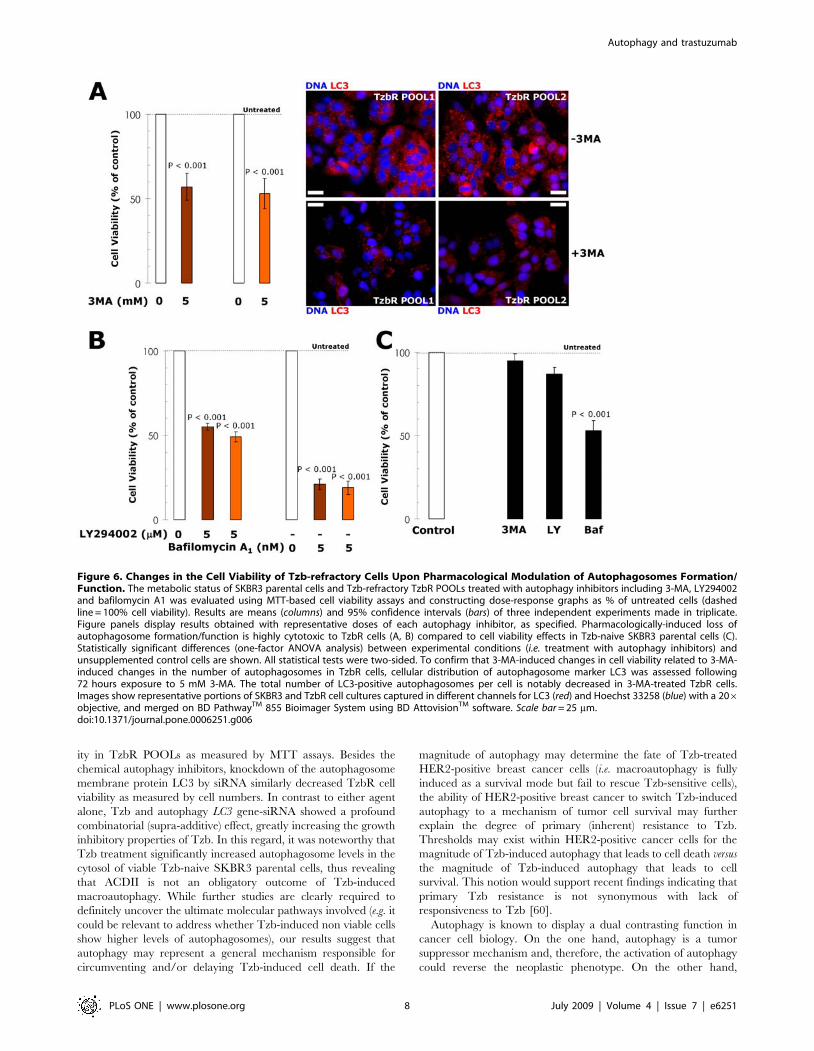
inhibitors [55]. Inhibition of the formation of pre-autophago-
somal structure upon treatment with 3-methyladenine (3-MA)
notably reduced cell viability in Tzb-refractory TzbR POOLs
but not in Tzb-naive SKBR3 parental cells (Figure 6). Since 3-
MA treatment may induce off-target effects, we further
confirmed that 3-MA treatment was efficient at reducing the
number of LC3-positive autophagosomes in TzbR cells.
Representative immuno-confocal images of Tzb-refractory
TzbR POOLs cultured in the absence or presence of 3-MA
are shown in Figure 6. Cultures of TzbR cells treated with 3-
MA likewise showed a significant increase in the number of cells
containing ,20 autophagosomes per cell. These findings,
altogether, strongly suggest that increased macroautophagy
actively provides a survival function to Tzb-refractory cells.
This notion was further supported when similar studies were
carried out in the presence of 2-(4-morpholinyl)-8-phenylchro-
Figure 2. Dynamics of Autophagosome Formation in SKBR3 cells-derived Tzb-Refractory POOLs. Immunoblotting. Autophagosomeformation in whole cell lysates of Tzb-naive SKBR3 parental cells and Tzb-refractory TzbR POOLs was detected with Western blot analysis using a LC3antibody. Top band (18 kDa) represents LC3-I. Bottom band (16 kDa) represents LC3-II, a typical marker of autophagosomes. Autophagosomeformation is robust in Tzb-refractory TzbR cells when compared to low to undetectable levels of LC3-I/LC3-II expression in Tzb-sensitive SKBR3parental cells. Figure shows a representative immunoblotting analysis. Equivalent results were obtained in three independent experiments.Immunofluorescence. After fixation and permeabilization, cellular distribution of autophagosome marker LC3 was assessed following staining with aLC3 antibody and Hoechst 33258 for nuclear counterstaining. SKBR3 parental cells display a homogenous but weak cytoplasmic LC3 staining, whichis typical of absent or low-level autophagosome formation. TzbR POOLs show a marked contrast enhancement in the punctated pattern ofendogenous LC3 expression, which is characteristic of autophagosome formation. Of note, Tzb exposure further increases autophagosome LC3pattern. Images show representative portions of SKBR3 and TzbR cell cultures captured in different channels for LC3 (red) and Hoechst 33258 (blue)with a 206objective, and merged on BD PathwayTM 855 Bioimager System using BD AttovisionTM software. Scale bar = 25 mm.doi:10.1371/journal.pone.0006251.g002
Autophagy and trastuzumab
PLoS ONE | www.plosone.org 4 July 2009 | Volume 4 | Issue 7 | e6251

mone (LY294002). In terms of cell viability, Tzb-refractory cells
were exquisitely sensitive to this agent that blocks phosphatidy-
linositol 3-kinase activity and prevents autophagic sequestration
when compared to SKBR3 parental cells (Figure 6). Bafilo-
mycin A1-prevented fusion of the autophagosomes and
lysosome, and was highly cytotoxic to TzbR cells. Although
less markedly, cell viability of Tzb-naive SKBR3 parental cells
was also significantly reduced in the presence of bafilomycin A1
(Figure 6).
To provide additional evidence that autophagy plays a critical
survival role in enabling Tzb-insensitive high-rates of cell
proliferation in Tzb-refractory cells and to avoid any off-target
side effects that may confound interpretation of the results
obtained with autophagy inhibitors, we used the potent and
highly sequence-specific mechanism of RNA interference (RNAi)
to block LC3-dependent autophagosome formation. Tzb-refrac-
tory TzbR POOLs transiently transfected with sequence-specific
double-stranded RNA oligonucleotides targeting Atg8/LC3 gene
demonstrated hypersensitivity to high-dose Tzb. Following
sequential exposure to LC3 siRNA and 200 mg/ml Tzb, TzbR
cells were extremely fragile and many of the cells died before the
216-hours harvest (data not shown), thus precluding analyses of
the nature of interaction. Interestingly, supra-additive (synergistic)
growth inhibitory interactions occurred at late time points (i.e. 144
and 216 hours) when RNAi-induced knock-down of LC3 was
followed by exposure to 10 mg/ml Tzb, an ineffective low-dose of
Tzb when used as single agent in TzbR cells (Figure 7). These
findings, altogether, clearly establish that hyperactivation of basal
autophagy plays an essential survival role in Tzb-refractory TzbR
cells re-challenged with Tzb.
Figure 3. Correlation Between Proliferative Profiles and Dynamics of Autophagosomal Formation in TzbR-refractory Cells. Toppanels. SKBR3 parental cells and SKBR3-derived Tzb-refractory POOLs were plated in 24-well plates at a density of 10,000 cells/well and cultured withregular medium in the absence or presence of Tzb (10 mg/mL Tzb in SKBR3 parental cells; 100 mg/mL Tzb in TzbR POOLs). The data presented aremean of number cells6105/well (circles) and 95% confidence intervals (bars) of three independent experiments made in duplicate obtained after 0, 3,6 and 9 days. Statistically significant differences (one-factor ANOVA analysis) between experimental conditions and unsupplemented control cells areshown. All statistical tests were two-sided. No statistically significant differences were observed in the number of TzbR cells growing in the presenceof 100 mg/mL Tzb up to 9 days whereas 10 mg/mL Tzb significantly reduced cell proliferation in SKBR3 parental cells as early as 3 days after Tzbexposure. Slope of the growth curves clearly denotes a faster proliferation of TzbR POOLs regardless Tzb exposure. Bottom panels. Images showrepresentative whole populations of SKBR3 cells and TzbR cells growing in individual wells that were captured using different channels for LC3 (red)and Hoechst 33258 (blue) as a 464 montage with a 206 objective on BD PathwayTM 855 Bioimager System, and merged using BD AttovisionTM
software. As discussed in Figure 2, TzbR POOLs notably exhibit a marked contrast enhancement in the punctated pattern of endogenous LC3expression, which is typical for autophagosomal formation. Scale bar = 200 mm.doi:10.1371/journal.pone.0006251.g003
Autophagy and trastuzumab
PLoS ONE | www.plosone.org 5 July 2009 | Volume 4 | Issue 7 | e6251

Activation of Autophagy Might Protect HER2-overexpressing Breast Cancer Cells Against Tzb
We finally speculated that macroautophagosome formation/
function may contribute to cell survival of Tzb-naive HER2-
positive breast cancer cells challenged to Tzb at first, thus limiting
its activity and promoting further resistance. Of note, few groups
of SKBR3 parental cells were found to accumulate some delicate
LC3 punctate structures following treatment with low-dose Tzb
(1 mg/ml Tzb). This increase in the number of autophagosomes
per cell was significantly higher in isolated ‘‘SKBR3 clones’’
capable to survive 72 hours treatment with high-dose (100 and
200 mg/ml) Tzb (Figure 8). In line with this, we found that p62
expression was concomitantly decreased after Tzb treatment in
this small fraction of SKBR3 cells. Tzb-promoted dynamics of
autophagosomal formation (i.e. punctate pattern of LC3) and
increased autophagic flux (i.e. p62 degradation) were confirmed
with Western blot analyses (Figure 8). Similar to our findings
when employing SKBR3 cells, autophagic activity was significant-
ly elevated in Tzb-naive HER2-dependent BT474 breast carci-
noma cells, whereas Tzb treatment failed to modulate autophago-
some-related LC3 expression, lysosomal function and p62
expression in HER2-negative MCF-7 cells, Together, these data
point to HER2 as required element in the cascade of events
leading to autophagy following exposure to the anti-HER2
monoclonal antibody Tzb. As previously noted in Tzb-refractory
pools, the nuclei of the adherent, autophagic surviving SKBR3
parental cells seemed viable. Although we did not evaluate if the
detached cells treated with Tzb showed irregular chromatin
condensation in the nucleus –which is typical of cells dying by
APCD (ACDII)-, the fact that Tzb-induced macroautophagy was
concomitant with a reduction in SKBR3 cell number provides
evidence that Tzb-induced cell death is not an obligatory outcome
of Tzb-induced macroautophagy. Therefore, macroautophagy
appears to facilitate survival in Tzb-naive HER2-overexpressing
human breast cancer cells, likely promoting their own resistance if
Tzb-induced HER2 blockade persists.
Discussion
We here report for the first time that the induction of autophagy
is closely related to the cell survival system triggered by HER2-gene
amplified human breast cancer cells in response to the anti-HER2
monoclonal antibody Tzb. Because a key point that needs to be
emphasized when monitoring macroautophagy is that there is a
difference between measurements that examine the numbers of
autophagosomes versus those that measure flux through the
autophagy pathway, we followed the ‘‘Guidelines for the use and
interpretation of assays for monitoring autophagy in higher eukaryotes’’ recently
presented by Klionsky and colleagues [45]. Thus, to unambiguously
demonstrate that enhanced basal autophagy causally functioned to
protect HER2-dependent human breast cancer cells from cell death
upon chronic exposure to Tzb, we firstly measured autophagosome
accumulation by fluorescence microscopy of endogenous LC3 (i.e.
increase in punctate LC3 -a hallmark of autophagy because it is the
first protein identified on the autophagosomal membrane-) and
LC3-II immunoblotting (i.e. increase in the amount of the lipidation
product of the autophagic conversion of LC3) [45–49]. Autophagy
flux was confirmed by fluorescence microscopy and immunoblot-
ting of p62/sequestosome-1 protein, which serves as a link between
LC3 and ubiquitinated substrates destined for autophagic degra-
dation [45,50–54]. To determine that autophagy induced by Tzb
provided an indispensable role in cell survival and facilitated the
development of acquired resistance to Tzb, we pharmacologically
impaired formation/function of macroautophagosomes by using
small-molecule autophagy inhibitors. Because it is now known that
pharmacological inhibitors of autophagy are not completely specific
to the autophagic process and thus may produce secondary or off-
target effects [55], we finally included the use of RNAi technology
for specific inhibition of autophagy. These combined studies not
only demonstrated that activation of basal autophagy was causally
related to the acquisition of Tzb resistance but further confirmed an
active role of ‘‘protective autophagy’’ in the maintenance of Tzb
insensitivity.
Figure 4. Dynamics of Autophagic Degradation in Tzb-refractory Cells. Immunoblotting. Autophagic degradation in whole cell lysates ofTzb-naive SKBR3 parental cells and Tzb-refractory TzbR POOLs was detected with Western blot analysis using a p62 antibody. Immunoblotting bands(64 kDa) represent p62/SQSTM1, a selective substrate of autophagy. Autophagic degradation (i.e. down-regulation of endogenous p62 proteinexpression) is significantly increased in Tzb-refractory TzbR cells when compared to Tzb-sensitive SKBR3 parental cells. Figure shows a representativeimmunoblotting analysis. Equivalent results were obtained in three independent experiments. Immunofluorescence. After fixation andpermeabilization, cellular distribution of p62 was assessed following staining with a p62 antibody and Hoechst 33258 for nuclear counterstaining.SKBR3 parental cells display a homogenous and strong cytoplasmic p62 staining, which is typical of absent or low-level of autophagic degradation.TzbR POOLs show a significant decrease in the cytoplasmic distribution of p62 that appears somewhat vesiculated, which is typical of enhancedautophagic degradation. Images show representative whole populations of SKBR3 and TzbR cells growing in individual wells that were capturedusing different channels for p62 (green) and Hoechst 33258 (blue) as a 464 montage with a 206 objective on BD PathwayTM 855 BioimagerSystem, and merged using BD AttovisionTM software. Scale bar = 200 mm.doi:10.1371/journal.pone.0006251.g004
Autophagy and trastuzumab
PLoS ONE | www.plosone.org 6 July 2009 | Volume 4 | Issue 7 | e6251

The typical punctate staining that accompanies the transloca-
tion of LC3 II from the cytosol to the autophagosome membrane
was detected at high levels detected in Tzb-refractory TzbR cells.
Formation of autophagosomes was further enhanced in the
presence of Tzb, thus suggesting that Tzb-refractory TzbR cells
are uniquely characterized by their ability to sustain high levels of
Tzb-induced macroautophagy without induction of cell death.
Because the polyubiquitin-binding protein p62/sequestosome-1
recognizes long-lived ubiquitinated protein bodies and presents
these to the autophagic machinery via LC3 (i.e. a large fraction of
p62-formed protein aggregates are degraded by autophagy [50–
54]), the fact that p62/SQSTM1 protein expression was reduced
in Tzb-refractory cells supported the notion that the catabolic
function of activated basal autophagy was playing a pro-survival
role in Tzb-refractory cells. Since p62 downregulation was
maintained in Tzb-refractory HER2-overexpressing cells chroni-
cally exposed to Tzb (i.e. twice weakly for a minimum of 5
months), it could be argued that reduced levels of p62/SQSTM1
may arise from reduced gene transcription or reduced translation
of this protein, rather than from autophagic degradation [45].
However, p62 expression has been shown also to remain
significantly decreased for several months in the chronic
autophagic maladaptive response that accompanies premature
aging in Zmpste24(2/2) mice [55,56]. Moreover, immunofluores-
cence microscopy analyses indicated that p62 downregulation was
concomitant with reduced immunoreactive for ubiquitin-positive
bodies (data not shown), further suggesting that Tzb-refractory
cells exhibit a significantly enhanced turnover of autophagic
substrates [57], including potentially toxic aggregate-prone
ubiquitinated proteins. This Tzb-resistance phenotype consistent
with a chronic increase in flux through autophagic clearance
pathways is in marked contrast with a newly discovered role for
LC3 in nonautophagic cytoplasmic vacuolation death of cancer
cells [58]. In this latter scenario, up-regulation and processing of
the autophagic marker LC3 is accompanied by a marked increase
in p62/SQSTM1 expression and dilation of endoplasmic
reticulum due to accumulation of ubiquitinated proteins. Both
chemical and genetic inhibition of autophagy demonstrated that
development of acquired resistance to Tzb was due, at least in
part, to activation of Autophagy in HER2-overexpressing breast
cancer cells chronically cultured in the presence of Tzb. Three
chemicals (3-MA, LY294002 and bafilomycin A1), which are
routinely used to inhibit autophagy at different stages of the
autophagosome maturation [59], significantly reduced cell viabil-
Figure 5. Dynamics of Autophagy Flux in Tzb-refractory cells. Since increased LC3-II levels together with a reduction of p62 protein levelscharacterize the occurrence of increased autophagic flux, SKBR3 parental cells and TzbR cells (untreated and treated with 100 mg/mL Tzb) were triplestained with antibodies against LC3 and p62 and with Hoechst 33258 for nuclear counterstaining. Tzb-refractory cells exhibit an exacerbatedautophagic clearance compared to Tzb-sensitive SKBR3 parental cells when considering overlapping fluorescence of LC3 and p62 as an indirectmarker of p62 presenting ubiquitinated protein bodies to the autophagic machinery in a LC3-dependent manner. Images show representativeportions of SKBR3 and TzbR cell cultures that were captured using different channels for LC3 (red), p62 (green) and Hoechst 33258 (blue) with a 206objective and merged on BD PathwayTM 855 Bioimager System using BD AttovisionTM software. Scale bar = 25 mm.doi:10.1371/journal.pone.0006251.g005
Autophagy and trastuzumab
PLoS ONE | www.plosone.org 7 July 2009 | Volume 4 | Issue 7 | e6251
ity in TzbR POOLs as measured by MTT assays. Besides the
chemical autophagy inhibitors, knockdown of the autophagosome
membrane protein LC3 by siRNA similarly decreased TzbR cell
viability as measured by cell numbers. In contrast to either agent
alone, Tzb and autophagy LC3 gene-siRNA showed a profound
combinatorial (supra-additive) effect, greatly increasing the growth
inhibitory properties of Tzb. In this regard, it was noteworthy that
Tzb treatment significantly increased autophagosome levels in the
cytosol of viable Tzb-naive SKBR3 parental cells, thus revealing
that ACDII is not an obligatory outcome of Tzb-induced
macroautophagy. While further studies are clearly required to
definitely uncover the ultimate molecular pathways involved (e.g. it
could be relevant to address whether Tzb-induced non viable cells
show higher levels of autophagosomes), our results suggest that
autophagy may represent a general mechanism responsible for
circumventing and/or delaying Tzb-induced cell death. If the
magnitude of autophagy may determine the fate of Tzb-treated
HER2-positive breast cancer cells (i.e. macroautophagy is fully
induced as a survival mode but fail to rescue Tzb-sensitive cells),
the ability of HER2-positive breast cancer to switch Tzb-induced
autophagy to a mechanism of tumor cell survival may further
explain the degree of primary (inherent) resistance to Tzb.
Thresholds may exist within HER2-positive cancer cells for the
magnitude of Tzb-induced autophagy that leads to cell death versus
the magnitude of Tzb-induced autophagy that leads to cell
survival. This notion would support recent findings indicating that
primary Tzb resistance is not synonymous with lack of
responsiveness to Tzb [60].
Autophagy is known to display a dual contrasting function in
cancer cell biology. On the one hand, autophagy is a tumor
suppressor mechanism and, therefore, the activation of autophagy
could reverse the neoplastic phenotype. On the other hand,
Figure 6. Changes in the Cell Viability of Tzb-refractory Cells Upon Pharmacological Modulation of Autophagosomes Formation/Function. The metabolic status of SKBR3 parental cells and Tzb-refractory TzbR POOLs treated with autophagy inhibitors including 3-MA, LY294002and bafilomycin A1 was evaluated using MTT-based cell viability assays and constructing dose-response graphs as % of untreated cells (dashedline = 100% cell viability). Results are means (columns) and 95% confidence intervals (bars) of three independent experiments made in triplicate.Figure panels display results obtained with representative doses of each autophagy inhibitor, as specified. Pharmacologically-induced loss ofautophagosome formation/function is highly cytotoxic to TzbR cells (A, B) compared to cell viability effects in Tzb-naive SKBR3 parental cells (C).Statistically significant differences (one-factor ANOVA analysis) between experimental conditions (i.e. treatment with autophagy inhibitors) andunsupplemented control cells are shown. All statistical tests were two-sided. To confirm that 3-MA-induced changes in cell viability related to 3-MA-induced changes in the number of autophagosomes in TzbR cells, cellular distribution of autophagosome marker LC3 was assessed following72 hours exposure to 5 mM 3-MA. The total number of LC3-positive autophagosomes per cell is notably decreased in 3-MA-treated TzbR cells.Images show representative portions of SKBR3 and TzbR cell cultures captured in different channels for LC3 (red) and Hoechst 33258 (blue) with a 206objective, and merged on BD PathwayTM 855 Bioimager System using BD AttovisionTM software. Scale bar = 25 mm.doi:10.1371/journal.pone.0006251.g006
Autophagy and trastuzumab
PLoS ONE | www.plosone.org 8 July 2009 | Volume 4 | Issue 7 | e6251

autophagy may contribute to tumor progression as a protective
mechanism against stressful microenvironmental conditions in-
cluding anti-cancer therapies [44]. From a clinical perspective, this
debate is crucial in order to preferentially promote the
development of therapeutic interventions that can either inhibit
or enhance autophagy in tumor cells. In the cellular response to
cancer therapy, a number of clinically available cancer therapeu-
tics and experimental anticancer treatment modalities, including
DNA-damaging chemotherapeutics, endocrine therapies (e.g.
tamoxifen) and radiation therapy have been found to induce
autophagy in cell culture and animal models [61–67]. Recent
investigations have found also the presence of autophagic
structures in response to molecular cancer therapies such as the
TKI imatinib mesylate (GleevecH) -the first approved drug to
directly turn off the signal of a protein known to cause a cancer-
and autophagy/autophagic cell death have been suggested as next
targets for elimination of the resistance to imatinib in chronic
myelogenous leukaemia (CML) and gastrointestinal stromal
tumors (GIST) [68–72]. For instance, imatinib-resistant cell lines
undergo cell death when concurrently treated with imatinib and
the autophagy inhibitor chloroquine. Further expanding the
cytoprotective role of autophagy following exposure to anti-cancer
therapies, we now add the anti-HER2 monoclonal antibody as a
novel molecularly targeted therapy that can trigger a pro-survival
function of autophagy in HER2-dependent human breast
carcinoma cells. Under experimental conditions described here,
increased basal autophagy related to an increased proliferative
capacity of Tzb-refractory cells translated into a significant
decrease in the doubling time of Tzb-refractory cells, whereas
inhibition of autophagy accelerated Tzb-induced cell death.
Because recent studies have uncovered significant interactions
between autophagic, apoptotic and proliferative signaling path-
ways [73,74], a potential contribution of enhanced autophagic
activity to efficiently maintain energy homeostasis and confer a
selective growth advantage under stress conditions imposed by
molecularly targeted therapies such as the anti-HER2 monoclonal
antibody Tzb strongly suggests that specific inhibition of
autophagic machinery may have a therapeutic role not only in
HER2-positive breast cancer patients with advanced disease
refractory to Tzb but also in the prevention or delay of Tzb
resistance in early HER2-positive breast cancer disease.
In summary, we provide compelling data that increased
autophagosome formation and function (i.e. enhanced autophagic
flux) induced by Tzb treatment plays a critical role in HER2-
positive breast cancer cell survival. HER2-overexpressing breast
cancer cells chronically exposed to Tzb exhibit a bona fide up-
regulation of the autophagic activity that efficiently works to
protect themselves from the growth-inhibitory effects of Tzb. Our
working model is that macroautophagosome formation and
catabolic function contributes to HER2-dependent breast cancer
Figure 7. Changes in the Cell Proliferation of Tzb-refractory Cells Upon siRNA-induced Knock Down of the Autophagosome MarkerLC3. Tzb-refractory TzbR POOLs were mock transfected, transfected with a non-specific control siRNA Pool (negative control) or transfected withsiRNA-targeting LC3. 72 h after transfection, one set of mock-, non-specific negative control-, and RNAi LC3-transfected cells were used forimmunoblotting analyses of LC3 expression. A second set of cells were harvested, re-cultured in 24-well plates at a density of 10,000 cells/well andtreated with regular medium in the absence or presence of Tzb (1 mg/mL Tzb in SKBR3 parental cells and 10 mg/mL Tzb in TzbR POOLs). There wereno significant changes in cell numbers when TzbR cells were treated with Tzb as single agent (omitted). siRNA-induced blockade of LC3 expressionsignificantly reduces cell proliferation rates in TzbR POOLs. More importantly, supra-additive growth inhibitory interactions occur in LC3-depletedTzb-treated TzbR POOLs. The data presented are mean of number cells 6105/well (circles) and 95% confidence intervals (bars) of three independentexperiments made in duplicate after 3, 6 and 9 days. Statistically significant differences (one-factor ANOVA analysis) between experimental conditions(i.e. LC3 siRNA6Tzb) and control cells (i.e. siRNA [-]) are shown. All statistical tests were two-sided.doi:10.1371/journal.pone.0006251.g007
Autophagy and trastuzumab
PLoS ONE | www.plosone.org 9 July 2009 | Volume 4 | Issue 7 | e6251

survival and facilitates a rapid development of Tzb resistance,
whereas blockade of autophagosome formation/function signifi-
cantly helps to enhance the growth inhibitory activity of Tzb
toward Tzb-refractory breast cancer cells. To our knowledge,
these are the first examples demonstrating a synergistic nature of
combining Tzb with autophagy inhibition, thus highlighting the
importance of investigating autophagy knock-down as a novel
means to sensitize Tzb-resistant HER2-positive breast carcinomas
to the growth inhibitory actions of Tzb. Chloroquine, a drug
initially developed for the treatment of malaria in the 1930s, and
recently tested as autophagy inhibitor in experimental models [75–
77], may be used in future clinical trials in combination with Tzb,
which should help clarify the importance of manipulating
autophagy for enhancing the therapeutic benefit of Tzb in
HER2-dependent breast carcinomas.
Materials and Methods
MaterialsTrastuzumab (Tzb; HerceptinH) -kindly provided by Hospital
Universitari de Girona Dr. Josep Trueta Pharmacy (Girona,
Spain)- was solubilized in bacteriostatic water for injection (USP, a
sterile, nonpyrogenic preparation of water for injection containing
1.1%–1.1 mg/mL- of benzyl alcohol added as a bacteriostatic
preservative)-, stored at 4uC (stock solution at 21 mg/mL) and
used within one month. 3-methyladenine (3-MA) was purchased
from Sigma-Chemicals (St. Louis, MO, USA) and solubilized
(stock solution at 1 M) in phosphate buffered saline (PBS).
Bafilomycin A1 and LY294002 were purchased from Sigma-
Chemicals (St. Louis, MO, USA) and Cell Signaling Technology,
respectively, and reconstituted in dimethyl sulphoxide (DMSO;
Figure 8. Dynamics of Autophagosome Formation and Autophagic Flux in Tzb-naive SKBR3 Cells Treated with Tzb. Tzb-naive SKBR3parental cells were exposed to graded concentrations of Tzb (0, 10, 100 and 200 mg/mL Tzb) for 72 hours. Immunofluorescence. After fixation andpermeabilization, cells were triple stained with LC3 and p62 antibodies and counterstained with Hoechst 33342 to visualize cell nuclei. Untreatedcontrol SKBR3 cells likewise show homogenous cytoplasmatic staining of LC3 and p62 (i.e. absent or low-level autophagosome formation). SurvivingSKBR3 cells following exposure to graded concentrations of Tzb notably exhibit a marked contrast enhancement in the punctated pattern of LC3 (i.e.autophagosome formation) concurrently accompanied by p62 down-regulation (i.e. enhanced autophagic flux). Images show representative portionsof SKBR3 and TzbR cell cultures captured in different channels for LC3 (red) and Hoechst 33258 (blue) with a 206 objective, and merged on BDPathwayTM 855 Bioimager System using BD AttovisionTM software. Scale bar = 25 mm. Immunoblotting. Autophagosome formation and autophagicdegradation in whole cell lysates of Tzb-treated Tzb-naive SKBR3 cells was confirmed with Western blot analyses using LC3 and p62 antibodies. High-dose Tzb notably increases LC3-II while reducing p62 expression, thus revealing an enhanced autophagic flux in the surviving fraction of Tzb-treatedSKBR3 cells. Figure shows a representative immunoblotting analysis. Equivalent results were obtained in three independent experiments.doi:10.1371/journal.pone.0006251.g008
Autophagy and trastuzumab
PLoS ONE | www.plosone.org 10 July 2009 | Volume 4 | Issue 7 | e6251

stock solution at 1 mM). Rabbit anti-light-chain 3 (LC3)
polyclonal antibody was purchased from MBL International
Corporation (Woburn, MA, USA; PD014). Mouse anti-
SQSTM1/p62 monoclonal antibody was purchased from Abcam
plc. (Cambridge, UK; ab56416).
Culture ConditionsSKBR3 breast cancer cells were obtained from the American
Type Culture Collection (ATCC) and were routinely grown in
Improved MEM (IMEM; Biosource International; Invitrogen
S.A., Barcelona, Spain) supplemented with 10% fetal bovine
serum (FBS) and 2 mM L-glutamine. Cells were maintained at
37uC in a humidified atmosphere of 95% air and 5% CO2. Cells
were screened periodically for Mycoplasma contamination. For
experimental use Tzb, 3-MA, bafilomycin A1 and LY294002 were
prepared freshly from stock solutions and diluted with growth
medium. Control cells were cultured in medium containing the
same concentration (v/v) as the experimental cultures with
treatments. The vehicle solutions had no noticeable influence on
the proliferation of experimental cells.
Establishment of Tzb-acquired Auto-resistance in HER2-positive SKBR3 breast Cancer Cells
To establish SKBR3/TzbR pools exhibiting secondary resis-
tance to the anti-HER2 monoclonal antibody Tzb, Tzb-naive
SKBR3 parental cells were exposed to increasing concentrations
of Tzb for a minimum of 10 months. Briefly, SKBR3 cells were
initially exposed to 20 mg/mL Tzb for 3 months (4 treatments
weekly) followed by 185 mg/mL Tzb for 2 months (twice weekly).
Two pools selected for further study (i.e. TzbR POOL1 and TzbR
POOL2), resisted continuous growth in 200 mg/mL Tzb (cells
were passaged at 70% confluence and Tzb-containing medium
was replaced twice weakly for a minimum of 5 months). The
resistant pools were maintained in medium without Tzb for at
least 2 days before each experiment.
Metabolic Status Assessment (MTT-based Cell ViabilityAssays)
Cell viability was determined using a standard colorimetric
MTT (3-4,5-dimethylthiazol-2-yl-2, 5-diphenyl-tetrazolium bro-
mide) reduction assay. Cells in exponential growth were harvested
by trypsinization and seeded at a concentration of ,2.56103
cells/200 mL/well into 96-well plates, and allowed an overnight
period for attachment. Then the medium was removed and fresh
medium along with Tzb, 3-MA, LY294002, or bafilomycin A1
was added to cultures, as specified. Control cells without agents
were cultured in parallel using the same conditions with
comparable media changes. Agents were not renewed during the
entire period of cell exposure. Following treatment (4–5 days), the
medium was removed and replaced by fresh drug-free medium
(100 mL/well), and MTT (5 mg/mL in PBS) was added to each
well at a 1/10 volume. After incubation for 2–3 hr at 37uC, the
supernatants were carefully aspirated, 100 mL of DMSO were
added to each well, and the plates agitated to dissolve the crystal
product. Optical density (OD) was measured at 570 nm using a
multi-well plate reader (Model Anthos Labtec 2010 1.7 reader).
Cell viability after exposure of cells to drugs was analyzed as
percentages of the control cell absorbances, which were obtained
from control wells treated with appropriate concentrations of the
agents’ vehicles that were processed simultaneously. For each
treatment, cell viability was evaluated as a percentage using the
following equation: (OD570 of treated sample/OD570 of untreated
sample) 6100.
Transient transfection of siRNAsThe siRNA sequences used for targeted silencing of human LC3
(MAP LC3b siRNA (h): sc-43390) were supplied by Santa Cruz
Biotechnology (Santa Cruz, CA, USA) as ready-to-use pools of 3
to 5 target-specific 19–25 nt double-stranded siRNAs designed to
efficiently knock down the expression of LC3 gene. siRNA A (sc-
37007), which consists of a scrambled sequence that will not lead
to the specific degradation of any known cellular mRNA, was
employed as negative control for experiments using LC3-targeted
siRNA transfection. Transfections were performed as described in
Santa Cruz technical bulletin. Briefly, cells at a confluence of 60 to
80% were transfected with the selected siRNAs using Santa Cruz
Biotechnology’s siRNA Transfection Reagent (sc-29528) and
siRNA Transfection Medium (sc-36868) following the manufac-
turer’s instructions.
Cell proliferation assaysCells were trypsinized and re-plated in 24-well plates at a
density of 10,000 cells/well. Cells were incubated for 18 h to allow
for attachment, after which a zero time point was determined.
Cells were then cultured in regular medium containing 5% FBS) in
the absence or presence of Tzb, and counted at days 0, 3, 6 and 9
with a Coulter Counter (Coulter Electronics, Inc., Hialeah, FL,
USA). All assays were performed at least three times in duplicate.
Immunofluorescence staining and high-content confocalimaging
Cells were seeded at approximately 5,000 cells/well in 96-well
clear bottom imaging tissue culture plates (Becton Dickinson
Biosciences; San Jose, CA, USA) optimized for automated imaging
applications. TritonH X-100 permeabilization and blocking,
primary antibody staining (1:50 dilution), secondary antibody
staining using Alexa FluorH 488/594 goat anti-rabbit/mouse IgGs
(Invitrogen, Molecular Probes, Eugene, Oregon, USA) and
counterstaining (using Hoechst 33258; Invitrogen) were performed
following BD Biosciences protocols. Images were captured in
different channels for Alexa FluorH 488 (pseudo-colored green),
Alexa FluorH 594 (pseudo-colored red) and Hoechst 33258
(pseudo-colored blue) on a BD PathwayTM 855 Bioimager System
(Becton Dickinson Biosciences, San Jose, California, USA) with
206 or 406 objectives (NA 075 Olympus). Merged images,
confocal Z stack acquisition and 3D visualization were obtained
according to the Recommended Assay Procedure using BD
AttovisionTM software.
Immunoblotting proceduresCells were washed twice with cold-PBS and then lysed in buffer
(20 mM Tris pH 7.5, 150 mM NaCl, 1 mM EDTA, 1 mM
EGTA, 1% TritonH X-100, 2.5 mM sodium pyrophosphate,
1 mM b-glycerolphosphate, 1 mM Na3VO4, 1 mg/mL leupeptin,
1 mM phenylmethylsulfonylfluoride, and complete protease
inhibitor cocktail [Sigma-Chemicals; St. Louis, MO, USA]) for
30 minutes on ice. The lysates were cleared by centrifugation in an
Eppendorff tube (15 minutes at 14,0006g, 4uC). Protein content
was determined against a standardized control using the Pierce
Protein Assay Kit (Rockford, IL, USA). Equal amounts of protein
(i.e. 50 mg) were resuspended in 56 Laemmli sample buffer
(10 min at 70uC), resolved by electrophoresis on 10% SDS-PAGE,
and transferred onto nitrocellulose membranes. Non-specific
binding on the nitrocellulose filter paper was minimized by
blocking for 1 h at RT with TBS-T buffer [25 mM Tris-HCl
(pH 7.5), 150 mM NaCl, 0.05% Tween 20] containing 5% (w/v)
nonfat dry milk. The treated filters were washed in TBS-T and
Autophagy and trastuzumab
PLoS ONE | www.plosone.org 11 July 2009 | Volume 4 | Issue 7 | e6251
then incubated with anti-LC3 (1:1000 dilution) or anti-p62 (1:1000
dilution) antibodies, as specified, in 5% w/v BSA, 16 TBS-T
buffer, 0.1% Tween-20 at 4uC with gentle shaking, overnight. The
membranes were washed in TBS-T, horseradish peroxidase-
conjugated secondary anti-mouse/rabbit IgGs in TBS-T was
added for 1 h, and immunoreactive bands were detected by
chemiluminiscence reagent (Pierce, Rockford, IL, USA). Experi-
ments involving immunoblotting procedures were repeated at least
three times and blots were re-probed with an antibody for b-actin
to control for protein loading and transfer. Densitometric values of
proteins bands were quantified using the Scion Image software
(Scion Corporation, Frederick, MD, USA).
StatisticsTwo-group comparisons were performed by the Student t test
for paired and unpaired values. Comparisons of means of $3
groups were performed by ANOVA, and the existence of
individual differences, in case of significant F values at ANOVA,
tested by Scheffe’s multiple comparisons.
Supporting Information
Video S1
Found at: doi:10.1371/journal.pone.0006251.s001 (0.16 MB
MPG)
Video S2
Found at: doi:10.1371/journal.pone.0006251.s002 (0.63 MB
MPG)
Video S3
Found at: doi:10.1371/journal.pone.0006251.s003 (0.65 MB
MPG)
Author Contributions
Conceived and designed the experiments: AVM JAM. Performed the
experiments: AVM COF. Analyzed the data: AVM COF JAM.
Contributed reagents/materials/analysis tools: JAM. Wrote the paper:
JAM.
References
1. Pegram MD, Konecny G, Slamon DJ (2000) The molecular and cellular biology
of HER2/neu gene amplification/overexpression and the clinical developmentof herceptin (trastuzumab) therapy for breast cancer. Cancer Treat Res 103:
57–75.
2. Lan KH, Lu CH, Yu D (2005) Mechanisms of trastuzumab resistance and their
clinical implications. Ann N Y Acad Sci 1059: 70–75.
3. Nahta R, Esteva FJ (2006) Herceptin: mechanisms of action and resistance.
Cancer Lett 232: 123–138.
4. Nahta R, Yu D, Hung MC, Hortobagyi GN, Esteva FJ (2006) Mechanisms of
disease: understanding resistance to HER2-targeted therapy in human breastcancer. Nat Clin Pract Oncol 3: 269–280.
5. Nahta R, Esteva FJ (2006) HER2 therapy: molecular mechanisms of
trastuzumab resistance. Breast Cancer Res 8: 215.
6. Nahta R, Esteva FJ (2007) Trastuzumab: triumphs and tribulations. Oncogene
26: 3637–3643.
7. Menendez JA, Lupu R (2007) Transphosphorylation of kinase-dead HER3 and
breast cancer progression: a new standpoint or an old concept revisited? BreastCancer Res 9: 111.
8. Jin Q, Esteva FJ (2008) Cross-talk between the ErbB/HER family and the type I
insulin-like growth factor receptor signaling pathway in breast cancer.
J Mammary Gland Biol Neoplasia 13: 485–498.
9. Price-Schiavi SA, Jepson S, Li P, Arango M, Rudland PS, et al. (2002) Rat Muc4
(sialomucin complex) reduces binding of anti-ErbB2 antibodies to tumor cell
surfaces, a potential mechanism for herceptin resistance. Int J Cancer 99: 783–791.
10. Nagy P, Friedlander E, Tanner M, Kapanen AI, Carraway KL, et al. (2005)Decreased accessibility and lack of activation of ErbB2 in JIMT-1, a herceptin-
resistant, MUC4-expressing breast cancer cell line. Cancer Res 65: 473–482.
11. Altundag O, Altundag K, Ozcakar B, Silay YS (2005) HER2/neu intragenic
kinase domain mutations may be major determinant of response to trastuzumabor specific kinase inhibitors in non-small cell lung cancer patients. Lung Cancer
49: 279–280.
12. Cappuzzo F, Bemis L, Varella-Garcia M (2006) HER2 mutation and response to
trastuzumab therapy in non-small-cell lung cancer. N Engl J Med 354:
2619–2621.
13. Wang SE, Narasanna A, Perez-Torres M, Xiang B, Wu FY, et al. (2006) HER2
kinase domain mutation results in constitutive phosphorylation and activation of
HER2 and EGFR and resistance to EGFR tyrosine kinase inhibitors. Cancer
Cell 10: 25–38.
14. Molina MA, Codony-Servat J, Albanell J, Rojo F, Arribas J, et al. (2001)
Trastuzumab (herceptin), a humanized anti-Her2 receptor monoclonal anti-
body, inhibits basal and activated Her2 ectodomain cleavage in breast cancer
cells. Cancer Res 61: 4744–4749.
15. Liu X, Fridman JS, Wang Q, Caulder E, Yang G, et al. (2006) Selective
inhibition of ADAM metalloproteases blocks HER-2 extracellular domain
(ECD) cleavage and potentiates the anti-tumor effects of trastuzumab. Cancer
Biol Ther 5: 648–656.
16. Scaltriti M, Rojo F, Ocana A, Anido J, Guzman M, et al. (2007) Expression of
p95HER2, a truncated form of the HER2 receptor, and response to anti-HER2
therapies in breast cancer. J Natl Cancer Inst 2007 99: 628–638.
17. Lu Y, Zi X, Zhao Y, Mascarenhas D, Pollak M (2001) Insulin-like growth factor-I receptor signaling and resistance to trastuzumab (Herceptin). J Natl Cancer
Inst 93: 1852–1857.
18. Lu Y, Zi X, Pollak M (2004) Molecular mechanisms underlying IGF-I-induced
attenuation of the growth-inhibitory activity of trastuzumab (Herceptin) on
SKBR3 breast cancer cells. Int J Cancer 108: 334–341.
19. Nahta R, Yuan LX, Zhang B, Kobayashi R, Esteva FJ (2005) Insulin-like growthfactor-I receptor/human epidermal growth factor receptor 2 heterodimerization
contributes to trastuzumab resistance of breast cancer cells. Cancer Res 65:11118–11128.
20. Esparıs-Ogando A, Ocana A, Rodrıguez-Barrueco R, Ferreira L, Borges J, et al.(2008) Synergic antitumoral effect of an IGF-IR inhibitor and trastuzumab on
HER2-overexpressing breast cancer cells. Ann Oncol 19: 1860–1869.
21. Ropero S, Menendez JA, Vazquez-Martın A, Montero S, Cortes-Funes H, et al.
(2004) Trastuzumab plus tamoxifen: anti-proliferative and molecular interac-tions in breast carcinoma. Breast Cancer Res Treat 86: 125–137.
22. Ocana A, Cruz JJ, Pandiella A (2006) Trastuzumab and antiestrogen therapy:focus on mechanisms of action and resistance. Am J Clin Oncol 29: 90–95.
23. du Manoir JM, Francia G, Man S, Mossoba M, Medin JA, et al. (2006)Strategies for delaying or treating in vivo acquired resistance to trastuzumab in
human breast cancer xenografts. Clin Cancer Res 12(3 Pt 1): 904–916.
24. Nahta R, Takahashi T, Ueno NT, Hung MC, Esteva FJ (2004) P27(kip1) down-
regulation is associated with trastuzumab resistance in breast cancer cells.Cancer Res 64: 3981–3986.
25. Nagata Y, Lan KH, Zhou X, Tan M, Esteva FJ, et al. (2004) PTEN activationcontributes to tumor inhibition by trastuzumab, and loss of PTEN predicts
trastuzumab resistance in patients. Cancer Cell 6: 117–127.
26. Lu CH, Wyszomierski SL, Tseng LM, Sun MH, Lan KH, et al. (2007)
Preclinical testing of clinically applicable strategies for overcoming trastuzumabresistance caused by PTEN deficiency. Clin Cancer Res 13: 5883–5888.
27. Park BH, Davidson NE (2007) PI3 kinase activation and response toTrastuzumab Therapy: what’s neu with herceptin resistance? Cancer Cell 12:
297–299.
28. Berns K, Horlings HM, Hennessy BT, Madiredjo M, Hijmans EM, et al. (2007)
A functional genetic approach identifies the PI3K pathway as a majordeterminant of trastuzumab resistance in breast cancer. Cancer Cell 12:
395–402.
29. Serra V, Markman B, Scaltriti M, Eichhorn PJ, Valero V, et al. (2008) NVP-
BEZ235, a dual PI3K/mTOR inhibitor, prevents PI3K signaling and inhibits thegrowth of cancer cells with activating PI3K mutations. Cancer Res 68: 8022–8030.
30. Eichhorn PJ, Gili M, Scaltriti M, Serra V, Guzman M, et al. (2008)Phosphatidylinositol 3-kinase hyperactivation results in lapatinib resistance that
is reversed by the mTOR/phosphatidylinositol 3-kinase inhibitor NVP-BEZ235.Cancer Res 68: 9221–9230.
31. Lum JJ, DeBerardinis RJ, Thompson CB (2005) Autophagy in metazoans: cellsurvival in the land of plenty. Nat Rev Mol Cell Biol 6: 439–448.
32. Huang J, Klionsky DJ (2007) Autophagy and human disease. Cell Cycle 6:1837–1849.
33. Høyer-Hansen M, Jaattela M (2008) Autophagy: an emerging target for cancertherapy. Autophagy 4: 574–580.
34. Jin S, White E (2008) Tumor suppression by autophagy through themanagement of metabolic stress. Autophagy; 4: 563–566.
35. Mizushima N, Levine B, Cuervo AM, Klionsky DJ (2008) Autophagy fightsdisease through cellular self-digestion. Nature 451: 1069–1075.
36. Kundu M, Thompson CB (2008) Autophagy: basic principles and relevance to
disease. Annu Rev Pathol 3: 427–455.
37. Galluzzi L, Morselli E, Vicencio JM, Kepp O, Joza N, et al. (2008) Life, death
and burial: multifaceted impact of autophagy. Biochem Soc Trans 36(Pt 5):
786–790.
38. Jin S, White E (2008) Tumor suppression by autophagy through themanagement of metabolic stress. Autophagy 4: 563–566.
Autophagy and trastuzumab
PLoS ONE | www.plosone.org 12 July 2009 | Volume 4 | Issue 7 | e6251
39. Jones RG, Thompson CB (2009) Tumor suppressors and cell metabolism: a
recipe for cancer growth. Genes Dev 23: 537–548.
40. Kondo Y, Kondo S (2006) Autophagy and cancer therapy. Autophagy 2: 85–90.
41. Hippert MM, O’Toole PS, Thorburn A (2006) Autophagy in cancer: good, bad,
or both? Cancer Res 66: 9349–9351.
42. Moretti L, Yang ES, Kim KW, Lu B (2007) Autophagy signaling in cancer and
its potential as novel target to improve anticancer therapy. Drug Resist Updat
10: 135–143.
43. Rubinsztein DC, Gestwicki JE, Murphy LO, Klionsky DJ (2007) Potential
therapeutic applications of autophagy. Nat Rev Drug Discov 6: 304–312.
44. Amaravadi RK, Thompson CB (2007) The roles of therapy-induced autophagy
and necrosis in cancer treatment. Clin Cancer Res 13: 7271–7279.
45. Klionsky DJ, Abeliovich H, Agostinis P, Agrawal DK, Aliev G, et al. (2008)
Guidelines for the use and interpretation of assays for monitoring autophagy in
higher eukaryotes. Autophagy 4: 151–175.
46. Tanida I, Minematsu-Ikeguchi N, Ueno T, Kominami E (2005) Lysosomal
turnover, but not a cellular level, of endogenous LC3 is a marker for autophagy.
Autophagy 1: 84–91.
47. Mizushima N, Yoshimori T (2007) How to interpret LC3 immunoblotting.
Autophagy 3: 542–545.
48. Tanida I, Yamaji T, Ueno T, Ishiura S, Kominami E, et al. (2008)
Consideration about negative controls for LC3 and expression vectors for four
colored fluorescent protein-LC3 negative controls. Autophagy 4: 131–134.
49. Kimura S, Fujita N, Noda T, Yoshimori T (2009) Monitoring autophagy in
mammalian cultured cells through the dynamics of LC3. Methods Enzymol 452:
1–12.
50. Bjørkøy G, Lamark T, Brech A, Outzen H, Perander M, et al. (2005) p62/
SQSTM1 forms protein aggregates degraded by autophagy and has a protective
effect on huntingtin-induced cell death. J Cell Biol 171: 603–614.
51. Bjørkøy G, Lamark T, Johansen T (2006) p62/SQSTM1: a missing link
between protein aggregates and the autophagy machinery. Autophagy 2:
138–139.
52. Pankiv S, Clausen TH, Lamark T, Brech A, Bruun JA, et al. (2007) p62/
SQSTM1 binds directly to Atg8/LC3 to facilitate degradation of ubiquitinated
protein aggregates by autophagy. J Biol Chem 282: 24131–24145.
53. Ichimura Y, Kominami E, Tanaka K, Komatsu M (2008) Selective turnover of
p62/A170/SQSTM1 by autophagy. Autophagy 4: 1063–1066.
54. Shvets E, Elazar Z (2008) Autophagy-independent incorporation of GFP-LC3
into protein aggregates is dependent on its interaction with p62/SQSTM1.
Autophagy 4: 1054–1056.
55. Marino G, Ugalde AP, Salvador-Montoliu N, Varela I, Quiros PM, et al. (2008)
Premature aging in mice activates a systemic metabolic response involving
autophagy induction. Hum Mol Genet 17: 2196–2211.
56. Marino G, Lopez-Otın C (2008) Autophagy and aging: new lessons from
progeroid mice. Autophagy 4: 807–809.
57. Kirkin V, McEwan DG, Novak I, Dikic I (2009) A role for ubiquitin in selective
autophagy. Mol Cell 34: 259–269.
58. Kar R, Singha PK, Venkatachalam MA, Saikumar P (2009) A novel role for
MAP1 LC3 in nonautophagic cytoplasmic vacuolation death of cancer cells.
Oncogene (doi: 10.1038/onc.2009.118).
59. Klionsky DJ, Cuervo AM, Seglen PO (2007) Methods for monitoring autophagy
from yeast to human. Autophagy 31: 181–206.
60. Narayan M, Wilken JA, Harris LN, Baron AT, Kimbler KD, et al. (2009)
Trastuzumab-induced HER reprogramming in ‘‘resistant’’ breast carcinomacells. Cancer Res 69: 2191–2194.
61. Kanzawa T, Germano IM, Komata T, Ito H, Kondo Y, et al. (2004) Role of
autophagy in temozolomide-induced cytotoxicity for malignant glioma cells. CellDeath Differ 11: 448–457.
62. Katayama M, Kawaguchi T, Berger MS, Pieper RO (2007) DNA damagingagent-induced autophagy produces a cytoprotective adenosine triphosphate
surge in malignant glioma cells. Cell Death Differ 14: 548–558.
63. Abedin MJ, Wang D, McDonnell MA, Lehmann U, Kelekar A (2007)Autophagy delays apoptotic death in breast cancer cells following DNA damage.
Cell Death Differ 14: 500–510.64. Carew JS, Nawrocki ST, Kahue CN, Zhang H, Yang C, et al. (2007) Targeting
autophagy augments the anticancer activity of the histone deacetylase inhibitorSAHA to overcome Bcr-Abl-mediated drug resistance. Blood 110: 313–322.
65. Samaddar JS, Gaddy VT, Duplantier J, Thandavan SP, Shah M, et al. (2008) A
role for macroautophagy in protection against 4-hydroxytamoxifen-induced celldeath and the development of antiestrogen resistance. Mol Cancer Ther 7:
2977–2987.66. Qadir MA, Kwok B, Dragowska WH, To KH, Le D, et al. (2008)
Macroautophagy inhibition sensitizes tamoxifen-resistant breast cancer cells
and enhances mitochondrial depolarization. Breast Cancer Res Treat 112:389–403.
67. Schoenlein PV, Periyasamy-Thandavan S, Samaddar JS, Jackson WH,Barrett JT (2009) Autophagy facilitates the progression of ERalpha-positive
breast cancer cells to antiestrogen resistance. Autophagy 5: 400–403.68. Samudio I, Kurinna S, Ruvolo P, Korchin B, Kantarjian H, et al. (2008)
Inhibition of mitochondrial metabolism by methyl-2-cyano-3,12-dioxooleana-
1,9-diene-28-oate induces apoptotic or autophagic cell death in chronic myeloidleukemia cells. Mol Cancer Ther 7: 1130–1139.
69. Mishima Y, Terui Y, Mishima Y, Taniyama A, Kuniyoshi R, et al. (2008)Autophagy and autophagic cell death are next targets for elimination of the
resistance to tyrosine kinase inhibitors. Cancer Sci 99: 2200–2208.
70. Miselli F, Negri T, Gronchi A, Losa M, Conca E, et al. (2008) Is autophagyrather than apoptosis the regression driver in imatinib-treated gastrointestinal
stromal tumors? Transl Oncol 1: 177–86.71. Shingu T, Fujiwara K, Bogler O, Akiyama Y, Moritake K, et al. (2009)
Inhibition of autophagy at a late stage enhances imatinib-induced cytotoxicity inhuman malignant glioma cells. Int J Cancer 124: 1060–1071.
72. Shingu T, Fujiwara K, Bogler O, Akiyama Y, Moritake K, et al. (2009) Stage-
specific effect of inhibition of autophagy on chemotherapy-induced cytotoxicity.Autophagy 5: 537–539.
73. Zakeri Z, Melendez A, Lockshin RA (2008) Detection of autophagy in cell death.Methods Enzymol 442: 289–306.
74. Yoshioka A, Miyata H, Doki Y, Yamasaki M, Sohma I, et al. (2008) LC3, an
autophagosome marker, is highly expressed in gastrointestinal cancers.Int J Oncol 33: 461–468.
75. Carew JS, Nawrocki ST, Cleveland JL (2007) Modulating autophagy fortherapeutic benefit. Autophagy 3: 464–467.
76. Amaravadi RK, Yu D, Lum JJ, Bui T, Christophorou MA, et al. (2007)Autophagy inhibition enhances therapy-induced apoptosis in a Myc-induced
model of lymphoma. J Clin Invest 117: 326–336.
77. Amaravadi RK (2008) Autophagy-induced tumor dormancy in ovarian cancer.J Clin Invest 118: 3837–3840.
Autophagy and trastuzumab
PLoS ONE | www.plosone.org 13 July 2009 | Volume 4 | Issue 7 | e6251