auxin binding protein: curiouser and curiouser
TRANSCRIPT
TRENDS in Plant Science Vol.6 No.12 December 2001
http://plants.trends.com 1360-1385/01/$ – see front matter © 2001 Elsevier Science Ltd. All rights reserved. PII: S1360-1385(01)02150-1
586 ReviewReview
Candace Timpte
Dept Biological Sciences,University of NewOrleans, New Orleans,LA 70148, USA.e-mail: [email protected]
‘Curiouser and curiouser!’ cried Alice. (As her body
grew after swallowing a potion, she realized that she
should give her now-distant feet a new pair of boots
for Christmas). Alice went on planning to herself how
she would manage it. ‘They must go by the carrier’,
she thought; ‘and how funny it will seem, sending
presents to one’s own feet! And how odd the
directions will look!’
Through the Looking Glass (Lewis Carrol)
The plant hormone auxin (indole-3-acetic acid, or
IAA) is central to diverse plant growth and
developmental responses. Some of the best-
characterized examples are tropic growth responses
(such as to gravity or light), stem elongation, lateral
branching of roots and shoots, and vascular
development1. These whole-plant responses are the
result of changes at the cellular level that include
elongation, division or differentiation. However, the
mechanisms of auxin perception and response are
understood poorly. Some responses are rapid and
others occur after a lag period, complicating the
situation further.
The first step in a classic hormone response
pathway is a receptor binding a hormone. Many
investigators have sought auxin receptors and several
good candidates have been isolated2. However, as well
as binding auxin, the receptor must also transduce the
auxin stimulus into the known responses. Collecting
evidence that the auxin–receptor interaction causes
direct changes in the cell has been difficult. The
immediate short-term auxin responses include
changes in protoplast electrophysiology, guard-cell
gating and early-response-gene induction. Longer-
term responses include cell elongation, cell division
and phenotypic changes in the whole plant. The choice
of assay is the key to establishing an auxin–receptor
interaction; one must remember that more than one
pathway might be activated by one receptor, and
direct cause-and-effect relations must be established.
Auxin-binding protein
Twenty years ago, an auxin-binding activity was
purified from maize coleoptiles by several groups2,3.
This auxin-binding protein, ABP1, was shown
by photoaffinity labeling to bind auxin4 (its
characterization is summarized in Ref. 2). The maize
ABP1 cDNA encodes a 201 amino acid protein, with a
38 residue signal sequence. The unglycosylated
protein is 20 kDa, whereas the mature protein is
22 kDa, containing a high-mannose-type
oligosaccharide2. ABP1 was the first plant protein
discovered with a C-terminal KDEL sequence, which
is an endoplasmic reticulum (ER) retention signal5.
ABP1 has no hydrophobic regions. Thus, to function
as a receptor, it probably associates with a
membrane-bound ‘docking’protein. ABP1 bears no
resemblance to well-known hormone receptors from
animal systems and does not have substantial
similarity to any mammalian gene. Yet, ABP1 has
been identified from many plant species including
maize, Arabidopsis, tobacco and radish2.
In spite of excellent research efforts, important
questions need to be answered if ABP1 is to be
established as the auxin receptor. First, does ABP1
ligand binding have biological relevance? The auxin
receptor must bind auxin but also must evoke
changes in the cell. Second, what is the structure of
this protein, and how does this structure relate to its
signaling mechanism? Third, where does ABP1
reside in the cell? Typically, a mammalian hormone
binds the target ligand at the plasma membrane,
although one exception is the steroid hormone
receptor. Paradoxically, the KDEL sequence of ABP1
suggests an ER, not a plasma membrane, location.
Could it be elsewhere in the cell?
ABP1 is crucial for embryogenesis
Recent genetic studies provide strong evidence for
ABP1 mediating responses leading to cell elongation
and embryogenesis. Arabidopsis has a single gene
encoding ABP1 (Ref. 6) and disruption of this gene is
expected to affect auxin signaling processes and to
reveal ABP1’s role in plant development. A ‘knockout’
plant harboring a T-DNA insertion in the first exon of
the ABP1 gene has been identified7. Homozygous
individuals were not recovered from this plant line,
strongly indicating that disruption of the ABP1 gene
is lethal. About 25% of the seeds in transgenic siliques
were white and nonviable, clear evidence of
segregation of a lethal homozygous phenotype.
Auxin is implicated in a variety of plant developmental processes, yet the
molecular mechanism of auxin response remains largely unknown. Auxin
binding protein 1 (ABP1) mediates cell expansion and might be involved in cell
cycle control. Structural modeling shows that it is a ββ-barrel dimer, with the
C terminus free to interact with other proteins. We do not know where ABP1
performs its receptor function. Most ABP1 is detected within the endoplasmic
reticulum but the evidence indicates that it functions at the plasma membrane.
ABP1 is established as a crucial component of auxin signaling, but its precise
mechanism remains unclear.
Auxin binding protein: curiouser and
curiouser
Candace Timpte

Addition of a transgenic, functional copy of ABP1
rescued the embryonic-lethal phenotype, suggesting
that normal embryo development requires at least
one copy of ABP1.
Examination of the nonviable embryos revealed
that ABP1 is required early in plant development:
embryos arrested after the globular stage. Newly
formed cross walls between cells were wrongly
oriented and cells failed to elongate, leading to
embryo death7. These results provide direct evidence
that ABP1 plays a crucial role in embryonic
morphogenesis. Whether the role is in cell elongation,
embryo polarity establishment or individual cell
polarity could not be determined. To address the
polarity versus elongation issue, antisense
suppression was used to create an ABP1 loss-of-
function mutation in the BY-2 tobacco cell line. The
results enabled the two types of expansion commonly
observed in cultured plant cells to be differentiated8.
Auxin-induced elongation to increase cell volume
beyond that of the divided cell was abolished in these
transgenic cells7. However, cell expansion to replace
cell volume following division was not affected in the
ABP1-antisense lines. Thus, elongation growth is the
crucial auxin response in cultured cells and failure to
elongate is the probable cause of embryo lethality in
the transgenic knockout plants.
ABP1 mediates cell expansion
The complementary approach, overproducing ABP1,
confirms the role of ABP1 in auxin-mediated cell
expansion. Tobacco was transformed with ABP1
under the control of an inducible promoter9. In control
plants, auxin only induced growth at the leaf tips,
whereas, in overproducing transgenic plants, it
induced growth throughout the leaf. Regions that are
not normally auxin responsive acquired inducible
growth that was strictly dependent on the presence
of auxin; a structurally similar inactive auxin did not
stimulate growth. Thus, overproducing ABP1
extended the range of auxin sensitivity in mature
leaf tissue9. A meticulous analysis of individual cells
from ABP1-overproducing plants reveals that
auxin-inducible cell expansion is a component of
this growth10. The abundance of ABP1 in each cell
correlates with the extent of auxin-induced cell
expansion and with cell size in transgenic plants.
Evidence from cultured cells supports a role for
ABP1 in cell expansion. Cultured maize cells
overproducing ABP1 expanded in an auxin-
dependent fashion and were greater in volume than
control cells9. Antisense-suppressed ABP1 tobacco
BY-2 cells had undetectable levels of ABP1 protein
and lacked auxin-induced cell expansion when
compared with wild-type cells10.
Auxin-induced cell division might involve ABP1
Auxin-mediated growth might also have a division
component. Cells from ABP1-overproducing tobacco
leaves were examined for nuclear division stage10.
The proportion of nuclei in G2 stage was double that
of the wild type. By sequential analysis of cells in
developing leaves, cell expansion was found to
precede the G2 advance in the cycle. The premature
G2 advance is probably an indirect effect of the
increased cell volume of transgenic plants10.
A conditional ABP1 knockout mutation has
been constructed by producing a transgenic
ABP1 antibody in the tobacco BY2 cell line
(C. Perrot-Rechenmann, pers. commun.). This
transgenic antibody presumably binds ABP1
in planta and limits its activity within the cell.
These knockout cells showed no significant change in
cell volume but arrested at the G1 phase of the cell
cycle. Thus, ABP1 might play a crucial role in the
regulation G1 and G2/M phases of the cell cycle.
Although the conclusions from these two
transgenic studies differ, the results indicate a
crucial role for ABP1 in plant cells. Furthermore,
either knocking out or overproducing ABP1 provides
crucial evidence that ABP1 mediates perception of
auxin in cultured cells and that disruption of this
signal causes changes in the cell cycle.
ABP1 triggers a plasma membrane electrical response
Hyperpolarization of the cell membrane occurs
within minutes after applying biologically active
auxin, providing a convenient assay for evaluating
auxin response at the outer face of the plasma
membrane. ABP1 has been implicated in this
response in many studies11. Synthetic peptides
corresponding to the C terminus of ABP1 were tested
in the hyperpolarization assay12,13 (Table 1). Peptide
Pz152-163 is a maize-derived sequence. Two others
are tobacco-derived peptides: the first, Nt-C15, is
most similar to the wild-type sequence whereas the
second, Nt-C12, lacks three conserved residues.
The maize and Nt-C15 peptides all induce
hyperpolarization, much as auxin does when applied
to tobacco protoplasts. The truncated Nt-C12 peptide
fails to induce the hyperpolarization response. This
study confirms previous results that exogenous
peptides derived from ABP1 can elicit an electrical
response. These results confirm that the homologous
system is more efficient than the heterologous
system, because peptides and membranes were
derived from the same species13.
TRENDS in Plant Science Vol.6 No.12 December 2001
http://plants.trends.com
587ReviewReview
Table 1. C-terminal sequences tested for hyperpolarization
Peptide Sequence Hyperpolarization?
Consensus WDE.C......KEDL Not known
Maize ABP1 WDEDCFEAA..KDEL Yes
Nicotiana tabacum ABP1 WDEECYQTTSWKDEL Yes
Pz152–163 .DEDCFEAA..KDEL Yes
Nt-C15 WDEECYQTTSWKDEL Yes
Nt-C12 ..ECYQTTSW.KDEL No
Mutation targets WDEECYQTTSWKDELa No
Deletion WDEECYQTTSW.... NoaText in bold indicates target residues for mutation.

The C-terminal charged residues in ABP1 were
mutagenized and the entire protein was tested in the
hyperpolarization assay14. The charged residues were
mutated to the cognate amine residues, singly and
paired, and a KDEL deletion mutant protein was
constructed (Table 1). The KDEL deletion evoked the
same hyperpolarization response as the wild type when
applied to cells. None of the charge-substituted mutant
proteins evoked a hyperpolarization response14. Thus,
the substitution of charged residues causes ABP1 to
fail to interact with the plasma membrane protein
that affects hyperpolarization, implicating a
charge–charge interaction between the proteins.
Alternatively, the mutated ABP1 might simply misfold
and fail to interact with the plasma membrane protein.
The electrical response of plant cells was affected by
antibodies directed against ABP1. Several monoclonal
antibodies induce hyperpolarization in tobacco cell
protoplasts and act as auxin agonists13. Three other
monoclonal antibodies act as antagonists and block
auxin action, either by recognizing the auxin-binding
site as the epitope13 or by immobilizing ABP1 in a non-
functional conformation. Similarly, antibodies affected
orchid cell stomatal opening. Both the D16 monoclonal
antibody, raised against the putative auxin binding
site of ABP1 (Ref. 15), and a monoclonal antibody
against an ABP1 peptide, induced stomatal opening
and acidification, similar to the effects of auxin16. A
monoclonal antibody that targets the C terminus of
ABP1 and the peptide Pz152-163 stimulated stomatal
closure and increased pH, similar to the mode of
abscisic acid. A Pz152-163 peptide lacking the KDEL
sequence had no effect. This result is curious because
other data suggests that the KDEL is not required
for hyperpolarization stimulation. However, small
changes in a peptide can cause great changes in
peptide structure. These immunological results
indicate that ABP1 transduces the auxin signal to
the plasma membrane to effect hyperpolarization,
perhaps by interacting with another protein.
The amount of ABP1 might be tightly regulated
in the cell. As the evidence above indicates,
increased production of ABP1 enhances auxin
sensitivity9,10,17,18. Examination at the molecular level
reveals that transgenic overexpression of wild-type
ABP1 generated a ~100-fold increase in expression by
RNA blotting but only a 30% increase in detectable
protein by immunoblotting18. In antisense transgenic
plants, maximal inhibition of ABP1 protein was
merely 50%, indicating that complete inhibition of
ABP1 might be detrimental to the plant18.
Structure of ABP1
The three-dimensional structure of ABP1 could give
clues about the mechanism of signaling or potential
protein–protein interactions. The first model for the
auxin-binding site of ABP1 was based on the
structure and interaction of 45 different auxin
analogs19. This model proposed a planar, indole ring-
binding platform, a charged carboxylic acid-binding
site and a hydrophobic transition region. By
photoaffinity labeling with azido IAA (Ref. 20) and
immunology21, two regions were implicated in auxin
binding. Structure mapping studies using a panel of
monoclonal antibodies further defined the identity
of residues forming the auxin-binding platform and
the carboxylic acid-binding site13.
β-Barrel dimerComparisons of amino acid sequences show that there
are several highly conserved residues between auxin
binding proteins in monocots and dicots2,22. An
augmented model has been proposed based on these
and additional comparisons with the cupin and vicilin
superfamily of proteins23. The structural basis of this
model relies on conserved residues corresponding to
β-barrel turn anchors in the germin protein structure24.
The proposed structure is a β-barrel homodimer,
containing β-sheets and no α-helix, consistent with
circular dichroism spectra25, and resembles the
pseudodimer symmetry of a vicilin monomer23.
Recently, ABP1 was crystallized, and X-ray diffraction
analysis to 1.9 Å resolution shows two glycosylated
homodimers in asymmetric units26. These crystal
structure data are consistent with a β-barrel (Fig. 1).
This level of resolution cannot confirm the auxin-
binding site. A conserved region might be analogous to
the metal-binding site of oxalate oxidase23 and thus
indicate that ABP1 has some unknown enzyme
function. This speculation is intriguing, because no
enzymatic activity has been reported for ABP1.
Mobile C-terminusExperimental evidence suggests that binding auxin
causes a conformational change involving the
C-terminus27. Interference mapping studies suggest
that the C-terminus interacts with the auxin-binding
site, perhaps through disulfide bonds13. Two
antibodies map to overlapping ABP1 regions but have
opposite electrochemical effects, one agonistic and the
other antagonistic to auxin action14. In the presence of
auxin, binding by one agonist antibody is completely
abolished and the other antibody has a weaker
interaction with ABP1 (Ref. 14). This result suggests
TRENDS in Plant Science Vol.6 No.12 December 2001
http://plants.trends.com
588 ReviewReview
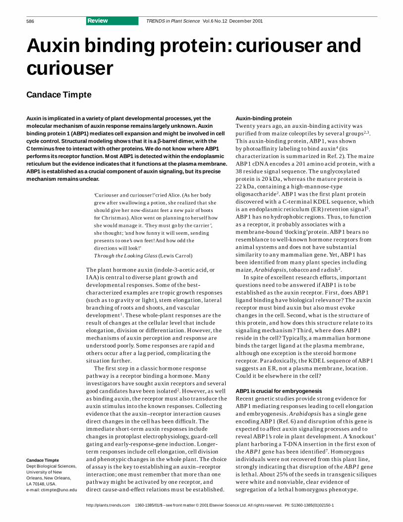
TRENDS in Plant Science
N-term
N-term
C-term
C-term
Fig. 1. Conceptual modelof ABP1. ABP1 is a β-barrelstructure modeled onsimilarity with cupinfamilies and concavalin Afor dimerization. ResidueW44 (red) and the clusterof residues forming theputative metal-bindingsite (blue) might form theplatform for auxin binding.The purple ribbonindicates conservedβ-turn anchor residues.Abbreviations: C-term,C-terminal; N-term,N-terminal.
that ABP1 undergoes a distinct conformational
change when auxin is bound, changing the epitope.
Circular dichroism data indicate a conformational
change upon auxin binding25. Although the
C-terminus did not have sufficient similarity to the
vicilin superfamily to model its location, a C-terminal
tryptophan or WDE sequence might occupy the
binding pocket in the absence of auxin23. The
experimental evidence presented above supports
the importance of the conserved WDE residues.
Conformational changes upon auxin binding might
release the C-terminus for signal propagation and
interaction with other proteins.
Localization versus site of action remains perplexing
The KDEL sequence of ABP1 appears to be effective
at localizing ABP1 to the endoplasmic reticulum (ER).
Paradoxically, the evidence indicates that ABP1 binds
auxin with low affinity at the pH of the ER (Ref. 28).
However, numerous visualization techniques show
that most, but not all, ABP1 is localized to the ER, not
the plasma membrane. ABP1 has been visualized at
the plasma membrane of maize cultured cells by
using immunogold labeling29. ABP1 has also been
detected throughout the Golgi and has been secreted
into culture medium29. A small population of ABP1
molecules has been detected at the plasma membrane
of maize coleoptile protoplasts using silver-enhanced
immunogold epipolarization microscopy30. The
number of ABP1 molecules at the surface was
estimated to be as low as 1000, which represents only
a small proportion of total cellular ABP1. A small
number of cell surface receptors requires less
hormone to achieve half-maximal occupancy.
Physiologically, this would allow the cell to be
sensitive to small amounts of hormone29.
Because antibodies are unlikely to enter intact
cells, the evidence that several antibodies trigger the
plasma membrane hyperpolarization response
indicates that at least some portion of the ABP1 pool
resides at the plasma membrane11,14–16. Because the
antibodies and peptides bind a protein in intact
protoplasts that affects membrane conductivity,
logically they must bind ABP1 localized to the
plasma membrane.
The C-terminal KDEL sequence was changed to
examine its role in the localization of ABP1 (Ref. 17).
KDEL was mutated to HDEL to enhance its retention
in the ER or mutated to either KDELGL or KEQL to
compromise its retention. As expected, the KDEL or
HDEL proteins localized to the ER, whereas the
KEQL and KDELGL proteins entered the Golgi
stacks. However, there was no difference in the cell
surface abundance of ABP1 in cells expressing
mutant proteins as examined by electron microscopy
or silver-enhanced immunogold epipolarization
microscopy17. Thus, even without the KDEL
sequence, quantities of ABP1 do not localize
massively to the plasma membrane.
Similar results confirming the major ER
localization were obtained by analysis of maize
coleoptile rolled leaves31. Neither immunofluorescence
nor immunogold labeling detected ABP1 at the
plasma membrane. Double-labeling experiments to
detect conformational changes that might sequester
the KDEL and allow transit to the plasma
membrane were negative. Under conditions of
auxin binding, ABP1 remained in the ER (Ref. 31).
Carbohydrate analysis further confirmed the ER
localization of ABP1. The ABP1 oligosaccharides
are high mannose types, not the more complex
carbohydrates expected if ABP1 traversed the
Golgi stacks. Less than 2% of ABP, by glycan
analysis, escaped the ER retention system31. At this
low level of escape, a special mechanism to avoid
ER retention need not be invoked. Bulk flow or
association with other proteins might be sufficient
to allow this tiny amount of ABP1 to escape to the
plasma membrane.
The mechanism of KDEL-mediated ER retention
and retrieval operates efficiently, such that an
alternative delivery mechanism to the vacuole was
proposed32. A KDEL terminus was attached to a
protein not normally localized to the ER. This
construct circumvented the Golgi to reach the
vacuole. Some such alternate mechanism might
operate for ABP1. Because overproduction of ABP1
increases the sensitivity of cells to auxin, the
presence of ABP1 at the plasma membrane might be
tightly regulated.
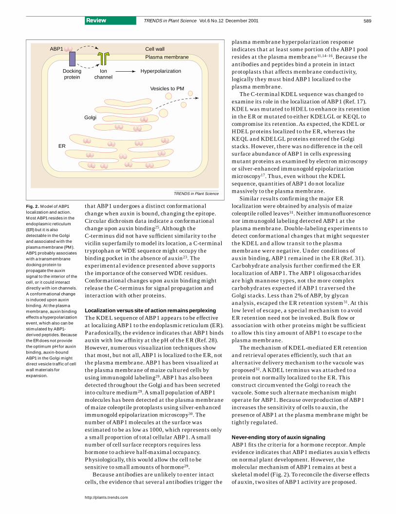
Never-ending story of auxin signaling
ABP1 fits the criteria for a hormone receptor. Ample
evidence indicates that ABP1 mediates auxin’s effects
on normal plant development. However, the
molecular mechanism of ABP1 remains at best a
skeletal model (Fig. 2). To reconcile the diverse effects
of auxin, two sites of ABP1 activity are proposed.
TRENDS in Plant Science Vol.6 No.12 December 2001
http://plants.trends.com
589ReviewReview
TRENDS in Plant Science
Golgi
ER
Dockingprotein
ABP1
Ionchannel
Hyperpolarization
Vesicles to PM
Cell wall
Plasma membrane
Fig. 2. Model of ABP1localization and action.Most ABP1 resides in theendoplasmic reticulum(ER) but it is alsodetectable in the Golgiand associated with theplasma membrane (PM).ABP1 probably associateswith a transmembranedocking protein topropagate the auxinsignal to the interior of thecell, or it could interactdirectly with ion channels.A conformational changeis induced upon auxinbinding. At the plasmamembrane, auxin bindingeffects a hyperpolarizationevent, which also can bestimulated by ABP1-derived peptides. Becausethe ER does not providethe optimum pH for auxinbinding, auxin-boundABP1 in the Golgi mightdirect vesicle traffic of cellwall materials forexpansion.

TRENDS in Plant Science Vol.6 No.12 December 2001
http://plants.trends.com
590 Review
Binding at higher or lower concentrations of auxin
might temper the response. Although ABP1 has no
hydrophobic regions, the auxin-binding signal must
be transmitted to the cell. To this end, ABP1 might
interact with a plasma membrane docking protein
(yet to be identified)3,33 or might interact directly
with the ion channel. Auxin binding induces a
conformational change in ABP1, enabling interaction
with the docking protein, or alters the ABP1–docking-
protein complex to transmit the signal. The docking
protein might be abundant at the plasma membrane;
excess docking protein would then be available to
interact with exogenously provided ABP1 or
peptides in assays, and to mediate membrane
hyperpolarization. Similarly, ER or Golgi-localized
ABP1 might interact with a transmembrane protein
to regulate the secretion of cell wall components to
mediate cell expansion.
Auxin-induced conformational changes in ABP1
might alter interactions with other membrane
proteins, perhaps heterotrimeric G-proteins. Auxin
is known to induce the transcription of several
auxin-regulated genes that are repressed by the
activation of a specific MAPK cascade34.
Furthermore, Arabidopsis cells overproducing the
plant heterotrimeric Gα protein mimic the auxin-
induced increase in cell division35. These results
suggest that the auxin signaling pathway might
involve the Gα protein to regulate cell cycle control.
However, given the varied plant responses to auxin,
there might be more than one type of auxin
receptor in the cell.
It has not been possible to cover all aspects of
auxin signaling in this article. The importance of
ABP1 in plant development is certain but more
pieces of the puzzle remain to be identified. Refining
the location and site of action of ABP1 might require
the use of non-plant in vivo systems, such as the
previously used COS cells28. The proposed docking
protein or other proteins that interact with ABP1
must be identified. Plant development, as the
ultimate result of cell division and cell elongation, is
affected by numerous hormone signals. The
mechanism of integration of these signals and
exactly where ABP1 fits into signaling cross-talk
remain to be discovered.
Review
References
1 Davies, P.J., ed. (1995) Plant Hormones:
Physiology, Biochemistry and Molecular Biology,
Kluwer Academic Publishers
2 Jones, A.M. (1994) Auxin-binding proteins. Annu.
Rev. Plant Physiol. 45, 393–420
3 Klambt, D. (1990) A view about the function of
auxin-binding proteins at plasma membranes.
Plant Mol. Biol. 14, 1045–1050
4 Jones, A.M. and Venis, M. (1989) Photoaffinity
labeling of indole-3-acetic acid binding proteins
in maize. Proc. Natl. Acad. Sci. U. S. A.
86, 6153–6156
5 Pelham, H.R.B. (1988) Evidence that luminal ER
proteins are sorted from secreted proteins in a
post-ER compartment. EMBO J. 7, 913–918
6 Palme, K. et al. (1992) Molecular analysis of an
auxin binding protein gene located on chromosome
4 of Arabidopsis. Plant Cell 4, 193–201
7 Chen, J-G. et al. (2001) ABP1 is required for
organized cell elongation and division in
Arabidopsis embryogenesis. Genes Dev.
15, 902–911
8 Hasezawa, S. and Syono, K. (1983) Hormonal
control of elongation of tobacco cells derived from
protoplasts. Plant Cell Physiol. 24, 127–132
9 Jones, A.M. et al. (1998) Auxin-dependent cell
expansion mediated by overexpressed auxin-
binding protein 1. Science 282, 1114–1117
10 Chen, J-G. et al. The role of auxin-binding
protein I in the cell expansion of tobacco leaf cells.
Plant J. (in press)
11 Barbier-Brygoo, H. (1995) Tracking auxin
receptors using functional approaches. Crit. Rev.
Plant. Sci. 14, 1–25
12 Leblanc, N. et al. (1999) The auxin-binding
protein Nt-Erabp1 alone activates an auxin-like
transduction pathway. FEBS Lett. 449, 57–60
13 Leblanc, N. et al. (1999) A novel immunological
approach establishes that the auxin binding
protein Nt-abp1 is an element involved in auxin
signaling at the plasma membrane. J. Biol. Chem.
274, 28314–28320
14 David, K. et al. (2001) Conformational dynamics
underlie the activity of the auxin binding protein,
Nt-ABP1. J. Biol. Chem. 276, 34517–34523
15 Venis, M.A. et al. (1992) Antibodies to a peptide
from the maize auxin-binding protein have auxin
agonist activity. Proc. Natl. Acad. Sci. U. S. A.
89, 7208–7212
16 Gehring, C.A. et al. (1998) Auxin binding protein
antibodies and peptides influence stomatal
opening and alter cytoplasmic pH. Planta
205, 581–586
17 Bauly, J.M. et al. (2000) Overexpression of
auxin binding protein enhances the sensitivity
of guard cells to auxin. Plant Physiol.
124, 1229–1238
18 Shimomura, S. et al. (1999) Characterization of
auxin-binding protein 1 from tobacco: content,
localization and auxin-binding activity. Planta
209, 118–125
19 Edgerton, M.D. et al. (1994) Modeling the auxin
binding site of auxin binding protein 1 of maize.
Phytochemistry 35, 1111–1123
20 Brown, J.C. and Jones, A.M. (1994) Mapping the
auxin-binding site of auxin-binding protein 1.
J. Biol. Chem. 269, 21135–21140
21 Napier, R.M. and Venis, M.A. (1990) Monoclonal
antibodies detect an auxin-induced
conformational change in the maize auxin-
binding protein. Planta 182, 313–318
22 Anai, T. et al. (1997) Comparison of ABP1
primary sequences from monocotyledonous and
dicotyledonous species. J. Plant Physiol.
151, 446–449
23 Warwicker, J. (2001) Modeling of auxin-binding
protein 1 suggests that its C-terminus and auxin
could compete for a binding site that incorporates
a metal ion and tryptophan residue 44. Planta
212, 343–347
24 Dunwell, J.M. et al. (2000) Microbial relatives
of the seed storage protein of higher plants:
conservation of structure and diversification of
function during evolution of the cupin
superfamily. Microbiol. Mol. Biol. Rev. 153–179
25 Shimomura, S. et al. (1986) Purification and
properties of an auxin-binding protein from
maize shoot membranes. J. Biochem.
99, 1513–1524
26 Woo, E-J. et al. (2000) Crystallization and
preliminary X-ray analysis of the auxin
receptor ABP1. Acta Crystallogr.
D56, 1476–1478
27 Walther, A. et al. (1997) Antibodies against
distinct ABP1 regions modify auxin binding to
ABP1 and change the physiological auxin
response of maize coleoptile sections. J. Plant
Physiol. 150, 110–114
28 Tian, H. et al. (1995) Auxin-binding protein 1 does
not bind auxin within the endoplasmic reticulum
despite this being the predominant subcellular
location for this hormone receptor. J. Biol. Chem.
270, 26962–26969
29 Jones, A.M. and Herman, E.M. (1993)
KDEL-containing auxin binding protein is
secreted to the plasma membrane and cell wall.
Plant Physiol. 101, 595–606
30 Diekman, W. et al. (1995) Auxins induce
clustering of the auxin-binding protein at the
surface of maize coleoptile protoplasts. Proc. Natl.
Acad. Sci. U. S. A. 92, 3425–3429
31 Henderson, J. et al. (1997) Retention of maize
auxin-binding protein in the endoplasmic
reticulum: quantifying escape and the role of
auxin. Planta 202, 313–323
32 Frigerio, L. et al. (2001) Influence of KDEL on the
fate of trimeric or assembly defective phaseolin:
selective use of an alternate route to vacuoles.
Plant Cell 13, 1109–1126
33 Macdonald, H. (1997) Auxin perception and
signal transduction. Physiol. Plant.
100, 423–430
34 Kovtun, Y. et al. (1998) Suppression of auxin
signal transduction by a MAPK cascade in higher
plants. Nature 395, 716–720
35 Ullah, H. et al. (2001) Modulation of cell
proliferation by heterotrimeric G protein in
Arabidopsis. Science 292, 2066–2069
Acknowledgements
I am grateful to BarbaraTriplett, Sarah Lingle,Hee-Jin Kim, Amy Hermanand several anonymousreviewers for comments.Thanks to Jim Nolan forthe structure figure. I thankAlan Jones and CatherinePerrot-Rechenmann forsharing unpublishedresults.