avian adenoviruses
TRANSCRIPT
Rev. sci. tech. Off. int. Epiz., 2000,19 (2), 589-601
Avian adenoviruses J.B. McFerran ( 1 ) & J.A. Smyth (2)
(1)19 Knocktern Gardens, Belfast BT4 3LZ, Northern Ireland, United Kingdom (2) (Corresponding author) Veterinary Sciences Division, Department of Agriculture and Rural Development for Northern Ireland, Stoney Road, Belfast BT4 3SD, Northern Ireland, United Kingdom
Summary Adenov i rus infect ions are ubiqui tous in commerc ia l l y fa rmed birds, and probably in all avian species. There is a w i d e range of v i ru lence , in some cases even w i th in the same serotype. Wh i l e many in fect ions are subc l in ica l and appear to be of l itt le economic or we l fa re impor tance, s igni f icant outbreaks of disease assoc ia ted w i t h adenovi rus do occur. These diseases are not of publ ic heal th s ign i f i cance.
Keywords Adenoviruses - Avian diseases - Egg drop syndrome - Hydropericardium syndrome -Inclusion body hepatitis - Marble spleen disease-Turkey haemorrhagic enteritis.
Introduction Classification From a veterinary viewpoint, the avian adenoviruses can be divided into three groups, i.e. groups I, II and III.
Group I, or conventional adenoviruses, share a common group antigen, distinct from the mammalian adenovirus group antigen. These viruses grow readily in avian cell cultures and have been isolated from chickens, turkeys, geese, ducks, quail, pigeons, ostriches and other avian species. Fowl adenoviruses can be divided into at least twelve serotypes. A major problem in classification has been the presence of prime strains and strains of broad antigenicity. Five groups (A-E) have also been distinguished on the basis of restriction endonuclease analysis using two enzymes (58). The fowl adenoviruses not only infect chickens, but also turkeys and many other species. Turkeys,geese and ducks are affected by adenoviruses that do not grow or only grow poorly in chicken cell cultures and require a homologous cell type. At least three serotypes have been isolated from turkeys, and these grow in turkey but not chicken cells. A study of the relationship between isolates found in the United States of America (USA) and Northern Ireland, and between these turkey isolates and other avian strains, remains to be undertaken.
Three serotypes have been isolated from geese (57) and one from Muscovy ducks (Cairina moschata) (4).
Group II adenoviruses include the viruses of turkey haemorrhagic enteritis (THE), marble spleen disease (MSD) and group II splenomegaly of chickens. These viruses share a
common antigen which is distinct from the group antigen of mammalian and group I avian adenoviruses.
Group III viruses, the egg drop syndrome (EDS) viruses, are widely distributed in waterfowl but can easily infect chickens, resulting in the production of abnormal eggshells.
Until recently, two genera have been recognised within the family Adenoviridae, namely: Mastadenovirus (mammalian strains including human strains) and Aviadenovirus. A third genus has recently been proposed, the genus Atadenovirus (2). Egg drop syndrome virus would be included in the Atadenovirus genus, together with bovine adenovirus 5, 6, 7 and 8, and ovine adenovirus isolate 287. The position of the avian group II (THE/MSD) viruses in this classification is unclear (1 , 1 9 , 2 0 ) .
A number of reviews have described group 1 ( 1 9 , 2 1 , 2 3 , 3 6 ) , group II (18, 31) and group III viruses (18, 20 , 47) . These should be consulted for a full reference listing, as only key and recent references are included here.
Aetiology The adenovirus virion is a non-enveloped icosahedral particle of 70 nm-90 nm in diameter. The particle has 252 capsomeres arranged in twelve triangular faces with six capsomeres along each edge. The nucleic acid is linear, double-stranded deoxyribonucleic acid. The virions have a density in caesium chloride of between 1.32 g/ml and 1.37 g/ml. Adenoviruses replicate in the nucleus, producing basophilic inclusions.
All adenoviruses are resistant to lipid solvents, sodium deoxycholate, trypsin, 2% phenol and 5 0 % alcohol. They are
590 Rev. sci. tech. Off. int. Epiz., 19 (2)
resistant to exposure at pH 3 to pH 9, but are inactivated by 1:1,000 formalin. The avian adenoviruses appear to be more resistant to thermal inactivation than mammalian adenoviruses. Some strains survive 60°C and even 70°C for 30 min, and an F l isolate was reported to survive 18 h at 56°C. At present, information on the effect of divalent cations
- is conflicting. Most workers accept that divalent cations destabilise adenoviruses, but some studies found no effect. Within the group 1 adenoviruses, only some strains of F l agglutinate rat erythrocytes.
Group I (conventional) adenoviruses Epidemiology and pathogenesis Adenoviruses are ubiquitous in chickens, as demonstrated by serological surveys and virological studies, and have been isolated from both sick and healthy birds. Adenoviruses have also been isolated from turkeys, geese, ducks, pigeons, budgerigars and a mallard duck (Anas platyrhynchos). Evidence of adenovirus infection has been recorded in gulls, psittacines, owls and hawks. Infection by adenoviruses is likely to occur in all species of birds.
Transmission Vertical transmission is a very important route. Chicks hatching from infected eggs may excrete virus in faeces from the rime of hatching, but more typically chicks do not excrete virus until two to four weeks of age. Presumably reactivation of latent virus does not occur until maternal antibody declines. In a broiler flock where chicks originate from different parent flocks, a massive interchange of strains occurs, and concurrent infections of one bird with two or even three serotypes is not unusual. Spread of virus in this way results in peak virus excretion in a flock between four and six weeks. In one study of a layer replacement flock, virus excretion was at a maximum between five and nine weeks, but 7 0 % of birds were still excreting after fourteen weeks. In another study, virus excretion again remained at a high level until fourteen weeks, and eight different serotypes were isolated from seven farms. Birds can re-excrete virus throughout life. Following a period of excretion, the virus appears to become latent, presumably due to the development of local immunity. When the local immunity is lost, after eight to twelve weeks, the virus is unmasked and excretion occurs. Humoral antibody does not appear to play a role in preventing excretion, as adult birds have been found to excrete virus despite high levels of neutralising antibody to the same serotype. Humoral antibody appears to offer little or no protection against infection with a different serotype. Adenoviruses are frequently isolated from hens during the period of peak egg production. This upsurge in virus activity ensures maximum transmission of virus to the next generation, through the egg.
Horizontal transmission is also important. The virus is excreted in high titres in the faeces. In addition, virus grows in the nasal and tracheal mucosa, conjunctiva and kidneys, and therefore virus could be present in other secretions or excretions. Virus could also be present in semen, which could be important where artificial insemination is used. Excretion of virus in the faeces follows a different pattern in juveniles and adults. In the juvenile, higher titres of virus are excreted for longer periods than in the adult. Lateral spread appears to occur principally by direct contact between birds or indirect contact by people, crates, egg trays and trolleys. Airborne spread probably only occurs over very short distances. True aerosol spread between farms is highly unlikely, but virus in contaminated poultry litter from a depopulated house could present a risk. In broiler houses, infection appears to spread very rapidly, but this is probably due to reactivation of latent virus in many birds throughout the house. When introduction of virus is minimal, as in a specific-pathogen-free (SPF) flock, spread can be very slow.
Disease A wide range of virulence has been reported within the adenoviruses and the viruses are ubiquitous. Many infections are subclinical, in some cases because birds still have some maternal immunity when infected, but in many cases because the viruses have low virulence. The lack of virulence of some strains is illustrated by the fact that many SPF flocks become infected, even during lay, without any signs being observed. However, because latent adenovirus infections often become apparent at approximately two to three weeks of age, and again around peak egg production (i.e. during periods when disease or production problems are rife), adenoviruses have been associated with a range of conditions such as respiratory disease, diarrhoea, reduced egg production, detrimental effects on feed conversion and arthritis. In most of these conditions, the role of the adenovirus, if any, is that of a helper or secondary pathogen, rather than a primary pathogen. Thus, a study in Denmark was unable to detect any effect of adenoviruses on broiler flock performance (16) . However, adenovirus is an important pathogen in some outbreaks of disease.
Inclusion body hepatit is
Inclusion body hepatitis (IBH) is usually seen in meat-producing birds between three and seven weeks of age, but has also been recorded in birds as young as seven days, and as old as twenty weeks. Classically, IBH is associated with sudden onset mortality which peaks within three to four days and ceases by days five to six, although in some outbreaks, deaths have continued for up to three weeks. Morbidity is low. Affected birds crouch, have ruffled feathers and die or recover within 48 h. Mortality usually ranges between 5% and 10%, but can reach 3 0 % . Within an integrated breeding organisation, disease episodes in broiler flocks have been associated with certain breeder flocks.
Rev. sci. tech. Off. int. Epiz., 19 (2) 591
The liver is the primary organ affected. Some reports suggest that the target organ is the haemopoietic system, but the aplastic anaemia described was probably due to simultaneous infection with chicken anaemia virus. The liver is pale, swollen and friable, and petechial or ecchymotic haemorrhages may be present. Haemorrhages may also be present in the musculature. Numerous eosinophilic intranuclear inclusions, and infrequently basophilic inclusions, are found in the hepatocytes. For many years, the role of adenoviruses in IBH has been unclear. Many serotypes have been associated with outbreaks of IBH. Adenoviruses are observed in the basophilic inclusions, but the eosinophilic inclusions are composed of fibrillar granular material. Experimental reproduction of IBH using adenoviruses has been inconclusive. Most workers have had no success, but some experimental infections have produced liver lesions and death following parenteral inoculation. However, the hepatocyte nuclei contained basophilic inclusions, rather than the eosinophilic inclusions typical of natural outbreaks.
Recent outbreaks of IBH have been described in Australia in birds under three weeks of age. Mortality was up to 3 0 % and basophilic nuclear inclusions predominated in the hepatocytes. Reproduction of the condition was possible using serotypes 6, 7 and 8 isolated from field cases, administered by natural routes. All isolates were genetically closely-related, possessing a group E genotype. The field isolates were further divided into hypervirulent and mildly pathogenic isolates, using nine endonucleases (10 , 29) . Recombination studies indicated that the fibre was responsible for the differences in virulence between isolates (30).
The serotypes isolated from severe outbreaks of IBH in New Zealand were principally F 8 and also F l and F12. In addition to the liver lesions where eosinophilic inclusions predominated, atrophy of the bursa and thymus was reported, together with aplastic bone marrow. These isolates all belonged to genotype E, but were distinct from the genotype found in Australia (40) .
Necrotising pancreatitis and intranuclear inclusions have been observed in natural cases of IBH, and pancreatitis has occurred in experimentally infected chickens. Gizzard erosions and/or ulceration were present, but no intranuclear inclusion bodies were detected in the gizzard epithelial cells in outbreaks of IBH. Focal necrotising pancreatitis and gizzard erosions with typical adenovirus inclusions containing virus particles in necrotic pancreatic acinar cells and gizzard epithelial cells have also been seen in the absence of IBH (49). The latter birds were also infected with chicken anaemia virus. Other workers have also noted gizzard erosions, necrotising pancreatitis and mild proventriculitis with wet unformed faeces, in birds orally infected with adenovirus (17).
Infection with infectious bursal disease virus (IBDV) has been suggested as a major predisposing factor in the development of IBH. However, in New Zealand, and in the early cases in Northern Ireland, IBDV was absent. Furthermore, spontaneous IBH has been reported in SPF birds free of IBDV.
Adenoviral IBH has been recorded in pigeons, kestrels and a merlin (Falco columbarius), and in day-old turkeys from which turkey adenovirus serotype 2 was recovered (15). Pancreatitis was also found in some of the pigeons.
Hydropericardium syndrome In 1987, a new syndrome named hydropericardium syndrome (HPS) or Angara disease was recognised in Pakistan (44). The disease has subsequently been recognised in India, Kuwait, Iraq, Mexico, Central and South America, Japan and Russia. The disease in Central and South America has been diagnosed as IBH (43). Hydropericardium syndrome differs from IBH only in that the mortality rate and the incidence of hydropericardium are much higher.
The disease principally affects meat-producing birds between three and six weeks of age, with mortality from 2 0 % to 8 0 % . Hydropericardium syndrome also occurs in breeding and laying flocks, with lower mortality rates. The disease is characterised by the accumulation of clear fluid (up to 10 ml) in the pericardium. Pulmonary oedema, an enlarged liver and pale enlarged kidneys are usually present. In addition, multifocal coagulative necrosis of the liver is observed, with mononuclear cell infiltration and intranuclear basophilic inclusions in the hepatocytes. The serological response to Newcastle disease vaccination is impaired.
The disease is considered to be the result of infection with adenovirus type 4 or 8 although some workers consider that other factors may be involved (44, 51) .
An HPS-like disease has been reported in pigeons, and broilers injected with liver from affected pigeons developed HPS (26).
Disease in turkeys Adenoviruses have been isolated from clinical outbreaks of respiratory disease, diarrhoea and depressed egg production and more recently, IBH in day-old turkeys (see above). Attempts to reproduce the diseases have generally been unsuccessful.
Disease in wa te r fow l Three serotypes isolated from geese failed to reproduce disease in experimentally infected goslings (57). In a disease outbreak with high mortality associated with hepatitis, adenovirus-like particles were observed in the liver (37).
In Canada, an isolated parent flock produced two hatches in which mortality in four- to eleven-day-old goslings reached 12% due to respiratory tract disease (38).- A diptheritic
592 Rev. sci. tech. Off. int. Epiz., 19 (2)
stenosing tracheitis with occasional bronchitis and pneumonia, in which tracheal epithelial cells contained numerous adenovirus particles, was reported in 1 0 % of seven- to twenty-one-day-old Muscovy ducks (3).
D i s e a s e in g u i n e a - f o w l
Pancreatitis and focal pancreatic necrosis with large basophilic and smaller eosinophilic inclusions have been associated with adenoviral infection of guinea-fowl. Pancreatitis and respiratory lesions have been induced by intranasal inoculation of adenovirus into day-old guinea-fowl. A haemorrhagic disease of guinea-fowl in which adenoviral inclusions were present in the spleen has been reported and reproduced experimentally (25) .
Disease in ostriches Adenoviruses have been associated with illness, diarrhoea, pancreatitis, death and poor hatchability in ostriches. An isolate from an ostrich produced pancreatitis in guinea-fowl (5, 13)- In a study where three-day-old ostrich chicks were inoculated with an ostrich-derived adenovirus, all inoculates died (33).
Quai l b r o n c h i t i s
Quail bronchitis is an acute, highly contagious disease of young bobwhite quail (Colinus virginianus). Disease is most severe in one- to three-week-old birds, with morbidity approaching 100% and mortality up to 5 0 % . Antibody has been detected in older birds and in wild quail. Disease has also been seen in Japanese quail (Coturnix coturnix japonica) (36) .
Quail bronchitis is caused by a type 1 fowl adenovirus which is indistinguishable from chicken isolates. No information is available regarding whether the F l strain behaves in quail as it does in chickens, where latency and vertical transmission occur. Chickens and turkeys may be experimentally infected with isolates from quail, but develop only very mild symptoms of disease.
Gross lesions in quail bronchitis include evidence of ocular and nasal discharge, mucoid tracheitis and airsacculitis. Occasionally, haemorrhagic exudate is present in the trachea. Histologically, a necrotising tracheitis, proliferative and necrotising bronchitis and pneumonia are observed. Basophilic intranuclear inclusions are common in tracheal epithelial cells. Multifocal necrotising hepatitis, splenitis and bursal lymphoid necrosis leading to atrophy are also seen (35).
Other d i s e a s e s in qua i l
Two cases of adenoviral inclusion body ventriculitis have been diagnosed in bobwhite quail (12) . In coturnix quail, gastrointestinal disease with inclusions in the digestive tract, particularly in the caeca, has been reported recently (52).
Diagnosis A detailed methodology has been described in the literature for group I adenoviruses (18, 22) and for quail bronchitis (36).
V i r u s i s o l a t i o n The preferred sample is faeces or colon with faeces. If a particular organ has obvious lesions, for example, the liver in IBH, or the trachea in quail bronchitis, this should also be included. Virus is frequently present in bursa of Fabricius, nasal mucosa, pharynx, trachea, lung and kidney. A 10% suspension of the specimen is made in cell culture media or bacteriological broth. In both cases, antimicrobial agents such as 1,000 international units (IU) of penicillin/ml and 1,000 µg streptomycin/ml should be added. The suspensions can be stored at 4°C or - 2 0 ° C or below until required. Isolation is usually undertaken in cell cultures. For chickens, chick embryo liver or chick kidney cells are best. Chick embryo fibroblasts are insensitive and chick embryo liver cell cultures must show a predominance of epithelial cells. These cells are also suitable for preliminary isolation attempts from other species. However, some adenoviruses that affect turkeys, and probably other avian species, only grow in the cells of homologous species. Therefore, where possible, the homologous cell type should be used, for example, turkey kidney when investigating turkeys. One difficulty is the lack of SPF eggs for most species other than chickens. Because of the widespread distribution of adenoviruses and the presence of virus in eggs, an SPF source is virtually essential. If unavailable, SPF chicken eggs may be the only choice. Following inoculation, the cell cultures should be observed for fourteen days before being discarded. This usually involves one blind passage. Uninoculated cells should be treated in the same manner, to check for the presence of latent virus. Both rolled cultures and flasks are equally sensitive. Frequently, more than one adenovirus serotype, or more than one virus is isolated, for example adenovirus and reovirus. To acquire pure cultures, the use of plaque purification or the limiting dilution techniques often associated with the use of
• neutralising antisera is necessary.
If adenovirus is present, round cells which detach from the glass are observed. As a routine practice, all isolates should be checked for the presence of haemagglutinins, to exclude Orthomyxoviridae and Paramyxoviridae. Adenoviruses of group I and II do not agglutinate fowl erythrocytes. The most rapid method of confirming the presence of adenovirus is indirect immunofluorescence. If available, direct examination of disrupted cell preparations with the electron microscope is also a rapid method of recognition, as the virus morphology is typical. However, if the serotype is to be established, the isolate must be typed against the standard antisera.
Embryonated eggs, inoculated by the allantoic route are not sensitive, except in the case of virus types 1 and 5. Laboratory isolates have been successfully propagated in eggs following inoculation of the yolk sac.
Rev. sci. tech. Off. int. Epiz., 19 (2) 593
Modern biochemical methods can be employed, but are of limited value. Polymerase chain reaction (PCR) techniques may be inappropriate because latency makes it impossible to determine if a positive result is due to the disease currently being investigated or an earlier infection. However, genotyping may be a valuable tool to distinguish between pathogenic and non-pathogenic strains.
Serological detect ion The double immunodiffusion (gel precipitation) test has been widely used. However, the low cost in materials and labour is probably the only advantage of this test. The main disadvantages are lack of sensitivity and detection of group antigen. The test has been used widely to monitor SPF flocks for freedom from adenovirus infection where only group antigen detection is required. However, in many cases, the test has remained negative when birds in SPF flocks have become infected. This has been confirmed by experimental studies which have demonstrated that birds undergoing a primary infection as a result of natural exposure may not respond with precipitin antibodies. The apparent sensitivity of the test in the field is a result of the birds being infected with two or more strains. The sensitivity of the test can be increased by using a pool of antigen prepared from three different serotypes.
The test of choice to monitor SPF flocks is the enzyme-linked immunosorbent assay (ELISA). Little benefit is derived from using a test to detect group antibody in commercial birds, given the widespread extent of infection.
The serum neutralisation test is used to detect type-specific antibody. This is time consuming and expensive, even using the microtitre technique, because a minimum of twelve serotypes must be used when testing chicken sera.
Public health importance Group I adenoviruses do not naturally infect mammals and therefore no public health implications exist.
Prevention and control The widespread distribution of group I adenoviruses throughout the world means that eradication would not be possible. Furthermore, some strains may be able to move between domestic and wild birds. Until recently, development of vaccines has not been a priority because of the absence of important diseases caused by adenoviruses. Since the recent outbreaks of IBH and HPS, development of vaccines has been attempted with varying success. A formalin inactivated liver suspension with liquid paraffin adjuvant is reported to be highly effective against HPS (39). Some other inactivated vaccines have also given good results (44).
No trade implications exist for infections with conventional adenoviruses. Obviously, movement of birds or eggs from flocks infected with the highly virulent viruses associated with HPS or the recent outbreaks of IBH to uninfected areas would
not be wise. However, at present, testing for these conditions is not possible. Thus, type 8 viruses belonging to restriction enzyme group E have been associated with new variant IBH, but similar viruses have also been isolated from normal, healthy birds. The best option is certification that the birds, or in the case of eggs, the parents, have not demonstrated signs of HPS or new variant IBH.
Group II adenoviruses Group II has three known members, namely: turkey haemorrhagic enteritis virus (THEV), marble spleen disease virus (MSDV) and avian adenovirus group II splenomegaly virus (AASV) of chickens. These viruses share a common antigen, which is distinct from that shared by the group I or conventional avian adenoviruses, and from mammalian adenoviruses.
Convalescent THEV serum protects pheasants against MSD. A single serological type of group II viruses appears to exist and isolates are classified only as to the source (e.g. THEV or MSDV). Isolates can be distinguished from one another by restriction endonuclease analysis and monoclonal antibody affinity.
Infectivity resists heating for 1 h at 65°C, but is destroyed after 1 h at 70°C. The viruses demonstrate a wide range of virulence, ranging from highly virulent to non-virulent.
Culture in conventional cell cultures such as turkey kidney or chick embryo liver is not possible. Growth occurs in a turkey lymphoblastoid B cell line derived from a Marek's disease induced tumour, the MDTC-RP19 cell line (28, 54) . Virus has also been grown in turkey peripheral blood leukocytes.
Disease Turkey haemorrhagic enteri t is Turkey haemorrhagic enteritis virus is distributed widely throughout the world. Antibody studies demonstrate that a high proportion of adult domestic turkeys have been infected, although a study of wild turkeys reported no evidence of infection. Guinea-fowl and psittacines may be naturally infected. Other gallinaceous birds such as peafowl, bobwhite quail and chukars can be infected. Lesions develop in the latter, but deaths have not been reported. A serological survey of forty-two species of wild birds indicated no evidence of a reservoir outside the Galliformes (9).
Turkey haemorrhagic enteritis usually occurs in turkeys between six and eleven weeks, although a case has been described in 2.5-week-old poults. Turkeys under thirteen days old appear to be resistant to infection in the absence of maternal immunity, presumably because target cells have not adequately matured. No upper age limit exists for infection.
594 Rev. soi. teck Off. int. Epk, 19 (2]
Transmission is faecal-oral. Virus is présent in faeces forseveral weeks and further bursts of excrétion may occur whenlocal antibody wanes. The virus is very résistant and can easilybe carried from farm to farm by humans. Infection is alsoliable to recur in successive flocks in the same house, unlesscleansing and disinfection is meticulous. No évidence of eggtransmission has been found.
The virus replicates initially in the lymphoid cells of theintestinal tract and bursa of Fabricius. Virus can be detectedone day post infection (dpi), peaks at 4 dpi-7 dpi and remainsdétectable up to 15 dpi in the intestinal tract. Virus isrecoverable from the bursa between 2 dpi and 7 dpi. Virus isprésent in plasma from 2 dpi, and virus replicates are detectedin the blood leukocytes from 3 dpi to 18 dpi. The spleen is themajor site of viral replication. Antigen is détectable in thespleen from 2 dpi, reaches a peak at 6 dpi and is no longerdétectable at 18 dpi (27). Reports as to the amount of antigenin the intestine are conflicting, and the intestinal pathologymay be immune-mediated (48). Apoptosis occurs inapproximately half of the immunoglobulin M+ cells at 3 dpibut not in cluster of differentiation 4+ (CD4+) and CD8+T lymphocytes, and occurrence of apoptosis is not restrictedto infected cells. The rôle of apoptosis in the pathogenesis ofTHEV is not clear, but this may be the cause of theimmunosuppression (34, 48).
Experimentally, the incubation period is five to six daysfollowing oral infection. In natural outbreaks, virtually ailbirds become infected, as demonstrated by the developmentof antibodies. Mortality ranges from zéro to over 60% with anaverage of 10%-15%.
Classically, the onset of disease is sudden. Birds are depressed,hâve bloody droppings and may die suddenly. Death usuallyoccurs within 24 h of the appearance of the first signs ofdisease, or the bird recovers. Signs of disease within a flocklast approximately six to ten days. Outbreaks due to lessvirulent strains are less spectacular. AU strains, includingthose previously thought to be apathogenic, areimmunosuppressive. Therefore, infection with THEV mayallow paramyxovirus type II, Chlamydia, Staphylococcus andE. coli to cause disease (11).
In a breeding organisation in Northern Ireland which has avery high standard of hygiène, breeding turkeys remaineduninfected until commencement of lay. The turkeysdeveloped a clinical condition similar to EDS, with loss ofeggshell colour, thin shelled and shell-less eggs, and this wasassociated with seroconversion to THEV. The birds remainedapparently healthy.
Birds which hâve died from THE are often pale due to bloodloss. Sudden death is often indicated by feed in the crop andgood body condition of the carcass. The small intestine isusually distended, the mucosa is congested and the lumen
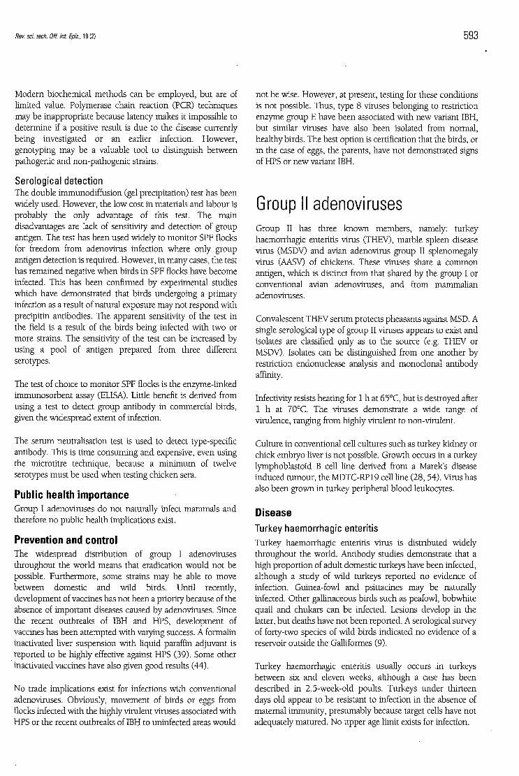
filled with feed and blood (Fig. 1). In some cases, a yellowfibrinonecrotic membrane may be présent. The lésions aremore prominent in the proximal small intestine. If sick birdsare killed, the spleens are found to be enlarged, friable andmarbled or mottled. Where birds hâve died as a resuit ofinfection, the spleens tend to be smaller and the mottling isless apparent.
Fig. 1Duodénum from a turkey with haemorrhagic enteritisThe upper spécimen shows a less acute form
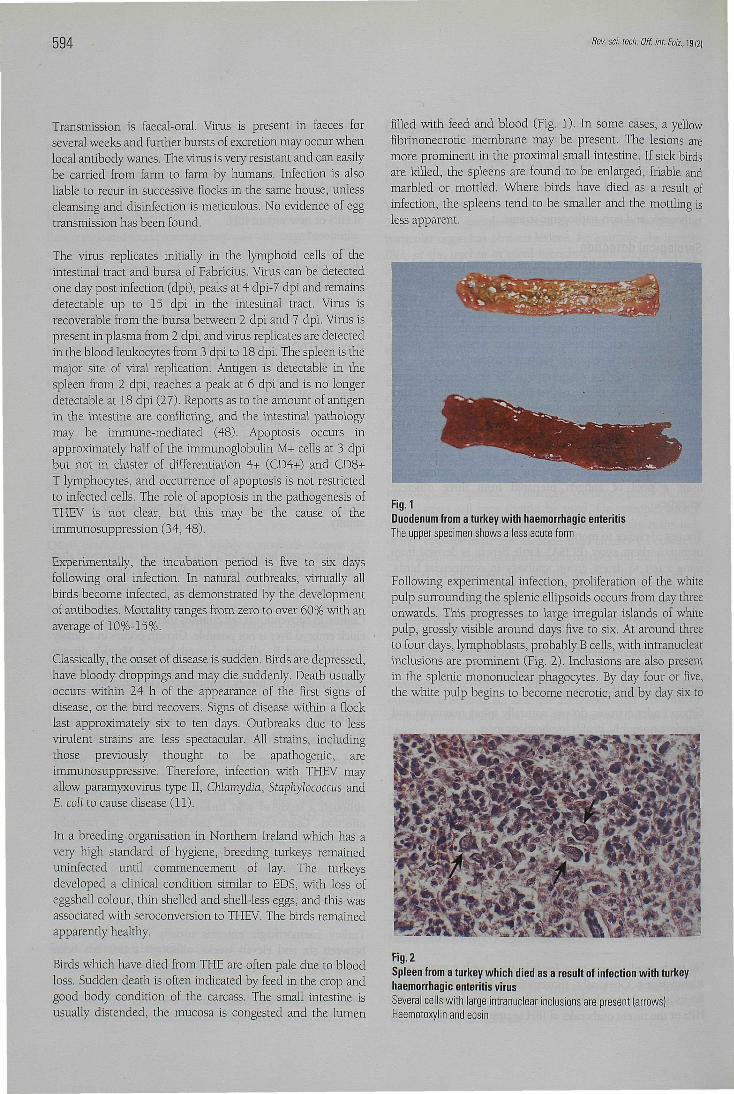
Following expérimental infection, prolifération of the whitepulp surrounding the splenic ellipsoids occurs from day threeonwards. This progresses to large irregular islands of whitepulp, grossly visible around days five to six. At around threeto four days, lymphoblasts, probably B cells, with intranuclearinclusions are prominent (Fig. 2). Inclusions are also présentin the splenic mononuclear phagocytes. By day four or five,the white pulp begins to become necrotic, and by day six to
Fig. 2Spleen from a turkey which died as a resuit of infection with turkeyhaemorrhagic enteritis virusSeveral cells with large intranuclear inclusions are présent (arrows)Haemotoxylin and eosin
Rev. sel tech. Off. int. Epiz., 19 (2) 595
seven the white pulp is completely involuted with onlyoccasional plasma cells appearing in the red pulp. Lymphoiddepletion also occurs in the thymus and bursa of Fabriciusbetween days three and nine (27).
Severe congestion of the intestinal mucosa, degeneration andsloughing of the villous epithelium and haemorrhages in thevillous tips are also observed. One group reported that theblood vessels in the lamina propria were intact and theerythrocytes appeared to escape from the vessels bydiapedesis (41). Increased numbers of lymphoreticular cellswith intranuclear inclusions are présent in the lamina propria,in addition to mast cells, plasma cells and heterophils.
DiagnosîsThe spleen is the preferred organ for virus isolation, but faecesalso contain large amounts of virus. The lymphoblastoidB-cell Une of turkeys (MSTC-RP19) is inoculated. If cellculture is not available, then five- to ten-week-oldantibody-free turkeys can be given material orally or by theintravenous route. Birds usually die approximately three daysafter the intravenous injection and five or six days after oralinfection. Birds which are infected but still alive at six daysusually hâve enlarged spleens.
Traditionally, diagnosis has been made using affected spleenas antigen in a double immunodiffusion test. More sensitivetests such as the immunofluorescent test, ELISA, restrictionendonuclease and PCR are now being used increasingly.
Antibody can be first detected three to four days after infectionusing the ELISA. This antibody is long lasting; in one flock,83% of the birds were still positive forty months after initialtesting. Due to lower sensitivity, the double immunodiffusiontest becomes positive only after two weeks. Further détails areprovided by Pierson et al. (32).
Enlargement of the spleen in turkeys can be caused by THEV,but can also be due to reticuloendotheliosis orlymphoproliferative diseases. Blood in the intestine gives astrong indication of THE, and démonstration of antigen in thespleen provides the proof.
Vaccines are used in many areas. A tissue culture attenuatedvaccine has been used extensively, but such vaccines hâvebeen reported to be immunosuppressive. Vaccines derivedfrom the spleen of birds with THE or MSD hâve been used asvaccines, but both types are also immunosuppressive. Arecombinant fowl pox vaccine which afforded goodprotection under laboratory conditions and which did notcause immunosuppression has recently been reported (6).
Marble spleen diseaseMarble spleen disease is observed in pheasant productionopérations throughout the world. Marble spleen diseaseoccurs naturally in three- to eight-month-old birds, but hasbeen experimentally reproduced in adult pheasants. Infection,
as indicated by antibody development, approaches 100%.Birds are often found dead, but dépression, weakness, nasaldischarge and dyspnoea may also be observed. Mortalityranges from 2% to 20%, usually occurring over a period oftento fourteen days, but can continue for several weeks.
Antigen is présent in spleen, liver, lung, bone marrow andkidney, but in contrast to THE infection, no antigen isdétectable in the intestine (11). Bursectomy protects againstthe disease and an age-related résistance occurs below sixweeks of âge, which is unconnected with the présence ofmaternai antibody. This indicates the importance of Blymphocytes in the disease process. T lymphocytes areimportant in controlling MSD infection (11). Infection withMSD impairs bodi the humoral and cell mediated responses.The effect on the humoral response is more pronounced andlasts several weeks.
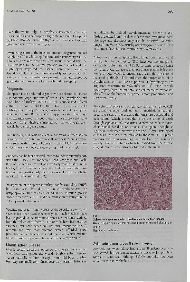
The spleens of pheasants which hâve died as a resuit of MSDare usually enlarged and mottled or marbled. In naturallyoccurring cases of the disease, the lungs are congested andoedematous (which is thought to be the cause of deaththrough asphyxiation). In contrast to HEV, no évidence existsof intestinal bleeding or lésions. The splenic weight issignificantly elevated between 6 dpi and 10 dpi. Histologicalchanges in the spleen are similar to those in THE. Splenicnecrosis and numerous large intranuclear inclusions areusually observed in birds which hâve died from the disease(Fig. 3). Necrosis may also be observed in the lungs.
Fig.3Spleen from a pheasant which died from marble spleen diseaseNecrosis (N) and numerous cells containing large intranuclear inclusions arevisibleHaemotoxylin and eosin
Avian adenovirus group II splenomegalyAntibody to avian adenovirus group II splenomegaly iswidespread, but associated disease is not a major problem.Mortality is unusual, although 8%-9% mortality has beenrecorded in mature chickens.
596 Rev. sci. tech. Off. int Epiz., 19 (2]
Infection is recognised as splenomegaly in broilers at slaughterand as splenomegaly with pulmonary oedema/congestion inadults. The disease can be important as a cause ofcondemnation at slaughter because of enlarged spleens.Histologically, the splenic lésions are similar to those reportedin THE.
Public health importanceThe group II adenoviruses pose no threat to public health, asno record exists of infection of mammals by thèse viruses.
ControlThe viruses are widely distributed throughout the world andtherefore import restrictions would not be justified.
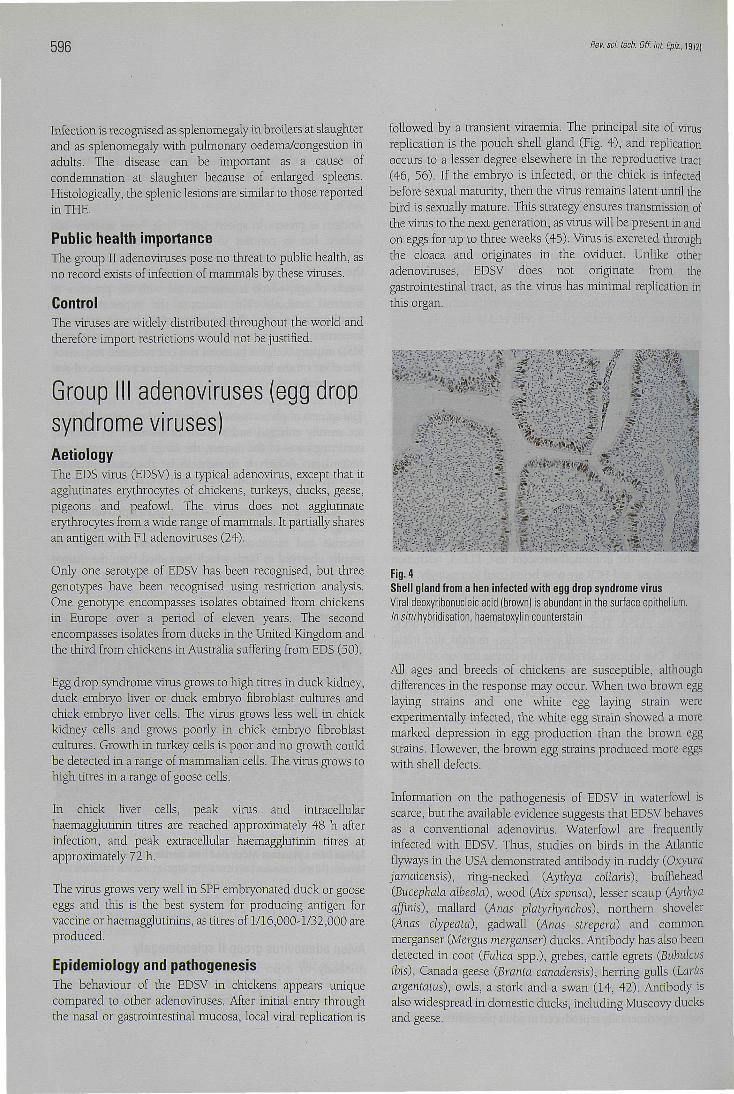
followed by a transient viraemia. The principal site of virusreplication is the pouch shell gland (Fig. 4), and replicationoccurs to a lesser degree elsewhere in the reproductive tract(46, 56). If the embryo is infected, or the chick is infectedbefore sexual maturity, then the virus remains latent until thebird is sexually mature. This strategy ensures transmission ofthe virus to the next génération, as virus will be présent in andon eggs for up to three weeks (45). Virus is excreted throughthe cloaca and originates in the oviduct. Unlike otheradenoviruses, EDSV does not originate from thegastrointestinal tract, as the virus has minimal replication inthis organ.
Group III adenoviruses (egg dropsyndrome viruses)AetiologyThe EDS virus (EDSV) is a typical adenovirus, except that itagglutinâtes erythrocytes of chickens, turkeys, ducks, geese,pigeons and peafowl. The virus does not agglutinateerythrocytes from a wide range of mammals. It partially sharesan antigen with FI adenoviruses (24).
Only one serotype of EDSV has been recognised, but threegénotypes hâve been recognised using restriction analysis.One génotype encompasses isolâtes obtained from chickensin Europe over a period of eleven years. The secondencompasses isolâtes from ducks in the United Kingdom andthe third from chickens in Australia suffering from EDS (50).
Egg drop syndrome virus grows to high titres in duck kidney,duck embryo liver or duck embryo fibroblast cultures andchick embryo liver cells. The virus grows less well in chickkidney cells and grows poorly in chick embryo fibroblastcultures. Growth in turkey cells is poor and no growth couldbe detected in a range of mammalian cells. The virus grows tohigh titres in a range of goose cells.
In chick liver cells, peak virus and intracellularhaemaggrutinin titres are reached approximately 48 h afterinfection, and peak extracellular haemagglutinin titres atapproximately 72 h.
The virus grows very well in SPF embryonated duck or gooseeggs and this is the best System for producing antigen forvaccine or haemagglutinins, as titres of 1/16,000-1/32,000 areproduced.
Epidemiology and pathogenesisThe behaviour of the EDSV in chickens appears uniquecompared to other adenoviruses. After initial entry throughthe nasal or gastrointestinal mucosa, local viral replication is
Fig. 4Shell gland from a hen infected with egg drop syndrome virusViral deoxyribonucleic acid (brown) is abundant in the surface epithelium.In situ hybridisation, haematoxylin counterstain
AU âges and breeds of chickens are susceptible, althoughdifférences in the response may occur. When two brown egglaying strains and one white egg laying strain wereexperimentally infected, the white egg strain showed a moremarked dépression in egg production than the brown eggstrains. However, the brown egg strains produced more eggswith shell defects.
Information on the pathogenesis of EDSV in waterfowl isscarce, but the available évidence suggests that EDSV behavesas a conventional adenovirus. Waterfowl are frequentlyinfected with EDSV. Thus, studies on birds in the Atlanticflyways in the USA demonstrated antibody in ruddy (Oxyurajamaicensis), ring-necked (Aythya collaris), bufflehead(Bucephala albeold), wood (Abc sponsa), lesser scaup (Aythyaaffinis), mallard (Anas platyrhynchos), northem shoveler(Anas clypeatà), gadwall (Anas strepera) and commonmerganser (Mergus merganser) ducks. Antibody has also beendetected in coot (Fulica spp.), grèbes, cattle egrets (Bubulcusibis), Canada geese (Branta canadensis), herring gulls (Larùsargentatus), owls, a stork and a swan (14, 42). Antibody isalso widespread in domestic ducks, including Muscovy ducksand geese.
Bev. sci. tecb. Off. int. Epiz., 19 (2) 597
Quail are susceptible and develop classical clinical signs (8).Although turkeys and pheasants can be experimentallyinfected, no signs of disease are observed. Guinea-fowl may benaturally infected and develop typical signs. However, in onestudy, guinea-fowl failed to show signs of disease after beinginfected with a fowl isolate (55).
Three syndromes are associated with EDS. The classical formwas seen when primary breeding stock became infected.Chicks derived from thèse flocks remained healthy and didnot produce antibody until reaching sexual maturity. At sometime between the onset of egg laying and peak production,abnormal eggs were produced and the birds producedantibody. This infection probably initially arose from the useof a vaccine grown in duck cells which contained latent EDSV(18). Infection has since been eradicated from primarychicken breeding stock (18). However, the viras subsequentlyinfected commercial egg-producing flocks and has becomeendémie in some areas. This is primarily due to the présenceof virus on the exterior of eggs, leading to contamination oftrays and trolleys. In many cases, this equipment is notadequately cleaned or disinfected before being returned fromthe egg packing plants to other faims at random. Infection canalso be transmitted from flock to flock by humans, such asgroup advisory staff and workers servicing equipment.
The third category is the sporadic outbreak. This occurs whenchickens corne into contact with domestic or wild waterfowl.Contact may be direct or through contaminated drinkingwater. Thèse outbreaks are self-limiting unless infection isspread to other flocks, when the outbreaks become the focusof an endémie cluster.
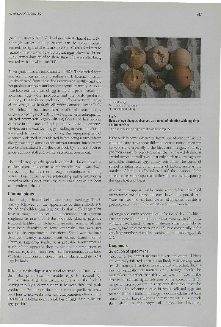
Clinical signsThe first sign is loss of shell colour in pigmented eggs. This isquickly followed by the appearance of thin shelled, softshelled or shell-less eggs (Fig. 5). The thin shelled eggs oftenhâve a rough sandpaper-like appearance or a granularroughness at one end. If the obviously affected eggs areremoved, fertility and hatchability are not affected. Small eggshâve been described in some outbreaks, but were notreported in expérimental infections. Some workers hâvedescribed watery albumen, but others found normalalbumen. Egg drop syndrome is probably a misnomer asmuch of the apparent drop is due to the production ofshell-less eggs, as the numbers of shell membranes in the litterwill testify, and consumption of the thin-shelled and shell-lesseggs by birds.
If the disease develops as a resuit of réactivation of latent virusthen the production of usable eggs is reduced byapproximately 40%; this usually occurs when the flock iscoming into lay and production is between 50% and peakproduction. Production does not return to predicted levelsuntil four to ten weeks later and compensation often occurslater in lay resulting in an overall loss of eggs of ten to sixteeneggs per bird.
S : shell-less eggM : ruptured shell membraneN : normal pigmented egg
Fig. 5Range of egg changes observed as a resuit of infection with egg dropsyndrome virusPale and thin-shelled eggs are shown in the top row
If the birds become infected by latéral spread when in lay, theclinical picture may appear différent because transmission canbe very slow, especially if the birds are in cages. Poor eggproduction may be reported rather than a marked décline. Acareful inspection will reveal that only birds in a few cages areproducing abnormal eggs at any one time. The speed ofspread is influenced by a number of factors, such as thenumber of birds initially infected and the position of theaffected cages with respect to the flow of the belts transportingthe eggs, feed and faeces.
Affected birds appear healthy; some workers hâve describedinappétence and dullness but most hâve not reported this.Transient diarrhoea has been described by some, but this isprobably exudate and fluid excrétion from the oviduct.
Although one study reported oral infection of day-old chickscausing increased mortality in the first week of life (7), mostworkers hâve found no signs of disease either in chicks orgrowing birds infected with virus (47), or commercially in thevery large numbers of chicks hatching from infected eggs (18,21).
DiagnosisSélection of spécimensSélection of the correct spécimen is very important. If birdsare vertically infected, then no antibody will develop untilsexual maturity. Therefore, to certify that a breeding flock isfree of vertically transmitted virus, testing should beundertaken no earlier than thirty-two weeks of âge. In theabsence of clinical signs, sélection of the correct bird forsampling poses a problem. In a cage unit, this problem can beovercome by selecting a cage in which affected eggs areprésent. If ail the birds in the cage are examined, then at leastsome birds will hâve antibody and may hâve virus. The pouchshell gland is the organ of choice for histology,
598 Rev. sci. tech. Off. int. Epiz., 19 (2)
immunochemistry or virus isolation, but pathognomonic lesions and viral antigen are present only for a short time. If blood is to be collected for serology, then the birds bled should be those from the cages in which defective eggs have been produced for the longest time. On litter, the problem is more difficult. To isolate virus, or to detect antigen or lesions, the simplest method is to feed affected eggs to antibody-free hens held individually in cages. The eggs produced by these birds should be examined daily and testing should be performed when a bird produces abnormal eggs. Examination of randomly selected cloacal swabs has been successful in some cases.
S e r o l o g i c a l t e s t s The haemagglutination inhibition (HI) test is the method of choice. A 1/10 serum dilution is mixed with an equal volume of a solution containing four haemagglutinating units of antigen. The mixture is allowed to react for 15 min at room temperature and then one volume of an 0.8% fowl erythrocyte suspension is added. Other tests, such as the ELISA and serum neutralisation, are available, but the HI test is rapid, inexpensive and accurate.
V i r u s i so l a t i on A 1 0 % suspension is made from the pouch shell gland and the supernatant inoculated onto cell cultures or embryonated duck eggs. Suitable cells, in order of preference, are duck cells, chick embryo liver or chick kidney cells. At least fourteen days incubation (one blind passage) are required after inoculation. If the cells degenerate, the supernatant should be checked for the presence of haemagglutinins using a 0.8% fowl erythrocyte suspension. If agglutination occurs, the isolate can be confirmed by an HI test using specific antiserum.
A n t i g e n d e t e c t i o n Antigen can be detected in the pouch shell gland, during the time that defective eggs are produced, using immunofluorescent techniques on frozen sections or the avidin-biotin-peroxidase technique on formalin fixed tissue sections. In situ hybridisation may also be used.
Public health importance Infection with EDSV has no public health significance.
Control Basic breeding stock should be free of infection, and many breeding organisations are free at all levels. Given that EDSV is transmitted vertically and that birds do not develop antibody until sexual maturity, certification of freedom from vertically transmitted virus is not possible until the flock has been in lay for a number of weeks. The HI test is satisfactory, but the
appropriate time for blood testing varies according to the type of parent. For broiler breeders, sampling at approximately thirty weeks of age would be acceptable, although thirty-five weeks was chosen in eradication programmes to allow a generous safety margin. Given the severe economic effects of the disease and the difficulty of excluding the virus if using an egg packing station serving infected flocks, many commercial egg producers routinely vaccinate flocks using a commercial inactivated vaccine which is very effective in controlling disease when administered correctly. Apparent failure of vaccines to protect appears to be due to poor vaccination techniques (53).
Risks from imported eggs and processed chickens Since avian adenoviruses may be vertically transmitted, imported hatching eggs could give rise to infected chicks. Although adenoviruses occur world-wide, diseases such as hydropericardium syndrome do not. Therefore prudent measures would include checking the history of the supply flock, and rejection of eggs from a region or organisation where serious adenoviral diseases are present. Similarly, non-fertile eggs and hatchery waste eggs may also be infected, and these should not be recycled into poultry food.
Viraemia usually occurs in the early stages of adenovirus infections, and since adenovirus may be found in many visceral organs, adenoviruses could theoretically be present in processed chicken. However, flocks infected with significant adenoviruses will show evidence of disease and accordingly should not be slaughtered for human consumption. Therefore, the risk of importing very pathogenic adenoviruses with processed chicken should be low. Adenoviruses have been recovered from the faeces of normal chickens and the potential exists for contamination of carcasses in the processing plant. However, while adenoviruses may remain viable for some time, in contrast to some significant bacterial contaminants, the virus will not multiply on the carcass. Thus, while the risk of acquiring significant infection from uncooked poultry meat or offal appears small, care should nonetheless be taken to ensure that such poultry meat or offal are not recycled to avian species.
Rev. sci. tech. Off. int. Epiz., 19 (2) 599
Adénovirus aviaires J.B. McFerran &J.A. Smyth
Résumé Les adénovirus sont des agents pathogènes ubiquistes a f fec tant les oiseaux des élevages industr ie ls, et probablement tou tes les espèces av iennes. La v i ru lence var ie cons idérab lement d'une souche à l 'autre, parfois au sein du même sérotype. Nombre d ' in fect ions sont inapparentes et leur inc idence économique ou sani ta i re semble fa ib le ; tou te fo is , les adénovi rus peuvent être à l 'or igine de graves épizooties. Ces dern ières sont sans conséquence pour la santé publ ique.
Mots-clés Adénovirus - Entérite hémorragique du dindon - Hépatite à corps d'inclusion - Maladie de la rate marbrée - Maladies aviaires - Syndrome de la chute de ponte - Syndrome de l'hydropéricarde.
Adenovirus aviares J.B. McFerran & J.A. Smyth
Resumen Las in fecc iones por adenovi rus son ubicuas entre las aves de explotac ión comerc ia l , e incluso posib lemente entre todas las especies de aves. Existen niveles de v i ru lenc ia muy var iab les, a veces inc luso para un mismo serot ipo. Aunque muchas de las in fecc iones son de carác ter subcl ín ico y parecen i r re levantes en términos económicos o de bienestar de las aves, a veces se dec laran brotes in fecc iosos de cierta cons iderac ión asociados a la presencia de adenovi rus. Estas enfermedades son de importancia menor en lo que a salud públ ica se ref iere.
Palabras clave Adenovirus - Enfermedad del bazo jaspeado - Enfermedades aviares - Enteritis hemorrágica del pavo - Hepatitis por cuerpos de inclusión - Síndrome de caída del huevo - Síndrome de hidropericardio.
References
1. Aghakhan S.M. (1974). - Avian adenoviruses. Vet. Bull, 44, 531-552.
2. Benko M. & Harrach B. (1998). - A proposal for a new (third) genus within the family Adenoviridae. Arch. Virol., 143, 829-837.
3. Bergmann V., Heidrich R. & Kinder E. (1985). -Pathomorphologische und - elektronenmikroskopische
Feststellung einer Adenovirus-Tracheitis bei Moschusenten (Cairina moschata). Monatsch. VetMed., 40 (9), 313-315.
4. Bouquet J.F., Moreau Y., McFerran J.B. & Connor T.J. (1982). - Isolation and characterisation of an adenovirus isolated from Muscovy ducks. Avian Pathol, 11, 301-307.
5. Capua I., Gough R.R., Scaramozzino P., Lelli R. & Gatti A. (1994). - Isolation of an adenovirus from an ostrich (Struthio

600 Rev. sci. tech. Off. int. Epiz.. 19 (2)
camelus) causing pancreatitis in an experimentally infected guinea fowl (Numida meleagris). Avian Dis., 38, 642-646.
6. Cardona C.J., Reed W.M., Witter R.L. & Silva R.F. (1999). -Protection of turkeys from hemorrhagic enteritis with a recombinant fowl poxvirus expressing the native hexon of hemorrhagic enteritis vims. Avian Dis., 43, 234-244.
7. Cook J.K.A. & Darbyshire J.H. (1981). - Longitudinal studies on the egg drop syndrome 1976 (EDS 76) in the fowl following experimental infection at 1-day old. Avian Pathol., 10, 449-459.
8. Das B.B. & Pradhan H.K. (1992). - Outbreaks of egg drop syndrome due to EDS-76 virus in quail (Coturnix coturnix japonica). Vet. Rec., 131 (12), 264-265.
9. Domermuth C.H., Forrester D.J., Trainer D.O. & Bigler W.J. (1977). - Serologic examination of wild birds for hemorrhagic enteritis of turkey and marble spleen disease of pheasants. J. Wildl. Dis., 13, 405-408.
10. Erny K.M., Barr D.A. & Fahey K.J. (1991). - Molecular characterisation of highly virulent fowl adenoviruses associated with outbreaks of inclusion body hepatitis. Avian Pathol, 20, 597-606.
11. Fitzgerald S.D. & Reed W.M. (1996). - Recent advances in understanding the pathogenesis of marble spleen disease vims infection. in Proc. International Symposium on adenovirus and reovirus infections in poultry (E.F. Kaleta & U. Heffels-Redmann, eds), 24-27 June, Rauischolzhausen, Germany. Institut für Geflügelkrankheiten, Justus-Liebig-Universität, Giessen, 105-108.
12. Goodwin M.A. (1993). - Adenovirus inclusion body ventriculitis in chickens and captive bobwhite quail (Colinus virginianus). Avian Dis., 37, 568-571.
13. Gough R.E., Drury S.E., Capua I., Courteney A.E., Sharp M.W. & Dick A.C.K. (1997). - Isolation and identification of adenoviruses from ostriches (Struthio camelus). Vet. Ree, 140, 402-403.
14. Gulka C.M., Piela T.H., Yates V.J. & Bagshaw C. (1984). -Evidence of exposure of waterfowl and other aquatic birds to the hemagglutinating duck adenovirus identical to EDS 76 virus. J . Wildl. Dis., 20, 1-5.
15. Guy J.S. & Barnes H.J. (1997). - Characterization of an avian adenovirus associated with inclusion body hepatitis in day-old turkeys. Avian Dis., 41 , 726-731.
16. Jorgensen P.H., Otte L., Nielsen O.L. & Bisgaard M. (1995). - Influence of subclinical virus infections and other factors on broiler flock performance. Br. Poult. Sci., 36, 455-463.
17. Lenz S.D., Hoerr F.J., Ellis A.C., Toivio-Kinnucan M.A. & Yu M. (1998). - Gastrointestinal pathogenicity of adenoviruses and reoviruses isolated from broiler chickens in Alabama.J. vet. diagn. Invest., 10, 145-151.
18. McCracken R.M. & Adair B.M. (1993). - Adenoviridae. In Vims infection of birds (J.B. McFerran & M.S. McNulty, eds). Elsevier Science Publishers BV, Amsterdam, 123-144.
19. McFerran J.B. (1981). - Adenoviruses of vertebrate animals. In Comparative diagnosis of viral diseases, Volume III (E. Kurstak & C. Kurstak, eds). Academic Press, New York, 102-165.
20. McFerran J.B. (1997). - Egg drop syndrome. In Diseases of poultry, 10th Ed. (B.W. Calnek with H.J. Barnes, C.W. Beard, L.R. McDougald & Y.M. Saif, eds). Iowa State University Press, Ames, 633-642.
21. McFerran J.B. (1997). - Group I adenovirus infections. In Diseases of poultry, 10th Ed. (B.W. Calnek with H.J. Barnes, C.W. Beard, L.R. McDougald & Y.M. Saif, eds). Iowa State University Press, Ames, 607-620.
22. McFerran J.B. (1998). - Adenoviruses. In A laboratory manual for the isolation and identification of avian pathogens, 4th Ed. (D.E. Swayne, J.R. Glisson, M.W. Jackwood, J.E. Pearson & W.M. Reed, eds). American Association of Avian Pathologists, Kennett Square, Pennsylvania, 100-106.
23. McFerran J.B. & Adair B.M. (1977). - Avian adenoviruses - a review. Avian Pathol., 6, 189-217.
24. McFerran J.B., Connor T.J. & Adair B.M. (1978). - Studies on the antigenic relationship between an isolate (127) from the egg drop syndrome 1976 and a fowl adenovirus. Avian Pathol, 7, 629-636.
25. Massi P., Gelmetti D., Sironi G., Dottori M., Lavazza A. & Pascucci S. (1995). - Adenovirus associated haemorrhagic disease in guinea fowl. Avian Pathol, 24, 227-237.
26. Naeem K. & Akram H.S. (1995). - Hydropericardium syndrome in a pigeon flock. Vet. Rec., 136, 296-297.
27. Nagaraja KV. & Hussain I. (1996). - Pathogenesis of hemorrhagic enteritis vims (adenovirus II) infection in turkeys. In Proc. International Symposium on adenovirus and reovirus infections in poultry (E.F. Kaleta & U. Heffels-Redmann, eds), 24-27 June, Rauischolzhausen, Germany. Institut für Geflügelkrankheiten, Justus-Liebig-Universität, Giessen, 51-59.
28. Nazerian K. & Fadly A. (1982). - Propagation of virulent and avirulent turkey hemorrhagic enteritis virus in cell culture. Avian Dis., 26, 816-827.
29. Pallister J.A. & Sheppard M. (1996). - Comparison by restriction enzyme analysis of three fowl adenoviruses of varying pathogenicity. Vet. Microbiol, 48, 155-163.
30. Pallister J.A., Wright P.J. & Sheppar M. (1996). - A single gene is responsible for virulence in the fowl adenoviruses. In Proc. International Symposium on adenovirus and reovirus infections in poultry (E.F. Kaleta & U. Heffels-Redmann, eds), 24-27 June, Rauischolzhausen, Germany. Institut für Geflügelkrankheiten, Justus-Liebig-Universität, Giessen, 24-25.
31. Pierson F.W. & Domermuth C.H. (1997). - Hemorrhagic enteritis, marble spleen disease and related infections. In Diseases of poultry, 10th Ed. (B.W. Calnek with H.J. Barnes, C.W. Beard, L.R. McDougald & Y.M. Saif, eds). Iowa State University Press, Ames, 624-633.
Rev. sci. tech. Off. int. Epiz., 19 (2) 601
32. Pierson F.W., Domermuth C.H. & Gross W.B. (1998). -Hemorrhagic enteritis of turkeys and marble spleen disease of pheasants. In A laboratory manual for the isolation and identification of avian pathogens, 4th Ed. (D.E. Swayne, J.R. Glisson, M.W. Jackwood, J.E. Pearson & W.M. Reed, eds). American Association of Avian Pathologists, Kennett Square, Pennsylvania, 106-110.
33. Raines A.M., Kocan A. & Schmidt R. (1997). - Experimental inoculation of adenovirus in ostrich chicks (Struthio camelus). J. avian Med. Surg., 11, 255-259.
34. Rautenschlein S., Suresh M., Neumann U. & Sharma J.M. (1996). - Pathogenic mechanisms of avian adenovirus type II. In Proc. International Symposium on adenovirus and reovirus infections in poultry (E.F. Kaleta & U. Heffels-Redmann, eds), 24-27 June, Rauischolzhausen, Germany. Institut für Geflügelkrankheiten, Justus-Liebig-Universität, Giessen, 41-50.
35. Reed W.M. & Jack S.W. (1996). - Quail bronchitis: pathology and pathogenesis. In Proc. International Symposium on adenovirus and reovirus infections in poultry (E.F. Kaleta & U. Heffels-Redmann, eds), 24-27 June, Rauischolzhausen, Germany. Institut für Geflügelkrankheiten, Justus-Liebig-Universität, Giessen, 98-104.
36. Reed W.M. & Jack S.W. (1997). - Quail bronchitis. In Diseases of poultry, 10th Ed. (B.W. Calnek with H.J. Barnes, C.W. Beard, L.R. McDougald & Y.M. Saif, eds). Iowa State University Press, Ames, 620-624.
37. Riddell C. (1984). - Vims hepatitis in domestic geese in Saskatchewan. Avian Dis., 28, 774-782.
38. Riddell C , van den Hurk J.V., Copeland S. & Wobeser G. (1992). - Virus tracheitis in goslings in Saskatchewan. Avian Dis., 36, 158-163.
39. Roy P., Koteeswaran A. & Manickam R. (1999). - Efficacy of an inactivated oil emulsion vaccine against hydropericardium syndrome in broilers. Vet. Rec., 145, 458-459.
40. Saifuddin M., Wilks C.R. & Murray A. (1992). -Characterisation of avian adenoviruses associated with inclusion body hepatitis. N.Z. vet. J . , 40, 52-55.
41. Saunders G.K., Pierson F W . & van den Hurk J.V. (1993). -Haemorrhagic enteritis virus in turkeys: a comparison of virulent and avirulent vims infections, and a proposed pathogenesis. Avian Pathol., 22, 47-58.
42. Schloer G.M. (1980). - Frequency of antibody to adenovirus 127 in domestic ducks and wild waterfowl. Avian Dis., 24, 91-98.
43. Shane S.M. (1996). - Hydropericardium-hepatitis syndrome: the current world situation. Zootechnica int., 19 (1), 20-27.
44. Shane S.M. & Jaffery M.S. (1997). - Hydropericardium-hepatitis syndrome (Angara disease). In Diseases of poultry, 10th Ed. (B.W. Calnek with H.J. Barnes, C.W. Beard, L.R. McDougald & Y.M. Saif, eds). Iowa State University Press, Ames, 1019-1022.
45. Smyth J.A. & Adair B.M. (1988). - Lateral transmission of egg drop syndrome 76 vims by the egg. Avian Pathol., 17, 193-200.
46. Smyth J.A., Platten M.A. & McFerran J.B. (1988). - A study of the pathogenesis of egg drop syndrome in laying hens. Avian Pathol., 17, 653-666.
47. Smyth J.A. & McFerran J.B. (1989). - Egg drop syndrome. In Nononcogenic avian viruses. Progress in veterinary microbiology and immunology, 5 (R. Pandey, ed.). Karger, Basel, 83-108.
48. Suresh M. & Sharma J.M. (1996). - Pathogenesis of type II avian adenovirus infection in turkeys: in vivo cell tropism and tissue distribution of the virus. J. Virol, 70 (1), 30-36.
49. Tanimura N., Nakamura K., Imai K., Maeda M., Gobo T., Nitta S., Ishihara T. & Amano H. (1993). - Necrotizing pancreatitis and gizzard erosion associated with adenovirus infection in chickens. Avian Dis., 37, 606-611.
50. Todd D., McNulty M.S. & Smyth J.A. (1988). -Differentiation of egg drop syndrome vims isolates by restriction endonuclease analysis of virus DNA. Avian Pathol., 17, 909-919.
51. Toro H., Prusas C., Raue R., Cerda L., Geisse C., Gonzalez C. & Hess M. (1999). - Characterisation of fowl adenoviruses from outbreaks of inclusion body hepatitis/ hydropericardium syndrome in Chile. Avian Dis., 43, 262-270.
52. Tsai S.S., Chang T.C, Chang G.N., Chern R.S. & Itakura C. (1998). - Naturally-occurring adenovirus-asociated gastrointestinal lesions in coturnix (Coturnix coturnix) quail. Avian Pathol, 27, 641-643.
53. Valks M.M.H. (1997). - Egg drop syndrome 76 is back. Misset World Poult., 13 (2), 47-49.
54. Van den Hurk J.V. (1985). - Propagation of hemorrhagic enteritis virus in normal (non-tumor derived) cell culture. J. Am. vet med. Assoc., 187, 307.
55. Watanabe T. & Ohmi H. (1983). - Susceptibility of guinea fowls to the vims of infectious laryngotracheitis and egg drop syndrome 1976. J. agric. Sci. (Japan), 28, 193-200.
56. Yamaguchi S., Imada T., Kawamura T., Taniguchi T. & Kawakami M. (1981). - Pathogenicity and distribution of egg drop syndrome 1976 vims (JPA-1) in inoculated laying hens. Avian Dis., 25, 642-649.
57. Zsak L. & Kisary J . (1984). - Characterisation of adenoviruses isolated from geese. Avian Pathol, 13, 253-264.
58. Zsak L. & Kisary J . (1984). - Grouping of fowl adenoviruses based upon the restriction patterns of DNA generated by BamHI and HindIII. Intervirology, 22 (2), 110-114.