avian infectious bronchitis - photo session
TRANSCRIPT
Avian Infectious Bronchitis IBPhoto Session
CLINICAL SIGNS - RESPIRATORY
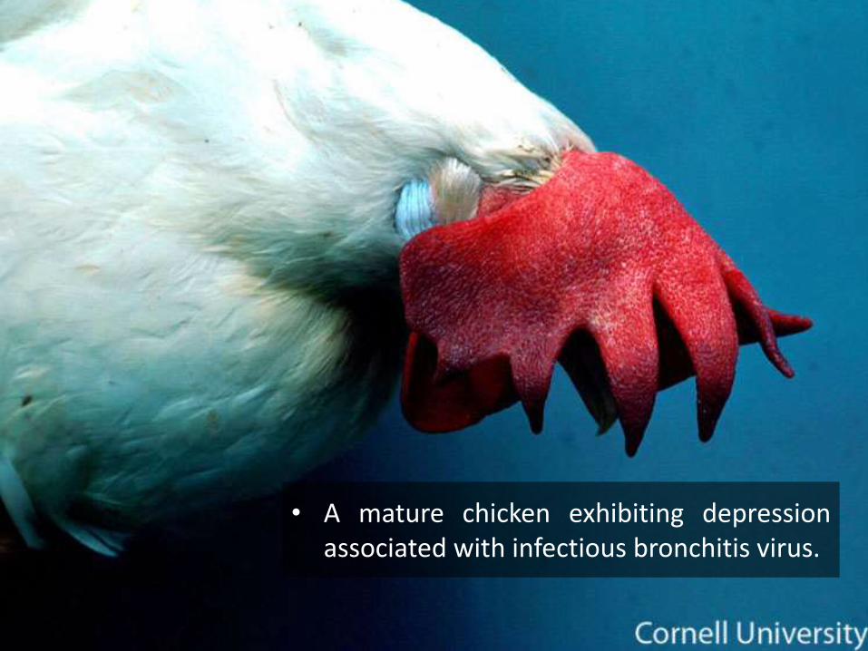
• A mature chicken exhibiting depressionassociated with infectious bronchitis virus.
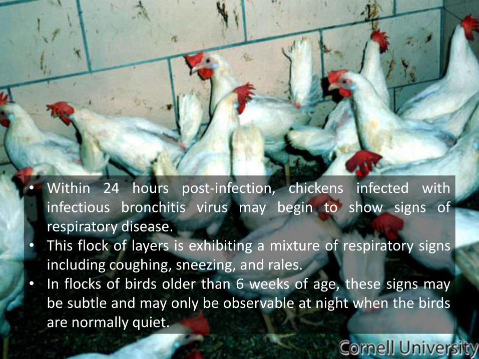
• Within 24 hours post-infection, chickens infected withinfectious bronchitis virus may begin to show signs ofrespiratory disease.
• This flock of layers is exhibiting a mixture of respiratory signsincluding coughing, sneezing, and rales.
• In flocks of birds older than 6 weeks of age, these signs maybe subtle and may only be observable at night when the birdsare normally quiet.
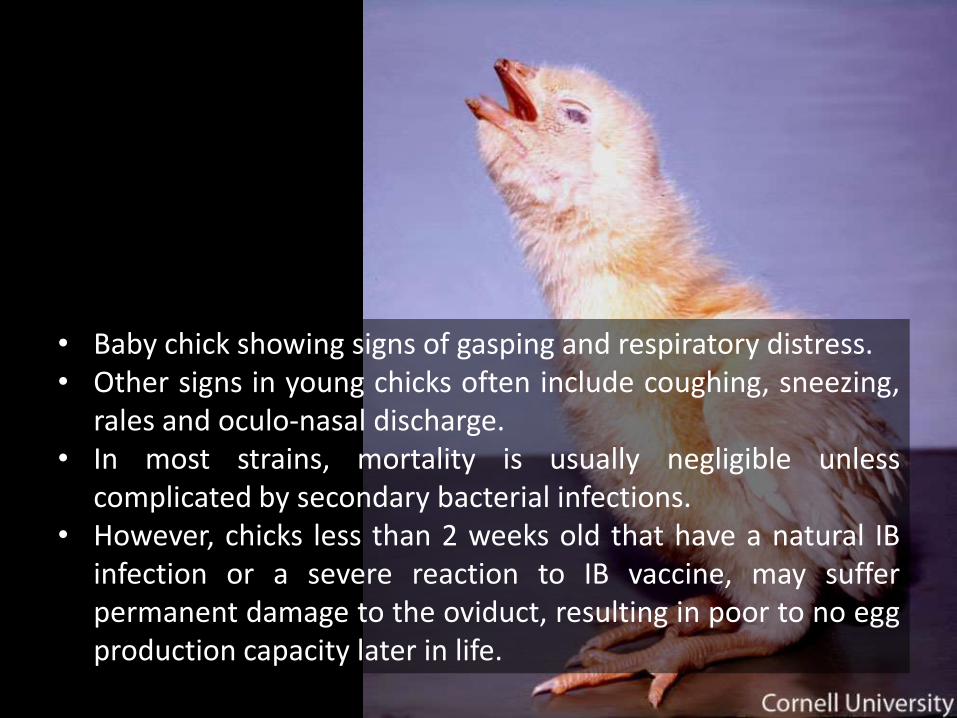
• Baby chick showing signs of gasping and respiratory distress.• Other signs in young chicks often include coughing, sneezing,
rales and oculo-nasal discharge.• In most strains, mortality is usually negligible unless
complicated by secondary bacterial infections.• However, chicks less than 2 weeks old that have a natural IB
infection or a severe reaction to IB vaccine, may sufferpermanent damage to the oviduct, resulting in poor to no eggproduction capacity later in life.
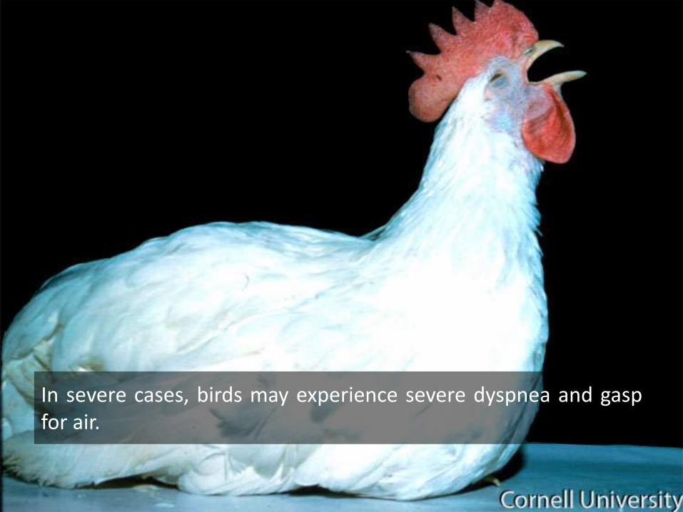
In severe cases, birds may experience severe dyspnea and gaspfor air.
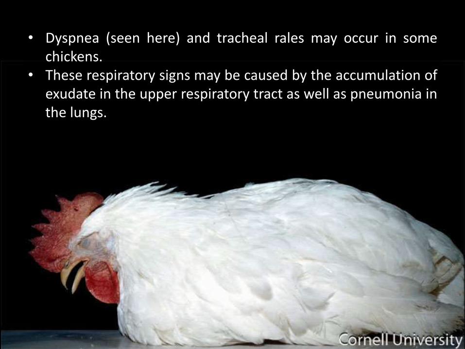
• Dyspnea (seen here) and tracheal rales may occur in somechickens.
• These respiratory signs may be caused by the accumulation ofexudate in the upper respiratory tract as well as pneumonia inthe lungs.

CLINICAL SIGNS - CONJUNCTIVITIS
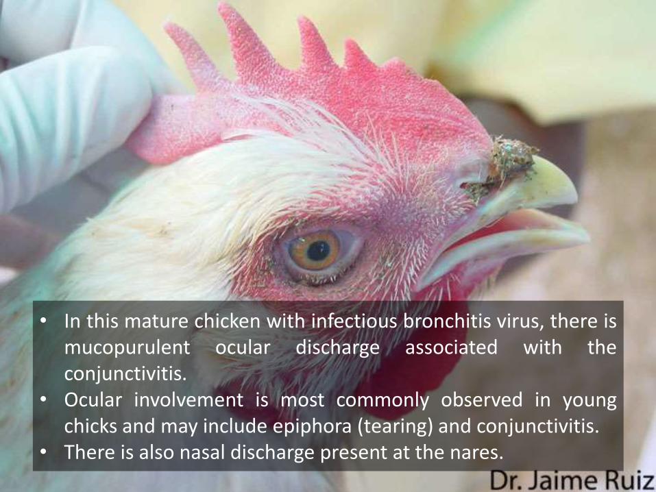
• In this mature chicken with infectious bronchitis virus, there ismucopurulent ocular discharge associated with theconjunctivitis.
• Ocular involvement is most commonly observed in youngchicks and may include epiphora (tearing) and conjunctivitis.
• There is also nasal discharge present at the nares.
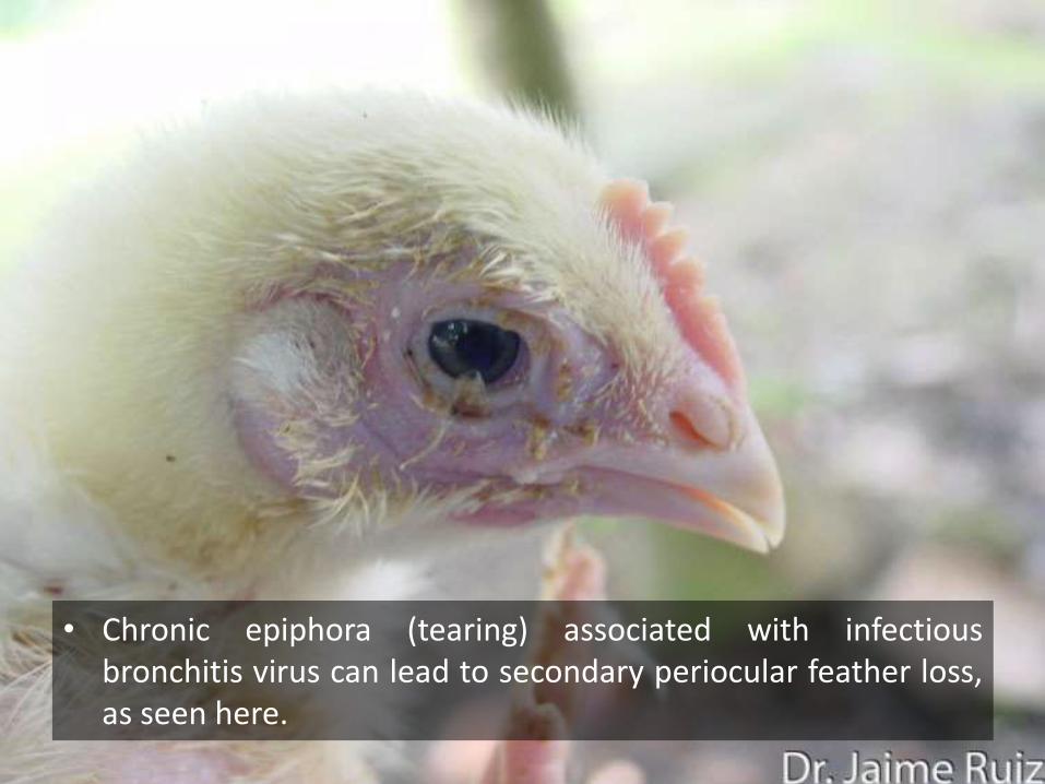
• Chronic epiphora (tearing) associated with infectiousbronchitis virus can lead to secondary periocular feather loss,as seen here.

CLINICAL SIGNS – EGG PRODUCTION
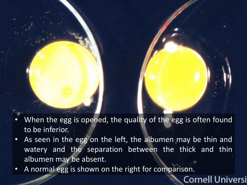
• When the egg is opened, the quality of the egg is often foundto be inferior.
• As seen in the egg on the left, the albumen may be thin andwatery and the separation between the thick and thinalbumen may be absent.
• A normal egg is shown on the right for comparison.
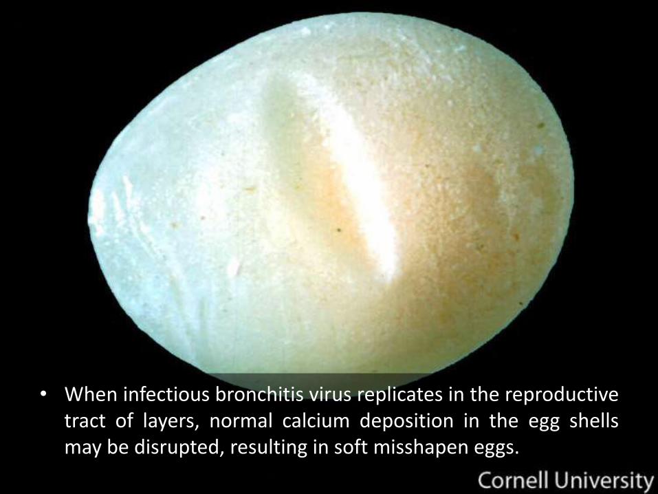
• When infectious bronchitis virus replicates in the reproductivetract of layers, normal calcium deposition in the egg shellsmay be disrupted, resulting in soft misshapen eggs.
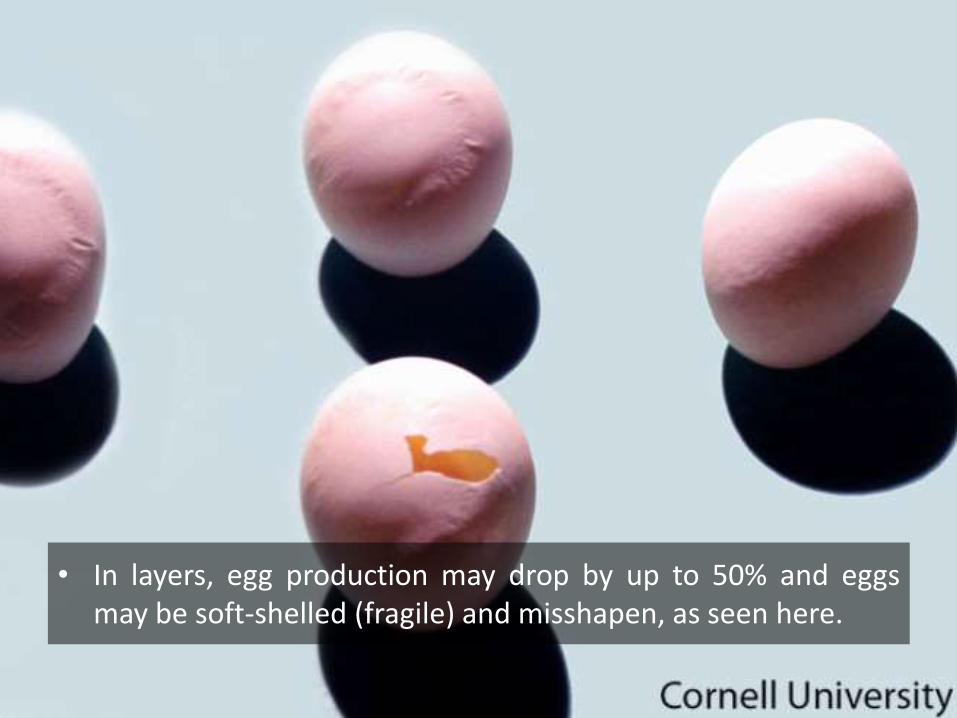
• In layers, egg production may drop by up to 50% and eggsmay be soft-shelled (fragile) and misshapen, as seen here.
PM LESIONS - RESPIRATORY
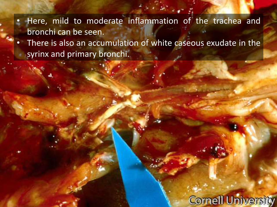
• Here, mild to moderate inflammation of the trachea andbronchi can be seen.
• There is also an accumulation of white caseous exudate in thesyrinx and primary bronchi.
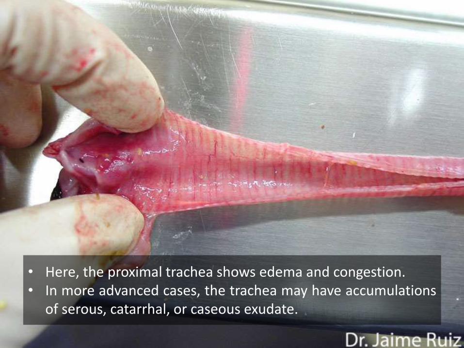
• Here, the proximal trachea shows edema and congestion.• In more advanced cases, the trachea may have accumulations
of serous, catarrhal, or caseous exudate.
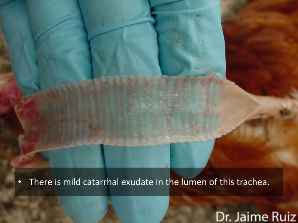
• There is mild catarrhal exudate in the lumen of this trachea.

PM LESIONS - EMBRYO
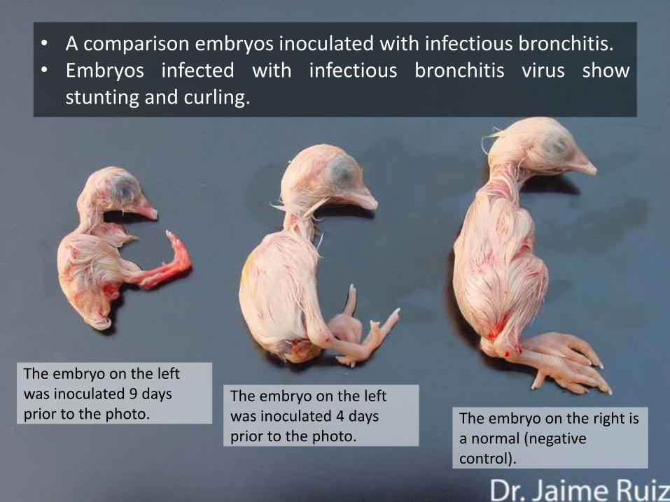
The embryo on the left was inoculated 9 days prior to the photo.
The embryo on the left was inoculated 4 days prior to the photo.
The embryo on the right is a normal (negative control).
• A comparison embryos inoculated with infectious bronchitis.• Embryos infected with infectious bronchitis virus show
stunting and curling.

• The embryo of the left shows stunting and dwarfing, resultingfrom the inoculation of a susceptible embryo with infectiousbronchitis virus.
• The amnion and allantois are usually thickened and closelyinvest the embryo.
• A normal embryo is shown on the right for comparison.
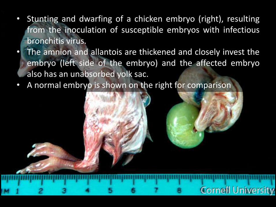
• Stunting and dwarfing of a chicken embryo (right), resultingfrom the inoculation of susceptible embryos with infectiousbronchitis virus.
• The amnion and allantois are thickened and closely invest theembryo (left side of the embryo) and the affected embryoalso has an unabsorbed yolk sac.
• A normal embryo is shown on the right for comparison

PM LESIONS - HEART
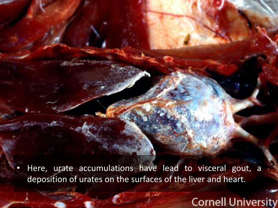
• Here, urate accumulations have lead to visceral gout, adeposition of urates on the surfaces of the liver and heart.
PM LESIONS - URINARY
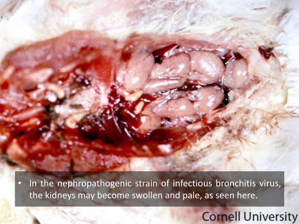
• In the nephropathogenic strain of infectious bronchitis virus,the kidneys may become swollen and pale, as seen here.
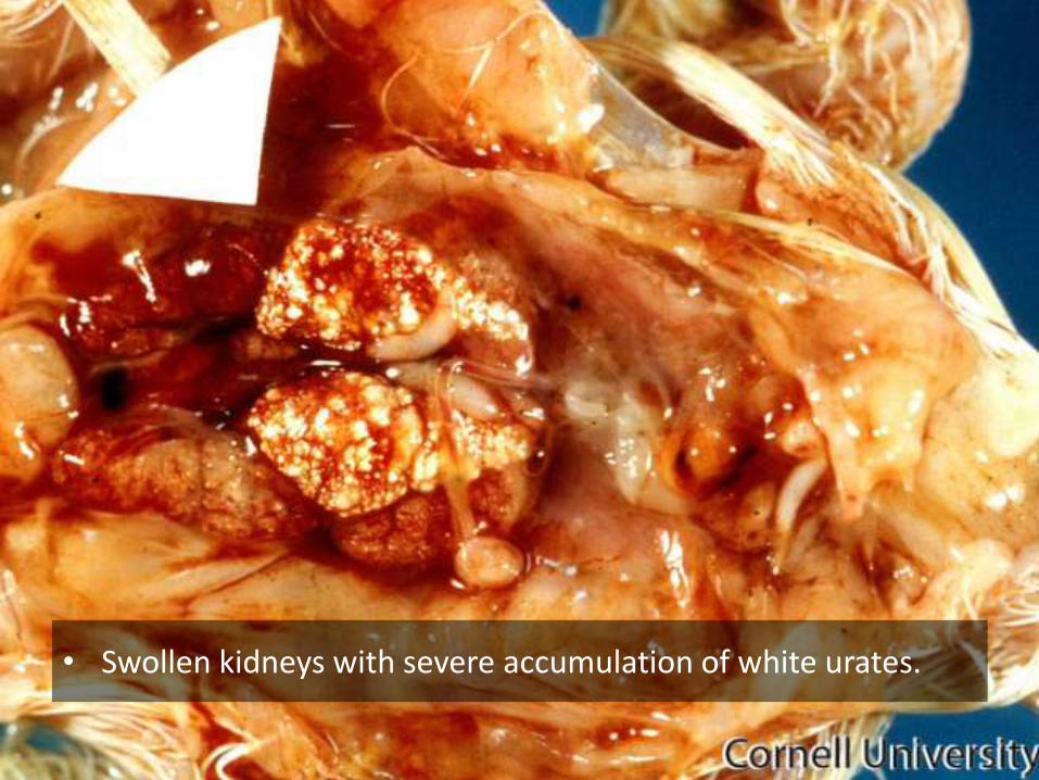
• Swollen kidneys with severe accumulation of white urates.
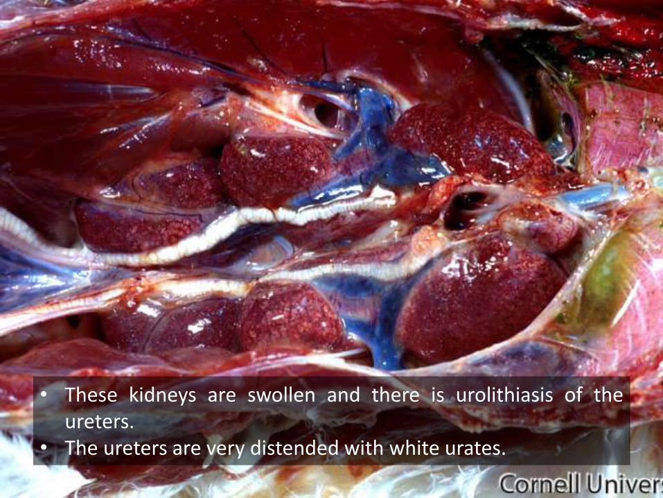
• These kidneys are swollen and there is urolithiasis of theureters.
• The ureters are very distended with white urates.
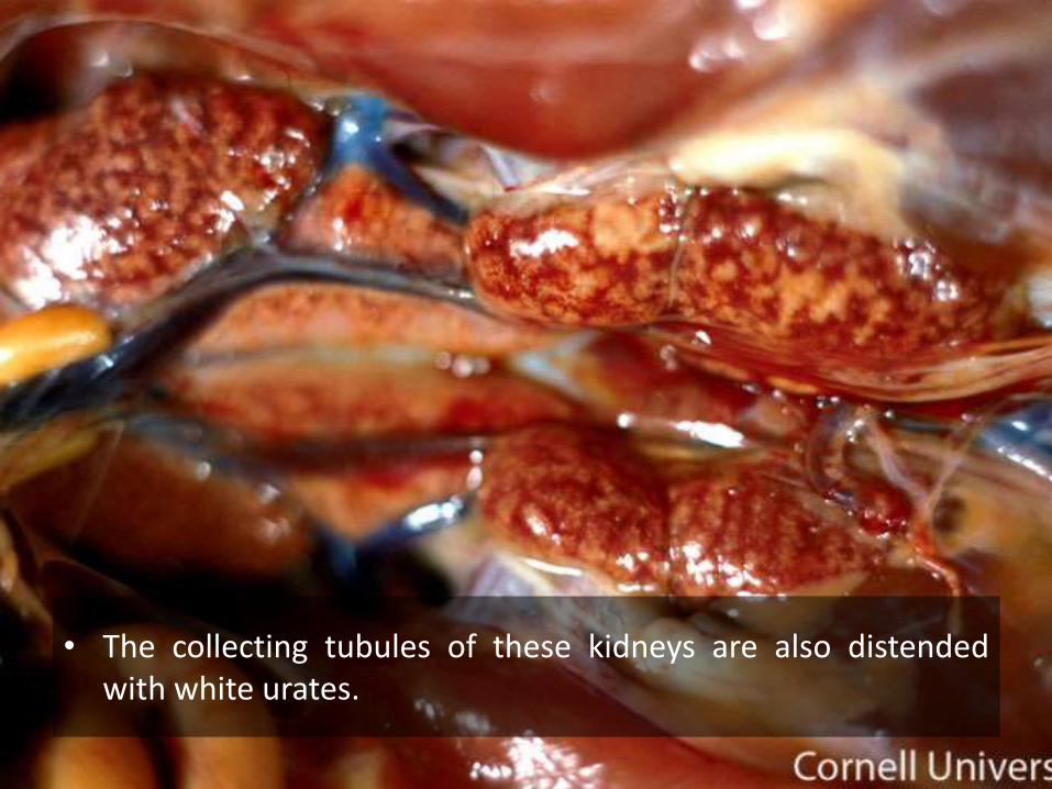
• The collecting tubules of these kidneys are also distendedwith white urates.