avianretrovirus pp32 dna-binding protein recognition ...jvi.asm.org/content/44/1/330.full.pdf ·...
TRANSCRIPT
JOURNAL OF VIROLOGY, Oct. 1982, P. 330-343 Vol. 44, No. 10022-538X/82/100330-14$02.00/0Copyright 0 1982, American Society for Microbiology
Avian Retrovirus pp32 DNA-Binding ProteinI. Recognition of Specific Sequences on Retrovirus DNA Terminal
RepeatsTAPAN K. MISRA,1 DUANE P. GRANDGENETT,l* AND J. THOMAS PARSONS2
Institute for Molecular Virology, St. Louis University Medical Center, St. Louis, Missouri 63110,1 andDepartment of Microbiology, University of Virginia Medical School, Charlottesville, Virginia 229082
Received 28 August 1981/Accepted 29 June 1982
The avian retrovirus pp32 protein possesses a DNA-nicking activity whichprefers supercoiled DNA as substrate. We have investigated the binding of pp32to avian retrovirus long terminal repeat (LTR) DNA present in both supercoiledand linear forms. The cloned viral DNA was derived from unintegrated Schmidt-Ruppin A (SRA) DNA. A subclone of the viral DNA in pBR322 (termed pPvuII-DG) contains some src sequences, tandem copies of LTR sequences, and partialgag sequences in the order src-U3 U5:U3 U5-gag. Binding of pp32 to supercoiledpPvuII-DG DNA followed by digestion of this complex with a multicut restrictionenzyme (28 fragments total) permitted pp32 to preferentially retain on nitrocellu-lose ifiters two viral DNA fragments containing only LTR DNA sequences. Inaddition, pp32 also preferentially retained four plasmid DNA fragments contain-ing either potential promoters or Tn3 "left-end" inverted repeat sequences.Mapping of the pp32 binding sites on viral LTR DNA was accomplished by usingthe DNase I footprinting technique. The pp32 protein, but not the avian retrovirusa4 DNA polymerase, is able to form a unique protein-DNA complex with selectedregions of either SRA or Prague A LTR DNAs. Partial DNase I digestion of a 275-base pair SRA DNA fragment complexed with pp32 gives upon electrophoresis indenaturing gels a unique ladder pattern, with regions of diminished DNase Isusceptibility from 6 to 10 nucleotides in length, in comparison with controldigests in the absence of protein. The binding of pp32 to this fragment also yieldsenhanced DNase I-susceptible sites that are spaced between the areas protectedfrom DNase I digestion. The protected region of this unique complex was astretch of 170 ± 10 nucleotides that encompasses the presumed viral promoter sitein U3, which is adjacent to the src region, extends through U5, and proceeds pastthe joint into U3 for about 34 base pairs. No specific protection or DNase Ienhancement by pp32 was observed in experiments with a 435-base pair SRADNA fragment derived from a part of U3 and the adjacent src region or a 55-basepair DNA fragment derived from another part of U3. The DNA sequence ofPrague A DNA at the fused LTRs differs from that of SRA DNA. The alteration inthe sequence at the juncture of the LTRs prevented pp32 from forming a stablecomplex in this region of the LTR. Our results are relevant to two aspects of theinteraction between pp32 and LTR DNA. First, the pp32 protein in the presenceof selected viral DNA restriction fragments possibly forms a higher orderoligomer analogous to Escherichia coliDNA gyrase-DNA complexes or eucaryot-ic nucleosome structures. Second, the specificity of the binding suggests a role forpp32 and the protected DNA sequences in the retrovirus life cycle. The preferredsequences to which pp32 binds include two adjacent 15-base pair invertedterminal repeats at the joint between U5 and U3 in SRA DNA. This region isinvolved in circularization of linear DNA and is perhaps the site that directsintegration into cellular DNA.
The analysis of avian cells infected with avian synthesis of a linear duplex DNA followed byretroviruses has shown that replication of viral the formation of a covalently closed circularDNA commences in the cytoplasm of the cell form found exclusively in the nucleus (26, 30, 40).within 1 h after infection and proceeds with the Linear cytoplasmic avian retrovirus DNA is
330
on July 9, 2018 by guesthttp://jvi.asm
.org/D
ownloaded from

AVIAN RETROVIRUS pp32 PROTEIN 331
slightly larger than unit length because it con-tains two long direct repeats (330 base pairs[bp]), one at each terminus (12, 29). Each longterminal repeat (LTR) is composed of nucleotidesequences unique to the 5' and 3' ends (U5 andU3) of the viral RNA genome. Possible mecha-nisms involved in the biosynthesis of linear viralDNA have been suggested by Gilboa et al. (4).The nucleus contains linear viral DNA as well as
two classes of covalently closed circular viralDNA. The circular DNAs differ only in thenumber of copies of the LTR. The smallerspecies contains a single copy of the LTR,whereas the larger species contains a tandemarrangement of LTRs joined together in thearrangement src-U3 U5:U3 U5-gag. Which of thethree viral DNA forms is the immediate precur-sor to the integrated provirus still remains to beclarified.
Retroviruses involved in the natural infectiousprocess integrate their DNA such that the inte-grated viral DNA is colinear with unintegratedlinear viral DNA (12, 26, 29). The cellular sitefor virus integration is apparently not specific,although direct sequencing of the cellular DNAaround the integrated retrovirus genome is nec-essary to rule out regional specificity (2, 32, 33,39).Molecular cloning of retrovirus DNA and sub-
sequent DNA sequencing studies have permit-ted a direct comparison of the structure of theretrovirus terminal repeats with the structures ofeucaryotic and procaryotic transposable ele-ments (1). The most striking revelation is theclose structural relationship which exists be-tween retrovirus DNA and transposable ele-ments (2, 32, 33, 37, 39). Transposable elementsand retrovirus DNA share the following proper-ties, although there are some variations withineach system. Both systems apparently utilizenonhomologous integration mechanisms; theirDNAs are flanked by either long inverted ordirect repeat elements that have short terminalinverted repeat structures; and upon integrationof transposon DNA or of retrovirus DNA thereis a loss of several base pairs from their termini,and a small number of base pairs at the recipientsite are reiterated at both ends of the insertedDNA.
Presently, no retrovirus proteins have beendirectly implicated in integration of retrovirusDNA. Our biochemical studies reported heresuggest that retroviruses encode a protein whichmight participate in viral DNA circularization orintegration because of its preferential binding toselected regions of retrovirus LTR DNA. Thepartially phosphorylated protein, termed pp32,is apparently derived from the ,B subunit of theavian retrovirus RNA-directed DNA polymer-ase by a proteolytic cleavage event in vivo (6,
27). It possesses a Mg2+-dependent DNA endo-nuclease activity which introduces nicks in su-percoiled DNA, its preferred substrate, generat-ing only unit-length DNA (5, 8, 27). A presumedvirus-coded DNA endonuclease biochemicallysimilar to pp32 isolated from Friend (22) andRauscher (16) murine leukemia viruses has re-cently been identified. We exploited the recentadvancement of DNase I footprinting (3) tostudy protein-DNA complexes and the nitrocel-lulose filter binding assay (14) to examine thebinding of pp32 to retrovirus LTR DNA andother nonviral DNA. DNase I footprinting iden-tifies regions of DNA that are protected frompartial DNase I digestion by bound protein. Wedemonstrated by the footprinting technique thatavian myeloblastosis virus (AMV) pp32 appearsto form a unique protein-nucleic acid complexwith selected regions of restriction enzyme frag-ments derived from cloned Schmidt-Ruppin A(SRA) or Prague A (PrA) viral DNA containingLTR sequences. Using the nitrocellulose filterbinding assay, we also showed that pp32 is ableto preferentially bind to several regions on asupercoiled subclone of the viral DNA inpBR322 (pPvuII-DG) containing tandem copiesof LTR DNA sequences (38). Digestion of pre-formed pp32-supercoiled pPvuII-DG complexesby a multicut restriction enzyme followed byfiltration on nitrocellulose filters permitted theidentification of specific pp32-bound DNA frag-ments. In addition to two DNA fragments con-taining viral LTR DNA sequences, pp32 alsopreferentially retained four pBR322 DNA frag-ments containing either promoters or Tn3 "left-end" inverted repeat sequences.
MATERIALS AND METHODS
DNAs. A subclone of SRA, pPvuII-DG, which con-tained two complete copies of the LTR DNA intandem (38), was generously supplied by J. M. Bishop(University of San Francisco, San Francisco, Calif.)and colleagues. A clone containing the complete PrARous sarcoma virus (RSV) genome (pSl-102) (10) anda subclone (pXBm-102) containing the src gene andtandem copies of LTR DNA were constructed in theplasmid vector pBR322 (Gilmartin and Parsons, un-published data).
Preparation of labeled DNAs. The SRA viral DNA,inserted into pBR322 DNA at the PvuII position, wascut with EcoRI. The fragments were end labeled with[.y-32PIATP (13) by T4 polynucleotide kinase. Thelabeled DNA was digested with PvuII, yielding threedifferent labeled viral DNA restriction fragments of330, 435, and 901 bp in length (see Fig. 1). Thefragments were separated on 5% acrylamide gels,eluted, and purified by DEAE-cellulose chromatogra-phy. The double-labeled 330-bp fragment was subse-quently cleaved with PvuI, generating 55- and 275-bpfragments. To generate the 275-bp DNA fragmentlabeled on the 5' end of the minus viral DNA strand,cloned DNA was first cleaved with PvuI, labeled with
VOL. 44, 1982
on July 9, 2018 by guesthttp://jvi.asm
.org/D
ownloaded from

332 MISRA, GRANDGENETT, AND PARSONS
[y-32P]ATP by T4 kinase, and digested with EcoRI.Restriction fragments from the PrA RSV clones wereprepared in a manner similar to that described above,except that an additional Hindlll site, which is locatedat the joint of the terminal repeats, was also labeled(10).The pPvuII-DG plasmid was uniformly labeled by
growing the bacteria in a low-phosphate-containingmedium with 32Pi. The plasmid DNA was purified bytwo successive CsCl buoyant density gradient sedi-mentation steps and by velocity sedimentation throughsucrose (8). The specific activity of the initially puri-fied supercoiled DNA varied from 20,000 to 35,000cpm per ,ug of DNA. The preparations of supercoiledDNA utilized were always greater than 95% super-coiled.
Nitroceliulose filter binding assay. Nitrocellulose fil-ters were treated in 0.4 M potassium hydroxide for 20min at 21°C, washed extensively with distilled water,and neutralized with 0.1 M Tris-hydrochloride (pH7.4) (17, 34). The filters were stored in sterile distilledwater, and each was washed with 6 ml of the appropri-ate wash buffer before use.The standard reaction mixture for DNA binding
contained 20 mM Tris-hydrochloride (pH 7.5 at 25C),0.1 M NaCl, 3 mM dithiothreitol, 0.2 mM EDTA, andbovine serum albumin at 50 ,ug per ml in a volume of100 ,ul. The appropriate DNA was added, and themixture was equilibrated at 37°C. The binding reactionwas initiated by the addition of the pp32 protein (1 to 5,ul). After incubation (usually 3 to 5 min), the reactionmixture was diluted with 1 ml of 20 mM Tris-hydro-chloride (pH 7.4)-0.1 M NaCl-3 mM dithiothreitol-0.2mM EDTA (wash buffer) and filtered at a flow rate of 1to 2 ml per minute. The filters were then washed withan additional 1 ml of 0.1 M NaCl buffer, dried, andcounted with an organic scintillation fluid. Back-ground counts due to DNA retained on the filter in theabsence of pp32 were always subtracted.The DNA retained by pp32 on the nitrocellulose
filters or the filtrates was also examined by polyacryl-amide gel electrophoresis. 32P-labeled DNA in thefiltrates was quantitatively coprecipitated with yeasttRNA (7.5 p.g) and the DNA retained by pp32 on thefilters was eluted at 37°C for 3 h with 0.3 ml of 0.2%sodium dodecyl sulfate-0.02 M Tris-hydrochloride(pH 7.5)-0.3 M sodium acetate containing yeast tRNA(4 ,ug). After ethanol precipitation, the pellets weresuspended in 50 ,ul of buffer and subjected to electro-phoresis on 5% polyacrylamide gels. The gels weredried and exposed to Kodak XR-5 X-ray film in thepresence or absence of DuPont Lightning Plus intensi-fying screens at -70°C.The binding of pp32 to supercoiled pPvuII-DG DNA
before digestion of this complex by HaeIII or otherrestriction enzymes was similar to the standard bind-ing reaction. The pp32 protein was complexed to 32p_labeled DNA at 37°C for 5 min in the absence ofdivalent metal ion followed by addition of a 5- to 10-fold excess of the HaeIII restriction enzyme as recom-mended by the supplier (New England Biolabs) todigest the DNA (-0.3 jig). The divalent metal ion,Mg2+ (100 ,u1 of a 2 mM solution), was immediatelyadded, and the reaction mixture was incubated for anadditional 5 to 10 min. The restriction enzyme diges-tion was stopped by the addition of 5 mM EDTA. Thesample was diluted with 1 ml of 0.1 M NaCl wash
buffer, filtered, and washed with an additional 1 ml ofbuffer. The DNA retained on the filter was eluted asdescribed above. As a control for nonspecific bindingof the restriction enzyme to DNA, the enzyme wasincubated with the DNA as described above in theabsence of pp32.DNase I footprinting. The partial DNase I digestion
procedures used were similar to those described forthe phage X integrase (24). Approximately 5 to 15 ng ofend-labeled DNA fragment was incubated with vari-ous amounts of pp32 DNA-binding protein (2.75 to 12,ug/ml) in 40 ,ul of reaction buffer containing 10 mMTris-hydrochloride (pH 7.5), 1 mM MgCl2, 5 mMdithiothreitol, 500 p.g of bovine serum albumin per ml,and 5% glycerol. The DNA samples were preincubat-ed at 20°C for several minutes followed by addition ofpp32. After an additional 3 min to allow for pp32binding to DNA, the DNA was digested with pancreat-ic DNase I (Worthington Diagnostics) at a concentra-tion of 0.25 ,ug/ml for either 2 or 2.5 min at 20°C. Thereaction was quenched with 3.4 p.g of sonicated calfthymus DNA in the presence of 10 p.g of Escherichiacoli tRNA and 10 mM EDTA and was followed byphenol extraction and ethanol precipitation. Theamount of end-labeled fragments layered on a particu-lar gel varied only by approximately 10% in any lane.In some experiments, the binding of pp32 to DNA waschallenged by the addition of heparin (2- to 5-foldmolar excess over pp32) for 2 min before DNase Idigestion.
Electrophoresis conditions. The chemical sequencemarkers for the various restriction fragments wereprepared as described by Maxam and Gilbert (20). TheDNA sequencing gels were 8, 10, or 15% polyacryl-amide-8 M urea (0.5 mm thick, 34 cm long). Theelectrophoresis buffer used was 90 mM Tris-borate(pH 8.0)-2 mM EDTA. The DNA samples were dena-tured with 7 M urea in the electrophoresis buffer at90°C for 5 min. Electrophoresis was carried out at1,200 V, and the gels were exposed at -70°C to X-rayfilm, with or without intensifying screens.
Purification of AMV pp32. The pp32 protein w-purified from AMV as described previously (8).
Purification ofAMV aj DNA polymerase. The AMVa,B DNA polymerase was purified by phosphocelluloseand heparin-Sepharose chromatography (7).
RESULTSRetention ofDNA restriction enzyme fragments
by AMV pp32 on nitrocellulose filters. To deter-mine whether AMV pp32 could selectively re-tain avian retrovirus LTR DNA fragments overplasmid DNA fragments on nitrocellulose fil-ters, pPvuII-DG was digested with HaeIII re-striction enzyme. The pBR322 DNA contains a1,670-bp viral insert (38) consisting of some srcsequences, tandem copies of LTR DNA, andgag sequences (Fig. 1). The HaeIII-digestedfragments were incubated with various amountsof pp32 under standard assay conditions (seeabove) and filtered on nitrocellulose (Fig. 2). Atlow protein-to-DNA ratios (Fig. 2; lanes B, C,and D), pp32 appears to retain six fragments(numbered 1, 4, 6, 8, 9, and 12) on the filterslightly better than other corresponding DNA
J. VIROL.
on July 9, 2018 by guesthttp://jvi.asm
.org/D
ownloaded from

VOL. 44, 1982
r A.V l l Tn 3 | Ori
65275 _04
436 --------- 901
PYu 11 Pvu Eco Rl Pvu Eco Rl
RI R330- LI U330.4 t- X
WC U3 US U3 U5 Pe
a a 17B 1!
AVIAN RETROVIRUS pp32 PROTEIN 333
23 dent at higher protein-to-DNA ratios (Fig. 2;25 lanes E and F). In the absence of pp32, little or
no DNA is retained on the filter (Fig. 2; lane A).------,, Specificity of pp32 binding sites on supercoiled
NV pPvuII-DG. The topography of the DNA, i.e.,supercoiling, may enhance pp32 recognitionof sequences contained in the six fragmentsdescribed above relative to the larger set of
^7 14 fragments (a total of 28). Supercoiled DNA is the-jX::V; - lpreferred substrate for the pp32-associated
DNA endonuclease activity (8). The pp32 pro-3 13 11 21 199 10 20 1SC 16 158 12
I I I 1 1, u I 1 1]22 24 26
FIG. 1. HaeIII restriction fragments of pPvuII-DGDNA. pPvuII-DG was digested with HaeIII, and thecombined SRA- and pBR322-generated fragments areillustrated on the blocked lines 1, 3, and 4 (35, 36, 38;J. M. Bishop, personal communication). Fragmentsare ordered by size. Fragment 1 contains the proposedpromoter for ampicillin (Amp) and proceeds throughthe Tn3-region, the origin of replication (Ori), and theviral insert and terminates at the tetracycline gene(Tet). The shaded region in line 3 indicates viral DNAsequences inserted at the PvuII position of pBR322.The second blocked line illustrates the fragments usedin our DNase I footprinting experiments. The 1,670-bpviral DNA subclone has two direct tandem repeatcopies designated R and L. The numbers Rl and R330refer to the terminal repeat sequences adjacent to thesrc region on the right end of the linear map ofunintegrated avian retrovirus DNA, whereas the num-bers LI and L330 refer to the terminal repeat adjacentto the gag region on the left end of the linear map.Only part of the src and gag sequences are present inthis subclone. The joint identifies the probable bluntend joining of linear DNA in vivo (38). The terminalrepeat consists of two identical copies (330 bp inlength), each of which contains the region unique tothe 3' terminus (U3) and the 5' terminus (U5) of theviral genome. The EcoRI, PvuI, and PvuII restrictionsites of this subcloned DNA are shown. End-labeledfragments identified as 901, 55, 275, and 435 bp wereobtained by cutting the inserted viral DNA withEcoRl, followed by dephosphorylation, end labeling,and cutting with the appropriate restriction enzymesas shown in the diagram. The 275- and 901-bp frag-ments were labeled on the plus viral DNA strand, andthe others were labeled on the minus viral DNAstrand. The dashed lines with arrows indicate thedirection for reading of sequencing gels.
fragments similar in size. Fragments 6 and 8retained by pp32 contain viral LTR DNA se-
quences (Fig. 1). The other four pBR322 frag-ments preferentially retained by pp32 containpromoter sequences for either ampicillin (frag-ment 1) or tetracycline (fragment 12), sequencesfor the left-end inverted repeat of Tn3 (fragment4), and sequences encoding the carboxyl termi-nal end of the ,B-lactamase gene (fragment 9) (35,36). The slight preferential retention of these sixfragments over the other fragments is less evi-
tein was bouna to supercoluea prvuni-LJu atseveral different protein-to-DNA ratios in theabsence of Mg2+ (Fig. 3). The quantity of pp32
A B C........ ....
of:
D E F
_v,. 4_0.:.:. '
*N n "am
G
-157
.4458395
8329
-9267
12 192
FIG. 2. Binding of pp32 to HaeIII restriction frag-ments of pPvuII-DG DNA. Various amounts of pp32protein were incubated with 0.5 ,ug of HaeIII frag-ments derived from pPvuII-DG DNA under standardassay condition (see text), except these filtered sam-ples were each washed with 3 ml of 0.1 M NaCl bufferinstead of the usual 1 ml buffer wash. The pp32-retained DNA was eluted and analyzed on 5% poly-acrylamide gels. The numbers on the right identify thesix fragments preferentially retained by pp32 and arenumbered according to size. The numbered fragmentsare 587, 458, 395, 329, 267, and 192 bp in length,respectively (indicated as subscripts to the fragmentnumber). The complete HaeIII digestion pattern ofpPvuII-DG DNA is shown in Fig. 1. The molecularweights of these fragments were compared to otherknown molecular-weight standards. Lane A, 0 ,ugof pp32; lane B, 0.13 pLg; lane C, 0.24 pLg; lane D, 0.36p.g; lane E, 0.50 ,ug; lane F, 0.63 ,ug. Lane G contains0.09 ,ug of control HaeIII-digested fragments whichwere not filtered.
.
15A 1 OA 17A
on July 9, 2018 by guesthttp://jvi.asm
.org/D
ownloaded from

334 MISRA, GRANDGENETT, AND PARSONS
A B C D E
-A Nowa:f.-- I59ak. 587
..,.-4458
395.K..:.
O8329
-9267
-12 192
FIG. 3. Binding of pp32 to supercoiled pPvuII-DGfollowed by HaeIII digestion. Supercoiled pPvuII-DGDNA (0.32 ,ug) was incubated with 0, 0.056, 0.11, and0.18 ,ug of pp32 (lanes A, B, C, and D, respectively).The pp32-DNA complex was digested with HaeIIIbefore filtration on nitrocellulose as described in thetext. The filter-bound DNA was eluted and analyzedon 5% polyacrylamide gels. Lane E is HaeIII-digestedDNA (0.09 jg DNA) only and was not filtered. Thenomenclature is the same as in Fig. 1 and 2.
utilized in these experiments was not saturatingwith respect to retention of supercoiled pPvuII-DG by the protein on nitrocellulose filters (un-published data). The pp32-DNA complex wasincubated at 37°C for 5 min followed by thesimultaneous addition of HaeIII restriction en-zyme and Mg2+. After 10 min of further incuba-tion, the samples were filtered on nitrocelluloseand washed. The DNA retained on the filterswas extracted and analyzed on 5% polyacryl-amide gels. The HaeIII enzyme retained littleDNA on the filter by itself (Fig. 3; lane A).Binding ofpp32 to supercoiled DNA followed bydigestion with a multicut restriction enzymeresulted in enhanced retention offragments 1, 4,6, 8, 9, and 12 by pp32 relative to the otherfragments (Fig. 3; lanes B, C, and D). Thepreferential retention of these fragments by pp32appears similar at several different protein-to-DNA ratios (10:1, 20:1, and 30:1) (Fig. 3; lanesB, C, and D, respectively). No fragments small-er than the fragment 12 specifically retained bypp32 were evident upon prolonged exposure ofthe gel. Quantitative analysis of gel scans in Fig.
3 or actual counting of the labeled DNA frag-ments revealed that pp32 binds to sequences inthese six fragments approximately two- to three-fold better (molar basis) than the other nonselec-tively retained fragments. The selectivity ofpp32 binding with the above filter assay to thesesix HaeIII fragments confirms data which willbe presented in greater detail elsewhere (V.Parsons and M. Golomb, submitted for publica-tion).Like numerous other highly specific nucleic
acid binding proteins which bind to DNA atrandom locations (9, 17, 28), pp32 retains someof the other 22 HaeIII DNA fragments on nitro-cellulose filters, but at a much decreased capaci-ty as compared to the 6 fragments describedabove (Fig. 2 and 3). This apparent nonspecificbinding to DNA by pp32 is more evident at highprotein-to-DNA ratios in the presence of HaeIIIdigestion fragments (Fig. 2; lanes E, F).Mapping of pp32 binding sites on SRA terminal
repeats. With the nitrocellulose filter bindingassay, pp32 retained only two viral DNA frag-ments derived from pPvuII-DG; both fragmentscontained LTR DNA sequences (Fig. 1 through3). The two fragments (fragments 6 and 8) re-tained by pp32 contained mostly U3 and U5DNA sequences (Fig. 1). We wanted to map thelocation of pp32 binding sites within these LTRDNA sequences by the DNase I footprintingmethod (3).We first investigated the interaction of pp32
with the 275-bp restriction enzyme fragmentderived from the internal region of the tandemLTR DNA copies (Fig. 1). This figure depictsboth copies of the SRA terminal repeat fusedtogether as found in circular DNA obtained frominfected cells (38). Each purified end-labeledDNA restriction fragment was incubated with orwithout purified pp32 protein and then analyzedby the DNase I footprinting procedure (3). Thebinding of pp32 to this 275-bp fragment gener-ates a unique pattern of enhanced and reducedsusceptibilities to DNase I nicking along theDNA (Fig. 4). A group of specific DNA frag-ments is produced in the presence of pp32which, on electrophoresis in denaturing gels,gives rise to a ladder pattern with protectedregions from 6 to 10 nucleotides in length. Protec-tion from DNase I digestion by pp32 is indicatedby the absence of bands or by their diminishedintensities in the presence of pp32 as comparedwith control digests (for example, compare lanesC, G, and H from R252 to R260, R284 to R291,etc., in Fig. 4). In addition to the observed effectof reduced sensitivity to DNase I in selectedareas on this 275-bp fragment, the association ofpp32 with this DNA results, in most cases, inenhanced DNase I cleavage between regions ofdiminished DNase I susceptibility. When similar
J. VIROL.
on July 9, 2018 by guesthttp://jvi.asm
.org/D
ownloaded from

AVIAN RETROVIRUS pp32 PROTEIN 335
A B C D E F G H
L83 ° wL6&8 °-
L56'°
L40o~~~~#L309
L40
R327-R3230 P:.
R313°.....r
R301° ___
R2900d*M
R281° _ _ _
R272- __R266o
R258B _ v
R2530U
R2370.
FIG. 4. DNase I footprints ofAMV pp32 binding tothe SRA 275-bp restriction fragment. The 275-bp frag-ment (Fig. 1, line 2) was subjected to various treat-ments (described below) and then analyzed on an 8%polyacrylamide DNA sequencing gel. Lane A, chemi-cal G (20); lane B, chemical C (20); lane C, approxi-mately 10 ng of end-labeled DNA in binding buffer (seetext) was digested by DNase I. The reaction wasterminated, and samples were analyzed on polyacryl-amide gels. Lane D was treated the same as lane Cexcept that the sample was treated with heparin (4.25i.g/ml, 2 min exposure) before DNase I digestion.Lane E, was treated the same as lane C except that theDNA was incubated with pp32 (2.7 ,ug/ml) for 3 minbefore DNase I digestion. Lane F, was treated thesame as lane E except heparin was added after pp32was incubated with DNA. Lane G, was treated thesame as lane E except that the concentration of pp32was 5.5 pg/ml. Lane H was treated the same as lane F
samples (as described in Fig. 4) were subjectedto electrophoresis in a 15% polyacrylamide gel,regions protected by pp32 between R190 and L4were also characterized (not shown; see Fig.12). Figure 5 contains the nucleotide sequenceinformation of the cloned SRA LTR DNA (38).The regions protected by pp32 are indicated bydiscontinuous underlining, with enhanced DNaseI nicking indicated by arrows. Starting in the U3region adjacent to src, the protected regionextends from the presumed promoter region(R194, Fig. 5) (38) in U3, through the U5 region,and then terminates approximately 34 bp pastthe fused terminal repeats, for a total of 170 ± 10bp. The exact nucleotides are not defined insome areas that are protected by pp32 fromDNase I digestion, because DNase I does notnick DNA uniformly. Also, DNase I digestionyields fragments with 3'-OH termini which mi-grate slightly slower than chemical sequencefragments that terminate in a 3'-PO4 (24).The observed changes in the DNase I diges-
tion pattern of the 275-bp fragment in the pres-ence of pp32 were reversed by the addition ofthe polyanion, heparin, before the addition ofDNase I (Fig. 4; compare lanes E and F or lanesG and H). Under these conditions, heparin doesnot appreciably affect DNase I digestion ascompared to control digests (Fig. 4; lanes C andD), but it causes the apparent detachment ofpp32 from the restriction fragment. Heparin hasbeen used to help distinguish bacterial and phageattachment sites for phage X integrase (11).The concentration of pp32 needed to generate
this unique DNA-protein complex on the 275-bpfragment was defined (Fig. 4). For example, inlane E of Fig. 4, the concentration of pp32 was2.7 ,ug per ml. There appears to be partialprotection of specific regions (see R252 to R260and other regions) in the 275-bp fragment at thisconcentration; also present are the readily iden-tifiable areas of enhanced DNase I cleavage.Increasing the concentration of pp32 by twofoldresults in almost complete protection of most ofthe specified areas (Fig. 4; lane G). If the con-centration of pp32 is increased to 12 ,ug per ml,no apparent changes in the observed protectionor DNase I enhancement patterns were ob-served (not shown). The near total absence ofsome bands in the digests containing pp32 sug-gests that the pp32 binding site is nearly saturat-ed. The same protection patterns were obtainedwith six different preparations of purified pp32which were greater than 95% homogeneous (8).Heat-inactivation studies demonstrated that the
except that the concentration of pp32 was 5.5 .Lg/ml.The numbers on the photograph identify specific nu-cleotides (see Fig. 5 for sequence).
VOL. 44, 1982
on July 9, 2018 by guesthttp://jvi.asm
.org/D
ownloaded from

336 MISRA, GRANDGENE1T, AND PARSONS
RIR U3
src*ATGTAGTCTTATGCAXR20 R40. R60 R80
LATACTCTTGTAGTCTTGCAACATGGTAACGATGAGTTAGCAACATGCCTTACAAGGAGAGAAAAAGCACCGTG
RIOO R120 R140 R160
CATGCCGATTGGTGGAAGTMGGTGGTACGATCGTGCCTTATTAGGMGGCAACAGACGGGTCTGACATGGATTGGACGAACCACTGMPvuI EcoRI
R180 R200 R220 U3 R I 5 R260
TTCCGCATTGCAGAGATATTGTATMrAAGTGCCTAGCTCGATACAATAAACGCCAMGACCATTCACCACi TGGTGTGCACCTGGGT
SIR ' SIRIR280 R300 R320 Us LI U3 L20
.4,4 * 4 4 4* 4 4* 4TGATGGCCGGACCGTTGATTCCCTGACGACTACGAGCACCTGCATGAAGCAGAAGGCTTCATT-MTGTAGTCTTATGCAATACTCTTGT
Joint
L40 L60 L80 L100
AGTCTTGCAACATGGTAACGATGAGTTAGCAACATGCCTTACAAGGAGAGAAAAAGCACCGTGCATGCCGATTGGTGGAAGTAAGGTGG
L120 L140 L160 L180 L200
TACGATCGTGCCTTATTAGGMGGCAACAGACGGGTCTGACATGGATTGGACGAACCACTGAATTCCGCATTGCAGAGATATTGTATTTPvuI EcoRI
L220 U31 R L260 L280** i * + *+; *++*~~~444 4 * 4
J SIR IL300 L320 U5!S~~ ~~~* *+ * gag
CGACTACGAGCACCTGCATGAAGCAGMGGCTTCATT
FIG. 5. Nucleotide sequence of SRA LTR DNA. The sequence information (plus strand of viral DNA) ispresented for two copies of LTR DNA fused together as found in in vivo circular DNA (38) and are numbered asdesignated in Fig. 1. The short inverted repeats at the ends of the LTR DNA are designated as SIR. The boldletter R identifies the terminal repeat present at both ends of the genomic RNA. The brackets identify U3 and U5sequences. The regions protected by pp32 are indicated by discontinuous underlining, and the arrows indicateareas of enhanced DNase I cleavage. The boundaries of protection and enhanced DNase I cleavage induced bypp32 were derived from analysis of numerous gels.
half-life for pp32 DNA-binding activity with theDNase I footprinting assay was -4 min at 60°C,comparable to that observed with pp32 DNAendonuclease activity or pp32 binding to super-coiled DNA (D. Grandgenett, T. Misra, and P.Hippenmeyer, submitted for publication).To better define the boundary of pp32 binding
on LTR DNA from about R315 to L40 (whichincludes the joint region containing both mis-matched 15-bp short inverted repeats, R315 toR330 and Li to L15), we cleaved the SRAsubclone with PvuI to generate an internal frag-ment of 330 bp in length (Fig. 1). The DNA wasend labeled and cleaved with EcoRI to generatea 275-bp molecule labeled at the L120 positionon the minus strand. The pp32 protein did notinduce any specific changes in the DNase Idigestion pattern from L83 to approximatelyL34; diminished DNase I susceptibility startedat approximately L34 and proceeded onwardinto the R terminal repeat (data not shown). Thesame general areas protected by pp32 on theplus strand (Fig. 5) also appear to be protectedwhen the DNA is labeled on the minus strand.
Since the terminal repeats contain two com-
plete copies of the U3 and U5 region joinedtogether, pp32 could possibly interact with re-gion L190 to L330 (Fig. 1; 901-bp fragment),which has the same sequence as region R190 toR330 (the 275-bp fragment). As observed in Fig.6, pp32 apparently protected the same regions(for examples, see L222 to L230, L252 to L260,etc.) and caused the enhanced DNase I cuttingpattern on the 901-bp fragment like that found onthe 275-bp fragment (Fig. 4 and 5). The preciseboundary of pp32 protection on the 901-bp frag-ment at the L330 position, adjacent to the primerbinding site, is as yet undefined. However, pp32can at least protect sequences from L325 to twonucleotides in on the adjacent primer site (Fig.6).
In a series of control experiments, we wantedto determine the specificity of pp32 binding toLTR DNA. First, in similar studies with theAMV a,1 DNA polymerase, the presumed pre-cursor to pp32, we did not observe any periodic-ity of protection and enhancement under condi-tions which permitted pp32 to form a uniquecomplex with the SRA 275- or 901-bp LTRDNA. One such analysis is shown in Fig. 7. A
J. VIROL.
on July 9, 2018 by guesthttp://jvi.asm
.org/D
ownloaded from

AVIAN RETROVIRUS pp32 PROTEIN 337
general reduction (Fig. 7; lanes D, E, and F) inDNase I-susceptible sites, suggesting nonspecif-ic protection ofDNA sequences by the polymer-ase.Second, we determined whether other viral
DNA restriction fragments permitted the forma-tion of these pp32-mediated specific DNA com-plexes. As indicated in the DNA sequence dia-gram (Fig. 5), pp32 does not protect certainareas of the U3 terminal repeat region. A 435-bpDNA fragment (Fig. 1) representing region R178
L3230
t3O90L301'
L281°t272,
1264-
L2530
A B C D E F
t2370*Th
L230J NW
L218 _
L2140
L209e ;R.i#t,
t2070
40PP
L199- Ahj__~~4
L1914
L188°
FIG. 6. DNase I footprint of pp32 binding to anSRA 901-bp restriction fragment. Conditions weresimilar to those described in Fig. 4 except that thepp32 concentrations were different and the sampleswere analyzed on 10o polyacrylamide gels. Lane A,chemical G (20); lane B, pp32 (7.0 ,ug/ml); lane C, pp32(7.0 ,ug/ml) challenged with heparin; lane D, pp32 (12FLg/ml); lane E, pp32 (12 ,ug/ml) challenged with hepa-rin.
comparison of Fig. 6 with Fig. 7 clearly revealsthat there is no protection or enhanced DNase Icleavage observed when oa3 DNA polymerase iscomplexed to SRA 901-bp LTR DNA. At pro-tein concentrations comparable to those usedwith pp32, the a1 DNA polymerase did notpromote the formation of specific complexes asobserved with pp32. Rather, increasing concen-trations of oa3 DNA polymerase promoted a
301
i281 Sx X
t253VEN
..4
L20. W
FIG. 7. DNase I footprints of AMV ot3 DNA poly-merase binding to the SRA 901-bp restriction frag-ment. Conditions were similar to those described inFig. 4 except that pp32 was replaced with purified c13DNA polymerase. Lane A, chemical G (20); lanes Band C, DNA only; lanes D, E, and F, oL3 polymerase at4.7, 7.3, and 9.5 p.g/ml, respectively.
VOL . 44, 1982
on July 9, 2018 by guesthttp://jvi.asm
.org/D
ownloaded from

338 MISRA, GRANDGENETT, AND PARSONS
P 3 )-.E F-I i, ...
,o
:,; ,Q
*~- 2 12.
:11 .. ,.
4
2Sit
4.
^.kW'..
4m
40 X4
i1 4 7 a}e
FIG. 8. DNase I footprints of ppSRA 435-bp restriction fragment. Tbeled on the minus strand. Conditionthose described in Fig. 4 except thalsubjected to electrophoresis for twothe same 15% polyacrylamide gel.chemical G (20); lanes B and G, DN)DNA plus heparin; lanes D and I, Ilanes E and J, pp32 (7.0 ,ug/ml)heparin. Some of the G-terminated frwere derived from other gels with di
to Ri, which includes one copy c
located short inverted repeat, wabinding of pp32 with this DNA.the gel in Fig. 8 reveals that pp32any specific change in the DNpattern of any unique U3 sequenbp fragment. Increasing the c(pp32 to 14 ,ug/ml (twofold increasin no specific change in the DNpattern. In addition, with pp32protection ofDNA or enhanced I
of the 55-bp fragment (L178 to L
ed in the U3 region (not shown). Finally, oneDNA fragment derived from the pol gene ofcloned RSV PrA DNA and labeled at the uniqueBglII site (10) did not permit the formation ofthese specific pp32-DNA complexes (data notshown).pp32 binding sites in PrA terminal repeats.
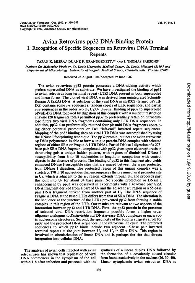
Does AMV pp32 bind to the same or differentregions on the terminal repeats of PrA RSVDNA relative to SRA DNA? To answer thisquestion, we analyzed a subclone of PrA DNAcontaining two copies of the terminal repeat.Figure 9 illustrates a restriction enzyme map of
'F 8 the PrA tandem repeats inserted into pBR322-:)4 DNA. This subclone (pXBmlO2) was derived
from a permutated genomic clone of PrA RSVDNA which contained two tandem copies of the"P14:.terminal repeat (10). This particular clone of-fered the advantage of a unique HindIII sitelocated at the joint of the fused repeats, enabling
is~'R examination of most of the U5 binding regionsfrom a different starting point than with SRADNA. In addition, the sequence at the junctionof the tandem repeat is different from that foundin SRA (Gilmartin, Pugatsch, and Parsons, un-published data). Specifically, the PrA DNA
-7 lacks seven nucleotides of the 14-bp invertedrepeat on the U5 terminus at the joint and theentire short inverted repeat on the adjacent U3
R 17: terminus (Fig. 10). The adjacent U3 short invert-'7 ed repeat is located 22 nucleotides downstream
at position L22. In total, the PrA DNA sequenceis considerably different from Li to about L60F1i4 (Fig. 10) as compared to SRA DNA (R322 toR330 and Li to approximately L44; see Fig. 5).This sequence alteration observed with PrADNA serves as a good control for SRA DNA to
32 binding to the determine whether sequences at the fused jointhe DNA was la- are involved in recognition of LTR DNA by the
Ithe sample was pp32 protein.different times on For a positive control, the pXBmlO2 clonedLanes A and F, DNA was cleaved with EcoRI and end labeledk; lanes C and H, with T4 kinase. A fragment of approximately 300pp32 (7.0 ,ug/ml); bp labeled at position L196 on the plus viralchallenged with DNA strand was isolated after SacI digestionragments (lane A) (Fig. 9). Binding of pp32 protein produced aistinct bands. similar ladder pattern and caused diminished
DNase I susceptibility in the same DNA regions)f the terminally on the PrA 300-bp restriction fragment (data notis used to study shown) as those observed on the SRA 901-bpExamination of restriction fragment (Fig. 5).2 did not induce We next investigated the interaction of--thease I digestion pp32 protein with a 285-bp PrA restriction frag-ices on the 435- ment (R165 to L144, labeled on the 5' end of plus)ncentration of strand DNA) which was similar in size andse) also resulted shared partial sequence homology with the SRAlase I digestion 275-bp fragment. As stated previously, the se-there was no quence of the PrA fragment deviates considera-
DNase I cutting bly from SRA DNA (Fig. 5) at positions Li to,118) also locat- approximately L60 (Fig. 10). The pp32 protein
J. VIROL.
-'s
Nk.,"kl;- -*N.
on July 9, 2018 by guesthttp://jvi.asm
.org/D
ownloaded from
AVIAN RETROVIRUS pp32 PROTEIN 339
JointR310 LI
U3 Uu5 It E i
PvuI EcoRI HindM285__-___
.,___145ah
~-- 300~ -10
I-le r _ _
FIG. 9. PrA DNA restriction fragments used in DNase I footprinting experiments. The PrA DNA (pXBm 102)inserted into pBR322 DNA was digested with the indicated restriction enzymes, and the fragments were endlabeled as described in Fig. 1. The -300-, 285-, and 197-bp restriction fragnents were labeled on the plus strand,whereas the 145-bp fragnent was labeled on the minus strand.
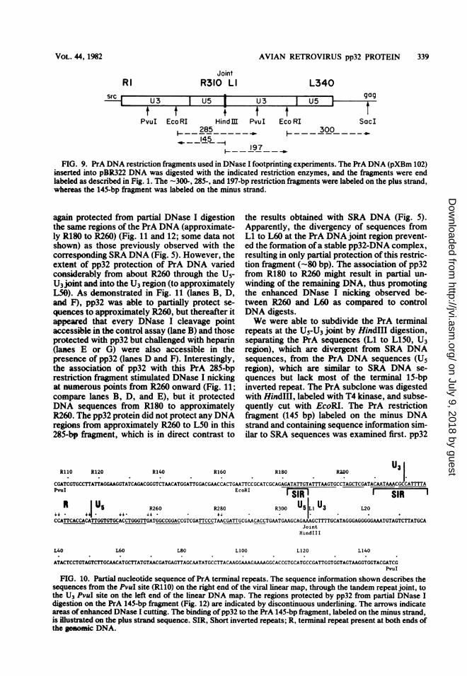
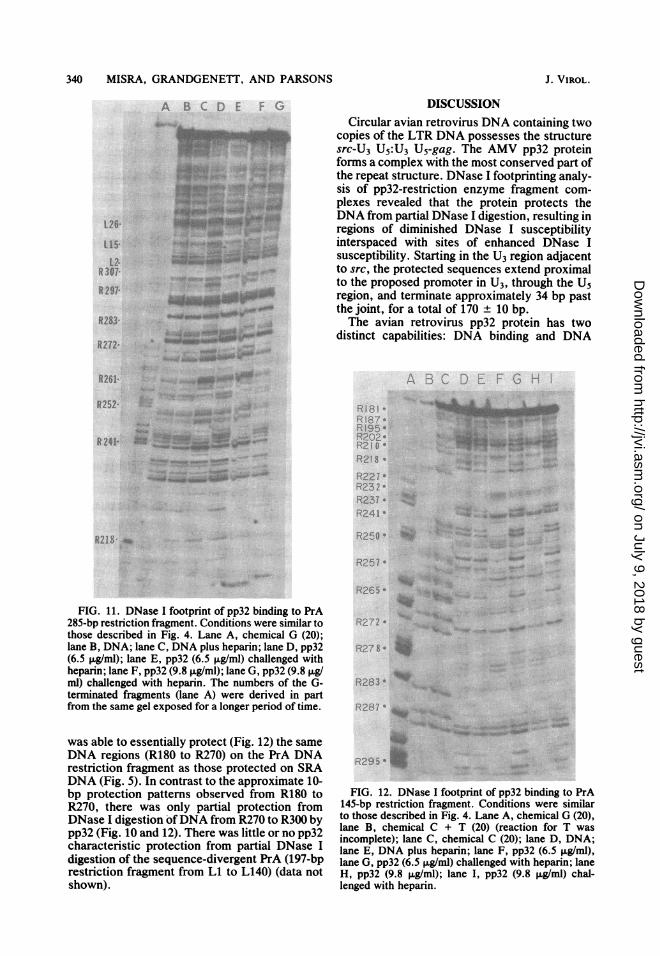
again protected from partial DNase I digestionthe same regions of the PrA DNA (approximate-ly R180 to R260) (Fig. 11 and 12; some data notshown) as those previously observed with thecorresponding SRA DNA (Fig. 5). However, theextent of pp32 protection of PrA DNA variedconsiderably from about R260 through the U5-U3 joint and into the U3 region (to approximatelyL50). As demonstrated in Fig. 11 (lanes B, D,and F), pp32 was able to partially protect se-quences to approximately R260, but thereafter itappeared that every DNase I cleavage pointaccessible in the control assay (lane B) and thoseprotected with pp32 but challenged with heparin(lanes E or G) were also accessible in thepresence of pp32 (lanes D and F). Interestingly,the association of pp32 with this PrA 285-bprestriction fragnent stimulated DNase I nickingat numerous points from R260 onward (Fig. 11;compare lanes B, D, and E), but it protectedDNA sequences from R180 to approximatelyR260. The pp32 protein did not protect any DNAregions from approximately R260 to L50 in this285-bp fraginent, which is in direct contrast to
the results obtained with SRA DNA (Fig. 5).Apparently, the divergency of sequences fromLi to L60 at the PrA DNA joint region prevent-ed the formation ofa stable pp32-DNA complex,resulting in only partial protection of this restric-tion fragment (-80 bp). The association of pp32from R180 to R260 might result in partial un-winding of the remaining DNA, thus promotingthe enhanced DNase I nicking observed be-tween R260 and L60 as compared to controlDNA digests.We were able to subdivide the PrA terminal
repeats at the U5-U3 joint by Hindlll digestion,separating the PrA sequences (LI to L150, U3region), which are divergent from SRA DNAsequences, from the PrA DNA sequences (U5region), which are similar to SRA DNA se-quences but lack most of the terminal 15-bpinverted repeat. The PrA subclone was digestedwith HindIll, labeled with T4 kinase, and subse-quently cut with EcoRI. The PrA restrictionfragment (145 bp) labeled on the minus DNAstrand and containing sequence information sim-ilar to SRA sequences was examined first. pp32
RI 10 R120 R140 R160 R180 R00U3
I.CGATCGTGCCTiTATTAGGAAGGTATCAGACGGGTCTAACATGGATTGGACGAACCACTGAATTCCGCATCGCAGAGATATTGTATTTAAGTGCCTAGCTCGATACAATAAACGCCATTTAPvuI EcoRI ISIR SIR
1 U5 R260 R280 R300 U5 i U3 L2044 - 41 . 44- &,& - * ; * 1CCATTCACCAC---GTGTGCACCTGGGTTGATGGCCGGACCGTCGATTCCCTAACGATTGCGAACACCTGAATGAAGCAGAAAGCTTTTGCATAGGGAGGGGGAAATGTAGTCTTATGCA
JointHindIII
L40 L60 L80 LIOO L120 L140
ATACTCCTGTAGTCTTGCAACATGCTTATGTAACGATGAGTTAGCAATATGCCTTACAAGGAAAGAAAAGGCACCGTGCATGCCGATTGGTGGTAGTAAGGTGGTACGATCG
FIG. 10. Partial nucleotide sequence of PrA terminal repeats. The sequence information shown describes thesequences from the PvuI site (R110) on the right end of the viral linear map, through the tandem repeat joint, tothe U3 PvuI site on the left end of the linear DNA map. The regions protected by pp32 from partial DNase Idigestion on the PrA 145-bp fragment (Fig. 12) are indicated by discontinuous underlining. The arrows indicateareas of enhanced DNase I cutting. The binding of pp32 to the PrA 145-bp fragment, labeled on the minus strand,is illustrted on the plus strand sequence. SIR, Short inverted repeats; R, terminal repeat present at both ends ofthe genomic DNA.
RI L340
U3
tPvuI
tEcolFRI SacI
VOL. 44, 1982
I U 5 --I 909
-f-srcr
on July 9, 2018 by guesthttp://jvi.asm
.org/D
ownloaded from
340 MISRA, GRANDGENETT, AND PARSONS
A B C L:i E 'F-
L26'
R 307-
R8297-
*'00_ ""M'.w.
R8283-
R272-
R261 a
R252: ...
R 241-
R283 ~ _-W
R218
FIG. 11. DNase I footprint of pp32 binding to PrA285-bp restriction fragment. Conditions were similar tothose described in Fig. 4. Lane A, chemical G (20);lane B, DNA; lane C, DNA plus heparin; lane D, pp32(6.5 ,ig/ml); lane E, pp32 (6.5 ,ug/ml) challenged withheparin; lane F, pp32 (9.8 ,ug/ml); lane G, pp32 (9.8 jig/ml) challenged with heparin. The numbers of the G-terminated fragments (lane A) were derived in partfrom the same gel exposed for a longer period of time.
was able to essentially protect (Fig. 12) the sameDNA regions (R180 to R270) on the PrA DNArestriction fragment as those protected on SRADNA (Fig. 5). In contrast to the approximate 10-bp protection patterns observed from R180 toR270, there was only partial protection fromDNase I digestion ofDNA from R270 to R300 bypp32 (Fig. 10 and 12). There was little or no pp32characteristic protection from partial DNase Idigestion of the sequence-divergent PrA (197-bprestriction fragment from Li to L140) (data notshown).
DISCUSSIONCircular avian retrovirus DNA containing two
copies of the LTR DNA possesses the structuresrc-U3 U5:U3 U5-gag. The AMV pp32 proteinforms a complex with the most conserved part ofthe repeat structure. DNase I footprinting analy-sis of pp32-restriction enzyme fragment com-plexes revealed that the protein protects theDNA from partial DNase I digestion, resulting inregions of diminished DNase I susceptibilityinterspaced with sites of enhanced DNase Isusceptibility. Starting in the U3 region adjacentto src, the protected sequences extend proximalto the proposed promoter in U3, through the Usregion, and terminate approximately 34 bp pastthe joint, for a total of 170 ± 10 bp.The avian retrovirus pp32 protein has two
distinct capabilities: DNA binding and DNA
.
7.1- ?O2e
'1. "- O2 0
R2|D o
R2'F8 -
8227i8232 -
RE^2 -a-n4j~~~~~~
R250 ~: _
Rir__
R265 ' ,
n:
R283
R 2;,' 7* }}
FIG. 12. DNase I footprint of pp32 binding to NrA145-bp restriction fragment. Conditions were similarto those described in Fig. 4. Lane A, chemical G (20),lane B, chemical C + T (20) (reaction for T wasincomplete); lane C, chemical C (20); lane D, DNA;lane E, DNA plus heparin; lane F, pp32 (6.5big/ml),lane G, pp32 (6.5 +g/ml) challenged with heparin; laneH, pp32 (9.8 ,ug/ml); lane I, pp32 (9.8 ,ug/ml) chal-lenged with heparin.
J. VIROL.
on July 9, 2018 by guesthttp://jvi.asm
.org/D
ownloaded from

AVIAN RETROVIRUS pp32 PROTEIN 341
endonuclease activity (8). In the presence ofMg2+, the enzyme nicks supercoiled DNA, gen-erating only unit-length DNA. Supercoiled viralDNA present in the nucleus of virus-infectedcells, containing either one or two copies of theterminal repeat, could possibly be the immediateprecursor to the integration event. The pp32-associated DNA-nicking activity could conceiv-ably generate a staggered cut in either form ofviral DNA, similar to mechanisms proposed forintegration of transposable elements (31) andretrovirus DNA (33). Efforts are currently un-derway to determine whether pp32 or ax, DNApolymerase (5) is able to nick supercoiled viralDNA containing tandem copies of the terminalrepeats at a specific location within the LTRs. Itis also possible that linear DNA is the immediateprecursor to the integrated provirus, eliminatingthe need for a site-specific nuclease.The DNase I footprinting data suggest that the
complex formed between the viral terminal re-peat DNA restriction fragments (Fig. 4 and 6)and pp32 resembles eucaryotic nucleosomestructures (19, 23) or procaryotic DNA gyrase-DNA complexes (15, 18, 21). The biologicalsignificance of these pp32-DNA complexes ispresently unknown. Protein cross-linking stud-ies should reveal a subunit structure for theprotein in solution and bound to DNA in theabove complexes. Evidence already exists indi-cating that the pp32 protein dimerizes in solution(8). By analogy with the other systems men-tioned previously, it is plausible that 170 ± 10 bpof viral DNA could be wrapped around oligo-mers of pp32 containing one or two tetramers ofpp32. Other physical techniques must be used toprovide direct supporting evidence that pp32forms nucleosome-like structures with LTRDNA.What mechanisms are involved which permit
pp32 to recognize selective regions (Fig. 5 and10) of the LTR DNA? Apparently, nucleotidesequences may play a functional role, becausepp32 does not form these complexes with certainregions of LTR DNA. A readily identifiablesequence common to all of the viral DNA frag-ments promoting pp32-DNA complexes is theterminally located 15-bp short inverted repeatstructures or promoter sequences. One shortinverted repeat structure (Rl to R15) located onthe SRA 435-bp fragment (Fig. 8) and containingonly U3 sequences was insufficient to permitformation of a complex. However, pp32 canform a complex with the 901-bp fragment (Fig.6), which contains one short inverted repeat(L316 to L330) and the entire U5 region, includ-ing the proposed promoter region of U3. Lastly,two adjacent short inverted repeats are presentat the joint of the LTRs of the SRA 275-bpfragment (Fig. 4 and 5). The pp32 protein appar-
ently is able to form a -170-bp pp32-DNAcomplex with this fragment. Interestingly, thedeletion of one short U3 inverted repeat andpartial deletion of the other U5 inverted repeatalong with a 21-bp insertion in this joint region ofthe PrA fragment prevented the formation of acomplete stable complex (Fig. 10 and 11). Thisresult suggests that the short inverted repeatsmay be necessary but not sufficient for theformation of a stable pp32-DNA complex. Thispreliminary observation that the short invertedrepeats are necessary for the formation of thesepp32-DNA complexes must be confirmed withother deletions in these repeats. Resection of theentire LTR may be necessary for identifying keysequences involved in formation of these pp32-DNA complexes. At present, we cannot excludethe possibility that pp32 may bind to otherselective regions of avian retrovirus genomicDNA not yet tested (see below).The preferential retention by pp32 on nitrocel-
lulose filters of viral DNA restriction fragmentscontaining LTR DNA sequences (Fig. 2 and 3)complements our DNase I footprinting analysis.What common parameters exist for pp32 recog-nition of viral LTR DNA and the four separateplasmid regions on supercoiled pPvuII-DG?The most outstanding common features amongthese six fragments (Fig. 1) are that they possesseither potential promoters, palindromic se-quences, or A-T-rich regions. We have not yetthoroughly analyzed which sequences or struc-tural features permit pp32 recognition of thesesix DNA regions on pPvuII-DG. We have con-firmed the mapping of the preferred binding sitesfor pp32 on supercoiled pPvuII-DG by the use ofrestriction enzymes Hinfl and HpaII (unpub-lished data). The pp32 protein does not preferen-tially retain viral DNA fragments containing srcor gag sequences in supercoiled pPvuII-DG(Fig. 1, 2, and 3; unpublished data). We arecurrently determining whether pp32 can bindpreferentially to other viral DNA fragments de-rived from the rest of the RSV DNA genome andto LTR DNA derievd from AMV (25). Althoughmost of the U3 region of AMV LTR DNA isdifferent from that of RSV, there is considerablehomology between both viral LTR DNAs, start-ing in the conserved part of the U3 region(promoter) and extending through the end of theU5 region (25).
Supercoiled DNAs are required or preferredin a number of biological processes such asrepair, recombination, and transcription. There-fore, a key to understanding the biological roleof pp32 may be directly related to defining theparameters of pp32 binding to supercoiled unin-tegrated viral DNA. We can only speculate onthe biological role, if any, pp32 has in the lifecycle of avian retroviruses. The observation of a
VOL. 44, 1982
on July 9, 2018 by guesthttp://jvi.asm
.org/D
ownloaded from

342 MISRA, GRANDGENETT, AND PARSONS
similar DNA endonuclease in mammalian retro-viruses (16, 22) lends support to the possibilitythat this particular protein is involved in theretrovirus life cycle. Our DNA binding data isconsistent with the interpretation that pp32might be involved in the transport of linear viralDNA from the cytoplasm to the nucleus or mightpromote the formation of a noncovalent circleby bringing the ends of the linear DNA together,or both. The binding of pp32 with selectiveregions of the terminal repeats of retrovirusDNA, which are actually involved in the in vivointegration event, is particularly suggestive of afunctional integrative role for pp32. We cannotexclude the possibility that pp32 functions inviral DNA synthesis, transcription, or otherundefined steps in the replication cycle. Func-tional virus mutants encoding a temperature-sensitive lesion in the pp32 moiety of the Psubunit will surely be needed to establish abiological role for pp32 as well as for the poly-merase-associated DNA endonuclease. Genera-tion of these temperature-sensitive virus mu-tants by site-directed in vitro mutagenesis ofcloned retrovirus DNA is currently underway.
ACKNOWLEDGMENTSWe thank M. Golomb for her suggestion of using HaeIII
restriction enzyme in the filter binding assay, Gary Gerard andHoward Nash for helpful discussions, A. C. Vora and MichaelPursley for technical assistance, and Ann K. Bergersen forsecretarial assistance on this manuscript.This work was supported by Public Health Service grants
CA-16312 (D.P.G.) and CA-27578 (J.T.P.) from the NationalCancer Institute and an American Cancer Society Researchgrant MV-25 (D.P.G.). T.K.M. is a recipient of an NIHPostdoctoral Research Fellowship. D.P.G. is a recipient of anAmerican Cancer Society Faculty Research Award.
LITERATURE CITED
1. Calos, M. P., and J. H. Miller. 1980. Transposable ele-ments. Cell 20:579-595.
2. Dhar, R., W. L. McClements, L. W. Enquist, and G. E.Vande Woude. 1980. Nucleotide sequences of integratedMoloney sarcoma provirus long terminal repeats and theirhost and viral junctions. Proc. Natl. Acad. Sci. U.S.A.77:3937-3941.
3. Galas, D. J., and A. Schmitz. 1978. DNase footprinting: asimple method for the detection of protein-DNA bindingspecificity. Nucleic Acids Res. 5:3157-3170.
4. Gilboa, E., S. W. Mtra, S. Goff, and D. Baltimore. 1979.A detailed model of reverse transcription and tests ofcrucial aspects. Cell 18:93-100.
5. Golomb, M., and D. P. Grandgenett. 1979. Endonucleaseactivity of purified RNA-directed DNA polymerase fromavian myeloblastosis virus. J. Biol. Chem. 254:1606-1613.
6. Golomb, M., D. P. Grandgenett, and W. Mason. 1981.Virus-coded DNA endonuclease from avian retrovirus. J.Virol. 38:548-555.
7. Golomb, M., A. C. Vora, and D. P. Grandgenett. 1980.Purification of reverse transcriptase from avian retrovirususing affinity chromatography on heparin-Sepharose. J.Virol. Methods 1:157-165.
8. Grandgenett, D. P., A. C. Vora, and R. D. Schiff. 1978. A32,000 dalton nucleic acid binding protein from avianretrovirus cores possesses DNA endonuclease activity.Virology 89:119-132.
9. Hamilton, D., R. Yuan, and Y. Kikucki. 1981. The nature
of the complexes formed between the Int protein andDNA. J. Mol. Biol. 152:163-169.
10. HIghfield, P. E., L. F. Rafield, T. M. Gilmer, and J. T.Parsons. 1980. Molecular cloning of avian sarcoma virusclosed circular DNA: structural and biological character-ization of three recombinant clones. J. Virol. 36:271-279.
11. Hsu, P.-L., W. Ross, and A. Landy. 1980. The A phage attsite: functional limits and interaction with Int protein.Nature (London) 285:85-91.
12. Hsu, T. W., J. L. Sabran, G. E. Mark, R. V. Guntaka,and J. M. Taylor. 1978. Analysis of unintegrated avianRNA tumor virus double-stranded DNA intermediates. J.Virol. 28:810-818.
13. Johnson, R. A., and T. F. Walseth. 1979. The enzymaticpreparation of [a-32P]ATP, [a-32PJGTP, [32PJcAMP, and[32P]cGMP, and their use in the assay of adenylate andguanylate cyclases and cyclic nucleotide phosphodiester-ases. Adv. Cyclic Nucleotide Res. 10:136-167.
14. Jones, 0. W., and P. Berg. 1966. Studies on the binding ofRNA polymerase to polynucleotides. J. Mol. Biol.22:199-209.
15. Klrkegaard, K., and J. C. Wang. 1981. Mapping thetopography of DNA wrapped around gyrase by nucleo-lytic and chemical probing of complexes of unique DNAsequences. Cell 23:721-729.
16. Kopchkck, J. J., J. Harless, B. S. Gelsser, R. KiHam, R. R.Hewitt, and R. B. Arlnhaus. 1981. Endodeoxyribonu-clease activity associated with Rauscher murine leukemiavirus. J. Virol. 37:274-283.
17. Lin, S. Y., and A. D. Riggs. 1972. Lac repressor bindingto non-operator DNA: detailed studies and comparison ofequilibrium and rate competition methods. J. Mol. Biol.72:671-690.
18. Lul, L. F., and J. C. Wang. 1978. DNA-DNA gyrasecomplex: the wrapping of the DNA duplex outside theenzyme. Cell 15:979-984.
19. Lutter, L. C. 1979. Precise location of DNase I cuttingsites in the nucleosome core determined by high resolu-tion gel electrophoresis. Nucleic Acids Res. 6:41-56.
20. Maxam, A. M., and W. Gilbert. 1980. Sequencing end-labeled DNA with base-specific chemical reactions. Meth-ods Enzymol. 65:499-560.
21. Morrison, A., and N. R. Cozzarelli. 1981. Contacts be-tween DNA gyrase and its binding site on DNA: featuresof symmetry and asymmetry revealed by protection fromnucleases. Proc. Natl. Acad. Sci. U.S.A. 78:1416-1420.
22. Nissen-Meyer, J., and I. New. 1980. Purification andpreparation ofDNA endonuclease associated with Friendleukemia virus. Nucleic Acids Res. 8:5043-5055.
23. Rhodes, D., and A. Klug. 1980. Helical periodicity ofDNAdetermined by enzyme digestion. Nature (London)286:573-578.
24. Ross, W., A. Landy, Y. Klkuchi, and H. Nash. 1979.Interaction of Int protein with specific sites on A att DNA.Cell 18:297-307.
25. Rushlow, K. E., J. A. Lautenberger, E. P. Reddy, L. M.Souza, M. A. Baluda, J. G. Chirlian, and T. S. Papas.1982. Nucleotide sequence analysis of the long terminalrepeat of avian myeloblastosis virus and adjacent hostsequences. J. Virol. 42:840-846.
26. Sabran, J. L., T. W. Hsu, C. Yeater, A. Kaji, W. S.Mason, and J. M. Taylor. 1979. Analysis of integratedavian RNA tumor virus DNA in transformed chicken,duck, and quail fibroblasts. J. Virol. 29:170-178.
27. Schiff, R. D., and D. P. Grandgenett. 1980. Partial phos-phorylation in vivo of the avian retrovirus pp32 DNAendonuclease. J. Virol. 36:889-893.
28. Schmitz, A., and D. J. Galas. 1980. Sequence-specificinteractions of the tight-binding I12-X86 lac repressor withnon-operator DNA. Nucleic Acids Res. 8:487-506.
29. Shank, P. R., S. H. Hughes, H. J. Kung, J. E. Majors, N.Qunitrell, R. V. Guntaka, J. M. Bibop, and H. E. Var-mus. 1978. Mapping unintegrated avian sarcoma virusDNA: termini of linear DNA bear 300 nucleotides presentonce or twice in two species of cellular DNA. Cell
J. VIROL.
on July 9, 2018 by guesthttp://jvi.asm
.org/D
ownloaded from

AVIAN RETROVIRUS pp32 PROTEIN 343
15:1383-1395.30. Shank, P. R., and H. E. Varnus. 1978. Virus-specific
DNA in the cytoplasm of avian sarcoma virus-infectedcells is a precursor to covalently closed circular viralDNA in the nucleus. J. Virol. 25:104-114.
31. Shapiro, J. A. 1979. Molecular model for the transpositionand replication of bacteriophage Mu and other transpos-able elements. Proc. Natl. Acad. Sci. U.S.A. 76:1933-1937.
32. Shlmotohno, K., S. Mizutani, and H. M. Temin. 1980.Sequence of retrovirus provirus resembles that of bacteri-al transposable elements. Nature (London) 285:550-554.
33. Shoemaker, C., S. Goff, E. Gilboa, M. Paskind, S. W.Mltra, and D. Baltdmore. 1980. Structure of a clonedcircular Moloney murine leukemia virus DNA moleculecontaining an inverted segment: implications for retro-virus integration. Proc. Nati. Acad. Sci. U.S.A. 77:3932-3936.
34. Strauss, H. S., R. S. Boston, M. T. Record, and R. R.Burgess. 1981. Variables affecting the selectivity andefficiency of retention of DNA fragments by E. coli RNApolymerase in the nitrocellulose-filter binding assay. Gene13:7587.
35. Sutcliffe, J. G. 1978. Complete nucleptide sequence of the
Escherichia coli plasmid pBR322. Cold Spring HarborSymp. Quant. Biol. 43:77-90.
36. Sutdlffe, J. G. 1978. pBR322 restriction map derived fromthe DNA sequence: accurate DNA size markers up to4361 nucleotides pairs long. Nucleic Acids Res. 5:2721-2728.
37. Sutcllffe, J. G., T. M. Shinnck, I. M. Verma, and R. A.Lerner. 1980. Nucleotide sequence of Moloney leukemiavirus: 3' end reveals details of replication, analogy tobacterial transposons, and an unexpected gene. Proc.Natl. Acad. Sci. U.S.A. 77:3302-3306.
38. Swanstrom, R., W. J. DeLorbe, J. M. Bishop, and H. E.Varmus. 1981. Nucleotide sequence of cloned unintegrat-ed avian sarcoma virus DNA: viral DNA contains directand inverted repeats similar to those in transposableelements. Proc. Natl. Acad. Sci. U.S.A. 78:124-128.
39. Van Beveren, C., J. G. Goddard, A. Berns, and I. M.Verma. 1980. Structure of Moloney murine leukemia viralDNA: nucleotide sequence of the 5' long terminal repeatand adjacent cellular sequences. Proc. Natl. Acad. Sci.U.S.A. 77:3307-3311.
40. Weinberg, R. 1977. Structure of the intermediate leadingto the integrated provirus. Biochim. Biophys. Acta473:39-56.
VOL. 44, 1982
on July 9, 2018 by guesthttp://jvi.asm
.org/D
ownloaded from