award number: contracting organization: defence … · usamrmc acuro proposal number 09272002. ......
TRANSCRIPT

Award Number: W81WXH-10-C-0044
TITLE: The utility of human plasma-derived butyrylcholinesterase (huBuChE) as a therapeutic measure in the absence of pre-treatment or conventional post-poisoning therapies against nerve agent. PRINCIPAL INVESTIGATOR: Helen Mumford
CONTRACTING ORGANIZATION: Defence Science and Technology Laboratory, Porton Down, Salisbury, Wiltshire UK.
REPORT DATE: October 2011 TYPE OF REPORT: Final
PREPARED FOR: U.S. Army Medical Research and Materiel Command Fort Detrick, Maryland 21702-5012 DISTRIBUTION STATEMENT: X Approved for public release; distribution unlimited The views, opinions and/or findings contained in this report are those of the author(s) and should not be construed as an official Department of the Army position, policy or decision unless so designated by other documentation.
REPORT DOCUMENTATION PAGE Form Approved
OMB No. 0704-0188 Public reporting burden for this collection of information is estimated to average 1 hour per response, including the time for reviewing instructions, searching existing data sources, gathering and maintaining the data needed, and completing and reviewing this collection of information. Send comments regarding this burden estimate or any other aspect of this collection of information, including suggestions for reducing this burden to Department of Defense, Washington Headquarters Services, Directorate for Information Operations and Reports (0704-0188), 1215 Jefferson Davis Highway, Suite 1204, Arlington, VA 22202-4302. Respondents should be aware that notwithstanding any other provision of law, no person shall be subject to any penalty for failing to comply with a collection of information if it does not display a currently valid OMB control number. PLEASE DO NOT RETURN YOUR FORM TO THE ABOVE ADDRESS.
1. REPORT DATE (DD-MM-YYYY) 1 October 2011
2. REPORT TYPEFinal
3. DATES COVERED (From - To)01-04-2010 – 30-09-2011
4. TITLE AND SUBTITLE The utility of human plasma-derived butyrylcholinesterase (huBuChE) as a therapeutic measure in the absence of pre-treatment or conventional post-poisoning therapies against nerve agent.
5a. CONTRACT NUMBER W81WXH-10-C-0044 5b. GRANT NUMBER 5c. PROGRAM ELEMENT NUMBER
6. AUTHOR(S) Helen Mumford
5d. PROJECT NUMBER
5e. TASK NUMBER
5f. WORK UNIT NUMBER
7. PERFORMING ORGANIZATION NAME(S) AND ADDRESS(ES)
8. PERFORMING ORGANIZATION REPORT
Defence Science and Technology Laboratory, Porton Down, Salisbury, Wiltshire UK.
9. SPONSORING / MONITORING AGENCY NAME(S) AND ADDRESS(ES) 10. SPONSOR/MONITOR’S ACRONYM(S)
U.S. Army Medical Research and Materiel Command Fort Detrick, Maryland 21702-5012
11. SPONSOR/MONITOR’S REPORT
NUMBER(S)
12. DISTRIBUTION / AVAILABILITY STATEMENT Approved for public release; distribution unlimited
13. SUPPLEMENTARY NOTES
14. ABSTRACT The aim of this study was to demonstrate, in guinea-pigs in vivo, the utility of huBuChE as a therapy against percutaneous (p.c.) nerve agent poisoning, following which there is a slower absorption of agent than by the inhalation route and consequently a longer “window of opportunity” for therapeutic intervention. The p.c. toxicity of VR was determined in Dunkin-Hartley guinea-pigs. The 24h LD50 was 0.45mg/kg (0.36 – 0.54 95% CI) and the 48-h LD50 was 0.38 mg/kg (0.31-0.45 95% CI). We used a telemetered guinea-pig model to assess the efficacy of post-exposure therapy using huBuChE. When therapy was administered by the intramuscular (i.m.) route within 2h of VR poisoning (1.6 LD50), high protection levels were achieved; 87.5% (7/8) of guinea-pigs survived to 7 days. Delaying the administration of huBuChE to the onset of systemic cholinergic signs of poisoning dramatically reduced the survival rate to 3/8 animals. Therapy using huBuChE via the intravenous (i.v.) route improved the survival rate compared to therapy via the i.m. route; 7/8 animals survived to the end of the experiment at 7 days. The physiological data suggest that huBuChE acts by preventing or mitigating the detrimental changes seen following p.c. VR exposure, e.g. seizure, hypothermia, bradycardia.
15. SUBJECT TERMS Bioscavenger; human plasma-derived butyrylcholinesterase; huBuChE; nerve agent; percutaneous; guinea-pig, VR, medical countermeasures
16. SECURITY CLASSIFICATION OF: 17. LIMITATION OF ABSTRACT
18. NUMBER OF PAGES
19a. NAME OF RESPONSIBLE PERSON USAMRMC
a. REPORT U
b. ABSTRACT U
c. THIS PAGEU
UU 30 19b. TELEPHONE NUMBER (include area code)
Standard Form 298 (Rev. 8-98) Prescribed by ANSI Std. Z39.18

Table of Contents
Page
Introduction .............................................................................................................. 5
Body ........................................................................................................................... 5
Key Research Accomplishments ......................................................................... 15
Reportable Outcomes ........................................................................................... 15
Conclusions ........................................................................................................... 15
References ............................................................................................................. 16
Appendix 1 – Physiological Data ........................................................................ 17
Appendix 2 – Research Personnel ..................................................................... 30

5
Introduction
Human butyrylcholinesterase (huBuChE) has investigational new drug (IND) status in the U.S. as a pretreatment against organophosphate poisoning in humans, and huBuChE pretreatment provides significant protection against the lethal effects of inhaled/acute nerve agents in animals. Less is known about the efficacy of huBuChE against percutaneous nerve agent challenge. The aim of this study was to determine, in vivo, the utility of huBuChE as a post-exposure therapy against percutaneous nerve agent poisoning, following which there is a slower absorption of agent than by the inhalation route and consequently a longer “window of opportunity” for therapeutic intervention. The aim of this project was to demonstrate the beneficial effects of huBuChE, in terms of efficacy and window of opportunity for therapeutic intervention, following percutaneous challenge with VR. The nerve agent VR (O-isobutyl S-[2-(diethylamino)ethyl] methyl phosphonothioate) is a structural analogue of VX, and information exists about the toxicity via a number of routes. The percutaneous toxicity had not previously been determined at our establishment in the particular strain of guinea-pigs used, therefore it was necessary for an LD50 to be determined in this study. This would inform the challenge doses used for the therapy experiments, and provide information regarding the time course of the onset of signs of poisoning which informed the therapy intervention times chosen in the subsequent huBuChE efficacy study.
The work program was divided into two tasks:
1. Conduct experiments to determine the LD50 of percutaneously-applied VR in guinea-pigs. 2. Conduct experiments to investigate the efficacy of huBuChE therapy following percutaneous VR. Use
telemetric monitoring to determine the effects of poisoning and huBuChE on selected physiological indicators.
Body
All experiments were conducted according to the terms and conditions of project licences issued by the UK Home Office under the Animals (Scientific Procedures) Act 1986. Project-specific protocols were also approved under USAMRMC ACURO proposal number 09272002.
Task 1
Task 1 Aim: To determine the LD50 of percutaneously-applied VR in guinea-pigs.
Task 1 Methods:
VR was synthesised by Detection Department, Dstl, Porton Down at 99% purity and supplied in isopropyl alcohol (IPA) at approximately 30mg/ml.
Male Dunkin-Hartley guinea-pigs (Harlan, UK) were implanted on arrival with a temperature/identity transponder and singly housed. Body weight and temperature were recorded daily throughout the experiment. The guinea-pigs were kept in standardised conditions throughout the study, according to U.K. Home Office guidelines (room temperature 21°C, humidity 50%). The lights were on from 06:00 to 18:00.
Historical VX data were used to identify the range of starting doses for VR, as the toxicity was expected to be similar. The experiment commenced with the chosen dose challenges. Interim analysis was undertaken after 12 animals, and an adaptive design was used to select additional doses for the final six animals to improve the precision of the probit analysis (Stallard, 2006; Russell et al, 2009). Animals were dosed in groups of 3, and on each experimental day a separate aliquot of stock VR was diluted to the appropriate concentrations in IPA and applied to a clipped area of the dorsal skin at a dose volume of 0.033 ml/kg. VR dosing was carried out in a fume cupboard in which the animals remained for 7h under continuous observation. Guinea-pigs were then returned to a laboratory and observed periodically thereafter. Signs of VR poisoning e.g. tremor, incapacitation, lachrymation and fasciculations were scored during the observation periods. Incapacitation was classified as normal, mild, moderate or

6
substantial (Wetherell et al, 2002). Survival was determined at 24h and 48h. A post-mortem examination was carried out on animals that died during the study and on animals euthanized at the end of the experiment at 48 hours.
The Probit regression model was applied to the experimental data using a Generalized Linear Model in the R Statistical Software package (http://www.r-project.org/). The LD50 metric was calculated from the Probit regression equation by rearranging the model formula in terms of dosage. Approximate standard errors for this dose estimate were calculated using the transformation method and confidence intervals using the Delta method.
Task 1 Results:
The toxicity determination used a total of 18 male guinea-pigs. On the day of dosing the average body weight was 387 ± 9g, (SD). The Probit regression model was fitted to all the data on completion of the study. Although the trials were designed for the 24 hour outcome, the 48 hour data were also well described by the model. The outcome provided an accurate estimate of the main metric of interest, the LD50, but additionally the LD95 and LD99 have been estimated with greater precision than was expected. The results are summarised in Tables 1 & 2.
95% Confidence Interval Prob Estimate
(mg/kg) Standard Error Lower Upper
LD50 0.45 0.04 0.36 0.54 LD95 0.67 0.10 0.47 0.87 LD99 0.76 0.14 0.49 1.03
Table 1. LD50, LD95 and LD99 estimates at 24 hours following percutaneous challenge with VR in the guinea-pig.
95% Confidence Interval Prob Estimate
(mg/kg) Standard Error Lower Upper
LD50 0.38 0.04 0.31 0.45 LD95 0.52 0.06 0.40 0.64 LD99 0.58 0.08 0.41 0.74
Table 2. LD50, LD95 and LD99 estimates at 48 hours following percutaneous challenge with VR in the guinea-pig.
These data were used to choose a challenge dose for the next study task. A suitable challenge dose would result in a high probability of animals dying within 48h in the absence of effective medical countermeasures. The chosen dose was 0.6 mg/kg. This represented 1.3 × the 24h LD50 of 0.45mg/kg and was approximately equal to the 48h LD99. Based on previous experience, the surgical procedures used to implant telemetry transmitters would not be expected to affect the toxicity of the VR, hence animals were not telemetered for this task.

7
Task 2
Task 2 Aim: To investigate the efficacy of huBuChE therapy following percutaneous VR.
Task 2 Methods:
Surgery: Animals were surgically prepared according to procedures previously published (Mumford et al, 2010). Briefly, 14 days prior to VX challenge, male Dunkin Hartley guinea-pigs (Harlan Interfauna) were implanted under isoflurane anaesthesia (1.5-2% with O2 0.8 l/min and N2O 0.8 l/min) with telemetry transmitters capable of measuring body temperature, locomotor activity, ECG and EEG (TL11M2-F40-EET, Data Sciences International, St Paul, USA). Animals were allowed to recover for approximately 2 weeks prior to nerve agent dosing. Body weight and temperature were recorded daily throughout the experiment. The guinea-pigs were kept in standardised conditions throughout the study, according to Home Office guidelines (room temperature 21°C, humidity 50%). The lights were on from 06:00 to 18:00.
In order to administer therapy via the intravenous route, some animals underwent a second procedure 48h prior to VR challenge, to implant a jugular venous cannula under halothane anaesthesia (Mumford et al, 2011). A total of 55 animals entered the study to make up the 48 animals in the VR challenged groups; thus a contingency of 7 animals was used out of the planned contingency of 10 animals.
A group size of 8 was chosen based on a Log Rank Test power analysis requiring a proportional difference of 0.6 in the number of animals surviving between the control and treated groups, to achieve statistical significance for a power of 70%. Animals were usually dosed in pairs and randomly assigned to receive either therapy or control treatment.
Nerve agent dosing: On the day of dosing the average body weight was 470 ± 32g, (SD). An area of skin on the back was close-clipped. Eighteen hours later VR (18.2 mg/ml in IPA or 22.42 mg/ml in IPA) was applied to the prepared area of skin at a dose volume of 0.033 ml/kg giving a dose of 0.6 mg/kg for the “signs of poisoning” groups or 0.74 mg/ml for the 2h dosing groups.
Saline vehicle (1ml/kg) or huBuChE (24.2 mg/kg) was administered (i.m.) in equal volumes into the thigh muscles of each of the hind legs or i.v. using a jugular venous cannula. Plasma-derived huBuChE (Baxter) was purified from out-dated blood under conditions of Good Manufacturing Practices (Kolarich et al, 2002), and was provided in saline at 24.2 mg/ml (619 units/mg, Lot No LB06CLNO24, used in experiments from September 2010 to April 2011) and at 20.9 mg/ml (648 units/mg, Lot No LB06CLN009). To achieve dose equivalence, (in units/kg) a dosing volume of 1.106 ml/kg was used for the second enzyme batch (14980 U/kg /648 U/mg = 23.12 mg/kg / 20.9 mg/ml= 1.106ml/kg). This change affected the final 11 animals in the study, 6 of which received huBuChE and 5 saline, the volume of which was adjusted to correspond with the revised huBuChE volume. These animals were all in the 2-hour dosing groups.
VR dosing was carried out in a fume cupboard. A 90-minute pre-dosing period was allowed for acclimatization. Following VR dosing, the animals remained in the fume cupboard for 7h under continuous observation. They were then returned to their usual laboratory and observed periodically thereafter. A post-mortem examination was carried out on animals that died during the study and on animals euthanized by anaesthetic overdose at the end of the experiment at 7 days. Tissue samples (plasma, erythrocytes and selected brain areas) were retained for cholinesterase activity determination.
Cholinesterase analysis: Post-mortem blood samples were collected in tubes containing the anticoagulant EDTA. The samples were centrifuged separated into red cells and plasma. Haematocrit (packed red cell volume) was measured in a sub-sample. The cells were washed and made up to the original volume with saline (0.9% w/v). The brains were dissected into the striatum, cortex, cerebellum, hippocampus, midbrain and medulla-pons regions. Tissue samples were stored at -18°C prior to assay. Previously-published brain area cholinesterase activity data from naïve weight-matched control animals were used for comparative purposes (Mumford et al, 2008). There is some

8
variability in these values, but inclusion of historical data was justified because it provided a reference point for animals unexposed to nerve agent. Acetylcholine enzyme activity was measured spectrophotometrically using a modification (330 µmolar DTNB, 1 mmolar acetylthiocholine) of the method described by Ellman (1961), in which the change in absorbance over 6 minutes was measured. The reaction rate was linear over this time period. The protein content of the supernatant was determined using the method detailed by Lowry et al. (1951). Enzyme activities were expressed as μmol acetylthiocholine hydrolysed per minute per ml of blood or per 100 mg brain tissue.
Data analysis: Telemetry data were collected using commercial software (Dataquest ATR V4.2, Data Sciences International, USA) with segment duration 60 sec. ECG and EEG were recorded at sampling frequencies of 500 Hz and 250 Hz respectively. Heart rates were derived from the ECG waveform by the computer software and expressed as mean beats per minute for each minute of data. Temperature was measured through a sensor in the implant body and recorded every minute. Continuous telemetry recording was begun at least 24h prior to VR dosing and continued for at least 48h following, unless the animal died. Control heart rate and temperature were taken as the mean of the last 30-minute predosing period for each individual animal. Bradycardia (reduced heart rate) was defined for the purposes of this study as a 25% fall from the 30-minute predosing average, sustained for at least 3 consecutive minutes of valid data. The time of first occurrence of bradycardia following VR was determined on an individual animal basis. Seizure was determined by visual examination of the EEG trace, either concurrently with observations of animal behaviour or retrospectively by viewing the saved waveforms, and characterised by regular high-amplitude spike activity, either continuous or in intermittent bursts, each of a few seconds duration. Individual animal physiology data are presented in Appendix 2.
Data are presented as mean ± standard deviation unless otherwise stated. Statistical comparisons were performed using GraphPad Prism (GraphPad Software, Inc., USA).
9
Task 2 Results:
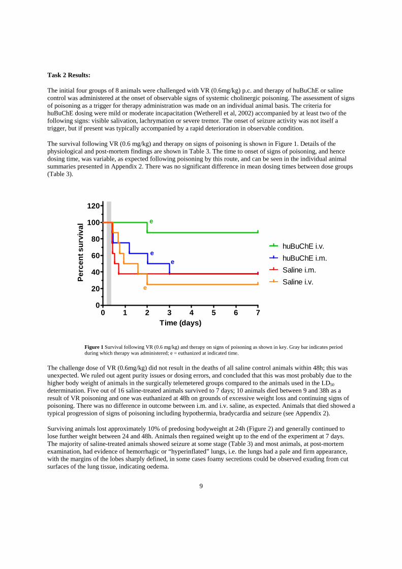
The initial four groups of 8 animals were challenged with VR (0.6mg/kg) p.c. and therapy of huBuChE or saline control was administered at the onset of observable signs of systemic cholinergic poisoning. The assessment of signs of poisoning as a trigger for therapy administration was made on an individual animal basis. The criteria for huBuChE dosing were mild or moderate incapacitation (Wetherell et al, 2002) accompanied by at least two of the following signs: visible salivation, lachrymation or severe tremor. The onset of seizure activity was not itself a trigger, but if present was typically accompanied by a rapid deterioration in observable condition.
The survival following VR (0.6 mg/kg) and therapy on signs of poisoning is shown in Figure 1. Details of the physiological and post-mortem findings are shown in Table 3. The time to onset of signs of poisoning, and hence dosing time, was variable, as expected following poisoning by this route, and can be seen in the individual animal summaries presented in Appendix 2. There was no significant difference in mean dosing times between dose groups (Table 3).
Figure 1 Survival following VR (0.6 mg/kg) and therapy on signs of poisoning as shown in key. Gray bar indicates period during which therapy was administered; e = euthanized at indicated time.
The challenge dose of VR (0.6mg/kg) did not result in the deaths of all saline control animals within 48h; this was unexpected. We ruled out agent purity issues or dosing errors, and concluded that this was most probably due to the higher body weight of animals in the surgically telemetered groups compared to the animals used in the LD50 determination. Five out of 16 saline-treated animals survived to 7 days; 10 animals died between 9 and 38h as a result of VR poisoning and one was euthanized at 48h on grounds of excessive weight loss and continuing signs of poisoning. There was no difference in outcome between i.m. and i.v. saline, as expected. Animals that died showed a typical progression of signs of poisoning including hypothermia, bradycardia and seizure (see Appendix 2).
Surviving animals lost approximately 10% of predosing bodyweight at 24h (Figure 2) and generally continued to lose further weight between 24 and 48h. Animals then regained weight up to the end of the experiment at 7 days. The majority of saline-treated animals showed seizure at some stage (Table 3) and most animals, at post-mortem examination, had evidence of hemorrhagic or “hyperinflated” lungs, i.e. the lungs had a pale and firm appearance, with the margins of the lobes sharply defined, in some cases foamy secretions could be observed exuding from cut surfaces of the lung tissue, indicating oedema.
0 1 2 3 4 5 6 70
20
40
60
80
100
120
Saline i.m.
huBuChE i.m.
Saline i.v.
huBuChE i.v.e
e
e
e
Time (days)
Pe
rce
nt
su
rviv
al
10
VR dose (mg/kg)
Therapy and administration time
Number dosed
Body weight on day of VR challenge (g) (mean ± SD)
Time of dosing (h) (mean ± SD)
Survival48h
Survival7 days
Brady-cardia
Seizure Abnormal lungs at post mortem
Abnormal GI tract at post mortem
Heart/lung wet weight as % of bodyweight (mean ± SD), n
0.6 huBuChE i.m. (signs of poisoning)
8 467.7 ± 27.7
7.25 ± 1.90
5/8 3/8 5/8 6/8 7/8 1/8 1.28 ± 0.18, 5
0.6 huBuChE i.v. (signs of poisoning)
8 472.6 ± 34.0
7.47 ± 1.53
8/8 7/8 4/8 6/8 5/8 0/8 1.44 ± 0.35, 8
0.6 Saline i.m. (signs of poisoning)
8 469.5 ± 44.2
7.36 ± 1.42
3/8 3/8 7/8 7/8 7/8 2/8 1.18 ± 0.01, 3
0.6 Saline i.v. (signs of poisoning)
8 464.7 ± 24.2
8.03 ± 1.30
3/8 2/8 7/8 7/8 6/8 4/8 1.77 ± 0.88, 3
0.74 huBuChE i.m. (2 hours post VR)
8 491.6 ± 35.9
n.a. 7/8 7/8 3/8 1/8 1/8 0/8 1.19 ± 0.16, 8
0.74 Saline i.m. (2 hours post VR)
8 492.2 ± 39.2
n.a. 0/8 0/8 8/8 8/8 8/8 3/8 1.10 ±0.60, 4
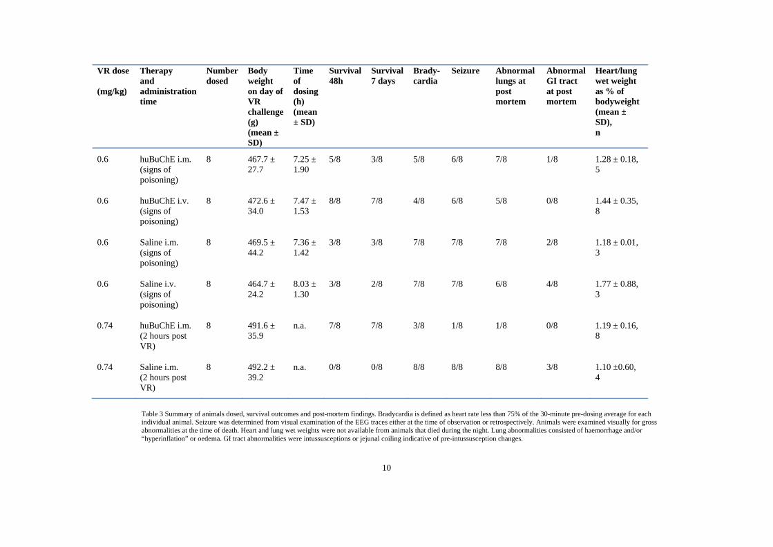
Table 3 Summary of animals dosed, survival outcomes and post-mortem findings. Bradycardia is defined as heart rate less than 75% of the 30-minute pre-dosing average for each individual animal. Seizure was determined from visual examination of the EEG traces either at the time of observation or retrospectively. Animals were examined visually for gross abnormalities at the time of death. Heart and lung wet weights were not available from animals that died during the night. Lung abnormalities consisted of haemorrhage and/or “hyperinflation” or oedema. GI tract abnormalities were intussusceptions or jejunal coiling indicative of pre-intussusception changes.

11
Figure 2 Bodyweight (relative to day of dosing) of surviving animals following VR challenge (0.6 mg/kg) and therapy on signs of poisoning as shown in key. Data shown as mean ± SD. Group sizes are shown on the graph to indicate the times at which animals died or were euthanized.
When huBuChE was administered by the i.m. route on signs of poisoning, 3/8 animals died between 10 and 29h as a result of VR poisoning; a further 2 were euthanized (at 48h and 72h) due to excessive weight loss. One of the animals that died, at 28h, had GI tract abnormalities consistent with the start of intussusception. All the animals that died or were euthanized showed hyperinflation or haemorrhage in the lungs on post-mortem examination. Three animals survived to the end of the experiment at 7 days, two of which had mildly haemorrhaghic lungs. A marked improvement in survival was obtained by the administration of therapy via the i.v. route (Table 3). In this group, all 8 animals were alive at 48h; at this time, one animal was euthanized due to a combination of excessive weight loss (24%) and poor condition. All remaining animals were in good condition at 48h, most having regained normal posture, and survived to the end of the experiment at 7 days, with 6 of the 7 having exceeded their dosing weight by that time (Figure 2). The incidence of seizure was similar in huBuChE-treated animals to saline controls, but following therapy the seizure activity resolved, more rapidly in the i.v. group (sees Appendix 2). No animals in the i.v. huBuChE group had GI tract abnormalities, and in 3 of the 7 surviving animals there were no gross abnormalities of the lungs seen at post-mortem examination at 7 days. This increase in survival was expected, since huBuChE administered by the i.v. route would be expected to have a higher bioavailability and a shorter time to achieve peak plasma concentrations. This is supported by the rapid reversal in physiological abnormalities observed following i.v. huBuChE in these animals (Appendix 2). These results confirm those seen in a similar study with VX-poisoned guinea-pigs (Mumford et al 2011).
For the final two dose groups, the aim was to investigate the protective effect of huBuChE when administered after VR poisoning, but prior to the onset of observable signs of systemic cholinergic poisoning. Based on the results from the first 4 groups, it was decided that a 2-hour dosing point would be appropriate. In view of the higher than expected survival in the saline-treated animals when challenged with 0.6 mg/kg, we increased the challenge dose in the 2-hour group to 0.74 mg/kg (the upper 95% CI of the 48-h LD99).
Following VR (0.74mg/kg) and saline (i.m.) at 2 hours, all animals died between 5 and 22h (Figure 3). The incidence of seizure was 100% and all animals had hyperinflated and/or haemorrhaghic lungs. Several animals (3/8)
-6 -4 -2 0 2 4 6 870
80
90
100
110
120
huBuChE im signs
saline im signs
huBUChE iv signs
saline iv signs4
32
3
6
5
4 3
8
73
7
2
3
Days
Bo
dyw
eig
ht
as
% o
f D
ay 0
12
had some visible abnormalities of the GI tract when examined at post-mortem; these generally consisted of jejunal coiling, sometimes accompanied by evidence of bleeding into the lumen of the GI tract (Table 3).
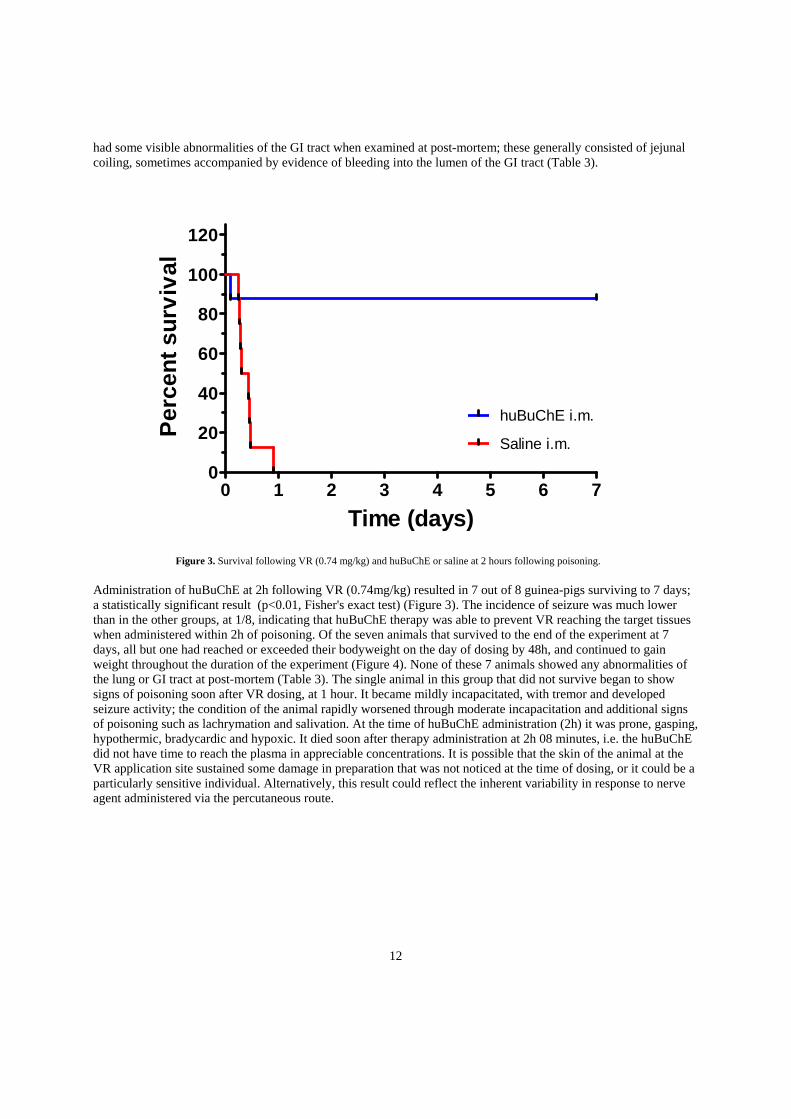
Figure 3. Survival following VR (0.74 mg/kg) and huBuChE or saline at 2 hours following poisoning.
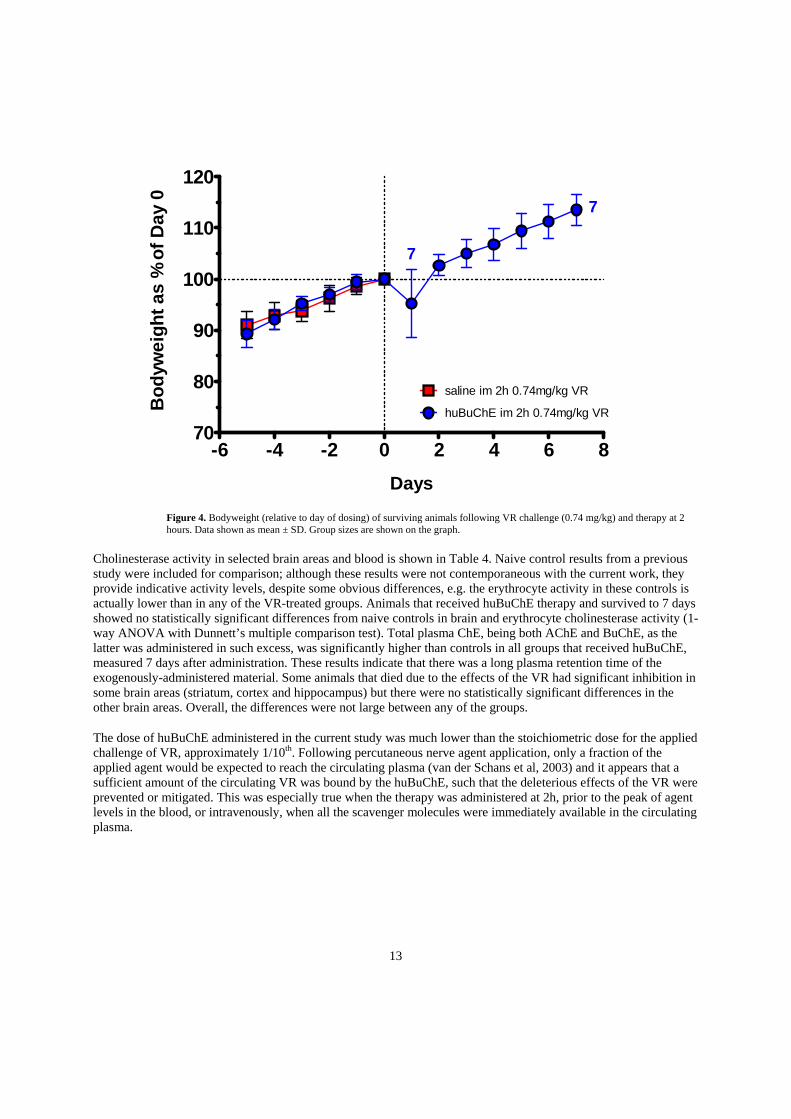
Administration of huBuChE at 2h following VR (0.74mg/kg) resulted in 7 out of 8 guinea-pigs surviving to 7 days; a statistically significant result (p<0.01, Fisher's exact test) (Figure 3). The incidence of seizure was much lower than in the other groups, at 1/8, indicating that huBuChE therapy was able to prevent VR reaching the target tissues when administered within 2h of poisoning. Of the seven animals that survived to the end of the experiment at 7 days, all but one had reached or exceeded their bodyweight on the day of dosing by 48h, and continued to gain weight throughout the duration of the experiment (Figure 4). None of these 7 animals showed any abnormalities of the lung or GI tract at post-mortem (Table 3). The single animal in this group that did not survive began to show signs of poisoning soon after VR dosing, at 1 hour. It became mildly incapacitated, with tremor and developed seizure activity; the condition of the animal rapidly worsened through moderate incapacitation and additional signs of poisoning such as lachrymation and salivation. At the time of huBuChE administration (2h) it was prone, gasping, hypothermic, bradycardic and hypoxic. It died soon after therapy administration at 2h 08 minutes, i.e. the huBuChE did not have time to reach the plasma in appreciable concentrations. It is possible that the skin of the animal at the VR application site sustained some damage in preparation that was not noticed at the time of dosing, or it could be a particularly sensitive individual. Alternatively, this result could reflect the inherent variability in response to nerve agent administered via the percutaneous route.
0 1 2 3 4 5 6 70
20
40
60
80
100
120
Saline i.m.
huBuChE i.m.
Time (days)
Per
cen
t su
rviv
al
13
Figure 4. Bodyweight (relative to day of dosing) of surviving animals following VR challenge (0.74 mg/kg) and therapy at 2 hours. Data shown as mean ± SD. Group sizes are shown on the graph.
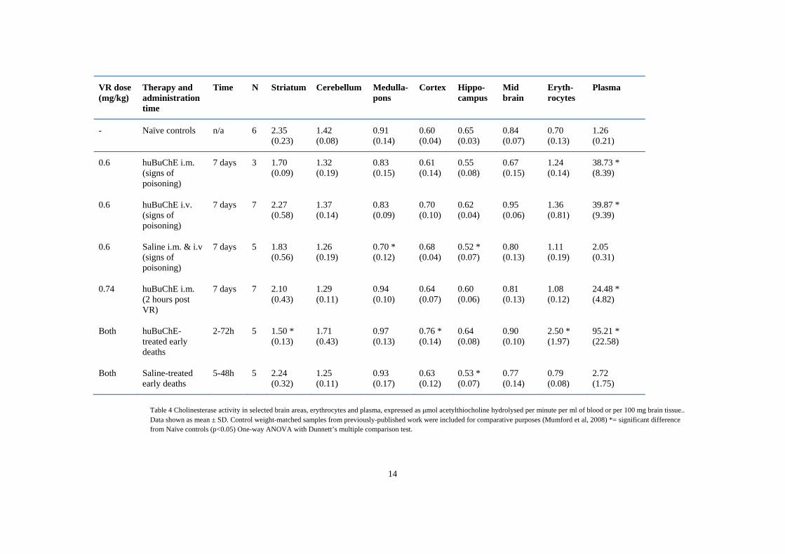
Cholinesterase activity in selected brain areas and blood is shown in Table 4. Naive control results from a previous study were included for comparison; although these results were not contemporaneous with the current work, they provide indicative activity levels, despite some obvious differences, e.g. the erythrocyte activity in these controls is actually lower than in any of the VR-treated groups. Animals that received huBuChE therapy and survived to 7 days showed no statistically significant differences from naive controls in brain and erythrocyte cholinesterase activity (1-way ANOVA with Dunnett’s multiple comparison test). Total plasma ChE, being both AChE and BuChE, as the latter was administered in such excess, was significantly higher than controls in all groups that received huBuChE, measured 7 days after administration. These results indicate that there was a long plasma retention time of the exogenously-administered material. Some animals that died due to the effects of the VR had significant inhibition in some brain areas (striatum, cortex and hippocampus) but there were no statistically significant differences in the other brain areas. Overall, the differences were not large between any of the groups.
The dose of huBuChE administered in the current study was much lower than the stoichiometric dose for the applied challenge of VR, approximately 1/10th. Following percutaneous nerve agent application, only a fraction of the applied agent would be expected to reach the circulating plasma (van der Schans et al, 2003) and it appears that a sufficient amount of the circulating VR was bound by the huBuChE, such that the deleterious effects of the VR were prevented or mitigated. This was especially true when the therapy was administered at 2h, prior to the peak of agent levels in the blood, or intravenously, when all the scavenger molecules were immediately available in the circulating plasma.
-6 -4 -2 0 2 4 6 870
80
90
100
110
120
huBuChE im 2h 0.74mg/kg VR
saline im 2h 0.74mg/kg VR
7
7
Days
Bo
dyw
eig
ht
as %
of
Day
0
14
VR dose (mg/kg)
Therapy and administration time
Time N Striatum Cerebellum Medulla-pons
Cortex Hippo-campus
Mid brain
Eryth-rocytes
Plasma
- Naïve controls n/a 6 2.35 (0.23)
1.42 (0.08)
0.91 (0.14)
0.60 (0.04)
0.65 (0.03)
0.84 (0.07)
0.70 (0.13)
1.26 (0.21)
0.6 huBuChE i.m. (signs of poisoning)
7 days 3 1.70 (0.09)
1.32 (0.19)
0.83 (0.15)
0.61 (0.14)
0.55 (0.08)
0.67 (0.15)
1.24 (0.14)
38.73 * (8.39)
0.6 huBuChE i.v. (signs of poisoning)
7 days 7 2.27 (0.58)
1.37 (0.14)
0.83 (0.09)
0.70 (0.10)
0.62 (0.04)
0.95 (0.06)
1.36 (0.81)
39.87 * (9.39)
0.6 Saline i.m. & i.v (signs of poisoning)
7 days 5 1.83 (0.56)
1.26 (0.19)
0.70 * (0.12)
0.68 (0.04)
0.52 * (0.07)
0.80 (0.13)
1.11 (0.19)
2.05 (0.31)
0.74 huBuChE i.m. (2 hours post VR)
7 days 7 2.10 (0.43)
1.29 (0.11)
0.94 (0.10)
0.64 (0.07)
0.60 (0.06)
0.81 (0.13)
1.08 (0.12)
24.48 * (4.82)
Both huBuChE-treated early deaths
2-72h 5 1.50 * (0.13)
1.71 (0.43)
0.97 (0.13)
0.76 * (0.14)
0.64 (0.08)
0.90 (0.10)
2.50 * (1.97)
95.21 * (22.58)
Both Saline-treated early deaths
5-48h 5 2.24 (0.32)
1.25 (0.11)
0.93 (0.17)
0.63 (0.12)
0.53 * (0.07)
0.77 (0.14)
0.79 (0.08)
2.72 (1.75)
Table 4 Cholinesterase activity in selected brain areas, erythrocytes and plasma, expressed as μmol acetylthiocholine hydrolysed per minute per ml of blood or per 100 mg brain tissue.. Data shown as mean ± SD. Control weight-matched samples from previously-published work were included for comparative purposes (Mumford et al, 2008) *= significant difference from Naïve controls (p<0.05) One-way ANOVA with Dunnett’s multiple comparison test.
15
Key Research Accomplishments
The percutaneous toxicity of VR was determined to have a 24h LD50 of 0.45mg/kg (0.36 – 0.54 95% CI) and a 48-h LD50 of 0.38 mg/kg (0.31-0.45 95% CI).
The efficacy of huBuChE as post-exposure therapy was determined following percutaneous VR challenge. Therapy administration prior to the onset of observable signs of systemic cholinergic signs of poisoning was more efficacious than when therapy was delayed. Therapy delivered intravenously was more efficacious that intramuscular huBuChE administration.
Brain cholinesterase activity was preserved in huBuChE-treated animals.
Plasma cholinesterase activity was significantly elevated at 7 days in huBuChE-treated guinea-pigs compared to control values, indicating a long plasma retention time of the bioscavenger.
Protection was afforded by a dose of huBuChE that was substantially lower than a stoichiometric ratio for the applied dose of agent.
Reportable Outcomes
An abstract (#244), titled "The Utility of Human Plasma-Derived Butyrylcholinesterase (huBuChE) as a Therapeutic Measure in the Absence of Pre-Treatment or Conventional Post-Poisoning Therapies Against Nerve Agent", was submitted and has been accepted as an ORAL PRESENTATION during the Chemical and Biological Defense Science and Technology (CBD S&T) Conference on 14-18 November 2011 in Las Vegas, Nevada.
Conclusions
This study has shown that, following percutaneous nerve agent poisoning, post-exposure therapy using huBuChE has potential as a treatment strategy. This finding complements recently published studies demonstrating the efficacy of huBuChE and recombinant butyrylcholinesterase following percutaneous VX poisoning in guinea-pigs and swine (Mumford et al, 2011, Mumford and Troyer, 2011, Lenz et al, 2010 and Tenn et al, 2008). When huBuChE therapy was given 2h following an increased challenge dose of 0.74 mg/kg VR, there was 87.5% survival in huBuChE-treated animals. Previous studies have demonstrated that bioscavenger afforded almost complete protection when administered prior to the onset of observable cholinergic signs of VX poisoning, (Mumford et al, 2011, Mumford and Troyer, 2011). The present study replicated this finding for p.c. VR challenge. There was a limited therapeutic window after percutaneous VR poisoning and the protective effect of huBuChE decreased when i.m. administration was delayed to the onset of signs of poisoning. The outcome was improved substantially when therapy was administered via the i.v. route. The challenge dose chosen (0.6 mg/kg, or 1.3 × LD50) produced the expected range of effects and onset times, but a higher than expected proportion of saline-treated animals survived the VR challenge. This result reflects the uncertainty in the derived LD50 values, and the inherent variability of the p.c. dosing route. The onset of observable signs of cholinergic poisoning used as the trigger for huBuChE dosing was between 5h and 9h following VR challenge. These results support other work showing that post-exposure therapy with bioscavenger is an effective treatment strategy, and that therapy using huBuChE via the i.v. route is more effective than therapy via the i.m. route.
16
References
Ellman,GL, Courtney,D, Andres,V, Featherstone,RM. A new and rapid colorimetric determination of acetylcholinesterase activity. Biochemical Pharmacology 7, 88-95. 14-11-1961.
Kolarich D, Weber A, Pabst M, Stadlmann J, Teschner W, Ehrlich H. Glycoproteornic characterization of butyrylcholinesterase from human plasma. Proteomics 2008;8:254-63.
Lenz DE, Clarkson ED, Schultz SM, Cerasoli DM. Butyrylcholinesterase as a therapeutic drug for protection against percutaneous VX. Chem Biol Interact 2010;187:249-52.
Lowry,OH, Rosenbrough,NJ, Lewis Farr,A, Randall,RJ. Protein measurement with folin phenol reagent. Biological Chemistry 193, 265-275. 28-5-1951.
Mumford H, Price ME, Wetherell JR. A novel approach to assessing percutaneous VX poisoning in the conscious guinea-pig. Journal of Applied Toxicology 2008;28:694-702.
Mumford H, Price ME, Cerasoli DM, Teschner W, Ehrlich H, Schwarz HP. Efficacy and physiological effects of human butyrycholinesterase as a post-exposure therapy against percutaneous poisoning by VX in the guinea-pig. Chem Biol Interact 2010;187:304-8.
Mumford, H., Price, M.E., Lenz, D.E., Cerasoli, D.M. Post-exposure therapy with human butyrylcholinesterase following percutaneous VX challenge in guinea-pigs. Clinical Toxicology 2011;49:287-297
Mumford, H., Troyer, J.K. Post-exposure therapy with recombinant human BuChE following percutaneous VX challenge in guinea-pigs. Toxicology Letters, 2011, 206,29-34.
Russell, K. G.; Eccleston, J. A.; Lewis, S. M.; Woods, D. C. Design considerations for small experiments and simple logistic regression. Journal of Statistical Computation and Simulation 2009;79:81-91.
Stallard, N. Optimal adaptive designs for acute oral toxicity assessment. Journal of Statistical Planning and Inference 2006,136:1781-1799.
Tenn CC, Mikler JR, Hill I, Weatherby K, Garrett M, Caddy N. Recombinant Human Butyrylcholinesterase as a Therapeutic Agent to Counteract the Effects of VX Toxicity in Domestic Swine. J Med Chem Def 2008;6.
van der Schans MJ, Lander BJ, van der Wiel H, Langenberg JP,Benschop HP. Toxicokinetics of the nerve agent (+/-)-VX in anesthetized and atropinized hairless guinea pigs and marmosets after intravenous and percutaneous administration. Toxicol Appl Pharmacol 2003; 191:48–62.
Wetherell J, Hall T, Passingham S. Physostigmine and hyoscine improves protection against the lethal and incapacitating effects of nerve agent poisoning in the guinea-pig. Neurotoxicology 2002;23:341-9.

17
Appendix 1 – Raw Physiology Data
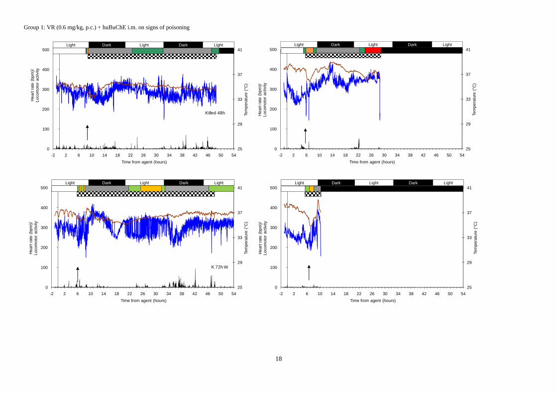
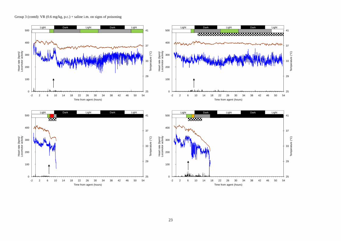
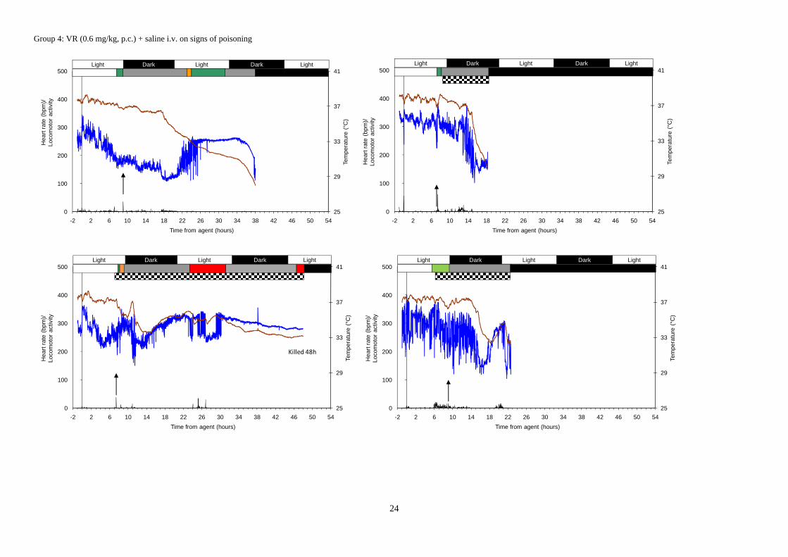
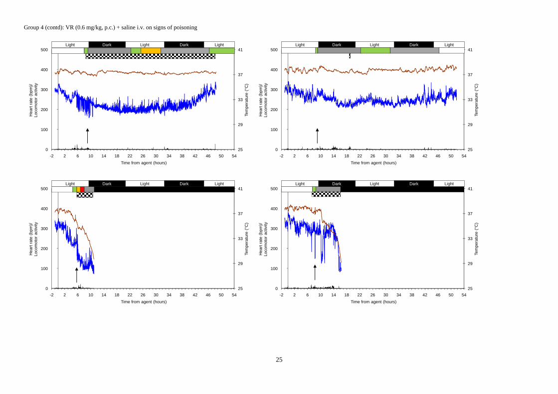
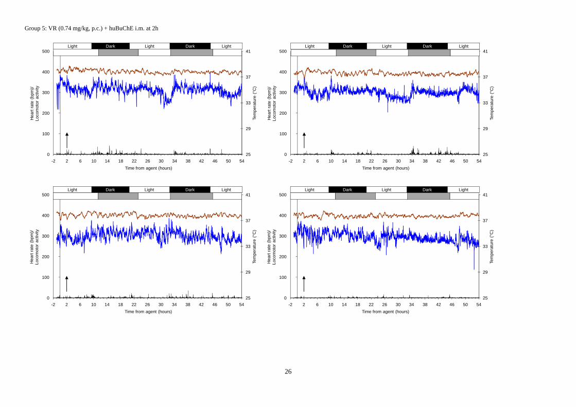
This appendix illustrates the responses of individual animals in terms of the physiological indicators measured.
Each panel represents one animal; the main panel shows heart rate (blue line, left axis); locomotor activity counts (arbitrary units) (black histogram, left axis) body temperature (brown line, right axis). At the top of each panel is the indication of the day:night periods; below that, the duration of incapacitation during observation periods is shown, according to the following key: white = not incapacitated; green = mildly incapacitated; orange = moderately incapacitated; red = substantially incapacitated; grey = not observed; black = dead. A hatched area below the incapacitation indicates the duration of seizure activity, if present, as determined from the EEG recordings. The time of therapy (or saline) administration is indicated by a vertical arrow in the panel.
18
Group 1: VR (0.6 mg/kg, p.c.) + huBuChE i.m. on signs of poisoning
25
29
33
37
41
0
100
200
300
400
500
-2 2 6 10 14 18 22 26 30 34 38 42 46 50 54
Tem
pera
ture
(°C
)
Hea
rt r
ate
(bpm
)/
Loco
mot
or a
ctiv
ity
Time from agent (hours)
DarkLightLight Dark Light
Killed 48h
25
29
33
37
41
0
100
200
300
400
500
-2 2 6 10 14 18 22 26 30 34 38 42 46 50 54
Tem
pera
ture
(°C
)
Hea
rt r
ate
(bpm
)/
Loco
mot
or a
ctiv
ity
Time from agent (hours)
DarkLightLight Dark Light
25
29
33
37
41
0
100
200
300
400
500
-2 2 6 10 14 18 22 26 30 34 38 42 46 50 54
Tem
pera
ture
(°C
)
Hea
rt r
ate
(bp
m)/
Lo
com
otor
act
ivity
Time from agent (hours)
DarkLightLight Dark Light
K 72h W
25
29
33
37
41
0
100
200
300
400
500
-2 2 6 10 14 18 22 26 30 34 38 42 46 50 54
Tem
pera
ture
(°C
)
Hea
rt r
ate
(bp
m)/
Lo
com
otor
act
ivity
Time from agent (hours)
DarkLightLight Dark Light

19
Group 1 (contd): VR (0.6 mg/kg, p.c.) + huBuChE i.m. on signs of poisoning
25
29
33
37
41
0
100
200
300
400
500
-2 2 6 10 14 18 22 26 30 34 38 42 46 50 54
Tem
pera
ture
(°C
)
Hea
rt r
ate
(bp
m)/
L
ocom
otor
act
ivity
Time from agent (hours)
DarkLightLight Dark Light
25
29
33
37
41
0
100
200
300
400
500
-2 2 6 10 14 18 22 26 30 34 38 42 46 50 54
Tem
pera
ture
(°C
)
Hea
rt r
ate
(bp
m)/
Lo
com
otor
act
ivity
Time from agent (hours)
DarkLightLight Dark Light
25
29
33
37
41
0
100
200
300
400
500
-2 2 6 10 14 18 22 26 30 34 38 42 46 50 54
Tem
pera
ture
(°C
)
Hea
rt r
ate
(bp
m)/
Lo
com
otor
act
ivity
Time from agent (hours)
DarkLightLight Dark Light
25
29
33
37
41
0
100
200
300
400
500
-2 2 6 10 14 18 22 26 30 34 38 42 46 50 54
Tem
pera
ture
(°C
)
Hea
rt r
ate
(bp
m)/
Lo
com
otor
act
ivity
Time from agent (hours)
DarkLightLight Dark Light

20
Group 2: VR (0.6 mg/kg, p.c.) + huBuChE i.v. on signs of poisoning
25
29
33
37
41
0
100
200
300
400
500
-2 2 6 10 14 18 22 26 30 34 38 42 46 50 54
Tem
pera
ture
(°C
)
Hea
rt r
ate
(bp
m)/
Lo
com
otor
act
ivity
Time from agent (hours)
DarkLightLight Dark Light
25
29
33
37
41
0
100
200
300
400
500
-2 2 6 10 14 18 22 26 30 34 38 42 46 50 54
Tem
pera
ture
(°C
)
Hea
rt r
ate
(bp
m)/
Lo
com
otor
act
ivity
Time from agent (hours)
DarkLightLight Dark Light
Killed 48h
25
29
33
37
41
0
100
200
300
400
500
-2 2 6 10 14 18 22 26 30 34 38 42 46 50 54
Tem
pera
ture
(°C
)
Hea
rt r
ate
(bp
m)/
Lo
com
otor
act
ivity
Time from agent (hours)
DarkLightLight Dark Light
25
29
33
37
41
0
100
200
300
400
500
-2 2 6 10 14 18 22 26 30 34 38 42 46 50 54
Tem
pera
ture
(°C
)
Hea
rt r
ate
(bp
m)/
Lo
com
otor
act
ivity
Time from agent (hours)
DarkLightLight Dark Light

21
Group 2 (contd): VR (0.6 mg/kg, p.c.) + huBuChE i.v. on signs of poisoning
25
29
33
37
41
0
100
200
300
400
500
-2 2 6 10 14 18 22 26 30 34 38 42 46 50 54
Tem
pera
ture
(°C
)
Hea
rt r
ate
(bpm
)/
Loco
mot
or a
ctiv
ity
Time from agent (hours)
DarkLightLight Dark Light
25
29
33
37
41
0
100
200
300
400
500
-2 2 6 10 14 18 22 26 30 34 38 42 46 50 54
Tem
pera
ture
(°C
)
Hea
rt r
ate
(bp
m)/
Lo
com
otor
act
ivity
Time from agent (hours)
DarkLightLight Dark Light
25
29
33
37
41
0
100
200
300
400
500
-2 2 6 10 14 18 22 26 30 34 38 42 46 50 54
Tem
pera
ture
(°C
)
Hea
rt r
ate
(bp
m)/
Lo
com
otor
act
ivity
Time from agent (hours)
DarkLightLight Dark Light
25
29
33
37
41
0
100
200
300
400
500
-2 2 6 10 14 18 22 26 30 34 38 42 46 50 54
Tem
pera
ture
(°C
)
Hea
rt r
ate
(bp
m)/
Lo
com
otor
act
ivity
Time from agent (hours)
DarkLightLight LightDark

22
Group 3: VR (0.6 mg/kg, p.c.) + saline i.m. on signs of poisoning
25
29
33
37
41
45
0
100
200
300
400
500
-2 2 6 10 14 18 22 26 30 34 38 42 46 50 54
Tem
pera
ture
(°C
)
Hea
rt r
ate
(bpm
)/
Loco
mot
or a
ctiv
ity
Time from agent (hours)
DarkLightLight Dark Light
25
29
33
37
41
0
100
200
300
400
500
-2 2 6 10 14 18 22 26 30 34 38 42 46 50 54
Tem
pera
ture
(°C
)
Hea
rt r
ate
(bp
m)/
Lo
com
otor
act
ivity
Time from agent (hours)
DarkLightLight Dark Light
25
29
33
37
41
0
100
200
300
400
500
-2 2 6 10 14 18 22 26 30 34 38 42 46 50 54
Tem
pera
ture
(°C
)
Hea
rt r
ate
(bp
m)/
Lo
com
otor
act
ivity
Time from agent (hours)
DarkLightLight Dark Light
25
29
33
37
41
0
100
200
300
400
500
-2 2 6 10 14 18 22 26 30 34 38 42 46 50 54
Tem
pera
ture
(°C
)
Hea
rt r
ate
(bp
m)/
Lo
com
otor
act
ivity
Time from agent (hours)
DarkLightLight Dark Light
23
Group 3 (contd): VR (0.6 mg/kg, p.c.) + saline i.m. on signs of poisoning
25
29
33
37
41
0
100
200
300
400
500
-2 2 6 10 14 18 22 26 30 34 38 42 46 50 54
Tem
pera
ture
(°C
)
Hea
rt r
ate
(bp
m)/
Lo
com
otor
act
ivity
Time from agent (hours)
DarkLightLight Dark Light
25
29
33
37
41
0
100
200
300
400
500
-2 2 6 10 14 18 22 26 30 34 38 42 46 50 54
Tem
pera
ture
(°C
)
Hea
rt r
ate
(bp
m)/
Lo
com
otor
act
ivity
Time from agent (hours)
DarkLightLight Dark Light
25
29
33
37
41
0
100
200
300
400
500
-2 2 6 10 14 18 22 26 30 34 38 42 46 50 54
Tem
pera
ture
(°C
)
Hea
rt r
ate
(bp
m)/
Lo
com
otor
act
ivity
Time from agent (hours)
DarkLightLight Dark Light
25
29
33
37
41
0
100
200
300
400
500
-2 2 6 10 14 18 22 26 30 34 38 42 46 50 54
Tem
pera
ture
(°C
)
Hea
rt r
ate
(bp
m)/
Lo
com
otor
act
ivity
Time from agent (hours)
DarkLightLight Dark Light
24
Group 4: VR (0.6 mg/kg, p.c.) + saline i.v. on signs of poisoning
25
29
33
37
41
0
100
200
300
400
500
-2 2 6 10 14 18 22 26 30 34 38 42 46 50 54
Tem
pera
ture
(°C
)
Hea
rt r
ate
(bpm
)/
Loco
mot
or a
ctiv
ity
Time from agent (hours)
DarkLightLight Dark Light
25
29
33
37
41
0
100
200
300
400
500
-2 2 6 10 14 18 22 26 30 34 38 42 46 50 54
Tem
pera
ture
(°C
)
Hea
rt r
ate
(bp
m)/
Lo
com
otor
act
ivity
Time from agent (hours)
DarkLightLight Dark Light
25
29
33
37
41
0
100
200
300
400
500
-2 2 6 10 14 18 22 26 30 34 38 42 46 50 54
Tem
pera
ture
(°C
)
Hea
rt r
ate
(bp
m)/
Lo
com
otor
act
ivity
Time from agent (hours)
DarkLightLight Dark Light
Killed 48h
25
29
33
37
41
0
100
200
300
400
500
-2 2 6 10 14 18 22 26 30 34 38 42 46 50 54
Tem
pera
ture
(°C
)
Hea
rt r
ate
(bp
m)/
Lo
com
otor
act
ivity
Time from agent (hours)
DarkLightLight Dark Light
25
Group 4 (contd): VR (0.6 mg/kg, p.c.) + saline i.v. on signs of poisoning
25
29
33
37
41
0
100
200
300
400
500
-2 2 6 10 14 18 22 26 30 34 38 42 46 50 54
Tem
pera
ture
(°C
)
Hea
rt r
ate
(bp
m)/
Lo
com
otor
act
ivity
Time from agent (hours)
DarkLightLight Dark Light
25
29
33
37
41
0
100
200
300
400
500
-2 2 6 10 14 18 22 26 30 34 38 42 46 50 54
Tem
pera
ture
(°C
)
Hea
rt r
ate
(bp
m)/
Lo
com
otor
act
ivity
Time from agent (hours)
DarkLightLight Dark Light
25
29
33
37
41
0
100
200
300
400
500
-2 2 6 10 14 18 22 26 30 34 38 42 46 50 54
Tem
pera
ture
(°C
)
Hea
rt r
ate
(bp
m)/
Lo
com
otor
act
ivity
Time from agent (hours)
DarkLightLight LightDark
25
29
33
37
41
0
100
200
300
400
500
-2 2 6 10 14 18 22 26 30 34 38 42 46 50 54
Tem
pera
ture
(°C
)
Hea
rt r
ate
(bp
m)/
Lo
com
otor
act
ivity
Time from agent (hours)
DarkLightLight LightDark
26
Group 5: VR (0.74 mg/kg, p.c.) + huBuChE i.m. at 2h
25
29
33
37
41
0
100
200
300
400
500
-2 2 6 10 14 18 22 26 30 34 38 42 46 50 54
Tem
pera
ture
(°C
)
Hea
rt r
ate
(bp
m)/
Lo
com
otor
act
ivity
Time from agent (hours)
DarkLightLight LightDark
25
29
33
37
41
0
100
200
300
400
500
-2 2 6 10 14 18 22 26 30 34 38 42 46 50 54
Tem
pera
ture
(°C
)
Hea
rt r
ate
(bp
m)/
Lo
com
otor
act
ivity
Time from agent (hours)
DarkLightLight LightDark
25
29
33
37
41
0
100
200
300
400
500
-2 2 6 10 14 18 22 26 30 34 38 42 46 50 54
Tem
pera
ture
(°C
)
Hea
rt r
ate
(bpm
)/
Loco
mot
or a
ctiv
ity
Time from agent (hours)
DarkLightLight LightDark
25
29
33
37
41
0
100
200
300
400
500
-2 2 6 10 14 18 22 26 30 34 38 42 46 50 54
Tem
pera
ture
(°C
)
Hea
rt r
ate
(bpm
)/
Loco
mot
or a
ctiv
ity
Time from agent (hours)
DarkLightLight LightDark
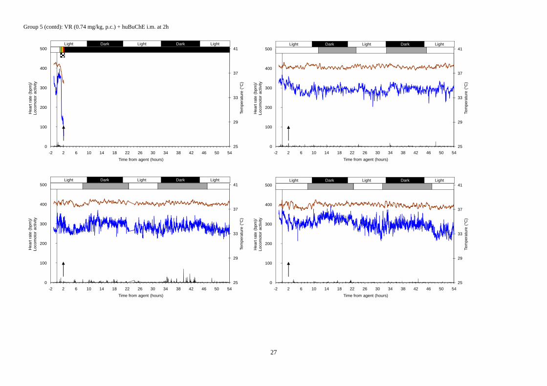
27
Group 5 (contd): VR (0.74 mg/kg, p.c.) + huBuChE i.m. at 2h
25
29
33
37
41
0
100
200
300
400
500
-2 2 6 10 14 18 22 26 30 34 38 42 46 50 54
Tem
pera
ture
(°C
)
Hea
rt r
ate
(bp
m)/
Lo
com
otor
act
ivity
Time from agent (hours)
DarkLightLight LightDark
25
29
33
37
41
0
100
200
300
400
500
-2 2 6 10 14 18 22 26 30 34 38 42 46 50 54
Tem
pera
ture
(°C
)
Hea
rt r
ate
(bpm
)/
Loco
mot
or a
ctiv
ity
Time from agent (hours)
DarkLightLight LightDark
25
29
33
37
41
0
100
200
300
400
500
-2 2 6 10 14 18 22 26 30 34 38 42 46 50 54
Tem
pera
ture
(°C
)
Hea
rt r
ate
(bpm
)/
Loco
mot
or a
ctiv
ity
Time from agent (hours)
DarkLightLight LightDark
25
29
33
37
41
0
100
200
300
400
500
-2 2 6 10 14 18 22 26 30 34 38 42 46 50 54
Tem
pera
ture
(°C
)
Hea
rt r
ate
(bpm
)/
Loco
mot
or a
ctiv
ity
Time from agent (hours)
DarkLightLight LightDark

28
Group 6: VR (0.74 mg/kg, p.c.) + saline i.m. at 2h
25
29
33
37
41
0
100
200
300
400
500
-2 2 6 10 14 18 22 26 30 34 38 42 46 50 54
Tem
pera
ture
(°C
)
Hea
rt r
ate
(bp
m)/
Lo
com
otor
act
ivity
Time from agent (hours)
DarkLightLight LightDark
25
29
33
37
41
0
100
200
300
400
500
-2 2 6 10 14 18 22 26 30 34 38 42 46 50 54
Tem
pera
ture
(°C
)
Hea
rt r
ate
(bp
m)/
Lo
com
otor
act
ivity
Time from agent (hours)
DarkLightLight LightDark
25
29
33
37
41
0
100
200
300
400
500
-2 2 6 10 14 18 22 26 30 34 38 42 46 50 54
Tem
pera
ture
(°C
)
Hea
rt r
ate
(bp
m)/
Lo
com
otor
act
ivity
Time from agent (hours)
DarkLightLight LightDark
25
29
33
37
41
0
100
200
300
400
500
-2 2 6 10 14 18 22 26 30 34 38 42 46 50 54
Tem
pera
ture
(°C
)
Hea
rt r
ate
(bpm
)/
Loco
mot
or a
ctiv
ity
Time from agent (hours)
DarkLightLight LightDark

29
Group 6 (contd): VR (0.74 mg/kg, p.c.) + saline i.m. at 2h
25
29
33
37
41
0
100
200
300
400
500
-2 2 6 10 14 18 22 26 30 34 38 42 46 50 54
Tem
pera
ture
(°C
)
Hea
rt r
ate
(bp
m)/
Lo
com
otor
act
ivity
Time from agent (hours)
DarkLightLight LightDark
25
29
33
37
41
0
100
200
300
400
500
-2 2 6 10 14 18 22 26 30 34 38 42 46 50 54
Tem
pera
ture
(°C
)
Hea
rt r
ate
(bp
m)/
Lo
com
otor
act
ivity
Time from agent (hours)
DarkLightLight LightDark
25
29
33
37
41
0
100
200
300
400
500
-2 2 6 10 14 18 22 26 30 34 38 42 46 50 54
Tem
pera
ture
(°C
)
Hea
rt r
ate
(bpm
)/
Loco
mot
or a
ctiv
ity
Time from agent (hours)
DarkLightLight LightDark
25
29
33
37
41
0
100
200
300
400
500
-2 2 6 10 14 18 22 26 30 34 38 42 46 50 54
Tem
pera
ture
(°C
)
Hea
rt r
ate
(bpm
)/
Loco
mot
or a
ctiv
ity
Time from agent (hours)
DarkLightLight LightDark

30
Appendix 2 – Research Personnel
This appendix lists the key personnel receiving pay from the research effort:
Helen Mumford (Principal Investigator)
Krystle L Gray
John E H Tattersall
Matthew E Price
E May Irwin