award number: w81xwh-11-2-0084 - apps.dtic.mil · lower after 4 hours in the lp-sw group compared...
TRANSCRIPT

AD_________________ Award Number: W81XWH-11-2-0084 TITLE: Optimization of Lyophilized Plasma for Use in Combat Casualties PRINCIPAL INVESTIGATOR: Martin A. Schreiber, MD CONTRACTING ORGANIZATION: Oregon Health & Science University Portland, OR 97239 REPORT DATE: January 2014 TYPE OF REPORT: Annual PREPARED FOR: U.S. Army Medical Research and Materiel Command Fort Detrick, Maryland 21702-5012 DISTRIBUTION STATEMENT: Approved for Public Release; Distribution Unlimited The views, opinions and/or findings contained in this report are those of the author(s) and should not be construed as an official Department of the Army position, policy or decision unless so designated by other documentation.
REPORT DOCUMENTATION PAGE Form Approved
OMB No. 0704-0188 Public reporting burden for this collection of information is estimated to average 1 hour per response, including the time for reviewing instructions, searching existing data sources, gathering and maintaining the data needed, and completing and reviewing this collection of information. Send comments regarding this burden estimate or any other aspect of this collection of information, including suggestions for reducing this burden to Department of Defense, Washington Headquarters Services, Directorate for Information Operations and Reports (0704-0188), 1215 Jefferson Davis Highway, Suite 1204, Arlington, VA 22202-4302. Respondents should be aware that notwithstanding any other provision of law, no person shall be subject to any penalty for failing to comply with a collection of information if it does not display a currently valid OMB control number. PLEASE DO NOT RETURN YOUR FORM TO THE ABOVE ADDRESS.
1. REPORT DATE
2. REPORT TYPE
Annual 3. DATES COVERED
29-Dec-2012 to 28-Dec-2013 4. TITLE AND SUBTITLE
Optimization of Lyophilized Plasma for Use in Combat Casualties
5a. CONTRACT NUMBER
5b. GRANT NUMBER
W81XWH-11-2-0084 5c. PROGRAM ELEMENT NUMBER
6. AUTHOR(S)
Martin A. Schreiber, Jerome Differding, Tim Lee, Sean McCully, Belinda McCully, Claire Sands, Elizabeth Rick, David Hampton, Scott Louis, and Nathan Anderson
5d. PROJECT NUMBER
5e. TASK NUMBER
email: [email protected]
5f. WORK UNIT NUMBER
7. PERFORMING ORGANIZATION NAME(S) AND ADDRESS(ES)
Oregon Health & Science University
P
rtland, Oregon 97239
AND ADDRESS(ES)
8. PERFORMING ORGANIZATION REPORT NUMBER
Portland, OR 97239
9. SPONSORING / MONITORING AGENCY NAME(S) AND ADDRESS(ES) 10. SPONSOR/MONITOR’S ACRONYM(S)
U.S. Army Medical Research and Materiel Command
Fort Detrick, Maryland 21702-5012
11. SPONSOR/MONITOR’S REPORT
NUMBER(S)
12. DISTRIBUTION / AVAILABILITY STATEMENT
Approved for Public Release; Distribution Unlimited
13. SUPPLEMENTARY NOTES
14. ABSTRACT Purpose: Low-volume ascorbic acid buffered reconstituted lyophilized plasma (LP) provides logistical advantages, reduces the risks of large
volume resuscitation, modulates inflammation, and is equally effective for hemostatic resuscitation as full-volume LP. We compared the physiologic effects of resuscitation using LP reconstituted with sterile water (LP-SW), lactated Ringer’s (LP-LR), normal saline (LP-NS), and Hextend® (LP-Hx). Scope: Plasma was collected from swine, lyophilized then reconstituted into four test solutions: LP-SW, LP-LR, LP-NS,
or LP-Hx. Forty swine were anesthetized and subjected to a validated model of polytrauma and hemorrhagic shock (including a Grade V liver injury), then randomized to receive one of the four test solutions. Physiologic parameters, blood loss, lactate and hematocrit were followed. Coagulation status was evaluated using thrombelastography. Inflammatory mediator expression was evaluated by RT-PCR and multiplex serum assay. Major Findings: Forty animals were included in the study (10 animals per group). One animal died following LP-Hx
resuscitation. There was less blood loss in the LP-SW and LP-LR groups compared to the LP-NS and LP-Hx groups (p<0.05). LP-SW had less early coagulopathic changes by TEG and LP-Hx had persistently elevated INR at the end of the study period (p < 0.05). Serum IL-6 was lower after 4 hours in the LP-SW group compared to LP-NS (p<0.05).Resuscitation using low-volume LP-SW and LP-LR buffered with ascorbic acid confers an anti-inflammatory benefit and results in less blood loss. Sterile water is a safe, cost effective, and universally available fluid for creating a low volume hemostatic LP resuscitation solution.
15. SUBJECT TERMS
Uncontrolled hemorrhage, resuscitation, animal model, coagulation, swine, lyophilized plasma
16. SECURITY CLASSIFICATION OF:
17. LIMITATION OF ABSTRACT
18. NUMBER OF PAGES
19a. NAME OF RESPONSIBLE PERSON
USAMRMC a. REPORT
U b. ABSTRACT
U c. THIS PAGE
U
UU
19b. TELEPHONE NUMBER (include area
code)
January 2014
13

Table of Contents
Page
Cover ............................................................................................................................... 1
SF 298 .............................................................................................................................. 2
Introduction ..................................................................................................................... 4
Body ................................................................................................................................ 5
Key Research Accomplishments ................................................................................... 8
Reportable Outcomes ..................................................................................................... 8
Conclusion ...................................................................................................................... 8
References ...................................................................................................................... 9
Supporting Data .............................................................................................................. 10
4
INTRODUCTION:
Trauma is the leading cause of death in persons under the age of 40 years old.1 Approximately 40% of traumatic deaths are associated with uncontrolled bleeding and occur within the first several hours following injury.2 Up to 25% of trauma patients are coagulopathic on arrival to the emergency department and coagulopathy alone is directly associated with increased mortality.3 Transfusion of high ratios of fresh frozen plasma (FFP) to platelets to red blood cell (RBC) transfusions results in improved survival in massively transfused trauma patients4-9 and an overall increase in the amount of FFP administered may alone be beneficial regardless of the final FFP:RBC ratio given.8,10,11 However, several logistical limitations associated with storage and thawing requirements hinder FFP’s availability outside of the hospital setting and preclude its use in remote rural settings and in the military theater. Lyophilized plasma (LP) has several logistical advantages compared to FFP. LP is freeze-dried plasma that is stable at room temperature for up to 15 months and up to 24 months if refrigerated.12 LP reconstituted to its original plasma resulted in less overall blood loss when transfused 1:1 with RBCs in a swine model of polytrauma and severe hemorrhage.13 Our previous study showed that reducing the volume needed to reconstitute LP by 50% resulted in a safe infusate with equivalent hemostatic efficacy when used in the same swine model.14 To further optimize the low volume LP solution, we proposed to compare the hemostatic efficacy and physiologic response of LP solutions reconstituted using sterile water, normal saline, lactated Ringer’s, and Hextend© .

5
BODY:
Specific Aim 2 Materials – To determine the optimal fluid at 50% normal volume for
reconstituting lyophilized plasma without reducing its efficacy in a multiple injury model of
hemorrhagic shock in swine.
This model was developed at Oregon Health & Science University (OHSU), and approved by the Institutional Animal Care and Use Committee. Female Yorkshire Crossbred swine underwent the following polytrauma protocol to assess the efficacy that lyophilized plasma reconstituted with a smaller volume will result in similar or better results than fully reconstituted lyophilized plasma.
Specific Aim 2 Methods –
Blood Collection for Plasma Preparation
All experimental procedures were done in accordance with the guidelines of the Institutional Animal Care and Use Committee at Oregon Health & Science University. Blood products were obtained from juvenile female Yorkshire crossbred swine. The carotid artery was sterilely cannulated, animals were exsanguinated and blood was collected into citrated blood donation bags (Teruflex; Terumo Medical Corp, Tokyo, Japan). Whole blood was centrifuged at 5000g for 9 minutes at 4°C. Plasma was removed using a plasma extractor (Baxter Healthcare, Deerfield, Illinois) and stored at -20°C for transport to a laboratory (HemCon Medical Technologies, Inc, Portland, Oregon) for lyophilization. LP was reconstituted to half its original plasma volume with one of the test solutions: sterile water, normal saline, lactated Ringer’s, and Hextend©. Ascorbic acid (AA) was added to all four fluids at a pre-determined ratio for pH adjustment.
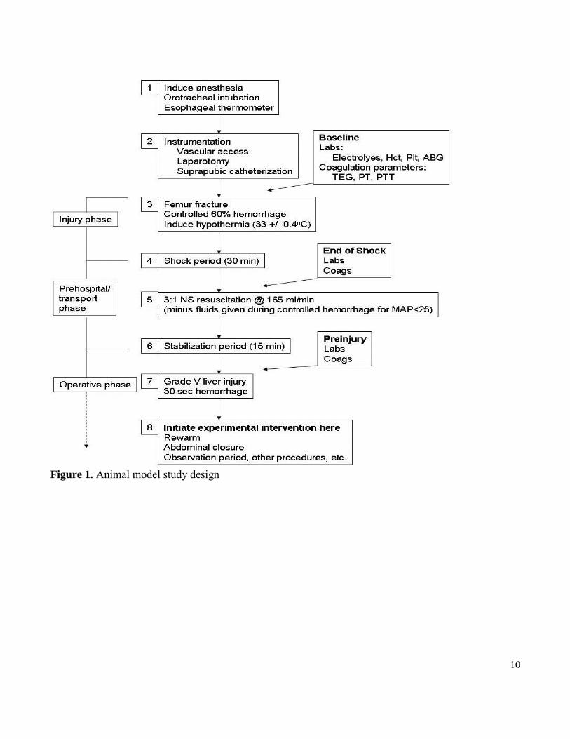
Animal Model:
Forty juvenile, female Yorkshire crossbred swine were subjected to a well-validated swine model of severe injury and hemorrhagic shock (Figure 1). Animals were fasted for 16 hours the day before surgery. Water was available ad libitum. A single vendor was utilized to eliminate potential differences in animal strain. Anesthesia
On the day of the experiment animals were given an induction agent consisting of 8 mg/kg Telazol® (tiletamine hydrochloride 50 mg/ml, zolazepam hydrochloride 50 mg/ml, Fort Dodge Animal Health, Fort Dodge, Iowa) given intramuscularly. Animals were placed in the supine position. Orotracheal intubation was performed and a 7.5mm internal diameter cuffed endotracheal tube was placed. The endotracheal tube was connected to the anesthesia machine with 1-3% isoflurane for anesthetic maintenance in 50% oxygen. Tidal volume was fixed at 10 ml/kg with a rate of 10 breaths per minute. An esophageal stethoscope, gastric tube and thermometer were inserted. An EKG monitor was secured and continuous monitoring started. Throughout the study, anesthesia was maintained to the clinical endpoints of reflexes and muscle relaxation as is done in humans. Monitoring, access and pre-experiment procedures
After swine were anesthetized a left cervical cutdown was performed and polyethylene catheters were inserted respectively into the left common carotid and left external jugular vein. The arterial line

6
was utilized for the controlled hemorrhage and blood sampling throughout the experiment while the venous line was used for administration of bolus resuscitation fluids and TXA. Finally, a proximal femoral cutdown was performed and the artery was cannulated for continuous blood pressure monitoring. Mean arterial pressure (MAP) was continuously recorded and averaged every 10 seconds with a blood pressure analyzer and digital data collection system (DigiMed, Louisville, KY). Baseline labs were collected and included electrolytes, lactate, spun hematocrit (Hct), activated clotting time (ACT), platelets, INR, partial thromboplastin time (PTT), and arterial blood gas (ABG). In addition, a baseline thrombelastogram (TEG, Haemoscope Corporation, Niles, IL) was performed. A celiotomy was then performed, at which time a suprapubic bladder catheter was placed to monitor urine output.
Injury Phase After needle localization, a captive bolt gun was used to fracture the femur and create a soft tissue injury at the mid-shaft of the left femur. A controlled hemorrhage was then initiated to remove 60% of the blood volume based on a published, standard equation relating blood volume to body weight for domestic swine. During hemorrhage if the mean arterial blood pressure (MAP) fell below 25mm/Hg, normal saline (NS) was infused at a rate of 165 ml/min to keep the MAP > 25 mm/Hg. The animal was also cooled to 33 ± 0.4oC using cooled intraperitoneal lavage with crystalloid as needed (most of the animals developed a degree of hypothermia spontaneously due to shock and infusion of IV fluids). These procedures were followed by a 30-minute shock period, representing time in the field prior to medical intervention.
Prehospital care/transport phase After the 30-minute shock period, electrolytes, spun hematocrit, ACT, ABG, and TEG were
again recorded. Blood samples were collected for platelet values and coagulation studies. After sample collection, the hemorrhage volume was replaced with a 3:1 ratio of NS infused at a rate of 165 ml/min, minus any given during the controlled hemorrhage to induce acidosis and coagulopathy. This reflects current civilian pre-hospital resuscitative practices.
Operative phase Following NS resuscitation, a 15-minute stabilization period was observed; during which a
baseline MAP was recorded and pre-weighed laparotomy sponges were placed in both paracolic gutters and in the pelvis for blood collection. Blood samples for point-of-care and laboratory studies were again collected, and a previously described grade V liver injury was created at the confluence of the right and middle hepatic veins using a specialized clamp. The liver injury was designed to provide a second stressor after initial injury and also to create a standardized injury that had the potential to re-bleed, both of which simulate a laparotomy after trauma in a patient with solid organ injury.
Thirty seconds of hemorrhage were then followed by evacuation of blood from the abdomen. Following the uncontrolled hemorrhage period, the liver was packed tightly with laparotomy sponges. Swine were randomized to receive one of four different low-volume LP solutions reconstituted with 1) sterile water (LP-SW), 2) normal saline (LP-NS), 3) lactated Ringer’s (LP-LR), or 4) Hextend© (LP-Hx). Study fluid resuscitation was initiated at the time of liver packing. The animal was also re-warmed to 37°C, and the abdomen closed with towel clips.
7
Follow-up Animals were monitored for 4 hours post injury or to death. Blood samples were collected at 1, 2, 3, and 4 hours. A MAP below 15 mmHg signified death, and the time of death was recorded. Animals surviving 4 hours were euthanized with Euthasol. Lung tissue was collected at the end of 4 hours or at declaration of death for rt-PCR analysis. Tissue was stored in RNA later. A necropsy was performed and the liver injury graded using the American Association for the Surgery of Trauma (AAST) liver injury grading system to ensure adequacy and similarity of injuries between groups. Heart (HR) rate and MAP were continuously recorded throughout the study. Blood loss following liver injury was carefully recorded with the use of pre-weighed laparotomy sponges and pre-weighed suction canisters.
Study Variables
Physiologic variables included survival, MAP, blood loss from the controlled hemorrhage, and blood loss due to the liver injury. Point-of-care laboratory values included TEG, Hct, lactate, platelets, ABG, and electrolytes. Additional assays completed after the experiment include INR, PTT, fibrinogen, IL-6, IL-8, IL-10, and TNF-α.
Statistical Analysis
Variables were assessed for normal distribution. Normally distributed data were reported as means with standard deviations. Comparisons between groups at various time points were analyzed by independent t-tests when the data were normally distributed. Paired-samples t-tests were used to compare same-group samples across various time points. Significance was denoted at p < 0.05. Data were analyzed utilizing SPSS statistical software, version 19.0 (IBM Corp. Released 2010. Armonk, NY).
Results
Of the 40 swine randomized in the study, one animal died shortly after infusion of the LP-Hx solution was completed. At baseline, animals were similar in weight, hematocrit, lactate, base excess, and pH (p>0.05, all comparisons).
There were no differences between groups in hemodynamic parameters (HR and MAP, Figure 2). There were significant differences in total blood loss between the LP-LR group and the LR-NS and LR-Hx groups (p<0.05, Figure 3). Additionally, the LR-SW group lost the least amount of blood over the study and was significantly lower compared to the LP-Hx group (p<0.05).
Prior to initiation of the LP study fluids, the changes in hematocrit were not different between study groups at any time point, (p>0.05, Figure 4). Following resuscitation, the hematocrit of the LP-Hx group was significantly lower than the LP-SW group at the 1-hour, 2-hour, 3-hour, and 4-hour time points (p<0.05, Figure 4). Serum lactate increased in all four fluid groups. There was a trend towards higher serum lactate levels in the LP-LR group at 1-hour; however, there was no statistically significant difference between groups at any time point (p>0.05, Figure 4). Prior to resuscitation with the LP study solutions, all animals became similarly acidotic following NS resuscitation following femur fracture and

8
controlled hemorrhage. Following LP study solution resuscitation the serum pH increased in all groups and was not different between LP fluid groups at any time point thereafter (p>0.05, Figure 4).
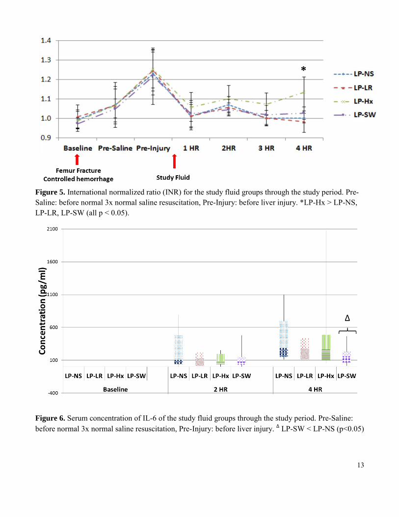
The INR was increased in all animals following femur fracture, controlled hemorrhage, and NS resuscitation (Figure 5). There was correction of the INR in all four study groups following resuscitation with the LP solutions following liver injury. However, the INR of the LP-Hx group remained significantly higher than the LP-NS, LP-LR, and LP-SW groups at 4-hours, (p<0.05).
There were no differences in the serum concentrations of IL-10 and TNF-α (p>0.05). IL-6 serum concentrations, however, were significantly lower in the LP-SW group compared to the LP-NS Group (p<0.05, Figure 6).
KEY RESEARCH ACCOMPLISHMENTS – Specific Aim 2 1. LP-SW showed less coagulopathic changes following injury compared to other groups. 2. Lower IL-6 concentrations at end of study in the LP-SW group. 3. Less total blood loss in the LP-SW group. 4. No statistically significant differences between the groups at baseline. 5. No statistically hemodynamic differences between animals following resuscitation.
REPORTABLE OUTCOMES – Specific Aim 2: Lee TH, McCully SP, McCully BH, Sands C, Hampton DA, Louis SG, Rick B, Anderson N, Differding J, Schreiber MA. “Comparison of the hemostatic efficacy of low-volume lyophilized plasma reconstituted using sterile water, lactated Ringer's, normal saline, and Hextend solutions.” J Trauma Acute Care Surg. 2014 Feb;76(2):264-72. doi: 10.1097/TA.0000000000000109. PMID: 24458032 [PubMed - in process]
CONCLUSION:
Previously, LP reconstituted to its original plasma volume was shown to be effective in hemostatic resuscitation using a swine model of polytrauma and hemorrhage.13 Minimizing full-volume LP solution into an equally effective low-volume solution improved LP’s logistical advantages over FFP.14 In this study, we aimed to further optimize low-volume LP solution by evaluating commonly used resuscitation fluids as potential reconstituting agents. Our results indicate that sterile water reconstituted LP is associated with less early and late coagulopathic changes, less inflammation, and similar hemodynamic benefits compared to the other reconstituting fluids tested.

9
References
1. Mattox KL, Moore EE, Feliciano DV. Trauma, 7th edition. McGraw-Hill Professional; 2012.
2. Sauaia A, Moore FA, Moore EE, et al. Epidemiology of trauma deaths: A reassessment. Journal of
Trauma-Injury Infection & Critical Care. 1995;38(2):185-193.
3. Hess JR, Brohi K, Dutton RP, et al. The coagulopathy of trauma: A review of mechanisms. J Trauma. 2008;65(4):748-754.
4. Borgman MA, Spinella PC, Perkins JG, et al. The ratio of blood products transfused affects mortality in patients receiving massive transfusions at a combat support hospital. Journal of Trauma-Injury
Infection & Critical Care. 2007;63(4):805-813.
5. Holcomb JB, Wade CE, Michalek JE, et al. Increased plasma and platelet to red blood cell ratios improves outcome in 466 massively transfused civilian trauma patients. Ann Surg. 2008;248(3):447-458.
6. Sperry JL, Ochoa JB, Gunn SR, et al. An FFP:PRBC transfusion ratio >/=1:1.5 is associated with a lower risk of mortality after massive transfusion. Journal of Trauma-Injury Infection & Critical Care. 2008;65(5):986-993.
7. Zink KA, Sambasivan CN, Holcomb JB, Chisholm G, Schreiber MA. A high ratio of plasma and platelets to packed red blood cells in the first 6 hours of massive transfusion improves outcomes in a large multicenter study. The American Journal of Surgery. 2009;197(5):565-570.
8. Murad MH, Stubbs JR, Gandhi MJ, et al. The effect of plasma transfusion on morbidity and mortality: A systematic review and meta-analysis. Transfusion. 2010;50(6):1370-1383.
9. Holcomb JB, del Junco DJ, Fox EE, et al. The prospective, observational, multicenter, major trauma transfusion (PROMMTT) study: Comparative effectiveness of a time-varying treatment with competing risks. JAMA Surg. 2013;148(2):127-136.
10. Johansson PI, Stensballe J. Hemostatic resuscitation for massive bleeding: The paradigm of plasma and platelets--a review of the current literature. Transfusion. 2010;50(3):701-710.
11. Curry NS, Davenport RA, Hunt BJ, Stanworth SJ. Transfusion strategies for traumatic coagulopathy. Blood Rev. 2012;26(5):223-232.
12. Bux J, Dickhorner D, Scheel E. Quality of freeze-dried (lyophilized) quarantined single-donor plasma. Transfusion. 2013.
13. Spoerke N, Zink K, Cho SD, et al. Lyophilized plasma for resuscitation in a swine model of severe injury. Arch Surg. 2009;144(9):829-834.
14. Lee TH, Van PY, Spoerke NJ, et al. The use of lyophilized plasma in a severe multi-injury pig model. Transfusion. 2013;53 Suppl 1:72S-79S.
10
Figure 1. Animal model study design

11
Figure 2. Mean arterial pressure (MAP) and heart rate (HR) of the study fluid groups through the study period. Pre-Saline: before normal 3x normal saline resuscitation, Pre-Injury: before liver injury.
Figure 3. Volume of blood loss following liver injury of study groups. 30 sec: Blood loss measured 30 seconds after uncontrolled hemorrhage following liver injury, After Liver Injury: Blood loss at the end of study, Total: Combined volume of 30 sec and After Liver Injury. *LP-LR < LP-NS and LP-LR < LP-Hx, p < 0.05; ΔLP-SW < LP-Hx, p < 0.05.

12
Figure 4. Hematocrit, serum pH, and serum lactate changes of the study fluid groups through the study period. Pre-Saline: before normal 3x normal saline resuscitation, Pre-Injury: before liver injury. *LP-Hx < LP-SW, all time points p < 0.05.
13
Figure 5. International normalized ratio (INR) for the study fluid groups through the study period. Pre-Saline: before normal 3x normal saline resuscitation, Pre-Injury: before liver injury. *LP-Hx > LP-NS, LP-LR, LP-SW (all p < 0.05).
Figure 6. Serum concentration of IL-6 of the study fluid groups through the study period. Pre-Saline: before normal 3x normal saline resuscitation, Pre-Injury: before liver injury. Δ LP-SW < LP-NS (p<0.05)