axl as a modulator of sunitinib response in glioblastoma...
TRANSCRIPT

Available online at www.sciencedirect.com
journal homepage: www.elsevier.com/locate/yexcr
E X P E R I M E N T A L C E L L R E S E A R C H 3 3 2 ( 2 0 1 5 ) 1 – 1 0
http://dx.doi.org/10.10014-4827/& 2015 E
nCorresponding au4710-057 Braga, Port
E-mail address: r
Research Article
AXL as a modulator of sunitinib response inglioblastoma cell lines
Olga Martinhoa,b,c, Luis Eduardo Zuccac, Rui Manuel Reisa,b,c,n
aLife and Health Sciences Research Institute (ICVS), Health Sciences School, University of Minho, Braga, PortugalbICVS/3B’s—PT Government Associate Laboratory, Braga/Guimarães, PortugalcMolecular Oncology Research Center, Barretos Cancer Hospital, Barretos, São Paulo, Brazil
a r t i c l e i n f o r m a t i o n
Article Chronology:
Received 21 June 2014Received in revised form15 January 2015Accepted 19 January 2015Available online 28 January 2015
Keywords:
GlioblastomaSunitinibBiomarkerRTKAXL
016/j.yexcr.2015.01.009lsevier Inc. All rights reser
thor at: Life and Health Scieugal. Fax: þ351 253 [email protected] (R
a b s t r a c t
Receptor tyrosine kinase (RTK) targeted therapy has been explored for glioblastoma treatment.However, it is unclear which RTK inhibitors are the most effective and there are no predictivebiomarkers available. We recently identified the RTK AXL as a putative target for the pan-RTKinhibitors cediranib and sunitinib, which are under clinical trials for glioblastoma patients. Here,we provide evidence that AXL activity can modulate sunitinib response in glioblastoma cell lines.We found that AXL knockdown conferred lower sensitivity to sunitinib by rescuing migratorydefects and inhibiting apoptosis in cells expressing high AXL basal levels. Accordingly, over-
activation of AXL by its ligand GAS6 rendered AXL positive glioblastoma cells more sensitive tosunitinib. AXL knockdown induced a cellular rewiring of several growth signaling pathwaysthrough activation of RTKs, such as EGFR, as well as intracellular pathways such as MAPK andAKT. The combination of sunitinib with a specific AKT inhibitor reverted the resistance of AXL-silenced cells to sunitinib. Together, our results suggest that sunitinib inhibits AXL and AXLactivation status modulates therapy response of glioblastoma cells to sunitinib. Moreover, itindicates that combining sunitinib therapy with AKT pathway inhibitors could overcomesunitinib resistance.
& 2015 Elsevier Inc. All rights reserved.
Introduction
Glioblastoma is the most common form of primary brain tumor andone of the most lethal and challenging human malignancies [1–4].Standard treatment consists in a combination of surgery, irradiationand temozolomide, which postpones progression and extends over-all survival in 5 years from 2% up to 10%, but these tumors univ-ersally recur and unrelentingly result in patient death [4–8].
ved.
nces Research Institute (IC0..M. Reis).
An increasing understanding of the molecular mechanismsunderlying glioblastoma [9–11] has led to the identification of anumber of promising therapeutic targets, including several mem-bers of the receptor tyrosine kinase (RTK) family [12–16]. Amongthe most interesting RTK targets are EGFR, PDGFRA and KIT,which have been intensively studied [17–20]. However, clinicaltrials with single-targeted agents that inhibit these moleculeshave shown only minimal therapeutic activity, with no significant
VS), School of Health Sciences, University of Minho, Campus de Gualtar,

E X P E R I M E N T A L C E L L R E S E A R C H 3 3 2 ( 2 0 1 5 ) 1 – 1 02
prolongation of survival [21–28]. Therefore, it is currently beli-eved that multi-kinase inhibitors simultaneously targeting severalRTKs may yield greater clinical efficacy in selected glioblastomapatients [29–32].AXL belongs to the TAM (Tyro3, AXL and Mer) receptor
subfamily of RTKs that also includes Tyro3 and Mer [33]. TheTAM receptors are characterized by a combination of two imm-unoglobin-like domains and dual fibronectin type III repeats inthe extracellular region and a cytoplasmic kinase domain. Theligands for TAM receptors are Gas6 (growth arrest-specific 6) andprotein S [34]. AXL was originally cloned from patients with chr-onic myelogenous leukemia and, when overexpressed, it exhibitstransforming potential [33]. AXL overexpression has been repo-rted in a variety of human cancers, being associated with tumorinvasiveness and metastasis [35–38].Regarding brain tumors, AXL has been implicated in gliomagen-
esis and chemoresistance [39]. Previous investigations found thatAXL is constitutively phosphorylated in many glioma cell lines,murine xenograft tumors and primary patient tumor samples [40].Immunohistochemical analysis of AXL and Gas6 demonstrated thatco-expression of these proteins correlates with tumor recurrenceand progression [41,42]. Furthermore, inhibition of AXL usingin vitro and in vivo models resulted in a reduction of tumor growth,migration and invasion, as well as prolonged overall animal sur-vival in xenografts containing dominant negative AXL [42]. Alto-gether the previous data has indicating that AXL targeted therapymay also diminish glioblastoma aggressiveness [43].In a recent pre-clinical study performed by our group, the eff-
ectiveness of two pan-RTK inhibitors (sunitinib and cediranib) inglioblastoma cell lines was assessed and AXL was found as a com-mon candidate target for both cediranib and sunitinib [31]. Otherstudies have also suggested AXL as target for sunitinib in othertumor types, such as in renal cell carcinoma [38,44]. Moreover, denovo activation of AXL was found in imatinib-treated gastrointest-inal stromal tumors (GIST) and in Her-2 positive breast cancer cellstreated with lapatinib and, in both cases, AXL was associated withtherapy resistance [45,46]. Nevertheless, the role of this protein asa putative modulator of sunitinib therapy response is still in needof in vitro and in vivo validation.Due to the importance of AXL in glioblastoma and its recent
implication in RTK inhibitor responses, in the present study weaimed to validate AXL as a cediranib and sunitinib target and todetermine whether it could act as a modulator of cediranib andsunitinib response in glioblastoma cell lines.
Materials and methods
Cell lines and cell culture
Eight immortalized glioblastoma cell lines were used: SW1088,SW1783, U-87 MG and A172 were obtained from ATCC (AmericanType Culture Collection), SNB-19 and GAMG were obtained fromDSMZ (German Collection of Microorganisms and Cell Cultures)and U251 and U373 were kindly provided by Professor JosephCostello. All cell lines were maintained in DMEM-10 at 37 1C and5% CO2 as previously described [31]. Authentication of cell lineswas performed by IdentiCell Laboratories (Department of Mole-cular Medicine (MOMA) at Aarhus University Hospital Skejby inÅrhus, Denmark), as described [31].
Generation of cell lines stably expressing short hairpin (sh)AXL
For the generation of cell lines stably expressing a short hairpin forAXL (shAXL), we used pRNA-U6.1/Neo vector containing a 19 bpshRNA. For AXL overexpression a complete AXL cDNA cloned intothe pcDNA3 vector was used. Both vectors were kindly provided byShuang-En Chuang from the National Health Research Institutes,Taiwan [47]. Electroporation with Neon Transfection System (Invi-trogen, Life Technologies) was used to introduce the shAXL/AXLand the empty vectors into the cells, following the manufacturer’sinstructions. Twenty-four hours after transfection, stable transfec-tants were selected with 300–450 mg/ml of G418 (Sigma Aldrich) incomplete DMEM medium.
Immunofluorescence analysis for AXL
For immunofluorescence analysis, the cells were plated in glasscoverslips placed into 12-well plates at a density of 6�104 cellsper well and allowed to adhere overnight. Then, the cells werefixed in cold methanol by 5 min at �20 1C. For block unspecificligations the cells were incubated with a solution of PBS contain-ing 10% FBS for 30 min at room temperature followed by incuba-tion with a primary antibody against AXL (dilution 1:50; inc-ubation ON at RT; AXL C-20 clone; Santa Cruz Biotechnology). Thecells were then washed in a PBS solution with 0.5% FBS and inc-ubated with a rabbit anti-goat antibody conjugated with TRITC(dilution 1:500, Life Technology) for 1 h at room temperature inthe dark. Finally the cells were counterstained with 40,6-diami-dino-2-phenylindole (DAPI) and images were obtained with theuse of fluorescence microscopy (BX16; Olympus).
Western blot and human RTK arrays
To assess the effect of the drugs on the intracellular signalingpathways and RTKs, the cells were cultured in T25 culture flasksor six-well plates, allowed to grow to 85% confluence, serum sta-rved for 2 h and then incubated with the drugs. When necessarythe cells were stimulated with 400 ng/ml of Gas6 (human rec-ombinant Gas6, R&D systems) for 15 min.
To assess apoptosis, the cells were incubated with increasingconcentrations of sunitinib for 24 h. At the indicated time points,the cells were washed and scraped in cold PBS and lysed in buffercontaining 50 mM Tris pH 7.6–8, 150 mM NaCl, 5 mM EDTA, 1 mMNa3VO4, 10 mM NaF, 10 mM NaPyrophosphate, 1% NP-40 and 1/7of protease cocktail inhibitors (Roche). Western blotting was per-formed using standard 10% SDS-PAGE, loading 20 mg of proteinper lane. All the antibodies were used as recommended by themanufacturer and as previously described [31].
For detection of AXL activation we used a specific antibody todetect AXL phosphorylation, phospho-AXL (Tyr702) (Cell SignalingTechnologies, D12B2). For calibration of the activation levels, anantibody to detect the total protein levels (AXL (C-20), Santa CruzBiotechnology) was used. A specific antibody to detect EphA7 pho-sphorylation (Tyr791) (MyBiosource) was also used. Anti-GAPDH(Santa Cruz Biotechnology) was used as a loading control. All theother antibodies were used as previously described [31].
For the human RTK arrays, 500 μg of fresh protein lysates wereincubated overnight at 4 1C with nitrocellulose membranes dottedwith duplicated spots for 42 anti-RTK and control antibodies. Bound

E X P E R I M E N T A L C E L L R E S E A R C H 3 3 2 ( 2 0 1 5 ) 1 – 1 0 3
phospho-RTKs were incubated with a pan anti-phosphotyrosine-HRP antibody for 2 h at room temperature [31].
Antibodies were detected by chemiluminescence (ThermoScientific Pierce ECL Western Blotting) using the ChemiDoc™XRSþ System (Bio-Rad).
Drugs
All the drugs (cediranib, sunitinib, imatinib, selumetinib andMK2206) used in this work were obtained from Selleck Chemicals,USA and prepared as 10 mM stock solutions in dimethyl sulfoxide(DMSO) and stored at �20 1C. To obtain an equal quantity of DMSO(1% final concentration) in each of the conditions studied, prior tothe final dilution of the drugs to an appropriate concentration inDMEM medium, the drugs were first prepared as intermediatedilutions in DMSO [31].
Cell viability assay
To determine the concentration at which 50% of the cell growth isinhibited by drug treatment (IC50 concentration), cells were plated
Fig. 1 – AXL is a target of cediranib and sunitinib therapy in gliobactivation (phosphorylated AXL: p-AXL) in eight glioblastoma cell lSNB-19 and U251 cell lines. (C) U251 and SNB-19 cells were treated(SU) for 2 h, and stimulated with 400 ng/ml of Gas6. The activationWestern blot. GAPDH was used as a loading control.
into 96-well plates at a density of 2�103 cells per well and allowedto adhere overnight in DMEM medium containing 10% FBS. Sub-sequently, the cells were treated with increasing concentrations ofthe drugs or with DMSO alone, both diluted in 0.5% FBS culturemedium to a final concentration of 1% DMSO. When necessary thecells were incubated simultaneously with 400 ng/ml of Gas6, orwith 2.5 mM of selumetinib or MK2206. After 72 h, cell viabilitywas quantified using the Cell Titer96 Aqueous cell proliferationassay (Promega). The results were expressed as the mean percen-tage7SD of viable cells relative to the DMSO alone (considered as100% viability). The IC50 concentration was calculated by nonlinearregression analysis using GraphPad Prism software.To assess the effect of a fixed concentration of the drug in cell-
ular viability over time, the cells were plated onto 96-well platesat a density of 1�103 cells per well and allowed to adhereovernight in complete DMEM medium. Next, the viable cells werequantified using the Cell Titer96 Aqueous cell proliferation assay(Promega), and used for time point 0. Then, the cells were inc-ubated with fixed concentrations of the drugs or with DMSOalone, both diluted in 0.5% FBS culture medium to a final con-centration of 1% DMSO, over 24, 48 and 72 h. At the end of each
lastoma cells. (A) Western blot analysis of AXL expression andines. (B) Immunofluoresnce analysis of AXL expression in A172,with increasing concentrations of cediranib (CD) and sunitiniblevels of AXL, MAPK (ERK) and AKT pathways were assessed by

E X P E R I M E N T A L C E L L R E S E A R C H 3 3 2 ( 2 0 1 5 ) 1 – 1 04
time point, cell viability was again assessed using the Cell Titer96Aqueous cell proliferation assay. The results were calibrated to thestarting viability (time 0 h, considered as 100% viability) and exp-ressed as the mean7SD. Both assays were performed in triplicateat least three times [31].
Wound healing migration assay
The cells were seeded on 6-well plates and cultured to at least95% confluence. Monolayer cells were washed with PBS and scr-aped with a plastic 200 ml pipette tip. The cells were incubatedwith fixed concentrations of the drugs or with DMSO alone, bothdiluted in 0.5% FBS culture medium to a final concentration of 1%DMSO. When necessary the cells were incubated simultaneouslywith 400 ng/ml of Gas6 or with 2.5 mM of selumetinib or MK2206.The “wounded” areas were photographed by phase contrastmicroscopy at 0 and 48 h time points. The relative migration dist-ance was calculated using the following formula: percentage ofwound closure (%)¼100 (A�B)/A, where A is the width of cellwounds before incubation and B is the width of cell wounds afterincubation. Results are expressed as the mean7SD. The assay wasperformed in triplicate at least three times.
Statistical analysis
Single comparisons between the different conditions studiedwere made using the Student’s t test and differences betweengroups were tested using two-way analysis of variance (ANOVA).
Fig. 2 – Role of AXL in the modulation of sunitinib, cediranib and iU251 cells were stably transfected with a shRNA for AXL (shAXL),(pRNAU6 or pcDNA3) were used as negative controls. Cell lysates weof transfected cells (shAXL, AXL and respective empty vectors: EV),IM: imatinib) by MTS after 72 h of treatment.
Statistical analysis was done using Graph Pad Prism version 5. Thelevel of significance in all the statistical analysis was set atpo0.05.
Results
Inhibition of AXL signaling by cediranib and sunitinib
In order to determine the expression and phosphorylation levelsof AXL in glioblastoma cell lines we performed western blot ana-lysis with specific antibodies (Fig. 1A). Despite the weak expres-sion of total AXL protein in all cell lines, we could confirm thepresence of AXL activation in seven of the cell lines, beingSW1088 the only one lacking AXL activity (Fig. 1A).
To further assess whether AXL is a cediranib and sunitinib target,we chose a cell line with high AXL phosphorylation levels (SNB-19)and a cell line with lower levels of AXL activation and expression(U251) (Fig. 1A and B). We found that in the U251 cell line, neithercediranib nor sunitinib were able to inhibit AXL activation uponligand (Gas6) stimulation. In contrast, in SNB-19 cells, AXL phos-phorylation was inhibited by both drugs (Fig. 1C). Specifically, weobserved that only high doses of cediranib inhibited Gas6-inducedAXL activation, while sunitinib was effective at lower doses. Reg-arding inhibition of intracellular signaling pathways, cediranib waseffective in blocking ERK activation in both cell lines (Fig. 1C). Incontrast, sunitinib was only able to inhibit AKT activation in SNB-19cells at high doses when AXL inhibition was complete, suggesting
matinib therapy response in glioblastoma cells. (A) SNB-19 andor full length AXL cDNA (AXL). The respective empty vectorsre analyzed by Western blot for AXL expression. (B) IC50 valueswere assessed for the three drugs (CD: cediranib; SU: sunitinib;

E X P E R I M E N T A L C E L L R E S E A R C H 3 3 2 ( 2 0 1 5 ) 1 – 1 0 5
that AKT could be dependent on AXL signaling in this cell line(Fig. 1C).
Role of AXL in cediranib and sunitinib therapy response
To determine whether AXL can modulate cediranib and sunitinibresponse in glioblastoma, we knocked down AXL in the U251 andSNB-19 cell lines, and overexpressed it in the U251 cell line(Fig. 2A). Following transfection of a short hairpin targeting AXL(shAXL), we observed a significant reduction of total and phos-phorylated AXL protein levels when compared to the cells tra-nsfected with the empty vector (pRNAU6) in both cell lines(Fig. 2A). U251 cells successfully overexpressed AXL upon trans-fection with AXL cDNA when compared with cells transfectedwith the empty vector (pcDNA3) (Fig. 2A).
To analyze the effectiveness of cediranib and sunitinib in theseAXL modulated cell lines, we determined the IC50 concentrationsof each drug after cell transfection (Fig. 2B). Imatinib was used asnegative control, since we have previously shown that this drugdoes not target AXL [20]. We found that in the U251 (low AXLactivity) cell line, neither AXL inhibition nor AXL overexpression
Fig. 3 – Role of AXL in glioma cells survival, migration and apoptocells were treated with 5 lM of cediranib (CD), 5 lM of sunitinib (SMTS. Data is represented as the mean7SD and differences in two-wstatistically significant (#). (B) For migration assessment the cells wEV—empty vector using student’s t test). (C) SNB-19 transfected ce(SU) for 24 h. Apoptosis was assessed by Western blot for PARP cleathat is specific for the cleaved form. GAPDH was used as a loading
altered the sensitivity of the cells to any of the agents tested. Incontrast, in the SNB-19 (high AXL activity) cell line, while no diff-erences were observed for imatinib, as expected, nor to cediranib,a statistically significant (po0.05) increase in the IC50 value wasfound for sunitinib on the shAXL cells (11.6 μM) when comparedwith the cells transfected with the empty vector (7.1 μM) (Fig. 2B).To exclude the possibility of metabolic adaptation of the cells
after transfection, we performed survival and migration assays atfixed doses of sunitinib (Fig. 3). AXL-silenced SNB-19 cells exhi-bited a survival advantage when treated over time with sunitinibcompared to the empty vector control cells (po0.05) (Fig. 3A). Asexpected, the treatment of SNB-19 cells with cediranib had noeffect on viability (Fig. 3A). Also, no significant differences wereobserved in the survival of U251 transfected cells (empty vectorversus AXL/shAXL cells) upon treatment with either drug over time.Using a wound healing migration assay, we observed that both
cediranib and sunitinib significantly reduced migration of both celllines when compared to the DMSO control. In contrast, sunitinib nolonger reduced migration in the AXL silenced SNB-19 cells com-pared to the DMSO control. Rather, we observed that shAXL tra-nsfected cells have a significantly higher migratory capability than
sis after sunitinib treatment. (A) SNB-19 and U251 transfectedU) or DMSO as a control to assess cellular viability over time byay analysis of variance (ANOVA) with a po0.05 were consideredere incubated for 48 h (*compared to DMSO and # compared tolls were incubated with increasing concentrations of sunitinibvage using two antibodies, one that detects total PARP and onecontrol.
E X P E R I M E N T A L C E L L R E S E A R C H 3 3 2 ( 2 0 1 5 ) 1 – 1 06
control cells after sunitinib treatment (Fig. 3B). Corroborating theseresults, we analyzed apoptosis by PARP cleavage and found thatknockdown of AXL in SNB-19 cells inhibited apoptosis induced bysunitinib treatment when compared to the empty vector trans-fected cells (Fig. 3C).These data indicate that AXL-inhibited cells are less sensitive to
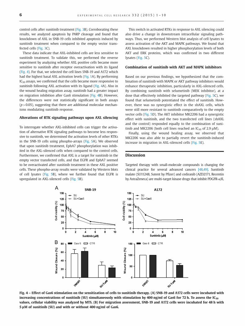
sunitinib treatment. To validate this, we performed the reverseexperiment by analyzing whether AXL positive cells became moresensitive to sunitinib after receptor overactivation with its ligand(Fig. 4). For that, we selected the cell lines SNB-19 and A172 whichhad the highest basal AXL activation levels (Fig. 1A). By performingIC50 assays, we confirmed that the cells became more responsive tosunitinib following AXL activation with its ligand (Fig. 4A). Also inthe wound healing migration assay, sunitinib had a greater impacton migration inhibition after Gas6 stimulation (Fig. 4B). However,the differences were not statistically significant in both assays(p40.05), suggesting that there are additional molecular mechan-isms modulating sunitinib response.
Alterations of RTK signaling pathways upon AXL silencing
To interrogate whether AXL-inhibited cells can trigger the activa-tion of alternative RTK signaling pathways to become less respon-sive to sunitinib, we determined the activation levels of other RTKsin the SNB-19 cells using phospho-arrays (Fig. 5A). We observedthat upon sunitinib treatment, EphA7 phosphorylation was inhib-ited in the AXL-silenced cells when compared to the control cells.Furthermore, we confirmed that AXL is a target for sunitinib in theempty vector transfected cells, and that EGFR and EphA7 seemedto be overactivated after sunitinib treatment in these AXL positivecells. These phospho-array results were validated by Western blotsof cell lysates (Fig. 5B), where we further found that EGFR isupregulated in AXL-silenced cells (Fig. 5B).
Fig. 4 – Effect of Gas6 stimulation on the sensitization of cells to suincreasing concentrations of sunitinib (SU) simultaneously with stvalues, cellular viability was analyzed by MTS. (B) For migration as5 lM of sunitinib (SU) and with or without 400 ng/ml of Gas6.
This switch in activated RTKs in response to AXL silencing couldalso drive a change in downstream intracellular signaling path-ways. Thus, we performed Western blot analysis of cell lysates toassess activation of the AKT and MAPK pathways. We found thatAXL knockdown resulted in higher phosphorylation levels of bothAKT and ERK proteins, which was confirmed in two differentlysates (Fig. 5C).
Combination of sunitinib with AKT and MAPK inhibitors
Based on our previous findings, we hypothesized that the com-bination of sunitinib with MAPK or AKT pathway inhibitors wouldenhance therapeutic inhibition, particularly in AXL-silenced cells.By combining sunitinib with selumetinib (MEK inhibitor), at adose that effectively inhibited the targeted pathway (Fig. 5C), wefound that selumetinib potentiated the effect of sunitinib. How-ever, there was no synergistic effect in the shAXL cells, whichwere still more resistant to sunitinib comparatively to the emptyvector cells (Fig. 5D). The AKT inhibitor MK2206 had a synergisticeffect with sunitinib, and the two transfected cell lines (shAXLand the control) responded equally to the combination of suni-tinib and MK2206 (both cell lines reached an IC50 of 2.9 mM).
Finally, using the wound healing assay, we observed thatMK2206 was also able to partially revert the sunitinib-inducedincrease in migration in AXL-silenced cells (Fig. 5E).
Discussion
Targeted therapy with small-molecule compounds is changing theclinical practice for several advanced cancers [48,49]. Sunitinibmalate (SU11248, Sutent by Pfizer) and cediranib (AZD2171, Recentinby AstraZeneca) are multi-target kinase drugs that inhibit PDGFR-α/ß,
nitinib therapy. (A) SNB-19 and A172 cells were incubated withimulation by 400 ng/ml of Gas6 for 72 h. To assess the IC50
sessment, SNB-19 and A172 cells were incubated for 48 h with

Fig. 5 – RTKs and intracellular pathway alterations in AXL negative cells. (A) Phospho-RTK arrays were performed for SNB-19transfected cells before and after sunitinib treatment (2 h, 5 lM). Each RTK is represented in duplicate on the arrays (two spotsside-by-side), and four pairs of phospho-tyrosine positive controls are located in the corners of each array. (B) Western blotvalidation of the arrays with specific antibodies to AXL, EGFR and EphA7 was performed on the same lysates. SNB-19 wild-type(WT) cells were also included as a control. GAPDH was used as a loading control. (C) Two independent lysates of the SNB-19transfected cell line were analyzed by Western blot for the levels of MAPK (ERK) and AKT pathway activation. On the right, SNB-19wild-type cell line was treated for 2 h with 2.5 lM of selumetinib (SE) or MK2206 (MK) to confirm their capability for inhibitingthe MAPK and AKT pathways, respectively. (D) Combination studies were done in the SNB-19 transfected cell line with sunitiniband fixed concentrations of MK or SE (2.5 lM) over 72 h. To assess the IC50 values, the cellular viability was determined by MTS. (E)To migration quantification, the cells were incubated for 48 h with 5 lM of sunitinib (SU) and with or without 2.5 lM of Gas6.
E X P E R I M E N T A L C E L L R E S E A R C H 3 3 2 ( 2 0 1 5 ) 1 – 1 0 7
VEGFR1-3, KIT, RET, FLT3, CSF1R, and VEGFR1-3, KIT, PDGFRα,respectively [50–52]. These therapeutic agents have shown interest-ing results in pre-clinical mouse glioma models, but failed todemonstrate benefit in progression free survival and overall survivalin clinical trials [26,53–65]. A recent phase III clinical trial withcediranib showed that this drug failed to meet its primary end pointof progression free survival prolongation, either as monotherapy orin combination with chemotherapy [26]. Hitherto, and in contrast toother tumor types [66], no predictive biomarkers for moleculartargeted therapy response were yet identified in glioblastoma,hampering the design of efficient tailored therapies for these patients[30], and justifying to some extent the failure of the clinical trialsuntil now.
In a previous work of our group, we reported that cediranibcould also inhibit EGFR, EphA7, AXL, MET, EphB2, and sunitinibcould target EphB2, ROR1 and AXL [31], besides the abovemen-tioned “classical” targets. Other authors have recently identifiedAXL as target for sunitinib [67,68]. In the present work we aimedto validate AXL as a cediranib and sunitinib target in glioblastomacells, using two glioblastoma cell lines with distinct AXL levels,namely U251 (low basal AXL levels) and SNB-19 (high basal AXLlevels). Initially, we assessed AXL inhibition by western blot whencells were stimulated with the ligand GAS6. We showed thatcediranib was able to inhibit AXL phosphorylation only at highdoses in the SNB-19 cell line. Sunitinib strongly inhibited AXL in a
dose-dependent manner and also inhibited the AKT pathway athigh doses in the SNB-19 cell line. In accordance, modulation ofAXL protein levels showed that the low levels of AXL activitycould be associated with the observed response of SNB-19 cells tosunitinib therapy, but not to cediranib or imatinib. These resultswere not observed in U251 cell line with or without AXL expr-ession. Several issues could explain these distinct drug responses.We have previously shown that the activation profile of RTKs inthese two particular cell lines is very similar, however U251 ismuch more sensitive to sunitinib than SNB-19 [31]. This can inpart explain the similarity in the sunitinib response after AXLmodulation in U251 cells, since they already exhibit a highlysensitive response. Additionally, these findings can also suggestthat sunitinib response is cell line-dependent and that AXL is notthe only factor modulating the cells’ responsiveness to this drug.Hence, the main predictive factor for the response to this drugremains to be discovered, as previously pointed out [56].Moreover, our present work suggests that AXL could be a
modulator of sunitinib response, at least for cell lines that presenthigh endogenous levels of this RTK activation, since we observedan increased responsiveness to sunitinib in cells activated with AXLreceptor ligand. A role for AXL in molecular targeted therapiesmodulation is not new and has been related with resistance. Forexample, de novo activation of AXL has been associated withimatinib, lapatinib treatment and mainly with resistance to EGFR

E X P E R I M E N T A L C E L L R E S E A R C H 3 3 2 ( 2 0 1 5 ) 1 – 1 08
inhibitors in several tumor models [45,46,69–72]. Interestingly, wefound that AXL-silenced cells exhibited higher activation levels ofthe MAPK and AKT pathways and that some RTKs, such as EGFR,are overactivated upon AXL inhibition. Several preclinical andclinical studies have illustrated that deregulation of one signalingpathway can sometimes alleviate or bypass the “addiction” to ano-ther pathway [73,74]. Thus, to overcome the activation of intracel-lular signaling pathways that render resistance to RTK inhibitors,several combinations of drugs are currently being tested to targetboth ERK/MAPK and PI3K pathways, and their use in combinationwith other targeted therapies holds great promise [75–77]. Ourin vitro studies showed that by combining sunitinib with AKTinhibitors the lower sensitivity of AXL-knockdown cells to sunitinibcould be reverted. This suggests that this therapeutic strategy maybe effective in glioblastoma patients, mainly for those with low AXLactivity who are less likely to be responsive to sunitinib trea-tment alone.In conclusion, we report that the levels of AXL activation could
be one of the modulators of sunitinib response in glioblastomapatients. In addition, we showed that in the absence of AXL acti-vation, the combination of sunitinib with specific inhibitors of theAKT pathway may overcome the eventual resistance phenotype ofglioblastoma cells to sunitinib.
Acknowledgments
This work was funded by Fundação para a Ciência e Tecnologia(FCT), Portugal (project: PTDC/SAU-TOX/114549/2009). Olga Mar-tinho is a recipient of a Post-Doc fellowship (UMINHO/BPD/32/2013) from QREN. We would like to acknowledge Dr. Shuang-EnChuang from the National Health Research Institute, Taiwan, forproviding AXL vectors, and Dr. Raquel Andrade for critical reviewof the manuscript.
r e f e r e n c e s
[1] S. Agnihotri, et al., Glioblastoma, a brief review of history,molecular genetics, animal models and novel therapeutic stra-tegies, Arch. Immunol. Ther. Exp. (Warsz) 61 (1) (2013) 25–41.
[2] Ostrom, Q.T., et al., The epidemiology of glioma in adults: a “stateof the science” review. Neuro. Oncol., 2014.
[3] M. Weller, et al., Molecular neuro-oncology in clinical practice: anew horizon, Lancet Oncol. 14 (9) (2013) e370–e379.
[4] S. Tanaka, et al., Diagnostic and therapeutic avenues for glio-blastoma: no longer a dead end?, Nat. Rev. Clin. Oncol. 10 (1)(2013) 14–26.
[5] L.J. Yang, C.F. Zhou, Z.X. Lin, Temozolomide and radiotherapy fornewly diagnosed glioblastoma multiforme: a systematic review,Cancer Invest. 32 (2) (2014) 31–36.
[6] R. Stupp, et al., Radiotherapy plus concomitant and adjuvanttemozolomide for glioblastoma, N. Engl. J. Med 352 (10) (2005)987–996.
[7] M. Weller, et al., EANO guideline for the diagnosis and treatmentof anaplastic gliomas and glioblastoma, Lancet Oncol. 15 (9)(2014) e395–e403.
[8] R. Stupp, et al., Effects of radiotherapy with concomitant andadjuvant temozolomide versus radiotherapy alone on survival inglioblastoma in a randomised phase III study: 5-year analysis ofthe EORTC-NCIC trial, Lancet Oncol. 10 (5) (2009) 459–466.
[9] P.Y. Wen, S. Kesari, Malignant gliomas in adults, N. Engl. J. Med.359 (5) (2008) 492–507.
[10] J.T. Huse, E.C. Holland, Targeting brain cancer: advances in themolecular pathology of malignant glioma and medulloblastoma,Nat. Rev. Cancer 10 (5) (2010) 319–331.
[11] Y. Zhu, L.F. Parada, The molecular and genetic basis of neurolo-gical tumours, Nat. Rev. Cancer 2 (8) (2002) 616–626.
[12] G. Konopka, A. Bonni, Signaling pathways regulating glioma-genesis, Curr. Mol. Med 3 (1) (2003) 73–84.
[13] G.S. Kapoor, D.M. O’Rourke, Mitogenic signaling cascades in glialtumors, Neurosurgery 52 (6) (2003) 1425–1434.
[14] T.F. Cloughesy, W.K. Cavenee, P.S. Mischel, Glioblastoma: frommolecular pathology to targeted treatment, Annu. Rev. Pathol.9 (2014) 1–25.
[15] Z. Wardak, K.S. Choe, Molecular pathways and potential ther-apeutic targets in glioblastoma multiforme, Expert Rev. Antic-ancer Ther. 13 (11) (2013) 1307–1318.
[16] B. Purow, D. Schiff, Advances in the genetics of glioblastoma: arewe reaching critical mass?, Nat. Rev. Neurol. 5 (8) (2009) 419–426.
[17] A.L. Gomes, et al., Molecular alterations of KIT oncogene ingliomas, Cell Oncol 29 (5) (2007) 399–408.
[18] O. Martinho, et al., Expression, mutation and copy number analysisof platelet-derived growth factor receptor A (PDGFRA) and itsligand PDGFA in gliomas, Br. J. Cancer 101 (6) (2009) 973–982.
[19] R.M. Reis, et al., Molecular characterization of PDGFR-alpha/PDGF-A and c-KIT/SCF in gliosarcomas, Cell Oncol. 27 (5-6)(2005) 319–326.
[20] M. Viana-Pereira, et al., Analysis of EGFR overexpression, EGFRgene amplification and the EGFRvIII mutation in Portuguesehigh-grade gliomas, Anticancer Res. 28 (2A) (2008) 913–920.
[21] E. Raymond, et al., Phase II study of imatinib in patients withrecurrent gliomas of various histologies: a European Organisa-tion for Research and Treatment of Cancer Brain Tumor GroupStudy, J. Clin. Oncol. 26 (28) (2008) 4659–4665.
[22] S. Baruchel, et al., A Canadian paediatric brain tumour consor-tium (CPBTC) phase II molecularly targeted study of imatinib inrecurrent and refractory paediatric central nervous systemtumours, Eur. J. Cancer 45 (13) (2009) 2352–2359.
[23] P.Y. Wen, et al., Phase I/II study of imatinib mesylate for recurrentmalignant gliomas: North American Brain Tumor ConsortiumStudy 99-08, Clin. Cancer Res. 12 (16) (2006) 4899–4907.
[24] A. Desjardins, et al., Phase II study of imatinib mesylate andhydroxyurea for recurrent grade III malignant gliomas, J. Neuro-Oncol. 83 (1) (2007) 53–60.
[25] M. Preusser, et al., Epithelial Growth Factor Receptor Inhibitorsfor treatment of recurrent or progressive high grade glioma: anexploratory study, J. Neurooncol. 89 (2) (2008) 211–218.
[26] T.T. Batchelor, et al., Phase III randomized trial comparing theefficacy of cediranib as monotherapy, and in combination withlomustine, versus lomustine alone in patients with recurrentglioblastoma, J. Clin. Oncol. 31 (26) (2013) 3212–3218.
[27] J.J. Raizer, et al., A phase II trial of erlotinib in patients with recurrentmalignant gliomas and nonprogressive glioblastoma multiformepostradiation therapy, Neuro Oncol 12 (1) (2010) 95–103.
[28] J.H. Uhm, et al., Phase II evaluation of gefitinib in patients withnewly diagnosed Grade 4 astrocytoma: Mayo/North CentralCancer Treatment Group Study N0074, Int. J. Radiat. Oncol. Biol.Phys. 80 (2) (2011) 347–353.
[29] A. Idbaih, et al., Therapeutic application of noncytotoxic mole-cular targeted therapy in gliomas: growth factor receptors andangiogenesis inhibitors, Oncologist 13 (9) (2008) 978–992.
[30] P.C. De Witt Hamer, Small molecule kinase inhibitors in glio-blastoma: a systematic review of clinical studies, Neuro. Oncol.12 (3) (2010) (304-16).
[31] O. Martinho, et al., In vitro and in vivo analysis of RTK inhibitorefficacy and identification of its novel targets in glioblastomas,Transl. Oncol. 6 (2) (2013) 187–196.
[32] Y. Zhang, et al., XL-184, a MET, VEGFR-2 and RET kinase inhibitorfor the treatment of thyroid cancer, glioblastoma multiforme andNSCLC, IDrugs 13 (2) (2010) 112–121.

E X P E R I M E N T A L C E L L R E S E A R C H 3 3 2 ( 2 0 1 5 ) 1 – 1 0 9
[33] J.P. O’Bryan, et al., axl, a transforming gene isolated from primaryhuman myeloid leukemia cells, encodes a novel receptor tyrosinekinase, Mol. Cell Biol. 11 (10) (1991) 5016–5031.
[34] B.C. Varnum, et al., Axl receptor tyrosine kinase stimulated bythe vitamin K-dependent protein encoded by growth-arrest-specific gene 6, Nature 373 (6515) (1995) 623–626.
[35] Y. Li, et al., Axl as a potential therapeutic target in cancer: role ofAxl in tumor growth, metastasis and angiogenesis, Oncogene 28(39) (2009) 3442–3455.
[36] X. Ye, et al., An anti-Axl monoclonal antibody attenuates xeno-graft tumor growth and enhances the effect of multiple antic-ancer therapies, Oncogene 29 (38) (2010) 5254–5264.
[37] V.A. Korshunov, Axl-dependent signalling: a clinical update, Clin.Sci. (London) 122 (8) (2012) 361–368.
[38] E.B. Rankin, et al., Direct regulation of GAS6/AXL signaling by HIFpromotes renal metastasis through SRC and MET, Proc. Natl.Acad. Sci. U.S.A. 111 (37) (2014) 13373–13378.
[39] A.K. Keating, et al., Inhibition of Mer and Axl receptor tyrosinekinases in astrocytoma cells leads to increased apoptosis andimproved chemosensitivity, Mol. Cancer Ther. 9 (5) (2010) 1298–1307.
[40] J.M. Stommel, et al., Coactivation of receptor tyrosine kinasesaffects the response of tumor cells to targeted therapies, Science318 (5848) (2007) 287–290.
[41] M. Hutterer, et al., Axl and growth arrest-specific gene 6 arefrequently overexpressed in human gliomas and predict poorprognosis in patients with glioblastoma multiforme, Clin. CancerRes. 14 (1) (2008) 130–138.
[42] P. Vajkoczy, et al., Dominant-negative inhibition of the Axlreceptor tyrosine kinase suppresses brain tumor cell growth andinvasion and prolongs survival, Proc. Natl. Acad. Sci. U.S.A 103(15) (2006) 5799–5804.
[43] K.H. Knubel, et al., MerTK inhibition is a novel therapeuticapproach for glioblastoma multiforme, Oncotarget 5 (5) (2014)1338–1351.
[44] A. Gustafsson, et al., Differential expression of Axl and Gas6 inrenal cell carcinoma reflecting tumor advancement and survival,Clin. Cancer Res 15 (14) (2009) 4742–4749.
[45] D. Mahadevan, et al., A novel tyrosine kinase switch is amechanism of imatinib resistance in gastrointestinal stromaltumors, Oncogene 26 (27) (2007) 3909–3919.
[46] L. Liu, et al., Novel mechanism of lapatinib resistance in HER2-positive breast tumor cells: activation of AXL, Cancer Res 69 (17)(2009) 6871–6878.
[47] J.D. Lay, et al., Sulfasalazine suppresses drug resistance andinvasiveness of lung adenocarcinoma cells expressing AXL,Cancer Res 67 (8) (2007) 3878–3887.
[48] L.K. Shawver, D. Slamon, A. Ullrich, Smart drugs: tyrosine kinaseinhibitors in cancer therapy, Cancer Cell 1 (2) (2002) 117–123.
[49] N. Papadopoulos, K.W. Kinzler, B. Vogelstein, The role of companiondiagnostics in the development and use of mutation-targetedcancer therapies, Nat. Biotechnol. 24 (8) (2006) 985–995.
[50] G.S. Papaetis, K.N. Syrigos, Sunitinib: a multitargeted receptortyrosine kinase inhibitor in the era of molecular cancer therapies,BioDrugs 23 (6) (2009) 377–389.
[51] S.R. Wedge, et al., AZD2171: a highly potent, orally bioavailable,vascular endothelial growth factor receptor-2 tyrosine kinaseinhibitor for the treatment of cancer, Cancer Res 65 (10) (2005)4389–4400.
[52] S.R. Brave, et al., Assessing the activity of cediranib, a VEGFR-2/3tyrosine kinase inhibitor, against VEGFR-1 and members of thestructurally related PDGFR family, Mol. Cancer Ther. 10 (5) (2011)861–873.
[53] T.T. Batchelor, et al., Phase II study of cediranib, an oral pan-vascular endothelial growth factor receptor tyrosine kinaseinhibitor, in patients with recurrent glioblastoma, J. Clin. Oncol.28 (17) (2010) 2817–2823.
[54] B. Neyns, et al., Phase II study of sunitinib malate in patients withrecurrent high-grade glioma, J. Neurooncol. 103 (3) (2011) 491–501.
[55] C. Balana, et al., Sunitinib administered prior to radiotherapy inpatients with non-resectable glioblastoma: results of a Phase IIstudy, Target Oncol. (2014).
[56] M. Hutterer, et al., A single-arm phase II Austrian/Germanmulticenter trial on continuous daily sunitinib in primaryglioblastoma at first recurrence (SURGE 01-07), Neuro Oncol. 16(1) (2014) 92–102.
[57] T.N. Kreisl, et al., Continuous daily sunitinib for recurrentglioblastoma, J. Neurooncol. 111 (1) (2013) 41–48.
[58] T.T. Batchelor, et al., AZD2171, a pan-VEGF receptor tyrosinekinase inhibitor, normalizes tumor vasculature and alleviatesedema in glioblastoma patients, Cancer Cell 11 (1) (2007) 83–95.
[59] W.S. Kamoun, et al., Edema control by cediranib, a vascularendothelial growth factor receptor-targeted kinase inhibitor,prolongs survival despite persistent brain tumor growth in mice,J. Clin. Oncol 27 (15) (2009) 2542–2552.
[60] A.C. Navis, et al., Effects of targeting the VEGF and PDGF path-ways in diffuse orthotopic glioma models, J. Pathol. 223 (5)(2011) 626–634.
[61] M. Chahal, et al., MGMT modulates glioblastoma angiogenesisand response to the tyrosine kinase inhibitor sunitinib, Neuro.Oncol. 12 (8) (2010) 822–833.
[62] S. de Bouard, et al., Antiangiogenic and anti-invasive effects ofsunitinib on experimental human glioblastoma, Neuro. Oncol. 9(4) (2007) 412–423.
[63] Q. Zhou, P. Guo, J.M. Gallo, Impact of angiogenesis inhibition bysunitinib on tumor distribution of temozolomide, Clin. CancerRes. 14 (5) (2008) 1540–1549.
[64] D.B. Mendel, et al., In vivo antitumor activity of SU11248, a noveltyrosine kinase inhibitor targeting vascular endothelial growthfactor and platelet-derived growth factor receptors: determina-tion of a pharmacokinetic/pharmacodynamic relationship, Clin.Cancer Res. 9 (1) (2003) 327–337.
[65] A.J. Schueneman, et al., SU11248 maintenance therapy preventstumor regrowth after fractionated irradiation of murine tumormodels, Cancer Res 63 (14) (2003) 4009–4016.
[66] C.R. Chong, P.A. Janne, The quest to overcome resistance to EGFR-targeted therapies in cancer, Nat. Med. 19 (11) (2013) 1389–1400.
[67] Sunitinib inhibits AXL Phosphorylation in Tumor Cells.[68] R. Kumar, et al., Myelosuppression and kinase selectivity of
multikinase angiogenesis inhibitors, Br. J. Cancer 101 (10) (2009)1717–1723.
[69] C.I. Lin, et al., Strategic combination therapy overcomes tyrosinekinase coactivation in adrenocortical carcinoma, Surgery 152 (6)(2012) 1045–1050.
[70] K.M. Giles, et al., Axl mediates acquired resistance of head andneck cancer cells to the epidermal growth factor receptorinhibitor erlotinib, Mol. Cancer Ther. 12 (11) (2013) 2541–2558.
[71] P.D. Dunne, et al., AXL is a key regulator of inherent andchemotherapy-induced invasion and predicts a poor clinicaloutcome in early-stage colon cancer, Clin. Cancer Res. 20 (1)(2014) 164–175.
[72] J.K. Rho, et al., MET and AXL inhibitor NPS-1034 exerts efficacyagainst lung cancer cells resistant to EGFR kinase inhibitorsbecause of MET or AXL activation, Cancer Res. 74 (1) (2014)253–262.
[73] J.A. Engelman, et al., MET amplification leads to gefitinib resis-tance in lung cancer by activating ERBB3 signaling, Science 316(5827) (2007) 1039–1043.
[74] A.M. Xu, P.H. Huang, Receptor tyrosine kinase coactivation net-works in cancer, Cancer Res 70 (10) (2010) 3857–3860.
[75] P.J. Roberts, C.J. Der, Targeting the Raf-MEK-ERK mitogen-activated protein kinase cascade for the treatment of cancer,Oncogene 26 (22) (2007) 3291–3310.
[76] S.M. Maira, et al., PI3K inhibitors for cancer treatment: where dowe stand?, Biochem. Soc. Trans. 37 (Pt 1) (2009) 265–272.

E X P E R I M E N T A L C E L L R E S E A R C H 3 3 2 ( 2 0 1 5 ) 1 – 1 010
[77] S.M. Maira, et al., Identification and characterization of NVP-BEZ235, a new orally available dual phosphatidylinositol 3-kinase/
mammalian target of rapamycin inhibitor with potent in vivoantitumor activity, Mol. Cancer Ther 7 (7) (2008) 1851–1863.