axoglial junctions: separate the channels or scramble the message
TRANSCRIPT
Dispatch R555
Axoglial junctions: Separate the channels or scramble the messagePeter J. Brophy
Axoglial junctions flank the nodes of Ranvier inmyelinated nerves. These large cell adhesion complexeshave an essential role in sequestering potassiumchannels located under the myelin sheath from nodalsodium channels. Recent studies have shed new lighton the composition and function of axoglial junctions.
Address: Department of Preclinical Veterinary Sciences, University ofEdinburgh, Edinburgh EH9 1QH, UK.E-mail: [email protected]
Current Biology 2001, 11:R555–R557
0960-9822/01/$ – see front matter © 2001 Elsevier Science Ltd. All rights reserved.
Fast and efficient communication is a characteristic featureof the complex nervous systems of higher vertebrates. Thistype of nerve impulse conduction requires ensheathment ofnerve fibres by oligodendrocytes and Schwann cells, themyelin-forming glia of the central and peripheral nervoussystem (CNS and PNS), respectively. In myelinated fibres,sodium channels concentrate in axolemmal domains at nodesof Ranvier that are delimited by axoglial adhesion zones, andthese specialized regions encourage the action potential tojump rapidly from node to node down the fibre — saltatoryconduction (Figure 1). Several recent papers ([1–3] and S.S.Scherer and E. Peles, personal communications) have madesubstantial contributions, not only to our understanding ofthe molecular composition of the paranodal axoglial junc-tion, but also to a clearer appreciation of its relationship tothe function of the node of Ranvier in health and disease.
The tight wraps of glial myelin expand at their extremitiesto form cytoplasm-filled paranodal loops, which adhere tothe neuronal plasma membrane, or axolemma. Here, theyform an adhesion zone with a characteristic septate-likemorphology (Figure 1). The complex appears to have aunique composition, with the axonal ‘contactin-associatedprotein’ caspr (or paranodin) as its first reported con-stituent [4,5]. Caspr/paranodin and the other major paran-odal axonal protein, contactin, form a cis complex in theparanodal axolemma of PNS fibres, and this interaction isalso necessary for delivery of caspr/paranodin to the cellsurface [2,6]. The considerable sequence similarity betweencaspr/paranodin and Drosophila neurexin IV prompted thesuggestion for a further name-change to NCP1, indicatingthat it is a member of a subfamily of neurexin-related pro-teins [1]. Last year, an alternatively spliced isoform of theneuronal cell adhesion molecule neurofascin, NF155, wasidentified as a counterpart of caspr/paranodin in the paran-odal loops of both oligodendrocytes and Schwann cells,where it is concentrated at the axoglial junction (Figure 1)[3,7]. However, no evidence has yet been found for anadhesive trans interaction between the two proteins.
What is the function of the axoglial junction? One proposalis that the establishment of tight contact between myelinat-ing processes and the axon during development might helpto cluster sodium channels at the node. In this model,sodium channels would accumulate at the edge of eachextending myelin process during ensheathment, such thatthe spreading processes would force axolemmal islands
Figure 1
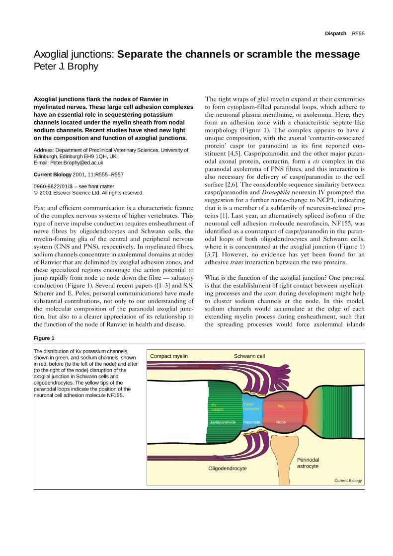
The distribution of Kv potassium channels,shown in green, and sodium channels, shownin red, before (to the left of the node) and after(to the right of the node) disruption of theaxoglial junction in Schwann cells andoligodendrocytes. The yellow tips of theparanodal loops indicate the position of theneuronal cell adhesion molecule NF155.
Kvcaspr2
Casprcontactin
Nav
Perinodalastrocyte
Current Biology
Schwann cellCompact myelin
Oligodendrocyte
Juxtaparanode Paranode Node

R556 Current Biology Vol 11 No 14
enriched in channels to coalesce at the nascent node ofRanvier [8]. Once the paranodal junctions are established,they could also serve to prevent the diffusion of sodiumchannels and restrict them to the node. However, severalnew studies on neurological mutants with myelinationdefects, either naturally occurring or engineered, have castthe function of axoglial adhesion in a new light. At the heartof all of these studies is the question: is the accumulation ofsodium channels at the node of Ranvier cell autonomous, ordoes it require glial contact at the axoglial junction?
Recent data from Scherer’s laboratory (S.S. Scherer,personal communication) have shown that sodium channelsin the CNS axons of myelin deficient (md) rats are clusteredin the absence of paranodal junctions. Mice with the rathermore subtle incapacity to synthesize the abundant galac-tolipids of myelin exhibit disrupted paranodal axoglialinteractions in the CNS and PNS, but still maintain nodalsodium channels [9]. If the junctional complex is targeteddirectly by inactivating the gene encoding caspr/paranodin,normal axoglial junctions do not form but the mice stilldisplay nodal concentrations of sodium channels, albeit in arather more diffuse fashion [1]. Mice deficient in contactinalso have disrupted junctional attachments; nevertheless,the absence of normal axoglial adhesion does not affectsodium channel clustering [2]. In spite of the presence ofsodium channels at the nodes of Ranvier, all of thesemutants have severe neurological phenotypes, which inmost cases have been shown to be linked to substantiallyreduced conduction velocities. If sodium channels persistat the node, how might the conduction of nerve impulsesbe compromised by the absence of normal adhesionbetween the paranodal glial loops and the axolemma?
In addition to sodium channels, myelinated axons also havepotassium channels. These channels are of the delayed-rec-tifier type first described in the Drosophila Shaker mutant.In mammalian myelinated axons, potassium channels arefirst detected during development within the nodal gap;subsequently they are sequestered from the node in thejuxtaparanodal region by the paranodal axoglial junction(Figure 1) [10]. This channel distribution was in fact firstproposed by Rosenbluth [11] based on the electron micro-scopic distribution of intramembranous particles. Shaker orKv potassium channels are believed to accelerate the rate ofrepolarization of the axolemma and play an important part inthe generation of action potentials before the myelin sheathhas been properly established during development. Theirexact function in the mature nerve has yet to be established.Nevertheless, clues have come from mice deficient in theKv1.1 channel, which display conduction failures at branchpoints and hyperexcitability induced by cooling [12].
In support of the importance of potassium channel seques-tration under the myelin sheath in the mature nerve, a key
feature of the deranged axoglial adhesion zones in the mdrat is the aberrant localization of potassium channels closerto the node (Figure 1; S.S. Scherer, personal communica-tion). Displacement of Kv channels from the juxtaparan-odal to the paranodal region of the axolemma is also afeature of mice deficient in caspr/paranodin, contactin orgalactocerebroside [1,2,9]. In each of these mutants, theKv channels approach the sodium channels so closely thatthey can sometimes be seen to overlap by immunofluores-cence microscopy. A protein closely related to caspr, caspr2,which associates with juxtanodal Kv channels, is also dis-placed to the paranodal region under these conditions [1].Thus, the common pathological change in a variety ofmutant mice is the desegregation of sodium and potassiumchannels as a result of a defective axoglial junction. Themechanism by which channel displacement might con-tribute to both ‘negative’ (conduction block) and ‘positive’(spontaneous bursts of impulses) consequences in demyeli-nating diseases is still not clear. Nevertheless, spontaneousfiring of action potentials is increasingly suspected of con-tributing to the development of neuropathic pain in somedemyelinating conditions [13].
Now that the molecular composition of the axoglialjunction is becoming clearer, increasing attention is beingpaid to how it might be assembled. Here, the actincytoskeleton appears to have an important role. Glialneurofascin has a well-characterized binding site for theactin-binding protein ankyrin, but it also interacts withezrin, a FERM (protein 4.1-ezrin-radixin-moesin) domainprotein (our unpublished data). FERM domain proteinscan act as linkers to the actin cytoskeleton. Interestingly,the closely related axonal protein L1 also has a functionalFERM domain binding site, in addition to an ankyrin-interaction domain, and Benson and colleagues (D. Benson,personal communication) have speculated these two modesof linkage to the actin cytoskeleton have distinct func-tions, with the ezrin site playing a more important partduring axonal development and pathfinding. The propertyof associating with FERM domain proteins is shared bycaspr/paranodin. Although, this binding site is dispensablefor targeting, it is required for stabilizing the protein at thejunction ([6] and E. Peles, personal communication). Itnow appears that the FERM domain protein with whichcaspr/paranodin interacts is protein 4.1B, and that whenthe caspr/paranodin–contactin complex is disrupted incontactin-deficient mice, protein 4.1B is mislocalized ([14]and E. Peles, personal communication).
The axoglial junction is the largest of the mammalian celladhesion complexes, and disruption of the interactionbetween myelin-forming glia and the axon is one of theearliest correlates of axonal dysfunction in demyelinatingdisease [15]. A sudden flurry of new data from several labshas given us a much better picture of the molecular

composition of the complex, and the generation of micelacking each of these components is helping to elucidatetheir function. In addition to identifying the rest of theplayers at this remarkable structure, a key issue which hasyet to be addressed is the possibility of signalling betweenaxons and glia at the site of axoglial interaction. Scott Bradyand colleagues [16] first showed several years ago that glialcontact influences the extent of neurofilament phosphory-lation in the axon, and axonal contact is well-known toinfluence Schwann cell gene expression in general, and dif-ferentiation in particular. The major site of physical contactbetween the glial cell and the axon is the junction; hence, itseems very likely that elucidation of the signal transductionpathways that function in the cytoplasm-filled paranodalloops, for example, will reveal a new dimension to cell–cellcommunication in the nervous system.
References1. Bhat MA, Rios JC, Lue Y, Garcia-Fresco GP, Ching W, St Martin M,
Li J, Einheber S, Chesler M, Rosenbluth J, et al.: Axon-gliainteractions and the domain organization of myelinated axonsrequires neurexin IV/caspr/paranodin. Neuron 2001, 30:369-383.
2. Boyle MET, Berglund EO, Murai KK, Weber L, Peles E, Ranscht B:Contactin orchestrates assembly of the septate-like junctions atthe paranode in myelinated peripheral nerve. Neuron 2001,30:385-397.
3. Tait S, Gunn-Moore F, Collinson JM, Huang J, Lubetzki C, Pedraza L,Sherman DL, Colman DR, Brophy PJ: An oligodendrocyte celladhesion molecule at the site of assembly of the paranodal axo-glial junction. J Cell Biol 2000, 150:657-666.
4. Einheber S, Zanazzi G, Ching W, Scherer S, Milner TA, Peles E,Salzer JL: The axonal membrane protein Caspr, a homologue ofneurexin IV, is a component of the septate-like paranodaljunctions that assemble during myelination. J Cell Biol 1997,139:1495-1506.
5. Menegoz M, Gaspar P, Le Bert M, Galvez T, Burgaya F, Palfrey C,Ezan P, Arnos F, Girault JA: Paranodin, a glycoprotein of neuronalparanodal membranes. Neuron 1997, 19:319-331.
6. Faivre-Sarrailh C, Gauthier F, Denisenko-Nehrbass N, Le Bivic A,Rougon G, Girault JA: The glycosylphosphatidyl inositol-anchoredadhesion molecule F3/contactin is required for surface transportof paranodin/contactin-associated protein (caspr). J Cell Biol2000, 149:491-502.
7. Collinson JM, Marshall D, Gillespie CS, Brophy PJ: Transientexpression of neurofascin by oligodendrocytes at the onset ofmyelinogenesis: implications for mechanisms of axon-glialinteraction. Glia 1998, 23:11-23.
8. Rasband MN, Peles E, Trimmer JS, Levinson SR, Lux SE, Shrager P:Dependence of nodal sodium channels clustering on paranodalaxoglial contact in the developing CNS. J Neurosci 1999,19:7516-7528.
9. Dupree JL, Girault JA, Popko B: Axo-glial interactions regulate thelocalization of axonal paranodal proteins. J Cell Biol 1999,147:1145-1151.
10. Vabnick I, Trimmer JS, Schwarz TL, Levinson SR, Risal D, Shrager P:Dynamic potassium channel distributions during axonaldevelopment prevent aberrant firing patterns. J Neurosci 1999,19:747-758.
11. Rosenbluth J: Role of glial cells in the differentiation and functionof myelinated axons. Int J Dev Neurosci 1988, 6:3-24.
12. Chiu SY, Zhou L, Zhang CL, Messing A: Analysis of potassiumchannel functions in mammalian axons by gene knockouts.J Neurocytol 1999, 8:349-364.
13. Gillespie CS, Sherman DL, Fleetwood-Walker SM, Cottrell DF, Tait S,Garry E, Ure J, Griffiths IR, Smith A, Brophy PJ: Peripheraldemyelination and neuropathic pain behavior in periaxin-deficientmice. Neuron 2000, 26:523-531.
14. Ohara R, Yamakawa H, Nakayama M, Ohara D: Type II brain 4.1(4.1B/KIAA0987), a member of the protein 4.1 family, is localizedto neuronal paranodes. Brain Res Mol Brain Res 2000, 85:41-52.
15. Griffin JW, Li CY, Macko C, Ho TW, Hsieh ST, Xue P, Wang FA,Cornblath DR, McKhann GM, Asbury AK: Early nodal changes in theacute motor axonal neuropathy pattern of the Guillain-Barresyndrome. J Neurocytol 1996, 25:33-51.
16. De Waegh SM, Lee VMY, Brady ST: Local modulation ofneurofilament phosphorylation, axonal caliber, and slow axonal-transport by myelinating Schwann cells. Cell 1992, 68:451-463.
Dispatch R557