azathioprine-induced carcinogenesis in mice according to
TRANSCRIPT

jnci.oxfordjournals.org JNCI | Article 1
DOI: 10.1093/jnci/djq389 © The Author 2010. Published by Oxford University Press. All rights reserved. For Permissions, please e-mail: [email protected].
Numerous studies have reported an increased risk of cancer among patients who received long-term therapy with drugs, including alkylating agents, topoisomerase inhibitors, and antimetabolites. Because of substantial improvements in drug therapy treatments, patients are now surviving longer and the number of iatrogenic cancers they develop has steadily increased. These cancers likely reflect not only late effects of therapy but also the individual patient’s genetic susceptibility, given that individual polymorphic variation in genes involved in carcinogen metabolism and detoxifi-cation and DNA repair pathways have been associated with the risk of developing iatrogenic cancers (1). It is therefore important to
identify the oncogenic mechanisms responsible for the develop-ment of therapy-related malignancies and to understand genetic susceptibility factors that may allow for the identification of indi-viduals at risk for such malignancies.
Among the frequently prescribed antimetabolites are the thio-purines, which comprise azathioprine, 6-mercaptopurine, and 6-thioguanine. Originally used in clinical practice to treat child-hood acute lymphoblastic leukemia, 6-mercaptopurine therapy has markedly improved the prognosis of patients with this malignancy (2). The introduction of azathioprine in the 1960s as an immuno-suppressant following organ transplantation has also contributed to
ARTICLE
Azathioprine-Induced Carcinogenesis in Mice According to Msh2 GenotypeAlexandra Chalastanis, Virginie Penard-Lacronique, Magali Svrcek, Valérie Defaweux, Nadine Antoine, Olivier Buhard, Sylvie Dumont, Bettina Fabiani, Isabelle Renault, Emmanuel Tubacher, Jean-François Fléjou, Hein te Riele, Alex Duval, Martine Muleris
Manuscript received February 2, 2010; revised August 6, 2010; accepted September 10, 2010.
Correspondence to: Alex Duval, MD, PhD, (e-mail: [email protected]) and Martine Muleris, PhD, (e-mail: [email protected]), Centre de Recherche Saint-Antoine (UMRS 938), Equipe “Instabilité des Microsatellites et Cancers,” Hôpital Saint-Antoine, Batiment Kourilsky, 184 rue du Faubourg St Antoine, Paris, France.
Background The thiopurine prodrug azathioprine is used extensively in cancer therapy. Exposure to this drug results in the selection of DNA mismatch repair–deficient cell clones in vitro. It has also been suggested that thiopurine drugs might constitute a risk factor for the emergence of human neoplasms displaying microsatellite instability (MSI) because of deficient DNA mismatch repair.
Methods Azathioprine was administered via drinking water (6–20 mg/kg body weight per day) to mice that were null (Msh2!/!; n = 27), heterozygous (Msh2"/!; n = 22), or wild type (Msh2WT; n = 18) for the DNA mismatch repair gene Msh2. Control mice (45 Msh2!/!, 38 Msh2"/!, and 12 Msh2WT) received drinking water lacking azathioprine. The effect of azathioprine on tumorigenesis and survival of the mice was evaluated by Kaplan–Meier curves using log-rank and Gehan–Breslow–Wilcoxon tests. Mouse tumor samples were characterized by histology and immunophenotyping, and their MSI status was determined by polymerase chain reaction analysis of three non-coding microsatellite markers and by immunohistochemistry. Msh2 status of tumor samples was assessed by loss of heterozygosity analyses and sequencing after reverse transcription–polymerase chain reaction of the entire Msh2 coding sequence. All statistical tests were two-sided.
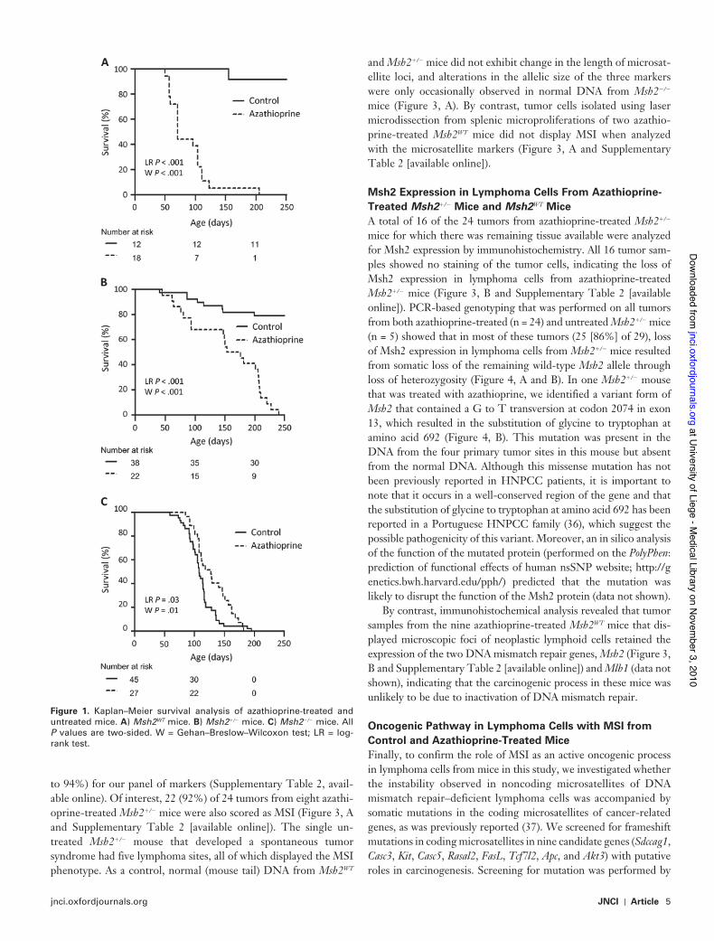
Results Most untreated Msh2WT and Msh2"/- mice remained asymptomatic and alive at 250 days of age, whereas azathi-oprine-treated Msh2WT and Msh2"/! mice developed lymphomas and died prematurely (median survival of 71 and 165 days of age, respectively). Azathioprine-treated Msh2"/! mice developed diffuse lymphomas lacking Msh2 expression and displaying MSI due to somatic inactivation of the functional Msh2 allele by loss of hetero-zygosity or mutation. By contrast, azathioprine-treated Msh2WT mice displayed no obvious tumor phenotype, but histological examination showed microscopic splenic foci of neoplastic lymphoid cells that retained Msh2 expression and did not display MSI. Both untreated and azathioprine-treated Msh2!/! mice had a reduced lifespan compared with untreated Msh2WT mice (median survival of 127 and 107 days of age, respectively) and developed lymphomas with MSI.
Conclusion Azathioprine-induced carcinogenesis in mice depends on the number of functional copies of the Msh2 gene.
J Natl Cancer Inst 2010;102:1–10
JNCI Journal of the National Cancer Institute Advance Access published October 5, 2010 at U
niversity of Liege - Medical Library on N
ovember 3, 2010
jnci.oxfordjournals.orgD
ownloaded from

2 Article | JNCI Vol. 102, Issue 22 | November 17, 2010
improvements in patient survival (3). Both drugs are also effective against autoimmune disorders, such as rheumatoid arthritis, auto-immune dermatological diseases, and inflammatory bowel diseases. Although it is now well established that prolonged treatment with thiopurine agents increases the risk of various cancer types, including non-Hodgkin lymphoma, cutaneous squamous cell carci-noma, hepatobiliary carcinoma, and mesenchymal tumors (4,5), the mechanism by which thiopurines contribute to carcinogenesis is not yet understood (6). To date, much of the increased risks of secondary cancers due to thiopurines and other immunosuppres-sants have been attributed to the effects of immunosuppression per se and to the subsequent involvement of oncogenic viruses (7). In vitro studies have also suggested that defects in the DNA mismatch repair system may be involved in thiopurine sensitivity because exposure of different cell lines to thiopurine agents consistently selects for cells with DNA mismatch repair defects (8–11). DNA mismatch repair is a system for recognizing and repairing the erro-neous insertion, deletion, and misincorporation of bases that can arise during DNA replication and recombination, as well as for processing some forms of DNA damage. In eukaryotes, the main protein components of this system are Msh2 and Mlh1. All three thiopurines are inactive prodrugs that are metabolized through a multistep pathway to generate 6-thiodeoxyguanosine, which becomes incorporated into DNA, causing the misincorporation of thymine during DNA synthesis. Recognition and processing of the subsequent mispaired bases by the DNA mismatch repair system results in cell death (12,13). DNA mismatch repair–deficient cells
are tolerant to thiopurines because they do not initiate lethal pro-cessing of the mispaired bases (14,15). Although DNA mismatch repair deficiency does not by itself cause cells to undergo malignant transformation, it is associated with an increased risk of cancers that exhibit a particular phenotype referred to as microsatellite insta-bility (MSI), which is defined as alterations in the length of short repeated (usually mono- or dinucleotide) sequences of DNA (ie, microsatellites) in tumor tissue relative to normal tissue (16–18).
The accumulation of somatic frameshift mutations defines a so-called mutator pathway that is likely to be oncogenic when it occurs in repeated sequences located within the coding sequence of genes involved in various biological pathways, such as the regu-lation of cell cycle and/or cell proliferation (eg, TGFBRII, IGFIIR, TCF-4, AXIN-2, PTEN, RIZ), the regulation of apoptosis (eg, BAX, CASP-5, BCL-10, APAF-1, FAS), or DNA damage signaling and repair (eg, RAD50, BLM, MSH3, MSH6, MBD4, MLH3, CHK1, ATR). The MSI phenotype characterizes tumors associated with the hereditary nonpolyposis colorectal cancer (HNPCC) syndrome and is also observed in approximately 10%–15% of spo-radic colorectal, gastric, and endometrial cancers (19). A high frequency of DNA mismatch repair deficiency has been reported in cancers that arise secondary to chemotherapy, particularly acute myeloid leukemia (AML) and myelodysplastic syndromes, in which the incidence of DNA mismatch repair deficiency is approx-imately 50%, whereas in de novo AML, it is less than 5% (20). Recent epidemiological studies have reported an association between DNA mismatch repair inactivation and the use of thiopu-rine regimens in AML and myelodysplastic syndromes that de-velop in patients treated for autoimmune disorders (5) and in AML and non-Hodgkin lymphomas that develop after organ transplan-tation (9,21,22). In other clinical contexts such as inflammatory bowel disease, there is no evidence that thiopurines contribute to the development of intestinal neoplasias with MSI (23). To date, there has been no formal in vivo demonstration that these drugs have an effect on MSI-driven oncogenesis.
In this study, we used a mouse model to examine the possible causal association between DNA mismatch repair and thiopurine sensitivity. Azathioprine was administered to mice that were null (Msh2!/!), heterozygous (Msh2"/!), or wild type (Msh2WT) for the DNA mismatch repair gene Msh2. The effect of azathioprine on tumorigenesis and survival of the mice was compared with that in untreated control mice.
Materials and MethodsMice and TreatmentMismatch repair–deficient mice carrying a targeted disruption in exon 12 of the Msh2 gene were described previously (24). Msh2"/! mice (129/Ola/FVB) (provided by Professor Hein te Riele) were intercrossed to obtain mice that were null, heterozygous, or wild type for the Msh2 gene. The genotypes of the mice were deter-mined by using polymerase chain reaction (PCR) analysis, as pre-viously described (25). Groups of mice aged 6 weeks (27 Msh2!/! mice, 22 Msh2"/! mice, and 18 Msh2WT mice) received azathioprine (Imurel 50 mg; GlaxoSmithKline, Marly-le-Roi, France) orally via their drinking water; the estimated dose was 6–20 mg/kg body weight per day, given that a mouse weighs approximately 20–30 g and
CONTEXT AND CAVEATS
Prior knowledgeThe thiopurine prodrug azathioprine is used extensively in cancer therapy and as an immunosuppressant in various clinical contexts. However, because exposure to azathioprine results in the selection of DNA mismatch repair–deficient cell clones in vitro, it may con-stitute a risk factor for the emergence of human neoplasms dis-playing microsatellite instability.
Study designAzathioprine was administered to mice that were null (Msh2!/!), heterozygous (Msh2"/!), or wild type (Msh2WT) for the DNA mis-match repair gene Msh2 to examine the possible causal associa-tion between DNA mismatch repair and thiopurine sensitivity. The effect of azathioprine on tumorigenesis and survival of the mice was compared with that in untreated control mice.
ContributionAzathioprine-induced carcinogenesis in mice depends on the number of functional copies of the Msh2 gene.
ImplicationsExposure to azathioprine may be an important risk factor for the development of tumors with microsatellite instability, especially for individuals who carry mutations in DNA mismatch repair genes.
LimitationsWhether these observations can be extended to other frequently prescribed immunosuppressants whose actions are independent of DNA mismatch repair is not known.
From the Editors
at University of Liege - M
edical Library on Novem
ber 3, 2010jnci.oxfordjournals.org
Dow
nloaded from

jnci.oxfordjournals.org JNCI | Article 3
drinks approximately 4–8 mL of water per day. This dosage of azathioprine corresponds to a human dose equivalent of 0.5–1.6 mg/kg body weight per day (26), which lies within the range of doses usually prescribed in human therapy (27–31). Control mice (45 Msh2!/!mice, 38 Msh2"/! mice, and 12 Msh2WT mice) received water that did not contain azathioprine. All experiments were con-ducted in accordance with the regulations controlling procedures in live animals in France, after approval of the Ethical Committee for Laboratory Animal Care of the Saint-Antoine research center (Paris, France).
Histopathological and Immunohistochemical StudiesMice that displayed signs of poor health including breath insuffi-ciency and posterior leg paralysis were killed by cervical disloca-tion and autopsied. At autopsy, the spleen, liver, and thymus were systematically removed together with other enlarged organs or lymph nodes, and a portion of each type of tissue was stored at −80°C. The remaining portions of tissue were formalin fixed, em-bedded in paraffin, and sectioned for histological analysis and im-munohistochemical staining for Mlh1 and Msh2. For histology, 4-µm sections were stained with hematoxylin–eosin. For Msh2 and Mlh1 immunohistochemistry, 4-µm sections were incubated with mouse monoclonal antibodies against MLH1 (mouse and human cross-reactive clone G168–728; 1:25 dilution; BD Pharmingen, San Diego, CA) and MSH2 (mouse and human cross-reactive clone FE11; 1:25 dilution; Calbiochem, Cambridge, MA) as previ-ously described (32), followed by incubation with the appropriate secondary antibodies (Trekkie biotinylated mouse link and trekavidin–HRP label; Biocare, Paris, France). Stromal and normal lymphocytes that were present in the section served as the control for positive staining. For immunophenotyping, 6-µm thick acetone-fixed frozen sections of spleen were incubated with a rat monoclonal antibody against mouse CD45, which recognizes hematopoietic cells (clone 30-F11; 1:50 dilution; BD Pharmingen), a rabbit monoclonal antibody against CD3, which recognizes T cells (mouse and human cross-reactive clone sp7; 1:100 dilution; Interchim, Montluçon, France), and a biotinylated rat monoclonal antibody against mouse CD19, which recognizes B cells (clone 6D5; 1:100 dilution; Abcam, Paris, France). Sections were then incubated with the following horseradish peroxidase–conjugated secondary antibodies: rabbit anti-rat immunoglobulin G (1:250 dilution; Abcam), amplification system anti-rabbit (Ready-to-use Envision+ system; Dako, Trappes, France), and horseradish peroxidase–streptavidin conjugate (1:500 dilution; Zymed, Invitrogen, Cergy Pontoise, France), respectively. Peroxidase activity (ie, bound antibody) was detected with the use of a DAB+ kit (Dako).
Microdissection of Mouse Tumor Tissue SectionsThe tumor cell component from two azathioprine-treated Msh2WT mice selected at random was collected from 10 hematoxylin- and eosin-stained 7-µm tissue sections per mouse with the use of a laser-capture microdissection system (PALM Laser; Zeiss, Le Pecq, France) and used for DNA extraction.
PCR-Based Msh2 Genotyping of Normal and Tumor DNANormal DNA (from mouse tail) and tumor DNA were extracted with the use of a QIAamp DNA extraction kit (Qiagen, Courtaboeuf,
France) according to the manufacturer’s instructions. Msh2 geno-type analysis was performed as previously described (25) by using a three-primer allele-specific PCR assay in which the wild-type and inactivated Msh2 alleles generated PCR products of 164 and 194 bp, respectively. Briefly, PCR was performed using Taq DNA Polymerase kit (Qiagen) with 0.5 mM MgCl2, 0.2 mM dNTP mix, and 0.5pmol/µL of each primer (primers are detailed in Supplementary Table 1, available online). PCR conditions were 94°C for 5 minutes followed by 35 cycles of 94°C for 30 seconds, 60°C for 30 seconds, and 72°C for 30 seconds, and a final exten-sion at 72°C for 7 minutes. The PCR products were resolved on agarose gels. To assess loss of heterozygosity of the Msh2 allele, quantitative densitometry scanning of the DNA bands on a 4% agarose gel was carried out with the use of GeneTools software (version 4.01; Syngene, Saint Quentin en Yvelines, France). Allelic loss was calculated using the following normalized allelic imbalance ratio (WT tumor to D tumor)/(WT normal to D normal), where WT and D correspond to the wild-type and disrupted alleles, re-spectively. An allelic imbalance ratio less than 0.6 was considered indicative of loss of heterozygosity based on the observation that some tumors contained an estimated 40% of normal stromal cells interspersed among the tumor cells.
Msh2 Gene SequencingReverse transcription–PCR was used to generate eight overlapping DNA fragments that covered the entire Msh2 coding sequence. Total RNA was extracted from nine mouse tissue samples four tumor sites and the corresponding normal tissue from mouse 36 (an azathioprine-treated Msh2"/! mouse that did not show loss of heterozygosity of the Msh2 allele), and tumor tissues from four other mice for controls (three azathioprine-treated Msh2"/! mice and one untreated Msh2!/! mouse) with the use of an RNeasy extraction kit (Qiagen) according to the manufacturer’s instruc-tions. For reverse transcription, we used 700 µg of RNA and murine leukemia virus reverse transcriptase and oligo dT from a GeneAmp RNA PCR kit (Perkin Elmer, Courtaboeuf, France). PCR was performed with the use of a HotStarTaq DNA Polymerase kit (Qiagen) with 3 mM MgCl2 and 0.3 µM of each primer (primers are detailed in Supplementary Table 1, available online). The PCR profile was 95°C for 15 minutes followed by 35 cycles of 95°C for 30 seconds, 63°C for 30 seconds, and 72°C for 1 mi-nute. Sequencing was done by a linear amplification with the use of a Big Dye Terminator v1.1 Cycle sequencing kit (Applera, Courtaboeuf, France) according to the manufacturer’s instruc-tions. PCR products were analyzed on an ABI PRISM 3100 Genetic Analyzer (Applera). Identified DNA sequence variants were con-firmed by sequencing both DNA strands.
Analysis of MSIWe used three noncoding mononucleotide repeats (all on chromosome 1) to determine tumor MSI status: A22, A24, and T40. PCR reactions were performed using DNA (50 ng) isolated from tumor and normal tissues from the same mouse, GoTaq DNA Polymerase (Promega, Charbonnieres, France), 1.5 mM MgCl2, and 0.3 µM of each primer (Supplementary Table 1, available online). The PCR profile was 94°C for 5 minutes followed by 35 cycles of 94°C for 30 seconds, 50°C (for A22 and A24) or 54°C (for T40) for 30
at University of Liege - M
edical Library on Novem
ber 3, 2010jnci.oxfordjournals.org
Dow
nloaded from

4 Article | JNCI Vol. 102, Issue 22 | November 17, 2010
seconds and 72°C for 30 seconds, with final extension at 72°C for 7 minutes. Amplified products were resolved on denaturing gels run in an ABI PRISM 3100 Genetic Analyzer and analyzed using Genescan 3.7 and Genotyper 2.1 software programs (Applera). We compared the predominant allele size observed in the tumor with that observed in normal tissue from the same mouse (ie, 190, 70, and 122 for markers A22, A24, and T40, respectively). Tumors were scored as MSI positive if the length of at least one marker in tumor DNA differed from that in normal tissue DNA.
Mutation Analysis of Target Genes for MSITo confirm the role of MSI as an active oncogenic process in lym-phoma cells, we screened for frameshift mutations in coding mic-rosatellites of cancer-related genes. Most coding microsatellite sequences that are contained within human genes are not con-served in the murine genome. We therefore constructed an in silico database by querying GenBank (October 2007) for murine genes that contain coding microsatellite sequences greater than seven nucleotides in length (available on request from the authors). From this database, we selected nine genes with involvement in tumorigenesis that were possible targets for mutation by MSI: Sdccag1, Casc3, Kit, Casc5, Rasal2, FasL, Tcf7l2, Apc, and Akt3. Only one of these genes, Tcf7l2 (also known as Tcf-4, the main effector of Wnt and Wingless signaling), was known to be mutated in a conserved microsatellite sequence in human colorectal tumors with MSI (33). Screening for mutation was performed by PCR using tumor or corresponding normal DNA (50 ng), primers spe-cific for each gene (at 0.3 µM; Supplementary Table 1, available online), and a Taq DNA Polymerase kit (Qiagen). The PCR pro-file was 94°C for 4 minutes, followed by 35 cycles of 94°C for 30 seconds, 60°C for 30 seconds, and 72°C for 1 minute, with a final extension at 72°C for 7 minutes. Tumors were scored as mutated if the length of the predominant allele in tumor DNA differed from that in normal tissue DNA.
Generation of Kaplan–Meier Survival Curves and Statistical AnalysisThe health of the mice was examined daily. Mice that presented with signs of poor health were killed by cervical dislocation according to the recommendations of our Ethical Committee. In our experi-ence, these signs are due to lymphomagenesis (34,35) and precede the time of death by a couple of days. The time of death was recorded for each mouse that was found dead or had been killed. Kaplan–Meier survival curves were generated with the use of Graphpad Prism software (version 4; Graphpad, Inc, San Diego, CA). Comparisons of median survivals were performed using log-rank and Gehan–Breslow–Wilcoxon tests. Comparisons of the mean number of tumors in Msh2!/! and treated Msh2"/! mice were performed using the Student t test. All statistical tests were two-sided.
ResultsEffect of Azathioprine on Mouse Tumorigenesis and Survival According to Msh2 GenotypePrevious studies demonstrated that heterozygous Msh2"/! mice have a normal lifespan, whereas most Msh2!/! mice spontaneously develop diffuse aggressive lymphomas and succumb to these by
6–8 months of age (34,35). In agreement with these findings, we observed that most (82%) of the untreated Msh2"/! mice remained free of disease, whereas all untreated Msh2!/! mice were dead by age 200 days (Figure 1). Msh2"/! and Msh2WT mice that received azathioprine via their drinking water showed statistically signifi-cantly shorter survival compared with the respective untreated mice: All treated mice were dead by age 250 days (log-rank test: P < .001; Gehan–Breslow–Wilcoxon test: P < .001). However, aza-thioprine-treated Msh2"/! mice had statistically significantly longer median survival compared with azathioprine-treated Msh2WT mice (165 vs 71 days, difference = 94 days, 95% confidence interval [CI] = 55 to 133 days; P < .001 [Gehan–Breslow–Wilcoxon test]; Figure 1). Conversely, azathioprine-treated Msh2!/! mice had slightly longer median survival compared with untreated Msh2!/! mice (127 vs 107 days, difference = 20 days, 95% CI = −9 to 49 days, P = .03 log-rank test; P = .01 [Gehan–Breslow–Wilcoxon test]; Figure 1).
All azathioprine-treated Msh2!/! mice (n = 15), untreated Msh2!/! mice (n = 10), and azathioprine-treated Msh2"/! mice (n = 8) that were autopsied were found to have developed a tumoral phenotype that was characterized by abnormal enlargement of the thymus, spleen, and liver, sometimes in association with adeno-megaly. Histological examination of all tumor masses revealed massive infiltration of organs by medium-to-large lymphoid cells (Figure 2, A and Supplementary Table 2 [available online]). Of note, the mean number of macroscopic tumor sites did not differ statisti-cally significantly between Msh2!/! mice (treated and untreated combined) and treated Msh2"/! mice (2 vs 3 sites, difference = 1 site, 95% CI = 0.94 to 1.06 sites; P = .13 [Student t test]) (Supplementary Table 2, available online). An identical syndrome was observed in one untreated Msh2"/! mouse. By contrast, none of the 11 azathio-prine-treated Msh2WT mice that were analyzed displayed obvious enlargement of organs; however, histological examination of these mice confirmed, in most instances (nine of 11 mice), the presence of microscopic foci comprising medium-to-large neoplastic lymphoid cells that were restricted to the spleen area located under the capsule (Figure 2, A). There were no histological or immunophenotypical differences between azathioprine-induced and spontaneous lym-phomas in mice with any Msh2 genotype (Figure 2, B and Supplementary Table 3 [available online]). All but one of the lym-phomas was of B-cell origin (ie, CD45 positive, CD19 positive, and CD3 negative); one tumor sample from an azathioprine-treated Msh2"/! mouse was of T-cell origin (ie, CD45 positive, CD19 negative, and CD3 positive).
MSI Status of Azathioprine-Induced Lymphomas in Msh2"/! and Msh2WT MiceWe next examined tumor DNA for three noncoding microsatellite markers to determine whether the tumors that developed in aza-thioprine-treated Msh2"/! and Msh2WT mice differed in terms of MSI status. Most of tumor DNA samples from Msh2!/! mice dis-played MSI (defined as a change in the length of one or more microsatellite loci compared with normal tissue DNA from the same mouse) (Figure 3, A). In all, 43 of 50 lymphomas from 25 Msh2 !/! mice (including 15 of 18 lymphomas from 10 untreated mice and 28 of 32 lymphomas from 15 azathioprine-treated mice) displayed MSI demonstrating a sensitivity of 86% (95% CI = 73%
at University of Liege - M
edical Library on Novem
ber 3, 2010jnci.oxfordjournals.org
Dow
nloaded from
jnci.oxfordjournals.org JNCI | Article 5
to 94%) for our panel of markers (Supplementary Table 2, avail-able online). Of interest, 22 (92%) of 24 tumors from eight azathi-oprine-treated Msh2"/! mice were also scored as MSI (Figure 3, A and Supplementary Table 2 [available online]). The single un-treated Msh2"/! mouse that developed a spontaneous tumor syndrome had five lymphoma sites, all of which displayed the MSI phenotype. As a control, normal (mouse tail) DNA from Msh2WT
and Msh2"/! mice did not exhibit change in the length of microsat-ellite loci, and alterations in the allelic size of the three markers were only occasionally observed in normal DNA from Msh2!/! mice (Figure 3, A). By contrast, tumor cells isolated using laser microdissection from splenic microproliferations of two azathio-prine-treated Msh2WT mice did not display MSI when analyzed with the microsatellite markers (Figure 3, A and Supplementary Table 2 [available online]).
Msh2 Expression in Lymphoma Cells From Azathioprine-Treated Msh2"/! Mice and Msh2WT MiceA total of 16 of the 24 tumors from azathioprine-treated Msh2"/! mice for which there was remaining tissue available were analyzed for Msh2 expression by immunohistochemistry. All 16 tumor sam-ples showed no staining of the tumor cells, indicating the loss of Msh2 expression in lymphoma cells from azathioprine-treated Msh2"/! mice (Figure 3, B and Supplementary Table 2 [available online]). PCR-based genotyping that was performed on all tumors from both azathioprine-treated (n = 24) and untreated Msh2"/! mice (n = 5) showed that in most of these tumors (25 [86%] of 29), loss of Msh2 expression in lymphoma cells from Msh2"/! mice resulted from somatic loss of the remaining wild-type Msh2 allele through loss of heterozygosity (Figure 4, A and B). In one Msh2"/! mouse that was treated with azathioprine, we identified a variant form of Msh2 that contained a G to T transversion at codon 2074 in exon 13, which resulted in the substitution of glycine to tryptophan at amino acid 692 (Figure 4, B). This mutation was present in the DNA from the four primary tumor sites in this mouse but absent from the normal DNA. Although this missense mutation has not been previously reported in HNPCC patients, it is important to note that it occurs in a well-conserved region of the gene and that the substitution of glycine to tryptophan at amino acid 692 has been reported in a Portuguese HNPCC family (36), which suggest the possible pathogenicity of this variant. Moreover, an in silico analysis of the function of the mutated protein (performed on the PolyPhen: prediction of functional effects of human nsSNP website; http://genetics.bwh.harvard.edu/pph/) predicted that the mutation was likely to disrupt the function of the Msh2 protein (data not shown).
By contrast, immunohistochemical analysis revealed that tumor samples from the nine azathioprine-treated Msh2WT mice that dis-played microscopic foci of neoplastic lymphoid cells retained the expression of the two DNA mismatch repair genes, Msh2 (Figure 3, B and Supplementary Table 2 [available online]) and Mlh1 (data not shown), indicating that the carcinogenic process in these mice was unlikely to be due to inactivation of DNA mismatch repair.
Oncogenic Pathway in Lymphoma Cells with MSI from Control and Azathioprine-Treated MiceFinally, to confirm the role of MSI as an active oncogenic process in lymphoma cells from mice in this study, we investigated whether the instability observed in noncoding microsatellites of DNA mismatch repair–deficient lymphoma cells was accompanied by somatic mutations in the coding microsatellites of cancer-related genes, as was previously reported (37). We screened for frameshift mutations in coding microsatellites in nine candidate genes (Sdccag1, Casc3, Kit, Casc5, Rasal2, FasL, Tcf7l2, Apc, and Akt3) with putative roles in carcinogenesis. Screening for mutation was performed by
Figure 1. Kaplan–Meier survival analysis of azathioprine-treated and untreated mice. A) Msh2WT mice. B) Msh2"/! mice. C) Msh2!/! mice. All P values are two-sided. W = Gehan–Breslow–Wilcoxon test; LR = log-rank test.
at University of Liege - M
edical Library on Novem
ber 3, 2010jnci.oxfordjournals.org
Dow
nloaded from

6 Article | JNCI Vol. 102, Issue 22 | November 17, 2010
comparing the predominant allele size of PCR product in tumor and corresponding normal DNA in a representative sample of tumors that displayed MSI (ie, 61 tumor samples from six azathio-prine-treated Msh2"/! mice, one Msh2"/! untreated mouse, 14 aza-thioprine-treated Msh2!/! mice, and four untreated Msh2!/! mice) (Figure 3, C). Frameshift mutations were found in eight of the target genes, of which the Sdccag1 (serologically defined colon can-cer antigen 1) and Casc3 (cancer susceptibility candidate 3) genes were mutated at relatively high frequencies in these tumors (58% and 21%, respectively; Figure 3, C). The mutation frequency for each of the nine target genes was similar in spontaneous and azathi-oprine-induced lymphomas (data not shown). The relatively high frequencies of frameshift mutations in Sdccag1 and Casc3 pointed to an active oncogenic MSI pathway in lymphomas from Msh2!/! and Msh2"/! mice. The human SDCCAG1 homolog has been identified as a tumor suppressor in non–small cell lung cancer cell lines (38,39). The Casc3 protein is a component of a splicing-dependent multi-protein junction complex that marks the position of the exon–exon junction in mature mRNA (40), thereby influencing downstream gene expression processes including mRNA splicing, nuclear mRNA export, subcellular mRNA localization, translation efficiency, and nonsense-mediated mRNA decay. Casc3 could then play a role in MSI-driven carcinogenesis, given that numerous frameshift mutation–derived mRNAs are processed by the non-sense-mediated mRNA decay machinery in MSI tumors (41,42). It is noteworthy that in a given mouse, some tumor locations dis-played a mutation in Sdccag1 or Casc3 genes, whereas other tumor sites did not, suggesting that the mutations occurred as indepen-dent molecular events during disease progression (Figure 3, D).
DiscussionThiopurines are effective anticancer agents and immunosuppres-sants in a variety of clinical conditions. However, they have been
reported to be associated with an increased incidence of secondary tumors (6). It is therefore important to identify the oncogenic mechanisms responsible for the development of iatrogenic neo-plasms because such information could allow the identification of patients who may be at risk of developing tumors when treated with these drugs. For this purpose, we investigated whether a murine model could be used to examine the causal involvement of DNA mismatch repair in thiopurine sensitivity. We administered azathioprine to mice that were wild type or inactivated in one or both copies of the DNA mismatch repair gene Msh2. The dosage of azathioprine used in our mouse model corresponds to a human dose equivalent of 0.5–1.6 mg/kg body weight per day (26) and lies within the range usually prescribed in human therapy. As a muta-gen and immunosuppressant, azathioprine was expected to induce tumor formation in mice, which turned out to be the case. However, it is interesting that we also found that the oncogenic pathway, the survival, and the tumoral phenotype of azathioprine-treated mice were influenced by their Msh2 status. Briefly, we observed that azathioprine-treated Msh2WT and Msh2"/! mice developed lymphomas and died prematurely (median survival of 71 vs 165 days of age, respectively) compared with the respective un-treated mice that mostly were alive at 250 days of age. However, we demonstrated that Msh2"/! mice developed diffuse lymphomas that lacked Msh2 expression and displayed MSI at both noncoding and coding microsatellite sequences because of somatic inactiva-tion of the remaining functional Msh2 allele in tumor cells. By contrast, Msh2WT mice presented with splenic microscopic foci of neoplastic lymphoid cells that retained both Msh2 and Mlh1 ex-pression and did not display MSI. Both untreated and azathio-prine-treated Msh2!/! mice developed diffuse MSI lymphomas as expected, but surprisingly, the former had slightly longer median survival (median survival of 127 vs 107 days of age, respectively). Our data thus clearly demonstrate the ability of azathioprine to trigger carcinogenesis through an MSI-driven process in vivo.
Figure 2. Histological and immunohistochemical characterization of murine lymphomas. A) Hematoxylin–eosin staining of splenic lym-phoma from azathioprine-treated Msh2!/!, Msh2"/!, and Msh2WT mice. Arrows indicate tumor cells; arrowheads indicate normal lym-phocytes. B) Immunohistochemistry of spleen tissue cryosections from two azathioprine-treated Msh2"/! mice. Adjacent sections of spleen from each mouse were stained with anti-bodies against CD45 to identify hematopoietic cells, against CD3 to identify T cells, or against CD19 to identify B cells. Brown indicates positive staining. Scale bars = 30 µm.
at University of Liege - M
edical Library on Novem
ber 3, 2010jnci.oxfordjournals.org
Dow
nloaded from

jnci.oxfordjournals.org JNCI | Article 7
Figure 3. Msh2 expression and instability analysis in noncoding and coding microsatellites in lymphomas from azathioprine-treated mice. A) Allelic profiles of noncoding microsatellite markers A22, A24, and T40 in normal (control) and tumor tissues from three mice. Shown are represen-tative profiles displaying shorter alleles (arrows) for the three markers in tumor DNA compared with the corresponding normal tissue DNA in an Msh2!/! mouse and an Msh2"/! mouse. In the representative profile for the Msh2WT mice, the sizes of the microsatellites did not differ between tumor and normal tissue. Length of the predominant allele (bp) is indi-cated in a box below each profile. Dashed vertical lines indicate the pre-dominant allele size observed in control DNA from all Msh2"/! and Msh2WT mice analyzed (reference value). Note that the size of the predom-inant allele of markers T40 and A22 differed from this reference value in the normal DNA from the Msh2!/! mouse. B) Immunohistochemical analysis of Msh2 expression in lymphomas. Shown are representative images of staining of tumor sections from an Msh2"/! mouse and an
Msh2WT mouse with an Msh2 antibody. Tumor cells were not stained in the Msh2"/! mice, whereas they stained positively in the Msh2WT mice. Normal lymphocytes showed a less pronounced Msh2 staining due to their lower proliferation rate. Red indicates positive staining. T = medium-to-large neoplastic lymphoid cells; N = normal lymphocytes. Scale bar = 30 µm. C) Mutation analysis of target genes for microsatellite instability (MSI). Analysis was performed by comparing the predominant allele size of the coding microsatellite in tumor DNA and corresponding normal tissue DNA. Two genes displayed frameshift mutations at relatively high frequencies, Sdccag1 and Casc3, pointing to an active oncogenic MSI pathway in lymphomas from Msh2!/! and Msh2"/! mice. D) Details of the Sdccag1 and Casc3 mutation status in individual tumors from Msh2!/! and Msh2"/! mice. Each row corresponds to a single mouse, each square represents a tumor site in that mouse, numbers correspond to mouse number, mice received azathioprine (Aza) treatment (1) or not (0). Black = mutated; white = wild type; gray = analysis not informative.
at University of Liege - M
edical Library on Novem
ber 3, 2010jnci.oxfordjournals.org
Dow
nloaded from

8 Article | JNCI Vol. 102, Issue 22 | November 17, 2010
To date, two mechanisms have been reported to underlie the cytotoxicity of azathioprine. First, azathioprine-induced purine starvation slows the rate of cell division, especially in lymphocytes, which, unlike other cell types, cannot use the salvage pathway of purine synthesis (43–45). Importantly, this effect has not been demonstrated to be dependent on Msh2 status. Second, DNA mis-match repair–dependent signaling of azathioprine-induced DNA damage results in apoptosis in DNA mismatch repair–proficient cells (12,13). By contrast, DNA mismatch repair–deficient cells display thiopurine tolerance and are therefore able to escape cell death when exposed to azathioprine (14,15) and subsequently un-dergo neoplastic cell transformation through the accumulation of genetic mutations caused by MSI. Moreover, the MSI-driven on-cogenic pathway has been shown previously to favor the emer-gence of a tumor phenotype in mice that is characterized by an enlarged thymus, spleen, and liver and which leads Msh2!/! mice to die from diffuse aggressive lymphomas (34,35). Taking all these elements into consideration, we propose the following explanations to account for the various oncogenic pathways, survival, and tumoral phenotypes observed in azathioprine-treated mice with different Msh2 status. First, untreated Msh2!/! mice exhibited the characteristic development of MSI lymphomas, which were ulti-mately responsible for their death (see above). Azathioprine-treated Msh2!/! mice displayed the same tumor phenotype as untreated mice, but had slightly longer survival, which may reflect reduced Msh2!/! lymphocyte proliferation due to purine starvation resulting from azathioprine treatment. Second, azathioprine-treated Msh2"/! mice were similar to both treated and untreated Msh2!/! mice with respect to tumor phenotype, histology, and MSI status of lymphoma cells. A reasonable explanation for this finding is that like (treated and untreated) Msh2!/! mice, azathioprine-treated Msh2"/! mice died from lymphomas that had MSI and an
active oncogenic mutator pathway. The longer median survival of azathioprine-treated Msh2"/! mice compared with azathioprine-treated Msh2!/! mice (165 vs 127 days) may be due to the time required for somatic inactivation of the remaining functional Msh2 allele in the Msh2"/! mice, in accordance with the Knudson two-hit model (46). Finally, azathioprine-treated Msh2WT mice exhibited a distinct tumor phenotype but no enlarged organs. Histological examination revealed microscopic foci of lymphoma cells express-ing Msh2 and Mlh1 under the splenic capsule, and careful exami-nation of genomic DNA extracted from laser-capture microdissected tumor tissue excluded the presence of an MSI phenotype. Strikingly, these mice exhibited the shortest median survival. We speculate that these mice may have died from lymphomas that arose from oncogenic mechanisms other than MSI, such as chromosomal in-stability. Alternatively, these mice may have died from general organ failure due to the greater cytotoxicity of azathioprine in Msh2WT cells. In support of the latter mechanism are data showing that human Msh2"/! lymphoblastoid cells are approximately four-fold more tolerant than wild-type cells to killing by temozolomide, a methylating agent known to involve a DNA mismatch repair–dependent signaling response to DNA damage (47).
Our findings could have important clinical implications for patients receiving azathioprine therapy. Msh2!/! mice spontane-ously develop lymphomas and display the phenotype of the rare patients with homozygous or biallelic inactivation of DNA mis-match repair genes, including a severely reduced lifespan and increased frequency of hematological malignancies compared with general population (48). We have shown that all azathioprine-induced lymphomas in Msh2"/! mice arise from an MSI-driven oncogenic process following inactivation of the Msh2 wild-type al-lele, mainly through loss of heterozygosity. Mice with one constitutively inactivated Msh2 allele are the murine equivalent of
Figure 4. Loss of heterozygosity analyses and sequencing of the Msh2 coding sequence in tumor tissues from Msh2"/! mice. DNA bands on the gel electrophoresis after polymerase chain reaction–based genotyp-ing are shown for all tumor samples from nine Msh2"/! mice (numbers correspond to mouse number). Some tumor sites showed complete loss of signal for the wild-type allele, whereas other sites showed re-sidual signal corresponding to contamination from non–tumor cell DNA. The intensity ratio of the two allelic bands (wild type and dis-rupted) in tumor DNA relative to that obtained in normal (control) DNA
was calculated and a ratio of 0.6 or less corresponding to a reduction of intensity of at least 40% in tumor DNA was used to score loss of hetero-zygosity. A) Loss of the wild-type Msh2 allele in all tumor sites for eight mice. LN = lymph node. B) Mutation of the wild-type Msh2 allele in tumor sites from mouse 36. In the single mouse in which the wild-type allele was retained at all four sites examined (left), a G to T transversion (asterisk) in exon 13 of the Msh2 gene was detected by sequencing after reverse transcription polymerase chain reaction analysis of the entire Msh2 coding sequence (right).
at University of Liege - M
edical Library on Novem
ber 3, 2010jnci.oxfordjournals.org
Dow
nloaded from

jnci.oxfordjournals.org JNCI | Article 9
the HNPCC syndrome, one of the most common genetic predispo-sitions to cancer in humans. However, unlike humans who carry a heterozygous mutation in a DNA mismatch repair gene, Msh2"/! mice do not spontaneously develop neoplasms with MSI. HNPCC is relatively prevalent, affecting 2%–5% of patients diagnosed with colorectal cancer (49). Given that the lifetime risk of colorectal cancer is approximately 6% in the United States (50), we estimate that HNPCC may affect approximately one of 300–800 individuals in the general population. Moreover, because the evaluated inci-dence of HNPCC relies on the early age at cancer diagnosis and family history of cancer, it is likely to be underestimated because the penetrance of the syndrome may be largely incomplete and HNPCC may also be characterized by the emergence of late-onset tumors in some patients (51). Finally, lymphomas that developed in azathioprine-treated Msh2WT mice did not display MSI, whereas we previously detected MSI in a minority (8%) of lymphomas that developed in a large cohort (n = 111) of non-HNPCC patients treated with azathioprine and/or other immunosuppressants (21). Together, the above observations indicate that azathioprine and, more broadly, the thiopurines constitute only a minor risk factor for the development of tumors with MSI in the general population. However, for individuals who carry mutations in DNA mismatch repair genes, exposure to azathioprine may be an important risk factor for the development of tumors with MSI. Our results could thus have important implications for the use of azathioprine in heterozygous carriers of DNA mismatch repair gene defects. On the basis of our findings, we recommend that more effort should be directed toward identifying possible cases of HNPCC among patients who are frequently prescribed thiopurines, such as trans-plant patients and patients suffering from inflammatory diseases. In addition, systematic screening for mutations in DNA mismatch repair genes in azathioprine-treated patients who subsequently developed DNA mismatch repair–deficient neoplasms could unmask previously unrecognized cases of HNPCC. Because earlier results by our group showed that MSI-associated lymphoprolifera-tive disorders in transplant patients were usually late onset (22), this possibility should be considered, especially when the tumor is diag-nosed long after the immunosuppressive therapy. Consequently, we recommend systematic screening for MSI in lymphomas and other neoplasms that arise in patients treated with thiopurine drugs. This screening could be particularly important because tumors with MSI usually have a different prognosis and sensitivity to chemothera-peutic agents compared with tumors that lack MSI, as has been shown for colorectal carcinomas (52).
To date, the mechanisms through which iatrogenic neoplasms arise in patients who are treated with different drugs in various clinical contexts are largely unknown. Here we show that mouse models can be highly relevant for the investigation of this issue because they can allow the identification of a known cancer path-way in relation to the prescription of a specific drug. Following on from these findings, we recommend that similar studies be carried out to determine whether other cancer-related genes play a role in the development of secondary neoplasms in response to other therapeutic agents. This study has provided further insight into the mechanisms that underlie azathioprine-induced carcino-genesis, including the ability of this drug to induce MSI-driven malignancy in Msh2"/! mice. The limitation of this study is that
we did not investigate if such observations are specific for thiopu-rines or whether they could be extended to other frequently pre-scribed immunosuppressants whose actions are independent of DNA mismatch repair. It is generally considered that MSI tumors are tightly controlled by the host’s immune system. Indeed, tumors that display MSI are characterized by a high level of lymphocytic infiltration due to their highly immunogenic nature (53), and it has been shown that aberrant neoantigenic peptides resulting from genes carrying frameshift mutations may elicit an immune response against tumor cells displaying MSI, which could then limit their proliferation (54,55). Further studies anal-ogous to this study involving other frequently prescribed immu-nosuppressants are required to determine whether a reduction in host immunosurveillance, by itself, plays a role in MSI-driven oncogenesis.
References 1. Allan JM, Rabkin CS. Genetic susceptibility to iatrogenic malignancy.
Pharmacogenomics. 2005;6(6):615–628. 2. Burchenal JH, Murphy ML, Ellison RR, et al. Clinical evaluation of a new
antimetabolite, 6-mercaptopurine, in the treatment of leukemia and allied diseases. Blood. 1953;8(11):965–999.
3. Elion GB. The purine path to chemotherapy. Science. 1989;244(4900):41–47. 4. Azathioprine. IARC Monogr. 1987;26(suppl 7:119. 5. Karran P, Offman J, Bignami M. Human mismatch repair, drug-induced
DNA damage, and secondary cancer. Biochimie. 2003;85(11):1149–1160. 6. Karran P, Attard N. Thiopurines in current medical practice: molecular
mechanisms and contributions to therapy-related cancer. Nat Rev Cancer. 2008;8(1):24–36.
7. Schulz TF. Cancer and viral infections in immunocompromised individ-uals. Int J Cancer. 2009;125(8):1755–1763.
8. Hawn MT, Umar A, Carethers JM, et al. Evidence for a connection between the mismatch repair system and the G2 cell cycle checkpoint. Cancer Res. 1995;55(17):3721–3725.
9. Offman J, Opelz G, Doehler B, et al. Defective DNA mismatch repair in acute myeloid leukemia/myelodysplastic syndrome after organ transplan-tation. Blood. 2004;104(3):822–828.
10. Borgdorff V, van Hees-Stuivenberg S, Meijers CM, de Wind N. Spontaneous and mutagen-induced loss of DNA mismatch repair in Msh2-heterozygous mammalian cells. Mutat Res. 2005;574(1–2):50–57.
11. Karran P, Bignami M. DNA damage tolerance, mismatch repair and genome instability. Bioessays. 1994;16(11):833–839.
12. Swann PF, Waters TR, Moulton DC, et al. Role of postreplicative DNA mismatch repair in the cytotoxic action of thioguanine. Science. 1996;273(5278):1109–1111.
13. Waters TR, Swann PF. Cytotoxic mechanism of 6-thioguanine: hMutSal-pha, the human mismatch binding heterodimer, binds to DNA containing S6-methylthioguanine. Biochemistry. 1997;36(9):2501–2506.
14. Thiopurines Karran P. DNA damage, DNA repair and therapy-related cancer. Br Med Bull. 2006. 79–80:153–170.
15. Coulthard S, Hogarth L. The thiopurines: an update. Invest New Drugs. 2005;23(6):523–532.
16. Ionov Y, Peinado MA, Malkhosyan S, Shibata D, Perucho M. Ubiquitous somatic mutations in simple repeated sequences reveal a new mechanism for colonic carcinogenesis. Nature. 1993;363(6429):558–561.
17. Thibodeau SN, Bren G, Schaid D. Microsatellite instability in cancer of the proximal colon. Science. 1993;260(5109):816–819.
18. Peltomaki P, Lothe RA, Aaltonen LA, et al. Microsatellite instability is associated with tumors that characterize the hereditary non-polyposis co-lorectal carcinoma syndrome. Cancer Res. 1993;53(24):5853–5855.
19. Boland CR, Thibodeau SN, Hamilton SR, et al. A National Cancer Institute Workshop on Microsatellite Instability for cancer detection and familial predisposition: development of international criteria for the deter-mination of microsatellite instability in colorectal cancer. Cancer Res. 1998;58(22):5248–5257.
at University of Liege - M
edical Library on Novem
ber 3, 2010jnci.oxfordjournals.org
Dow
nloaded from

10 Article | JNCI Vol. 102, Issue 22 | November 17, 2010
20. Casorelli I, Offman J, Mele L, et al. Drug treatment in the development of mismatch repair defective acute leukemia and myelodysplastic syndrome. DNA Repair (Amst). 2003;2(5):547–559.
21. Duval A, Raphael M, Brennetot C, et al. The mutator pathway is a feature of immunodeficiency-related lymphomas. Proc Natl Acad Sci U S A. 2004;101(14):5002–5007.
22. Borie C, Colas C, Dartigues P, et al. The mechanisms underlying MMR deficiency in immunodeficiency-related non-Hodgkin lymphomas are different from those in other sporadic microsatellite instable neoplasms. Int J Cancer. 2009;125(10):2360–2366.
23. Svrcek M, El-Bchiri J, Chalastanis A, et al. Specific clinical and biological features characterize inflammatory bowel disease associated colorectal can-cers showing microsatellite instability. J Clin Oncol. 2007;25(27):4231–4238.
24. de Wind N, Dekker M, Berns A, Radman M, te Riele H. Inactivation of the mouse Msh2 gene results in mismatch repair deficiency, methylation tolerance, hyperrecombination, and predisposition to cancer. Cell. 1995;82(2):321–330.
25. Toft NJ, Winton DJ, Kelly J, et al. Msh2 status modulates both apoptosis and mutation frequency in the murine small intestine. Proc Natl Acad Sci U S A. 1999;96(7):3911–3915.
26. Reagan-Shaw S, Nihal M, Ahmad N. Dose translation from animal to human studies revisited. FASEB J. 2008;22(3):659–661.
27. Anstey AV, Wakelin S, Reynolds NJ. Guidelines for prescribing azathio-prine in dermatology. Br J Dermatol. 2004;151(6):1123–1132.
28. Yip JS, Woodward M, Abreu MT, Sparrow MP. How are azathioprine and 6-mercaptopurine dosed by gastroenterologists? Results of a survey of clinical practice. Inflamm Bowel Dis. 2008;14(4):514–518.
29. Opelz G, Dohler B. Critical threshold of azathioprine dosage for mainte-nance immunosuppression in kidney graft recipients. Collaborative Transplant Study. Transplantation. 2000;69(5):818–821.
30. Fabre MA, Jones DC, Bunce M, et al. The impact of thiopurine S-methyltransferase polymorphisms on azathioprine dose 1 year after renal transplantation. Transpl Int. 2004;17(9):531–539.
31. Cuffari C, Hunt S, Bayless T. Utilisation of erythrocyte 6-thioguanine metabolite levels to optimise azathioprine therapy in patients with inflam-matory bowel disease. Gut. 2001;48(5):642–646.
32. Jourdan F, Sebbagh N, Comperat E, et al. Tissue microarray technology: validation in colorectal carcinoma and analysis of p53, hMLH1, and hMSH2 immunohistochemical expression. Virchows Arch. 2003;443(2):115–121.
33. Duval A, Gayet J, Zhou XP, Iacopetta B, Thomas G, Hamelin R. Frequent frameshift mutations of the TCF-4 gene in colorectal cancers with microsatellite instability. Cancer Res. 1999;59(17):4213–4215.
34. Reitmair AH, Redston M, Cai JC, et al. Spontaneous intestinal carcinomas and skin neoplasms in Msh2-deficient mice. Cancer Res. 1996;56(16):3842–3849.
35. de Wind N, Dekker M, van Rossum A, van der Valk M, te Riele H. Mouse models for hereditary nonpolyposis colorectal cancer. Cancer Res. 1998;58(2):248–255.
36. Isidro G, Veiga I, Matos P, et al. Four novel MSH2/MLH1 gene muta-tions in portuguese HNPCC families. Hum Mutat. 2000;15(1):116.
37. Lowsky R, Magliocco A, Ichinohasama R, et al. MSH2-deficient murine lymphomas harbor insertion/deletion mutations in the transforming growth factor beta receptor type 2 gene and display low not high frequency microsatellite instability. Blood. 2000;95(5):1767–1772.
38. Carbonnelle D, Jacquot C, Lanco X, et al. Up-regulation of a novel mRNA (NY-CO-1) involved in the methyl 4-methoxy-3-(3-methyl-2-butenoyl) benzoate (VT1)-induced proliferation arrest of a non-small-cell lung carcinoma cell line (NSCLC-N6). Int J Cancer. 2001;92(3):388–397.
39. Bi X, Jones T, Abbasi F, et al. Drosophila caliban, a nuclear export medi-ator, can function as a tumor suppressor in human lung cancer cells. Oncogene. 2005;24(56):8229–8239.
40. Degot S, Le Hir H, Alpy F, et al. Association of the breast cancer protein MLN51 with the exon junction complex via its speckle localizer and RNA binding module. J Biol Chem. 2004;279(32):33702–33715.
41. El-Bchiri J, Buhard O, Penard-Lacronique V, Thomas G, Hamelin R, Duval A. Differential nonsense mediated decay of mutated mRNAs in mismatch repair deficient colorectal cancers. Hum Mol Genet. 2005;14(16):2435–2442.
42. El-Bchiri J, Guilloux A, Dartigues P, et al. Nonsense-mediated mRNA decay impacts MSI-driven carcinogenesis and anti-tumor immunity in colorectal cancers. PLoS One. 2008;3(7):e2583.
43. Hutchinson P, Jose M, Atkins RC, Holdsworth SR. Ex vivo lymphocyte proliferative function is severely inhibited in renal transplant patients on mycophenolate mofetil treatment. Transpl Immunol. 2004;13(1):55–61.
44. Fairbanks LD, Bofill M, Ruckemann K, Simmonds HA. Importance of ribo-nucleotide availability to proliferating T-lymphocytes from healthy humans. Disproportionate expansion of pyrimidine pools and contrasting effects of de novo synthesis inhibitors. J Biol Chem. 1995;270(50):29682–29689.
45. Quemeneur L, Gerland LM, Flacher M, Ffrench M, Revillard JP, Genestier L. Differential control of cell cycle, proliferation, and survival of primary T lymphocytes by purine and pyrimidine nucleotides. J Immunol. 2003;170(10):4986–4995.
46. Knudson AG Jr. Mutation and cancer: statistical study of retinoblastoma. Proc Natl Acad Sci U S A. 1971;68(4):820–823.
47. Marra G, D’Atri S, Corti C, et al. Tolerance of human MSH2+/- lympho-blastoid cells to the methylating agent temozolomide. Proc Natl Acad Sci U S A. 2001;98(13):7164–7169.
48. Felton KE, Gilchrist DM, Andrew SE. Constitutive deficiency in DNA mismatch repair. Clin Genet. 2007;71(6):483–498.
49. Hampel H, Frankel WL, Martin E, et al. Feasibility of screening for lynch syndrome among patients with colorectal cancer. J Clin Oncol. 2008;26(35):5783–5788.
50. Levin KE, Dozois RR. Epidemiology of large bowel cancer. World J Surg. 1991;15(5):562–567.
51. Jenkins MA, Baglietto L, Dowty JG, et al. Cancer risks for mismatch repair gene mutation carriers: a population-based early onset case-family study. Clin Gastroenterol Hepatol. 2006;4(4):489–498.
52. Popat S, Hubner R, Houlston RS. Systematic review of microsatellite insta-bility and colorectal cancer prognosis. J Clin Oncol. 2005;23(3):609–618.
53. Smyrk TC, Watson P, Kaul K, Lynch HT. Tumor-infiltrating lympho-cytes are a marker for microsatellite instability in colorectal carcinoma. Cancer. 2001;91(12):2417–2422.
54. Saeterdal I, Bjorheim J, Lislerud K, et al. Frameshift-mutation-derived peptides as tumor-specific antigens in inherited and spontaneous colo-rectal cancer. Proc Natl Acad Sci U S A. 2001;98(23):13255–13260.
55. Ishikawa T, Fujita T, Suzuki Y, et al. Tumor-specific immunological recognition of frameshift-mutated peptides in colon cancer with microsat-ellite instability. Cancer Res. 2003;63(17):5564–5572.
FundingThis work was partly supported by grants to Alex Duval from the Association pour la Recherche contre le Cancer (ARC 1041) and to Martine Muleris from the GEFLUC (Groupement des Entreprises Françaises dans la LUtte contre le Cancer). Alexandra Chalastanis is a recipient of an MESR fellowship (Ministère de l’Enseignement Supérieur et de la Recherche).
NotesThe authors wish to thank Dominique Wendum for providing access to the laser-capture microdissection platform (Institut de Recherche en Santé Saint-Antoine - IFR 65) and Agathe Guilloux (Laboratoire de statistique théorique et appliquée, UPMC, Paris) for expert assistance in statistical analyses. The funders did not have any role in the in the design of the study, analysis or interpretation of the data, the writing of the manuscript, or the decision to submit the manu-script for publication.
Affiliations of authors: INSERM, UMRS 938, Paris, France (AC, MS, OB, J-FF, AD, MM); UPMC-Paris 6, France (AC, MS, OB, SD, J-FF, AD, MM); INSERM, U985, Paris, France (VP-L); Université Paris Descartes, Paris, France (VP-L); AP-HP Hôpital Saint-Antoine, Service d’Anatomie Pathologique, Paris, France (MS, BF, J-FF); Human Histology Laboratory, Faculty of Medicine (VD) and Laboratory of Animal Histology and Embryology, Faculty of Veterinary Medicine, University of Liège, Liège, Belgium (NA); IFR 65, Paris, France (SD, IR); CEPH, Paris, France (ET); Division of Molecular Biology, The Netherlands Cancer Institute, Amsterdam, the Netherlands (HtR).
at University of Liege - M
edical Library on Novem
ber 3, 2010jnci.oxfordjournals.org
Dow
nloaded from