babesiosis infection journal club
TRANSCRIPT
BABESIOSIS A CASE REPORT
Presented by : Dr.Asmita Desai
Moderated by: Dr.Hoogar MB

ABSTRACT• Babesiosis is an emerging, tick-transmitted, zoonotic disease
caused by hematotropic parasites of the genus Babesia.
• Most cases of Babesial infections in humans have been acquired in temperate regions of the United States, Europe, France and England.
• A few cases of Babesiosis have been described in other parts of the world, including China, Taiwan, Egypt, South Africa, and Mexico.
• The first case of human babesiosis is a NORMOSPLENIC, previously healthy individual from INDIA.
• Babesiosis, caused by infection with intraerythrocytic parasites of the genus Babesia,
It is one of the most common infections of free-living animals worldwide and is gaining increasing interest as an emerging zoonosis in humans.

Mode of transmission
• All Babesial parasites described to date are transmitted by Ixodid ticks to their vertebrate hosts, but the infection can spread by blood transfusion and perinatally.
• The parasites replicate in the vertebrate host’s red blood cells and are called PIROPLASMS due to their pear-shaped appearance when within the infected host cells.

• Without passing through pre-erythrocytic stage the Babesia parasites enter the erythrocytes and multiply resulting in rupture of the RBCs followed by infection of other RBCs.
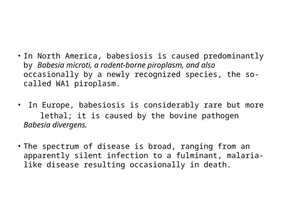
• In North America, babesiosis is caused predominantly by Babesia microti, a rodent-borne piroplasm, and also occasionally by a newly recognized species, the so-called WA1 piroplasm.
• In Europe, babesiosis is considerably rare but more lethal; it is caused by the bovine pathogen Babesia divergens.
• The spectrum of disease is broad, ranging from an apparently silent infection to a fulminant, malaria-like disease resulting occasionally in death.
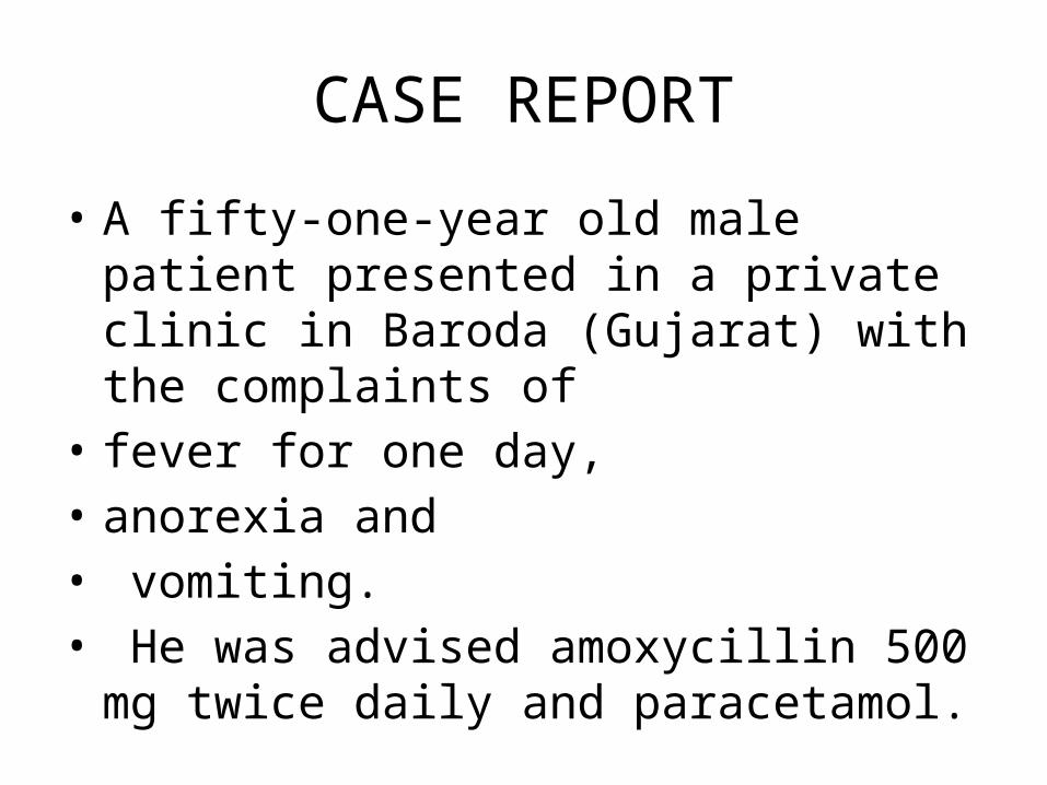
CASE REPORT
• A fifty-one-year old male patient presented in a private clinic in Baroda (Gujarat) with the complaints of
• fever for one day, • anorexia and• vomiting.• He was advised amoxycillin 500 mg twice
daily and paracetamol.
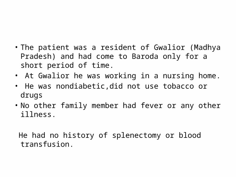
• The patient was a resident of Gwalior (Madhya Pradesh) and had come to Baroda only for a short period of time.
• At Gwalior he was working in a nursing home.• He was nondiabetic,did not use tobacco or drugs• No other family member had fever or any other
illness. He had no history of splenectomy or blood transfusion.
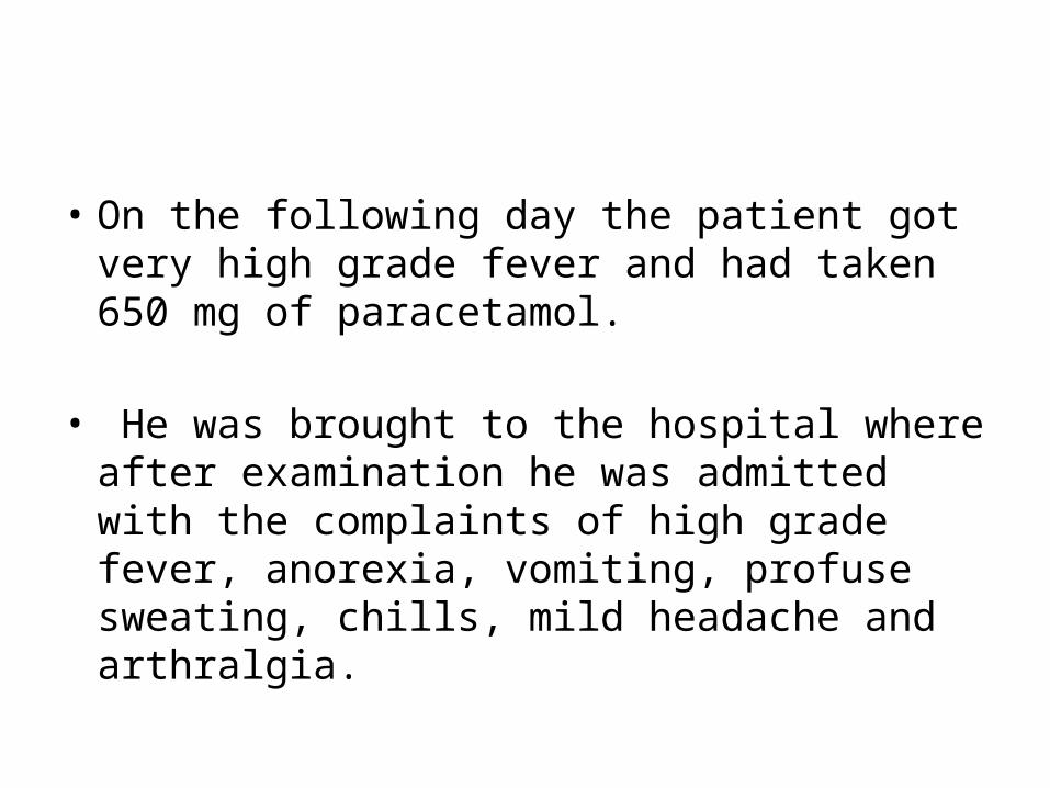
• On the following day the patient got very high grade fever and had taken 650 mg of paracetamol.
• He was brought to the hospital where after examination he was admitted with the complaints of high grade fever, anorexia, vomiting, profuse sweating, chills, mild headache and arthralgia.
ON EXAMINATION
• On examination, the patient had fever of 102⁰ F, pulse rate 97/minute, blood pressure of 110/70 mm. of Hg.
• The liver and spleen were palpable. The patient had scleral icterus.
• He was passing dark coloured urine
• The peripheral blood smear was stained with Leishman’s stain. The ring forms visible within erythrocytes varied greatly and were confused with Plasmodium species especially with falciparum .
• The patient was reported of having P.falciparum
and was treated with anti-malarial drugs.

• The density of parasite was about 5%.
The typical pairs and tetrads of pear-shaped trophozoites were seen at many places in the erythrocytes. Also were seen schizont forms devoid of haemozoin pigment (Fig. d), the extra cellular merozoites and intra erythrocytic multiple merozoites.
• After complete and careful examination of smears the parasites were identified by their morphology as Babesia species.
• To rule out co-infection with Plasmodium species, tests were performed to detect P. falciparum antigen (HRP II) and other Plasmodium specific antigen, which turned out negative.
• The serum was also negative for HIV (I & II) antibodies.
• The patient was put on clindamycin 250 mg four times daily and quinine 300 mg. per day for seven days and was also given folinic acid 15 mg diluted in 100 mL saline for the low WBC counts and thrombocytopenia.
• The patient became afebrile after two days. The repeat peripheral smears after seven days did not show any blood parasites, the total WBC and platelet counts became normal.
• The patient was discharged.
DISCUSSION• Human babesiosis is caused by one of several babesial species that
have distinct geographic distribution based on the presence of competent hosts.
• The spectrum of disease is broad, ranging from an apparently silent infection to a fulminant, malaria-like disease resulting occasionally in death.
• Various determinants are involved in the severity of disease manifestation; among those identified are
age,immunocompetence and coinfection with other pathogenic organisms.
• The diagnosis of babesiosis should begin with a descriptive history, which might include appropriate clinical manifestations,
• history of travel to an area where it is endemic,
• tick bite or exposure to a tick-infested area,• recent blood transfusion and• splenectomy.
• Babesia microti is an intracellular parasite within the erythrocytes; therefore, diagnosis of human babesiosis has relied heavily upon the determination of the presence of the erythrocytic stage of the organism.
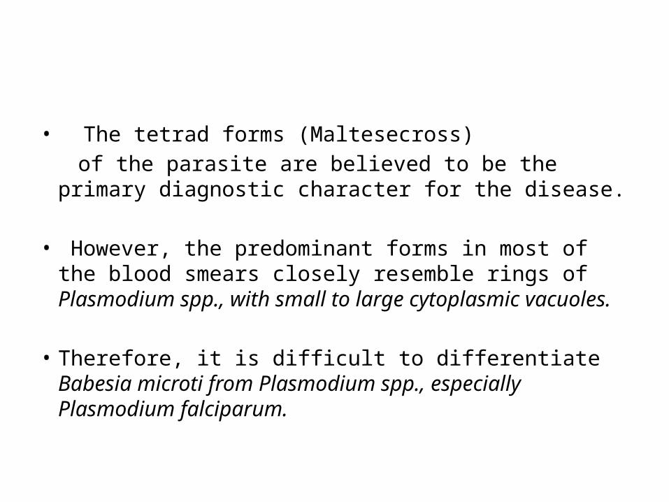
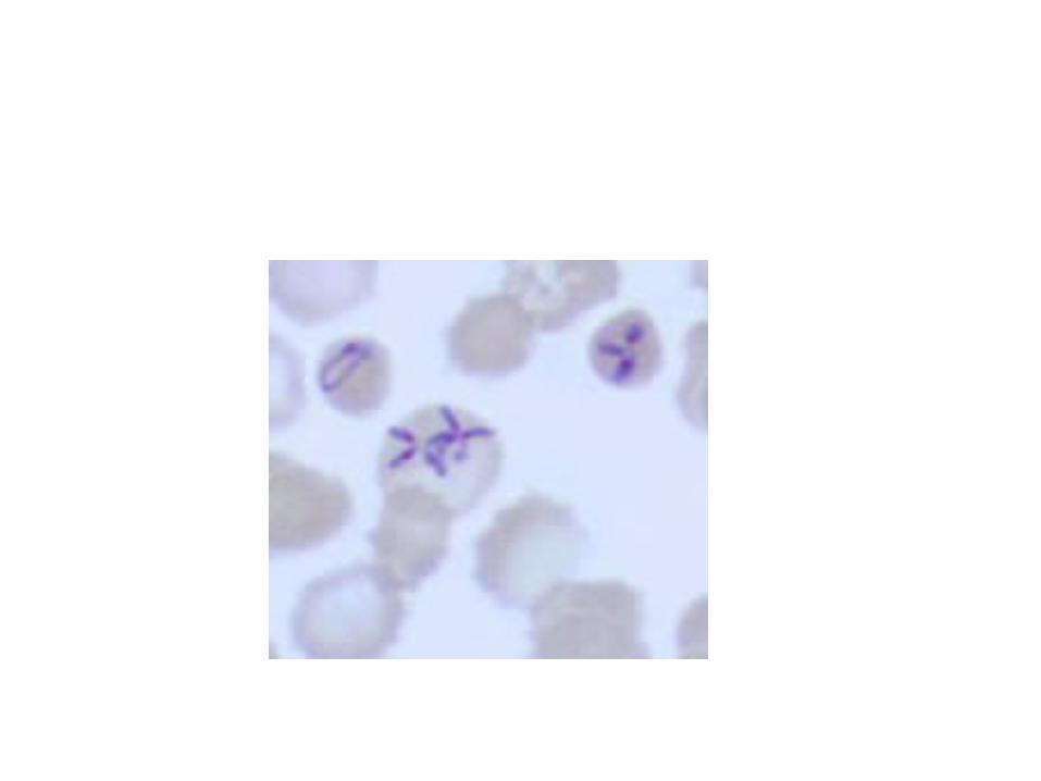
• The tetrad forms (Maltesecross)of the parasite are believed to be the primary diagnostic character for the disease.
• The tetrad forms (Maltesecross) of the parasite are believed to be the primary diagnostic
character for the disease.
• However, the predominant forms in most of the blood smears closely resemble rings of Plasmodium spp., with small to large cytoplasmic vacuoles.
• Therefore, it is difficult to differentiate Babesia microti from Plasmodium spp., especially Plasmodium falciparum.


DIAGNOSIS• It is recommended that diagnosis of clinical case of humanbabesiosis be made by a combination of criteria including the presence of intense parasitaemia (1-50%), erythrocytes infected by multiple basket-shaped parasites and the presence of extracellular merozoites.
In most instances, however, an• accurate patient history,• clinical presentation, and • Observation of characteristic morphologic features are sufficient to
establish the appropriate diagnosis;• otherwise molecular techniques may be used.
• The serologic tests may be performed if the parasitaemia is low but does not replace smear examination because of cross reaction of antibodies with other blood parasites.
The expensive technique like PCR is performed only by the reference laboratories for the diagnosis of the species of Babesia.
COMPLICATIONS
• ARDS• Anaemia requiring transfusions• Congestive heart failure• DIC• HYPOTENSION OR SHOCK• MI• RENAL FAILURE

• Our patient did not give history of tick bite or recent visit to any of the endemic areas which suggests an indigenous source of infection.
• During the smear examination at many places the paired pear-shaped merozoites, tetrads, multiple merozoites, absence of hemozoin pigment in the schizont forms and extra cellular merozoites were observed.
• The absence of HRP II, jaundice, low total WBC count and thrombocytopaenia and unremarkable history of patient suggested an infection with a different intraerythrocytic blood parasite.
Thorough review of the literature on babesiosis and the comparative smear examination led us to the diagnosis of this rare and probably the first case of human babesiosis in INDIA.

• Malaria is endemic in many parts of India and the intraerythrocytic parasites are frequently reported as plasmodia.
• Careful examination of peripheral blood smears can only differentiate the Babesia from malarial parasites.
• Thorough peripheral smear examination and serological surveys may be necessary to know the actual prevalence of human babesiosis in India.