bacillus calmette-guérin suppresses asthmatic responses ... · supernatant, the pellet was...
TRANSCRIPT
201© Copyright The Korean Academy of Asthma, Allergy and Clinical Immunology • The Korean Academy of Pediatric Allergy and Respiratory Disease http://e-aair.org
INTRODUCTION
Asthma is a chronic inflammatory disease that is characterized by infiltration of inflammatory cells such as eosinophils, airway hypersensitivity, and airflow obstruction.1,2 Th2 cells play a par-ticularly crucial role in the development of asthma by secreting interleukin (IL)-4, IL-5, and IL-13: IL-4 causes naïve T cells to differentiate into Th2 cells while IL-5 induces eosinophil infil-tration.3
The hygiene hypothesis proposes that westernized culture and its associated improvements in sanitation cause the microbial exposure of the population to be reduced. This decreases Th1 immune responses that are induced, the lack of which causes the immune system to become dominated by Th2 immune re-sponses.4-6 It has been suggested that this skewing of the immune system has caused the incidence of allergic diseases such as asthma to rise rapidly in the last few decades.7-9 However, recent studies suggest that the incidence of Th1-related diseases such as type 1 diabetes mellitus and multiple sclerosis is also increas-
Bacillus Calmette-Guérin Suppresses Asthmatic Responses via CD4+CD25+ Regulatory T Cells and Dendritic CellsYoung-Joon Kim,1 Ha-Jung Kim,1 Mi-Jin Kang,1 Ho-Sung Yu,1 Ju-Hee Seo,2 Hyung-Young Kim,2 Seoung-Ju Park,3 Yong- Chul Lee,3* Soo-Jong Hong2*
1Asan Institute for Life Sciences, University of Ulsan College of Medicine, Seoul, Korea2 Department of Pediatrics, Childhood Asthma Atopy Center, Research Center for Standardization of Allergic Diseases, Asan Medical Center, University of Ulsan College of Medicine, Seoul, Korea
3Department of Internal Medicine, Research Center for Pulmonary Disorders, Chonbuk National University Medical School, Jeonju, Korea
ing along with Th2-related diseases such as asthma.10 This indi-cates that other mechanisms apart from Th1/Th2 immune re-sponses participate in the modulation of allergic inflammatory responses. One of these mechanisms may involve regulatory T cells (Tregs): several studies report that Tregs regulate immune system by suppression of the Th1/Th2 responses.11-13 Tregs, which are widely considered to be crucial suppressor T cells, act by re-leasing the immunosuppressive cytokines IL-10 and transform-
Original ArticleAllergy Asthma Immunol Res. 2014 May;6(3):201-207.
http://dx.doi.org/10.4168/aair.2014.6.3.201pISSN 2092-7355 • eISSN 2092-7363
Purpose: Bacillus Calmette-Guérin (BCG) is known to suppress the asthmatic responses in a murine model of asthma and to induce dendritic cells (DCs) maturation. Mature DCs play a crucial role in the differentiation of regulatory T cells (Tregs), which are known to regulate allergic inflammato-ry responses. To investigate whether BCG regulates Tregs in a DCs-mediated manner, we analyzed in a murine model of asthma. Methods: BALB/c mice were injected intraperitoneally with BCG or intravenously with BCG-stimulated DCs and then sensitized and challenged with ovalbumin (OVA). Mice were analysed for bronchial hyperresponsiveness (BHR), the influx of inflammatory cells in the bronchoalveolar lavage (BAL) fluid, and histo-pathological changes in the lung. To identify the mechanisms, IgE, IgG1 and IgG2a in the serum were analysed and the CD25+ Tregs in the mice were depleted with anti-CD25 monoclonal antibody (mAb). Results: BCG and the transfer of BCG-stimulated DCs both suppressed all aspects of the asthmatic responses, namely, BHR, the production of total IgE and OVA-specific IgE and IgGs, and pulmonary eosinophilic inflammation. Anti-CD-25mAb treatment reversed these effects. Conclusions: BCG can attenuate the allergic inflammation in a mouse model of asthma by a Tregs-relat-ed mechanism that is mediated by DCs.
Key Words: BCG; dendritic cells; T-lymphocytes, regulatory; asthma; allergic inflammation; mouse
This is an Open Access article distributed under the terms of the Creative Commons Attribution Non-Commercial License (http://creativecommons.org/licenses/by-nc/3.0/) which permits unrestricted non-commercial use, distribution, and reproduction in any medium, provided the original work is properly cited.
Correspondence to: Soo-Jong Hong, MD, PhD, Department of Pediatrics, Childhood Asthma Atopy Center, Research Center for Standardization of Allergic Diseases, Asan Medical Center, University of Ulsan College of Medicine, 88 Olympic-ro 43-gil, Songpa-gu, Seoul 138-736, Korea.Tel: +82-2-3010-3379; Fax: +82-2-473-3725; E-mail: [email protected]
Yong-Chul Lee, MD, PhD, Department of Internal Medicine, Chonbuk National University Medical School, 20 Geonji-ro, Deokjin-gu, Jeonju 561-712, Korea.Tel: +82-63-250-1664; Fax: +82-63-254-1609; E-mail: [email protected]: October 26, 2012; Revised: April 12, 2013; Accepted: April 24, 2013•There are no financial or other issues that might lead to conflict of interest.

Kim et al.
Allergy Asthma Immunol Res. 2014 May;6(3):201-207. http://dx.doi.org/10.4168/aair.2014.6.3.201
Volume 6, Number 3, May 2014
202 http://e-aair.org
ing growth factor (TGF)-β14 and by cell-cell contact mechanism that is mediated cytotoxic lymphocyte associated antigen-4 (CTLA-4) expressed by CD4+CD25+ Tregs, which interacts with CD80 and/or CD86 on the surface of antigen-presenting cells (APCs).15
Bacillus Calmette-Guérin (BCG) is a mycobacterial vaccine organism that is used to prevent tuberculosis. It is known to modulate allergic inflammatory responses.16,17 For example, in-fants with a BCG scar have a higher anti-inflammatory response, namely their ratio of IL-10-producing T cells to IL-4-producing T cells is higher than the ratio of controls who lack a BCG scar.18 Significantly, analyses of mice who were immunised with BCG in the neonatal period revealed that their splenocytes had a higher frequency of CD4+CD25+ Tregs, expressed more Foxp3 and CTLA-4, and produced more IL-10 and TGF-β.19 Thus, BCG may be an example of the microbial exposure that is proposed in the hygiene hypothesis to prevent susceptibility to allergic in-flammation.
Dendritic cells (DCs) are APCs that play an important role in modulating immune responses.20-23 DCs can be divided into immature and mature DCs. Contact with antigens can cause immature DCs to differentiate into mature DCs that express on their cell surfaces CD80, CD86, CD40, and major histocompati-bility (MHC) class II markers.
Many studies have reported that depending on the stimuli, mature DCs can prime naïve T cells toward Tregs. Mature bone marrow-derived DCs (BMDCs) were able to induce the prolif-eration of CD4+CD25+ T cells in the presence of a polyclonal stimulus and in the absence of exogenous IL-2.24 Thymic stro-mal lymphopoietin-treated thymic DCs express CD80/86 and MHC class II and promote the conversion of CD4+CD25- thy-mocytes into CD4+CD25+Foxp3+ Tregs.25 Mature human mono-cytes-derived DCs upregulate the immune-inhibitory enzyme, indoleamine 2, 3-dioxygenase and expand CD4+CD25bright
Foxp3+CD127neg Tregs.26
We confirmed previously that vaccination with BCG can sup-press asthma-related inflammatory responses in a mouse mod-el of asthma.27 The aim of the present study was to investigate the mechanism by which BCG vaccination inhibits the inflam-matory allergic responses of this mouse model; to do so, the roles of DCs and Tregs were analysed.
MATERIALS AND METHODS
AnimalsFemale BALB/c mice were purchased from Orient Bio (Seong-
nam, Korea) and cared for and used in accordance with the guidelines of the Institutional Animal Care and Use Committee (IACUC) at Asan Medical Center and Ulsan University College of Medicine. The mice were housed in specific pathogen-free facility.
Antigen sensitization and challenge The mice (6 weeks of age, 5 mice/group) were sensitized by 2
intraperitoneal injections with 10 µg ovalbumin (OVA; chicken egg albumin grade V, Sigma Chemical Co., St. Louis, USA) dis-solved in 100 µL of phosphate-buffered saline (PBS) adsorbed to an equal volume of alum solution (Alumimject, Pierce, IL, USA) on days 0 and 7. On days 14-16, all mice were challenged daily for 30 minutes via an ultrasonic sprayer (Nescosonic UN-511, Alfresa, Osaka, Japan) with aerosolized 1% OVA (50 µg in 50 µL saline). The mice were divided into 5 groups of 5 mice each.
BCG vaccination Freeze-dried living BCG vaccine was obtained from Japan BCG
laboratory (Tokyo, Japan). The mice were injected intraperito-neally with BCG Tokyo 172 strain 5 days before the first sensiti-zation (day -5) at a dose of 1×10⁶ CFU/200 µL.
DCs preparation, BCG treatment, and injection into mice BMDCs were generated from BALB/c (6-8-weeks-old). Brief-
ly, bone marrow cells were cultured in Iscove’s Modified Dul-becco’s Medium (IMDM) supplemented with 10% fetal bovine serum (FBS), 1% L-glutamine, antibiotic-antimycotic, and 10 ng/mL recombinant murine Granulocyte-macrophage colony-stimulating factor (GM-CSF) (R&D systems, Minneapolis, MN, USA). The culture media were replaced with fresh media con-taining GM-CSF on days 3 and 6. On day 6, BMDCs were col-lected and stained for CD11c. then, the CD11c+ cells were sort-ed by using autoMACS (Miltenyi Biotec) and prepared at 1×10⁶ cells/10 mL, and stimulated with BCG (1×10⁷ CFU) for 48 hours in vitro. The mice were injected intravenously with BCG-stimulated DCs or non-stimulated DCs (1×10⁶/200 µL) 5 days before the first sensitization (day -5).
In vivo treatment with anti-CD25 monoclonal antibody The administration of anti-CD25 Abs is a commonly used
method to deplete Tregs in vivo.28 To investigate the mechanism by which Tregs participate in the allergy-suppressive effects of vaccination with BCG or BCG-stimulated DCs, the OVA-sensi-tized mice were injected intraperitoneally with 250 µg of anti-CD25 monoclonal antibody (mAb) (clone PC61, eBioscience, San Diego, USA) 1 day before the first challenge with 1% OVA.
Measurement of bronchial hyperresponsiveness (BHR) Twenty-four hours after the final OVA challenge, BHR was
measured in conscious unrestrained mice by using a barometric whole-body plethysmograph (one chamber plethysmography, All Medicus, Anyang, Korea), as described previously.29 Briefly, after inhalation of saline and each concentration of methacho-line (MeCh), enhanced pause (Penh) was measured at 10-sec-onds intervals for 3 minutes, averaged, and expressed as abso-lute values.

BCG Inhibits Asthma via DCs and Tregs
Allergy Asthma Immunol Res. 2014 May;6(3):201-207. http://dx.doi.org/10.4168/aair.2014.6.3.201
AAIR
203http://e-aair.org
Bronchoalveolar lavage (BAL) and determination of the cellular composition of the BAL fluid
After measurement of BHR, mice were anaesthetized. BAL was performed as described previously.29 Briefly, the trachea was im-mediately exposed after anesthesia. Then, BAL was performed through a catheter inserted into the exposed trachea following instillation of normal saline (2 mL) at 37°C. The BAL fluid was centrifuged at 2,000 rpm for 5min at 4°C. After discarding the supernatant, the pellet was resuspended in 100 µL PBS. Total BAL cell counts were performed using a haemacytometer. To count the different BAL fluid cells, cytospin slides were prepared, stained with Wright stain, and the different cell types were iden-tified on the basis of their standard morphology under a light microscope. Five sections per slide were evaluated.
Quantitation of the serum levels of immunoglobulins Serum was collected upon sacrifice 24 hours after last 1% OVA
challenge and the serum immunoglobulins were quantified by sandwich ELISA as described previously.29 Briefly, serum was separated from the blood clot by centrifugation at 2,500 rpm for 10 minutes at 4°C. Absorbance of Total IgE, OVA-specific IgE, IgG1 and IgG2a was measured using ELISA reader. The optical density was measured at 450 nm.
Histopathological analysis of the lungs To histologically evaluate the lung tissues, lung samples were
prepared as described previously.30 Inflammation was scored by 2 independent blinded investigators. The degree of peribron-chial and perivascular inflammation was evaluated on a sub-jective scale of 0-3 as described elsewhere.30 Briefly, a value of 0 meant that no inflammation was detectable, a value of 1, occa-sional cuffing with inflammatory cells, a value of 2, most bron-chi or vessels were surrounded by thin layer (1 to 5 cells thick) of inflammatory cells, and a value of 3, most bronchi or vessels were surrounded by a thick layer (more than 5 cells thick) of in-flammatory cells. The cellular infiltration in 5 randomly select-ed fields was assessed under a Zeiss Axiophot microscope (Carl Zeiss Inc., Thornwood, USA) at 100×.
Statistical analysis The each group were compared by using Mann-Whitney test.
The data were expressed as means±standard deviation (SD). All statistical analyses were performed by using SPSS 18.0 for Windows (SPSS Inc., Chicago, IL, USA). A P value that was less than 0.05 was considered significant.
RESULTS
Transfer of BCG-stimulated DCs inhibits BHR and airway inflammatory cells
The MeCh-BHR in response to aerosolized MeCh was mea-sured and the cells in the BAL fluid were identified and counted.
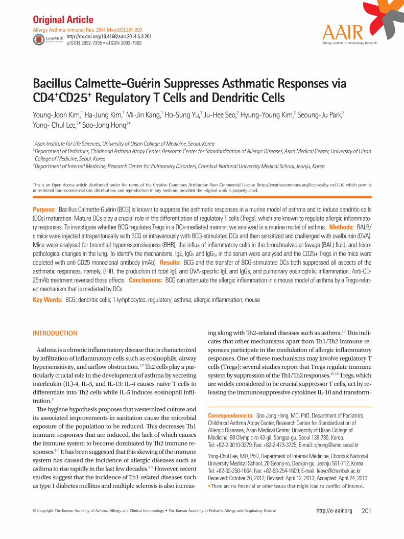
Mean Penh value at maximum MeCh dose of the BCG-stimulat-ed DCs mice was half those of mice, which had only been sen-sitized and challenged with OVA (OVA mice) (P<0.05, Fig. 1A). By contrast, the mean Penh values at maximum MeCh dose of the mice who had received non-stimulated DCs before OVA sensitization and challenge were equivalent to those of the OVA mice. In terms of the BAL fluid analyses, the mice receiving the BCG-stimulated DCs had significantly fewer infiltrating cells overall than the OVA mice (P<0.01, Fig. 1B), whereas the mice receiving non-stimulated DCs were similar to the OVA mice. With respect to specific cell types in the BAL fluid, the mice re-ceiving the BCG-stimulated DCs had significantly fewer eosin-ophils than the OVA mice and the mice receiving non-stimulat-ed DCs (P<0.01, Fig. 1B).
Transfer of BCG-stimulated DCs suppresses the inflammation of lung
Histological analysis of the lungs of the mice described above revealed that the recruitment to the lungs of peribronchial and perivascular inflammatory cells was low in mice who had re-ceived the BCG-stimulated DCs when compared to the OVA mice (Fig. 1C). In contrast, the recruitment of the mice that re-ceived the non-stimulated DCs was equivalent to that of the OVA mice (Fig. 1C).
To quantify the infiltration of inflammatory cells, peribronchi-al and perivascular histopathological inflammation scores were determined. Compared to the OVA mice, the inflammation score was low in the mice that had received BCG-stimulated DCs (P< 0.01, Fig. 1D). By contrast, the scores of the mice that had received the non-stimulated DCs were similar to those of the OVA mice (Fig. 1D).
Transfer of BCG-stimulated DCs reduces the systemic immune response to OVA
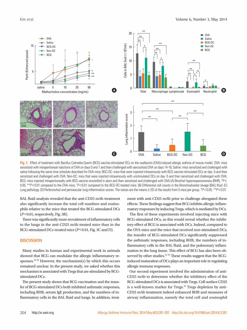
The levels of total IgE, OVA-specific IgE, OVA-specific IgG1, and OVA-specific IgG2a in the sera of each group were measured. The OVA mice had elevated levels of total IgE and OVA-specific IgE. Both measures were significantly lower in the mice that re-ceived the BCG-stimulated DCs but not in the mice that received the non-stimulated DCs (P<0.05, P<0.01, respectively, Fig. 2). Compared to the OVA mice, the mice that received the BCG-stimulated DCs also tended to have lower OVA-specific IgG2a levels and their OVA-specific IgG1 levels were significantly low-er (P<0.05, Fig. 2). By contrast, the OVA-specific IgG1 and OVA-specific IgG2a levels of the mice that received the non-stimulat-ed DCs were similar to those of the OVA mice (Fig. 2).
Anti-CD25 mAb treatment prior to OVA challenge can abrogate the effect of BCG-stimulated DCs transfer
BHR analysis revealed that the mean Penh values at maximum MeCh dose of the CD25-depleted mice were significantly high-er than those of the BCG-DCs-injected mice (P<0.05, Fig 3A).
Kim et al.
Allergy Asthma Immunol Res. 2014 May;6(3):201-207. http://dx.doi.org/10.4168/aair.2014.6.3.201
Volume 6, Number 3, May 2014
204 http://e-aair.org
BAL fluid analysis revealed that the anti-CD25 mAb treatment also significantly increase the total cell numbers and eosino-phils relative to the mice that treated the BCG-stimulated DCs (P<0.01, respectively, Fig. 3B).
There was significantly more recruitment of inflammatory cells to the lungs in the anti-CD25 mAb treated mice than in the BCG-stimulated DCs-treated mice (P<0.01, Fig. 3C and D).
DISCUSSION
Many studies in human and experimental work in animals showed that BCG can modulate the allergic inflammatory re-sponses.16-18 However, the mechanism(s) by which this occurs remained unclear. In the present study, we asked whether this mechanism is associated with Tregs that are stimulated by BCG- stimulated DCs.
The present study shows that BCG vaccination and the trans-fer of BCG-stimulated DCs both inhibited asthmatic responses, including BHR, serum IgE production, and the numbers of in-flammatory cells in the BAL fluid and lungs. In addition, treat-
ment with anti-CD25 mAb prior to challenge abrogated these effects. These findings suggest that BCG inhibits allergic inflam-matory responses by inducing Tregs, which is mediated by DCs.
The first of these experiments involved injecting mice with BCG-stimulated DCs, as this would reveal whether the inhibi-tory effect of BCG is associated with DCs. Indeed, compared to the OVA mice and the mice that received non-stimulated DCs, the transfer of BCG-stimulated DCs significantly suppressed the asthmatic responses, including BHR, the numbers of in-flammatory cells in the BAL fluid, and the pulmonary inflam-mation in the lung tissue. This effect of BCG has also been ob-served by other studies.31-33 These results suggest that the BCG-induced maturation of DCs plays an important role in regulating allergic immune responses.
Our second experiment involved the administration of anti-CD25 mAb to determine whether the inhibitory effect of the BCG-stimulated DCs is associated with Tregs. Cell-surface CD25 is a well-known marker for Tregs.34 Tregs depletion by anti-CD25 mAb treatment indeed enhanced BHR and measures of airway inflammation, namely the total cell and eosinophil
Fig. 1. Effect of treatment with Bacillus Calmette-Guerin (BCG) vaccine-stimulated DCs on the ovalbumin (OVA)-induced allergic asthma of mouse model. OVA: mice sensitized with intraperitoneal injections of OVA on days 0 and 7 and then challenged with aerosolized OVA on days 14-16; Saline: mice sensitized and challenged with saline following the same time schedule described for OVA mice; BGC-DC: mice that were injected intravenously with BCG vaccine-stimulated DCs on day -5 and then sensitized and challenged with OVA; Non-DC: mice that were injected intravenously with unstimulated DCs on day -5 and then sensitized and challenged with OVA; BCG: mice injected intraperitoneally with BCG vaccine emulsified in alum and then sensitized and challenged with OVA (A) Brochial hyperresponsiveness (BHR), *P< 0.05, **P<0.01 compared to the OVA mice; †P<0.01 compared to the BCG-DC-treated mice. (B) Differential cell counts in the Bronchoalvoelar lavage (BAL) fluid. (C) Lung pathology. (D) Peribronchial and perivascular lung inflammation scores. The values are the means±SD of the results from 5 mice per group. *P<0.05, **P<0.01.
Lung
infla
mm
atio
n sc
ore
OVA Saline BCG-DC Non-DC BCG
3
2
1
0
**
** ******
Cells
in B
AL fl
uid
( ×10
5 /mL)
Total Macrophage Lymphocyte Eosinophil Neutrophil
20
15
10
5
0
**
**
**
**
**
**
**
**
***
*
*
*
*
OVASalineBCG-DCNon-DCBCG
Penh
(Enh
ance
d pa
use)
15
5
saline 5 10 25 50Methacholine concentration (mg/mL)
OVASalineBCG-DCNon-DCBCG
*****
†
A B
OVA
BCG-DC
Saline
Non-DC
BCG
C D
BCG Inhibits Asthma via DCs and Tregs
Allergy Asthma Immunol Res. 2014 May;6(3):201-207. http://dx.doi.org/10.4168/aair.2014.6.3.201
AAIR
205http://e-aair.org
counts in the BAL fluid and the infiltration of inflammatory cells in the lung. Thus, it appears that the asthma-inhibiting ef-fects of BCG are associated with Tregs. These observations are consistent with those of other studies that have used mouse models.35-37 For example, BCG-primed DCs were shown to en-hance the expression of Foxp3+ Tregs, IL-10, and inducible co-stimulator.35 However, interestingly, BCG-primed DCs have also been shown to upregulate Th1-associated neutrophilic inflam-mation; this was more effective when the Pasteur strain was used instead of the Copenhagen strain.35 In the present study, the Tokyo strain of BCG was used and neutrophil increases were not observed. Thus, this difference may be related to the type of BCG strain that is used. Supporting this is a study that found considerable differences in the immunogenicity of vari-ous BCG strains, namely their ability to modulate T-cell prolifer-ation and cytokine production.38
This study demonstrated that matured DCs by BCG in vitro could suppress allergic inflammation in mouse model of asth-ma with adoptive transfer. In light of these, our study provides a new perspective of the mechanisms by which the allergic in-flammation in a mouse model of asthma can be modulated.
Further studies are needed to refine several aspects of this study. CD25 is not a specific marker of Tregs and it may be ex-pressed by activated T and B cells. Therefore, the use of anti-
CD25 may not be sufficient to explain the mechanism of Tregs.39 And we did not investigate the expression of Tregs in the airway or thymus or regional lymph node and the functional cytokine production. However, BCG treatment is likely to elevate Foxp3+ Tregs which is a master transcription factor of Tregs in vivo and in vitro.35,36 In addition, recent study has reported that DCs stim-ulated by BCG release IL-10 and induce differentiation of naïve T cells into Tregs.39
BCG is also recognized as a strong Th1 immune response in-ducer.41,42 A study has reported that the interaction between BCG and toll-like receptor (TLR) 2 on DCs may affect the differentia-tion of naïve T cell mainly to Th1-like cells with the help of IL-10, IL-12, and so forth.43 In addition, recently, some studies have reported that IFN-γ is essential to suppress allergic re-sponses.44 It would be worth to perform the study of BCG-DCs on damping allergic inflammation in IFN-γ- or TLR2-deficient mice.
In summary, the present study showed that BCG treatment inhibited the allergic responses of a mouse model of asthma; the study with BCG-stimulated DCs was consistent with these observations. In addition, anti-CD25 mAb treatment reversed the effects of BCG/BCG-DCs. These results suggest that the in-hibitory effect of BCG in our mouse model of asthma is mediat-ed by DCs, which induce Tregs.
Fig. 2. Effect of treatment with BCG vaccine-stimulated DCs on the serum levels of immunoglobulins in mouse model of allergic asthma. The plot legends are as described in Fig. 1. BCG vaccine-stimulated DC transfer inhibited total IgE, OVA-specific IgE and OVA- specific IgG1 levels compared to OVA mice. The values are the means±SD of the results from 5 mice per group. *P<0.05, **P<0. 01.
Tota
l IgE
(A 45
0 nm)
OVA Saline BCG-DC Non-DC BCG
0.2
0.1
0.0
**
*
A
OVA-
IgG 1
(A 45
0 nm)
OVA Saline BCG-DC Non-DC BCG
0.2
0.1
0.0
**
**
C
OVA-
IgG 2
a (A
450 n
m)
OVA Saline BCG-DC Non-DC BCG
0.2
0.1
0.0
****
D
OVA-
IgE
(A 45
0 nm)
OVA Saline BCG-DC Non-DC BCG
0.3
0.2
0.1
0.0
****
*
B

Kim et al.
Allergy Asthma Immunol Res. 2014 May;6(3):201-207. http://dx.doi.org/10.4168/aair.2014.6.3.201
Volume 6, Number 3, May 2014
206 http://e-aair.org
ACKNOWLEDGMENTS
All authors declare there are no conflicts of interest. This study was supported by a grant from the Korea Healthcare Technolo-gy R&D Project of the Ministry for Health, Welfare and Family Affairs, Republic of Korea (A084144).
REFERENCES
1. Walker C, Bode E, Boer L, Hansel TT, Blaser K, Virchow JC Jr. Aller-gic and nonallergic asthmatics have distinct patterns of T-cell acti-vation and cytokine production in peripheral blood and bronchoal-veolar lavage. Am Rev Respir Dis 1992;146:109-15.
2. Lee SH, Park JS, Park CS. The search for genetic variants and epi-genetics related to asthma. Allergy Asthma Immunol Res 2011;3: 236-44.
3. Wang JM, Rambaldi A, Biondi A, Chen ZG, Sanderson CJ, Man-tovani A. Recombinant human interleukin 5 is a selective eosino-phil chemoattractant. Eur J Immunol 1989;19:701-5.
4. Strachan DP. Hay fever, hygiene, and household size. BMJ 1989;299: 1259-60.
5. Riedler J, Braun-Fahrländer C, Eder W, Schreuer M, Waser M, Maisch S, Carr D, Schierl R, Nowak D, von Mutius E; ALEX Study Team. Exposure to farming in early life and development of asth-ma and allergy: a cross-sectional survey. Lancet 2001;358:1129-33.
6. Ege MJ, Mayer M, Normand AC, Genuneit J, Cookson WO, Braun-Fahrländer C, Heederik D, Piarroux R, von Mutius E; GABRIELA Transregio 22 Study Group. Exposure to environmental microor-ganisms and childhood asthma. N Engl J Med 2011;364:701-9.
7. Eder W, Ege MJ, von Mutius E. The asthma epidemic. N Engl J Med 2006;355:2226-35.
8. Hong SJ, Ahn KM, Lee SY, Kim KE. The prevalences of asthma and al-lergic diseases in Korean children. Korean J Pediatr 2008;51:343-50.
9. Lee SI. Prevalence of childhood asthma in Korea: international study of asthma and allergies in childhood. Allergy Asthma Immu-nol Res 2010;2:61-4.
10. Bach JF. The effect of infections on susceptibility to autoimmune and allergic diseases. N Engl J Med 2002;347:911-20.
11. Seroogy CM, Gern JE. The role of T regulatory cells in asthma. J Al-lergy Clin Immunol 2005;116:996-9.
12. Curotto de Lafaille MA, Kutchukhidze N, Shen S, Ding Y, Yee H, La-faille JJ. Adaptive Foxp3+ regulatory T cell-dependent and -indepen-dent control of allergic inflammation. Immunity 2008;29:114-26.
13. Robinson DS, Larché M, Durham SR. Tregs and allergic disease. J
Fig. 3. Effect of anti-CD25 monoclonal antibody (mAb) treatment on the allergic asthma of mice injected with BCG vaccine-stimulated DCs and then sensitized and challenged with OVA. The mice were injected intraperitoneally with anti-CD25 mAb before challenge with OVA. The plot legends are as described in Fig. 1 except that BCG-DC+ anti-CD25 indicate mice that received BCG-DCs and the anti-CD25 mAb. (A) BHR, *P<0.05; **P<0.01 compared to the OVA mice; †P<0.05 compared to the BCG-DC-treated mice. (B) Differential cell counts in the BAL fluid. (C) Lung pathology. (D) Peribronchial and perivascular lung inflammation scores. The values shown are the means±SD of the results from 5 mice per group. *P<0.05; **P<0.01.
Lung
infla
mm
atio
n sc
ore
OVA Saline BCG-DC BCG-DC + Anti-CD25
BCG
3
2
1
0
*
**
**
Cells
in B
AL fl
uid
( ×10
5 /mL)
Total Macrophage Lymphocyte Eosinophil Neutrophil
20
15
10
5
0
*
**
**
**
**
**
**
**
*** *
*
*
*
**
**
OVASalineBCG-DCBCG-DC + Anti-CD25BCG
Penh
(Enh
ance
d pa
use)
15
5
0 5 10 25 50Methacholine concentration (mg/mL)
OVASalineBCG-DCBCG-DC + Anti-CD25BCG
****
†
A B
OVA
BCG-DC
Saline
BCG-DC + Anti-CD25
BCG
C D
BCG Inhibits Asthma via DCs and Tregs
Allergy Asthma Immunol Res. 2014 May;6(3):201-207. http://dx.doi.org/10.4168/aair.2014.6.3.201
AAIR
207http://e-aair.org
Clin Invest 2004;114:1389-97.14. Kwon HK, Lee CG, So JS, Chae CS, Hwang JS, Sahoo A, Nam JH,
Rhee JH, Hwang KC, Im SH. Generation of regulatory dendritic cells and CD4+Foxp3+ T cells by probiotics administration suppresses immune disorders. Proc Natl Acad Sci U S A 2010;107:2159-64.
15. Read S, Malmström V, Powrie F. Cytotoxic T lymphocyte-associated antigen 4 plays an essential role in the function of CD25(+)CD4(+) regulatory cells that control intestinal inflammation. J Exp Med 2000;192:295-302.
16. Hopfenspirger MT, Agrawal DK. Airway hyperresponsiveness, late allergic response, and eosinophilia are reversed with mycobacteri-al antigens in ovalbumin-presensitized mice. J Immunol 2002;168: 2516-22.
17. Erb KJ, Holloway JW, Sobeck A, Moll H, Le Gros G. Infection of mice with Mycobacterium bovis-Bacillus Calmette-Guérin (BCG) sup-presses allergen-induced airway eosinophilia. J Exp Med 1998;187: 561-9.
18. Jason J, Archibald LK, Nwanyanwu OC, Kazembe PN, Chatt JA, Norton E, Dobbie H, Jarvis WR. Clinical and immune impact of My-cobacterium bovis BCG vaccination scarring. Infect Immun 2002; 70:6188-95.
19. Li Q, Shen HH. Neonatal bacillus Calmette-Guérin vaccination inhib-its de novo allergic inflammatory response in mice via alteration of CD4+CD25+ T-regulatory cells. Acta Pharmacol Sin 2009;30:125-33.
20. Hammad H, Lambrecht BN. Dendritic cells and epithelial cells: linking innate and adaptive immunity in asthma. Nat Rev Immu-nol 2008;8:193-204.
21. Geijtenbeek TB, Torensma R, van Vliet SJ, van Duijnhoven GC, Adema GJ, van Kooyk Y, Figdor CG. Identification of DC-SIGN, a novel dendritic cell-specific ICAM-3 receptor that supports prima-ry immune responses. Cell 2000;100:575-85.
22. Steinman RM, Hawiger D, Nussenzweig MC. Tolerogenic dendritic cells. Annu Rev Immunol 2003;21:685-711.
23. Banchereau J, Steinman RM. Dendritic cells and the control of im-munity. Nature 1998;392:245-52.
24. Brinster C, Shevach EM. Bone marrow-derived dendritic cells re-verse the anergic state of CD4+CD25+ T cells without reversing their suppressive function. J Immunol 2005;175:7332-40.
25. Watanabe N, Wang YH, Lee HK, Ito T, Wang YH, Cao W, Liu YJ. Hassall’s corpuscles instruct dendritic cells to induce CD4+CD25+ regulatory T cells in human thymus. Nature 2005;436:1181-5.
26. Chung DJ, Rossi M, Romano E, Ghith J, Yuan J, Munn DH, Young JW. Indoleamine 2,3-dioxygenase-expressing mature human monocyte-derived dendritic cells expand potent autologous regu-latory T cells. Blood 2009;114:555-63.
27. Kim YJ, Kim HJ, Kang MJ, Kwon JW, Seo JH, Kim BJ, Yu J, Hong SJ. Effects of Bacillus Calmette-Guérin (BCG) vaccination according to time point of administration on asthma in a murine model. Ko-rean J Asthma Allergy Clin Immunol 2010;30:307-13.
28. Choi MS, Park S, Choi T, Lee G, Haam KK, Hong MC, Min BI, Bae H. Bee venom ameliorates ovalbumin induced allergic asthma via modulating CD4+CD25+ regulatory T cells in mice. Cytokine 2013; 61:256-65.
29. Yu J, Jang SO, Kim BJ, Song YH, Kwon JW, Kang MJ, Choi WA, Jung HD, Hong SJ. The effects of Lactobacillus rhamnosus on the pre-vention of asthma in a murine model. Allergy Asthma Immunol Res 2010;2:199-205.
30. Tournoy KG, Kips JC, Schou C, Pauwels RA. Airway eosinophilia is not a requirement for allergen-induced airway hyperresponsive-
ness. Clin Exp Allergy 2000;30:79-85.31. Soualhine H, Deghmane AE, Sun J, Mak K, Talal A, Av-Gay Y,
Hmama Z. Mycobacterium bovis bacillus Calmette-Guérin secret-ing active cathepsin S stimulates expression of mature MHC class II molecules and antigen presentation in human macrophages. J Immunol 2007;179:5137-45.
32. Liu E, Law HK, Lau YL. BCG promotes cord blood monocyte-de-rived dendritic cell maturation with nuclear Rel-B up-regulation and cytosolic I kappa B alpha and beta degradation. Pediatr Res 2003;54:105-12.
33. Demangel C, Bean AG, Martin E, Feng CG, Kamath AT, Britton WJ. Protection against aerosol Mycobacterium tuberculosis infection using Mycobacterium bovis Bacillus Calmette Guérin-infected dendritic cells. Eur J Immunol 1999;29:1972-9.
34. Han Y, Guo Q, Zhang M, Chen Z, Cao X. CD69+ CD4+ CD25- T cells, a new subset of regulatory T cells, suppress T cell prolifera-tion through membrane-bound TGF-beta 1. J Immunol 2009;182: 111-20.
35. Ahrens B, Gruber C, Rha RD, Freund T, Quarcoo D, Awagyan A, Hutloff A, Dittrich AM, Wahn U, Hamelmann E. BCG priming of dendritic cells enhances T regulatory and Th1 function and sup-presses allergen-induced Th2 function in vitro and in vivo. Int Arch Allergy Immunol 2009;150:210-20.
36. Lagranderie M, Abolhassani M, Vanoirbeek JA, Lima C, Balazuc AM, Vargaftig BB, Marchal G. Mycobacterium bovis bacillus Calmette-Guérin killed by extended freeze-drying targets plasmacytoid den-dritic cells to regulate lung inflammation. J Immunol 2010;184: 1062-70.
37. Lewkowich IP, Herman NS, Schleifer KW, Dance MP, Chen BL, Di-enger KM, Sproles AA, Shah JS, Köhl J, Belkaid Y, Wills-Karp M. CD4+CD25+ T cells protect against experimentally induced asth-ma and alter pulmonary dendritic cell phenotype and function. J Exp Med 2005;202:1549-61.
38. Lagranderie MR, Balazuc AM, Deriaud E, Leclerc CD, Gheorghiu M. Comparison of immune responses of mice immunized with five different Mycobacterium bovis BCG vaccine strains. Infect Im-mun 1996;64:1-9.
39. Fehérvari Z, Sakaguchi S. Development and function of CD25+ CD4+ regulatory T cells. Curr Opin Immunol 2004;16:203-8.
40. Gao X, Bai H, Cheng J, Fan Y, Wang S, Jiao L, Xiu N, Yang X. CD8α+ and CD8α- DC subsets from BCG-infected mice inhibit allergic Th2-cell responses by enhancing Th1-cell and Treg-cell activity re-spectively. Eur J Immunol 2012;42:165-75.
41. Orme IM, Andersen P, Boom WH. T cell response to Mycobacteri-um tuberculosis. J Infect Dis 1993;167:1481-97.
42. Christ AP, Rodriguez D, Bortolatto J, Borducchi E, Keller A, Mucida D, Silva JS, Leite LC, Russo M. Enhancement of Th1 lung immunity induced by recombinant Mycobacterium bovis Bacillus Calmette-Guérin attenuates airway allergic disease. Am J Respir Cell Mol Biol 2010;43:243-52.
43. Barlan I, Bahceciler NN, Akdis M, Akdis CA. Bacillus Calmette-Guérin, Mycobacterium bovis, as an immunomodulator in atopic diseases. Immunol Allergy Clin North Am 2006;26:365-77.
44. Fonseca DM, Paula MO, Wowk PF, Campos LW, Gembre AF, Tura-to WM, Ramos SG, Dias-Baruffi M, Barboza R, Gomes E, Horn C, Marchal G, Arruda LK, Russo M, Bonato VL. IFN-γ-mediated effi-cacy of allergen-free immunotherapy using mycobacterial antigens and CpG-ODN. Immunol Cell Biol 2011;89:777-85.