bacteria-induced gap junctions in tumors favor antigen ...2010).pdftumor immunology bacteria-induced...
TRANSCRIPT

DOI: 10.1126/scitranslmed.3000739, 44ra57 (2010);2 Sci Transl Med, et al.Fabiana Saccheri
Cross-Presentation and Antitumor ImmunityBacteria-Induced Gap Junctions in Tumors Favor Antigen
http://stm.sciencemag.org/content/2/44/44ra57.full.htmlcan be found at:
and other services, including high-resolution figures,A complete electronic version of this article
http://stm.sciencemag.org/content/suppl/2010/08/09/2.44.44ra57.DC1.htmlcan be found in the online version of this article at: Supplementary Material
http://stm.sciencemag.org/content/2/44/44ra57.full.html#ref-list-1, 17 of which can be accessed free:cites 38 articlesThis article
http://stm.sciencemag.org/content/2/44/44ra57.full.html#related-urls1 articles hosted by HighWire Press; see:cited by This article has been
http://www.sciencemag.org/about/permissions.dtl in whole or in part can be found at: article
permission to reproduce this of this article or about obtaining reprintsInformation about obtaining
is a registered trademark of AAAS. Science Translational Medicinerights reserved. The title NW, Washington, DC 20005. Copyright 2010 by the American Association for the Advancement of Science; alllast week in December, by the American Association for the Advancement of Science, 1200 New York Avenue
(print ISSN 1946-6234; online ISSN 1946-6242) is published weekly, except theScience Translational Medicine
on
Nov
embe
r 9,
201
0st
m.s
cien
cem
ag.o
rgD
ownl
oade
d fr
om

R E S EARCH ART I C L E
TUMOR IMMUNOLOGY
Bacteria-Induced Gap Junctions in Tumors FavorAntigen Cross-Presentation and Antitumor ImmunityFabiana Saccheri,1 Chiara Pozzi,1 Francesca Avogadri,2 Sara Barozzi,1 Mario Faretta,1
Paola Fusi,3 Maria Rescigno1*
(Published 11 August 2010; Volume 2 Issue 44 44ra57)Nov
embe
r 9,
201
0
Antigen-presenting dendritic cells (DCs) trigger the activation of cytotoxic CD8 T cells that target and eliminate cellswith the antigen on their surface. Although DCs usually pick up and process antigens themselves, they can also receivepeptide antigens from other cells via gap junctions. We demonstrate here that infection with Salmonella can induce, inboth human and murine melanoma cells, the up-regulation of connexin 43 (Cx43), a ubiquitous protein that forms gapjunctions and that is normally lost during melanoma progression. Bacteria-treated melanoma cells can establish func-tional gap junctions with adjacent DCs. After bacterial infection, these gap junctions transferred preprocessed antigenicpeptides from the tumor cells to the DCs, which then presented those peptides on their surface. These peptides acti-vated cytotoxic T cells against the tumor antigen, which could control the growth of distant uninfected tumors. Mel-anoma cells in which Cx43 had been silenced, when infected in vivo with bacteria, failed to elicit a cytotoxic antitumorresponse, indicating that this Cx43 mechanism is the principal one used in vivo for the generation of antitumor re-sponses. The Cx43-dependent cross-presentation pathway is more effective than standard protocols of DC loading(peptide, tumor lysates, or apoptotic bodies) for generating DC-based tumor vaccines that both inhibit existing tumorsand prevent tumor establishment. In conclusion, we exploited an antimicrobial response present in tumor cells toactivate cytotoxic CD8 T cells specific for tumor-generated peptides that could directly recognize and kill tumor cells.
on
stm
.sci
ence
mag
.org
Dow
nloa
ded
from
INTRODUCTIONDendritic cells (DCs), key players in the activation of T cells (1), are en-dowed with the ability to present exogenous antigens that have not beengeneratedwithinDCs for the activation of T cells via the cross-presentationpathway. Cross-presentation is required for the initiation of effective anti-tumorT cell responses (2), and a repertoire of presented tumorpeptides iscrucial to activate T cells that will recognize and kill tumor cells (3). How-ever, the antigen presentation machinery, and in particular the protea-some, differs between tumor cells and DCs (4). Thus, DCs can processand present peptides that are different from those presented by tumor cells,initiating a tumor-specific response that will not recognize the tumor (4).
Gap junctions (GJs) are channels that connect the cytoplasm of twoadjacent cells (5). They allow the transfer of small molecules, includingions, second messengers, and metabolites, up to 1 kD (5). GJ intercel-lular communication (GJIC) participates in many physiological eventssuch as cell cycle control, differentiation, cell synchronization, and meta-bolic coordination (5, 6). GJs are formed by two hemichannels, calledconnexons, each made of six connexin proteins. There are at least 21connexins, most of which are tissue-specific except connexin 43 (Cx43),which is ubiquitously expressed (7). Loss of GJIC is a common featureof many human tumors and can occur early during tumorigenesis (8, 9).GJs also play a prominent role in the immune system (7). They are re-quired for B and T cell differentiation, antibody secretion by B cells, Tregulatory cell activity (10), and DC activation (11, 12). GJs are also in-volved in antigen cross-presentation, where they allow the spreading ofsmall linear peptides (up to 16 amino acids long) between neighboring
1Department of Experimental Oncology, European Institute of Oncology, Via Ripamonti435, Milan 20141, Italy. 2Department of Immunology, Memorial Sloan-Kettering CancerCenter, 1275 York Avenue, New York, NY 10021, USA. 3Department of Biotechnology andBiosciences, University of Milano-Bicocca, Milan 20126, Italy.*To whom correspondence should be addressed. E-mail: [email protected]
www.Sci
cells (13), including apoptotic cells (14). The transferred peptides can beloaded onto major histocompatibility complex (MHC) class I moleculesand presented on the surface of acceptor cells. This mechanism may beused to kill a ring of noninfected cells surrounding infected tissue for“sanitation” of the tissue. In addition, it may be used by the immunesystem to activate T cells specific for the infectious agent via transferof antigenic peptides from infected cells to noninfected professionalantigen-presenting cells (13). This pathway is also involved in killingof endothelial cells by tumor-specific cytotoxic T cells (CTLs) after trans-fer of peptides from tumor cells to endothelial cells in vitro (15). How-ever, the in vivo relevance of GJ cross-presentation pathway and itsexploitation in antitumor immunity have not been addressed.
Bacteria have been proposed as anticancer agents (16). The Gram-negative bacterium Salmonella typhimurium is particularly appealingfor its ability to home preferentially to tumor sites (17). Salmonella canbe used as a delivery vector for cytokines (18), chemokines (19), tumorantigens (20), and DNA-based vaccines (21, 22). The intratumoralinjection of S. typhimurium allows breaking ignorance and tolerancetomelanoma. It acts both locally, by recruiting immune cells that lead tothe elimination of the treated mass (23), and systemically, where it favorsthe development of an antitumor response via the cross-presentation oftumor antigens (24). Here, we examined how S. typhimurium infectionfacilitates the cross-presentation of tumor antigens and demonstrate thatit can be used for generation of a DC-based tumor vaccine.
RESULTS
Bacteria induce the up-regulation of Cx43 in severaltumor cell linesThe first requirement for the establishment of GJs between cells is theexpression of connexins. Cx43 is ubiquitous and is implicated in im-
enceTranslationalMedicine.org 11 August 2010 Vol 2 Issue 44 44ra57 1

R E S EARCH ART I C L E
on
Nov
embe
r 9,
201
0g.
org
mune responses (7) but is also down-regulated during melanoma pro-gression (25). Indeed, GJIC is lost in many tumors, enabling the au-tonomous cell behavior of transformed cells (9). Here, we used thehighly aggressive and low immunogenic B16F10 melanoma model(named B16 hereafter). We first tested whether B16 cells expressedCx43. Although untreated B16 cells contained little Cx43, it was up-regulated after infection with Salmonella, as assessed by immuno-fluorescence and Western blot analysis at 24 and 48 hours (Fig. 1,A and B). The same up-regulation of Cx43 was observed when B16cells were incubated with purified bacterial components, such as lipo-polysaccharide (LPS), lipoteichoic acid (LTA), and flagellin (Fig. 1C).A DC line, called DC1, treated with LPS was used as a positive controlfor Cx43 up-regulation (Fig. 1B). Because interferon-g (IFN-g) can al-so up-regulate Cx43 in immune cells (12), we tested whether IFN-gcould induce Cx43 in B16 cells. We found that IFN-g not only up-regulated Cx43 but also synergized with the bacterial components(Fig. 1C). We then analyzed whether Cx43 was up-regulated in tumorsinfected with Salmonella in vivo. Mice were injected subcutaneouslywith 105 B16 cells in the right flank. After 10 days, when the tumorreached an area of 0.5 cm2, tumors were infected with an avirulentstrain of S. typhimurium (SL3261AT).One and 3 days later, tumorswereresected, and Cx43 expression was analyzed by fluorescence-activatedcell sorting (FACS) analysis. Salmonella treatment induced the up-regulation of Cx43 (Fig. 1D).We then analyzed whether IFN-g expres-sion was induced in vivo after intratumoral bacterial infection. IFN-gwas already detectable in infected tumors 1 day after infection, reachinga peak at 7 days (Fig. 1E). These results suggest that bacterial compo-
www.Sci
nents and IFN-g may synergize in vivo for the up-regulation of Cx43and that noninfected cells could express Cx43 in the inflamed en-vironment via the action of free bacterial components or IFN-g. To eval-uatewhether this was a peculiar characteristic of the tumor cell line thatwe used, we extended the analysis to two additional murine melanomacell lines (B16BL6 and C57B1) and to several human melanoma celllines (WM-115, WM-266.4, SK-MEL-31, CHL-1, IGR-1, IGR-37,IGR-39, MEWO, and RPMI-7951). All tested murine tumors and halfof the human cell lines showed behavior similar to that of B16 in re-sponse to bacteria, whereas the other human cell lines expressed higherbasal levels of Cx43, which were either unchanged or down-regulatedin response to bacteria (fig. S1). Thus, whereas tumor cells generallylose Cx43, treatment with bacteria and IFN-g resulted in Cx43 up-regulation in all murine and half of the humanmelanoma cell lines thatwe tested.
Cx43 up-regulation correlates with the generation offunctional gap junctionsWe next tested whether Cx43 up-regulation correlated with the for-mation of functional GJ pores. Infected or noninfected B16 cells weremicroinjected with a mixture of a GJ-diffusible dye (Lucifer yellow)and a GJ-nondiffusible dye (dextran–Texas Red, 70 kD) (13). After bac-terial infection, Lucifer yellow was able to diffuse to adjacent cells (Fig.2A) at a level comparable to that of the untreated NIH 3T3 cells usedas a positive control (26). We then addressed whether similar inter-cellular communication was established with adjacent DCs. Twenty-four hours after the infection, infected and noninfected B16 tumor
stm
.sci
ence
ma
Dow
nloa
ded
from
NT SL
- - -24 h
48 h24 h-- -
A B C
D
Vinculin
LPS
SL
B16DC1
1 day 3 days 7 days
400
800
1200
1600
2000
0
B16 NTB16 SL
IFN
-γ p
g/m
g t
ota
l pro
tein
Cx43
-
IFN-γ - + + + + +- - - -NT SL Flagel LPS LTA
Vinculin
Cx430
1
2
3
4
5
0
20
40
60
80
100
1 day 3 days
B16 NTB16 SL
% C
x43
po
s ce
lls
**
E
Inte
nsi
ty(a
rbit
rary
un
its)
0
10
20
30
40
3 days
Geo
met
ric
mea
n in
ten
sity
*
*
1 day
NT SL
Fig. 1. Bacteria and IFN-g up-regulate Cx43 expression in tumor cells. (A)B16 cells were left untreated (NT) or incubated with bacteria [Salmonella
to vinculin are shown. Bars show band quantification in the presence (blackbars) or absence (white bars) of IFN-g. (D and E) B16-established tumors
(SL)] for 2 hours. After 24 hours, Cx43 expression was evaluated by immu-nofluorescence (red, Cx43; blue, DAPI). (B) B16 cells or DC1 DCs were leftuntreated or incubated with bacteria (Salmonella) for 2 hours or LPS for 24hours. Western blot after 24 and 48 hours with an antibody to Cx43 or tovinculin is shown. (C) B16 cells were untreated or incubated with Salmonel-la or bacterial products [flagellin (Flagel), LPS, and LTA] in the presence orabsence of IFN-g. Western blots after 24 hours with an antibody to Cx43 or
were treated (B16 SL, black bars) or not (B16 NT, white bars) with Salmo-nella. (D) One and 3 days later, mice were killed, tumors were smashed, andcells were analyzed for Cx43 expression by FACS. Percentage of Cx43+ cells(left) and geometric mean intensity (right) are shown. (E) One, 3, and 7 daysafter Salmonella infection, mice were killed, tumors were smashed, andIFN-g production was measured by ELISA. Error bars, SD. Each experimentwas repeated three or four times with similar results.
enceTranslationalMedicine.org 11 August 2010 Vol 2 Issue 44 44ra57 2

R E S EARCH ART I C L E
on
Nov
embe
r 9,
201
0st
m.s
cien
cem
ag.o
rgD
ownl
oade
d fr
om
cells were labeled with the GJ-diffusible dye calcein-acetoxymethylester (calcein-AM), and LPS-treatedDCs were labeled with the nontransferable dye 7-hydroxy-9H-(1,3-dichloro-9,9-dimethylacridin-2-one) (DDAO). We plated the cells separately but inclose proximity in the same petri dish and moni-tored the transfer of dye from the B16 cells to DCsby real-time confocal videomicroscopy. As shownin movie S1 and in the sequence of frames collectedfrom movie S1 (Fig. 2B), starting at 20 min, thetransfer of the calcein dye was observed from tumorcells (white) to DCs (red), which gradually be-came yellow. The DCs were also able to transferthe dye among themselves, indicating that GJICwas forming among the DCs. To quantify the trans-fer of material occurring between tumor cells andDCs, we used a cytofluorimetric method to measureGJIC using the same dyes to distinguish donor fromacceptor cells. After 1 hour of coculture, the numberof DDAO+calcein-AM+ cells was significantly in-creased when tumor cells were pretreated withbacteria (Fig. 2C). This assay also allowed us toevaluate whether the transfer of the dye was indeedoccurring via GJs because the GJ uncoupler hepta-nol, which is a pleiotropic lipophilic agent thatblocks electrical cell-to-cell communication, com-pletely abolished the transfer of calcein from B16cells to DCs (Fig. 2C). This indicates that func-tional GJs can form between tumor cells and DCsfor cell-cell communication and that this mech-anism may be used for the transfer of antigenicmaterial.
DCs present preprocessed tumor-derivedpeptide without the need of phagocytosisTo follow the transfer of antigenic material frominfected tumor cells to DCs, we used a B16 cell line ex-pressing the antigen ovalbumin (B16-OVA). We ana-lyzed the appearance of the OVA(257–264) SIINFEKLpeptide in association with MHC class I (Kb) mole-cules using the specific antibody 25-D1.16 (27) on thesurface of DCs. To assess whether DCs acquired thepeptide after phagocytosis of infected B16-OVA cells,we labeled the latter with the vital dye carboxyflu-orescein diacetate succinimidyl ester (CFSE) and in-cubated the stained cells with DCs for 24 hours. Wedetected DCs positive for the 25-D1.16 antibody pri-marily in the fraction of cells that were negative forCFSE (CD11c+CFSE− cells) and only after infectionof tumor cells (Fig. 3A). The low percentage ofKbOVA-positive cells in the CFSE+ DC fraction afterincubation with B16 control is likely due to non-specific binding of the antibody. This result suggeststhat phagocytosis of intact tumor cells or apoptoticbodies is not required for the exchange of the tumor-associated peptide from infected tumor cells to DCs.To address the nature of the transfer, we added hep-tanol to the cell culture. Heptanol completely abol-
B16 cells
Dextran Texas Red
70 kD
Lucifer yellow457.25 daltons
A
15 s
20 min
40 min
60 min
80 min
100 min
120 min
B
C
B16 NT + DC1 B16 SL + DC1
No Hept Hept
0 h
1 h
No Hept Hept0
1
2
3
4
5
% C
alce
in-A
M+
/DD
AO
+ c
ells
*
B16 NT
B16 SL
NIH
50 m
Fig. 2. Bacteria-induced up-regulation of Cx43 expression correlates with establishment offunctional gap junctions. (A) B16 cells were infected (B16 SL) or not (B16 NT) in vitro with
Salmonella and, after 24 hours, microinjected with a mix of the GJ-diffusible dye Lucifer yel-low (green) and the nontransferable dye dextran–Texas Red (red, to mark microinjected cells).Cells were fixed immediately after microinjecting the last cell and observed for dye transferby fluorescence microscopy. Untreated NIH 3T3 cells were used as positive control. Magnifi-cation, ×40. Scale bar, 20 mm. (B) Confocal microscopy analysis. B16 cells were infected withSalmonella and, after 24 hours, stained with calcein-AM (green/white), whereas DCs, previous-ly treated with LPS for 1 hour, were stained with DDAO (red). A drop of each population wasplated onto a microscope slide close to each other, and the cells were coincubated for 1 hour.The cells were analyzed by confocal microscopy for 2 hours. Representative single framestaken from the movie every 20 min are shown. (C) Only Salmonella-infected cells can com-municate with DCs via a GJ-dependent mechanism. B16 cells were infected or not with Sal-monella, pulsed with the GJ-diffusible dye calcein-AM, and incubated with DDAO-labeled DCsin the presence or absence of heptanol (Hept). One hour later, the percentage of cells double-positive for DDAO and calcein-AM, representing the DCs that received the dye from tumorcells, was evaluated by FACS (black bars). Error bars, SD. *P < 0.05. Each experiment was re-peated twice with similar results.www.ScienceTranslationalMedicine.org 11 August 2010 Vol 2 Issue 44 44ra57 3

R E S EARCH ART I C L E
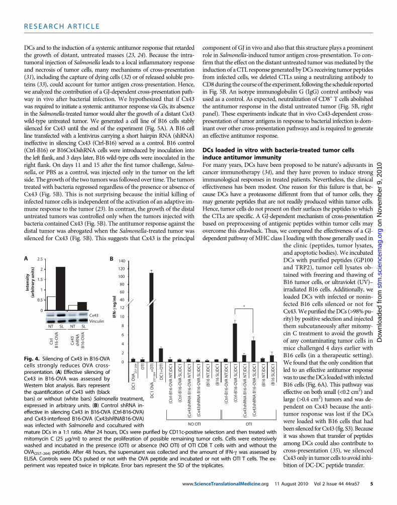
ished the presentation of the peptide, indicating that the OVA peptide islikely transferred via GJIC (Fig. 3A). Because only linear peptides of upto 16 amino acids can be transferred via GJs, the exploitation of thispathway requires processing of the antigenic material within tumorcells. Thus, we examined whether proteasome-dependent degradationin tumor cells was necessary for the presentation of the OVA peptideby DCs. Pretreatment of B16-OVA cells with lactacystin, a cell-perme-able, irreversible 20S or 26S proteasome inhibitor (28), prevented theexposure of the Kb-OVA complex on the DC surface (Fig. 3B). We thenevaluated whether the presentation of peptides via GJ led to T cell ac-tivation and whether this mechanism was dependent on Cx43 expressionby tumor cells. We generated a clone of B16-OVA cells that were stablysilenced for the expression of Cx43. We used four different lentivirus
on
Nov
embe
r 9,
201
0st
m.s
cien
cem
ag.o
rgD
ownl
oade
d fr
om
constructs but only three were effective in silencingCx43; hence, we used the noneffective one as a neg-ative control. In Fig. 4A, the extent of Cx43 silenc-ing is shown. We then tested T cell activation interms of IFN-g release using purified naïve OTI Tcells that carry a T cell receptor (TCR) specific forthe complex of Kb-OVA(257–264)SIINFEKL peptide.We confirmed that pretreatment of B16-OVA cellswith Salmonella favored the cross-presentation ofOVA peptide by DCs (Fig. 4B). This presentationwas partly dependent on Cx43 because its silencingin the B16-OVAcells reduced the capacity ofDCs toactivateOTI T cells (Fig. 4B). The residual activationof T cells may be due to alternative cross-presentationpathways or to incomplete silencing of Cx43 in theB16-OVAcells. Together, these data suggest that bac-terial infection promotes the transfer of a processedpeptide via a GJ-dependent mechanism.
Intratumoral bacterial injectionincreases the percentage of Cx43+ DCsin lymph nodesThe transfer of GJ-diffusible dye occurs not onlybetween tumor cells and DCs but also amongDCs (movie S1 and Fig. 2B). These additional GJscould increase the number of DCs capable of pre-senting tumor-associated antigens in the draininglymph nodes. Hence, we tested the frequency ofCD11c+Cx43+ DCs in lymph nodes that did or didnot drain the tumor site; the tumor had been treatedwith Salmonella or phosphate-buffered saline (PBS) asa control.Weobserved that by24hours after infection,the frequency ofCD11c+Cx43+CD4+DCswas alreadyincreased in both lymphnodes that drained the tumorsite and those thatdidnot, but only if the tumor site (orthe skin) was infected with Salmonella (fig. S2A). Atlater time points (3 days), the CD8+ subset of DCs ex-pressing Cx43 was also increased in frequency (fig.S2A). This coincided with an increase in activatedDCs, as shown by the up-regulation of the activationmarker CD86 (fig. S2B). The activated DCs werethose that expressed Cx43 (fig. S2C). We werepuzzled to observe an increase in Cx43+ DCs innon–tumor-draining lymph nodes because we previ-ously showed that Salmonella remains confined to
www.Sci
the infected site through the generation of a granuloma-like structure(29, 30). However, we found an increased concentration of IFN-g in theserum of mice whose tumors were infected (fig. S2D), possibly explain-ing the rise of Cx43+DCs innon–tumor-draining nodes, because IFN-g canautonomously up-regulate Cx43. We did not see IFN-g in the serum ofSalmonella-infectedmice that were not carrying tumors. This differencemay be due to the nature of melanoma, which is highly vascularized,thereby giving IFN-g a route to the bloodstream.
Cx43-dependent cross-presentation is the major mechanismof tumor-antigen cross-presentation in vivoIn our previous studies, we observed that intratumoral injection ofSalmonella led to increased cross-presentation of tumor antigens by
0.26 0.75
0.22 0
0.16 5.78
0.07 0.07
B16
B16-OVA
NT SL
B16
B16-OVA
NT SL
CFSE neg
1.35 2.38
1.34 2.20
0.78 1.43
2.53 3.01
CD
11
c
0
2
4
6
8
10
12
14
16
A
B
% K
bO
VA
+/C
D1
1c+
Kb
OVA
B16-OVA NT
B16-OVA SL
B16-OVA NT + lac
B16-OVA SL + lac
- Heptanol
+ Heptanol
*
CFSE pos
0.26 0.75
0.22 0
0.16 5.78
0.07 0.07
B16
B16-OVA
NT SL
B16
B16-OVA
NT SL
CFSE neg
1.35 2.38
1.34 2.20
0.78 1.43
2.53 3.01
CD
11
c
0
2
4
6
8
10
12
14
16
A
B
% K
bO
VA
+/C
D1
1c+
Kb
OVA
B16-OVA NT
B16-OVA SL
B16-OVA NT + lac
B16-OVA SL + lac
- Heptanol
+ Heptanol
*
CFSE pos
Fig. 3. Bacterial infection facilitatescross-presentation of preprocessedtumor-associated antigens throughgap junctions. (A) B16 or B16-OVAcells were infected (SL) or not (NT)with Salmonella. Twenty-four hourslater, they were stained with CFSEand incubated with mature DCs inthe presence or absence of heptanolfor 24 hours. Dot plots show cells pos-itive for CD11c and KbOVA in theCFSE− (left) and CFSE+ (right) gate.Numbers show the percentage ofCD11c KbOVA double-positive cellsin the gate. One representative ex-
periment of three is shown. (B) Preprocessing in tumor cells is required for effective cross-presentation. B16-OVA cells were treated as above but in the presence or absence of theirreversible proteasome inhibitor lactacystin (lac). The percentage of KbOVA/CD11c double-positive cells is shown. Error bars, SD. *P < 0.05. One experiment representative of two isshown. The percentage of CD11c+KbOVA+ cells varied between experiments (compare A toB), but the trend was always similar.enceTranslationalMedicine.org 11 August 2010 Vol 2 Issue 44 44ra57 4
R E S EARCH ART I C L E
on
Nov
embe
r 9,
201
0g.
org
DCs and to the induction of a systemic antitumor response that retardedthe growth of distant, untreated masses (23, 24). Because the intra-tumoral injection of Salmonella leads to a local inflammatory responseand necrosis of tumor cells, many mechanisms of cross-presentation(31), including the capture of dying cells (32) or of released soluble pro-teins (33), could account for tumor antigen cross-presentation. Hence,we analyzed the contribution of a GJ-dependent cross-presentation path-way in vivo after bacterial infection. We hypothesized that if Cx43was required to initiate a systemic antitumor response via GJs, its absencein the Salmonella-treated tumor would alter the growth of a distant Cx43wild-type untreated tumor. We generated a cell line of B16 cells stablysilenced for Cx43 until the end of the experiment (Fig. 5A). A B16 cellline transfected with a lentivirus carrying a short hairpin RNA (shRNA)ineffective in silencing Cx43 (Ctrl-B16) served as a control. B16 control(Ctrl-B16) or B16Cx43shRNA cells were introduced by inoculation intothe left flank, and 3 days later, B16 wild-type cells were inoculated in theright flank. On days 11 and 15 after the first tumor challenge, Salmo-nella, or PBS as a control, was injected only in the tumor on the leftside. The growth of the two tumorswas followed over time. The tumorstreated with bacteria regressed regardless of the presence or absence ofCx43 (Fig. 5B). This is not surprising because the initial killing ofinfected tumor cells is independent of the activation of an adaptive im-mune response to the tumor (23). In contrast, the growth of the distaluntreated tumors was controlled only when the tumors injected withbacteria contained Cx43 (Fig. 5B). The antitumor response against thedistal tumor was abrogated when the Salmonella-treated tumor wassilenced for Cx43 (Fig. 5B). This suggests that Cx43 is the principal
www.Sci
component of GJ in vivo and also that this structure plays a prominentrole in Salmonella-induced tumor antigen cross-presentation. To con-firm that the effect on the distant untreated tumor wasmediated by theinduction of aCTL response generated byDCs receiving tumor peptidesfrom infected cells, we deleted CTLs using a neutralizing antibody toCD8during the course of the experiment, following the schedule reportedin Fig. 5B. An isotype immunoglobulin G (IgG) control antibody wasused as a control. As expected, neutralization of CD8+ T cells abolishedthe antitumor response in the distal untreated tumor (Fig. 5B, rightpanel). These experiments indicate that in vivo Cx43-dependent cross-presentation of tumor antigens in response to bacterial infection is dom-inant over other cross-presentation pathways and is required to generatean effective antitumor response.
DCs loaded in vitro with bacteria-treated tumor cellsinduce antitumor immunityFor many years, DCs have been proposed to be nature’s adjuvants incancer immunotherapy (34), and they have proven to induce strongimmunological responses in treated patients. Nevertheless, the clinicaleffectiveness has been modest. One reason for this failure is that, be-cause DCs have a proteasome different from that of tumor cells, theymay generate peptides that are not readily produced within tumor cells.Hence, tumor cells do not present on their surfaces the peptides to whichthe CTLs are specific. A GJ-dependent mechanism of cross-presentationbased on preprocessing of antigenic peptides within tumor cells mayovercome this drawback. Thus, we compared the effectiveness of a GJ-dependent pathway ofMHC class I loading with those generally used in
enceTranslationalMedicine.org 11 A
stm
.sci
ence
ma
Dow
nloa
ded
from
the clinic (peptides, tumor lysates,and apoptotic bodies). We incubatedDCs with purified peptides (GP100and TRP2), tumor cell lysates ob-tained with freezing and thawing ofB16 tumor cells, or ultraviolet (UV)–irradiated B16 cells. Additionally, weloaded DCs with infected or nonin-fected B16 cells silenced or not forCx43.Wepurified theDCs (>98%pu-rity) by positive selection and injectedthem subcutaneously after mitomy-cin C treatment to avoid the growthof any contaminating tumor cells inmice challenged 4 days earlier withB16 cells (in a therapeutic setting).We found that the only condition thatled to an effective antitumor responsewas touse theDCs loadedwith infectedB16 cells (Fig. 6A). This pathway waseffective on both small (<0.2 cm2) andlarge (>0.4 cm2) tumors and was de-pendent on Cx43 because the anti-tumor response was lost if the DCswere loaded with B16 cells that hadbeen silenced forCx43 (fig. S3). Becauseit was shown that transfer of peptidesamong DCs could also contribute tocross-presentation (35), we silencedCx43 only in tumor cells to avoid inhi-bition of DC-DC peptide transfer.
0
2
4
6
8
10
12
40
60
80
100
120
140
DC
1 O
VA25
7-26
4
OTI
DC
1 O
VA25
7-26
4+O
TI
DC
1+O
TI
(Ctr
l-B
16-O
VA N
T)D
C1
(Ctr
l-B
16-O
VA S
L)D
C1
(Cx4
3sh
RNA
B16
-OVA
NT
)DC
1
(Cx4
3sh
RNA
B16
-OVA
SL)
DC
1
NO OTI OTI
(B16
NT
)DC
1
(B16
SL)
DC
1
(Ctr
l-B
16-O
VA N
T)D
C1
(Ctr
l-B
16-O
VA S
L)D
C1
(Cx4
3sh
RNA
B16
-OVA
NT
)DC
1
(Cx4
3sh
RNA
B16
-OVA
SL)
DC
1
(B16
NT
)DC
1
(B16
SL)
DC
1
A B
IFN
-γ n
g/m
l
*
Ctr
l
B16
-OVA
Cx4
3 s
hRN
A B
16-O
VA
NT SL
Cx43
VinculinNT SL
2
2.5
0.5
1
1.5
0
Inte
nsi
ty(a
rbit
rary
un
its)
Fig. 4. Silencing of Cx43 in B16-OVAcells strongly reduces OVA cross-presentation. (A) Effective silencing ofCx43 in B16-OVA was assessed byWestern blot analysis. Bars representthe quantification of Cx43 with (blackbars) or without (white bars) Salmonella treatment,expressed in arbitrary units. (B) Control shRNA in-effective in silencing Cx43 in B16-OVA (Ctrl-B16-OVA)and Cx43-interfered B16-OVA (Cx43shRNAB16-OVA)was infected with Salmonella and cocultured withmature DCs in a 1:1 ratio. After 24 hours, DCs were purified by CD11c-positive selection and then treated with
mitomycin C (25 mg/ml) to arrest the proliferation of possible remaining tumor cells. Cells were extensivelywashed and incubated in the presence (OTI) or absence (NO OTI) of OTI CD8 T cells with and without theOVA(257–264) peptide. After 48 hours, the supernatant was collected and the amount of IFN-g was assessed byELISA. Controls were DCs pulsed or not with the OVA peptide and incubated or not with OTI T cells. The ex-periment was repeated twice in triplicate. Error bars represent the SD of the triplicates.ugust 2010 Vol 2 Issue 44 44ra57 5

R E S EARCH ART I C L E
www.ScienceTranslationalMe
9, 2
010
Even more pronounced was the effect observedin a preventive setting. If we vaccinated the micewith bacteria-treated B16 cells twice (on days 0 and4) before the challenge with B16 cells (day 21), tumorgrowth was inhibited and 100% of the mice remainedtumor-free and survived 50 days after tumor chal-lenge (Fig. 6B). This effect was also dependent onCx43 because it was abolished when we used Cx43-silenced B16 cells to load the DCs (Fig. 6B). Whenthe tumor cells were injected at earlier time points(day 8 after vaccination), tumors grew, but moreslowly, in mice receiving DCs loaded with bacteria-treated B16 cells; 50% of the mice survived (fig. S4).Note that tumor growth is shown until day 40 whenmice carrying 4-cm2 tumors were killed.
Together, these results indicate that the transferof antigenic peptides from tumor cells to DCs throughGJs is far more effective than standard pathways of DCloading in generating protective DC-based vaccines.
on
Nov
embe
r st
m.s
cien
cem
ag.o
rgD
ownl
oade
d fr
om
DISCUSSION
Here, we have provided evidence that it is possibleto exploit an immune response to infection to in-hibit tumor growth by bypassing tumor immuneevasion mechanisms. Tumor cells that generallydown-regulate GJIC and thereby escape control bythe environment up-regulate Cx43 in response toinfection with Salmonella or purified bacterial com-ponents such as LPS, flagellin, or LTA. The up-regulation of Cx43 in tumor cells is accompaniedby the generation of functional GJ both betweentumor cells and between tumor cells and DCs.These newly formed GJs can transfer processed an-tigenic material from tumor cells to the DCs for Tcell activation. This is a dominant pathway of cross-presentation, which leads to the establishment ofeffective antitumor immunity in vivo. The prepro-cessing of the antigen by the tumor guarantees thatDCs will present a repertoire of peptides that arealso presented by tumor cells.
This feature was recently also described forapoptosis-dependent cross-presentation pathwayof virally infected cells (36). It is likely that virusesalso up-regulate GJ proteins in infected cells and,because apoptotic cells can establish GJs for inter-cellular communication (14), virus-infected apoptoticcells may also allow transfer of antigenic material toacceptor cells. Thus, the mechanism we have de-scribed here may be commonly used during thosebacterial or viral infections that do not lead to ap-optosis of the infected cells.
Tumors develop several immune evasion strate-gies that allow the generation of more aggressivecancers with reduced immune responsiveness (37).GJ down-regulation may be a mechanism of immuneevasion that occurs early during tumorigenesis. This
A
Ctrl-B16 PBSCtrl-B16 SLCx43shRNA B16 PBSCx43shRNA B16 SL
Cx43shRNA B16 PBSCx43shRNA B16 SL
IgG control Anti-CD8
1
2
3
4
5
6
8
0 10 15 20 25 30
25 300 5 10 15 20 0 5 10 15 20 25 30
0 5 10 15 20 25 30
2
4
1
3
2
4
2
4
6
8
*
Primary tumor:Ctrl-B16 and Cx43shRNA B16
Distal tumor:B16
i.t. SL
Days
0 310 14
B
11 1518 21 24 27
Anti-CD8
0
Tum
or
size
(cm
2)
DaysDays
NT SL NT SL
0
0.2
0.4
0.6
0.8
1
Cx43
Vinculin
Cx43Vinculin
Primary tumor
Distal tumor
In vitro In vivo
0
Tum
or
size
(cm
2)
Inte
nsi
ty (a
rbit
rary
un
its)
0
0.2
0.4
0.6
0.8
1
* *
Inte
nsi
ty (a
rbit
rary
un
its)
Ctr
l-B
16
Ctrl-B16 PBSCtrl-B16 SL
Cx4
3sh
RNA
B16
Ctr
l-B
16
Cx4
3sh
RNA
B16
Fig. 5. Cx43 expression by tumor cells is required for the initiation of antitumor immunity. (A)Effective knockdown of Cx43 was determined by Western blot on control B16 (Ctrl-B16; with an
shRNA ineffective in silencing Cx43) or on Cx43shRNA B16 cells infected (SL, black bars) or not(NT, white bars) with Salmonella in vitro (left) and on established B16Cx43shRNA tumors in vivo(right). Bars represent the quantification of Cx43 expressed in arbitrary units. (B) Mice receivedtwo tumor inoculations following the indicated schedule. B16 control or Cx43shRNAB16 cellswere injected into the left flank for the generation of the primary tumor, whereas the distaltumor was formed by B16 wild-type cells injected in the right flank. Bacteria or PBS was injectedonly in the primary tumor. Groups of eight mice were injected intraperitoneally with neutralizingantibody to CD8 (right graphs) or isotype control (left graphs) at the days indicated in the sched-ule. The growth of the tumors is shown over time (upper graphs, primary tumors; lower graphs,distal tumors). Error bars, SE. *P < 0.05, when comparing the growth of the primary tumor whenit was injected with Salmonella or PBS (upper graphs). *P < 0.05, when comparing the growth ofthe distal tumor when the primary tumor was either B16 control or Cx43shRNAB16, bothinjected with Salmonella (lower graph). One of three independent experiments is shown.dicine.org 11 August 2010 Vol 2 Issue 44 44ra57 6

R E S EARCH ART I C L E
on
Nov
embe
r 9,
201
0st
m.s
cien
cem
ag.o
rgD
ownl
oade
d fr
om
would inhibit tumor antigen cross-presentation and prevent the develop-ment of antitumor immunity. The Cx43 up-regulation in response toinfection, however, would not be eliminated by selective pressure as thetumor develops and would therefore be retained. Upon infection, tumorswould still be able to initiate the anti-infection program leading to Cx43up-regulation and GJIC, as described here.
This cross-presentation pathway can be used ex vivo for the genera-tion of potent DC-based vaccines.Whenwe compared different pathwaysof tumor antigen cross-presentation such as direct loading with tumorpeptides, tumor lysates, and apoptotic bodies, we found that the Cx43-dependent mechanism was the only one having a benefit in a therapeuticsetting. In addition,when tested in a preventive experimental design such aswould be appropriate for patients having undergone surgical resection ofthe tumor, vaccination with DCs loaded with infected tumor cells led to100% of the mice being free of tumor at the end of the experiment.
It remains to be established whether this strategy could be translat-able to humans and to other types of tumors and whether there wouldstill be a benefit in tumors that did not show Cx43 up-regulation afterinfection. Given the ability of DCs to exchange material via GJ (35), thispathway could lead to the amplification of specific responses and couldbe widely applied.
MATERIALS AND METHODS
Mice, cells, and bacterial strainFive-week-old female C57BL/6J and OTI OVA-TCR transgenicmice were purchased from Charles River and maintained in specific
www.Sci
pathogen-free animal houses. Mouse studies were conducted accordingto the Italian law on approved experimental protocols.
The murine melanoma B16F10, B16F10OVA (called throughoutthe paper B16 and B16-OVA, respectively; a gift from P. Dellabona),and B16BL6 were cultured in RPMI 1640 medium supplemented with10% fetal bovine serum, 2 mM glutamine, penicillin (100 U/ml), strep-tomycin (100 mg/ml), and 50 mM 2-mercaptoethanol (complete RPMI).B16-OVA was cultured in the presence of hygromycin B (100 mg/ml).The murine melanoma C57/B1 was grown in minimum essential medi-um (MEM) supplemented as above plus 1% nonessential amino acids(complete MEM). The human melanoma cell lines were cultured eitherin complete MEM supplemented with 1% sodium pyruvate {WM-115[American Type Culture Collection (ATCC), CRL-1675], WM-266.4(European Collection of Cell Cultures, 91061233), MEWO (Interlab CellLine Collection, HTL97019), and SK-MEL-31 (ATCC, HTB-73)} or inDulbecco’s MEM supplemented as above {IGR-1 [German Resource Cen-tre for Biological Material (DSMZ), ACC 236], IGR-37 (DSMZ, ACC237), IGR-39 (DSMZ, ACC 239), RPMI-7951 (DSMZ, ACC 66), andCHL-1 (ATCC, CRL-9446)}. The murine DC line DC1 was generatedin our laboratory following an established methodology (38).
S. typhimurium SL3261AT is an aroAmetabolically defective strainon SL1344 background and is grown at 37°C in Luria broth.
In vitro infection with bacteria and treatment withpathogen-associated molecular patternsSingle bacterial colonies were grown overnight and restarted the nextday to reach an absorbance at 260 nm (A600) of 0.6 corresponding to0.6 × 109 colony-forming units (CFUs)/ml. Murine and human mela-noma cells were incubated with bacteria for 2 hours, at a cell-to-bacteria
* ****
*
**
*
0
DC1 DC1(B16 NT) DC1(B16 SL)DC1(B16 lysate)DC1(B16 UV-treated) DC1(TRP2/GP100)
Tum
or
size
(cm
2)
Days
1
234
56789
0 5 10 15 20 25 300
0.2
0.4
0.6
0.8
1.0
Surv
ival
(%)
15 20 25 30 35 40
Days
DC1DC1 +(Ctrl-B16 NT)
PBS
0
0.2
0.4
0.6
0.8
1.0
15 20 25 30 35 40
DC1 +(Ctrl-B16 SL)
DC1DC1 +(Cx43shRNA B16 NT)
PBSDC1 +(Cx43shRNA B16 SL)
1
2
3
4
5
0 5 10 15 20 25 30 35 40
1
2
3
4
5
0 5 10 15 20 25 30 35 40Days
Tum
or
size
(cm
2)
Tum
or
size
(cm
2)
DC1
DC1+(Ctrl-B16 NT)
DC1+(Ctrl-B16 SL)
PBS
DC1
DC1+(Cx43shRNA B16 NT)
DC1+(Cx43shRNA B16 SL)
PBS
Surv
ival
(%)
45 50
45 50
*
NS
B16DC1 vaccin
Days
0 4 8
A BB16
DC1 vaccin Days
0 4 21
Fig. 6. (A) Gap junction–based vaccinationis more effective to induce retardation ofthe tumor than the current methods of vac-cination. C57BL/6J mice were injected withB16 cells and, 4 and 8 days later, vaccinatedwith DCs loaded as described below. Thedagger indicatesmice that died before the end of the experiment. Diamonds, PBS as control. Twenty-one days later,micewere injectedwith B16 cells. Upper
unloaded DCs (DC1); triangles, DCs incubated with Salmonella-treated B16cells (B16 SL); squares, DCs incubated with untreated B16 cells (B16 NT); Xs,DCs loaded with B16 lysate; asterisks, DCs loaded with B16 UV–treated cells;circles, DCs loadedwith amix of TRP2 andGP100peptide (TRP2/GP100). (B)Loading of DCs with bacteria-treated tumor cells results in efficient anti-tumor vaccination in a preventive setting. On days 0 and 4, mice (n = 8 pergroup) were vaccinated with DCs loaded as described below or injected withe
graphs: PBS (dashed line), unloaded DCs (DC1, filled diamonds), and DCs in-cubated with Salmonella-treated (Ctrl-B16 SL, filled triangles) or untreated(Ctrl-B16NT, filled squares) control B16cells. Lowergraphs: PBS (dashed line) andDCs loadedwithbacteria-treated (DC1+Cx43shRNAB16SL, empty triangles) oruntreated (DC1 + Cx43shRNAB16 NT, empty squares) Cx43shRNAB16. Errorbars, SE. *P < 0.05, when comparing the growth of the tumors of mice vac-cinated with DC1 + Ctrl-B16 SL versus DC1 + Ctrl-B16 NT. NS, not significant.
nceTranslationalMedicine.org 11 August 2010 Vol 2 Issue 44 44ra57 7

R E S EARCH ART I C L E
on
Nov
embe
r 9,
201
0st
m.s
cien
cem
ag.o
rgD
ownl
oade
d fr
om
ratio of 1:50, in the appropriate medium without antibiotics. Cells werewashed with PBS and incubated in medium supplemented with genta-micin (50 mg/ml) for 12 or 24 hours to kill extracellular bacteria with orwithout IFN-g (100 U/ml).
B16 cells and DC1 DCs were plated in six-well plates (2 × 10⁵ cellsper well) and grown for 18 hours. Cells were incubated with pathogen-associated molecular patterns (PAMPs) in complete medium with orwithout IFN-g (100 U/ml) for 24 hours. The PAMPs used were LPS(1 mg/ml, Sigma), flagellin (0.1 mg/ml, Alexis), and LTA (10 mg/ml,Sigma).
Immunoblot analysisTwenty-four hours after infection or treatment with PAMPs, melanomacells were scraped in ELB buffer [250 mM NaCl, 0.5% NP-40, 50 mMHepes (pH 7.0), 5 mM EDTA] containing 1 mM dithiothreitol, 0.5 mMphenylmethylsulfonyl fluoride, and protease inhibitors. Cell lysates wererun on SDS page and transferred onto Immobilon polyvinylidene diflu-oride membranes. Membranes were probed with a polyclonal rabbit an-tibody to Cx43 (Sigma), a monoclonal mouse antibody to Cx43 (Zymed),or a mouse antibody to vinculin (Sigma) overnight at 4°C and incu-bated with horseradish peroxidase–conjugated antibodies to mouseIgG (Calbiochem) or to rabbit IgG (Bio-Rad) for 1 hour. Visualizationwas carried out with enhanced chemiluminesence (GE Healthcare).Bands were quantified by densitometry with National Institutes ofHealth Image–based software Scion Image.
Immunofluorescence analysisB16 cells were plated onto coverslips (5 × 10⁴ cells per coverslip) andgrown for 18 hours. Cells were infected with Salmonella. After 24 hours,the cells were fixed in 4% (w/v) paraformaldehyde in PBS for 15 min.Blocking and permeabilization were carried out by incubating the cellsin PBS, 3% (w/v) bovine serum albumin, and 0.1% (w/v) Triton X-100for 20 min. The cells were then stained with polyclonal rabbit primaryantibody to Cx43 (1:2000, Sigma) and cyanine 3–conjugated second-ary antibody to rabbit (1:400, Jackson ImmunoResearch Laboratories).Cell nuclei were stained with 4′,6-diamidino-2-phenylindole (DAPI).Confocal microscopy was carried out with a Leica TCS-SP2 (LeicaMicrosystem).
shRNA-mediated knockdownTo generate stable clones of B16 and B16-OVA cells silenced for Cx43,we infected tumor cells with MISSION Lentiviral Transduction Particlesspecific for Cx43 (Sigma) following the recommended protocol. Fourdifferent lentiviruses were tested, of which three successfully knockeddown Cx43. One of the successful lentiviruses and the unsuccessful len-tivirus were cloned by limiting dilution and tested for silencing beforeand after bacterial infection. The unsuccessful clone was used as a neg-ative control for the experiments (Ctrl-B16 and Ctrl-B16-OVA).
Mice treatmentsB16 control cells (105) (Ctrl-B16; unsuccessfully silenced for Cx43) or 3 ×105 Cx43-interfered B16 cells (Cx43shRNAB16) were subcutaneouslyinjected in the left flank of C57BL/6J mice (day 0), and after 3 days,5 × 104 B16 wild-type cells were subcutaneously injected in the rightflank. On days 11 and 15, 108 CFUs of Salmonella or PBS as a controlwere intratumorally injected in the left-flank tumors. The given doseof Salmonella was confirmed by plating bacterial dilutions on terrificbroth agar plates. For the depletion of CD8+ T cells, 100 mg of rat anti-
www.Sci
body to CD8 or isotype control antibody was injected in the perito-neum of the mice on days 10, 14, 18, 21, 24, and 27. Tumor growthwas monitored by measuring the two visible dimensions with a caliperevery 2 to 3 days. Statistical significance at each time point was calculatedas described below.
For therapeutic vaccination, 105 B16 cells were subcutaneouslyinjected into the left flank of C57BL/6J mice (day 0), and after 4and 8 days (or 12 and 16 days in case of large tumors >0.4 cm2),3.5 × 105 DC1 DCs were subcutaneously injected into the right flank.
For preventive vaccination, 3.5 × 10⁵ DC1 DCs were subcuta-neously injected in the right flank of C57BL/6J mice (on days 0 and 4),and on day 4 (or day 21), 105 B16 cells were subcutaneously injectedinto the left flank.
The day before vaccination, Ctrl-B16 and Cx43shRNAB16 cellswere infected in vitro with or without Salmonella and then coculturedwith DC1 DCs in a 1:1 ratio. After 24 hours, DCs were purified byCD11c+ selection [magnetic cell sorter (MACS), Miltenyi Biotec] andtreated with mitomycin C (25 mg/ml) (Sigma) for 20 min at 37°C to arrestthe proliferation of possible remaining tumor cells. For the comparisonexperiment of different vaccination protocols, DCs were also incubatedwith B16 lysate after three freezing and thawing cycles, B16 UV–treatedcells, or a mix of TRP2 and GP100 peptide (1 mg/ml each).
Dye transfer assaysFor microinjection assay, B16 cells were infected or not with Salmo-nella, and after 24 hours, 400 cells were microinjected under an invertedphase-contrast microscope (Axiovert 100, Zeiss) with a mixture of 2%Lucifer yellow CH (457.25 daltons, Sigma) and 1% tetramethylrhodaminedextran (70 kD, Molecular Probes) with an automated microinjectionsystem at a pressure of 1200 hPa applied for 0.2 s.
For FACS assay, DCs were labeled with 10 mM DDAO (MolecularProbes) for 15 min at room temperature in the dark and then exten-sively washed with PBS. B16 cells were infected or not with Salmonellaand, after 24 hours, labeled with 0.5 mM calcein-AM (Molecular Probes)in serum-free medium for 30 min at 37°C and then cocultured withDCs at a ratio of 2:1. Calcein transfer between tumor cells and DCswas evaluated by cytofluorimetry. The coculture of B16 or DCs wascarried out in the presence or absence of 3.5 mM heptanol (Sigma).
For confocal assay, B16 cells were infected or not with Salmonellaand, 24 hours later, stained with 3 mM calcein-AM in serum-free me-dium for 20 min at 37°C, whereas DCs, previously treated with LPS for1 hour, were stained with 1 mM DDAO for 10 min at room tempera-ture. After the staining, a drop of each population was plated in prox-imity to one another onto microscope slides and cells were coincubatedfor 1 hour. Dye transfer was visualized by a Leica SP2 Visible LaserConfocal Microscope. The movie was generated with Imaris software.
In vitro cross-presentation assayB16 and B16-OVA cells were treated or not with Salmonella and, 24hours later, stained with 5 mM CFSE for 20 min at 37°C in the darkand then washed with cold PBS. Where stated, B16 and B16-OVAcells were treated with 10 mM lactacystin (Sigma) overnight. Tumorcells were then added to DCs previously matured with LPS (1 mg/ml)and IFN-g (100 U/ml) in a 1:1 ratio and left in coculture for 24 hours inthe presence or absence of heptanol (3.5 mM). After 24 hours, cross-presentation of OVA peptide by DCs was analyzed by cytofluorimetryon CD11c+ cells with an antibody that recognizes the OVA SIINFEKLpeptide in association with MHC I (anti-KbOVA, 25-D1.16).
enceTranslationalMedicine.org 11 August 2010 Vol 2 Issue 44 44ra57 8

R E S EARCH ART I C L E
on
Nov
embe
r 9,
201
0g.
org
Analysis of IFN-g in the tumor massB16 control and Cx43shRNAB16-bearing mice were intratumorallyinjected with Salmonella or PBS as a control, and after 1, 3, and 7 days,tumors were removed and smashed in 1 ml of PBS containing 0.5%Triton X-100, incubated on ice for 1 hour, and centrifuged at 13,000rpm at 4°C for 15 min. Supernatants were analyzed for the presence ofIFN-g by enzyme-linked immunosorbent assay (ELISA) (R&DSystems) according to the manufacturer’s instructions.
Analysis of IFN-g produced in vitro by OVA-specificCD8 T cellsB16-OVA control (Ctrl-B16-OVA) and Cx43-interfered B16-OVA(Cx43shRNAB16-OVA) were infected with Salmonella and coculturedwith DCs previously matured with LPS (1 mg/ml) and IFN-g (100 U/ml)in a 1:1 ratio. After 24 hours, CD11c+ DCs were purified (MACS) andtreated with mitomycin C (25 mg/ml) for 20 min at 37°C to arrest theproliferation of any remaining tumor cells. DCs (2 × 10⁴) were cul-tured with 2 × 10⁵ CD8 T cells purified from OTI mice with and with-out 1 mM OVA(257–264) peptide. After 48 hours, culture supernatantwas assessed for IFN-g by ELISA.
Statistical analysisStudent’s paired t test was used to determine the statistical signifi-cance of the data. Significance was defined as P < 0.05 (two-tailed testand two-sample equal variance parameters). Statistic calculationsand Kaplan-Meier survival curves were performed with JMP 5.1software (SAS).
stm
.sci
ence
ma
nloa
ded
from
SUPPLEMENTARY MATERIAL
www.sciencetranslationalmedicine.org/cgi/content/full/2/44/44ra57/DC1Fig. S1. All of the murine and half of the human melanoma cell lines up-regulate Cx43 inresponse to Salmonella.Fig. S2. Intratumoral bacterial injection increases the percentage of Cx43+ DCs in lymph nodes.Fig. S3. Loading of DCs with bacteria-treated tumor cells results in efficient antitumor vacci-nation in a therapeutic setting.Fig. S4. Loading of DCs with bacteria-treated tumor cells results in efficient antitumor vacci-nation in a preventive setting.Movie S1. Bacteria-induced up-regulation of Cx43 expression correlates with establishment offunctional gap junctions.
Dow
REFERENCES AND NOTES
1. R. M. Steinman, J. Banchereau, Taking dendritic cells into medicine. Nature 449, 419–426(2007).
2. P. van der Bruggen, B. J. Van den Eynde, Processing and presentation of tumor antigensand vaccination strategies. Curr. Opin. Immunol. 18, 98–104 (2006).
3. A. Paschen, S. Eichmuller, D. Schadendorf, Identification of tumor antigens and T-cellepitopes, and its clinical application. Cancer Immunol. Immunother. 53, 196–203 (2004).
4. L. Chapatte, M. Ayyoub, S. Morel, A. L. Peitrequin, N. Lévy, C. Servis, B. J. Van den Eynde,D. Valmori, F. Lévy, Processing of tumor-associated antigen by the proteasomes of dendriticcells controls in vivo T-cell responses. Cancer Res. 66, 5461–5468 (2006).
5. B. J. Nicholson, Gap junctions—from cell to molecule. J. Cell Sci. 116, 4479–4481 (2003).6. G. Meşe, G. Richard, T. W. White, Gap junctions: Basic structure and function. J. Invest.
Dermatol. 127, 2516–2524 (2007).7. J. Neijssen, B. Pang, J. Neefjes, Gap junction-mediated intercellular communication in the
immune system. Prog. Biophys. Mol. Biol. 94, 207–218 (2007).8. T. J. King, J. S. Bertram, Connexins as targets for cancer chemoprevention and chemo-
therapy. Biochim. Biophys. Acta 1719, 146–160 (2005).9. M. Mesnil, S. Crespin, J. L. Avanzo, M. L. Zaidan-Dagli, Defective gap junctional intercellular
communication in the carcinogenic process. Biochim. Biophys. Acta 1719, 125–145 (2005).
www.Sci
10. T. Bopp, C. Becker, M. Klein, S. Klein-Hessling, A. Palmetshofer, E. Serfling, V. Heib, M. Becker,J. Kubach, S. Schmitt, S. Stoll, H. Schild, M. S. Staege, M. Stassen, H. Jonuleit, E. Schmitt, Cyclicadenosine monophosphate is a key component of regulatory T cell–mediated suppression.J. Exp. Med. 204, 1303–1310 (2007).
11. E. Oviedo-Orta, W. Howard Evans, Gap junctions and connexin-mediated communicationin the immune system. Biochim. Biophys. Acta 1662, 102–112 (2004).
12. H. Matsue, J. Yao, K. Matsue, A. Nagasaka, H. Sugiyama, R. Aoki, M. Kitamura, S. Shimada,Gap junction-mediated intercellular communication between dendritic cells (DCs) is re-quired for effective activation of DCs. J. Immunol. 176, 181–190 (2006).
13. J. Neijssen, C. Herberts, J. W. Drijfhout, E. Reits, L. Janssen, J. Neefjes, Cross-presentation byintercellular peptide transfer through gap junctions. Nature 434, 83–88 (2005).
14. B. Pang, J. Neijssen, X. Qiao, L. Janssen, H. Janssen, C. Lippuner, J. Neefjes, Direct antigenpresentation and gap junction mediated cross-presentation during apoptosis. J. Immunol.183, 1083–1090 (2009).
15. H. Benlalam, A. Jalil, M. Hasmim, B. Pang, R. Tamouza, M. Mitterrand, Y. Godet, N. Lamerant,C. Robert, M. F. Avril, J. Neefjes, T. Tursz, F. Mami-Chouaib, C. Kieda, S. Chouaib, Gap junctioncommunication between autologous endothelial and tumor cells induce cross-recognitionand elimination by specific CTL. J. Immunol. 182, 2654–2664 (2009).
16. D. Bermudes, L. M. Zheng, I. C. King, Live bacteria as anticancer agents and tumor-selectiveprotein delivery vectors. Curr. Opin. Drug Discov. Devel. 5, 194–199 (2002).
17. J. M. Pawelek, K. B. Low, D. Bermudes, Tumor-targeted Salmonella as a novel anticancervector. Cancer Res. 57, 4537–4544 (1997).
18. M. Loeffler, G. Le’Negrate, M. Krajewska, J. C. Reed, IL-18-producing Salmonella inhibit tu-mor growth. Cancer Gene Ther. 15, 787–794 (2008).
19. M. Loeffler, G. Le’Negrate, M. Krajewska, J. C. Reed, Salmonella typhimurium engineered toproduce CCL21 inhibit tumor growth. Cancer Immunol. Immunother. 58, 769–775 (2009).
20. J. Fensterle, B. Bergmann, C. L. Yone, C. Hotz, S. R. Meyer, S. Spreng, W. Goebel, U. R. Rapp,I. Gentschev, Cancer immunotherapy based on recombinant Salmonella enterica serovarTyphimurium aroA strains secreting prostate-specific antigen and cholera toxin subunit B.Cancer Gene Ther. 15, 85–93 (2008).
21. U. Pertl, H. Wodrich, J. M. Ruehlmann, S. D. Gillies, H. N. Lode, R. A. Reisfeld, Immuno-therapy with a posttranscriptionally modified DNA vaccine induces complete protectionagainst metastatic neuroblastoma. Blood 101, 649–654 (2003).
22. A. G. Niethammer, R. Xiang, J. M. Ruehlmann, H. N. Lode, C. S. Dolman, S. D. Gillies, R. A. Reisfeld,Targeted interleukin 2 therapy enhances protective immunity induced by an autologous oralDNA vaccine against murine melanoma. Cancer Res. 61, 6178–6184 (2001).
23. F. Avogadri, C. Martinoli, L. Petrovska, C. Chiodoni, P. Transidico, V. Bronte, R. Longhi, M. P.Colombo, G. Dougan, M. Rescigno, Cancer immunotherapy based on killing of Salmonella-infected tumor cells. Cancer Res. 65, 3920–3927 (2005).
24. F. Avogadri, D. Mittal, F. Saccheri, M. Sarrafiore, M. Ciocca, P. Larghi, R. Orecchia, M. Rescigno,Intra-tumoral Salmonella typhimurium induces a systemic anti-tumor immune responsethat is directed by low-dose radiation to treat distal disease. Eur. J. Immunol. 38, 1937–1947 (2008).
25. N. K. Haass, K. S. Smalley, M. Herlyn, The role of altered cell–cell communication in mel-anoma progression. J. Mol. Histol. 35, 309–318 (2004).
26. C. J. Wei, R. Francis, X. Xu, C. W. Lo, Connexin43 associated with an N-cadherin-containingmultiprotein complex is required for gap junction formation in NIH3T3 cells. J. Biol. Chem.280, 19925–19936 (2005).
27. A. Porgador, J. W. Yewdell, Y. Deng, J. R. Bennink, R. N. Germain, Localization, quantitation, andin situ detection of specific peptide–MHC class I complexes using a monoclonal antibody.Immunity 6, 715–726 (1997).
28. G. Fenteany, R. F. Standaert, W. S. Lane, S. Choi, E. J. Corey, S. L. Schreiber, Inhibition of protea-some activities and subunit-specific amino-terminal threonine modification by lactacystin.Science 268, 726–731 (1995).
29. G. Rotta, E. W. Edwards, S. Sangaletti, C. Bennett, S. Ronzoni, M. P. Colombo, R. M. Stein-man, G. J. Randolph, M. Rescigno, Lipopolysaccharide or whole bacteria block the con-version of inflammatory monocytes into dendritic cells in vivo. J. Exp. Med. 198, 1253–1263 (2003).
30. G. Rotta, G. Matteoli, E. Mazzini, P. Nuciforo, M. P. Colombo, M. Rescigno, Contrasting rolesof SPARC-related granuloma in bacterial containment and in the induction of anti–Salmonellatyphimurium immunity. J. Exp. Med. 205, 657–667 (2008).
31. T. A. Groothuis, J. Neefjes, The many roads to cross-presentation. J. Exp. Med. 202, 1313–1318(2005).
32. M. L. Albert, B. Sauter, N. Bhardwaj, Dendritic cells acquire antigen from apoptotic cells andinduce class I-restricted CTLs. Nature 392, 86–89 (1998).
33. A. L. Ackerman, C. Kyritsis, R. Tampé, P. Cresswell, Access of soluble antigens to the endoplasmicreticulum can explain cross-presentation by dendritic cells. Nat. Immunol. 6, 107–113 (2005).
34. J. Banchereau, A. K. Palucka, Dendritic cells as therapeutic vaccines against cancer. Nat.Rev. Immunol. 5, 296–306 (2005).
35. A. Mendoza-Naranjo, P. J. Saéz, C. C. Johansson, M. Ramírez, D. Mandakovic, C. Pereda, M. N.López, R. Kiessling, J. C. Sáez, F. Salazar-Onfray, Functional gap junctions facilitate melanoma
enceTranslationalMedicine.org 11 August 2010 Vol 2 Issue 44 44ra57 9

R E S EARCH ART I C L E
antigen transfer and cross-presentation between human dendritic cells. J. Immunol. 178,6949–6957 (2007).
36. N. E. Blachère, R. B. Darnell, M. L. Albert, Apoptotic cells deliver processed antigen to dendriticcells for cross-presentation. PLoS Biol. 3, e185 (2005).
37. G. P. Dunn, A. T. Bruce, H. Ikeda, L. J. Old, R. D. Schreiber, Cancer immunoediting: From im-munosurveillance to tumor escape. Nat. Immunol. 3, 991–998 (2002).
38. C. Winzler, P. Rovere, M. Rescigno, F. Granucci, G. Penna, L. Adorini, V. S. Zimmermann,J. Davoust, P. Ricciardi-Castagnoli, Maturation stages of mouse dendritic cells in growthfactor–dependent long-term cultures. J. Exp. Med. 185, 317–328 (1997).
39. Acknowledgments:We thank M. Chieppa for technical assistance in confocal microscopy.Funding: M.R. was supported by grants from the Association for International Cancer Re-search, the Associazione Italiana per la Ricerca sul Cancro, the European Molecular BiologyOrganization Young Investigator Programme, the European Commission (7th frameworkprogramme), and the Italian Ministry of Health (Ricerca Finalizzata). Author contributions:
www.Scien
F.S. did most of the experiments, with significant assistance from C.P. F.S. and C.P. analyzedall data. M.R. designed the study, analyzed all data and wrote the paper, with editorial inputfrom F.S. and P.F. F.A. performed some of the experiments. S.B. and M.F. designed and per-formed microinjection experiments. Competing interests: The authors declare that theyhave no competing interests.
Submitted 16 December 2009Accepted 23 July 2010Published 11 August 201010.1126/scitranslmed.3000739
Citation: F. Saccheri, C. Pozzi, F. Avogadri, S. Barozzi, M. Faretta, P. Fusi, M. Rescigno, Bacteria-induced gap junctions in tumors favor antigen cross-presentation and antitumor immunity.Sci. Transl. Med. 2, 44ra57 (2010).
ceTranslationalMedicine.org 11 August 2010 Vol 2 Issue 44 44ra57 10
on
Nov
embe
r 9,
201
0st
m.s
cien
cem
ag.o
rgD
ownl
oade
d fr
om