bacterial cell patogenity
TRANSCRIPT

E-mail: [email protected]: 224435355
Bacterial cellPatogenity
Jan Tkadlec
Dept of Medical Microbiology
2. LF UK a FN Motol

Non-textbook literature

Introduction:Impact of mikrobiology on the
medicine

Medicine before and now
Major surgery is amputation or dissection
Surgery (kardio, neuro, plastic, …)Transplantation (heart, lungs, …)Oncologic treatment (chemoterapy, radioterapy…)Immunosupresion
Modern medicine would be imposible without disinfection, antiseptics and antibiotics
What is the crucial difference?
Identification of microorganisms as a cause of infectious diseases!!!

Understanding microbes: increasing life expectansy
1908 – water chlorination
Twenties- vaccinationcampaings
30. – 40ies. Antibiotics
Deaths by infectious diseases Mean life expectancy

Reality: 6 bilions
Penicilin itself saved more than200 milions lifes
Projection based on previous trend: 3 bilions
Difference made by understanding microbes as a cause of diseases
Understanding microbes: human population
Is there any other scientific discipline that could claim to save so many lifes?

Louis Pasteur Robert Koch Alexander Fleming
Disinfection and antiseptics Microbes as the cause of diseases Antibiotics
Joseph ListerIgnaz Semmelweis

Cause of death per year :
Infectious diseases
Cancer
Cardiovasvular diseases
Total
Violence/injuries
Chronic lung diseases
Pregnancy related
Other
Git disorders
Neuro-psychologic disorders
Still lot of work for microbiologists, infectionists and epidemiologists

Bacterial cell

Size – the advantage of bacteria
• Size in micrometers (µm)
• Specific surface: Surface/volume – how big part of membrane per unit of biomass
• Surface:• Acquisition of nutrients
• Membrane based metabolic processes
• surface vs. volume=r2 vs. r3
• Rapid metabolism of bacteria is allowed by relatively large membrane per the volume of biomass

Many shapes of bacterial cell
e
• rods (bacily) (a)
• Vibrio –curved rods (e)
• cocci (b)• Diplo-• Strepto- (Chains)• Staphylo (bunch of
grapes)
• Spirillum(c)
• spirochete (d)
• Pleiomorphic bacteria• Non-uniform shape
• Species specific or culture condition based
• E.g. Helicobacter pylori

Bacterial cell=prokaryoteDefined by what it lacks:• Chromosome is not separate
from cytoplasm
• No membrane organelles (mitochondria, endoplasmic reticulum, Golgi…)
• No cytoskeleton
Eukaryote – Plants, animals and fungi

Bacterial ribosomTranslation=protein syntesis according the mRNA
Size - 70S (S =Svedberg unit of sedimentation)
(Eukaryotic are bigger - 80S)
Composition
• protein (40%) and rRNA (60%)
• Large (50S) and small (30S) subunit
Targett of the antibiotics

Antibiotics: finding the difference
Antibiotics targetts the structures orprocesses that are either missing ordifferent in Eukaryotic cell. Most of these processes are conected with growth of the cell
Dormant cells and spores are hard to treat

Surface structures of the bacterial cell

Cell wall• Determines shape of the cell
• Protects bacteria from• irradiation, desiccation, mechanical damage, inner pressure
• polymer – peptidoglycan (polysaccharide)• N-acetylglucosamine
• N-acetylmuramic acid
• Filaments are cross-linked to form a net
• permeable
• Rigid
• Synthesis – transpeptidases (PBP – penicillin binding proteins) reconnects peptidoglycan filaments during the growth – target of the beta lactam antibiotics

Gram positive bacteria
• Thick peptidoglycan layer
• (lipo)teichoic acid
e.g.:
• Staphylococcus
• Streptococcus

Gram negative bacteria• Only thin layer of peptidoglycan
• Periplasmic space
• Outer membrane
• Chemical protection• Resistance to glycopeptides
and macrolides
• Lipopolysacharide:
• Lipid A = endotoxin
• Core polysaccharide
• O-antigen
E. coli, Klebsiella, Pseudomonas, etc.

LPS = endotoxin
• endotoxin – part of thecell
• toxic component: lipid A
• septic (endotoxic) shock• Rapid induction of
immune responce(cytokine storm)
• Systemic inflamatoryresponce
• Multiorgan failure
• High mortality

• Gram negative• Thin cell wall (peptidoglycan)
• outer membrane – protects bacteria, contains lipopolysacharide
• Gram positive• Thick peptidoglycan layer, contains teichoic acid

Gram staining principle
• v
1. Crystal violet (CV) penetrates into the cell2. Iodine solution (mordant) – formatio of precipitates – binding to the cell wall3. Alcohol wash– decolorizing gram negatives – thin cell wall could not keep the stain. Grampositive keep
its color (purple)4. Counter staining of gram negatives with carbolfuchsin (pink)
Wilhelm et al 2015 ACS Chem. Biol.

Capsule
• Additional protective layer - polysacharide
• Protection from desication and immune system• Hides surface antigens from phagocytes, complement and antibodies
• Frequently found in serious patogens• Neisseria meningitidis, Haemophilus influenzae, Streptococcus
pneumoniae, Bacillus anthracis a další
• Needed for full virulence

Capsule – S. pneumoniae• S. pneumoniae (pneumococcus) – pneumonia,
meningitidis, otitis media
• Major virulence factor, but not toxic itself
• Strains laking capsule are non virulent
• Variable (80-100 antigenic variants)
• Vaccine against S. pneumoniae is targetting capsule
Vaccines against Neisseria meningitidis and Haemophilus influenzaealso targetts capsule

Biofilm• Bacteria dont form a tissue
• But they could form a biofilm!!!
• Biofilm is attached structured consorcium of bacteria enveloped by extracelularmatrix from polysacharides, proteins and extracelular DNA secreted by these bacteria
• Base of the biofilm is protected from• antibodies• complement• phagocytes• antibiotics
• Regeneration from the base

Biofilm – Clinical impact• Majority of patogenic bacteria forms
biofilm
• Protects from antibiotics and immunesystem
• Infection of foreign bodies:• implants
• prostetics
• dental implants
• catheters• vascular
• urinary
• Dental plaque
• Biofilm is impossible to eradicate

Bacterial small talks
• Quorum sensing• Regulates biofilm formation, virulence factor
production

Flagella• Different from eukaryotic one
• Anchored in the membrane
• screw-propeller like mechanism
• Energy from proton gradient
• Some bacteria lacks flagella (streptococci, staphylococci)
• Spirochetes flagellum (=axial filament) corkscrew like motion
• Highly antigenic• Serotyping of enterobacteria: H antigen

Chemotaxis – where are you going?Dva mody podle směru rotace bičíku• Forward • Tumbling

Movement on the solid surfaceSwarming e.g. Proteus• CLED agar – inhibition of swarming
• Otherwise Proteus will overgrow otherbacteria

Movement of non-motile bacteria
• Intermicrobial Hitchhiking
Staphylococcus aureus and Pseudomonas aeruginosa

• S. aureus and Candida
• Mechanical inoculation ofS. aureus by growing of theyeast

Fimbria
• syn. pili (pilus)
• Shorter than flagella, around thesurface of the cell
• Made of proteins
• Main function is adhesion• Top of pili – adhesins
• adhesin bind sacharide on the surface ofhuman cells
• Tissue specific

Fimbrie
• Escherichia coli urinary tract infection• PAP fimbria (Pyelonephritis-Associated Pili)
• Home made medicine: cranberries
• Sacharides from cranberries goes into urine and blocksadhesins on fimbria

Spore• Dormant stage
• Not metabolising
• Resistance• Chemical (e.g. Alcohol disinfection)
• Physical (temperature, irradiation)
• Viable for decades
• endospore – forms inside of the cell– G+ (Bacillus a Clostridium)
• exospore – on the cell surface
• Sporulation
• Germination=activation of the spore

Examples of sporulating bacteria:
• Clostridium• C. botulinum – botulotoxin
• C. tetani - tetanus
• C. difficile – post-antibiotic diarhoea
• C. perfringens
• Bacillus• B. anthracis – antrax
• B. cereus

Bacteria and the oxygena) Aerobic (e.g. Mycobacterium)
• Requires oxygen
• respiratory metabolism
• Oxygen is final electron acceptor
b) Anaerobic (e.g. Clostridium)
• Hates the oxygen (fermentation), oxygen makes them sick
c) facultative anaerobic (most of the bacteria)
• Prefer oxygen (respiration), but are OK withoutit (fermentation)
d) Microearophilic (e.g. Campylobacter)
• Requires oxygen bur not too much (cca 2 %)
e) Aerotolerant - (e.g.. Streptococcus)
• Dont care about oxygen (fermentation)

• Reactive oxygen species are toxic
• Result of metabolismprocesses
• Damage to the proteins, DNA/RNA and lipids
• Detoxifying enzymes• Superoxid dismutase
• Peroxidase
• catalasa (Staphylococci)
• Diferentiation staphylococci fromstreptococci
Oxygen as a poison

Anaerobní bakterieLacks enzyme that deals with reactive oxygen species
Dying in oxygen presence – transport of thesample
Anaerobiosis
• Chemical reaction
• Supplementation with nitrogen

Surface structure of bacteria and the immune system

Bacterial surface• Exposed to interaction with immunity mechanisms
• Targetted by both innate and adaptive immunity
• Innate immunity• present in all animals (snails, insectsetc.)
• Recognizes general features of bacterial cell surface
• rapid
• adaptive immunity• „invented“ by vertebrates
• Mediated by antibodies and T-cells
• Days to full response

howto tell a bacterium from a human cell?→molecules exclusive for bacteria: PAMPs (Pathogen-AssociatedMolecular Pattern)
Recognized by Phagocytes (macrophages and neutrophils) via TLR(Toll-
likereceptor) Complement
phagocytosis→eliminationofbacteria
PG peptidoglycanLPS lipopolysacharideLTA lipoteichoic acidDNA unmethylated DNA

• Cana ttack subtle antigenic variants, virtually any possiblestructure can be targeted
• Different antibody response towards different bacteria of the samespecies –serotypes
• bacterial surface antigens are sometimes similar to human proteins(similar sequence of aminoacids-antigenic mimicry)• autoimmunediseases
• Rheumatic fever• Streptococcus pyogenes
• rheumatic arthritis + periodontitis• Porphyromonas gingivalis
• ankylosing spondylitis• Klebsiella pneumoniae

Microbe host interaction


Symbiotic (comensal) bacteria• Symbiotic
• Mutual benefits – space for live in exchange for nutrients or vitaminsproduction
• vitamin B12 and vitamin K
• Commensalism• Did no harm and could occupy the space where the patogens would
like to sit - competition
• Probiotics• Living symbiotic or commensal bacteria – part of healthy microbiom
(Lactobacillus, Bifidobacterium, Streptococcus (non patogenic!), yeasts
• Prebiotics• Indigestible nutrients promote the growth of symbiotic bacteria• Oligosacharides – inulin, lactulose

Intestinal Bacillus sp colonisation could eliminate carriage of S. aureus• Probiotics for MRSA decolonisation or treatment?
Piewngam et al Nature 2018


Other examples of commensal bacteria competing with pathogens

Patogenic bacteria
• Patogenic bacteria = cause disease
• Primary (obligate) pathogen• Infection of healthy people
• Streptococcus pyogenes, Treponema pallidum, Bordetella pertussis, Salmonella typhi, Neisseriagonorrhoeae, Bacillus anthracis, Mycobacteriumtuberculosis, Yersinia pestis…
• Oportunnic patogens• Infection when immunity is impaired
• E. coli, S. aureus, and many others

• Goal of the patogen is not to kill the host but to reproduce
• If the host is kill to early patogen risks that it will die with host
• In the long term evolution selects for decreased virulence, patogen and host will find the equilibrum
• However if the patogen ha s external reservoir, or the population ofhost is big enough the virulence is not problem – this way epidemicsstarts

Virulence factors
• Allows bacteria to cause a disease
Could be divided• From the bacterial perspective:
• Adhesins – helps to colonise the host/tissue
• Invasins – helps to overcome mechanical barriers in the body – e.g. cytotoxiny
• Evasins– helps to evade immune response
• From the perspective of the host• Invasivity – adhesion and colonisation
• Toxicity – direct damage to host tissues
• Immune dependent factors – the damage is mediated by the reaction of the immunesystem

Adhesins
• Gram negatives – adhesins on fimbria
• Gram positives• Lipoteichoic acid
• F-protein (Str. pyogenes)
• Fibronectin binding proteins
• Capsule
• Biofilm

Toxins

Exotoxins
• = „true“ toxins
• Extracelullar protein moleculles (compare to endotoxin – outer membrane G-)
• Direct and serious damage to host
• Diferentiation – variable criteria:• chemical structure (single molecule or macromolecullar complex)
• Targett structure (cell surface or intracellular)
• Mechanism (neurotoxic, enterotoxic, cytotoxic)

Exoenzymes• enzymatic destruction of host cells and tisues
• Penetration of mechanical barriers (epitelia, mucous membranes) – extracelularmatrix• hyaluronidase
• kolagenase, gelatinase
• elastase
• streptokinase, staphylokinase
• cytolytic enzymes• (phospho)lipases
• Degradation of phospholipids in membranes
• Cell lysis

Cytotoxins
Pore forming toxins1. Mechanism of action2. Subunits recognise receptor3. Polymerisation of subunits4. Insertion of the pore into the
mebrane5. Ion leakage6. Cell lysis
Cytotoxins S. aureus
lukED, luk AB, PVL - leukocidins

Toxins based on mechanism of action
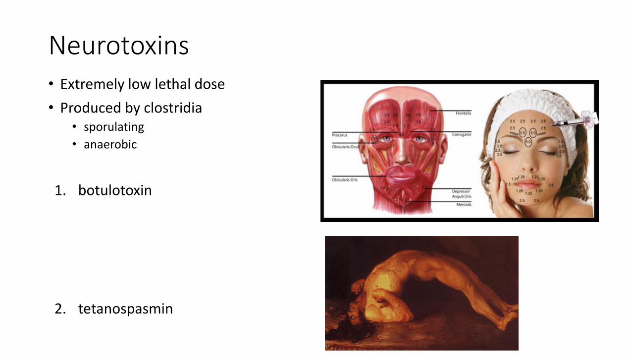
Neurotoxins• Extremely low lethal dose
• Produced by clostridia• sporulating
• anaerobic
1. botulotoxin
2. tetanospasmin

Neurotoxins• Mechanism of action: inhibition of fusion of synaptic vesiculles with presynaptic
membrane• botulotoxin: inhibits acetylcholin release → muscle relaxation
• tetanospasmin: inhibits GABA and glycine release→ muscle contraction
• Disruption of respiration

Enteric toxins
• Many patogenic bacteria• E. coli, Shigella, Salmonella, Vibrio cholerae, Campylobacter,
Clostridium difficile, S. aureus
• Poissoning of the intestinal epithelium→ diarrhoea• Typical symptom of intestinal infection
• Diarrhoea• patogen: rapid spread in host population
• Lots of liquid stool filled with bacteria
• host: cleaning of the intestines
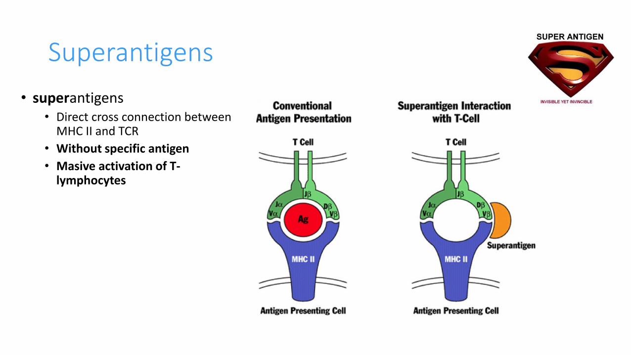
Superantigens
• superantigens• Direct cross connection between
MHC II and TCR
• Without specific antigen
• Masive activation of T-lymphocytes

Superantigens
• Staphylococcus aureus: 23 superantigens
• Streptococcus pyogenes: 11 superantigens
• For example:• Pyrogenic exotoxins of Streptococcus pyogenes
• Toxin shock syndrome toxin of S. aureus
• Staphylococal enterotoxins• Food poissoning

Toxic shock
• Caused by superantigens
• TSST-1 of S. aureus (less frequently causedby S. pyogenes)
• Similar to septic shock
• Leads to massive cytokine production by activated T-lymfocytes
• fever• Colaps of immune and regulátory homeostasis• Systemic patological changes
• CZ 1983-2011 - 159 cases, (47 menstrual form). letality 11 %(staphylococci), 50% (streptococci)
biofilm on tampon fibres reservoir ofstaphylococci producing toxic shock toxin

Symptoms of toxic shock

Toxin detection
• Detection of toxin moleculles (protein)• Specific antibodies
• latex bead agglutination
• ELISA
• Rapid antigen tests
• Detection of toxin genes(DNA)• PCR specific for selected genes (primers)