bacterial contamination of blood components
TRANSCRIPT

Bacterial Contamination of Blood Components
SUMMARY
INTRODUCTION
TRANSFUSION-TRANSMITTED BACTERIAL INFECTION OF RED CELLS
TRANSFUSION-TRANSMITTED BACTERIAL INFECTION OF PLATELETS
Clinical Presentation
TRANSFUSION-TRANSMITTED BACTERIAL INFECTION OF PLASMA AND
CRYOPRECIPITATE
SOURCES OF CONTAMINATION
METHODS TO REDUCE THE RISK OF POST-TRANSFUSION SEPSIS
Avoidance
Donor screening.
Skin preparation.
Diversion of the initial blood draw.
Apheresis versus whole-blood-derived platelet concentrates.
Bacterial Detection
Timing of detection.
Bacterial culture.
Molecular biology techniques.
Detection systems recently approved in Europe.
Detection systems under development.
Implementation of detection systems: problems and considerations. (i) Platelet
concentrates and prestorage pooling.
(ii) Anaerobic organisms.
(iii) Potential for reduced shelf-life.
(iv) Use of noncleared detection systems.
Bacterial Elimination
Pathogen reduction technologies.
Growth Inhibition
THE PROSPECT OF SEVEN-DAY STORAGE WITH DETECTION
CONCLUSIONS
1


SUMMARY
Blood for transfusion is a potential source of infection by a variety of known and
unknown transmissible agents. Over the last 20 years, astounding reductions in the risk of
viral infection via allogeneic blood have been achieved. As a result of this success,
bacterial contamination of blood products has emerged as the greatest residual source of
transfusion-transmitted disease. This paper summarizes the current status of detection,
prevention, and elimination of bacteria in blood products for transfusion.
INTRODUCTION
Allogeneic blood for transfusion is a potential source of infection by a variety of known
and unknown transmissible agents. Since the 1980s, with the recognition of human
immunodeficiency virus contamination of the blood supply, only a blood supply with a
zero risk to recipients has been politically acceptable. This approach has led to the
concept of "creeping precautionism" in the blood supply. The precautionary principle has
grown out of the environmental movement. Briefly, it states "...where there are threats of
serious or irreversible damage to the environment, lack of full scientific certainty should
not be used as a reason for postponing cost-effective measures to prevent environmental
degradation.To this end, astounding reductions in the viral risk of allogeneic blood has
been accomplished. However, a number of transfusion safety measures have been
implemented to achieve this level of safety, despite relatively poor cost-effectiveness, in
order to assuage the public's fears.
Given the reduction of viral transmission via allogeneic blood, the low but known risk of
bacterial contamination has emerged as the greatest residual threat of transfusion-
transmitted disease (Fig. 1).
3
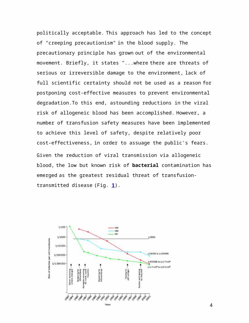
FIG. 1. Risk of infectious disease transmission per unit transfused by year, 1983 to 2001.
TRANSFUSION-TRANSMITTED BACTERIAL INFECTION OF RED CELLS
Each year, approximately 13,898,000 units of red cells or whole blood are transfused in
the United States alone. This equates to one unit being transfused every 2.3 s. Despite this
large number, sepsis associated with the transfusion of bacterially contaminated red cell
blood components is generally regarded as a very rare event. From 1976 through
September 1998, 26 fatalities thought to be secondary to contaminated whole blood or red
cells were reported to the U.S. Food and Drug Administration (FDA). Thus,
approximately one red cell-related death per year has been reported. The majority of
deaths reported to the FDA involved Yersinia enterocolitica. The highest reported
incidence of Y. enterocolitica contamination was reported in New Zealand, with an
incidence rate of 1 in 65,000 and a fatality rate of 1 in 104,000 red cell units transfused.
Unrecognized cases, underreporting, and regional variation may account for observed
differences in this incidence.
Interestingly, more recent passive reporting studies of bacterially contaminated red cells
from the United States, France, and the United Kingdom that caused symptoms of
infection show a relative paucity of Yersinia cases (Table 1). Of the reported deaths, one
was due to a coagulase-negative Staphylococcus strain and seven were due to a variety of
gram-negative organisms (including Serratia liquefaciens in three cases). These
4

organisms are all capable of growth at 1 to 6°C. Sepsis associated with the transfusion of
red cells contaminated with gram-negative bacteria is typically severe and rapid in onset.
Patients frequently develop high fever (temperatures as high as 109°F have been
observed) and chills during or immediately following transfusion. From 1987 to February
1996, 20 recipients of Yersinia-infected red cells were reported to the Centers for Disease
Control (S. T. Cookson, M. J. Arduino, S. M. Aguero, W. R. Jarvis, and the Yersinia
Study Group, Program Abstr. 36th Intersci. Conf. Antimicrob. Agents Chemother., abstr.
237, 1996). Twelve of the 20 recipients died, and the median time to death was only 25 h.
Of the seven who developed disseminated intravascular coagulopathy, six died.
TABLE 1. Organisms isolated from red cells implicated in transfusion-associated
infectionsa
Organism
No. of contaminated units in:
Total no.United
States
United
Kingdom
France (29 isolates, 25
implicated units)
Gram positive
Coagulase-negative
staphylococci
2 (1) 2 3 7 (1)
Streptococcus spp. 4d 4
Staphylococcus aureus 2b 2
Enteroccocus faecalis 1c 1
Bacillus cereus 2 2
Propionibacterium acnes 1 1
Subtotal 2 (1 or
50%)
2 (0 or 0%) 13 (0 or 0%) 17 (1 or
6%)
Gram negative
Serratia liquifaciens 2 (2) 1 2 (1) 5 (3)
Serratia marcescens 1 1
Yersinia enterocolitica 1 (1) 1 2 (1)
5

Enterobacter spp. 1 (1) 1 (1)
Acinetobacter spp. 5 (1)b 5 (1)
Pseudomonas spp. 2 (1) 2 (1)
Escherichia coli 3c 3
Klebsiella pneumoniae 1 1
Proteus mirabilis 1 1
Subtotal 3 (2 or
67%)
2 (1 or 50%) 16 (4 or 25%) 21 (7 or
33%)
Total 5 (3 or
60%)
4 (1 or 25%) 29 (4 or 14%) 38 (8 or
21%)
a Summary of organisms identified in the BACON, SHOT, and BACTHEM studies.
Numbers and percentages of fatalities are given in parentheses.
b In one case, S. aureus and A. baumannii were both isolated from the implicated bag.
c In one case, E. faecalis and E. coli were both isolated from the implicated bag.
d In two cases, two isolates of Streptococcus were isolated from the implicated bag. Of
the eight reported fatalities, seven (87.5%) were due to gram-negative organisms.
Prospective bacterial cultures of whole blood or red cells, however, have shown a much
higher incidence of bacterial contamination (2 to 4 per 1,000 units); however, the
organisms commonly cultured are Staphylococcus or Propionibacterium spp., which
generally proliferate poorly during storage at 1 to 6°C.
TRANFUSION –TRANSMITTED BACTERIAL INFECTION OF PLATELETS
Each year, within the United States approximately 4 million platelet units are transfused
(1 x 10⁶ single-donor apheresis platelets and 3 million whole-blood-derived platelet
concentrates). This equates to one platelet unit being transfused every 8 s. Unlike red cell
or whole-blood components, which are stored at 1 to 6°C, platelets are stored at 20 to
24°C to preserve function and survival. Such storage makes them an excellent growth
medium for a broad spectrum of bacteria. Multiple aerobic-culture surveillance studies
6

have demonstrated that 1 in 1,000 to 2,000 platelet units are bacterially contaminated.
Thus, 2,000 to 4,000 bacterially contaminated units would be expected to be transfused
per year. Estimates of the fraction of cases that result in patient symptoms have been as
low as 1 in 10 cases. However, in the only study that has prospectively cultured platelets
that were transfused, eight culture-positive but Gram stain-negative platelet pools that
were bacterially contaminated resulted in symptoms in three patients (37.5%) [A.
Dykstra, G. Hoeltge, M. Jacobs, J. Miller, R. Domen, and R. Yomtovian, Transfusion
38(Suppl.):104S, 1998]. Notably, six Gram stain-positive pools (which would be
expected to have had the highest bacterial load) were interdicted and never transfused.
Thus, clinical sepsis would be expected in at least 1 in 10 to 2 in 5 contaminated
transfusions (200 to 1,600 cases). National passive-reporting studies from the United
States, the United Kingdom, and France (Table 2) suggest that one-fifth to one-third
would result in death (40 to 533 deaths per year). This translates to a risk of death from a
transfusion of a bacterially contaminated platelet unit of between 1 in 7,500 and 1 in
100,000. Clinical observations from university hospitals actively pursuing suspected cases
of platelet-related sepsis confirm these estimates. Ness et al. from Johns Hopkins
University, reported a fatality rate of 1 in 17,000 with pooled whole-blood-derived
platelets and 1:61,000 with single-donor-apheresis-derived platelets. Similarly, University
Hospitals of Cleveland observed a fatality rate of 1 in 48,000 per random platelet unit.
The French BACTHEM study documented a fatality rate due to bacterially contaminated
platelets of 7 per 106 (1 in 140,000).
7

TABLE 2. Organisms implicated in infections associated with platelet transfusionsa
Organism
No. of contaminated units in:
Total no.United
States
United
Kingdom France
Gram positive
Bacillus cereus 1 4 (1) 2 7 (1)
Coagulase-negative
staphylococci
9 6 (1) 5 20 (1)
Streptococcus spp. 3 (1) 2 5 (1)
Staphylococcus aureus 4 2 (1) 6 (1)
Propionibacterium acnes 3 3
Subtotal 17 (1 or 6%)14 (3 or 21%) 10 (0 or 0%)41 (4 or
10%)
Gram negative
Klebsiella spp. 2 (1) 2 (1)
Serratia spp. 2 (2) 1 (1) 3 (3)
Escherichia coli 5 (1) 2 (1) 1 8 (2)
Acinetobacter spp. 1 1
Enterobacter spp. 2 (1) 1 (1) 1 4 (2)
Providencia rettgeri 1 (1) 1 (1)
Yersinia enterocolitica 1 1
Subtotal 11 (5 or
45%)
3 (2 or 67%) 6 (2 or 33%)20 (9 or
45%)
Total 28 (6 or
21%)
17 (5 or 29%) 16 (2 or
13%)
58 (11 or
19%)
a Summary of organisms identified in the BACON, SHOT, and BACTHEM studies.
Numbers and percentages of fatalities are given in parentheses.
8
From 1976 through September 1998, 51 fatalities thought to be secondary to
contaminated platelets were reported to the U.S. FDA. Gram-negative organisms
accounted for the majority of deaths (59.7%). Similarly, passive surveillance studies from
the United Kingdom, the United States, and France show that gram-positive organisms
were implicated in 41 (71%) of 58 of cases but gram-negative organisms (mostly
members of the Enterobacteriaceae) account for the majority (9 [82%] of 11) of the
fatalities (Table 1).
Clinical Presentation
Clinical sequelae of the infusion of bacterially contaminated platelets are variable and
may be acute or delayed (contributing to missed diagnoses). Ironically, the majority
(85%) of platelets are transfused to patients in the medical oncology, hematology,
pediatric hematology/oncology, and bone marrow transplantation settings, all typically
immunosuppressed patients. A study of platelet transfusions in autologous bone marrow
transplant patients found that 1 in 2,000 units of platelet concentrates associated with a
febrile episode (within 24 h of transfusion) were linked to bacterially contaminated
platelets. Of the 10 patients known to have received a bacterially contaminated unit, 4
experienced septic shock.
Because of the severity of the patient's underlying disease and the variability in
presentation and timing, sepsis resulting from bacterially contaminated platelets may go
unrecognized. For example, an outbreak of Salmonella enterica serovar Choleraesuis
sepsis in seven patients was traced to one repeat platelet donor, subsequently found to
have occult chronic osteomyelitis. One patient died, and two had long-term recurrences of
Salmonella sepsis. The time to the onset of illness ranged from 5 to 12 days (mean, 8.6
days). In many cases, the platelet-related bacterial sepsis is discovered only in retrospect.
Investigations of two Serratia liquefaciens-contaminated red cell units led to platelet
products from the whole-blood donations and to the recognition of an associated septic
death in one recipient and bacteremia in the other recipient.
9

TRANSFUSION-TRANSMITTED BACTERIAL INFECTION OF PLASMA AND
CRYOPRECIPITATE
Cell-free products such as plasma and cryoprecipitate are stored frozen and thus are rarely
associated with contamination. However, in some cases Pseudomonas cepacia and P.
aeruginosa have been cultured from cryoprecipitate and plasma thawed in contaminated
water baths.
SOURCES OF CONTAMINATION
Gram-positive skin commensals such as Staphylococcus epidermidis and Bacillus cereus
are the organisms most often recovered from donated blood (and implicated in bacterial
contamination of platelets). Such contamination is thought to occur principally during
phlebotomy, as a result of incomplete disinfection and/or skin core removal (including
skin appendages where the skin disinfectants may not penetrate) by the collection needle.
These organisms typically do not grow at 1 to 6°C but survive and multiply readily at the
platelet storage temperature of 20 to 24°C.
In the case of gram-negative bacterial contamination, asymptomatic donors with
transient bacteremia are presumed to be responsible for most cases of contamination. For
example, in the case of Y. enterocolitica contamination of red cells, implicated donors
typically are found to have elevated antibody titers (immunoglobulin M or G) to Y.
enterocolitica, implying a recent infection.
10

An outbreak of Serratia marcescens contamination of red cells in Denmark and Sweden
was thought to involve the manufacturing process since the sterile bag sets were
autoclaved and put in a clean but not sterile outer plastic package. It was thought that S.
marcescens present in the dust in the factory contaminated the outside of the containers
and that in the presence of moisture and a nutrient (the plasticizer diethylhexyl phthalate)
the bacteria proliferated and gained entry into the bag.
In rare cases, unusual circumstances have been identified as the source of bacterial
contamination. For example, a donor with a pet boa constrictor, who reported fever and
bloody diarrhea treated with antibiotics 13 days prior to his donation, was linked to
Salmonella enterica platelet sepsis in two patients. Scarring or dimpling of the
venipuncture site (due to prior donations) can result in recessed pits that are difficult to
adequately sterilize. A repeat single donor with a "dimpled" venipuncture site resulted in
four cases of gram-positive bacterial platelet sepsis in recipients. Another unusual
association was made between one donor in a platelet pool and the death of a leukemic
recipient from septic shock. The contaminating organism was identified as C. perfringens
and was thought to originate from a donor who had two young children and frequently
changed their diapers. As stated above, an outbreak of S. enterica serovar Choleraesuis
platelet sepsis in seven patients at the National Institutes of Health Clinical Center was
traced to a repeat platelet donor with an occult osteomyelitis. However, in most cases the
source of the bacterial contamination remains unidentified.
METHODS TO REDUCE THE RISK OF POST-TRANSFUSION SEPSIS
There are multiple strategies to reduce transfusion-related bacterial infection. The
various approaches can be grouped into four major categories: avoidance, bacterial
detection, bacterial elimination, and growth inhibition.
11

Avoidance
Donor screening. Screening of donors to detect a possible risk of infection or transient
bacteremia (e.g., from recent dental procedures) is routinely performed. However,
questioning about symptoms suggestive of infection are often problematic; for example,
gastrointestinal symptoms have not been a specific predictor (13% of donors have had
gastrointestinal symptoms in the previous 30 days). Such questioning also lacked
sensitivity, since only 13 of 20 donors associated with Y. enterocolitica-contaminated red
blood cells recalled a history of gastrointestinal symptoms; Cookson et al., Prog. Abstr.
36th ICAAC, 1996).
Skin preparation. Skin disinfection reduces the skin bacterial load, but a sterile
venipuncture cannot be guaranteed due to inaccessibility of organisms present in
sebaceous glands and hair follicles; organisms present in skin fragments may be drawn
into the component bag. Multiple studies have evaluated skin disinfection in the context
of blood donation. A summary of the effectiveness of some of the more commonly used
skin disinfection methods is given in Table 3. In general, either iodine or povidone iodine
are used. In donors with allergies to iodine, chlorhexidine or double isopropyl alcohol
skin disinfection is employed. The difference noted in Table 3 for two different isopropyl
alcohol scrubs was attributed to the abrasive nature of the sponge used in one of the
preparations.
TABLE 3. Percentage and mean count of donors with bacteria after skin disinfectiona
No. of CFU/plate
% of contaminated donors after disinfection with:
Povidone iodine x 2 IPAb and iodine IPA x 2 (sponge) IPA x 2 (swab)
0 39 79 0 29
10 57 93 69 50
100 75 100 86 68
1,000 96 100 97 89
Mean count 175 3 161 237
12

a Data are from reference 43.
b IPA, isopropyl alcohol.
Diversion of the initial blood draw. Several studies have demonstrated the partial
effectiveness of diverting the first 10 to 30 ml of blood from the initial collection. A study
performed with 22,000 samples of donated blood by the Red Cross in The Netherlands, in
which the first 10 ml of donor blood was diverted from the primary bag, showed that 16
of the first 5-ml aliquots were bacterially contaminated while only 2 of the second 5-ml
aliquots were positive after culture. A second study in France showed that removal of the
first 10 ml of donor blood was associated with a significant decrease in bacterial
contamination (18,263 collections with 0.39% contamination compared with 7,115
collections with 0.21% contamination; P < 0.05).
Such methods are most effective with respect to skin contaminants. However, it has been
said that "one should not be diverted by diversion," since most fatal cases of bacterial
contamination involve gram-negative organisms and thus would not minimized by
diversion. Interestingly, the addition of a diversion bag to whole-blood collections in the
United States has been initially complicated by hemolysis with certain sample tubes (from
Pall, East Hills, N.Y.) and dilution of the sample with anticoagulant (from Baxter, Round
Lakes, Ill), requiring redesign of both diversion systems (data presented at the HHS
Advisory Committee on Blood Safety and Availability, Washington, D.C.
Apheresis versus whole-blood-derived platelet concentrates. Therapeutic doses of
platelets can be obtained from two sources. Platelets can be collected from a donor by the
use of an automated cell separator (apheresis); such collections can yield one to three
therapeutic doses. Alternatively, platelets can be obtained from whole-blood donations
(whole blood platelet concentrates [WBPCs]); however, a therapeutic dose requires the
pooling of four to six WBPCs. Current estimates suggest that 69% of all therapeutic doses
of platelets transfused in the United States are apheresis derived. Usage varies
considerably by country.
13

Since bacterial contamination may result from a contaminated venipuncture or an
asymptomatic bacteremic donor, it would be expected that pooled platelet products would
be associated with a higher risk of contamination than a single donor product. From 1986
to 1998, the incidence of septic events related to platelets at Johns Hopkins University
Hospital decreased from 1 in 4,818 to 1 in 15,098 as the hospital went from transfusing
51.7% to 99.4% single-donor apheresis units. The overall clinical sepsis rate was
approximately 1 in 2,500 for WBPC pools and 1 in 13,400 for single-donor apheresis
transfusion. The related mortality rate was approximately 1 in 17,000 for WBPC pools
and 1 in 61,000 for single-donor apheresis platelets. It was concluded that the use of
single-donor apheresis platelets is a simple means of reducing septic platelet transfusion
reactions. Similarly, the University Hospitals of Cleveland have monitored the
contamination rates of both single-donor and whole-blood-derived concentrates [A.
Dykstra, M. Jacobs, and R. Yomtovian, Transfusion (Suppl.):104S, 1998]. The rate of
bacterial contamination of apheresis-derived platelets was 1 in 2,184. While the rate for
random-donor platelets was similar at 1 in 1,861, random-donor platelet concentrates in a
pool of six units would be expected to result in a contamination rate of 1 in 310.
Bacterial Detection
Timing of detection. Culture studies performed both on red cell units and platelets have
shown that culture on the day of collection invariably misses bacterially contaminated
units that would possibly achieve dangerous levels of overgrowth during storage of the
blood components [ M. A. Blajchman, A. Ali, P. Lyn, L. Bardossy, and H. Richardson,
Transfusion (Suppl.):7S, 1997]. This is in contrast to viral contamination of blood
components, in which the virus or the immune response to the virus (antibodies) is
detected from a sample obtained at the time of donation. Bacterial contamination of
blood components generally requires time for the organisms to proliferate before being
detectable in a small representative sample of the product. Therefore, knowledge of the
growth characteristics of bacteria in blood components is essential before one considers
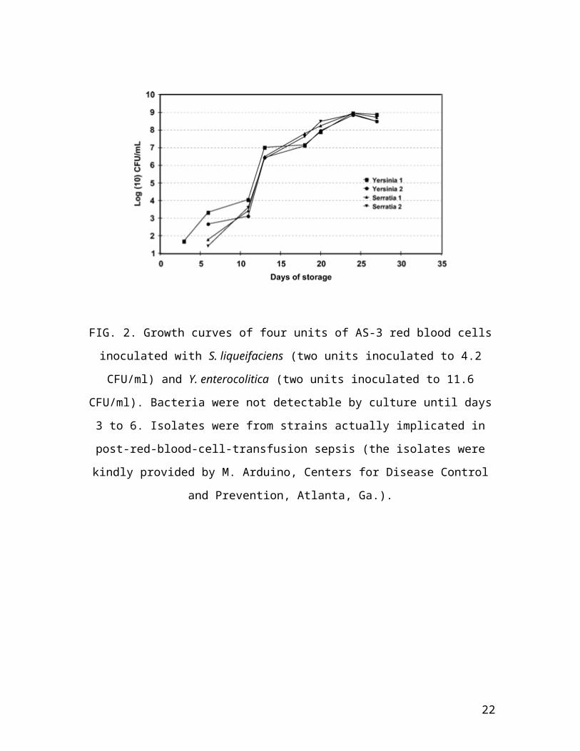
implementation of a detection strategy (Fig. 2 and 3).
14

FIG. 2. Growth curves of four units of AS-3 red blood cells inoculated with S.
liqueifaciens (two units inoculated to 4.2 CFU/ml) and Y. enterocolitica (two units
inoculated to 11.6 CFU/ml). Bacteria were not detectable by culture until days 3 to 6.
Isolates were from strains actually implicated in post-red-blood-cell-transfusion sepsis
(the isolates were kindly provided by M. Arduino, Centers for Disease Control and
Prevention, Atlanta, Ga.).
FIG. 3. Growth curves of six bacteria species (S. marcescens, n = 7; K. pneumoniae, n =
15

21; S. epidermidis, n = 21; Pseudomonas sp., n = 15; B. cereus, n = 9; S. aureus, n = 22)
in 95 platelet units. All bacteria were inoculated on day zero at 10-50 CFU/ml (10).
Growth characteristics of 165 platelet units inoculated (to 1 to 50 or 1,000 CFU/ml)
within 1 day of collection with one of the following: B. cereus, P. aeruginosa, K.
pneumoniae, S. marcescens, S. aureus, and S. epidermidis showed that after 1 day of
storage all samples of B. cereus, P. aeruginosa, and K. pneumoniae had concentrations of
10 CFU/ml (the level of detection), while only 71.4 and 84.2% of the S. marcescens and
S. aureus samples were at concentrations of 10 CFU/mL. By day 3 of storage, all units
inoculated with B. cereus, P. aeruginosa, K. pneumoniae, S. marcescens, and S. aureus
contained 105 CFU/ml. Units contaminated with S. epidermidis showed slower and more
varied growth.
Bacterial culture. Although there are a variety of automated and semi-automated
systems, only two are FDA cleared for quality control of bacterial culture in platelets in
the United States. The first is an automated liquid culture system (BacT/ALERT;
bioMérieux, Durham, N.C.) that uses broth bottles with a colorimetric sensor which
changes color as a consequence of increasing CO2 produced by bacterial proliferation.
The machine monitors both the rate of change of the colorimetric sensor and the absolute
color change of the sensor. Generally, one aerobic (and in some cases one aerobic and one
anaerobic) bottle is inoculated with 4 ml of the platelet product. This system is not
completely closed, since a needle is used to inoculate the bottles. It has been extensively
validated by tests with a wide range of potentially contaminating organisms. The method
reliably detects contamination of platelets inoculated to 10 CFU/ml and in many cases to
5 CFU/ml (e.g., B. cereus, S. marcescens, C. perfringens, S. epidermidis, S. pyogenes, E.
coli, K. pneumoniae, S. aureus, and viridans streptococci) in 12 to 26 h.
Propionibacterium acnes (which is of questionable clinical significance in this setting)
required longer incubations. Recently, our laboratory completed an 11-month pilot
program in which we cultured 2,397 apheresis-derived platelets on day 2 of storage
16

(collection day = day 0) and again at the time of issue (or following outdate: days 6 to 8).
This study involved the inoculation of 9,588 bottles; one "machine" false-positive bottle
(negative subculture, Gram stain, and repeat culture), and one inadvertent contamination
false-positive bottle (Propionibacterium sp.) were detected. Thus, the overall false-
positive rate was 2 in 4,794 samplings (0.04%, or 1 in 2,397 samplings) or 2 in 9,588
bottles (0.02% or 1 in 4,794 bottles). During this period, we successfully interdicted three
apheresis-derived units (one collection) contaminated with S. epidermidis but were unable
to prevent the transfusion of four apheresis units contaminated with Propionibacterium
sp., for which the culture bottles were reactive after 6 days of incubation. Thus, the
overall true-positive rate was 7 of 2,397 apheresis units (0.29%), with a true-positive rate
for aerobic organisms of 3 of 2,397 (0.13% or 1 of 799 units) and an anaerobic true-
positive rate of 4 of 2,397 (0.17% or 1 of 599 units). Within our facility, transition from a
research setting to a clinical microbiology laboratory has resulted in a higher false-
positive (laboratory contamination) rate of 0.6% per unit (with one aerobic and one
anaerobic bottle) (unpublished data). More recent experience from three large blood
centers is included in Table 4.
The second culture system is the Pall Bacterial Detection System (BDS). Sampling is
obtained so as to maintain an effectively closed system. Approximately 5 to 6 ml of
leukocyte-reduced platelet-rich plasma is removed from the bag and passed through a
filter that removes residual white cells and platelets but allows approximately 50% of the
bacteria (variable by organism) and plasma to pass into an incubation bag. The incubation
bag holds 2 to 3 ml and contains sodium polyanethol sulfonate, which facilitates the
growth of gram-negative organisms by inhibiting both complement and plasma lysosomal
enzymes and by antagonizing the action of growth-retarding lipoproteins normally
present in plasma. The incubation bag is then incubated at 35°C for at least 24 h before
the oxygen concentration of the bag's headspace is measured with an oxygen analyzer. A
decrease in the oxygen concentration to 19.5% or lower is an indication of bacterial
growth. Thus, this system does not detect obligate anaerobic organisms. Leukocyte-
17

reduced platelets challenged with 10 known platelet contaminants at doses of 100 to 500
CFU/ml were detected at a rate of 96.5% at 24 h and 100% at 30 h. External validation
studies have shown lower than expected sensitivity for S. epidermidis and S. agalactiae
[K.-A. T. Nguyen, T. Yamamoto, and N. Hirschler, Transfusion (Suppl.):S63-030K,
2003; H. C. Pleasant, G. A. Geele, and M. P. Holub, Transfusion (Suppl.):S62-030K,
2003 (abstract)]. Most recently, an enhanced version of this system has been cleared by
the FDA, it includes modifications to the configuration (with removal of the filter, which
removed some bacteria), and the incubation bag now contains a platelet-aggregating agent
and tryptic soy medium to enhance the sensitivity and decrease the incubation time.
Less sensitive methods of detection such as Gram or Wright stains or the use of glucose
and pH determination (in some case with multireagent strips) are used for rapid detection
of bacteria in platelet components prior to transfusion. However, the overall sensitivity is
only 106 to 107 CFU/ml. Werch et al., from the M. D. Anderson Cancer Center, reported
on the screening of 3,093 platelet concentrates, in which they found that multireagent
strips for 30 bags had glucose levels or pH outside of the reference range. Two of these
platelet units were culture positive for B. cereus. This screening method resulted in 1 unit
per 110 being wasted but prevented two patients from receiving contaminated platelets.
Molecular biology techniques. Because of the high sensitivity and specificity and the
rapidity of obtaining results, molecular biological approaches are theoretically appealing
for the detection of contaminating organisms in blood components. One such approach is
the use of a nonamplified chemiluminescence-linked universal bacterial rRNA probe.
This technique was shown to detect four bacterial species in the range of 104 to 105
CFU/ml. In some cases, S. aureus was detected at levels as low as 102 to 103 CFU/ml.
Other investigators have pursued the use of PCR-based amplification for a limited
number of organisms.
However, bacterial DNA contamination of nucleotide amplification reagents
(particularly the bacterially derived enzymes) and laboratory materials and the presence
of bacterial DNA sequences commonly found in human blood complicate the usefulness
of universal bacterial amplification techniques. It also remains to be seen if complex
18
molecular biology techniques could be successfully deployed to a wide range of
transfusion services.
Detection systems recently approved in Europe. Recently, two bacterial detection
systems were approved in Europe. The Scansystem (Hemosystem, Marseilles, France)
involves a 70-min solid-phase laser cytometry. In a few studies, the Scansystem detected
11 organisms in the range of 10 to 103 CFU/ml [P. Morel, M. Deschaseaux, X. Bertrand,
C. Naegelen, and D. Talon, Transfusion (Suppl.):SP13, 2003]. Positive samples require a
technologist to perform a microscopic confirmation.
A second "rapid" method recently approved detects bacteria by using dielectrophoresis
(Cell Analysis, Slough, United Kingdom). Platelet concentrates inoculated with six
organisms at concentrations of 103 to 105 CFU/ml were detected after 30 min of
dielectrophoresis by image analysis as they were released from the electrodes when the
electric field was discontinued [C. P. McDonald, R. Smith, J. Colvin, S. Robbins, J.
Barbara, W. Betts, R. Anthony, K. Chan, A. Brown, and K. Gregory, Transfusion
(Suppl.):S119-040H, 2001].
Detection systems under development. Currently, there are at least three other rapid-
detection systems under development. Genpoint (Oslo, Norway) has developed a unique
system that uses paramagnetic particles to concentrate bacteria (BUGS'n BEADS)
coupled with a PCR for highly conserved DNA. The target sensitivity of this assay is
1,000 CFU/ml, and the total time is 2.5 h. Immunetics, Inc. (Cambridge, Mass.) is
developing a 96-well 1-h enzyme immunoassay which employs proteins and derived
peptides that bind to the peptidoglycan component of the bacterial cell wall. Preliminary
results suggest a sensitivity of 103 to 104 CFU/ml (or lower) for a variety of organisms [L.
A. Beausang and A. Levin, Transfusion (Suppl.):20A, 2003; D. Kagan, and A. E. Levin,
Transfusion (Suppl.):S118-040H, 2001]. Verax Biomedical (Worcester, Mass.) is
developing an immunoassay embodied in a lateral-flow format. Preliminary results
suggest a sensitivity of 103 to 104 CFU/ml for a variety of organisms [F. Kenny, C.
Woodbury, M. Pelak, J. Weinberg, and J. A. Hall, Transfusion (Suppl.):10A, 2003].
19

Implementation of detection systems: problems and considerations. (i) Platelet
concentrates and prestorage pooling. Bacterial detection of whole-blood-derived
platelet concentrates presents logistical problems for systems that do not allow the
prestorage pooling of platelet concentrates (e.g., United States, Canada, and Hong Kong).
In Europe, whole-blood-derived buffy coat platelets are prepooled prior to storage; this
allows for the culturing of platelets by the methods described above. If prepooling is not
allowed, culturing of each individual platelet sample is generally not thought to be
practical due to the loss of volume from each bag required per culture and the high cost of
culture for each individual bag. In the absence of prepooling, many centers have chosen
or are contemplating the use of less sensitive bacterial detection strategies such as Gram
stains or glucose or pH measurement (or the use of multireagent strips).
(ii) Anaerobic organisms. Anaerobic organisms such as Propionibacterium sp. can be
identified in platelets; however, it is not known whether anaerobic organisms such as
propionibacteria are clinically significant since growth in the aerobic environment of
platelet storage is slow and not favorable. Although a few cases of Propionibacterium-
contaminated units have been infused into patients, no long-term sequelae have been
reported. Debate continues as to the value of an anaerobic culture in this context. It is
worth noting that some aerobic organisms or isolates (e.g., viridans streptococci) can be
more rapidly detected in an anaerobic culture system.
(iii) Potential for reduced shelf-life. Some centers have chosen to incubate platelet
cultures for 12 to 24 h prior to release. This has in many cases led to a decrease in the
available shelf-life of the platelets. Given that the current platelet products have a shelf-
life of only 5 days (and that, for 1 to 2 days, such products are not available, pending
completion of viral testing), any further reduction in available shelf-life can impact
inventory control and hospital supply.
(iv) Use of noncleared detection systems. Use of non-FDA-cleared detection systems
(e.g., alternative automated culture systems) would at a minimum require internal
validation. Examples of validation of detection systems are available in the literature.
Patent issues currently preclude certain companies from actively pursing this market. Use
20

of a non-FDA-cleared device for bacterial quality control of platelets, when cleared
devices are available, may place an institution at higher legal risk should a patient die
after receiving bacterially contaminated platelets.
Bacterial Elimination
Pathogen reduction technologies. Continued efforts to create a "zero-risk" blood supply
have led to various pathogen reduction technologies. A variety of photoactive sensitizers
have been investigated for use in the reduction of viruses, bacteria, and parasites.
The best studied of these methods is the use of amotosalen HCL (S-59), a synthetic
psoralen combined with a photochemical treatment process that causes permanent cross-
links in DNA and RNA (INTERCEPT Platelet System; Baxter Healthcare Corp., Round
Lake, Ill.). However, organisms capable of spore formation may be resistant to
photochemical inactivation. The SPRINT trial, a multi-center phase III trial, has evaluated
the hemostatic effectiveness of S-59 pathogen-inactivated platelets. Treated platelets were
found to be as effective as conventional platelets for the prevention and treatment of
bleeding; however, the treated platelets resulted in a lower post-transfusion platelet
response, resulting in the need for 35% more platelets to support transfusion-dependent
patients. Concerns also remain regarding mutagenicity and teratogenicity associated with
such technologies.
Growth Inhibition
The addition of antibiotics to blood components has been considered but has not been
adopted due to the added risk of drug reactions (trading one infrequent reaction for
another) and the potential for the development of antibiotic resistance if small amounts of
antibiotics were infused with every transfusion.
THE PROSPECT OF SEVEN-DAY STORAGE WITH DETECTION
Currently, within the United States platelet storage is limited to 5 days due to the fear of
bacterial overgrowth during storage at 20 to 24°C. In 1983, platelet storage in the United
21
States was extended to 7 days; however, in 1986 the FDA shortened the outdate to 5 days
following reports of bacterial sepsis with older units. With the prospect of bacterial
detection, the possibility of an extended shelf-life is being reconsidered. European reports
have described the use of bacterial culturing of platelets on days 1 to 3 of storage in
order to extend the shelf-life of platelets to 7 days, thereby reducing the number of
outdates [M. Ollgaard, I. Albjerg, and J. Georgen, Vox Sang. (Suppl. 1):1126, 1998; D.
Vuetic, J. Taseki, B. Balint, and V. Mirovic, Vox Sang. 80(Suppl. 1):P371, 2000]. Within
the United States, premarket multicenter studies (or possibly postmarket studies) are
currently under discussion to provide data that may allow the extension of storage to 7
days. It would be expected that the cost of culturing would be mitigated by the savings
obtained by the extension of platelet storage time. For example, in Denmark, when
bacterial culture was implemented in 1997, it was anticipated to be cost neutral if the
outdating could be reduced from 18 to 8% by extending the storage time to 7 days. The
actual outdating rate decreased to 3 to 6%.
CONCLUSIONS
Regulatory oversight in the United States and Canada has generally concentrated on new
emerging threats such as West Nile virus (leading to nationally mandated efforts to screen
the blood supply with West Nile virus nucleic acid testing). However, there is no similar
mandate in these countries for bacterial screening. To place these risks into some
perspective, in 2002 23 transfusion-transmitted cases of West Nile virus were identified in
the United States. Of these 23 recipients, 7 died, but only 5 of these deaths were
associated with West Nile virus meningoencephalitis. During the same period, 17 deaths
from bacterial contamination of blood components were reported to the FDA.
Contaminated apheresis-derived platelets and pooled platelets were the products most
often implicated. Gram-negative bacteria were the causative agents in the majority of
these fatalities. It is perplexing that similar (or greater) government emphasis has not been
placed on the detection of bacteria.
In blood banking and transfusion medicine, our paramount concern is to improve
transfusion safety for patients, in our attempt to achieve a zero-risk blood supply. Unlike
22

other recent threats (existing or theoretical) to blood safety, the movement for reducing
bacterial contamination has come from the blood-banking and the transfusion medicine
community. In this case, voluntary accrediting agencies such as the American Association
of Blood Banks and the College of American Pathologists have taken the lead by
developing standards to reduce the risk of bacterial contamination of blood components.
Our patients deserve no less.
REFERENCES
1
23

1. American Association of Blood Banks. 2003. Comprehensive report on
blood collection and transfusion in the United States in 2001. National Blood
Data Resource Center, Bethesda, Md.
2. American Association of Blood Banks. Bulletin. Guidance on
implementation of new bacteria reduction and detection standard.
http://www.aabb.org/members_only/archives/association_bulletins/ab03-
10.htm.
3. Anderson, K. C., M. A. Lew, B. C. Gorgone, J. Martel, C. B. Leamy, and
B. Sullivan. 1986. Transfusion-related sepsis after prolonged platelet storage.
Am. J. Med. 81:405-411.[CrossRef] [Medline]
4. Brecher, M. E. 2002. Bacterial contamination of blood products, p. 789. In
T. L. Simon (ed.), Rossi's principles of transfusion medicine, 3rd ed.
Lippincott, Williams and Wilkins, Baltimore, Md.
5. Brecher, M. E., G. Boothe, and A. Kerr. 1993. The use of a
chemiluminescence-linked universal bacterial ribosomal RNA gene probe
and blood gas analysis for the rapid detection of bacterial contamination in
white cell reduced and nonreduced platelets. Transfusion 33:450-457.
[CrossRef] [Medline]
6. Brecher, M. E., M. Foster, and D. Mair. 2001. Glucose and hemolysis as a
rapid screen for Yersinia and Serratia RBC contamination. Vox Sang. 81:136-
138. (Letter.)[CrossRef] [Medline]
7. Brecher, M. E., S. N. Hay, and S. J. Rothenberg. 2003. Monitoring of
apheresis platelet bacterial contamination with an automated liquid culture
system: a university experience. Transfusion 43:974-978.[CrossRef]
[Medline]
8. Brecher, M. E., S. N. Hay, and S. J. Rothenberg. 2004. Evaluation of a new
plastic culture bottle using an automated microbial detection system for 9
common contaminating organisms found in platelet components. Transfusion
44:359-363.[CrossRef] [Medline]
24

9. Brecher, M. E., D. Heath, S. Hay, S. Rothenberg, and L. C. Stutzman.
2002. Evaluation of a new generation culture bottle using the BacT/ALERT
3D® Microbial Detection System on 9 common contaminating organisms
found in platelet components. Transfusion 42:774-779.[CrossRef] [Medline]
10. Brecher, M. E., J. J. Hogan, G. Boothe, A. Kerr, L. McClannan, M. R.
Jacobs, R. Yomtovian, V. Chongokolwatana, G. Tegtmeier, S.
Henderson, A. Pineda, V. Halling, M. Kemper, K. Kuramato, P. V.
Holland, and M. Longiaru. 1994. Platelet bacterial contamination and the
use of a chemiluminscence-linked universal bacterial ribosomal RNA gene
probe. Transfusion 34:750-755.[CrossRef] [Medline]
11. Brecher, M. E., P. V. Holland, A. Pineda, G. E. Tegtmeier, and R.
Yomtovian. 2000. Growth of bacteria in inoculated platelets: implications for
bacterial detection and the extension of platelet storage. Transfusion
40:1308-1312.[CrossRef] [Medline]
12. Brecher, M. E., N. Means, C. S. Jere, D. Heath, S. Rothenberg, and L. C.
Stutzman. 2001. Evaluation of the BacT/ALERT 3D® Microbial Detection
System for platelet bacterial contamination: an analysis of 15 contaminating
organisms. Transfusion 41:477-482.[CrossRef] [Medline]
13. Bruneau, C., P. Perez, M. Chassaigne, P. Allouch, A. Audurier, C.
Gulian, G. Janus, G. Boulard, P. De Micco, L. R. Salmi, and L. Noel.
2001. Efficacy of a new collection procedure for preventing bacterial
contamination of whole-blood donations. Transfusion 41:74-81.[CrossRef]
[Medline]
14. Burstain, J. M., M. E. Brecher, K. Workman, M. Foster, G. H. Faber,
and D. Mair. 1997. Rapid identification of bacterially contaminated platelets
using reagent strips: glucose and pH analysis as markers of bacterial
metabolism. Transfusion 37:255-258.[CrossRef] [Medline]
15. CBER. 2002. Annual Report FY. 2002 (1 October 2001 through 30
September 2002). http://www.fda.gov/cber/inside/annrptpart3.htm.
25
16. Casewell, M. W., N. G. P. Slater, and J. E. Cooper. 1981. Operating theatre
water-baths as a cause of Pseudomonas septicemia. J. Hosp. Infect. 2:237-
240.[CrossRef] [Medline]
17. Centers for Disease Control and Prevention. 1997. Red blood cell
transfusions contaminated with Yersinia enterocolitica—United States, 1991-
1996, and initiation of a national study to detect bacteria-associated
transfusion reactions. Morb. Mortal. Wkly. Rep. 46:553-555.[Medline]
18. Centers for Disease Control and Prevention. 1991. Update: Yersinia
enterocolitica bacteremia and endotoxin shock associated with red blood cell
transfusion—United States. 40:176-178.
19. Chiu, E. K. W., K. Y. Yuen, A. K. W. Lie, R. Liang, Y. L. Lau, A. C. Lee,
Y. L. Kwong, S. Wong, M. H. Ng, and T. K. Chan. 1994. A prospective
study of symptomatic bacteremia following platelet transfusion and of its
management. Transfusion 34:950-954.[CrossRef] [Medline]
20. Commission on Laboratory Accreditation. Transfusion medicine
checklist. TRM.44955 phase I. College of American Pathologists,
Northfield, Ill. 2003.
http://www.cap.org/html/checklist_html/transfusionmedicine_1202.html.
Last accessed 1 September 2004.
21. Corash, L. Technology for inactivation of pathogens in cellular blood
products used for transfusion. Presented at the FDA/CBER Workshop on
Safety and Efficacy of Methods for Reducing Pathogens in Cellular Blood
Products Used in Transfusion. www.fda.gov/cber/minutes/workshop-
min.htm.
22. DeKorte, D., J. H. Marcelis, A. J. Verhoeven, and A. M. Soeterboek.
2002. Diversion of first blood volume results in a reduction of bacterial
contamination for whole-blood collections. Vox Sang. 83:13-16.[CrossRef]
23. Dumont, L. J., J. P. AuBuchon, P. Whitey, L. H. Herschel, A. Johnson, D.
McNeil, S. Sawyer, and J. C. Roger. 2002. Seven-day storage of single-
26

donor platelets: recovery and survival in an autologous transfusion study.
Transfusion 42:847-854.[CrossRef] [Medline]
24. Engelfriet, C. P., H. W. Reesink, M. A. Blajchman, L. Muylle, J.
Kjeldsen-Kragh, R. Kekomaki, R. Yomtovian, P. Hocker, G. Stiegler, H.
G. Klein, K. Soldan, Barbara J, A. Slopecki, A. Robinson, and H.
Seyfried. 2000. Bacterial contamination of blood components. Vox Sang.
78:59-67.[CrossRef] [Medline]
25. Fredricks, D. N., and D. A. Relman. 1998. Improved amplification of
microbial DNA from blood cultures by removal of the PCR inhibitor sodium
polyanetholesulfonate. J. Clin. Microbiol. 36:2810-
2816.[Abstract/Free Full Text]
26. Fridey, J. (ed.). 2003. Standards for blood banks and transfusion services,
22nd ed. American Association of Blood Banks, Bethesda, Md.
27. Georgsen, J. 2003. Detection of bacterial contamination of platelet
concentrates. International Forum 5. Vox Sang. 85:229-230.[CrossRef]
[Medline]
28. Gibson, T., and W. Norris. 1998. Skin fragments removed by injection
needles. Lancet 2:983-985.[CrossRef]
29. Goldman, M., G. Roy, N. Frechette, F. Decary, L. Massicotte, and G.
Delage. 1997. Evaluation of donor skin disinfection methods. Transfusion
37:309L-312L.[CrossRef]
30. Goodnough, L. T., A. Shander, and M. E. Brecher. 2003. Transfusion
medicine: looking to the future. Lancet 361:161-169.[CrossRef] [Medline]
31. Grossman, B. I., P. Kollins, P. M. Lau, J. L. Perreten, R. J. Bowman, S.
Malcolm, and W. M. Palko. 1991. Screening blood donors for
gastrointestinal illness: a strategy to eliminate carriers of Yersinia
entercolitica. Transfusion 31:500-501.[CrossRef] [Medline]
32. Heltberg, O., F. Show, P. Gerner-Smidt, H. J. Kolmos, E. Dybkjer, E.
Gutschik, D. Jerne, O. B. Jepsen, M. Weischer, W. Frederiksen, and H.
Sorensen. 1993. Nosocomial epidemic of Serratia marcescens septicemia
27

ascribed to contaminated blood transfusion bags. Transfusion 33:221-227.
[CrossRef] [Medline]
33. Hogman, C. F., H. Fritz, and L. Sandberg. 1993. Post-transfusion Serratia
marcescens septicemia (editorial). Transfusion 33:189-191.[CrossRef]
[Medline]
34. Jackson, B. R., M. P. Busch, S. L. Stramer, and J. P. AuBuchon. 2003.
The cost-effectiveness of NAT for HIV, HCV, and HBV in whole-blood
donations. Transfusion 43:721-729.[CrossRef] [Medline]
35. Hoppe, P. A. 1992. Interim measures for detection of bacterially
contaminated red cell components. Transfusion 32:199-201. (Editorial.)
[CrossRef] [Medline]
36. Jafari, M., J. Forsberg, R. O. Gilcher, J. W. Smith, J. M. Crutcher, M.
McDermott, B. R. Brown, and J. N. George. 2002. Salmonella sepsis
caused by a platelet transfusion from a donor with a pet snake. N. Engl. J.
Med. 347:1075-1078.[Free Full Text]
37. Knutson, F., R. Alfonso, K. Dupuis, V. Mayaudon, L. Lin, L. Corash, and
C. F. Hogman. 2000. Photochemical inactivation of bacteria and HIV in
buffy-coat-derived platelet concentrates under conditions that preserve in vitro
platelet function. Vox Sang. 78:209-216.[CrossRef] [Medline]
38. Kuehnert, M. J., V. R. Roth, N. R. Haley, K. R. Gregory, K. V. Elder, G.
B. Schreiber, M. J. Arduino, S. C. Holt, L. A. Carson, S. N. Banerjee, and
W. R. Jarvis. 2001. Transfusion-transmitted bacterial infection in the United
States, 1998 through 2000. Transfusion 41:1493-1499.[CrossRef] [Medline]
39. Lee, J. 1999. U.S. Food and Drug Administration. Presented at the
FDA/CBER Workshop on Bacterial Contamination of Platelets.
www.FDA.gov/CBER/minutes/bact092499.pdf.
40. Leiby, D. A., K. L. Kerr, J. M. Campos and R. Y. Dodd. 1997. A
retrospective analysis of microbial contaminants in outdated random-donor
platelets from multiple sites. Transfusion 37:259-263.[CrossRef] [Medline]
28

41. Macauley, A., A. Chandrasekar, G. Geddis, K. G. Morris, and W. M.
McClelland. 2003. Operational feasibility of routine bacterial monitoring of
platelets. Transfusion Med. 13:189-195.[CrossRef] [Medline]
42. McCullough, J., D. H. Vesole, R. J. Benjamin, S. J. Slichter, A. Pineda, E.
Snyder, E. A. Stadtmauer, I. Lopez-Plaza, S. Coutre, R. G. Strauss, L. T.
Goodnough, J. L. Fridey, T. Raife, R. Cable, S. Murphy, F. Howard IV,
K. Davis, J. S. Lin, P. Metzel, L. Corash, A. Koutsoukos, L. Lin, D. H.
Buchholz, and M. G. Conlan. 2004. Therapeutic efficacy and safety of
platelets treated with a photochemical process for pathogen inactivation: the
SPRINT Trial. Blood 104:1534-1541.[Abstract/Free Full Text]
43. McDonald, C. P., S. Hartley, K. Orchard, G. Hughes, M. M. Brett, P. E.
Hewitt, and J. A. Barbara. 1998. Fatal Clostridium perfringens sepsis from
a pooled platelet transfusion. Transfusion Med. 8:19-22.[CrossRef] [Medline]
44. McDonald, C. P., P. Lowe, A. Roy, S. Robbins, S. Hartley, J. F. Harrison,
A. Slopecki, N. Verlander, and J. A. Barbara. 2001. Evaluation of donor
arm disinfection techniques. Vox Sang. 80:135-141.[CrossRef] [Medline]
45. McDonald, C. P., A. Rogers, M. Cox, R. Smith, A. Roy, S. Robbins, S.
Hartley, J. A. Barbara, S. Rothenberg, L. Stutzman, and G. Widders.
2002. Evaluation of the 3D BacT/ALERT automated culture system for the
detection of microbial contamination of platelet concentrates. Transfusion
Med. 12:303-309[CrossRef] [Medline]
46. Morel, P., and P. Herve. 2003. Detection of bacterial contamination of
platelet concentrates. International Forum 6. Vox Sang. 85:230-232.
[CrossRef] [Medline]
47. Moreno, J. D. 2003. "Creeping precautionism" and the blood supply.
Transfusion 43:840-842.[CrossRef] [Medline]
48. Nikkari, S., I. J. McLaughin, W. Bi, D. E. Dodge, and D. A. Relman.
2001. Does blood of healthy subjects contain bacterial ribosomal DNA? J.
Clin. Microbiol. 39:1956-1959.[Abstract/Free Full Text]
29

49. Ness, P. M., H. G. Braine, K. King, C. Barrasso, T. Kickler, A. Fuller,
and N. Blades. 2001. Single donor platelets reduce the risk of septic
transfusion reactions. Transfusion 41:857-861.[CrossRef] [Medline]
50. Ortolano, G. A., L. F. Freundlich, S. Holme, R. L. Russell, M. A. Cortus,
K. Wilkins, H. Nomura, C. Chong, R. Carmen, A. Capetandes, and B.
Wenz. 2003. Detection of bacteria in WBC-reduced PLT concentrates using
percent oxygen as a marker for bacteria growth. Transfusion 43:1276-1285.
[CrossRef] [Medline]
51. Pealer, L. N., A. A. Marfin, L. R. Petersen, R. S. Lanciotti, P. L. Page, S.
L. Stramer, M. G. Stobierski, K. Signs, B. Newman, H. Kapoor, J. L.
Goodman, M. E. Chamberland, and the West Nile Virus Transmission
Investigation Team. 2003. Transmission of West Nile virus through blood
transfusion in the United States in 2002. N. Engl. J. Med. 349:1236-1245.
[Abstract/Free Full Text]
52. Perez, P., L. R. Salmi, G. Follea, J. L. Schmit, B. de Barbeyrac, P. Sudre,
R. Salamon, the BACTHEM Group, and the French Haemovigilance
Network. 2001. Determinants of transfusion-associated bacterial
contamination: results of the French BACTHEM case-control study.
Transfusion 41:862-871.[CrossRef] [Medline]
53. Rhame, F. S., R. K. Root, J. D. MacLowry, T. A. Dadisman, and J. V.
Bennett. 1973. Salmonella septicemia from platelet transfusions. Study of an
outbreak traced to a hematogenous carrier of Salmonella choleraesuis. Ann.
Intern. Med. 78:633-641.
54. Roth, V. R., M. J. Arduino, J. Nobiletti, S. C. Holt, L. A. Carson, C. F.
Wolf, B. A. Lenes, P. M. Allison, and W. R. Jarvis. 2000. Transfusion-
related sepsis due to Serratia liquefaciens in the United States. Transfusion
40:931-935.[CrossRef] [Medline]
55. Rudi, K., H. K. Hoidal, T. Katla, B. K. Johansen, J. Nordal, and K. S.
Jakobsen. 2004. Direct real-time PCR quantification of Campylobacter jejuni
in chicken fecal and cecal samples by integrated cell concentration and DNA
30

purification. Appl. Environ. Microbiol. 70:790-
797.[Abstract/Free Full Text]
56. Schneider, T., D. Breviere, M. F. Taillefer, A. Pujol-Rey, and J. J. Huart.
2000. Bacterial contamination of platelet concentrates by Propionibacterium
acnes. Transfusion Clin. Biol. 7:540-546.[CrossRef] [Medline]
57. Sen, K. 2000. Rapid identification of Yersinia enterocolitica in blood by the
5' nuclease PCR assay. J Clin. Microbiol. 38:1953-
1958.[Abstract/Free Full Text]
58. SHOT. 2002. Serious Hazards of Transfusion. SHOT report for 2000-2001.
(Cumulative data 01/10/1995-30/09/2002.)
http://www.shot.demon.co.uk/toc.htm.
59. Szewzyk, U., R. Szewzyk, and T. A. Stenstrom. 1993. Growth and survival
of Serratia marcescens under aerobic and anaerobic conditions in the
presence of materials from blood bags. J. Clin. Microbiol. 31:1826-1830.
[Abstract/Free Full Text]
60. Theakston, E. P., A. J. Morris, S. J. Streat, B. W. Baker, and D. G.
Woodfield. 1997. Transfusion transmitted Yersinia enterocolitica infection in
New Zealand. Aust. N. Z. J. Med. 27:62-67.[Medline]
61. Tipple, M. A., L. A. Bland, J. J. Murphy, M. J. Arduino, A. L. Panlilio, J.
J. Farmer III, M. A. Tourault, C. R. Macpherson, J. E. Menitove, A. J.
Grindon, P. S. Johnson, R. G. Strauss, J. A. Bufill, P. S. Ritch, J. R.
Archer, O. C. Tablan, and W. R. Jarvis. 1990. Sepsis associated with
transfusion of red cells contaminated with Yersinia enterocolitica.
Transfusion 30:207-213.[CrossRef] [Medline]
62. University Health System Consortium. UHC technology assessment.
Platelet transfusion guidelines, 1998. http://www.uhc.org.
63. Wagner, S. J., and D. Robinette. 1996. Evaluation of swirling, pH, and
glucose tests for the detection of bacterial contamination in platelet
concentrates. Transfusion 36:989-993.[CrossRef] [Medline]
31

64. Werch, J. B., P. Mhawech, C. E. Stager, E. I. Banez, and B. Lichtiger.
2002. Detecting bacteria in platelet concentrates by use of reagent strips.
Transfusion 42:1027-1031.[CrossRef] [Medline]
65. Wollowitz, S. 2001. Fundamentals of the psoralen-based Helinx technology
for inactivation of infectious pathogens and leukocytes in platelets and
plasma. Semin. Hematol. 38(Suppl. 11):4-11.
66. Yomtovian, R. 2002. Bacterial contamination an overview of key issues.
Presented at the FDA/CBER Safety and Efficacy of Methods for Reducing
Pathogens in Cellular Blood Products Used in Transfusion Workshop.
www.fda.gov/cber/minutes/workshop-min.htm.
67. Yomtovian, R., H. M. Lazarus, L. T. Goodnough, N. V. Hirschler, A. M.
Morrissey, and M. R. Jacobs. 1993. A prospective microbiologic
surveillance program to detect and prevent the transfusion of bacterially
contaminated platelets. Transfusion 33:902-909.[CrossRef] [Medline]
68. Zuck, T. F. 1987. Greetings—a final look back with comments about a policy
of a zero-risk blood supply. Transfusion 27:447-448.[CrossRef] [Medline]
32