bacterial rna:dna hybrids are activators of the nlrp3 ... · bacterial rna:dna hybrids are...
TRANSCRIPT

Bacterial RNA:DNA hybrids are activators of theNLRP3 inflammasomeSivapriya Kailasan Vanajaa,b, Vijay A. K. Rathinamb, Maninjay K. Atianandb, Parisa Kalantarib, Brian Skehanb,Katherine A. Fitzgeraldb,c,1,2, and John M. Leonga,1,2
aDepartment of Molecular Biology and Microbiology, Tufts University School of Medicine, Boston, MA 02111; bDivision of Infectious Diseases andImmunology, Department of Medicine, University of Massachusetts Medical School, Worcester, MA 01605; and cCentre of Molecular Inflammation Research,Department of Cancer Research and Molecular Medicine, Norwegian University of Science and Technology (NTNU), 7491 Trondheim, Norway
Edited by Ruslan Medzhitov, Yale University School of Medicine, New Haven, CT, and approved April 18, 2014 (received for review January 3, 2014)
Enterohemorrhagic Escherichia coli (EHEC) is an extracellular path-ogen that causes hemorrhagic colitis and hemolytic uremic syn-drome. The proinflammatory cytokine, interleukin-1β, has beenlinked to hemolytic uremic syndrome. Here we identify the nucle-otide-binding domain and leucine rich repeat containing family,pyrin domain containing 3 (NLRP3) inflammasome as an essentialmediator of EHEC-induced IL-1β. Whereas EHEC-specific virulencefactors were dispensable for NLRP3 activation, bacterial nucleicacids such as RNA:DNA hybrids and RNA gained cytosolic accessand mediated inflammasome-dependent responses. Consistentwith a direct role for RNA:DNA hybrids in inflammasome activa-tion, delivery of synthetic EHEC RNA:DNA hybrids into the cy-tosol triggered NLRP3-dependent responses, and introduction ofRNase H, which degrades such hybrids, into infected cells specifi-cally inhibited inflammasome activation. Notably, an E. coli rnhAmutant, which is incapable of producing RNase H and thus harborsincreased levels of RNA:DNA hybrid, induced elevated levels ofNLRP3-dependent caspase-1 activation and IL-1β maturation. Col-lectively, these findings identify RNA:DNA hybrids of bacterial or-igin as a unique microbial trigger of the NLRP3 inflammasome.
Enterohemorrhagic Escherichia coli (EHEC) is an importantextracellular pathogen that causes diarrheal illness. EHEC
colonizes the surface of colonic epithelial cells using a type IIIsecretion system (T3SS) to inject dozens of bacterial effectors intocells to modulate a variety of signaling pathways (1). In addition,EHEC harbors virulence factors such as shiga toxin (Stx) anda pO157 plasmid-encoded enterohemolysin, both implicated insevere manifestations of EHEC disease such as hemorrhagic colitisand hemolytic uremic syndrome (HUS), the life-threatening triadof hemolytic anemia, thrombocytopenia, and renal failure (1).Along with Stx, inflammation is thought to contribute to both
hemorrhagic colitis and HUS in EHEC infection (2). Coadmini-stration of LPS greatly potentiates the action of Stx in a murineHUSmodel, indicating that innate immune activation during EHECinfection may play an important role in the development of systemicdisease (3). In addition, elevated serum levels of the proinflamma-tory cytokine IL-1β have been observed in human EHEC infectionand are a major risk factor for HUS development (4).The enormous proinflammatory potential of IL-1β is coun-
tered by critical regulatory checkpoints that control its productionin immune cells. In contrast to the majority of proinflammatorycytokines, IL-1β is regulated at both the transcriptional andposttranslational levels. It is synthesized as an inactive proformfollowing engagement of receptors such as Toll-like receptors (5)and then processed by a cysteine protease, caspase-1 (IL-1β con-verting enzyme; ICE). Enzymatically active caspase-1 is itselfconverted from the inactive procaspase-1 form by the inflam-masome complex (5). Such complexes consist of a cytosolic sensor[which can be either a nucleotide-binding domain and leucine-rich repeat containing (NLR) protein or AIM2], the adaptorapoptosis-associated speck-like protein containing CARD (ASC)and the effector molecule procaspase-1. In addition to IL-1β,active caspase-1 also regulates IL-18 maturation and a form of
inflammatory cell death referred to as pyroptosis (6). During in-fection, microbial products engage the activation of one or moreNLR and/or AIM2 inflammasomes to coordinate IL-1β pro-duction. Nucleotide-binding domain and leucine rich repeatcontaining family, pyrin domain containing 3 (NLRP3), the best-studied inflammasome, plays a key role in host defense and isactivated by numerous bacterial, viral, and fungal pathogens, aswell as products associated with noninfectious inflammatorydiseases, such as uric acid and silica crystals (6). Recently,bacterial mRNA was identified as a viability-associated patho-gen-associated molecular pattern (vita-PAMP) important in pro-moting the assembly of the NLRP3 inflammasome (7, 8).Despite the importance of IL-1 production in the serious
manifestations of EHEC infection, the microbial element(s) thattriggers inflammasome activation has not been identified. In thisstudy, we found that EHEC triggers NLRP3 inflammasome acti-vation. Bacterial factors that have been previously implicated ininflammatory responses, such as an EHEC pore-forming toxinor its T3SS, were dispensable for EHEC-induced inflammasomeactivation. Rather, bacterial nucleic acids, such as RNA:DNAhybrids and RNA, are the triggers of NLRP3 activation in EHECinfection. Following phagocytosis, RNA:DNA hybrids of bacterial
Significance
The nucleotide-binding domain and leucine rich repeat con-taining family, pyrin domain containing 3 (NLRP3) inflamma-some regulates capase-1-dependent maturation of interleukin-1β during infection with Gram-negative bacterial pathogenssuch as enterohemorrhagic Escherichia coli. Here we identifiedbacterial RNA:DNA hybrids as well as RNA as critical mediatorsof these responses. RNA:DNA hybrids and RNA gained accessto the cytosol from phagolysosomal compartments duringinfection, leading to the assembly of NLRP3 inflammasomecomplex. Delivery of synthetic RNA:DNA hybrids into thecytosol triggered NLRP3-dependent responses, whereas in-troduction of RNase H, which degrades hybrids, abolishedinflammasome activation. Notably, an E. coli rnhA mutant, in-capable of producing RNase H, induced elevated levels ofNLRP3-dependent inflammasome activation. Collectively, thesestudies define bacterial RNA:DNA hybrids as a new microbe-associated molecular pattern with innate immune stimulatoryactivity during microbial infections.
Author contributions: S.K.V., V.A.K.R., K.A.F., and J.M.L. designed research; S.K.V., V.A.K.R.,M.K.A., and P.K. performed research; B.S. contributed new reagents/analytic tools; S.K.V.,V.A.K.R., K.A.F., and J.M.L. analyzed data; and S.K.V., V.A.K.R., K.A.F., and J.M.L. wrotethe paper.
The authors declare no conflict of interest.
This article is a PNAS Direct Submission.1K.A.F. and J.M.L. contributed equally to this work.2To whom correspondence may be addressed. E-mail: [email protected] [email protected].
This article contains supporting information online at www.pnas.org/lookup/suppl/doi:10.1073/pnas.1400075111/-/DCSupplemental.
www.pnas.org/cgi/doi/10.1073/pnas.1400075111 PNAS | May 27, 2014 | vol. 111 | no. 21 | 7765–7770
IMMUNOLO
GY

origin were observed as discrete foci and colocalized with cytosolicinflammasome complexes containing NLRP3, ASC, and caspase-1. Collectively, these studies identify bacterial RNA:DNA hybridsas a unique class of microbe-associated molecular pattern withinnate immune stimulatory activity during microbial infection.
ResultsEHEC Activates the NLRP3 Inflammasome Independent of Its MajorVirulence Factors. To identify host determinants required for theproteolytic maturation of IL-1β in response to EHEC, we infectedbone marrow-derived macrophages (BMDMs) and dendritic cells(BMDCs) from wild-type mice or mice deficient in various in-flammasome components with EHEC. EHEC-induced IL-1β pro-duction was abrogated in ASC-, caspase-1-, and NLRP3-deficient
macrophages and dendritic cells (Fig. 1 A–C and Fig. S1A).Among the upstream cellular events involved in NLRP3 activa-tion, Ca2+ mobilization was not required for EHEC-induced IL-1βproduction whereas reactive oxygen species production and K+
efflux were found to be required (Fig. S1 B–D). In contrast toNLRP3, the NLRC4/IPAF inflammasome, which senses T3SS,and the AIM2 inflammasome, which senses cytosolic DNA, werelargely dispensable for IL-1β secretion in EHEC-infected macro-phages (Fig. S1 E and F). Although recent studies identified animportant role for protein kinase R (PKR), a dsRNA-activatedprotein kinase with well-documented antiviral activities, in theactivation of inflammasomes (9), we found no role for PKR inregulating inflammasome-dependent IL-1β production in re-sponse to EHEC, nigericin, or poly(dA:dT) (Fig. S1G).To test the role of known EHEC virulence factors in inflam-
masome activation, we infected macrophages with EHEC strainsdeficient in a variety of virulence genes. Consistent with our datafrom NLRC4/IPAF-deficient cells, deletion of the EHEC T3SSonly marginally affected IL-1β induction (Fig. 1D). Stx, a majorvirulence factor required for EHEC-induced intestinal and renaldisease, did not contribute to IL-1β production (Fig. 1D). EHEClacking plasmid pO157, which encodes enterohemolysin, a pore-forming toxin previously implicated in NLRP3 activation (4),showed only a marginal defect in IL-1β production (Fig. 1D).Finally, a panel of EHEC strains lacking different O-islands (OI),i.e., segments of the EHEC genome that are lacking in the ge-nome of the laboratory strain E. coli K12 that often encode pu-tative or documented virulence factors (10), induced IL-1β that iscomparable to wild-type EHEC-induced IL-1β (Fig. 1D). Notably,IL-1β responses elicited by pO157- and stx-deficient mutantswere still entirely NLRP3 dependent (Fig. 1E), indicating theexistence of a predominant, presumably virulence-independent,mechanism by which EHEC activates the NLRP3 inflammasome.Consistent with this idea, avirulent strains of E. coli such as K12,BL21, as well as different “pathotypes” such as enterotoxigenicE. coli (ETEC) harboring virulence genes distinct from those ofEHEC also elicited IL-1β production in an NLRP3-dependentfashion (Fig. 1F). Heat-killing the bacteria, or blocking bacterialphagocytosis or lysosomal acidification using cytochalasin D andbafilomycin A, respectively, inhibited EHEC-induced IL-1β se-cretion (Fig. 1 G−I), suggesting that phagocytosis of viablebacteria followed by their degradation within the lysosomal com-partment is an important event upstream of NLRP3 activation.
EHEC RNA Accesses the Cytosol and Activates the NLRP3 Inflammasome.As EHEC activates the NLRP3 inflammasome, a molecular plat-form localized in the cytosolic compartment, we asked whether,following phagocytosis and lysosomal killing, EHEC componentssuch as nucleic acids translocate from the lysosomes to an in-tracellular location to trigger inflammasome activation. BacterialmRNA has been shown to induce NLRP3 inflammasome acti-vation and IL-1 responses (7, 8). However, it is not clear if bac-terial nucleic acids, including mRNA, are released from thelysosome into the cytosol following bacterial killing and deg-radation. To determine whether EHEC RNA is released intothe cytosol of BMDMs during infection, we performed subcellularfractionation of infected and uninfected cells (11). The purity ofthe cytosolic fraction was verified by the lack of LAMP1, a lyso-somal marker (Fig. 2A). RNA from cytosolic or organellar (in-cluding lysosomal) fractions was subjected to quantitative real-time PCR to measure EHEC representatives of ribosomal (rrsencoding16s rRNA) or messenger (eae, encoding intimin) RNA.As predicted, cytosolic and lysosomal fractions of uninfectedcells did not contain EHEC RNA, whereas lysosomal fractions ofinfected cells contained both transcripts (Fig. 2 B and C). Re-markably, rrs and eae RNA of EHEC were also detected in thecytosolic fraction of infected macrophages, suggesting that a
MediumEHEC
stxpO
157T3
SSOI-8
OI-44OI-5
0OI-5
2OI-8
0OI-1
15OI-1
72OI-1
44OI-3
60
200400600800
1000
IL-1
pg/
ml
MediumEHEC
pO15
7 Stx0
1000200030004000
IL-1
pg/
ml C57BL/6
NLRP3 -/-
E
MediumEHEC
K12K12
-F'BL2
1ETEC
010002000300040005000 C57BL/6
IL-1
pg/
ml NLRP3-/-
F
MediumEHEC
HK EHEC0
1000200030004000
IL-1
pg/
ml
G
EHEC
EHEC+Cyt
D
EHEC+DMSO
0500
100015002000
IL-1
pg/
ml
H
Bafilomycin (nM)0 50
10
0 15
0 0
500100015002000
IL-1
pg/
ml
I
A
MediumEHEC
Nigeric
in0
1000200030004000
C57BL/6
IL-1
pg/
ml ASC -/-
Medium
EHEC
Poly(dA
:dT)
01000200060008000
C57BL/6
IL-1
pg/
ml
Casp1-/-
MediumEHEC
Nigeric
in
poly(
dA:dT
)0
1000200030004000
C57BL/6 -/-
IL-1
pg/
ml NLRP3
B C
D
Fig. 1. EHEC activates NLRP3 inflammasome independent of its major virulencefactors. (A−G) IL-1β in supernatants of BMDMs from indicated genotypes infectedwith EHEC or various E. coli strains or heat-killed EHEC (HK EHEC) at a multiplicityof infection (MOI) of 25 for 8 h or treated with poly(dA:dT) for 6 h or nigericin for1 h. (H and I) IL-1β in the supernatants of C57BL/6 BMDMs treated with cyto-chalasin D (CytD; 2 μM), DMSO, or bafilomycin A (50−150 nM) or left untreatedfor 30min or 1 h before infectingwith EHEC (MOI = 25) for 8 h. Data presented asmean SEM are from one experiment representative 2–3 experiments.
7766 | www.pnas.org/cgi/doi/10.1073/pnas.1400075111 Kailasan Vanaja et al.

significant fraction of bacterial RNA is translocated to the cy-tosol following phagocytosis (Fig. 2 B and C).To attempt to visualize the cytosolic bacterial RNA, we la-
beled live EHEC with a clickIT RNA labeling system, whichlabels newly synthesized RNA, and analyzed the cells by confocalmicroscopy at various time points following infection. EHECwere phagocytosed within 30 min of infection (Fig. S2A) and, by 2 hpostinfection EHEC RNA (Fig. 2D and Fig. S2A) began to trans-locate to the cytosol from the lysosomes. Consistent with the large
fraction of the rrs and eae transcripts found in the cytosol by frac-tionation, ∼20–25% of cells displayed one or more foci of bacterialRNA in the cytosol by 2 h of infection. These foci of RNA were notassociated with bacteria, and, in fact, intact bacteria were not foundin the cytosol, consistent with the fact that EHEC is an extracellularbacterium excluded from the cytosolic compartment by the bacte-ricidal activity of the phagosomal machinery. We also observedspecks of EHEC DNA in the cytosol of infected BMDMs (Fig.S2B), but, as mentioned above, the DNA sensor AIM2 was notrequired for EHEC-induced IL-1β production. Consistentwith the hypothesis that lysosomal acidification is required forthis translocation, bafilomycin A inhibited detectable release ofnucleic acids into the macrophage cytosol following phago-cytosis of live bacteria (Fig. S2C).Notably, EHEC RNA released into the cytosol colocalized with
NLRP3 specks (Fig. 2E), which are indicative of the assembly of themacromolecular NLRP3 inflammasome complex (12). Furthermore,transfection of macrophages with purified EHEC RNA potentlyactivated IL-1β production in an NLRP3-dependent manner (Fig.2F), and transfection with RNase A protein [but not a controlprotein R-phycoerythrin (R-PE)] into the cytosol before EHEC in-fection resulted in a marked reduction in IL-1β production, but notTNF-α, consistent with a role for cytosolic EHEC RNA in NLRP3activation by EHEC (Fig. 2G). Thus, EHEC RNA has a significantrole in EHEC-induced inflammasome activation. Interestingly,although colocalization of EHEC RNA and NLRP3 was frequent(25% of cells), we also observed NLRP3 specks in ∼20% ofEHEC-infected cells that were not colocalized with EHEC RNA(Fig. S2D), indicating the presence of an additional bacterialstimulus that likely elicits NLRP3 complex formation in cells.
Bacterial RNA:DNA Hybrids Access the Cytosol and Colocalize with theNLRP3 Inflammasome. In addition to RNA and DNA species,prokaryotic cells possess RNA:DNA hybrids, which are generatedduring DNA replication and transcription (13, 14). RNA:DNAhybrid molecules are a relatively stable and abundant species ofnucleic acids in prokaryotes. The presence of both bacterialRNA and DNA in the cytosol raised the possibility that EHECRNA:DNA hybrids might be released into the cytoplasm fromkilled phagosomal bacteria to trigger an innate immune response.Using a well-characterized monoclonal antibody that specificallydetects RNA:DNA hybrids (15), we detected RNA:DNA hybridsin the nucleus of almost all infected and uninfected cells, con-sistent with their generation and accumulation during normaleukaryotic DNA replication and transcription (Fig. 3A and Fig.S3A). In addition to eukaryotic hybrids in the nucleus, RNA:DNAhybrids were observed within the phagosomes of infected but notof uninfected cells (Fig. S3B). Notably, starting 1 h after infec-tion, we detected bacterial RNA:DNA hybrids in the cytosolof infected macrophages (Fig. 3A). Importantly, both the anti-RNA:DNA hybrid antibody and the clickIT system, which stainsnewly synthesized bacterial RNA and thus the RNA in RNA:DNAhybrids formed during transcription, detected the same hybridspecks in the cytosol, indicating that these hybrids are in fact ofbacterial, not eukaryotic, origin (Fig. 3B).To determine if bacterial RNA:DNA hybrids, like bacterial
RNA species, colocalized with inflammasomes in the cytosol, wecostained cells for hybrids, NLRP3, ASC, and caspase-1 (Fig. 3Cand Fig. S4A). At 2 h of infection, ∼40% of the cells containedRNA:DNA hybrid specks in the cytosol and, notably, 75% ofthese RNA:DNA hybrid specks colocalized with NLRP3 andcaspase-1, implicating RNA:DNA hybrids as important inflam-masome activators in EHEC infection (Fig. 3D and Fig. S4B).Approximately 10% of the cells contained NLRP3-caspase-1specks that did not colocalize with RNA:DNA hybrids. Givenour finding that EHEC RNA is found in the cytoplasm, thesehybrid-negative specks could be NLRP3 inflammasomes trig-gered by EHEC RNA.
A
Medium
LAMP1
EHEC
β-Actin
Medium
EHEC
Organelle(Lysosome) Cytosol
Medium
EHEC
EHEC RNA
EHEC DNA 0
1000
2000
6000 C57BL/6
IL-1
pg/
ml
NLRP3 -/-
EHEC
EHEC+RNas
e A
EHEC+R-P
E0
2000400060008000 IL-1
pg/
ml TNF-
Medium
EHEC
Medium
EHEC0
500000100000015000002000000
mR
NA
fold
incr
ease
ov
er u
ninf
ecte
d ce
lls
rrs
CytosolLysosomes
G
B
Medium
EHEC
Medium
EHEC0
1000200030004000
eae
CytosolLysosomes
C
EHEC RNA Lysosomes
Plasma membrane Merged
EH
EHEC+RNas
EHEC+REHEC RNA NLRP3
Lysosomes Plasma membrane Merged
FD
E
Fig. 2. EHEC RNA accesses cytosol after phagocytosis and activates theNLRP3 inflammasome. (A) Immunoblots for LAMP1 and β-actin in the cytosolicand organellar (Lysosome) fractions of EHEC-infected and uninfected BMDMs.(B and C) Quantitative real-time PCR for detection of rrs and eaemRNA in thecytosolic and organellar (Lysosome) fractions of EHEC-infected and uninfectedBMDMs. (D and E ) Confocal microscopy of C57BL/6 BMDMs infected withEU-labeled EHEC at MOI = 10 for 2 h. EHEC RNA is visualized using clickITreagent (green), lysosomes using anti-LAMP1 antibody (red), NLRP3 usinganti-NLRP3 antibody (blue), and plasma membrane using cholera toxin B(magenta). (F) IL-1β in the supernatants of BMDMs from C57BL/6 or NLRP3−/−
mice infected with EHEC at MOI = 25 or transfected with 1 μg of EHEC RNAor EHEC DNA for 8 h. (G) IL-1β and TNFα in the supernatants of C57BL/6BMDMs transfected with 1 μg of RNase A or R-PE (control protein) or leftuntreated for 2 h before infectionwith EHEC atMOI= 25 for 8 h. Data presentedas mean SEM are from one experiment representative 2–3 experiments.
Kailasan Vanaja et al. PNAS | May 27, 2014 | vol. 111 | no. 21 | 7767
IMMUNOLO
GY
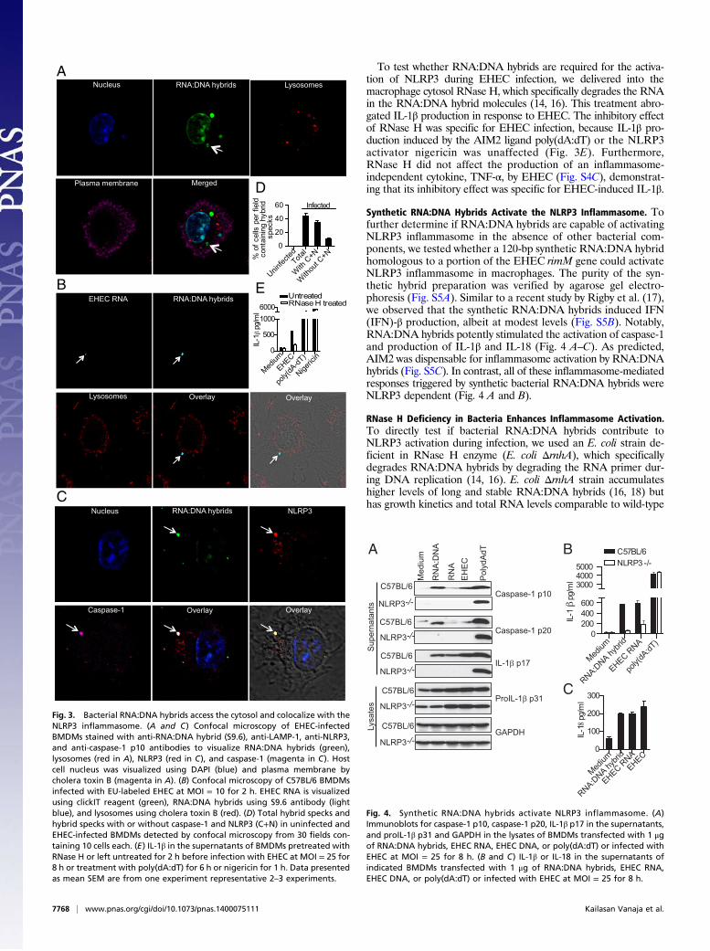
To test whether RNA:DNA hybrids are required for the activa-tion of NLRP3 during EHEC infection, we delivered into themacrophage cytosol RNase H, which specifically degrades the RNAin the RNA:DNA hybrid molecules (14, 16). This treatment abro-gated IL-1β production in response to EHEC. The inhibitory effectof RNase H was specific for EHEC infection, because IL-1β pro-duction induced by the AIM2 ligand poly(dA:dT) or the NLRP3activator nigericin was unaffected (Fig. 3E). Furthermore,RNase H did not affect the production of an inflammasome-independent cytokine, TNF-α, by EHEC (Fig. S4C), demonstrat-ing that its inhibitory effect was specific for EHEC-induced IL-1β.
Synthetic RNA:DNA Hybrids Activate the NLRP3 Inflammasome. Tofurther determine if RNA:DNA hybrids are capable of activatingNLRP3 inflammasome in the absence of other bacterial com-ponents, we tested whether a 120-bp synthetic RNA:DNA hybridhomologous to a portion of the EHEC rimM gene could activateNLRP3 inflammasome in macrophages. The purity of the syn-thetic hybrid preparation was verified by agarose gel electro-phoresis (Fig. S5A). Similar to a recent study by Rigby et al. (17),we observed that the synthetic RNA:DNA hybrids induced IFN(IFN)-β production, albeit at modest levels (Fig. S5B). Notably,RNA:DNA hybrids potently stimulated the activation of caspase-1and production of IL-1β and IL-18 (Fig. 4 A–C). As predicted,AIM2 was dispensable for inflammasome activation by RNA:DNAhybrids (Fig. S5C). In contrast, all of these inflammasome-mediatedresponses triggered by synthetic bacterial RNA:DNA hybrids wereNLRP3 dependent (Fig. 4 A and B).
RNase H Deficiency in Bacteria Enhances Inflammasome Activation.To directly test if bacterial RNA:DNA hybrids contribute toNLRP3 activation during infection, we used an E. coli strain de-ficient in RNase H enzyme (E. coli ΔrnhA), which specificallydegrades RNA:DNA hybrids by degrading the RNA primer dur-ing DNA replication (14, 16). E. coli ΔrnhA strain accumulateshigher levels of long and stable RNA:DNA hybrids (16, 18) buthas growth kinetics and total RNA levels comparable to wild-type
D
Uninfec
tedTota
l
With
C+N
With
out C
+N0
20
40
60
% o
f cel
ls p
er fi
eld
cont
aini
ng h
ybrid
spe
cks
Infected
Nucleus RNA:DNA hybrids Lysosomes
Plasma membrane Merged
Lysosomes
RNA:DNA hybrids EHEC RNA
Overlay Overlay
Caspase-1 Overlay Overlay
Nucleus RNA:DNA hybrids NLRP3
A
B
C
Medium
EHEC
poly(
dA-dT
)
Nigeric
in0
500
10006000
Untreated
IL-1
pg/
ml
RNase H treated
E
Fig. 3. Bacterial RNA:DNA hybrids access the cytosol and colocalize with theNLRP3 inflammasome. (A and C) Confocal microscopy of EHEC-infectedBMDMs stained with anti-RNA:DNA hybrid (S9.6), anti-LAMP-1, anti-NLRP3,and anti-caspase-1 p10 antibodies to visualize RNA:DNA hybrids (green),lysosomes (red in A), NLRP3 (red in C), and caspase-1 (magenta in C). Hostcell nucleus was visualized using DAPI (blue) and plasma membrane bycholera toxin B (magenta in A). (B) Confocal microscopy of C57BL/6 BMDMsinfected with EU-labeled EHEC at MOI = 10 for 2 h. EHEC RNA is visualizedusing clickIT reagent (green), RNA:DNA hybrids using S9.6 antibody (lightblue), and lysosomes using cholera toxin B (red). (D) Total hybrid specks andhybrid specks with or without caspase-1 and NLRP3 (C+N) in uninfected andEHEC-infected BMDMs detected by confocal microscopy from 30 fields con-taining 10 cells each. (E) IL-1β in the supernatants of BMDMs pretreated withRNase H or left untreated for 2 h before infection with EHEC at MOI = 25 for8 h or treatment with poly(dA:dT) for 6 h or nigericin for 1 h. Data presentedas mean SEM are from one experiment representative 2–3 experiments.
IL-1β p17
Caspase-1 p20
Sup
erna
tant
s
Med
ium
RN
A:D
NA
RN
AE
HE
C
Pol
ydA
dT
C57BL/6
NLRP3-/-
C57BL/6
C57BL/6
Caspase-1 p10
NLRP3-/-
NLRP3-/-
C57BL/6
NLRP3-/-ProIL-1β p31
C57BL/6
NLRP3-/-GAPDHLy
sate
s
A
Medium
RNA:DNA hy
brid
EHEC RNA
poly(
dA:dT
)0
200400600
300040005000
C57BL/6
IL-1
pg/
ml
NLRP3 -/-
Medium
RNA:DNA hy
brid
EHEC RNAEHEC
0
100
200
300
IL-18
pg/m
l
B
C
Fig. 4. Synthetic RNA:DNA hybrids activate NLRP3 inflammasome. (A)Immunoblots for caspase-1 p10, caspase-1 p20, IL-1β p17 in the supernatants,and proIL-1β p31 and GAPDH in the lysates of BMDMs transfected with 1 μgof RNA:DNA hybrids, EHEC RNA, EHEC DNA, or poly(dA:dT) or infected withEHEC at MOI = 25 for 8 h. (B and C) IL-1β or IL-18 in the supernatants ofindicated BMDMs transfected with 1 μg of RNA:DNA hybrids, EHEC RNA,EHEC DNA, or poly(dA:dT) or infected with EHEC at MOI = 25 for 8 h.
7768 | www.pnas.org/cgi/doi/10.1073/pnas.1400075111 Kailasan Vanaja et al.

(WT) E. coli (Fig. S6 A and B). In addition, the rnhA mutant waskilled within macrophages at a rate indistinguishable from WT(Fig. S6C). Supporting our hypothesis, the overproduction ofRNA:DNA hybrids by E. coli resulted in enhanced NLRP3 acti-vation as infection of macrophages with E. coli ΔrnhA resulted ina marked increase in the production of IL-1β and IL-18 as well ascleavage of caspase-1 and IL-1β relative to its parent strain (Fig.5 A–C). This enhanced macrophage response was specific forNLRP3 activation, because E. coli ΔrnhA induce inflammasome-independent cytokines such as TNF and IL-6 at levels similar toWT (Fig. S6 D and E). The elevated IL-1β response elicited byΔrnhA E. coli was completely dependent on ASC and NLRP3(Fig. 5D). Importantly, complementation of E. coli ΔrnhA withplasmid-encoded RNase H (“prnhA”) attenuated the increase incaspase-1 and IL-1β processing and the secretion of IL-1β andIL-18 to WT levels (Fig. 5 A–C). Although the number of hybridspecks visualized after infection with E. coli ΔrnhA was not sig-nificantly higher than upon infection by WT E. coli (Fig. S6F),
delivery of RNase H enzyme into ΔrnhA-infected reduced IL-1βproduction to levels equivalent to those of WT-infected mac-rophages, verifying that the enhanced inflammasome responseinduced by ΔrnhA E. coli was in fact due to higher levels ofRNA:DNA hybrids (Fig. 5E). E. coli ΔrnhA appeared to inducegreater than WT levels of oligomeric forms of ASC in an ASColigomerization assay, raising the possibility that increased levelsof RNA:DNA hybrids in the mutant may lead to increased in-flammasome assembly (Fig. S6G). The recognition of RNA:DNAduplexes was not exclusive to macrophages; upon infection ofdendritic cells, E. coli ΔrnhA induced greater inflammasomeactivation, characterized by IL-1β and IL-18 secretion and caspase-1and IL-1β maturation (Fig. 5 F–H). As above, genetic comple-mentation of E. coli ΔrnhA restored caspase-1 activation, IL-1βmaturation, and IL-1β as well as IL-18 secretion to WT levels(Fig. 5 F–H). Overall, the bacterial genetic complementation andcytosolic RNase H delivery approaches provide direct evidencefor RNA:DNA hybrids being bona fide bacterial componentscontributing to NLRP3 inflammasome activation during bacterialinfection.
DiscussionIL-1β is a potent inflammatory cytokine implicated in the out-come of EHEC disease (19). The production of bioactive IL-1β isregulated by cytosolic inflammasome complexes (6). In thisstudy, we report that EHEC-induced IL-1β maturation andproduction occurred independently of known virulence factors;rather, induction of IL-1β secretion required uptake of EHECand acidification of phagosomes, implicating EHEC breakdownproducts such as nucleic acids in this process.Microbial nucleic acids have long been known to trigger host
responses. Bacterial and viral RNA and DNA are potent acti-vators of type I IFNs and IL-1 cytokines. Bacterial mRNA hasbeen shown to induce NLRP3 inflammasome activation and IL-1responses (7, 8). Using both microscopic and biochemical ap-proaches, we have shown here that EHEC RNA was deliveredinto the host cell cytosol, colocalized with NLRP3 foci andtriggered inflammasome activation in an NLRP3-dependentmanner. Cytosolic delivery of exogenous RNase A attenuated theEHEC-driven IL-1β response.Another class of nucleic acid species abundant in bacteria are
RNA:DNA hybrids, which are formed during bacterial DNAreplication and transcription (13, 14). During bacterial DNAsynthesis, short RNA strands serve as primers for both leading andlagging strand (Okazaki fragments) synthesis, and RNA:DNAhybrids are formed every 100–200 nucleotides during laggingstrand synthesis (20, 21). RNA:DNA hybrids are also formedduring transcription, when a newly formed mRNA transcript ispaired with one strand of duplex DNA (13). Notably, RNA:DNAhybrids have a relatively stable structure and withstand acidicconditions up to pH 5.5 (22, 23). Several lines of evidence in thisstudy indicated that RNA:DNA hybrids of bacterial origin playa key role in NLRP3 inflammasome activation by EHEC. First,EHEC RNA:DNA hybrids were released into the cytosol ofinfected cells and colocalized with NLRP3 inflammasome. Sec-ond, depletion of EHEC RNA:DNA hybrids in the infected cellsby the cytosolic delivery of RNase H enzyme specifically inhibitedNLRP3 activation by EHEC. Third, an E. coli strain harboringhigher levels RNA:DNA hybrids due to the lack of RNase H in-duced NLRP3 activation more efficiently than WT bacteria. Thefindings of this study thus collectively identify bacterial RNA:DNAhybrids as a unique microbe-associated molecular pattern triggeringNLRP3 inflammasome activation. Interestingly, RNA:DNA hybridshave been recently shown to induce type I IFNs in a TLR9-MyD88-dependent manner (17).Collectively, our findings highlight an emerging theme in host−
pathogen interactions that the cytosolic immune surveillancemechanism is central to sensing extracellular or vacuolar bacteria
MediumW
TrnhA
rnhA/prnhA
0200400600800
IL-18
pg/m
l *
MediumW
TrnhA
rnhA/prnhA
0
5000
10000
15000
IL-1
pg/
ml *
A
IL-1β p17
Caspase-1 p10
Sup
erna
tant
s
Med
ium
WT
Δrnh
A
Δrnh
A/p
rnhA
NLRP3
Lysa
tes
GAPDH
IL-1β p17
Caspase-1 p20
Sup
erna
tant
sMed
ium
WT
ΔrnhA
ΔrnhA
/prnhA
ProIL-1β p31
Lysa
tes
GAPDH
Medium W
TrnhA
rnhA/prnhA
0
2000
4000
IL-1
pg/
ml
C57BL/6NLRP3 -/-C5
ASC-/-
F
MediumW
TrnhA
rnhA/prnhA
0
5000
10000
15000
20000
IL-1
pg/
ml *
D
MediumW
TrnhA
rnhA/prnhA
0
100
200
300
400
IL-18
pg/m
l *
B C
Medium W
Trnh
A
rnhA/prnhA
0500
100015002000 Untreated
IL-1
pg/
ml RNase H treated
E
G H
Fig. 5. RNase H deficiency in bacteria enhances inflammasome activation.(A, B, F, and G) IL-1β and IL-18 in the supernatants of C57BL/6 BMDMs (A andB) or BMDCs (F and G) infected with indicated strains of E. coli at MOI = 25for 8 h. (C and H) Immunoblots for cleaved caspase-1 and IL-1β in thesupernatants or NLRP3, proIL-1β p31 and GAPDH in the lysates of C57BL/6BMDMs (C) or BMDCs (H) infected with indicated E. coli strains for 8 h. (D) IL-1β in the supernatants of BMDMs indicated genotypes infected with in-dicated E. coli strains for 8 h. (E) IL-1β in the supernatants of BMDMs pre-treated with RNase H for 2 h or left untreated before infecting withindicated E. coli strains for 8 h. Asterisk (*) indicates P < 0.05 for WT vs.ΔrnhA or WT vs. ΔrnhA/prnhA as determined by one-way ANOVA followedby Bonferroni test. Data presented as mean SEM are from one experimentrepresentative 2–3 experiments.
Kailasan Vanaja et al. PNAS | May 27, 2014 | vol. 111 | no. 21 | 7769
IMMUNOLO
GY

through detection of microbial products that access the cytosol fromphagosomes. Although EHEC is excluded from the cytosol owingto its susceptibility to the microbicidal nature of phagolysosomes,cytosolic sensing of EHEC is yet an integral part of innate im-mune responses to EHEC. The inflammasome-dependent cyto-solic surveillance of EHEC relies heavily on the detection ofbacterial nucleic acids in the cytosol rather than on virulencefactors. Considering that nonpathogenic laboratory strains ofE. coli, as well as other pathogenic strains of E. coli such as ETEC,stimulated NLRP3 inflammasome activation, the recognition ofnucleic acids such as RNA or RNA:DNA hybrids may be ageneral mechanism by which the cytosolic innate immune systemrecognizes E. coli irrespective of virulence factor armament andpathogenic potential (8).NLR inflammasomes such as NLRP1b and NLRC4/IPAF are
activated by specific interaction with ligands such as anthraxtoxin and bacterial T3SS/flagellin, respectively (6). In contrast,NLRP3 activation is triggered by a plethora of stimuli of diversenature such as bacterial mRNA, pore forming toxins, crystallineparticles, and cellular perturbations such as ionic imbalance. Theprevailing consensus is that these disparate stimuli converge onone or more common elements or events upstream of NLRP3(6). Although bacterial RNA has been shown to activate theNLRP3 inflammasome, the mechanism by which this occurs isnot yet known. PKR represented an attractive candidate re-sponsible for RNA-mediated inflammasome activation, but incontrast to published work (9), we found that PKR-deficient cellswere not compromised for EHEC-induced NLRP3 inflamma-some signaling. Similar observations were reported by Nuñezand colleagues (24). Interestingly, a recent study (25) demon-strated that DHX33, a DExD/H-box RNA helicase family member,is a sensor for viral dsRNA and it activates inflammasome bydirectly binding to NLRP3. It is possible therefore that eitherDHX33 or a similar cytosolic sensor(s) for RNA and RNA:DNAhybrids activates NLRP3 in EHEC infection, or that NLRP3 maysense these bacterial nucleic acids directly.Our identification of bacterial RNA:DNA hybrids as activa-
tors of innate immunity has broad implications for microbialrecognition and host immunity. RNA:DNA hybrids are also
formed as replication intermediates in the life cycle of retro-viruses (26), and it is possible that such hybrids generated duringactive retroviral replication also contribute to NLRP3 inflam-masome-mediated immune responses. As with other nucleic acidsensing mechanisms, the ability of the innate immune system todetect RNA:DNA hybrids may also contribute to autoimmunedisease. Eukaryotic cells generate RNA:DNA hybrids duringtranscription, genomic replication, mitochondrial DNA replica-tion, and Ig class switching in B cells (13). Cytosolic exclusion ofRNA:DNA hybrids, like that of self-DNA, by nuclear and mi-tochondrial compartmentalization could be a regulatory mech-anism responsible for preventing self-RNA:DNA hybrids fromactivating cell-intrinsic innate immune responses. Degradation ofself-RNA:DNA by RNase H in the nucleus may provide an ad-ditional mechanism to avoid recognition of this product. Indeed,Aicardi Goutieres syndrome (AGS), a hereditary neurodegen-erative disorder characterized by autoimmune activation and typeI IFN production, has been associated with mutations in the hu-man RNASEH2B, which encodes an important subunit of theRNase H enzyme (27). Although the role of NLRP3 activationin AGS remains undefined, the identification of RNA:DNAhybrids as triggers of innate immunity provides new insightsinto cytosolic immune surveillance mechanisms with potentiallyimportant implications for host defense and autoimmunity.
Materials and MethodsA detailed materials and methods is given in SI Materials and Methods.
Briefly, BMDMs and BMDCs were generated fromWT and knockout mice andwere stimulated with bacteria (Table S1) or other activators as described before(28). Inflammasome responses were analyzed by ELISA and immunoblotting forIL-1β and caspase-1 or confocal microscopy as described before (28, 29).
ACKNOWLEDGMENTS. The authors thank Andrew Wright (Tufts University)for the E. coli RNase H mutant, Steve Leppla and Clinton Leysath (NationalInstitute of Allergy and Infectious Diseases) for anti-RNA:DNA hybrid anti-body, and members of the J.M.L. and K.A.F. labs for helpful discussions. Thiswork is supported by National Institutes of Health Grants AI46454 (to J.M.L.)and AI083713 (to K.A.F.) and a New England Regional Center for Excellence(NERCE) Post-Doctoral Fellowship Award U54 AI057159 (to S.K.V.).
1. Croxen MA, Finlay BB (2010) Molecular mechanisms of Escherichia coli pathogenicity.Nat Rev Microbiol 8(1):26–38.
2. King AJ (2002) Acute inflammation in the pathogenesis of hemolytic-uremic syn-drome. Kidney Int 61(4):1553–1564.
3. Keepers TR, Psotka MA, Gross LK, Obrig TG (2006) A murine model of HUS: Shigatoxin with lipopolysaccharide mimics the renal damage and physiologic response ofhuman disease. J Am Soc Nephrol 17(12):3404–3414.
4. Taneike I, Zhang HM, Wakisaka-Saito N, Yamamoto T (2002) Enterohemolysin operonof Shiga toxin-producing Escherichia coli: A virulence function of inflammatory cy-tokine production from human monocytes. FEBS Lett 524(1-3):219–224.
5. Yu HB, Finlay BB (2008) The caspase-1 inflammasome: A pilot of innate immune re-sponses. Cell Host Microbe 4(3):198–208.
6. Rathinam VA, Vanaja SK, Fitzgerald KA (2012) Regulation of inflammasome signaling.Nat Immunol 13(4):333–342.
7. Kanneganti TD, et al. (2006) Bacterial RNA and small antiviral compounds activatecaspase-1 through cryopyrin/Nalp3. Nature 440(7081):233–236.
8. Sander LE, et al. (2011) Detection of prokaryotic mRNA signifies microbial viabilityand promotes immunity. Nature 474(7351):385–389.
9. Lu B, et al. (2012) Novel role of PKR in inflammasome activation and HMGB1 release.Nature 488(7413):670–674.
10. Perna NT, et al. (2001) Genome sequence of enterohaemorrhagic Escherichia coliO157:H7. Nature 409(6819):529–533.
11. Ramsby M, Makowski G (2005) Proteomics Protocols Handbook (Springer, New York).12. Manji GA, et al. (2002) PYPAF1, a PYRIN-containing Apaf1-like protein that assembles
with ASC and regulates activation of NF-kappa B. J Biol Chem 277(13):11570–11575.13. Aguilera A, García-Muse T (2012) R loops: From transcription byproducts to threats to
genome stability. Mol Cell 46(2):115–124.14. Chapados BR, et al. (2001) Structural biochemistry of a type 2 RNase H: RNA primer
recognition and removal during DNA replication. J Mol Biol 307(2):541–556.15. Phillips DD, et al. (2013) The sub-nanomolar binding of DNA-RNA hybrids by the
single-chain Fv fragment of antibody S9.6. J Mol Recognit 26(8):376–381.16. Kogoma T (1986) RNase H-defective mutants of Escherichia coli. J Bacteriol 166(2):
361–363.
17. Rigby RE, et al. (2014) RNA:DNA hybrids are a novel molecular pattern sensed byTLR9. EMBO J 33(6):542–558.
18. von Meyenburg K, Boye E, Skarstad K, Koppes L, Kogoma T (1987) Mode of initiationof constitutive stable DNA replication in RNase H-defective mutants of Escherichia coliK-12. J Bacteriol 169(6):2650–2658.
19. Palermo M, et al. (2000) Pretreatment of mice with lipopolysaccharide (LPS) orIL-1beta exerts dose-dependent opposite effects on Shiga toxin-2 lethality. ClinExp Immunol 119(1):77–83.
20. Baker TA, Kornberg A (1988) Transcriptional activation of initiation of replication fromthe E. coli chromosomal origin: an RNA-DNA hybrid near oriC. Cell 55(1):113–123.
21. Masukata H, Tomizawa J (1990) A mechanism of formation of a persistent hybridbetween elongating RNA and template DNA. Cell 62(2):331–338.
22. Shaw NN, Xi H, Arya DP (2008) Molecular recognition of a DNA:RNA hybrid: Sub-nano-molar binding by a neomycin-methidium conjugate. Bioorg Med Chem Lett 18(14):4142–4145.
23. Chien YH, Davidson N (1978) RNA:DNA hybrids are more stable than DNA:DNA du-plexes in concentrated perchlorate and trichloroacetate solutions. Nucleic Acids Res5(5):1627–1637.
24. He Y, Franchi L, Núñez G (2013) The protein kinase PKR is critical for LPS-induced iNOSproduction but dispensable for inflammasome activation in macrophages. Eur J Im-munol 43(5):1147–1152.
25. Mitoma H, et al. (2013) The DHX33 RNA helicase senses cytosolic RNA and activatesthe NLRP3 inflammasome. Immunity 39(1):123–135.
26. Broecker F, Andrae K, Moelling K (2012) Premature activation of the HIV RNase H drivesthe virus into suicide: A novel microbicide? AIDS Res Hum Retroviruses 28(11):1397–1403.
27. Crow YJ, et al. (2006) Mutations in genes encoding ribonuclease H2 subunits causeAicardi-Goutières syndrome and mimic congenital viral brain infection. Nat Genet38(8):910–916.
28. Rathinam VA, et al. (2012) TRIF licenses caspase-11-dependent NLRP3 inflammasomeactivation by gram-negative bacteria. Cell 150(3):606–619.
29. Hornung V, et al. (2008) Silica crystals and aluminum salts activate the NALP3 in-flammasome through phagosomal destabilization. Nat Immunol 9(8):847–856.
7770 | www.pnas.org/cgi/doi/10.1073/pnas.1400075111 Kailasan Vanaja et al.