bacterial template synthesis of multifunctional
TRANSCRIPT

Research ArticleBacterial Template Synthesis of MultifunctionalNanospindles for Glutathione Detection and EnhancedCancer-Specific Chemo-Chemodynamic Therapy
Yan-Wen Bao, Xian-Wu Hua, Jia Zeng, and Fu-Gen Wu
State Key Laboratory of Bioelectronics, School of Biological Science and Medical Engineering, Southeast University, 2 Sipailou Road,Nanjing 210096, China
Correspondence should be addressed to Fu-Gen Wu; [email protected]
Received 15 August 2019; Accepted 12 February 2020; Published 26 March 2020
Copyright © 2020 Yan-Wen Bao et al. Exclusive Licensee Science and Technology Review Publishing House. Distributed under aCreative Commons Attribution License (CC BY 4.0).
Biological synthetic methods of nanoparticles have shown great advantages, such as environmental friendliness, low cost, mildreaction conditions, and enhanced biocompatibility and stability of products. Bacteria, as one of the most important livingorganisms, have been utilized as bioreducing nanofactories to biosynthesize many metal nanoparticles or compounds. Here,inspired by the disinfection process of KMnO4, we for the first time introduce bacteria as both the template and the reducing agentto construct a novel tumor microenvironment-responsive MnOx-based nanoplatform for biomedical applications in variousaspects. It is found that the bacterium/MnOx-based nanospindles (EM NSs) can efficiently encapsulate the chemotherapeutic agentdoxorubicin (DOX), leading to the fluorescence quenching of the drug. The as-formed DOX-loaded EM NSs (EMD NSs) areproven to be decomposed by glutathione (GSH) and can simultaneously release DOX and Mn2+ ions. The former can be utilizedfor sensitive fluorescence-based GSH sensing with a limit of detection as low as 0.28μM and selective cancer therapy, while thelatter plays important roles in GSH-activated magnetic resonance imaging and chemodynamic therapy. We also demonstrate thatthese nanospindles can generate oxygen in the presence of endogenous hydrogen peroxide to inhibit P-glycoprotein expressionunder hypoxia and can achieve excellent tumor eradication and tumor metastasis inhibition performance. Taken together, thiswork designs a multifunctional bacterially synthesized nanomissile for imaging-guided tumor-specific chemo-chemodynamiccombination therapy and will have implications for the design of microorganism-derived smart nanomedicines.
1. Introduction
Chemotherapy is one of the most common strategiesemployed in oncology [1]. Doxorubicin (DOX), a UnitedStates Food and Drug Administration-approved anthracy-cline antibiotic, is clinically used as a chemotherapeutic agentfor the treatment of various malignant tumors [2, 3].However, its short systemic circulation time, undesirable bio-distribution, and nonspecific toxicities to cancer/normal cellslead to not only relatively low anticancer efficacy but alsosevere side effects [4, 5]. To address these issues, variousnanocarriers such as liposomes [6], micelles [7], dendrimers[8], metal–organic frameworks (MOFs) [9], carbon nanoma-terials [10], and gold nanomaterials [11, 12] have beendesigned for the efficient delivery of DOX to cancer cells.Among them, conventional nanocarriers which tend to
release the encapsulated DOX molecules passively cannotrealize controllable drug release and fail to achieve satisfac-tory therapeutic efficacy [13]. Therefore, increasing effortshave been devoted to designing intelligent nanocarriers fordrug delivery, which can respond sensitively to internal/ex-ternal stimuli (e.g., pH, redox, enzymes, temperature, ultra-sound, and light) for on-demand imaging/therapy [14–18].
It is well known that tumor microenvironment (TME)has unique physiological characteristics such as acidic pH,hypoxia, and elevated levels of hydrogen peroxide (H2O2)and glutathione (GSH) [18, 19]. Hypoxia, resulting from animbalance between oxygen (O2) supply and consumption,can directly lead to the resistance of cancer cells to chemo-therapy, photodynamic therapy, or radiotherapy [20–24].Hypoxic adaptation is mainly mediated by a family of tran-scriptional regulators called hypoxia-inducible factors
AAASResearchVolume 2020, Article ID 9301215, 15 pageshttps://doi.org/10.34133/2020/9301215

(HIFs), including HIF-1α, which is involved in angiogenesis,invasion, and metastasis of cancer cells [25–27]. The upregu-lated HIF-1α has been reported to be associated with theoverexpression of P-glycoprotein (P-gp), an important pro-tein of the plasma membrane that recognizes external thera-peutic agents and pumps them out of the cells, resulting inpoor therapeutic outcomes [21, 28, 29]. The GSH level incancer cells/tissues is reported to be higher than that in nor-mal cells/tissues [16, 30–32], and hence, GSH may representan important signal molecule to improve the specificity ofcancer diagnosis and therapy. Besides, the overexpressionof GSH in cancer cells can consume the reactive oxygen spe-cies (ROS), thus protecting the cancer cells [33–35]. There-fore, it is highly desirable to develop a TME-responsivenanosystem with selective drug release, GSH depletion, andO2 generation capabilities for efficient cancer therapy.
In nanotechnology, there are mainly three approaches fornanoparticle fabrication including physical, chemical, andbiological methods. Compared with the former two tradi-tional approaches, biosynthesis, during which nanoparticlesare synthesized within the living or dead organisms, is mild,environmentally friendly, and usually inexpensive [36].Importantly, the biosynthesized nanoparticles exhibitenhanced biocompatibility and stability. Various biologicalorganisms have been utilized to biosynthesize many metalnanoparticles or compounds [37, 38]. For instance, Cuiet al. synthesized the fluorescent CdSe quantum dots usingliving yeast cells as a controllable biosynthesizer [39]. Tianet al. biofabricated the selenium-containing nanoparticles inShewanella oneidensisMR-1 cells using an extracellular elec-tron transfer regulation strategy [40]. Bacteria, as one of themost important living organisms, are considered as the opti-mal bioreducing nanofactories due to their simple culturingconditions and rapid propagation [41]. However, there hasbeen almost no research on the bacterium-biosynthesizedmanganese- (Mn-) containing nanoparticles.
In recent years, MnOx-based nanostructures haveattracted substantial attention as a unique type of TME-responsive theranostic agents [42–46]. Among them, manga-nese dioxide (MnO2) nanosheets have been reported to effi-ciently quench the fluorescence (FL) emission of adsorbedfluorophores, and the quenching effect will be reversed byGSH, which can be utilized for the quantification of GSHconcentration [47–50]. On the other hand, it has beenreported that MnOx-based nanostructures can be decom-posed by reaction with either H+ or GSH in the TME, gener-ating Mn2+ ions that can significantly enhance T1-magneticresonance (MR) imaging contrast for tumor-specific imagingand detection [48, 51]. Meanwhile, MnO2 nanostructures areable to trigger the decomposition of H2O2 existing in theTME into water and O2, so as to relieve tumor hypoxia[52]. Additionally, chemodynamic therapy (CDT), as anemerging therapeutic strategy using the in situ Fenton reac-tion or Fenton-like reaction to generate hydroxyl radical(⋅OH) to induce cell apoptosis, has attracted much attentiondue to its high selectivity and lethality activated by internalstimuli [53–57]. Apart from the commonly used iron ionsthat can induce Fenton reaction, other metal ions includingMn2+, Co2+, and Cu2+ also show Fenton-like activity [58,
59], which can be developed to be new types of chemody-namic nanoagents.
In this work, inspired by the disinfection process ofthe strong oxidizer potassium permanganate (KMnO4),during which MnO4
− is rapidly reduced to MnO2 accom-panied by the release of ROS that can kill bacteria [60], weput forward a novel biosynthetic strategy to construct aTME-responsive MnOx-based nanoplatform for cancertheranostics (Figure 1). Specifically, Escherichia coli (E. coli)bacterial cells are directly used as both the template and thereducing agent to react with the KMnO4 aqueous solutionvia ultrasonication at room temperature. The as-formednanomaterials possess the spindle-like morphology and areaccordingly named as E. coli-MnOx nanospindles (abbrevi-ated as EM NSs). After DOX loading, the resultant EMNSs–DOX (abbreviated as EMD NSs) show GSH-triggeredDOX release for rapid GSH detection and GSH depletion-enhanced chemotherapy, as well as Fenton-like Mn2+ deliv-ery for enhanced MR imaging and CDT. Meanwhile, EMDNSs can react with H2O2 to generate a significant amountof O2, overcoming hypoxia-induced chemoresistance. Lastbut not least, we also demonstrate that EMD NSs can notonly effectively eliminate the primary tumors but also inhibittumor metastasis. To the best of our knowledge, there havebeen few reports that realize simultaneously the TME-responsive chemotherapy and the Fenton-like metal-basedCDT. Besides, the present work may also represent the firstexample of fabricating microorganism-derived and GSH-responsive smart nanomedicines.
2. Results and Discussion
2.1. Preparation and Characterization of EM NSs and EMDNSs. EM NSs were facilely prepared via the room-temperature ultrasonication of KMnO4-treated E. coli cellsfollowed by purification. As it was observed that the suspen-sion turned from purple to red in the KMnO4 incubationprocess, we considered that the structure of the bacterial cellswas destroyed and the Mn element was completely or par-tially reduced. In the ultrasonication process, the Mn elementmight be further reduced and embedded in the oxidized bac-terial fragments to form the final nanoparticles. As shown inthe transmission electron microscopy (TEM) results(Figures 2(a) and 2(b), and Figure S1), the obtained EMNSs presented the spindle-like morphology with an averagelength of ~13.4nm and an average width of ~1.7 nm, whichwere both smaller than the average hydrodynamic size(~31.0nm, measured by dynamic light scattering (DLS); seeFigure S2) due to the presence of hydration layers. The rod-shaped E. coli bacterial cells were chosen because they areGram-negative bacteria with cell walls composed of thinpeptidoglycan layers, which may be destructed after KMnO4and ultrasonication treatments, resulting in the formation ofthe spindle-like nanoparticles. We also selected the rod-shaped Bacillus subtilis (B. subtilis) and sphericalStaphylococcus aureus (S. aureus) cells, the two kinds ofGram-positive bacteria with thick cell walls, and tried toprepare nanomaterials via similar KMnO4 andultrasonication treatments. Unfortunately, after these
2 Research

KMnO4
Ultra-sonication
E. coli bacteria EM NSs
DOX
GSH
EMD NSs DOX release/enhanced MR imaging
Off On
MR imaging
NS
FL imaging Intravenousinjection
GSH-responsive
release
NucleusGolgi apparatus
Lysosomes
MitochondriaER
GSH
H2O2O2 •OHMn2+
Figure 1: Schematic illustration of the fabrication of TME-responsive EMDNSs and their applications in rapid GSH detection and enhancedchemo-chemodynamic combination therapy for efficient tumor ablation.
50 nm
l w
Size (nm)
Freq
uenc
y (%
)
13.4 ± 2.520
1.7 ± 0.3
2𝜃 (degree)
Inte
nsity
(a.u
.)
Wavenumber (cm-1)
Abs
orba
nce (
a.u.)
𝜈(O–H)/𝜈(N–H)𝜈(C=O)/𝛿(H2O)
δ(N–H)/𝜈(C=C)
𝜈(C–H) 𝜈(C
–O)
𝜈(C
–N)
Atomic percent (%)C 1s 63.0O 1s 27.5N 1s 8.9Mn 2p 0.6
Mn 2p
O 1s
N 1s
C 1s
Binding energy (eV)
Inte
nsity
(a.u
.)
Binding energy (eV)290
6000
4000
2000
288 286 284 282
Inte
nsity
(a.u
.)
(a) (b) (c)
(d) (e) (f)
10
0
20
30 200
100
0
10
010 15 20 1 2 20 40 60
10001000200030004000 800
30000
400000.3
20000
10000
0600 400 200
C 1sFitted curveC=O (287.7 eV)C-O/C-N (286.0 eV)C-O/C=C (284.6 eV)
0.2
0.1
0.0
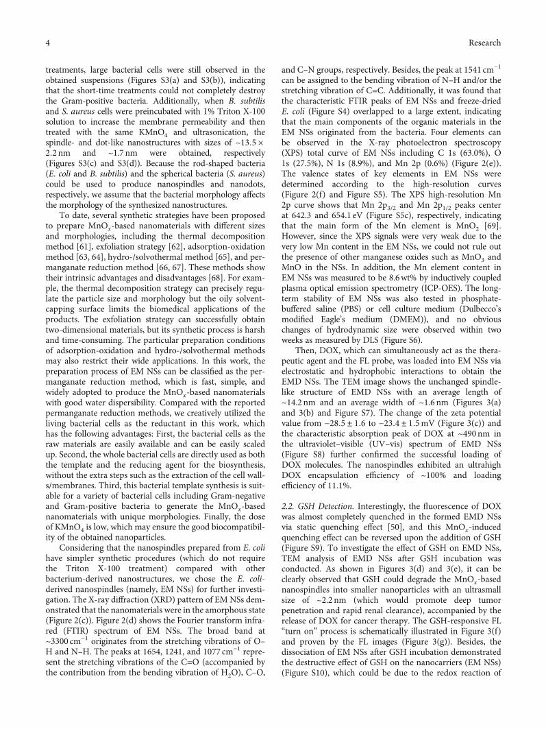
Figure 2: (a) TEM image of EM NSs. An enlarged version of this image was given in Figure S1 for clearer observation. (b) Correspondinglength (l) and width (w) histograms. (c) Powder XRD pattern of EM NSs. (d) FTIR spectrum of EM NSs. (e) XPS total and (f) high-resolution C 1s curves of EM NSs.
3Research
treatments, large bacterial cells were still observed in theobtained suspensions (Figures S3(a) and S3(b)), indicatingthat the short-time treatments could not completely destroythe Gram-positive bacteria. Additionally, when B. subtilisand S. aureus cells were preincubated with 1% Triton X-100solution to increase the membrane permeability and thentreated with the same KMnO4 and ultrasonication, thespindle- and dot-like nanostructures with sizes of ~13:5 ×2:2nm and ~1.7 nm were obtained, respectively(Figures S3(c) and S3(d)). Because the rod-shaped bacteria(E. coli and B. subtilis) and the spherical bacteria (S. aureus)could be used to produce nanospindles and nanodots,respectively, we assume that the bacterial morphology affectsthe morphology of the synthesized nanostructures.
To date, several synthetic strategies have been proposedto prepare MnOx-based nanomaterials with different sizesand morphologies, including the thermal decompositionmethod [61], exfoliation strategy [62], adsorption-oxidationmethod [63, 64], hydro-/solvothermal method [65], and per-manganate reduction method [66, 67]. These methods showtheir intrinsic advantages and disadvantages [68]. For exam-ple, the thermal decomposition strategy can precisely regu-late the particle size and morphology but the oily solvent-capping surface limits the biomedical applications of theproducts. The exfoliation strategy can successfully obtaintwo-dimensional materials, but its synthetic process is harshand time-consuming. The particular preparation conditionsof adsorption-oxidation and hydro-/solvothermal methodsmay also restrict their wide applications. In this work, thepreparation process of EM NSs can be classified as the per-manganate reduction method, which is fast, simple, andwidely adopted to produce the MnOx-based nanomaterialswith good water dispersibility. Compared with the reportedpermanganate reduction methods, we creatively utilized theliving bacterial cells as the reductant in this work, whichhas the following advantages: First, the bacterial cells as theraw materials are easily available and can be easily scaledup. Second, the whole bacterial cells are directly used as boththe template and the reducing agent for the biosynthesis,without the extra steps such as the extraction of the cell wall-s/membranes. Third, this bacterial template synthesis is suit-able for a variety of bacterial cells including Gram-negativeand Gram-positive bacteria to generate the MnOx-basednanomaterials with unique morphologies. Finally, the doseof KMnO4 is low, which may ensure the good biocompatibil-ity of the obtained nanoparticles.
Considering that the nanospindles prepared from E. colihave simpler synthetic procedures (which do not requirethe Triton X-100 treatment) compared with otherbacterium-derived nanostructures, we chose the E. coli-derived nanospindles (namely, EM NSs) for further investi-gation. The X-ray diffraction (XRD) pattern of EMNSs dem-onstrated that the nanomaterials were in the amorphous state(Figure 2(c)). Figure 2(d) shows the Fourier transform infra-red (FTIR) spectrum of EM NSs. The broad band at~3300 cm−1 originates from the stretching vibrations of O–H and N–H. The peaks at 1654, 1241, and 1077 cm−1 repre-sent the stretching vibrations of the C=O (accompanied bythe contribution from the bending vibration of H2O), C–O,
and C–N groups, respectively. Besides, the peak at 1541 cm−1
can be assigned to the bending vibration of N–H and/or thestretching vibration of C=C. Additionally, it was found thatthe characteristic FTIR peaks of EM NSs and freeze-driedE. coli (Figure S4) overlapped to a large extent, indicatingthat the main components of the organic materials in theEM NSs originated from the bacteria. Four elements canbe observed in the X-ray photoelectron spectroscopy(XPS) total curve of EM NSs including C 1s (63.0%), O1s (27.5%), N 1s (8.9%), and Mn 2p (0.6%) (Figure 2(e)).The valence states of key elements in EM NSs weredetermined according to the high-resolution curves(Figure 2(f) and Figure S5). The XPS high-resolution Mn2p curve shows that Mn 2p3/2 and Mn 2p1/2 peaks centerat 642.3 and 654.1 eV (Figure S5c), respectively, indicatingthat the main form of the Mn element is MnO2 [69].However, since the XPS signals were very weak due to thevery low Mn content in the EM NSs, we could not rule outthe presence of other manganese oxides such as MnO3 andMnO in the NSs. In addition, the Mn element content inEM NSs was measured to be 8.6wt% by inductively coupledplasma optical emission spectrometry (ICP-OES). The long-term stability of EM NSs was also tested in phosphate-buffered saline (PBS) or cell culture medium (Dulbecco’smodified Eagle’s medium (DMEM)), and no obviouschanges of hydrodynamic size were observed within twoweeks as measured by DLS (Figure S6).
Then, DOX, which can simultaneously act as the thera-peutic agent and the FL probe, was loaded into EM NSs viaelectrostatic and hydrophobic interactions to obtain theEMD NSs. The TEM image shows the unchanged spindle-like structure of EMD NSs with an average length of~14.2 nm and an average width of ~1.6 nm (Figures 3(a)and 3(b) and Figure S7). The change of the zeta potentialvalue from −28:5 ± 1:6 to −23:4 ± 1:5mV (Figure 3(c)) andthe characteristic absorption peak of DOX at ~490nm inthe ultraviolet–visible (UV–vis) spectrum of EMD NSs(Figure S8) further confirmed the successful loading ofDOX molecules. The nanospindles exhibited an ultrahighDOX encapsulation efficiency of ~100% and loadingefficiency of 11.1%.
2.2. GSH Detection. Interestingly, the fluorescence of DOXwas almost completely quenched in the formed EMD NSsvia static quenching effect [50], and this MnOx-inducedquenching effect can be reversed upon the addition of GSH(Figure S9). To investigate the effect of GSH on EMD NSs,TEM analysis of EMD NSs after GSH incubation wasconducted. As shown in Figures 3(d) and 3(e), it can beclearly observed that GSH could degrade the MnOx-basednanospindles into smaller nanoparticles with an ultrasmallsize of ~2.2 nm (which would promote deep tumorpenetration and rapid renal clearance), accompanied by therelease of DOX for cancer therapy. The GSH-responsive FL“turn on” process is schematically illustrated in Figure 3(f)and proven by the FL images (Figure 3(g)). Besides, thedissociation of EM NSs after GSH incubation demonstratedthe destructive effect of GSH on the nanocarriers (EM NSs)(Figure S10), which could be due to the redox reaction of
4 Research

Mn element in high valence states in EM NSs, as shown inequation (1). We demonstrated that the “turn on” FLresponse of EMD NSs could be used for the sensitive andselective GSH detection. As shown in Figure S11, after theaddition of varied concentrations of GSH, the FL intensityof EMD NSs increased under the 365nm UV irradiation.The GSH concentration-dependent FL enhancement ofEMD NSs was also shown in the FL spectra under 500nmexcitation (Figure 3(h)). Figure 3(i) shows the correspondingchange of FL intensity (ΔI/I0) at 555nm as a function ofGSH concentration ([GSH]). The relationship between ΔI/I0and [GSH] could be described by the following equationwhen [GSH] is in the range of 0–200μM: ΔI/I0 = 0:2396 +0:0470 × ½GSH� (R2 = 0:9925), with a detection limit of0.28μM (at a signal to noise ratio of 3). When [GSH] ishigher than 200μM, the ΔI/I0 value increased nonlinearlywith increasing [GSH]. Importantly, the response of EMD
NSs was highly selective towards GSH relative to othersubstances (common ions and small molecules) inphysiological solutions. As shown in Figure S12, the FLintensity of EMDNSs markedly increased after GSH addition,while other substances such as ions (including K+, Na+, Ca2+,and Mg2+) and small molecules (including glucose,dithiothreitol (DTT), and amino acids like L-glutamic acid(Glu), glycine (Gly), L-serine (Ser), L-arginine (Arg), L-lysine(Lys), D,L-homocysteine (Hcy), and L-cysteine (Cys)) did notcause distinct FL enhancements, which was ascribed tothe selective reducing ability of GSH towards the EMDNSs. It was worth noting that Hcy and Cys, the twobiological thiols that may severely affect GSH detection,did not interfere with the GSH detection in our system.
MnO2 + 2GSH + 2H+ ⟶Mn2+ + GSSG + 2H2O ð1Þ
50 nm
Zeta
pot
entia
l (m
V)
EM NSs
20
Free DOXEMD NSs
Low High
Free
DO
X
EM N
Ss
EM N
Ss+
GSH
EMD
NSs
EMD
NSs
+ G
SH
l w
14.2 ± 2.4 1.6 ± 0.2
30 nm
Size (nm)1.5
30
2.0 2.5 3.0 3.5
Freq
uenc
y (%
) d = 2.2 ± 0.3
2000150012001000700500400300200150
[GSH] (𝜇M)10070503020105210
Wavelength (nm)
FL in
tens
ity (a
.u.)
[GSH] (𝜇M)
∆I/I 0
y = 0.0470x + 0.2396R2 = 0.9925
(a) (b) (c)
(d) (e) (f)
GSH
EMD NSs DOXrelease
Off On
(g) (h) (i)
Size (nm)
Freq
uenc
y (%
) 20
10
0
20
40
010 15 20 1 2
10
0
–10
–20
–30
20
10
0
0520
300
560 600 640 680
15
5000
0
4
8
50 100 150 2001000 1500 2000
10
5
0
200
100
0
Figure 3: (a) TEM image of EMD NSs and (b) corresponding length (l) and width (w) histograms. An enlarged version of (a) was given inFigure S7 for clearer observation. (c) Zeta potential results of EM NSs, free DOX, and EMD NSs. (d) TEM image and (e) corresponding sizehistogram of EMD NSs after incubation with GSH (2.0mM). (f) Schematic illustration of EMD NSs before and after GSH incubation. (g) FLimages of different solutions/suspensions as indicated. Ex = 500 nm and Em = 620 nm. (h) GSH concentration- ([GSH]-) dependent FLspectra of EMD NSs. Ex = 500 nm. (i) Relationship between ΔI/I0 (at 555 nm) and [GSH] (0–2000μM). Inset: linear fitting result ofΔI/I0 versus [GSH] in the [GSH] range of 0–200μM.
5Research

2.3. In Vitro O2 Generation. Since MnO2 is known to be anexcellent catalyst to trigger the decomposition of H2O2 intoH2O and O2, we tested whether EM NSs and EMD NSscan generate O2 in vitro upon reaction with H2O2 at
endogenous levels (e.g., 100μM). As shown in Figure 4(a),both EM NSs and EMD NSs could trigger rapid O2 gener-ation within the H2O2 solution, demonstrating the catalyticproperty of EM NSs and EMD NSs to react with H2O2 and
Free DOX EMD NSsFree DOX EMD NSs
Mn concentration (mM)
1/T
1 (s–1
)
r1 = 1.79 mM–1 s–1r 1 = 10.82 mM
–1 s–1 0 0.15 0.3 0.6 0.9 1.2
GSH(–)
GSH(+)
Mn concentration (mM)
Time (s)0
12 15
10
5
050 100 150 200 250 0.0 0.4 0.8
4000.0
0.5
1.0
1.5
2.0
500 600 700 800
1.2
Diss
olve
d O
2 (m
g L–1
)
MCF-10A
8
MCF-7
DO
XH
oech
st33
342
Mer
ged
FL in
tens
ity (1
04 )
Time (h)
Am
ount
of
DO
X re
leas
e (%
)
1: MB (H2O) 2: MB + H2O23: MB + H2O2 + Mn2+ (H2O)4: MB + H2O2 + Mn2+
5: MB + H2O2 + Mn2+ + GSH6: MB + H2O2 + EM NSs7: MB + H2O2 + EM NSs + GSH
Wavelength (nm)A
bsor
banc
e (a.u
.)
1 2 3 4 5 6 7
EMD NSs + GSHEMD NSs
H2O2EM NSs + H2O2EMD NSs + H2O2
pH 7.4
pH 7.4, with GSHpH 6.5, with GSH
pH 6.5
⁎⁎⁎
⁎⁎⁎
⁎⁎⁎
11
10
9
8
7
123
456
7
6
4
2
0302520151050
0
20
40
60
80
100Co
ntro
l
Free
DO
X
EM N
Ss
EMD
NSs
MCF-7MCF-10A
⁎⁎
(b) (c)
(d)
(a)
(e)
(h)(g)(f)
Figure 4: (a) O2 generation in H2O2 solutions (100 μM) after addition of EM or EMD NSs (Mn concentration: 0.6mM). (b) Linear fitting of1/T1 versus Mn concentration for EMD NS suspensions and (c) T1-weighted MR images of EMD NS suspensions at varied Mnconcentrations without (−) or with (+) GSH (2.0mM). (d) Photographs and (e) UV–vis absorption spectra of MB-containing solutionsafter different treatments as indicated. The groups 1 and 3 were prepared in H2O, and the groups 2 and 4–7 were prepared inNa2CO3/NaHCO3 buffer solutions. (f) DOX release kinetics of EMD NSs in the presence or absence of GSH (2.0mM) in solutions withdifferent pH values (7.4 or 6.5). (g) Confocal images of MCF-7 cancer cells and MCF-10A normal cells after treatment with free DOX orEMD NSs followed by Hoechst 33342 staining (to visualize nuclei). (h) FL intensities of cells treated without (control) or with free DOX,EM NSs, and EMD NSs. ∗∗P < 0:01 and ∗∗∗P < 0:001.
6 Research

trigger its decomposition for O2 generation (equation (2)),relieving the tumor hypoxia.
MnO2 + H2O2 + 2H+ ⟶Mn2+ + O2 + 2H2O ð2Þ
2.4. MR Imaging, GSH Depletion, and Drug ReleaseBehavior. Because the Mn element is an excellent T1-short-ening agent in MR imaging, we investigated the ability ofEMD NSs as GSH-activated MR imaging contrast agents.The in vitro longitudinal relaxation rate (1/T1) as a func-tion of the Mn concentration of EMD NSs before and afterGSH reduction was evaluated (Figures 4(b) and 4(c)). AfterGSH addition, the produced Mn2+ ions exhibited a strongerenhancement in T1-weighted MR imaging than the EMDNSs. Longitudinal relaxivity r1, obtained by measuring therelaxation rate as a function of Mn concentration, exhibiteda 6-fold enhancement when EMD NSs were reduced toMn2+ ions by GSH.
On the other hand, we selected methylene blue (MB), adye that can be degraded by ⋅OH, as an indicator of ⋅OH gen-eration. By comparing the results of groups 1–4 inFigures 4(d) and 4(e), we inferred that H2O2, Mn2+ ions,and carbonate/bicarbonate (CO3
2−/HCO3−) buffer solution
are all necessary to fade the blue MB solution, which was inaccordance with the previous work [58]. Additionally, theresults of groups 4 and 5 indicated that the added GSH elim-inated the formed ⋅OH and inhibited the MB degradation.Based on the above results, we investigated the chemody-namic activity of our nanoagents. When Mn2+ ions werereplaced by EM NSs (group 6), partial MB moleculesdegraded due to the catalytic activity of EM NSs. Moreimportantly, in the presence of GSH, EM NSs exhibited aneven higher MB degradation efficiency (group 7), whichcould be explained by the GSH-stimulatedMn2+ release fromEM NSs accompanied with GSH depletion. These resultsindicated that the EMD NSs consisting of DOX and EMNSs can serve as both chemotherapeutic and chemodynamicnanoagents.
Furthermore, the in vitro drug release properties ofEMD NSs in the presence or absence of GSH displayedthe sustained and GSH-responsive DOX release fromEMD NSs (Figure 4(f)), which is determined by the elec-trostatic interaction between positively charged DOX mol-ecules and negatively charged EM NSs. Besides, at 24 h,the slightly higher cumulative drug release of EMD NSsat pH 6.5 than that at pH 7.4 might be ascribed to thegradual protonation of DOX under the acidic condition.Based on the overexpressed chemical entities in the cancerTME, such as intracellular H2O2 and GSH, the aboveH2O2-/GSH-responsive characters of EMD NSs are veryimportant in selective cancer therapy.
2.5. Different Effects of EMD NSs on Cancer/Normal Cells.Considering that the GSH concentration in cancer cells ismuch higher than that in normal cells [30, 31], which was alsoverified by our cellular total GSH assay results (Table S1), wewould like to investigate whether the EMD NSs respond tothe GSH in the two types of cells differently. In the cancer(human breast cancer MCF-7)/normal (human normal
mammary epithelial MCF-10A) cell experiments(Figures 4(g) and 4(h)), it can be seen that after treatmentwith EMD NSs, stronger FL signals were observed in the cellnuclei of MCF-7 cells compared with those in the cell nucleiof MCF-10A cells. These results revealed the excellentcancer/normal cell discrimination ability of EMD NSs, whichmainly resulted from their GSH-responsive DOX release.Based on the different DOX release behaviors of EMD NSsin cancer/normal cells, we further assessed the anticancerefficacy of EMD NSs towards three different cancer/normalcell groups by a 3-(4,5-dimethylthiazol-2-thiazolyl)-2,5-diphenyl-2H-tetrazolium bromide (MTT) assay. As shownin Figure 5(a), EMD NSs exhibited a strongerinhibitory/killing effect towards cancerous MCF-7 cells thannormal MCF-10A cells. According to the dose-dependentcell growth curves, the half maximal inhibitoryconcentration (IC50) value of EMD NSs for MCF-7 cells wascalculated to be 2:6 ± 0:1μgmL−1, which was much smallerthan that (30:9 ± 2:8μgmL−1) for MCF-10A cells(Figure 5(d)). Additionally, we also observed the strongercancer cell inhibitory/killing effects in two other pairs ofcancer/normal cells (Figures 5(b) and 5(c)), and thedifferences were quantified by the IC50 values of EMD NSs:human lung cancer A549 cells: 14:1 ± 1:2μgmL−1,normal human pulmonary alveolar epithelial HPAEpiCcells: 84:7 ± 21:4μgmL−1, human liver cancer HepG2cells: 0:7 ± 0:1μgmL−1, and human normal liver L02cells: 2:5 ± 0:2μgmL−1 (Figures 5(e) and 5(f)).
2.6. In Vitro Anticancer Treatment and Mechanism. On thebasis of the above results, we eventually selected MCF-7 cellsfor our subsequent experiments to study the anticancer effectsof EMD NSs. First, the cytotoxicity of the TME-responsivenanocarriers, EMNSs, was proven to be low by theMTT assay(Figure 5(g)). The viability of MCF-7 cells was concentration-dependent and remained >80% in the EM NS concentrationrange of 0–160μgmL−1. Importantly, using a total GSH detec-tion kit, it was observed that the total GSH content decreasedfrom 1.75mM in the cells without treatment (Table S1) to0.58mM in the EM NS-treated cells, suggesting the GSH-depleting ability of EM NSs to enhance the CDT efficiency.Next, at low DOX concentrations (0–5μgmL−1), EMD NSsexhibited the similar cytotoxicity against MCF-7 cellscompared with free DOX (Figure 5(h)). We also carried outadditional experiments to explore the anticancermechanisms of EMD NSs. After the MCF-7 cells weretreated with free DOX, EM NSs, or EMD NSs for 24h, thecaspase-3 levels are shown in Figure 5(i). The results showedthat the apoptosis level induced by EMD NSs was higherthan that in the control group and close to that in the “freeDOX” group at the same DOX concentration. According tothe cell apoptosis detection results (Figure 5(j)), comparedwith the control group, the cells treated with EMD NSsshowed significant early apoptosis (39.25%) and lateapoptosis/necrosis (54.29%), which were similar to thosetreated with free DOX (early apoptosis: 43.14%; lateapoptosis/necrosis: 49.52%). Additionally, the evidentincrease of G2/M phase in cell cycle distribution wasobserved in both “free DOX” and “EMD NSs” groups
7Research

⁎⁎⁎
0
100
0.5 1 2 5 10 15 20[DOX] (𝜇g mL–1)
Cel
l via
bilit
y (%
)IC
50 d
ose (
𝜇g
mL–1
)
MCF-7
30
MCF-10A
⁎⁎
IC50 d
ose (μg
mL–1
)
A549 HPAEpiC
⁎⁎⁎
IC50 d
ose (μg
mL–1
)
HepG2 L02(cancer cells) (normal cells) (cancer cells) (normal cells) (cancer cells) (normal cells)
⁎
⁎⁎⁎⁎⁎
Cas
pase
-3 ac
tivity
(fold
incr
ease
)
Frac
tion
of ea
chph
ase o
f cel
l cyc
le (%
)
Control
Free DOX
EM NSs
EMD NSs
100
80
Cou
nt
FITC-H103.5 105 106 107 107.6
0.56% 2.89%
96.23% 0.32%
0.80% 49.52%
6.54% 43.14%
1.00% 5.80%
91.95% 1.25%
0.86% 54.29%
5.60% 39.25%
Control Free DOX EM NSs EMD NSs
PI
Annexin V-FITC
[EM NSs] ( 𝜇g mL –1) 0 4 8 16 40 80 120 160
⁎⁎⁎⁎⁎⁎⁎⁎⁎
0 0.5 1 2 5 10 15 20[DOX] (𝜇g mL –1)
(l)(k)
(j)
(m)
(h)(g) (i)
(b)(a) (c)
(e)(d) (f)
P-gp
GAPDH
Nor
mox
ia
Hyp
oxia
Hyp
oxia
+EM
NSs
Ratio
of P
-gp
to G
APD
H
80
60
40
20
00
100
0.5 1 2 5 10 15 20[DOX] (𝜇g mL–1)
Cel
l via
bilit
y (%
)
80
60
40
20
0
MCF-10AMCF-7
HPAEpiCA549
0
100
0.5 1 2 5 10 15 20[DOX] (𝜇g mL–1)
Cel
l via
bilit
y (%
)
80
60
40
20
0
L02HepG2
20
10
0
90
120
60
30
0
2
3
1
0
2
4
0
Free DOXEMD NSs
Cel
l via
bilit
y (%
)
100
80
60
40
20
0
Cel
l via
bilit
y (%
)
100
80
60
40
20
0
Con
trol
Free
DO
X
EM N
Ss
EMD
NSs
Con
trol
Free
DO
X
EM N
Ss
EMD
NSs
100
0
80
4020
60
0.0
0.30.20.1
G2/MSG0/G1
60
40
20
0
Figure 5: Dose-dependent growth inhibition curves of (a) MCF-7/MCF-10A, (b) A549/HPAEpiC, and (c) HepG2/L02 cells after exposure toEMD NSs at varied DOX concentrations for 24 h. (d–f) Corresponding IC50 values of EMD NSs for the above three pairs of cancer/normalcells. (g) Viabilities of MCF-7 cells treated with varied concentrations of EM NSs. (h) Concentration-dependent cytotoxicities of free DOXand EMD NSs against MCF-7 cells. DOX and EM NS concentrations were abbreviated as [DOX] and [EM NSs], respectively, in (a–c, g,and h). Effects of free DOX, EM NSs, and EMD NSs on (i) caspase-3 level, (j) apoptosis/necrosis, and (k) cell cycle distribution of MCF-7cells. (l) Flow cytometry analysis results of cellular ROS levels after various treatments as indicated. The cells without any treatment wereset as the control group. (m) Images and corresponding statistical histogram of western blot results of P-gp expression (withglyceraldehyde-3-phosphate dehydrogenase (GAPDH) as the loading control) of normoxic cells, hypoxic cells, and hypoxic cells treatedwith EM NSs for 24 h. ∗P < 0:05, ∗∗P < 0:01, and ∗∗∗P < 0:001.
8 Research

(Figure 5(k)). Therefore, the anticancer effect of EMD NSsmainly resulted from the released DOX molecules(evidenced by the confocal imaging results in Figure 4(g)) inthis TME-responsive nanosystem. However, at high DOXconcentrations (10–20μgmL−1), EMD NSs showed therelatively higher cytotoxicity than free DOX (Figure 5(h)), sowe assumed that some other factors may also play roles inthe EMD NS-induced cell death. Although EM NSs causednegligible changes in the cell viabilities, the substantialaccumulation of ROS was observed in the EM NS- or EMDNS-treated cells (Figure 5(l) and Figure S13), which could beattributed to the conversion of endogenous H2O2 to highlyreactive ⋅OH (one of the most harmful forms of ROS)induced by the Mn2+-mediated Fenton-like reaction [58].Importantly, based on the in vitro EM NS-assisted O2generation results (Figure 4(a)), EM NSs showed greatpromise in downregulating the P-gp expression byincreasing the O2 level under hypoxia. Therefore, P-gpexpression was also measured by western blot analysis afterdifferent treatments. As shown in Figure 5(m), the untreatedhypoxic MCF-7 cells exhibited high P-gp expression, whichcould promote the efflux of DOX. After 24h incubation ofEM NSs, the P-gp expression in hypoxic cells decreased to arelatively low level similar to the expression level innormoxic cells, which indicated that the EM NS-basednanoplatform could inhibit P-gp expression under hypoxia,possessing the potential to reduce drug efflux and thusovercome hypoxia-induced chemoresistance. Taken together,all these observations proved that EMD NSs can efficientlykill cancer cells by the GSH consumption- and O2generation-enhanced chemo-chemodynamic combinationtherapy.
2.7. In Vivo Tumor Imaging and Chemo-ChemodynamicCancer Therapy. Motivated by the in vitro results, we thenperformed in vivo theranostic experiments using tumor-bearing mice via the tail vein injection with free DOX orEMD NSs. It was observed that free DOX was distributednonspecifically and rapidly to various organs and tissuesafter intravenous (i.v.) administration, leading to a rapidelimination from the circulation (Figure 6(a)). Meanwhile,the FL intensity of EMD NSs in the tumor areas showed acontinuous increase and reached the maximum at 12 hpostinjection followed by a slight decrease, whereas freeDOX exhibited very weak FL signals within the wholeobservation time period (Figure 6(b)). Such a direct FLcomparison verifies the excellent tumor targeting abilityof EMD NSs, which can be attributed to their proper size(~14nm in length) that endows them with excellent pas-sive tumor targeting ability via the enhanced permeabilityand retention effect. Importantly, since the tumor tissuesshow at least 4-fold higher concentrations of GSH levelscompared with normal tissues in the tumor-bearing mice[70], the GSH-triggered “off–on” DOX release of EMDNSs was demonstrated in vivo. As shown in Figure S14,a healthy tissue and a tumor tissue in a mouse wereinjected with EMD NSs, respectively. The negligible FLintensity in the normal tissue and the strong FL intensityin the tumor region provided a solid proof for the
selective tumor imaging capability of the nanoagents.Apart from the in vivo FL imaging, the MR images of atumor-bearing mouse were also acquired before and afterthe i.v. injection of EMD NSs. As shown in Figure 6(c),a detectable T1-weighted contrast enhancement wasobserved in the tumor area at 12 h post i.v. injection,which proved that the obtained EMD NSs can be usedas promising contrast agents for MR imaging application.In addition, the mice intravenously injected with EMDNSs were sacrificed at 1, 3, and 7d postinjection,respectively. Ex vivo images and quantified FL intensitiesrevealed that EMD NSs were distributed mainly intumors and partially in livers and kidneys at 1 and 3dpostinjection and were still retained in the tumor at 7 dpostinjection without evident distributions in otherorgans (Figures 6(d) and 6(e)), which indicated theirexcellent tumor accumulation, prolonged tumor retention,and good biosafety. Besides, the FL intensities in thekidneys were higher than those in the livers, implyingthat EMD NSs might be mainly captured andmetabolized by kidneys due to their small size.
Subsequently, inspired by the high tumor accumulationof EMD NSs, the in vivo tumor suppression activity of theTME-responsive nanoagents was evaluated using tumor-bearing mice. As shown in Figures 7(a) and 7(c), the tumorgrowth of the mice intravenously injected with EMD NSswas almost completely suppressed during the period of ourobservation (14 days), whereas free DOX did not exhibitnoticeable therapeutic efficacy. Additionally, the tumorgrowth of mice treated with EM NSs was also not effectivelysuppressed, which might be due to the inadequate admin-istration of EM NSs. The hematoxylin and eosin- (H&E-)stained tumor slices from the EMD NS-treated groupshowed that most cells were severely damaged after treat-ment (Figure 7(b)), further confirming the satisfactoryin vivo therapeutic outcome of the nanoagents. Further-more, the tumor lung metastasis was examined on the30th day after various treatments. As revealed by the pho-tographs of India ink-stained whole lungs and the H&E-stained lung slices (Figures 7(e) and 7(f)), a large numberof tumor nodules were observed in the lungs of untreated,DOX-treated, and EM NS-treated tumor-bearing mice,while no obvious lung metastasis was observed in theEMD NS-treated group, suggesting the excellent inhibitoryeffect of EMD NSs on tumor metastasis.
We also investigated the potential side effects of EMDNSs. First, no evident body weight loss was observed duringthe treatments (Figure 7(d)), suggesting the negligible toxiceffect of EMD NSs on the animals. Next, according to the tis-sue slices from the EMD NS-treated mice at 14 d postinjec-tion (Figure S15), compared with the control group, noobvious cell apoptosis/necrosis was observed in the majororgans, indicating the good biosafety of the nanoagents,which may result from the efficient excretion of thenanoagents by the mice. Besides, even at the high DOXconcentration of 200μgmL−1, EMD NSs did not inducehemolysis (Figures S16(a) and S16(b)), and noabnormalities in the detected blood indices were seen in theblood routine analysis for the mice sacrificed on the 14th
9Research

day after EMDNS treatment (Figures S16(c–n)), showing thegood hemocompatibility of EMDNSs. Collectively, the aboveresults demonstrated that EMD NSs can be used as safeantitumor nanoagents.
3. Conclusion
In summary, we have developed a TME-responsive, bacte-rium-derived, andMnOx-based nanoplatformwhich can effi-ciently load the anticancer drug DOX for GSH detection aswell as chemo-chemodynamic combination therapy. Interest-ingly, during the synthesis of the MnOx-based smart drugcarrier, the E. coli bacterial cells were used as both the tem-plate and the reducing agent. The as-obtained nanocarriersexhibit a spindle-like morphology with an average size of13:4 × 1:7nm, good colloidal stability, and multipleresponses. The DOX-loaded bacterium/MnOx-based nanos-pindles with high drug encapsulation efficiency (~100.0%)
and drug loading efficiency (11.1%) show the followingadvantages: (1) The loaded DOX can be used not only as ananticancer drug but also as a fluorescent probe. Based on theFL quenching of DOX by EM NSs and the FL recovery trig-gered by GSH, EMDNSs can serve as a selective and sensitiveGSH sensor. (2) The GSH-triggered DOX release and Mn2+
production can realize the FL and T1-weighted MR imaging,respectively, which is highly beneficial for the accuracy andeffectiveness of cancer treatment. (3) EMD NSs exhibit bothFenton-like Mn2+ delivery and GSH depletion properties forenhanced CDT. (4) The in situ generated O2 resulting fromEM NS-triggered decomposition of tumor endogenousH2O2 can inhibit the expression of P-gp, which is crucial forovercoming hypoxia-associated chemoresistance. (5) Sincethe DOX molecule can be replaced by other small moleculedrugs (such as photosensitizers, photothermal molecules,and other chemotherapeutics), the MnOx-based nanoplat-form holds great potential for universal drug delivery and
Spleen
Free
DO
XEM
D N
SsPre 1 h 3 h 6 h 9 h 12 h 18 h 24 h H
igh
Low
1 d 3 d 7 dHeart
Liver
Lung
Kidneys
Tumor
Hig
hLo
w
Hea
rt
Live
r
Sple
en
Lung
Kidn
eys
Tum
or
FL in
tens
ity (1
08 )Befo
rein
ject
ion
Afte
rin
ject
ion
(a) (b)
(e)(d)(c)
30
25
20
15
10
5
0
1 d3 d7 d
⁎⁎⁎⁎⁎⁎
⁎⁎⁎
⁎⁎
⁎⁎
⁎⁎⁎⁎
Pre
FL in
tens
ity (1
08 )
Time (h)
20
105 15 20 250
EMD NSsFree DOX
16
12
8
4
⁎⁎⁎
Figure 6: (a) In vivo FL images of tumor-bearing mice before (“Pre”) and after i.v. injection of free DOX or EMD NSs (DOX concentration:2mgkg−1) for different time periods. The blue dotted circles mark the tumor regions. (b) Corresponding FL intensities in the tumor regionsin (a). (c) In vivo MR images of a tumor-bearing mouse before and after i.v. injection of EMD NSs (DOX concentration: 2mgkg−1) for 12h.The red dotted circles mark the tumor regions. (d) Ex vivo FL images of major organs and tumor tissues of mice sacrificed at 1, 3, and 7 dpost i.v. injection of EMD NSs (DOX concentration: 2mgkg−1). (e) Corresponding FL intensities in organs or tissues in (d). ∗∗P < 0:01 and∗∗∗P < 0:001.
10 Research

various disease treatments. Importantly, the present workdevelops a facile bacterium-based biosynthetic strategy toconstruct smart drug nanocarriers for loading chemothera-peutics and realizing chemo-chemodynamic combinationtherapy, which we believe may find applications in researchareas such as nanomedicine and biosensing.
4. Materials and Methods
4.1. Materials. E. coli and S. aureus were purchased fromChina Center of Industrial Culture Collection (CICC, Bei-jing, China). KMnO4 was purchased from Chengdu KelongChemical Inc. (Chengdu, China). Doxorubicin hydrochlo-
ride (DOX) was purchased from Beijing Huafeng UnitedTechnology Co., Ltd. (China). L-Glutathione (GSH) waspurchased from Sigma-Aldrich (St. Louis, MO). Dialysismembranes (Spectra/Por® 6, regenerated cellulose) with theMWCO of 10 kDa were purchased from SpectrumLabs, Inc.(Rancho Dominguez, CA). All solutions/suspensions wereprepared with deionized (DI) water (18.2MΩ·cm) purifiedby a Milli-Q system (Millipore, Billerica, MA). The AnnexinV-Fluorescein Isothiocyanate (FITC) Apoptosis DetectionKit containing annexin V/propidium iodide (PI), Caspase-3Colorimetric Assay Kit, Annexin V-FITC Apoptosis Detec-tion Kit, Cell Cycle Detection Kit, and ROS Detection Kitwere all purchased from Nanjing KeyGen Biotech Co. Ltd.
Time (day)
Rela
tive t
umor
volu
me (
V/V
0)
Time (day)
Body
wei
ght (
g)Control Free DOX EM NSs EMD NSs
(a) (c)
(d)(b)
(e)
(f)
Healthy Control Free DOX EMD NSsEM NSs
Control Free DOX EM NSs EMD NSs
Day
1D
ay 3
Day
7D
ay 1
4
ControlFree DOX EMD NSs
EM NSs
ControlFree DOX EMD NSs
EM NSs
0
20
2 4 6 8 10 12 14
16
12
8
4
0
0
16
2 4 6 8 10 12 14 16
12
8
4
0
⁎⁎⁎
Figure 7: (a) Photographs of differently treated mice taken at 1, 3, 7, and 14 d. (b) H&E-stained slices of tumor issues taken on the 14th day.Scale bar = 100μm. (c) Relative tumor growth curves and (d) body weight curves in the three groups. ∗∗∗P < 0:001. (e) Photographs of Indiaink-filled whole lungs from mice in various groups and (f) corresponding H&E-stained lung slices. The red dashed circles mark the tumormetastasis sites. Scale bar = 100 μm.
11Research

4.2. Instruments and Characterization. The TEM experimentwas performed using a transmission electron microscope(JEM-2100, JEOL Ltd., Japan). Before measurements, thesamples were prepared by dropping the sample suspensionsonto the ultrathin carbon-coated copper grids and air-drying. The size distribution was obtained by counting atleast 100 particles from one TEM image per sample usingan image analysis software (Nano Measurer 1.2). For XRDanalysis, the fine EM NS powder was first obtained byfreeze-drying and grinding. Then, the powder was placedinto the sample holder and gently pressed using a glass slideto make the surface smooth. XRD patterns were thenrecorded on an XRD system (D8 Discover, Bruker, Ger-many). The FTIR spectra of the freeze-dried EM NS powderand bacteria (E. coli) were collected with a Nicolet iS50 FTIRspectrometer (Thermo Fisher Scientific Co., Waltham,MA, USA). Additionally, the EM NS powder was sprin-kled onto the surface of sticky carbon conductive tapefor XPS analysis using an X-ray photoelectron spectrome-ter (Quantera 2000, Ulvac-Phi Inc., Japan). UV–vis spectrawere obtained using a UV–vis spectrophotometer (UV-2600, Shimadzu, Japan). The FL spectra were obtainedon a spectrophotometer (RF-5301PC, Shimadzu, Japan).The hydrodynamic diameter and zeta potential measure-ments were conducted on a Zetasizer instrument (NanoZS, Malvern Instruments, UK). To determine the Mn con-centration in the EM NS suspension, the sample was firstdigested with aqua regia at 60°C overnight and thendiluted with DI water for analysis using ICP-OES (Per-kin-Elmer Optima 2100DV, USA).
4.3. Animal Model. Female BALB/c nude mice were obtainedfrom Yangzhou University Medical Center (Yangzhou,China) and used under protocols approved by the SoutheastUniversity Laboratory Animal Center. 100μL of PBS con-taining 4T1 cells (2 × 10⁶) was subcutaneously inoculatedonto the back of each mouse to build the xenograft tumormodel.
Conflicts of Interest
The authors declare no competing financial interests.
Authors’ Contributions
Yan-Wen Bao and Xian-Wu Hua contributed equally to thiswork. Yan-Wen Bao, Xian-Wu Hua, and Fu-Gen Wudesigned the experiments. Yan-Wen Bao, Xian-Wu Hua,and Jia Zeng performed the experiments. Yan-Wen Bao,Xian-Wu Hua, and Fu-Gen Wu analyzed the data and wrotethe manuscript. Fu-Gen Wu supervised the project.
Acknowledgments
This work was supported by the National Natural ScienceFoundation of China (21673037), the Postgraduate Researchand Practice Innovation Program of Jiangsu Province(KYCX18_0159), and the Fundamental Research Funds forthe Central Universities.
Supplementary Materials
Table S1: GSH levels in various cancer/normal cells. FigureS1: enlarged version of Figure 2(a) for clearer observation.Figure S2: size distribution of EM NSs dispersed in PBS asmeasured by DLS. Figure S3: TEM images of the suspensionsobtained by (a) B. subtilis and (b) S. aureus cells treated withKMnO4 and ultrasonication. TEM images of the suspensionsobtained by (c) B. subtilis and (d) S. aureus cells that werefirst preincubated with 1% Triton X-100 solution overnightand then treated with KMnO4 and ultrasonication. FigureS4: FTIR spectrum of the freeze-dried E. coli bacteria. FigureS5: XPS high-resolution (a) O 1s, (b) N 1s, and (c) Mn 2pcurves. Figure S6: hydrodynamic size changes of EM NSsdispersed in PBS or DMEM for a duration of two weeks asmeasured by DLS. Figure S7: enlarged version ofFigure 3(a) for clearer observation. Figure S8: UV–visabsorption spectra of aqueous solutions containing freeDOX, EM NSs, EMD NSs, or EMD NSs+GSH. Figure S9:FL emission spectra of aqueous solutions containing freeDOX, EM NSs, EMD NSs, or EMD NSs + GSH. Ex = 500nm. Figure S10: TEM image (left) and corresponding sizehistogram (right) of EM NSs after treatment with GSH(2.0mM). Figure S11: photographs of EMD NS suspensionsafter mixing with various concentrations of GSH under aUV lamp (365 nm excitation). The final GSH concentrationsare indicated in the figure. Figure S12: FL responses of EMDNSs to different guest molecules. The concentrations of allguests were 1.0mM. Figure S13: flow cytometry-based FLanalysis of cellular ROS levels after various treatments asindicated. ∗∗∗P < 0:001. Figure S14: in vivo FL image(inset) and corresponding FL analysis result of a mouseinjected with EMD NSs. “1” and “2” indicate the normaland tumor areas injected with EMD NSs, respectively. ∗∗
P < 0:01. Figure S15: histological analysis of major organscollected from mice sacrificed on the 14th day post i.v.injection with PBS (control), free DOX, EM NSs, orEMD NSs. Scale bar = 100μm. Figure S16: (a) photographsof RBC suspensions after hemolysis assay. (b) Hemolysis per-centages of RBCs treated with PBS (negative control) and Tri-ton X-100 (positive control), or varied concentrations of freeDOX or EMD NSs. DOX concentration was abbreviated as[DOX] in (a and b). (c–n) Blood routine analysis results ofmice after treatment with PBS (control), free DOX, or EMDNSs. The blood indexes including WBC, LYM, MONO,NEUT, RBC, HGB, HCT, MCV, MCH, MCHC, PLT, andMPV indicate numbers of white blood cells, lymphocytes,monocytes, neutrophilic granulocytes, red blood cells, concen-tration of hemoglobin, hematocrit, mean corpuscular volume,mean corpuscular hemoglobin, mean corpuscular hemoglobinconcentration, platelet count, and mean platelet volume,respectively. (Supplementary Materials)
References
[1] Y. Li, Y. Chen, W. Pan et al., “Nanocarriers with multi-lockedDNA valves targeting intracellular tumor-related mRNAs forcontrolled drug release,” Nanoscale, vol. 9, no. 44, pp. 17318–17324, 2017.
12 Research

[2] C. Du, D. Deng, L. Shan et al., “A pH-sensitive doxorubicinprodrug based on folate-conjugated BSA for tumor-targeteddrug delivery,” Biomaterials, vol. 34, no. 12, pp. 3087–3097,2013.
[3] C. Yu, Q. Zhou, F. Xiao et al., “Enhancing doxorubicin deliverytoward tumor by hydroxyethyl starch-g-polylactide partnernanocarriers,” ACS Applied Materials & Interfaces, vol. 9,no. 12, pp. 10481–10493, 2017.
[4] P. Huang, W. Wang, J. Zhou et al., “Amphiphilic polyelectro-lyte/prodrug nanoparticles constructed by synergetic electro-static and hydrophobic interactions with cooperative pH-sensitivity for controlled doxorubicin delivery,” ACS AppliedMaterials & Interfaces, vol. 7, no. 11, pp. 6340–6350, 2015.
[5] L. Lin, X. Liang, Y. Xu, Y. Yang, X. Li, and Z. Dai, “Doxorubi-cin and indocyanine green loaded hybrid bicelles for fluores-cence imaging guided synergetic chemo/photothermaltherapy,” Bioconjugate Chemistry, vol. 28, no. 9, pp. 2410–2419, 2017.
[6] J. Szebeni, P. Bedőcs, R. Urbanics et al., “Prevention of infusionreactions to PEGylated liposomal doxorubicin via tachyphy-laxis induction by placebo vesicles: a porcine model,” Journalof Controlled Release, vol. 160, no. 2, pp. 382–387, 2012.
[7] D. Sun, J. Ding, C. Xiao, J. Chen, X. Zhuang, and X. Chen,“Preclinical evaluation of antitumor activity of acid-sensitivePEGylated doxorubicin,” ACS Applied Materials & Interfaces,vol. 6, no. 23, pp. 21202–21214, 2014.
[8] C. C. Lee, E. R. Gillies, M. E. Fox et al., “A single dose ofdoxorubicin-functionalized bow-tie dendrimer cures micebearing C-26 colon carcinomas,” Proceedings of the NationalAcademy of Sciences of the United States of America, vol. 103,no. 45, pp. 16649–16654, 2006.
[9] W.-H. Chen, W.-C. Liao, Y. S. Sohn et al., “Stimuli-responsivenucleic acid-based polyacrylamide hydrogel-coated metal–organic framework nanoparticles for controlled drug release,”Advanced Functional Materials, vol. 28, no. 8, article 1705137,2018.
[10] Z. Liu, A. C. Fan, K. Rakhra et al., “Supramolecular stacking ofdoxorubicin on carbon nanotubes for in vivo cancer therapy,”Angewandte Chemie International Edition, vol. 48, no. 41,pp. 7668–7672, 2009.
[11] M. S. Yavuz, Y. Cheng, J. Chen et al., “Gold nanocages coveredby smart polymers for controlled release with near-infraredlight,” Nature Materials, vol. 8, no. 12, pp. 935–939, 2009.
[12] T. Cui, J.-J. Liang, H. Chen et al., “Performance ofdoxorubicin-conjugated gold nanoparticles: regulation of druglocation,” ACS Applied Materials & Interfaces, vol. 9, no. 10,pp. 8569–8580, 2017.
[13] M. Zan, J. Li, M. Huang et al., “Near-infrared light-triggereddrug release nanogels for combined photothermal-chemotherapy of cancer,” Biomaterials Science, vol. 3, no. 7,pp. 1147–1156, 2015.
[14] L. Feng, K. Li, X. Shi, M. Gao, J. Liu, and Z. Liu, “Smart pH-responsive nanocarriers based on nano-graphene oxide forcombined chemo- and photothermal therapy overcomingdrug resistance,” Advanced Healthcare Materials, vol. 3,no. 8, pp. 1261–1271, 2014.
[15] W. Fan, W. Bu, B. Shen et al., “Intelligent MnO2 nanosheetsanchored with upconversion nanoprobes for concurrentpH-/H2O2-responsive UCL imaging and oxygen-elevatedsynergetic therapy,” Advanced Materials, vol. 27, no. 28,pp. 4155–4161, 2015.
[16] R. Cheng, F. Meng, C. Deng, H.-A. Klok, and Z. Zhong, “Dualand multi-stimuli responsive polymeric nanoparticles for pro-grammed site- specific drug delivery,” Biomaterials, vol. 34,no. 14, pp. 3647–3657, 2013.
[17] S. Wang, G. Yu, Z. Wang et al., “Hierarchical tumormicroenvironment-responsive nanomedicine for pro-grammed delivery of chemotherapeutics,” Advanced Mate-rials, vol. 30, no. 40, article 1803926, 2018.
[18] X. Hu, F. Li, S. Wang, F. Xia, and D. Ling, “Biological stimulus-driven assembly/disassembly of functional nanoparticles fortargeted delivery, controlled activation, and bioelimination,”Advanced Healthcare Materials, vol. 7, no. 20, article1800359, 2018.
[19] P. Prasad, C. R. Gordijo, A. Z. Abbasi et al., “Multifunctionalalbumin-MnO2 nanoparticles modulate solid tumor microen-vironment by attenuating hypoxia, acidosis, vascular endothe-lial growth factor and enhance radiation response,” ACS Nano,vol. 8, no. 4, pp. 3202–3212, 2014.
[20] P. Vaupel and L. Harrison, “Tumor hypoxia: causative factors,compensatory mechanisms, and cellular response,” The Oncol-ogist, vol. 9, no. S5, pp. 4–9, 2004.
[21] H. Tian, Z. Luo, L. Liu et al., “Cancer cell membrane-biomimetic oxygen nanocarrier for breaking hypoxia-induced chemoresistance,” Advanced Functional Materials,vol. 27, no. 38, article 1703197, 2017.
[22] J. Xu, W. Han, P. Yang et al., “Tumor microenvironment-responsive mesoporous MnO2-coated upconversion nano-platform for self-enhanced tumor theranostics,” AdvancedFunctional Materials, vol. 28, no. 36, article 1803804,2018.
[23] J.-n. Liu, W. Bu, and J. Shi, “Chemical design and synthesisof functionalized probes for imaging and treating tumorhypoxia,” Chemical Reviews, vol. 117, no. 9, pp. 6160–6224, 2017.
[24] T. Huang, M. Zhao, Q. Yu et al., “De novo design of polymericcarrier to photothermally release singlet oxygen for hypoxictumor treatment,” Research, vol. 2019, article 9269081, 11pages, 2019.
[25] G. L. Semenza, “Oxygen sensing, hypoxia-inducible factors,and disease pathophysiology,” Annual Review of Pathology:Mechanisms of Disease, vol. 9, no. 1, pp. 47–71, 2014.
[26] D. M. Gilkes, G. L. Semenza, and D. Wirtz, “Hypoxia and theextracellular matrix: drivers of tumour metastasis,” NatureReviews Cancer, vol. 14, no. 6, pp. 430–439, 2014.
[27] K. Balamurugan, “HIF-1 at the crossroads of hypoxia, inflam-mation, and cancer,” International Journal of Cancer, vol. 138,no. 5, pp. 1058–1066, 2016.
[28] D. Samanta, D. M. Gilkes, P. Chaturvedi, L. Xiang, and G. L.Semenza, “Hypoxia-inducible factors are required for chemo-therapy resistance of breast cancer stem cells,” Proceedings ofthe National Academy of Sciences of the United States of Amer-ica, vol. 111, no. 50, pp. E5429–E5438, 2014.
[29] Z. Luo, H. Tian, L. Liu et al., “Tumor-targeted hybrid proteinoxygen carrier to simultaneously enhance hypoxia-dampenedchemotherapy and photodynamic therapy at a single dose,”Theranostics, vol. 8, no. 13, pp. 3584–3596, 2018.
[30] M. H. Lee, Z. Yang, C. W. Lim et al., “Disulfide-cleavage-trig-gered chemosensors and their biological applications,” Chem-ical Reviews, vol. 113, no. 7, pp. 5071–5109, 2013.
[31] F. Gong, L. Cheng, N. Yang et al., “Bimetallic oxideMnMoOX-Nanorods for in vivo photoacoustic imaging of GSH and
13Research

tumor-specific photothermal therapy,” Nano Letters, vol. 18,no. 9, pp. 6037–6044, 2018.
[32] R. Cheng, F. Feng, F. Meng, C. Deng, J. Feijen, and Z. Zhong,“Glutathione-responsive nano-vehicles as a promising plat-form for targeted intracellular drug and gene delivery,” Journalof Controlled Release, vol. 152, no. 1, pp. 2–12, 2011.
[33] X. Zhang, X. Chen, Y.-W. Jiang et al., “Glutathione-depletinggold nanoclusters for enhanced cancer radiotherapy throughsynergistic external and internal regulations,” ACS AppliedMaterials & Interfaces, vol. 10, no. 13, pp. 10601–10606, 2018.
[34] H. Fan, G. Yan, Z. Zhao et al., “A smart photosensitizer–man-ganese dioxide nanosystem for enhanced photodynamic ther-apy by reducing glutathione levels in cancer cells,”Angewandte Chemie International Edition, vol. 55, no. 18,pp. 5477–5482, 2016.
[35] E. Ju, K. Dong, Z. Chen et al., “Copper(II)–graphitic carbonnitride triggered synergy: improved ROS generation andreduced glutathione levels for enhanced photodynamic ther-apy,” Angewandte Chemie International Edition, vol. 55,no. 38, pp. 11467–11471, 2016.
[36] X. Fang, Y. Wang, Z. Wang, Z. Jiang, and M. Dong, “Microor-ganism assisted synthesized nanoparticles for catalytic applica-tions,” Energies, vol. 12, no. 1, p. 190, 2019.
[37] A. Schröfel, G. Kratošová, I. Šafařík, M. Šafaříková, I. Raška,and L. M. Shor, “Applications of biosynthesized metallic nano-particles—a review,” Acta Biomaterialia, vol. 10, no. 10,pp. 4023–4042, 2014.
[38] N. I. Hulkoti and T. C. Taranath, “Biosynthesis of nanoparti-cles using microbes—a review,” Colloids and Surfaces B: Bioin-terfaces, vol. 121, pp. 474–483, 2014.
[39] R. Cui, H.-H. Liu, H.-Y. Xie et al., “Living yeast cells as a con-trollable biosynthesizer for fluorescent quantum dots,”Advanced Functional Materials, vol. 19, no. 15, pp. 2359–2364, 2009.
[40] L.-J. Tian, W.-W. Li, T.-T. Zhu et al., “Directed biofabricationof nanoparticles through regulating extracellular electrontransfer,” Journal of the American Chemical Society, vol. 139,no. 35, pp. 12149–12152, 2017.
[41] M. Hasan, I. Ullah, H. Zulfiqar et al., “Biological entities aschemical reactors for synthesis of nanomaterials: progress,challenges and future perspective,” Materials Today Chemis-try, vol. 8, pp. 13–28, 2018.
[42] Q. Chen, L. Feng, J. Liu et al., “Intelligent Albumin-MnO2Nanoparticles as pH-/H2O2-responsive dissociable nanocar-riers tomodulate tumorhypoxia for effective combination ther-apy,” AdvancedMaterials, vol. 28, no. 33, pp. 7129–7136, 2016.
[43] G. Yang, L. Xu, Y. Chao et al., “Hollow MnO2 as a tumor-microenvironment-responsive biodegradable nano-platformfor combination therapy favoring antitumor immuneresponses,”Nature Communications, vol. 8, no. 1, p. 902, 2017.
[44] W. Zhu, Z. Dong, T. Fu et al., “Modulation of hypoxia in solidtumor microenvironment with MnO2 Nanoparticles toenhance photodynamic therapy,” Advanced Functional Mate-rials, vol. 26, no. 30, pp. 5490–5498, 2016.
[45] J. Liu, Q. Chen, W. Zhu et al., “Nanoscale-coordination-poly-mer-shelled manganese dioxide composite nanoparticles: amultistage redox/pH/H2O2-responsive cancer theranosticnanoplatform,” Advanced Functional Materials, vol. 27,no. 10, article 1605926, 2017.
[46] C. Chu, H. Lin, H. Liu et al., “Tumor microenvironment-triggered supramolecular system as an in situ nanotheranostic
generator for cancer phototherapy,” Advanced Materials,vol. 29, no. 23, article 1605928, 2017.
[47] R. Deng, X. Xie, M. Vendrell, Y.-T. Chang, and X. Liu, “Intra-cellular glutathione detection using MnO2-nanosheet-modi-fied upconversion nanoparticles,” Journal of the AmericanChemical Society, vol. 133, no. 50, pp. 20168–20171, 2011.
[48] Z. Zhao, H. Fan, G. Zhou et al., “Activatable fluorescence/MRIbimodal platform for tumor cell imaging via MnO2 nanosheet−aptamer nanoprobe,” Journal of the American Chemical Soci-ety, vol. 136, no. 32, pp. 11220–11223, 2014.
[49] Z.-Z. Dong, L. Lu, C.-N. Ko et al., “AMnO2 nanosheet-assistedGSH detection platform using an iridium(III) complex as aswitch-on luminescent probe,” Nanoscale, vol. 9, no. 14,pp. 4677–4682, 2017.
[50] W. Zhai, C. Wang, P. Yu, Y. Wang, and L. Mao, “Single-layerMnO2 nanosheets suppressed fluorescence of 7-hydroxycou-marin: mechanistic study and application for sensitive sensingof ascorbic acid in vivo,” Analytical Chemistry, vol. 86, no. 24,pp. 12206–12213, 2014.
[51] P. Mi, D. Kokuryo, H. Cabral et al., “A pH-activatable nano-particle with signal-amplification capabilities for non-invasive imaging of tumour malignancy,” Nature Nanotech-nology, vol. 11, no. 8, pp. 724–730, 2016.
[52] C. R. Gordijo, A. Z. Abbasi, M. A. Amini et al., “Design ofhybrid MnO2-polymer-lipid nanoparticles with tunable oxy-gen generation rates and tumor accumulation for cancer treat-ment,” Advanced Functional Materials, vol. 25, no. 12,pp. 1858–1872, 2015.
[53] Z. Tang, Y. Liu, M. He, and W. Bu, “Chemodynamic therapy:tumour microenvironment-mediated Fenton and Fenton-likereactions,” Angewandte Chemie International Edition,vol. 58, no. 4, pp. 946–956, 2019.
[54] C. Zhang, W. Bu, D. Ni et al., “Synthesis of iron nanometallicglasses and their application in cancer therapy by a localizedFenton reaction,” Angewandte Chemie International Edition,vol. 55, no. 6, pp. 2101–2106, 2016.
[55] G. Guan, X. Wang, B. Li et al., “‘Transformed’ Fe3S4 tetragonalnanosheets: a high-efficiency and body-clearable agent formagnetic resonance imaging guided photothermal and che-modynamic synergistic therapy,” Nanoscale, vol. 10, no. 37,pp. 17902–17911, 2018.
[56] Y. Liu,W. Zhen, L. Jin et al., “All-in-one theranostic nanoagentwith enhanced reactive oxygen species generation and modu-lating tumor microenvironment ability for effective tumoreradication,” ACS Nano, vol. 12, no. 5, pp. 4886–4893, 2018.
[57] Y. Hu, T. Lv, Y. Ma et al., “Nanoscale coordination polymersfor synergistic NO and chemodynamic therapy of liver can-cer,” Nano Letters, vol. 19, no. 4, pp. 2731–2738, 2019.
[58] L.-S. Lin, J. Song, L. Song et al., “Simultaneous Fenton-like iondelivery and glutathione depletion by MnO2-based nanoagentto enhance chemodynamic therapy,” Angewandte ChemieInternational Edition, vol. 57, no. 18, pp. 4902–4906, 2018.
[59] A. D. Bokare and W. Choi, “Review of iron-free Fenton-likesystems for activating H2O2 in advanced oxidation processes,”Journal of Hazardous Materials, vol. 275, pp. 121–135, 2014.
[60] J. Pan, Y. Wang, H. Pan et al., “Mimicking drug–substrateinteraction: a smart bioinspired technology for the fabricationof theranostic nanoprobes,” Advanced Functional Materials,vol. 27, no. 3, article 1603440, 2017.
[61] J. Rockenberger, E. C. Scher, and A. P. Alivisatos, “A new non-hydrolytic single–precursor approach to surfactant–capped
14 Research

nanocrystals of transition metal oxides,” Journal of the Ameri-can Chemical Society, vol. 121, no. 49, pp. 11595–11596, 1999.
[62] J. Li, D. Li, R. Yuan, and Y. Xiang, “Biodegradable MnO2Nanosheet-Mediated signal amplification in living cellsenables sensitive detection of down-regulated intracellularmicroRNA,” ACS Applied Materials & Interfaces, vol. 9,no. 7, pp. 5717–5724, 2017.
[63] W. Tang, W. Fan, W. Zhang et al., “Wet/sono-chemical syn-thesis of enzymatic two-dimensional MnO2 Nanosheets forsynergistic catalysis-enhanced phototheranostics,” AdvancedMaterials, vol. 31, no. 19, article 1900401, 2019.
[64] W. Zhen, W. Wang, Z. Ma et al., “Multienzyme-mimickingnanocomposite for tumor phototheranostics and normal cellprotection,” ChemNanoMat, vol. 5, no. 1, pp. 101–109, 2019.
[65] Q. Jia, J. Ge, W. Liu et al., “A magnetofluorescent carbon dotassembly as an acidic H2O2-driven oxygenerator to regulatetumor hypoxia for simultaneous bimodal imaging andenhanced photodynamic therapy,” Advanced Materials,vol. 30, no. 13, article 1706090, 2018.
[66] J. Liu, P. Du, T. Liu et al., “A black phosphorus/manganesedioxide nanoplatform: oxygen self-supply monitoring, photo-dynamic therapy enhancement and feedback,” Biomaterials,vol. 192, pp. 179–188, 2019.
[67] X. Jing, Y. Xu, D. Liu et al., “Intelligent nanoflowers: a fulltumor microenvironment-responsive multimodal cancer ther-anostic nanoplatform,” Nanoscale, vol. 11, no. 33, pp. 15508–15518, 2019.
[68] B. Ding, P. Zheng, P. Ma, and J. Lin, “Manganese oxide nano-materials: synthesis, properties, and theranostic applications,”Advanced Materials, vol. 32, no. 10, article 1905823, 2020.
[69] C. D. Wagner, W. M. Riggs, L. E. Davis, and J. F. Moulder,Handbook of X-Ray Photoelectron Spectroscopy, Perkin-Elmer, 1979.
[70] P. Kuppusamy, H. Li, G. Ilangovan et al., “Noninvasive imag-ing of tumor redox status and its modification by tissue gluta-thione levels,” Cancer Research, vol. 62, no. 1, pp. 307–312,2002.
15Research