bacteriology development of an immunoassay using
TRANSCRIPT

325
*Correspondence to: Lai, I-H.: [email protected], [email protected]©2020 The Japanese Society of Veterinary Science
This is an open-access article distributed under the terms of the Creative Commons Attribution Non-Commercial No Derivatives (by-nc-nd) License. (CC-BY-NC-ND 4.0: https://creativecommons.org/licenses/by-nc-nd/4.0/)
FULL PAPERBacteriology
Development of an immunoassay using recombinant outer membrane protein A and flagellin for diagnosis of goats with melioidosisShu-Hwae LEE1), Yi-Ping LU2), Wen-Ling SHIH3), Ching-Dong CHANG4), Yang-Chang TU2) and I-Hsiang LAI5)*
1)School of Veterinary Medicine, National Taiwan University, Taipei, 10617, Taiwan2)Animal Health Research Institute, Council of Agriculture, Executive Yuan, New Taipei City, 25158, Taiwan3)Department of Biological Science and Technology, National Pingtung University of Science and Technology,
Pingtung, 91201, Taiwan4)Department of Veterinary Medicine, National Pingtung University of Science and Technology, Pingtung,
91201, Taiwan5)General Research Service Center, National Pingtung University of Science and Technology, Pingtung, 91201,
Taiwan
ABSTRACT. Among domestic animals, melioidosis is one of the most common diseases reported in goat, sheep, and swine. To evaluate the specific antibodies in goats with melioidosis, we developed a serology test using recombinant outer membrane protein A (OmpA) and flagellin (FliC) of Burkholderia pseudomallei as antigens. DNA corresponding to each antigen was cloned into a pET32a vector and expressed in Escherichia coli. Essentially, the recombinant OmpA and FliC were expressed in a soluble form that could be isolated with 95% homogeneity. Both recombinants could be recognized by rabbit antibodies prepared against heat-inactivated B. pseudomallei (1:1,000) on a Western blot. Subsequently, we demonstrated that both recombinants could capture the antibodies present in goat with naturally occurring melioidosis (optimized titer 1:40) while not cross-reacting with the serum samples of goats naturally infected by Corynebacterium pseudotuberculosis or Staphylococcus aureus. Finally, an enzyme-linked immunosorbent assay (ELISA) using 20 goat serum samples without melioidosis and 10 goat serum samples with melioidosis demonstrated that the infected group has significantly higher antibody titer levels than the normal group (P<0.001) when using either OmpA or FliC as an antigen. However, the sensitivity (100%) of the assay using OmpA was superior to that (90%) from using FliC. Serological tests that are commonly used often rely on antigens from crude cell extracts, which pose risks for laboratory-acquired infections and inconsistency in their preparation; however, use of recombinant OmpA is safe; it can potentially be used as a reagent in testing for goat melioidosis.
KEY WORDS: enzyme-linked immunosorbent assay (ELISA), flagellin, melioidosis, outer membrane protein A, serology test
Melioidosis is an infectious and emerging disease caused by Burkholderia pseudomallei that results in high domestic animal mortality [22]. The disease occurs mainly in Southeast Asia, Northern Australia, and other tropical areas of the globe [26]. Livestock most commonly affected include goats, sheep, and swine [16, 17, 21]. On rare occasions, the bacteria could also infect cattle and horses [16, 22, 27]. It is contagious to humans and poses a major threat to public health [19, 26]. Infection is often associated with suppurative or caseous lesions, comprising a mixed purulent and granulomatous response in any part of a body organ, including the lungs, spleen, liver, and associated lymph nodes [22, 26].
The definitive diagnosis of melioidosis in an animal is made by the direct isolation of B. pseudomallei from lesions and discharges; however, cultures often require 48 hr or longer to be detected following incubation [20, 24]. Several serological techniques, including indirect hemagglutination (IHA) [6, 10, 12], immunofluorescent assay [4, 9], and enzyme-linked immunosorbent assay (ELISA) [5, 23] have been developed using either B. pseudomallei antigens or specific antibodies. However,
Received: 11 February 2019Accepted: 17 January 2020Advanced Epub: 29 January 2020
J. Vet. Med. Sci. 82(3): 325–332, 2020doi: 10.1292/jvms.19-0072

S.-H. LEE ET AL.
326doi: 10.1292/jvms.19-0072
such assays employed crude whole-cell preparations or extracts from the bacteria, which could increase the potential risks of laboratory-acquired infections. Several studies have demonstrated that vaccines prepared from outer membrane protein A (OmpA) and flagellin (FliC) of B. pseudomallei exhibited improved immunogenicity and protected against B. pseudomallei infection in mice [7, 11, 18]. The results further suggest that OmpA or FliC are immunodominant and could be used as potential probes for the early-phase diagnosis of melioidosis in humans [1, 2, 8, 14]. Allwood et al. and Arora et al. have demonstrated that OmpA is not only a potential vaccine candidate but could also be used for serodiagnosis of human melioidosis in endemic regions. It could be an indispensable tool for the serodiagnosis of melioidosis in the large-scale rapid screening of clinical samples. The OmpA-based indirect ELISA exhibited a sensitivity of 82.6–95% and a specificity of 93.75–98% [1, 2]. In addition, Chen et al. and Wajanarogana et al. demonstrated that indirect ELISA applying FliC as the antigen achieved 82.7–93.8% sensitivity and 94.6–96.3% specificity and offered a more efficient serodiagnosis of melioidosis [8, 25]. However, the differences in specificity and sensitivity between the two proposed probes in goat melioidosis remain unknown. Therefore, the objectives of the present study were to produce recombinant OmpA and FliC using an Escherichia coli expression system and to develop an ELISA for the diagnosis of melioidosis in affected goats.
MATERIALS AND METHODS
Bacterial strain and goat clinical samplesA clinical isolate of B. pseudomallei (strain Q1149, isolated in an outbreak of melioidosis in Taiwan in 2007) was obtained
from the Animal Health Research Institute, Council of Agriculture, Executive Yuan, Taiwan. The isolate was identified as B. pseudomallei based on the API 20NE test, diagnostic PCR, and 16S rRNA gene sequencing. Melioidostic goat tissues, culture-confirmed goat serum with and without melioidosis (n=10 and 20, respectively), and goat serum naturally infected with Corynebacterium pseudotuberculosis and Staphylococcus aureus were obtained from the Animal Health Research Institute, Council of Agriculture, Executive Yuan, Taiwan.
Gene amplification and plasmid constructionGene fragments coding for B. pseudomallei full-length OmpA and truncated FliC (central region encoding amino
acid residues 158 to 304 of flagellin) were amplified by PCR using proofreading DNA polymerase and oligonucleotide primers. The primer designs were based on the published OmpA and FliC gene sequences of B. pseudomallei K96243 (GenBank accession No. NC 006350) [13]. OmpA (609 bp) was amplified from genomic DNA with a forward primer (5′-CGGGATCCCAGTCGGTGCCGGCGTCGCGACAA-3′) containing a restriction site for BamHI (underlined) and a reverse primer (5′- GCCTCGAGTTACTGCGCCGGAACGGTCGTCTT-3′) containing a restriction site for XhoI (underlined). Truncated FliC (441 bp) was amplified from genomic DNA with a forward primer (5′-ACGGGATCCGTCGACCTCACGCAAAGCATG-3′) containing a restriction site for BamHI (underlined) and a reverse primer (5′-GCAAGCTTACGATACCATCGCCTGGTACGC-3′) containing a restriction site for HindIII (underlined). Amplification was performed using Platinum Pfx DNA polymerase (Invitrogen, Carlsbad, CA, USA) (35 cycles each consisting of 15 sec at 94°C, 30 sec at 55°C, and 50 sec at 68°C). The PCR products of OmpA and FliC were then cloned into the BamHI/XhoI or BamHI/HindIII sites of pET32a (Novagen, Madison, WI, U.S.A.) to generate pET32-OmpA and pET32-FliC expression vectors, respectively. The plasmids were screened in competent E. coli strain DH5α cells (Yeastern, Taipei, Taiwan). Finally, the expression vectors (pET32-OmpA and pET32-FliC) were isolated from the cultured cells and analyzed by DNA sequencing. For expression, each plasmid was transformed into competent E. coli strain BL21 (DE3) cells (Novagen).
Expression of recombinant OmpA and FliCThe induction of expression of recombinant OmpA and FliC was performed using a pET system (Novagen) according to
the manufacturer’s instructions. Briefly, a single recombinant E. coli BL21 (DE3) colony containing the insert was cultured in Luria-Bertani (LB) medium containing ampicillin (50 µg/ml) and incubated overnight at 37°C with gentle shaking (225 rpm). The following day, overnight cultures were inoculated at a ratio of 1:50 in 1-l of culture medium and grown at 37°C with shaking until the optical density reached 0.6 at 600 nm. Protein expression was induced by adding isopropyl β-D-1-thiogalactopyranoside (IPTG) to a final concentration of 1 mM and further grown at 37°C for 4 hr with shaking. At the end of protein induction, cells were harvested from cultures by centrifugation at 4,000× g for 10 min at 4°C, followed by resuspension in lysis buffer (50 mM NaH2PO4, 300 mM NaCl, 10 mM imidazole, pH 8.0). Soluble proteins were extracted by disrupting the cell suspension with sonication and cellular debris was removed by centrifugation at 12,000× g for 15 min at 4°C. Finally, the soluble supernatant was passed through a syringe filter (0.45 µm), after which purification steps could commence.
Purification of recombinant OmpA and FliCRecombinant proteins containing an N-terminal histidine tag were recovered in a soluble fraction and purified using
immobilized-metal affinity chromatography (IMAC) using ProBond nickel-chelating resin (Invitrogen) according to the manufacturer’s protocol. Initially, the soluble protein extract was loaded onto a nickel-chelating column pre-equilibrated with lysis buffer at a flow rate of 1 ml/min. The column was then washed with 20× column volumes using a washing buffer (50 mM NaH2PO4, 300 mM NaCl, 20 mM imidazole, pH 8.0). Bound recombinant protein was eluted completely with an elution buffer (50 mM NaH2PO4, 300 mM NaCl, 300 mM imidazole, pH 8.0), and 1-ml fractions were collected. The fractions containing

IMMUNODIAGNOSIS OF GOAT MELIOIDOSIS
327doi: 10.1292/jvms.19-0072
the recombinant protein were pooled, dialyzed against phosphate-buffered saline (PBS; 137 mM NaCl, 2.7 mM KCl, 10 mM Na2HPO4, 1.76 mM KH2PO4, pH 7.4), and stored at −20°C. Protein concentrations were determined using Bradford’s method [3] based on a standard curve calibrated with bovine serum albumin.
SDS-PAGE and Western blot analysesRecombinant OmpA and FliC were characterized using NuPAGE Novex 4–12% Bis-Tris polyacrylamide gel electrophoresis
(Invitrogen). In general, the protein samples were incubated at 70°C for 10 min in a NuPAGE LDS sample buffer (Invitrogen) before being loaded onto to the gel. The proteins were visualized by Coomassie blue staining or electrotransferred onto an Immobilon-P membrane (Millipore, Bedford, MA, USA) and processed for Western blot analysis as described previously [15]. In brief, electrotransferred and blocked membrane was incubated with a relevant primary antibody (rabbit antiserum against B. pseudomallei [1:1,000 dilution], mouse monoclonal antibody against His-tag [Invitrogen] [1:1,000 dilution], or a culture-confirmed melioidosis goat serum [1:100 dilution]), followed by washes with PBS containing 0.05% Tween 20 (PBS-T) and incubation with the relevant secondary antibody, goat anti-rabbit IgG peroxidase conjugate (KPL, Gaithersburg, MD, USA), goat anti-mouse IgG peroxidase conjugate (KPL), or rabbit anti-goat IgG peroxidase conjugate (KPL; 1:5,000 dilution). The membrane was finally developed using 3,3′-di-aminobenzidine (DAB) (Sigma-Aldrich, St. Louis, MO, USA) substrate containing 0.01% H2O2.
Immunization of rabbitsNew Zealand white rabbits were immunized with heated inactivated B. pseudomallei or purified recombinant antigen (OmpA
or FliC) to raise polyclonal antisera. Each rabbit was immunized intramuscularly with 200 µg protein at 0 and 4 weeks and again boosted with 100 µg protein at week 6. The first dose was administered in Freund’s complete adjuvant (Sigma-Aldrich) while the latter two doses were administered in Freund’s incomplete adjuvant. Animals were bled at week 8 and serum samples were collected. Titers and binding specificity were assessed using an ELISA and Western blotting, respectively. Antibodies were purified from serum using a Melon gel antibody purification kit (Pierce, Rockford, IL, USA). Ethical approval was received for the animal experiments performed in the present study (approval number: 98022) from the Animal Health Research Institute, Council of Agriculture, Executive Yuan, Taiwan.
Preparation of crude antigenCrude antigens were prepared from B. pseudomallei (strain Q1149) aqueous extracts grown on Trypticase soy agar at 37°C for 2
days. Bacteria from Trypticase soy agar plates were harvested into PBS (pH 7.4) and heat killed at 80°C for 1 hr. The preparation was centrifuged at 10,000× g for 10 min and the supernatant was used as the antigen. The crude B. pseudomallei antigen-ELISA assay was performed as described by Chantratita et al. [5].
ELISA procedureThe ELISA was performed in duplicate using 96-well ELISA plates (Greiner, Frickenhausen, Germany). Wells were coated
with 100 µl of each antigen (0.5 µg/ml) in a coating buffer (PBS, pH 7.4) and incubated at 37°C for 1 hr. The plate was washed with PBS and blocked with a blocking buffer (5% skim milk in PBS) at 37°C for 1 hr. After being washed three times with PBS-T, 100 µl of serum sample diluted in LowCross buffer (Candor, Weissensberg, Germany) (1:10–1:320 dilution) was added and wells incubated at 37°C for 1 hr. After three washes with PBS-T and incubation at 37°C for 1 hr with 100 µl of a 1:5,000 dilution of rabbit antigoat IgG peroxidase conjugate (KPL), wells were washed again. One hundred microliters of 2,2-azino-bis (3-ethylbenz-thiazoline-6-sulfonic acid) (ABTS) peroxidase substrate (KPL) was added to the plate followed by exposure for 30 min. The reaction was terminated with 1% SDS and the OD415nm values were determined using a Power Wave 340 microplate reader (Bio-Tek, Winooski, VT, USA). The assay was also evaluated using goat serum naturally infected with C. pseudotuberculosis and S. aureus.
Determination of the cut-off value for ELISAAnalysis of results was performed as described by Chen et al. [8] and Allwood et al. [1]. Briefly, each serum sample was
considered positive if the mean absorbance value was greater than the mean value of the negative control plus three standard deviation units. Sensitivity and specificity of this ELISA test were analyzed by receiver operating characteristic (ROC) analysis.
Localization of B. pseudomallei in melioidosis infection liver tissue of goatImmunohistochemical procedures were performed on sections (5 µm) cut from paraffin-embedded tissues. Each section was
dewaxed with xylene and rehydrated using decreasing concentrations of ethanol. Antigen retrieval was performed by immersion of the slide in 10 mM citrate buffer (pH 6) and treatment at 120°C for 20 min. Endogenous peroxidase activity was blocked by 3% H2O2 for 10 min at room temperature. After being blocked with SuperBlock T20 PBS Blocking Buffer (Pierce), the section was incubated with unlabeled rabbit anti-OmpA or anti-FliC antiserum for 1 hr. Tissue staining was carried out using a nonbiotin polymeric system (Super Sensitive Polymer HRP IHC Detection Systems; Bio-Genex, San Ramon, CA, USA). After being washed thrice with PBS-T, each section was incubated for 20 min at room temperature with super enhancer reagent (Bio-Genex). After being washed thrice with PBS-T, the section was incubated with horseradish peroxidase polymeric complexes (Bio-Genex) for 30 min at room temperature and developed using peroxide plus 3-amino-9-ethylcarbazole (AEC) as a substrate (Dakocytomation, Glostrup, Denmark). It was then rinsed in distilled water and finally counterstained with hematoxyline.
S.-H. LEE ET AL.
328doi: 10.1292/jvms.19-0072
RESULTS
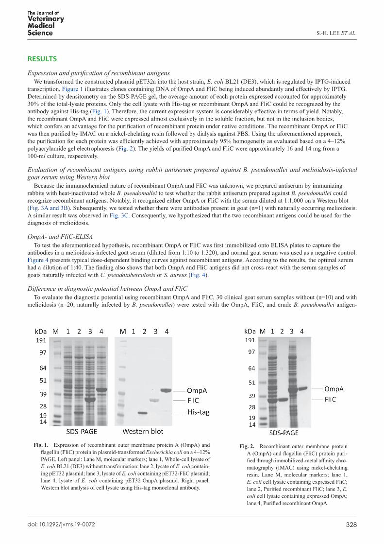
Expression and purification of recombinant antigensWe transformed the constructed plasmid pET32a into the host strain, E. coli BL21 (DE3), which is regulated by IPTG-induced
transcription. Figure 1 illustrates clones containing DNA of OmpA and FliC being induced abundantly and effectively by IPTG. Determined by densitometry on the SDS-PAGE gel, the average amount of each protein expressed accounted for approximately 30% of the total-lysate proteins. Only the cell lysate with His-tag or recombinant OmpA and FliC could be recognized by the antibody against His-tag (Fig. 1). Therefore, the current expression system is considerably effective in terms of yield. Notably, the recombinant OmpA and FliC were expressed almost exclusively in the soluble fraction, but not in the inclusion bodies, which confers an advantage for the purification of recombinant protein under native conditions. The recombinant OmpA or FliC was then purified by IMAC on a nickel-chelating resin followed by dialysis against PBS. Using the aforementioned approach, the purification for each protein was efficiently achieved with approximately 95% homogeneity as evaluated based on a 4–12% polyacrylamide gel electrophoresis (Fig. 2). The yields of purified OmpA and FliC were approximately 16 and 14 mg from a 100-ml culture, respectively.
Evaluation of recombinant antigens using rabbit antiserum prepared against B. pseudomallei and melioidosis-infected goat serum using Western blot
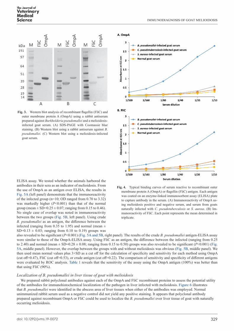
Because the immunochemical nature of recombinant OmpA and FliC was unknown, we prepared antiserum by immunizing rabbits with heat-inactivated whole B. pseudomallei to test whether the rabbit antiserum prepared against B. pseudomallei could recognize recombinant antigens. Notably, it recognized either OmpA or FliC with the serum diluted at 1:1,000 on a Western blot (Fig. 3A and 3B). Subsequently, we tested whether there were antibodies present in goat (n=1) with naturally occurring melioidosis. A similar result was observed in Fig. 3C. Consequently, we hypothesized that the two recombinant antigens could be used for the diagnosis of melioidosis.
OmpA- and FliC-ELISATo test the aforementioned hypothesis, recombinant OmpA or FliC was first immobilized onto ELISA plates to capture the
antibodies in a melioidosis-infected goat serum (diluted from 1:10 to 1:320), and normal goat serum was used as a negative control. Figure 4 presents typical dose-dependent binding curves against recombinant antigens. According to the results, the optimal serum had a dilution of 1:40. The finding also shows that both OmpA and FliC antigens did not cross-react with the serum samples of goats naturally infected with C. pseudotuberculosis or S. aureus (Fig. 4).
Difference in diagnostic potential between OmpA and FliCTo evaluate the diagnostic potential using recombinant OmpA and FliC, 30 clinical goat serum samples without (n=10) and with
melioidosis (n=20; naturally infected by B. pseudomallei) were tested with the OmpA, FliC, and crude B. pseudomallei antigen-
Fig. 1. Expression of recombinant outer membrane protein A (OmpA) and flagellin (FliC) protein in plasmid-transformed Escherichia coli on a 4–12% PAGE. Left panel: Lane M, molecular markers; lane 1, Whole-cell lysate of E. coli BL21 (DE3) without transformation; lane 2, lysate of E. coli contain-ing pET32 plasmid; lane 3, lysate of E. coli containing pET32-FliC plasmid; lane 4, lysate of E. coli containing pET32-OmpA plasmid. Right panel: Western blot analysis of cell lysate using His-tag monoclonal antibody.
Fig. 2. Recombinant outer membrane protein A (OmpA) and flagellin (FliC) protein puri-fied through immobilized-metal affinity chro-matography (IMAC) using nickel-chelating resin. Lane M, molecular markers; lane 1, E. coli cell lysate containing expressed FliC; lane 2, Purified recombinant FliC; lane 3, E. coli cell lysate containing expressed OmpA; lane 4, Purified recombinant OmpA.
IMMUNODIAGNOSIS OF GOAT MELIOIDOSIS
329doi: 10.1292/jvms.19-0072
ELISA assay. We tested whether the animals harbored the antibodies in their sera as an indicator of melioidosis. From the use of OmpA as an antigen over ELISA, the results in Fig. 5A (left panel) demonstrate that the immunoreactivity of the infected group (n=10; OD ranged from 0.70 to 3.32) was markedly higher (P<0.001) than that of the normal group (mean ± SD=0.25 ± 0.07; ranging from 0.15 to 0.46). No single case of overlap was noted in immunoreactivity between the two groups (Fig. 5B, left panel). Using crude B. pseudomallei as an antigen, the difference between the infected (ranging from 0.35 to 1.95) and normal (mean ± SD=0.13 ± 0.03; ranging from 0.10 to 0.19) groups was also revealed to be significant (P<0.001) (Fig. 5A and 5B, right panel). The results of the crude B. pseudomallei antigen-ELISA assay were similar to those of the OmpA-ELISA assay. Using FliC as an antigen, the difference between the infected (ranging from 0.25 to 2.40) and normal (mean ± SD=0.28 ± 0.08; ranging from 0.15 to 0.50) groups was also revealed to be significant (P<0.001) (Fig. 5A, middle panel). However, the overlap between the groups with and without melioidosis was obvious (Fig. 5B, middle panel). We then used mean normal values plus 3×SD as a cut off for the calculation of specificity and sensitivity for each method using OmpA (cut off=0.47), FliC (cut off=0.53), or crude antigen (cut off=0.22). The comparison of sensitivity and specificity of different antigens were evaluated by ROC analysis. Table 1 reveals that the sensitivity of the assay using the OmpA antigen (100%) was better than that using FliC (90%).
Localization of B. pseudomallei in liver tissue of goat with melioidosisWe prepared rabbit polyclonal antibodies against each of the OmpA and FliC recombinant proteins to assess the potential utility
of the antibodies for immunohistochemical localization of the pathogen in liver infected with melioidosis. Figure 6 illustrates that B. pseudomallei were identified in the abscess area of liver tissues when either of the antibodies was employed. Normal unimmunized rabbit serum used as a negative control did not yield any positive staining. It appears that polyclonal antibody prepared against recombinant OmpA or FliC could be used to localize the B. pseudomallei over liver tissue of goat with naturally occurring melioidosis.
Fig. 3. Western blot analysis of recombinant flagellin (FliC) and outer membrane protein A (OmpA) using a rabbit antiserum prepared against Burkholderia pseudomallei and a melioidosis-infected goat serum. (A) SDS-PAGE with Coomassie blue staining. (B) Western blot using a rabbit antiserum against B. pseudomallei. (C) Western blot using a melioidosis-infected goat serum.
Fig. 4. Typical binding curves of serum reactive to recombinant outer membrane protein A (OmpA) or flagellin (FliC) antigen. Each antigen was coated on an enzyme-linked immunosorbent assay (ELISA) plate to capture antibody in the serum. (A) Immunoreactivity of OmpA us-ing melioidosis positive and negative serum, and serum from goats naturally infected with C. pseudotuberculosis or S. aureus. (B) Im-munoreactivity of FliC. Each point represents the mean determined in triplicate.

S.-H. LEE ET AL.
330doi: 10.1292/jvms.19-0072
DISCUSSION
In the present study, ELISA was developed using a culture-confirmed melioidosis goat serum at a 1:40 dilution. Presented based on mean normal values plus 3×SD as a cut off value, our data revealed that ELISA captured by OmpA antigen resulted in higher sensitivity (100%) than that observed when using FliC as the antigen (90%) (Table 1). In previous reports, sensitivity of FliC ELISA was compareble to that of OmpA ELISA in human melioidosis. The reason for the discrepancy is not readily known. Certainly, it was not due to the amount of goat serum used because increased serum amounts for the test up from 1:40 to 1:10 yielded similar sensitivity and specificity (data not shown). Notably, Fig. 5 illustrates that the means of melioidosis positive values (antibody binding) were approximately seven-fold higher than the basal normal values obtained using OmpA antigen, and they were approximately 4.5-fold higher than the values obtained using FliC antigen. In addition, the deviation of the melioidosis group (antibody binding) determined using OmpA antigen was lower (40%) than that determined using FliC antigen (65%). Overall, the results could explain the high sensitivity associated with the OmpA antigen.
In immunohistochemical analyses using rabbit polyclonal antibodies prepared against each specific recombinant antigen, B.
Fig. 5. Serologic test using recombinant outer membrane protein A (OmpA) (left panel), flagellin (FliC) (middle panel), and crude Burkholderia pseudomallei antigen (right panel) as an antigen. Goat serum samples without (n=20) and with melioidosis (n=10) were tested for the presence of the antibodies (1:40) reactive to OmpA, FliC, and crude B. pseudomallei antigen. A: mean values ± SD. B: means of each of the individual values determined in triplicate. *P<0.001 compared with the normal group.
Table 1. Sensitivity and specificity of OmpA- and FliC-ELISA
ELISA Cutoff index Sensitivity (True positive rate)
False negative rate
Flase positive rate
Specificity (True negative rate)
OmpA-ELISA Mean normal values plus 2 SD 100% (10/10) 0% (0/10) 5% (1/20) 95% (19/20)Mean normal values plus 3 SD 100% (10/10) 0% (0/10) 0% (0/20) 100% (20/20)
FliC-ELISA Mean normal values plus 2 SD 90% (9/10) 10% (1/10) 5% (1/20) 95% (19/20)Mean normal values plus 3 SD 90% (9/10) 10% (1/10) 0% (0/20) 100% (20/20)
Crude antigen-ELISA Mean normal values plus 2 SD 100% (10/10) 0% (0/10) 5% (1/20) 95% (19/20)Mean normal values plus 3 SD 100% (10/10) 0% (0/10) 0% (0/20) 100% (20/20)
SD, standard deviation.

IMMUNODIAGNOSIS OF GOAT MELIOIDOSIS
331doi: 10.1292/jvms.19-0072
pseudomallei were primarily located at the abscess areas of liver tissue with melioidosis, which indicated that in general, the polyclonal antibodies were applicable in the immunohistochemical localization of B. pseudomallei in infected tissues. We were able to localize the bacterium in the lymph nodes, lungs, spleen, and mammary glands (data not shown).
In conclusion, the recombinant OmpA and FliC antigens could be used as probes to explore the presence of serum antibodies against B. pseudomallei in goats with melioidosis. Our results indicate that OmpA is a potentially superior reagent to FliC for melioidosis serology tests.
ACKNOWLEDGMENTS. We thank members of the Epidemiology Division of Animal Health Research Institute (Council of Agriculture, Executive Yuan, Taipei, Taiwan) for their enthusiastic support. We also thank Chu-Ting Yang (Bioresource Collection and Research Center, Food Industry and Development Institute, Hsinchu, Taiwan) for her research assistance.
REFERENCES
1. Allwood, E. M., Logue, C. A., Hafner, G. J., Ketheesan, N., Norton, R. E., Peak, I. R. and Beacham, I. R. 2008. Evaluation of recombinant antigens for diagnosis of melioidosis. FEMS Immunol. Med. Microbiol. 54: 144–153. [Medline] [CrossRef]
2. Arora, S., Thavaselvam, D., Kumar, A., Prakash, A., Barua, A. and Sathyaseelan, K. 2015. Cloning, expression and purification of outer membrane protein (OmpA) of Burkholderia pseudomallei and evaluation of its potential for serodiagnosis of melioidosis. Diagn. Microbiol. Infect. Dis. 81: 79–84. [Medline] [CrossRef]
3. Bradford, M. M. 1976. A rapid and sensitive method for the quantitation of microgram quantities of protein utilizing the principle of protein-dye
Fig. 6. Localization of Burkholderia pseudomallei infecting within the liver tissue of goat with melioidosis. Liver tissues of goat with meli-oidosis were incubated with purified rabbit antibody produced against outer membrane protein A (OmpA) (A, B and C) and rabbit antibody produced against flagellin (FliC) (D, E, and F). Normal nonimmuned rabbit serum was used as a negative control (G, H, and I). Afterward, immunostaining (in red) followed by a counter staining (in blue) were performed as described in the Materials and Methods section. A, D, and G (40× magnification). B, E, and H (100× magnification). C, F, and I (200× magnification).

S.-H. LEE ET AL.
332doi: 10.1292/jvms.19-0072
binding. Anal. Biochem. 72: 248–254. [Medline] [CrossRef] 4. Chantratita, N., Tandhavanant, S., Wongsuvan, G., Wuthiekanun, V., Teerawattanasook, N., Day, N. P. J., Limmathurotsakul, D. and Peacock, S. J.
2013. Rapid detection of Burkholderia pseudomallei in blood cultures using a monoclonal antibody-based immunofluorescent assay. Am. J. Trop. Med. Hyg. 89: 971–972. [Medline] [CrossRef]
5. Chantratita, N., Wuthiekanun, V., Thanwisai, A., Limmathurotsakul, D., Cheng, A. C., Chierakul, W., Day, N. P. and Peacock, S. J. 2007. Accuracy of enzyme-linked immunosorbent assay using crude and purified antigens for serodiagnosis of melioidosis. Clin. Vaccine Immunol. 14: 110–113. [Medline] [CrossRef]
6. Chaichana, P., Jenjaroen, K., Amornchai, P., Chumseng, S., Langla, S., Rongkard, P., Sumonwiriya, M., Jeeyapant, A., Chantratita, N., Teparrukkul, P., Limmathurotsakul, D., Day, N. P. J., Wuthiekanun, V. and Dunachie, S. J. 2018. Antibodies in Melioidosis: The role of the indirect hemagglutination assay in evaluating patients and exposed populations. Am. J. Trop. Med. Hyg. 99: 1378–1385. [Medline] [CrossRef]
7. Chen, Y. S., Hsiao, Y. S., Lin, H. H., Liu, Y. and Chen, Y. L. 2006. CpG-modified plasmid DNA encoding flagellin improves immunogenicity and provides protection against Burkholderia pseudomallei infection in BALB/c mice. Infect. Immun. 74: 1699–1705. [Medline] [CrossRef]
8. Chen, Y. S., Shiuan, D., Chen, S. C., Chye, S. M. and Chen, Y. L. 2003. Recombinant truncated flagellin of Burkholderia pseudomallei as a molecular probe for diagnosis of melioidosis. Clin. Diagn. Lab. Immunol. 10: 423–425. [Medline]
9. Dulsuk, A., Paksanont, S., Sangchankoom, A., Ekchariyawat, P., Phunpang, R., Jutrakul, Y., Chantratita, N. and West, T. E. 2016. Validation of a monoclonal antibody-based immunofluorescent assay to detect Burkholderia pseudomallei in blood cultures. Trans. R. Soc. Trop. Med. Hyg. 110: 670–672. [Medline]
10. Gilmore, G., Barnes, J., Ketheesan, N. and Norton, R. 2007. Use of antigens derived from Burkholderia pseudomallei, B. thailandensis, and B. cepacia in the indirect hemagglutination assay for melioidosis. Clin. Vaccine Immunol. 14: 1529–1531. [Medline] [CrossRef]
11. Hara, Y., Mohamed, R. and Nathan, S. 2009. Immunogenic Burkholderia pseudomallei outer membrane proteins as potential candidate vaccine targets. PLoS One 4: e6496. [Medline] [CrossRef]
12. Harris, P. N., Ketheesan, N., Owens, L. and Norton, R. E. 2009. Clinical features that affect indirect-hemagglutination-assay responses to Burkholderia pseudomallei. Clin. Vaccine Immunol. 16: 924–930. [Medline] [CrossRef]
13. Holden, M. T., Titball, R. W., Peacock, S. J., Cerdeño-Tárraga, A. M., Atkins, T., Crossman, L. C., Pitt, T., Churcher, C., Mungall, K., Bentley, S. D., Sebaihia, M., Thomson, N. R., Bason, N., Beacham, I. R., Brooks, K., Brown, K. A., Brown, N. F., Challis, G. L., Cherevach, I., Chillingworth, T., Cronin, A., Crossett, B., Davis, P., DeShazer, D., Feltwell, T., Fraser, A., Hance, Z., Hauser, H., Holroyd, S., Jagels, K., Keith, K. E., Maddison, M., Moule, S., Price, C., Quail, M. A., Rabbinowitsch, E., Rutherford, K., Sanders, M., Simmonds, M., Songsivilai, S., Stevens, K., Tumapa, S., Vesaratchavest, M., Whitehead, S., Yeats, C., Barrell, B. G., Oyston, P. C. and Parkhill, J. 2004. Genomic plasticity of the causative agent of melioidosis, Burkholderia pseudomallei. Proc. Natl. Acad. Sci. USA 101: 14240–14245. [Medline] [CrossRef]
14. Koosakulnirand, S., Phokrai, P., Jenjaroen, K., Roberts, R. A., Utaisincharoen, P., Dunachie, S. J., Brett, P. J., Burtnick, M. N. and Chantratita, N. 2018. Immune response to recombinant Burkholderia pseudomallei FliC. PLoS One 13: e0198906. [Medline] [CrossRef]
15. Lai, I. H., Tsai, T. I., Lin, H. H., Lai, W. Y. and Mao, S. J. 2007. Cloning and expression of human haptoglobin subunits in Escherichia coli: delineation of a major antioxidant domain. Protein Expr. Purif. 52: 356–362. [Medline] [CrossRef]
16. Limmathurotsakul, D., Thammasart, S., Warrasuth, N., Thapanagulsak, P., Jatapai, A., Pengreungrojanachai, V., Anun, S., Joraka, W., Thongkamkoon, P., Saiyen, P., Wongratanacheewin, S., Day, N. P. and Peacock, S. J. 2012. Melioidosis in animals, Thailand, 2006-2010. Emerg. Infect. Dis. 18: 325–327. [Medline] [CrossRef]
17. Musa, H. I., Hassan, L., Shamsuddin, Z. H., Panchadcharam, C., Zakaria, Z., Abdul Aziz, S. and Rachmat, R. F. 2015. Case-control investigation on the risk factors of melioidosis in small ruminant farms in Peninsular Malaysia. J. Appl. Microbiol. 119: 331–341. [Medline] [CrossRef]
18. Nithichanon, A., Gourlay, L. J., Bancroft, G. J., Ato, M., Takahashi, Y. and Lertmemongkolchai, G. 2017. Boosting of post-exposure human T-cell and B-cell recall responses in vivo by Burkholderia pseudomallei-related proteins. Immunology 151: 98–109. [Medline] [CrossRef]
19. Perumal Samy, R., Stiles, B. G., Sethi, G. and Lim, L. H. K. 2017. Melioidosis: Clinical impact and public health threat in the tropics. PLoS Negl. Trop. Dis. 11: e0004738. [Medline] [CrossRef]
20. Simpson, A. J., Howe, P. A., Wuthiekanun, V. and White, N. J. 1999. A comparison of lysis centrifugation, pour plate, and conventional blood culture methods in the diagnosis of septicaemic melioidosis. J. Clin. Pathol. 52: 616–619. [Medline] [CrossRef]
21. Soffler, C., Bosco-Lauth, A. M., Aboellail, T. A., Marolf, A. J. and Bowen, R. A. 2014. Pathogenesis of percutaneous infection of goats with Burkholderia pseudomallei: clinical, pathologic, and immunological responses in chronic melioidosis. Int. J. Exp. Pathol. 95: 101–119. [Medline] [CrossRef]
22. Sprague, L. D. and Neubauer, H. 2004. Melioidosis in animals: a review on epizootiology, diagnosis and clinical presentation. J. Vet. Med. B Infect. Dis. Vet. Public Health 51: 305–320. [Medline] [CrossRef]
23. Suttisunhakul, V., Wuthiekanun, V., Brett, P. J., Khusmith, S., Day, N. P., Burtnick, M. N., Limmathurotsakul, D. and Chantratita, N. 2016. Development of rapid enzyme-linked immunosorbent assays for detection of antibodies to Burkholderia pseudomallei. J. Clin. Microbiol. 54: 1259–1268. [Medline] [CrossRef]
24. Tiangpitayakorn, C., Songsivilai, S., Piyasangthong, N. and Dharakul, T. 1997. Speed of detection of Burkholderia pseudomallei in blood cultures and its correlation with the clinical outcome. Am. J. Trop. Med. Hyg. 57: 96–99. [Medline] [CrossRef]
25. Wajanarogana, S., Nimnuch, P., Thongmee, A. and Kritsiriwuthinan, K. 2013. Potential of recombinant flagellin fragment from Burkholderia thailandensis as an antigen for melioidosis antibody detection by indirect ELISA. Mol. Cell. Probes 27: 98–102. [Medline] [CrossRef]
26. Wiersinga, W. J., Currie, B. J. and Peacock, S. J. 2012. Melioidosis. N. Engl. J. Med. 367: 1035–1044. [Medline] [CrossRef] 27. Yang, S. 2000. Melioidosis research in China. Acta Trop. 77: 157–165. [Medline] [CrossRef]