basic fibroblast growth factor activates mek/erk cell...
TRANSCRIPT
Basic Fibroblast Growth Factor Activates MEK/ERK CellSignaling Pathway and Stimulates the Proliferation ofChicken Primordial Germ CellsJin Won Choi1, Sujung Kim1, Tae Min Kim1, Young Min Kim1, Hee Won Seo1, Tae Sub Park2, Jae-Wook
Jeong3, Gwonhwa Song1, Jae Yong Han1*
1 WCU Biomodulation Major, Department of Agricultural Biotechnology, Seoul National University, Seoul, Korea, 2 Avicore Biotechnology Institute, Optifarm Solution Inc.,
Gyeonggi-do, Korea, 3 Department of Molecular and Cellular Biology, Baylor College of Medicine, Houston, Texas, United States of America
Abstract
Background: Long-term maintenance of avian primordial germ cells (PGCs) in vitro has tremendous potential because it canbe used to deepen our understanding of the biology of PGCs. A transgenic bioreactor based on the unique migration ofPGCs toward the recipients’ sex cord via the bloodstream and thereby creating a germline chimeric bird has many potentialapplications. However, the growth factors and the signaling pathway essential for inducing proliferation of chicken PGCs areunknown.
Methodology/Principal Findings: Therefore, we conducted this study to investigate the effects of various combinations ofgrowth factors on the survival and proliferation of PGCs under feeder-free conditions. We observed proliferation of PGCs inmedia containing bFGF. Subsequent characterization confirmed that the cultured PGCs maintained expression of PGC-specific markers, telomerase activity, normal migrational activity, and germline transmission. We also found that bFGFactivates the mitogen-activated protein kinase kinase/extracellular-signal regulated kinase (MEK/ERK) signaling. Also, theexpression of 133 transcripts was reversibly altered by bFGF withdrawal.
Conclusions/Significance: Our results demonstrate that chicken PGCs can be maintained in vitro without any differentiationor dedifferentiation in feeder free culture conditions, and subsequent analysis revealed that bFGF is one of the key factorsthat enable proliferation of chicken PGCs via MEK/ERK signaling regulating downstream genes that may be important forPGC proliferation and survival.
Citation: Choi JW, Kim S, Kim TM, Kim YM, Seo HW, et al. (2010) Basic Fibroblast Growth Factor Activates MEK/ERK Cell Signaling Pathway and Stimulates theProliferation of Chicken Primordial Germ Cells. PLoS ONE 5(9): e12968. doi:10.1371/journal.pone.0012968
Editor: Laszlo Orban, Temasek Life Sciences Laboratory, Singapore
Received June 18, 2010; Accepted September 2, 2010; Published September 23, 2010
Copyright: � 2010 Choi et al. This is an open-access article distributed under the terms of the Creative Commons Attribution License, which permits unrestricteduse, distribution, and reproduction in any medium, provided the original author and source are credited.
Funding: This work was supported by Korea Research Foundation program (KRF-2006-311-F00087) and also supported by World Class University (WCU) program(R31-10056) through the National Research Foundation of Korea funded by the Ministry of Education, Science and Technology. The funders had no role in studydesign, data collection and analysis, decision to publish, or preparation of the manuscript.
Competing Interests: The authors have declared that no competing interests exist.
* E-mail: [email protected]
Introduction
Germ cells play important roles in species continuation by
delivering genetic information to the next generation. In many
animal species, they arise from a small population of cells known as
primordial germ cells (PGCs) [1,2,3]. In chickens, PGCs are
initially localized to the central zone of the area pellucida in stage X
embryos [4]. They migrate to the germinal crescent at stage 4 (18–
19 h after incubation) [5] and, between stages 10 and 12, move
into blood vessels and begin circulating in the bloodstream [6,7].
Via the circulatory system, PGCs finally migrate to the genital
ridge [8,9]. During migration, PGCs proliferate: about 30 PGCs
are found in a stage X embryo, 200–250 in the germinal crescent
[4], and more than 1,000 at stage 31 (7 days of incubation) [10].
The basic fibroblast growth factor (bFGF) is a member of the
fibroblast growth factor family that plays diverse roles in regulating
cell proliferation, migration, and differentiation during embryonic
development [11,12,13]. In mammals, it appears to be important
for self-renewal of human embryonic stem cells [14] and mouse
spermatogonial stem cells (SSCs) [15]. FGF signaling is critical to
PGC migration and thereby controls germ cell numbers in mice
[16]. In chickens, bFGF is one of the factors supporting the
proliferation of preblastodermal cells [17], embryonic germ cells
(EGCs) [18], and PGCs [19]. However, it remains to be
determined whether bFGF is essential for the proliferation of
chicken PGCs.
Studies of the maintenance and proliferation of avian PGCs in
vitro offer tremendous potential in understanding the physiology of
PGCs and the production of transgenic bioreactors. A recent
report showed that PGCs from the blood of stage 14–17 chicken
embryos could be expanded when cultured on a feeder layer of
Buffalo rat liver (BRL) cells or Sandoz inbred mouse-derived
thioguanine-resistant and ouabain-resistant (STO) cells, in an
undefined medium conditioned with BRL cells containing
leukemia inhibitory factor (LIF), stem cell factor (SCF), and bFGF
[19]. However, specific growth factors that are essential for PGC
proliferation remain to be identified, and the complex and
undefined parameters arising from the use of conditioned media
PLoS ONE | www.plosone.org 1 September 2010 | Volume 5 | Issue 9 | e12968
have made the roles of individual growth factors impossible to
evaluate.
In this report, we describe the development of a feeder-free
PGC culture system that excludes the effects of undefined
molecules from the feeder layer. By using this culture system,
the effect of individual growth factors, including LIF, SCF, and
bFGF, on the proliferation of chicken PGCs in vitro was evaluated.
Results
bFGF is Essential for The Proliferation of Chicken PGCs invitro
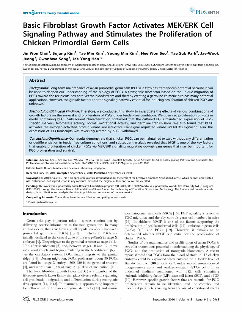
Whole blood cells containing PGCs from chicken embryos at
stage 14–15 were isolated (Fig. 1A) and cultured in the presence of
LIF, SCF, and bFGF. After 7–14 days of growth, most of the
blood cells were dead and PGC colonies formed and loosely
attached to culture plate (Fig. 1B). The PGC colonies were
detached by gentle pipetting and disaggregated with an appropri-
ate enzyme (Fig. 1C). The disaggregated PGCs again grew and
aggregated to form colonies in 3–4 days (Fig. 1D). The PGCs were
then subcultured every 3–4 days. During subsequent passages,
cultured PGCs did not attach to the culture plate and grew in
suspension.
To identify the essential growth factors for PGC growth, we
cultured PGCs under various combinations of growth factors. As
illustrated in Fig. 1E, PGC colonies were only observed for cells
grown in media containing bFGF. We next examined the effects of
different growth conditions on PGC culture expansion. PGCs
were cultured for 32 days (to passage 5) in media supplemented
with LIF, SCF, and bFGF, and then continued to be cultured for a
further 12 days under different conditions (Fig. 1F). When bFGF
was absent from the culture medium, the number of cells
decreased. In contrast, when bFGF alone or a combination of
LIF, SCF, and bFGF was added to the culture medium, cell
numbers increased at a constant rate over three passages. The
results show that of the three growth factors tested, bFGF is
essential for PGC proliferation.
The effects of bFGF on PGC proliferation were examined in
detail using cells that were cultured for more than 32 days. PGCs
were cultured in the presence or absence of bFGF (10 ng?ml21),
and cell morphology and recovery were examined 4 days later. In
the presence of bFGF, the proliferating PGCs formed colonies. In
contrast, in the absence of bFGF, PGC growth was limited and a
large number of fragmented cells were observed (Fig. 2A–2B). The
total number of cells increased about threefold in the presence of
bFGF, but decreased in the absence of bFGF (p,0.01; Fig. 2C).
When PGCs were treated with different concentrations of bFGF,
cell recovery increased in a dose-dependent manner (Fig. 2D).
Low cell recovery in untreated cells may have resulted from
increased apoptosis and reduced growth rates in the absence of
bFGF. Thus, we next analyzed cell cycle progression and apoptosis
of PGCs in the presence and absence of bFGF. Removal of bFGF
for 24 h decreased the proportion of cells in the S and G2/M
phase and increased the proportion of cells in the G1 phase
(Fig. 2E). An increased number of apoptotic cells were observed by
a TUNEL assay when bFGF was removed for 24 h (Fig. 2F–2J).
Taken together, these results indicate that a single growth factor,
bFGF, may support PGC proliferation. Therefore, we cultured
PGCs in media containing bFGF and characterized them in detail.
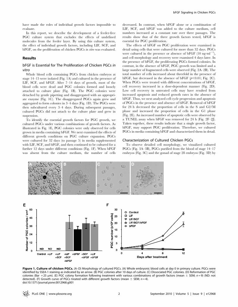
Characterization of Cultured Chicken PGCsTo observe detailed cell morphology, we visualized cultured
PGCs (Fig. 3A–3B), PGCs purified from the blood of stage 14–17
embryos (Fig. 3C) and the gonad of stage 28 embryos (Fig. 3D) by
Figure 1. Culture of chicken PGCs. (A–D) Morphology of cultured PGCs. (A) Whole embryonic blood cells at day 0 in primary culture. PGCs wereidentified by SSEA-1 staining as indicated by an arrow. (B) PGC colonies after 10 days of culture. (C) Dissociated PGC colonies. (D) Reformation of PGCcolonies (Bar = 25 mm). (E) PGC colony formation following treatment with various combinations of growth factors (mean 6 SEM; n = 9) (ND: notdetected). (F) Growth curve of PGCs treated with different growth factors (mean 6 SEM; n = 4).doi:10.1371/journal.pone.0012968.g001
bFGF Signaling in Chicken PGCs
PLoS ONE | www.plosone.org 2 September 2010 | Volume 5 | Issue 9 | e12968

scanning electron microscopy. All examined PGCs were approx-
imately 9–12 mm in diameter and sphere-shaped with numerous
microvilli. Moreover, when cultured on Matrigel, PGCs attached
to the surface of the matrix. Pseudopodia-like structures were
observed (Fig. 3E–3F).
Immunocytochemical analysis was performed to characterize
cultured PGCs in detail. These cells were positive for the chicken
PGC markers SSEA-1, ITGA6, and ITGB1 [20,21] (Fig. 3G–3I)
but negative for SSEA-3 and SSEA-4 (data not shown). To
examine PGC-specific gene expression, RT-PCR analysis was
performed (Fig. 3J). Expression of NANOG (NM_001146142.1)
and POUV (NM_001110178), two genes known to be expressed in
the germ cells of early-stage chicken embryos [22], were detected.
The germ cell-specific genes CVH (NM_204708) [4], DAZL
(NM_204218) [23], and KIT (NM_204361.1) [24] were also
expressed in cultured PGCs. These same genes were similarly
expressed in purified PGCs. Because a previous study showed that
cultured PGCs have telomerase activity [19], we tested telomerase
activity in PGCs cultured for 126 days. The result showed that the
cultured PGCs used in our study also have telomerase activity
(Fig. 3K). These data suggest that cultured PGCs are immortalized
cells that maintain expression of both surface markers and PGC-
specific genes.
Avian PGCs initially migrate to the germinal crescent before
migrating to the genital ridge via the bloodstream [8,25]. In
addition, when injected into the dorsal aorta of stage 14–17
embryos, donor PGCs migrate to the gonads and contribute to the
germ line [26,27]. We therefore tested the migrational activity of
cultured PGCs by two different strategies. First, PKH26-labeled
PGCs that had been cultured for 82 days were injected into the
subgerminal cavities of stage X blastoderm embryos that were
then observed at stage 6 by fluorescence microscopy. Localization
Figure 2. Effects of bFGF on PGC proliferation. (A–B) Morphology of PGCs in the presence of bFGF and 4 days after bFGF withdrawal (Bar= 100 mm). (C) Effect of bFGF on cell recovery after 4 days of culture (mean 6 SEM; n = 4) (*significant differences following treatment; p,0.01). (D)Dose-dependent effect of bFGF on the proliferation of PGCs (mean 6 SEM; n = 3) (superscripts indicate significant differences between treatments;p,0.05). (E) Analysis of cell cycle distribution of PGCs in the presence of bFGF (left) and 24 h after bFGF withdrawal (right). (F–I) TUNEL assayperformed on PGCs cultured with bFGF and 24 h after bFGF withdrawal (J) Number of apoptotic cells after bFGF withdrawal (*significant differencebetween treatments; p,0.01). Statistical analyses were conducted with a Student’s t-test (C and J) or ANOVA using SAS software (D).doi:10.1371/journal.pone.0012968.g002
bFGF Signaling in Chicken PGCs
PLoS ONE | www.plosone.org 3 September 2010 | Volume 5 | Issue 9 | e12968
of the injected PGCs was restricted to the germinal crescent
(Fig. 3L). Next, PKH26-labeled PGCs that had been cultured for
82 days were injected into the bloodstream of recipient stage 14–
17 embryos that were subsequently observed at stage 30. Labeled
cells were detected in the embryonic gonad (Fig. 3M–3N).
Moreover, the number of cells that migrated did not significantly
differ between cultured PGCs and PGCs purified from the gonad
of stage 28 embryos when the they were injected into the
subgerminal cavity of blastoderm embryos (p = 0.4759, Fig. 3O)
and the dorsal aorta of stage 14–17 embryos (p = 0.5031, Fig. 3P).
These results suggest that cultured PGCs have normal migrational
activity.
When PGCs are injected into the blood vessels of recipient
embryos during stages 13–17, germline chimera are produced
[27,28]. To confirm that the cultured PGCs can contribute to the
germline, cultured PGCs (i/i) were injected into WL (I/I) recipient
embryos. The putative germline chimeric chicken was sexually
matured and donor-derived offspring with black plumage color
were produced after artificial insemination with KO semen
(Fig. 3Q and Table S1). To examine whether the cultured PGCs
Figure 3. Characterization of cultured PGCs. Scanning electron microscopy of cultured PGCs (A–B), blood PGCs (C), and gonadal PGCs (D). (E, F)PGCs cultured on Matrigel (arrows indicate pseudopod-like structures). Bars: 2 mm (A, C, and D); 200 nm (B); 100 mm (E); and 25 mm (F). (G–I)Immunocytochemical analysis of cultured PGCs. PGCs cultured for 60 days were immunostained with antibodies raised against SSEA-1 (G), ITGA6 (H),and ITGB1 (I). (J) RT-PCR analysis of NANOG, POUV, CVH, DAZL, and KIT in cultured PGCs (passages 10 and 20) [–: negative control (no template)]. (K)Telomerase activity in PGCs. Repeated sequences were observed in PGCs and DT40 (positive control cells). Chicken embryonic fibroblast (CEF) andbuffer (–) were used as negative controls. Arrow indicates the 56-bp internal control template band. (L) Migration of cultured PGCs into the germinalcrescent. Approximately 500 PGCs, cultured for 82 days or purified by MACS, were labeled with PKH26 and then transferred into the subgerminalcavities of blastoderm embryos. Labeled cells (red) were detected in the germinal crescent (arrows) (GC: germinal crescent, AP: area pellucida). (M–N)Gonadal migration of culture PGCs. Approximately 500 PGCs, cultured for 82 days or purified by MACS, were labeled with PKH26 and then injectedinto blood vessels of recipient embryos at stage 14–17. Labeled cells (red) were detected in the embryonic gonad. (O) Numbers of PGCs that hadmigrated into the germinal crescent of stage 6 embryos that had, as stage X recipient embryos, been injected with 500 PGCs (mean 6 SEM; n = 12 forpurified PGCs, n = 11 for cultured PGCs). No significant difference was observed between cultured and purified PGCs. (P) Number of PGCs that hadmigrated into the gonads of stage 28 embryos that had, as stage 14 recipient embryos, been injected (i.v.) with 500 PGCs (mean 6 SEM; n = 12 forpurified PGCs, n = 10 for cultured PGCs). No significant difference was observed between cultured and purified PGCs. Statistical analysis wasconducted with the general linear model (PROC-GLM) of SAS software. (Q) Germline transmission of cultured PGCs. Donor KO (i/i) PGCs cultured formore than 50 days were injected into the dorsal aorta of WL (I/I) recipient embryos (female), and after sexual maturation, progeny were derived fromthe donor KO PGCs (black plumage color, black arrow). Progeny that derived from the endogenous WL PGCs are noted by a white arrow. The whiteegg indicates that the recipient chicken is WL because KO chickens lay brown eggs as indicated by the blue arrow.doi:10.1371/journal.pone.0012968.g003
bFGF Signaling in Chicken PGCs
PLoS ONE | www.plosone.org 4 September 2010 | Volume 5 | Issue 9 | e12968
were dedifferentiated into EGCs during long-term culture, we also
injected cultured PGCs into the subgerminal cavities of a stage X
WL blastoderm, and 18 chicks subsequently hatched. However,
we could not observe any somatic chimerism in the hatched
chicks. These results confirm that the cultured PGCs are
functionally normal.
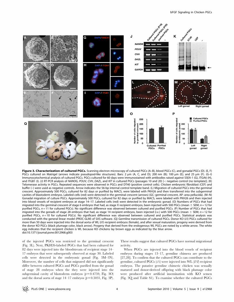
Activation of MEK/ERK Signaling by bFGF in PGCsWe examined the effect of bFGF on phosphorylation of AKT
and three MAPKs: ERK1/2, p38, and JNK. PGCs were treated
with bFGF, and the phosphorylation of MAPK and AKT was
detected by a Western blot (Fig. 4A). Western blot analyses of
whole PGC extracts using antibodies raised against p-(activated)
target proteins showed that bFGF increased the level of p-ERK1/
2 over the basal level. However, p-p38 and p-JNK MAPKs could
not be detected (data not shown). AKT was phosphorylated on
T380 and S473 independent of bFGF treatment.
We then examined the ERK1/2 signaling pathway in PGCs in
more detail. In ERK signaling cascades, extracellular signals are
transmitted by MEK1/2 [29]. Thus, we analyzed ERK1/2
Figure 4. bFGF signaling in chicken PGCs. (A) Activation of MAPK and PI3K/AKT signaling pathways by bFGF (10 ng?ml21). NT: no treatment (B)Dose-dependent activation of MEK/ERK signaling by bFGF. NT: no treatment (C) Temporal effect of bFGF (10 ng?ml21) on activation of MEK/ERKsignaling. NT: no treatment (D) Inhibition of MEK/ERK signaling by a specific inhibitor of FGFRs (PD173074). NT: no treatment (E) Inhibition of ERKactivation by PD0325901 in cultured PGCs (PD173074 was used as a control). (F) Cell recovery 4 days after PD0325901 treatment (mean 6 SEM, n = 4).bFGF (10 ng?ml21) was treated in all groups. Superscripts indicate significant differences between treatment groups (p,0.05). Statistical analyseswere conducted with ANOVA using SAS software. (G–H) Morphology of PGCs 4 days after treatment with PD0325901 (0.4 mM) (bar = 100 mm). bFGF(10 ng?ml21) was treated in all groups.doi:10.1371/journal.pone.0012968.g004
bFGF Signaling in Chicken PGCs
PLoS ONE | www.plosone.org 5 September 2010 | Volume 5 | Issue 9 | e12968

signaling by simultaneously assessing the phosphorylation of both
MEK1/2 and ERK1/2. Based on preliminary dose–response data,
10 ng?ml21 bFGF was selected as the dose to be used in all
experiments in the present study (Fig. 4B). As shown in Fig. 4C,
bFGF stimulated increases in p-MEK1/2 and p-ERK1/2 levels
within 15 min that were sustained for 6 h and 1 h, respectively. To
determine the cell signaling pathways mediating the effects of bFGF
on MEK1/2 and ERK1/2, PGCs were pretreated with a
pharmacological inhibitor of FGFR (PD173074). Induction of p-
MEK1/2 and p-ERK1/2 by bFGF was blocked by FGFR
inhibition (Fig. 4D). These results suggest that MEK/ERK cascades
are downstream targets of the FGF pathway in chicken PGCs. In
addition, to examine whether inhibition of MEK/ERK signaling
affects PGC survival, PGCs were treated with PD0325901, a
specific inhibitor of MEK [30]. Activation of p-ERK1/2 by bFGF
was dose-dependently inhibited by PD0325901 (Fig. 4E). Further-
more, PD0325901 significantly and dose-dependently reduced
PGC recovery 4 days after bFGF treatment (p,0.05) (Fig. 4F). As
shown in Fig. 4G–4H, most PGCs treated with PD0325901 were
fragmented, and cell colonies were rare. Collectively, these results
suggest that stimulation of PGC proliferation by bFGF requires
activation of MEK1/2–ERK1/2 signaling.
Effect of Withdrawal of bFGF for 24 h on the Potential ofPGCs
To examine the effect of bFGF on the potential of PGCs, PGCs
were characterized following withdrawal of bFGF for 24 h.
Antibody staining showed that bFGF withdrawal did not affect
the expression of the markers SSEA-1, ITGA6, and ITGB1
(Fig. 5A–5F). In contrast, quantitative RT-PCR analysis demon-
strated that expression of PGC marker genes including NANOG,
POUV, CVH, and DAZL was downregulated following bFGF
withdrawal (Fig. 5G). However, injection of PGCs into the blood
vessels of recipient embryos revealed that their migrational activity
was not significantly affected by bFGF withdrawal (Fig. 5H). These
data suggest that the withdrawal of bFGF for 24 h does not
significantly affect the potential of PGC.
Identification of bFGF-Regulated Genes in PGCsTo identify transcriptional genes regulated by bFGF in chicken
PGCs, we cultured chicken PGCs without bFGF for 24 h (-bFGF)
and re-added bFGF for another 24 h (+bFGF). We compared the
gene transcription profiles between PGCs before bFGF withdraw-
al (RAW, 0 h), without bFGF (-bFGF, 24 h) and re-adding of
bFGF (+bFGF, 48 h) through the microarray analysis (Fig. 6A). In
Figure 5. Effect of bFGF withdrawal on the potential of PGC. bFGF withdrawal did not significantly affect the potential of PGC. (A–F)Immunocytochemical analysis of cultured PGCs 24 h after bFGF withdrawal. (G) Gene expression analysis of cultured PGCs 24 h after bFGFwithdrawal (mean 6 SEM; n = 3). (H–I) Migrational activity of PGCs 24 h after bFGF withdrawal.doi:10.1371/journal.pone.0012968.g005
bFGF Signaling in Chicken PGCs
PLoS ONE | www.plosone.org 6 September 2010 | Volume 5 | Issue 9 | e12968

microarray analyses, a correlation matrix showed the between-
group variation to be greater than the within-group variation
(Fig. 6B). Following bFGF withdrawal, 162 transcripts were
downregulated and 91 upregulated. When bFGF was replaced,
132 transcripts were upregulated and 58 downregulated (Table
S2). In total, we identified 310 transcripts whose expression was
changed at least 1.2-fold by bFGF withdrawal or replacement.
Moreover, of the gene expression changes that followed bFGF
withdrawal, 133 were reversed by bFGF replacement (Fig. 6C).
Hierarchical clustering analysis also showed that most of the gene
expression changes that followed bFGF withdrawal were reversed
by bFGF replacement, though in some cases only partially
(Fig. 6D).
We next categorized the bFGF-regulated genes into specific
functional groups according to gene ontology (GO). GO groups that
were enriched in the lists of genes whose expression was altered by
bFGF withdrawal or replacement included cancer, cell division
process, cell death, apoptosis, differentiation, proliferation, develop-
mental process, and mobilization (Fig. 6E and Table S3). The
microarray results showed that many genes involved in cell survival
and proliferation were regulated by bFGF. These include SPRY2
(NM_204800.1), PPAP2A (XM_424730.2), GJA1 (NM_204586.1), and
TMEFF2 (XM_001231528.1), all of which are involved in cell cycle or
proliferation and IL17RD (NM_204515.1), DUSP6 (NM_204354.1),
SGK1 (NM_204476.1), and ITGB5 (NM_204483.1), which are
classified as cell death genes (Table S4). We validated the microarray
data by quantitative RT-PCR. The results showed agreement between
the microarray expression profile and quantitative RT-PCR data
(Fig. 6F). Collectively, these data suggest that bFGF’s regulation of
genes with roles in the control of cell proliferation and survival may
promote PGC population expansion in vitro.
Discussion
Specific growth factors and feeder cells are reportedly required
for the culture of PGCs in vitro. In zebrafish, epidermal growth
factor, bFGF, Kit ligand-a, stromal cell-derived factor-1b, and
RTS34st feeder cells were used to culture PGCs [31]. In mice
and humans, three growth factors, namely LIF, SCF, and bFGF,
and STO feeder cells are required for the culture of EGCs [32,33].
Figure 6. Identification of bFGF-regulated transcripts in PGCs. (A) Schematic representation of sample preparation. RNA samples wereextracted at three time points: before bFGF withdrawal (RAW), 24 h after bFGF withdrawal (-bFGF), and 24 h after bFGF replacement (+bFGF). (B)Pearson’s correlation matrix of microarray data. (C) Venn diagram distribution of bFGF-regulated genes. (D) Hierarchical clustering of bFGF-regulatedtranscripts. Clustering was performed on 322 transcripts whose expression changed at least 1.2-fold among the three groups. (E) Functionalcategorization of genes whose expression changed following bFGF withdrawal and replacement. (F) Validation by quantitative RT-PCR of themicroarray data relating to genes with roles in the control of apoptosis, cell cycle, and proliferation (mean 6 SEM; n = 3).doi:10.1371/journal.pone.0012968.g006
bFGF Signaling in Chicken PGCs
PLoS ONE | www.plosone.org 7 September 2010 | Volume 5 | Issue 9 | e12968

Chicken PGCs have been cultured in media supplemented with
the same growth factors (except for LIF, which is produced from
BRL-conditioned media) and feeder cells as mammalian EGCs
[19]. In contrast to mammalian EGCs and zebrafish PGCs, which
attach to the culture surface, the chicken PGCs in our system grew
in suspension without physical interaction with the feeder layer.
We therefore reasoned that the major role of the feeder layer
might be to supply growth factors, and hypothesized that the
addition of essential growth factors to the culture medium might
be enough to support in vitro proliferation of PGCs. To test our
hypothesis, PGCs were cultured in media supplemented with
different growth factors under feeder-free condition. Moreover, we
did not use BRL-conditioned media. As a result, we identified
bFGF as being an essential factor for in vitro PGC proliferation
because PGCs grew in media supplemented with bFGF alone and
maintained PGC characteristics. In mammals, bFGF also acts as a
mitogen for PGCs in vitro [33]. However, a recent study showed
that bFGF is also a key factor in reprogramming PGCs to become
EGCs [34]. bFGF downregulates Blimp1 (which plays a critical
role in the specification and maintenance of the early germ cell
lineage [35]) resulting in upregulation of Blimp1 targets including c-
Myc and Klf4, which are the key factors in promoting
reprogramming somatic cells to become pluripotent [36].
However, we did not, in the present study, observe EGC-like
colonies during culture of more than 150 days.
van de Lavoir et al. [19] reported in vitro culture of chicken
PGCs and they used STO cells as a feeder and BRL-conditioned
media. But in our culture condition, chicken PGCs can be
maintained without feeder layer (a feeder-free) and BRL-
conditioned media. In this paper, we found that FGF signaling
is more essential than a feeder or BRL-conditioned media for in
vitro proliferation of chicken PGCs.
PGCs grew to form cell colonies under the growth conditions
we used, which contradicts a previous report in which PGCs
grew as single cells [19]. Growth factors may not be the major
cause of the colony formation as PGCs formed colonies with all
growth factor combinations we tested. This discrepancy possibly
stems from the other differences in the two different culture
methods for PGCs. The essential differences are the use of feeder
layers and BRL-conditioned media. Further study will be needed
to reveal what factors cause PGCs to form colonies. However,
colony formation may be normal. A study in mice found that
PGCs aggregate together and that interactions between PGCs
play a role in the accumulation of PGCs in the genital ridge
[37]. In chickens, PGCs were previously shown to aggregate to
form cell colonies not attaching to the surface provided during
culture [38].
Morphological analysis of PGCs in a previous report demon-
strated that PGCs were sphere-shaped with numerous microvilli
[39]. Moreover, it has been reported than when PGCs adhered to
a collagen layer, the majority of the PGCs produced small
pseudopodia [40]. For our data, PGCs also had numerous
microvilli and generated pseudopodia when attached to Matrigel.
These results suggest that cultured PGCs maintain similar
morphological characters to those of PGCs in vivo.
In the present study, PGCs expressed SSEA-1, ITGA6, and
ITGB1 but not SSEA-3 or SSEA-4. In a previous study, we
reported that SSEA-3 and SSEA-4 were markers for PGCs and
EGCs, assuming that PGCs were cultured for fewer than 10 days
to maintain the characteristics of PGCs [20]. This disparity could
arise from the different culture conditions employed that may have
resulted in different glycochain expression. Alternatively, the
PGCs might have differentiated into EGCs within 10 days of
culture in the previous study. Indeed, a recent study in mice
showed that differentiation of PGCs into EGCs takes approxi-
mately 10 days [34].
Migrational activity is a key characteristic of PGCs. In stage X
blastoderms, about 30 PGCs are scattered in the central zone of
the area pellucida [4]. After segregation from the epiblast, PGCs are
known to passively relocate to the germinal crescent during
gastrulation [25,41]. Although we were unable to define the exact
mechanism by which PGCs translocate to the germinal crescent,
our results somewhat support the notion that PGCs move actively
to the germinal crescent as most of the exogenous PGCs that had
been introduced localized to the germinal crescent. Had they
moved passively, injected PGCs would be expected to be
distributed evenly throughout other areas.
Many signaling pathways may support the biology of stem cells.
In mouse embryonic stem cells (ESCs), signaling via LIF/STAT3
[42], BMP/ID [43], PI3K/AKT [44,45], and Src [46] plays a
critical role in maintaining the capacity for self-renewal. In human
ESCs, MEK/ERK signaling, which lies downstream of the FGF
receptor, is required for the maintenance of pluripotency, while
PI3K/AKT signaling regulates cell proliferation and survival [47].
In mouse SSCs, glial cell-derived neurotrophic factor (GDNF)
activates downstream signals that mediate self-renewal via the
PI3K/AKT pathway [48] and also promotes mSSC proliferation
by upregulating c-Fos transcription via the ERK/MEK pathway
[49]. In the present study, we examined MAPK and PI3K/AKT
signaling molecules as candidate downstream effectors of bFGF
responses as FGFR is a receptor tyrosine kinase whose activation
induces cell proliferation and differentiation via the MEK/ERK
pathway or PI3K/AKT pathway during early development in
vertebrates [50]. Two other MAPK pathways, p38 and JNK, were
also found to be activated by FGF signaling in different cell types
[51,52,53]. Our data collectively show that MEK/ERK is a
downstream target of bFGF that activates a diverse range of
second messengers and supports cell proliferation in chicken
PGCs.
Although bFGF stimulates MEK/ERK signaling and induces
the proliferation of PGCs, MEK/ERK alone was not enough to
prevent PGCs from losing their unique potential because bFGF
withdrawal did not significantly alter marker expression or
migrational activity. In addition to MEK/ERK signaling, other
signaling pathways may play important roles in maintaining PGC
characteristics.
We also identified genes whose expression in PGCs was altered
by bFGF. Of them, IL17RD was the most markedly changed gene
(by both bFGF withdrawal and replacement). IL17RD (also known
as SEF) is a spatial regulator of RAS/MAPK signaling. SEF
specifically blocks ERK nuclear translocation without inhibiting its
activity in the cytoplasm, consequently inhibiting phosphorylation
and activation of the nuclear ERK substrate, ELK-1 [54].
Therefore, MEK/ERK signaling may activate cytoplasmic
substrates such as RSK2. The expression profiles of two negative
feedback regulators of MEK/ERK, SPRY2 and DUSP6 (also
known as MKP3 [50]) were similar to that of IL17RD, suggesting
that excessive activation of ERK adversely affects the potential of
PGC. The alteration in DUSP6 expression may also relate to our
observation that phosphorylation of ERK1/2 decreased 6 h after
bFGF treatment. In addition, the bFGF-regulated genes included
several with cellular functions important for cell survival and
proliferation, including ion transport (SGK1) [55], glycolipid
metabolism (PPAP2A) [56], and cell proliferation [57]. However,
functional studies are needed to confirm their precise functions in
PGCs.
We conclude that bFGF is an essential growth factor for the in
vitro culture of chicken PGCs under feeder-free conditions, whose
bFGF Signaling in Chicken PGCs
PLoS ONE | www.plosone.org 8 September 2010 | Volume 5 | Issue 9 | e12968

effects are mediated by MEK/ERK. Our results provide novel
insights into the physiology of germ cells in other species as
chickens are the only vertebrate in which unlimited expansion of
PGCs is feasible. PGCs may become a versatile tool for generating
transgenic bioreactors and avian models.
Materials and Methods
Experimental Animals and Animal CareThe care and experimental use of chickens were approved by
the Institute of Laboratory Animal Resources, Seoul National
University (SNU-070823-5). Korean Oge (KO) and White
Leghorn (WL) chickens were maintained according to a standard
management program at the University Animal Farm, Seoul
National University, Korea. The procedures for animal manage-
ment, reproduction, and embryo manipulation adhered to the
standard operating protocols of our laboratory.
Culture of PGCsApproximately 2 ml of whole blood cells taken from the dorsal
aorta of stage 14–15 (H&H) (50–54 h of incubation) KO chicken
embryos (mixed-sex) were mixed and cultured in media compris-
ing knockout Dulbecco’s modified Eagle’s medium (Invitrogen,
Carlsbad, CA), 7.5% fetal bovine serum (Hyclone, Logan, UT),
2.5% chicken serum (Sigma-Aldrich, St. Louis, MO), 2 mM
GlutaMAX-I Supplement (Invitrogen), 16nucleosides (Millipore,
Temecula, CA), 16nonessential amino acids, b-mercaptoethanol,
and combinations of the following growth factors: 2 ng?ml21
human LIF (Sigma-Aldrich), 5 ng?ml21 human SCF (Sigma-
Aldrich), and 10 ng?ml21 human bFGF (Sigma-Aldrich). Cells
were cultured in a CO2 incubator maintained at 37uC in an
atmosphere of 5% CO2 in air with 60%–70% relative humidity.
The cultured PGCs were subcultured at 3- to 4-day intervals by
dissociating cell colonies using Accutase (Millipore). To generate
pseudopodia, PGCs were cultured on hESC-qualified Matrigel
(BD Biosciences, San Jose, CA).
Cell cycle analysisCultured cells were fixed with 70% ethanol at 4uC overnight
and incubated with RNase (100 mg?ml21; Sigma-Aldrich) for
5 min. After addition of propidium iodide (50 mg?ml21 in PBS;
Sigma-Aldrich), DNA content was analyzed using a FACS Calibur
flow cytometer (BD Biosciences).
TUNEL AssayCells were washed and concentrated on glass slides. After
fixation in a formalin-ethanol fixative (4% formalin in 95%
ethanol) for 10 min, the cells were incubated in a permeabilization
solution (0.1% Triton X-100 in PBS). Apoptotic cells were
identified using an In Situ Cell Death Detection Kit, TMR red
(Roche Applied Science, Basel, Switzerland) that stains apoptotic
cells red. Cells were counterstained with DAPI, mounted, and
analyzed under a fluorescence microscope.
Scanning Electron MicroscopyCultured PGCs were fixed in 2% glutaraldehyde in 0.1 M
sodium cacodylate buffer (SCB; pH 7.2) and post-fixed in 1%
osmium tetroxide in SCB at 4uC for 2 h. After dehydration in a
graded series of increasing concentrations of ethanol, the samples
were immersed in hexamethyldisilazane and then dried. The
samples were observed using a SUPRA 55VP field emission
scanning electron microscope (Carl Zeiss, Oberkochen, Germany)
at the National Instrumentation Center for Environmental
Management (NICEM) at Seoul National University.
ImmunocytochemistryA protocol adapted from a previous report [20] was used for
immunocytochemistry. Briefly, cultured PGCs were fixed in 3.7%
paraformaldehyde solution for 10 min, washed three times with
phosphate-buffered saline (PBS) and blocked with blocking buffer,
consisting of PBS containing 5% (v/v) goat serum and 1% bovine
serum albumin, for 30 min and then incubated with primary
antibodies diluted 1:200 in blocking buffer at 4uC overnight.
Primary antibodies raised against SSEA-1 (Santa Cruz Biotech-
nology, Santa Cruz, CA), SSEA-3 (Santa Cruz Biotechnology),
SSEA-4 (Santa Cruz Biotechnology), ITGA6 (Millipore), and
ITGB1 (Millipore) were used. Following three washes with PBS,
cells were incubated with secondary antibodies labeled with
phycoerythrin or fluorescein isothiocynate (Santa Cruz Biotech-
nology) for 1 h at room temperature. Cells were finally mounted
with ProLongH Gold antifade reagent (with DAPI, or 49,6-
diamidino-2-phenylindole) (Invitrogen) and analyzed under a
fluorescence microscope. For PGCs in whole embryonic blood
at day 0, staining was carried out using the SSEA-1 antibody and
DAKO Universal LASBH kit, Peroxidase (DAKO, Carpinteria,
CA) according to the manufacture’s instruction.
RT-PCR AnalysisTotal RNA samples were prepared using an RNeasy Mini Kit
(Qiagen, Valencia, CA) and cDNA synthesized using AccuPowerHRT PreMix (Bioneer, Daejeon, Korea). RT-PCR was performed
using specific primer sets (Table S5). Reactions comprised 35
cycles at 95uC for 20 s, 60uC for 40 s, and 72uC for 1 min. RNA
was extracted from purified PGCs that were isolated by magnetic-
activated cell sorting (MACS) [58] from gonadal cells from stage
27–28 chicken embryos using an anti-SSEA-1 antibody, and this
RNA was used as a control. Primer set information is listed in
Table S5.
Detection of Telomerase ActivityCultured PGCs were pelleted and frozen at 270uC before
analysis. Telomerase activity was detected using a TRAPEZEHXL Telomerase Detection Kit (Millipore) according to the
manufacturer’s protocol. DT40 was used as a positive control,
and chicken embryonic fibroblasts (CEFs) and lysis buffer as
negative controls.
Migration AssayPGCs cultured for 82 days were used for the migration assay. To
assay migration into the germinal crescent, cultured PGCs were
labeled with PKH26 fluorescent dye (Sigma-Aldrich) and then
transferred into the subgerminal cavity of stage X embryos. After they
were sealed with Parafilm, eggs were further incubated until stage 6
(24 h of incubation). Embryos were cut away from the yolk with the
aid of filter paper and microdissecting scissors and then the number of
fluorescent PGCs in the germinal crescent of excised embryos was
counted under a fluorescence microscope (IX-70, Olympus, Tokyo,
Japan). To assay migration into the gonads, PKH26-labeled PGCs
were injected into the dorsal aorta of stage 14–17 embryos. After they
were sealed with Parafilm, eggs were further incubated until stage 30.
Gonads from the recipient embryos were retrieved, and then the
number of fluorescent PGCs in the gonad was counted under a
fluorescence microscope (IX-70, Olympus).
Production of Germline Chimeric ChickenPGCs cultured for more than 50 days were used for germ cell
transfer. A small window was made at the pointed end of the
recipient egg, and 2 ml (containing approximately 3,000 cells) was
bFGF Signaling in Chicken PGCs
PLoS ONE | www.plosone.org 9 September 2010 | Volume 5 | Issue 9 | e12968
injected into the upper portion of the dorsal aorta of the stage 13
WL embryo (50 h of incubation) using a micropipette. The
window was then sealed twice with Parafilm, and the egg was
incubated with the pointed end down until hatching. Putative
germline chimeric chickens that reached sexual maturity were
then testcrossed by mating with KO (i/i) chickens of the opposite
sex. Donor PGC-derived offspring could be identified based on
their color: donor PGC-derived progeny (i/i) had black feathers,
whereas the progeny (I/i) from endogenous WL PGC (I/I) had
white feathers.
Western Blot AnalysisTotal protein was extracted using a QproteomeTM Mammalian
Protein Prep Kit, separated on a 10% polyacrylamide gel and then
transferred to a polyvinylidene fluoride membrane (Millipore).
The following primary antibodies were used: rabbit anti-ERK1/2,
anti-phospho (p)-ERK1/2, anti-MEK1/2, anti-p-MEK1/2, anti-
AKT, anti-p-AKT (Thr308), and anti-p-AKT (Ser473) (Cell
Signaling Technology, Danvers, MA); and mouse anti-b-actin
(Santa Cruz Biotechnology). Peroxidase-conjugated anti-rabbit
IgG and anti-mouse IgG (Santa Cruz Biotechnology) were used as
secondary antibodies. The blots were treated with the ECL
substrate solutions (AbFrontier, Seoul, Korea) and exposed in a
ChemiDoc XRS System (Bio-Rad Laboratories, Hercules, CA) to
detect chemiluminescence.
MicroarrayMicroarray analysis was performed using Affymetrix Gene-
ChipH Chicken Genome Arrays (Affymetrix, Santa Clara, CA).
Data were generated by the Seoulin Bioscience Cooperation
(Seoul, Korea). Total RNA was extracted from three different
treated cell samples using an RNeasy Total RNA Isolation Kit
(Qiagen). All experiments were repeated three times. Briefly, we
used dChip software [59]. We selected differentially expressed
genes at each time point by two-sample comparisons using the
following criteria: lower boundary of a 90% confidence interval for
fold-changes greater than 1.2, and absolute value of differences
between group means greater than 50. Differentially regulated
genes identified in the microarray analyses were analyzed using
Ingenuity Pathways Analysis software (Ingenuity Systems, Moun-
tain View, CA). Canonical pathway analyses identified the
pathways from the Ingenuity Pathways Analysis library of
canonical pathways that were most highly represented in the data
set. The raw data has been deposited in a MIAME compliant
format in the GEO database, accession number GSE22592.
Quantitative RT-PCRTotal RNA samples were prepared using RNeasy Mini Kits
(Qiagen) and cDNA was synthesized using AccuPowerH RT
PreMix (Bioneer). Gene expression levels were measured using
EvagreenH dye (Biotium Inc., Hayward, CA) and a CFX96 Real-
Time PCR Detection System (Bio-Rad Laboratories). Quantifica-
tion of relative gene expression was calculated using the following
formula: 22DDCt, where DDCt = (Ct of the target gene - Ct of
GAPDH)treatment -(Ct of the target gene - Ct of GAPDH)control.
Primer set information is listed in Table S6.
Statistical AnalysisEffects of bFGF on cell recovery and apoptosis were analyzed
with a Student’s t-test. To determine if any significant differences
in recovery existed between cells treated with different concentra-
tions of bFGF and PD0325901, an analysis of variance (ANOVA)
was performed using SAS software (SAS Institute, Cary, NC). To
determine if any significant differences existed between cultured
PGCs and purified PGCs in the migration assay, the data were
analyzed with the general linear model (PROC-GLM) of SAS
software. If the main effect was significant, the effects of individual
treatments were compared by the least-significant difference (LSD)
method. A p value of less than 0.05 was considered to be
statistically significant.
Supporting Information
Table S1 Progeny test of donor-derived chicks from putative
germline chimeric chickens produced by transplantation of
cultured PGCs. aPortion of all offspring that were donor-derived
(i/i). Values in parentheses are percentages.
Found at: doi:10.1371/journal.pone.0012968.s001 (0.03 MB
DOC)
Table S2 Number of genes changed in each comparison of the
treatments in the microarray analysis.
Found at: doi:10.1371/journal.pone.0012968.s002 (0.03 MB
DOC)
Table S3 Categorization of the bFGF-regulated genes into
specific functional groups by gene ontology terms.
Found at: doi:10.1371/journal.pone.0012968.s003 (0.05 MB
DOC)
Table S4 Regulation of cell proliferation and survival genes by
bFGF. aFrom microarray data from three samples of cultured
PGCs after bFGF withdrawal. bFrom microarray data from three
samples of cultured PGCs after bFGF replacement.
Found at: doi:10.1371/journal.pone.0012968.s004 (0.09 MB
DOC)
Table S5 Information of the primer sets used for RT-PCR
analysis.
Found at: doi:10.1371/journal.pone.0012968.s005 (0.03 MB
DOC)
Table S6 Information of the primer sets used for quantitative
RT-PCR analysis.
Found at: doi:10.1371/journal.pone.0012968.s006 (0.04 MB
DOC)
Author Contributions
Conceived and designed the experiments: JWC GS JYH. Performed the
experiments: JWC SK TMK YMK. Analyzed the data: JWC TMK HWS
JJ GS JYH. Wrote the paper: JWC TSP GS JYH.
References
1. Han JY (2009) Germ cells and transgenesis in chickens. Comp Immunol
Microbiol Infect Dis 32: 61–80.
2. Strome S, Lehmann R (2007) Germ versus soma decisions: lessons from flies and
worms. Science 316: 392–393.
3. Hayashi K, de Sousa Lopes SM, Surani MA (2007) Germ cell specification in
mice. Science 316: 394–396.
4. Tsunekawa N, Naito M, Sakai Y, Nishida T, Noce T (2000) Isolation of chicken
vasa homolog gene and tracing the origin of primordial germ cells. Development
127: 2741–2750.
5. Nieuwkoop PD, Sutasurya LA (1979) Primordial germ cells in the chordates:
embryogenesis and phylogenesis. Cambridge Eng.; New York: Cambridge
University Press. xi, 187 p.
6. Ukeshima A, Yoshinaga K, Fujimoto T (1991) Scanning and transmission electron
microscopic observations of chick primordial germ cells with special reference to
the extravasation in their migration course. J Electron Microsc 40: 124–128.
7. Ando Y, Fujimoto T (1983) Ultrastructural evidence that chick primordial germ
cells Leave the blood-vascular system prior to migrating to the gonadal anlagen.
Dev Growth Differ 25: 345–352.
bFGF Signaling in Chicken PGCs
PLoS ONE | www.plosone.org 10 September 2010 | Volume 5 | Issue 9 | e12968

8. Meyer DB (1964) The migration of primordial germ cells in the chick embryo.
Dev Biol 10: 154–190.9. Hamburger V, Hamiliton HL (1951) A series of normal stages in development of
the chick. J Morphol 88: 49–92.
10. Zaccanti F, Vallisneri M, Quaglia A (1990) Early aspects of sex differentiation inthe gonads of chick embryos. Differentiation 43: 71–80.
11. Delaune E, Lemaire P, Kodjabachian L (2005) Neural induction in Xenopusrequires early FGF signalling in addition to BMP inhibition. Development 132:
299–310.
12. Feldman B, Poueymirou W, Papaioannou VE, DeChiara TM, Goldfarb M(1995) Requirement of FGF-4 for postimplantation mouse development. Science
267: 246–249.13. Reifers F, Bohli H, Walsh EC, Crossley PH, Stainier DYR, et al. (1998) Fgf8 is
mutated in zebrafish acerebellar (ace) mutants and is required for maintenanceof midbrain-hindbrain boundary development and somitogenesis. Development
125: 2381–2395.
14. Levenstein ME, Ludwig TE, Xu RH, Llanas RA, VanDenHeuvel-Kramer K,et al. (2006) Basic fibroblast growth factor support of human embryonic stem cell
self-renewal. Stem Cells 24: 568–574.15. Kubota H, Avarbock MR, Brinster RL (2004) Growth factors essential for self-
renewal and expansion of mouse spermatogonial stem cells. Proc Natl Acad
Sci U S A 101: 16489–16494.16. Takeuchi Y, Molyneaux K, Runyan C, Schaible K, Wylie C (2005) The roles of
FGF signaling in germ cell migration in the mouse. Development 132:5399–5409.
17. Park HJ, Park TS, Kim TM, Kim JN, Shin SS, et al. (2006) Establishment of anin vitro culture system for chicken preblastodermal cells. Mol Reprod Dev 73:
452–461.
18. Park TS, Han JY (2000) Derivation and characterization of pluripotentembryonic germ cells in chicken. Mol Reprod Dev 56: 475–482.
19. van de Lavoir MC, Diamond JH, Leighton PA, Mather-Love C, Heyer BS, et al.(2006) Germline transmission of genetically modified primordial germ cells.
Nature 441: 766–769.
20. Jung JG, Kim DK, Park TS, Lee SD, Lim JM, et al. (2005) Development ofnovel markers for the characterization of chicken primordial germ cells. Stem
Cells 23: 689–698.21. Motono M, Ohashi T, Nishijima K, Iijima S (2008) Analysis of chicken
primordial germ cells. Cytotechnology 57: 199–205.22. Lavial F, Acloque H, Bertocchini F, Macleod DJ, Boast S, et al. (2007) The Oct4
homologue PouV and Nanog regulate pluripotency in chicken embryonic stem
cells. Development 134: 3549–3563.23. Extavour CG, Akam M (2003) Mechanisms of germ cell specification across the
metazoans: epigenesis and preformation. Development 130: 5869–5884.24. Robinson LL, Gaskell TL, Saunders PT, Anderson RA (2001) Germ cell specific
expression of c-kit in the human fetal gonad. Mol Hum Reprod 7: 845–852.
25. Ginsburg M (1997) Primordial germ cell development in avians. Poultry Sci 76:91–95.
26. Tajima A, Naito M, Yasuda Y, Kuwana T (1993) Production of germ linechimera by transfer of primordial germ cells in the domestic chicken (Gallus
domesticus). Theriogenology 40: 509–519.27. Naito M, Tajima A, Yasuda Y, Kuwana T (1994) Production of germline
chimeric chickens, with high transmission rate of donor-derived gametes,
produced by transfer of primordial germ cells. Mol Reprod Dev 39: 153–161.28. Park TS, Jeong DK, Kim JN, Song GH, Hong YH, et al. (2003) Improved
germline transmission in chicken chimeras produced by transplantation ofgonadal primordial germ cells into recipient embryos. Biol Reprod 68:
1657–1662.
29. Rubinfeld H, Seger R (2005) The ERK cascade: a prototype of MAPKsignaling. Mol Biotechnol 31: 151–174.
30. Bain J, Plater L, Elliott M, Shpiro N, Hastie CJ, et al. (2007) The selectivity ofprotein kinase inhibitors: a further update. Biochem J 408: 297–315.
31. Fan L, Moon J, Wong TT, Crodian J, Collodi P (2008) Zebrafish primordial
germ cell cultures derived from vasa::RFP transgenic embryos. Stem Cells Dev17: 585–597.
32. Shamblott MJ, Axelman J, Wang S, Bugg EM, Littlefield JW, et al. (1998)Derivation of pluripotent stem cells from cultured human primordial germ cells.
Proc Natl Acad Sci U S A 95: 13726–13731.33. Matsui Y, Zsebo K, Hogan BL (1992) Derivation of pluripotential embryonic
stem cells from murine primordial germ cells in culture. Cell 70: 841–847.
34. Durcova-Hills G, Adams IR, Barton SC, Surani MA, McLaren A (2006) Therole of exogenous fibroblast growth factor-2 on the reprogramming of primordial
germ cells into pluripotent stem cells. Stem Cells 24: 1441–1449.
35. Ohinata Y, Payer B, O’Carroll D, Ancelin K, Ono Y, et al. (2005) Blimp1 is a
critical determinant of the germ cell lineage in mice. Nature 436: 207–213.
36. Durcova-Hills G, Tang F, Doody G, Tooze R, Surani MA (2008)
Reprogramming primordial germ cells into pluripotent stem cells. PLoS One3: e3531.
37. Gomperts M, Garciacastro M, Wylie C, Heasman J (1994) Interactions between
primordial germ cells play a role in their migration in mouse embryos.
Development 120: 135–141.
38. Fritts-Williams ML, Meyer DB (1972) Isolation and culture of homogenous
populations of primordial germ cells in chick. Exp Cell Res 75: 512–514.
39. England MA, Matsumura G (1993) Primordial germ cells in the primitive streakstages chick embryo as studied by scanning electron microscopy. J Anat 183:
67–73.
40. Kuwana T, Miyayama Y, Kajiwara Y, Fujimoto T (1987) Behavior of chick
primordial germ cells moving toward gonadal primordium in vitro: scanning
electron microscopic study. Anat Rec 219: 164–170.
41. Eyal-Giladi H, Ginsburg M, Farbarov A (1981) Avian primordial germ cells areof epiblastic origin. J Embryol Exp Morphol 65: 139–147.
42. Niwa H, Burdon T, Chambers I, Smith A (1998) Self-renewal of pluripotentembryonic stem cells is mediated via activation of STAT3. Gene Dev 12:
2048–2060.
43. Ying QL, Nichols J, Chambers I, Smith A (2003) BMP induction of Id proteins
suppresses differentiation and sustains embryonic stem cell self-renewal incollaboration with STAT3. Cell 115: 281–292.
44. Paling NRD, Wheadon H, Bone HK, Welham MJ (2004) Regulation ofembryonic stem cell self-renewal by phosphoinositide 3-kinase-dependent
signaling. J Biol Chem 279: 48063–48070.
45. Jirmanova L, Afanassieff M, Gobert-Gosse S, Markossian S, Savatier P (2002)
Differential contributions of ERK and PI3-kinase to the regulation of cyclin D1expression and to the control of the G1/S transition in mouse embryonic stem
cells. Oncogene 21: 5515–5528.
46. Anneren C, Cowan CA, Melton DA (2004) The Src family of tyrosine kinases is
important for embryonic stem cell self-renewal. J Biol Chem 279: 31590–31598.
47. Li J, Wang G, Wang C, Zhao Y, Zhang H, et al. (2007) MEK/ERK signaling
contributes to the maintenance of human embryonic stem cell self-renewal.Differentiation 75: 299–307.
48. Lee J, Kanatsu-Shinohara M, Inoue K, Ogonuki N, Miki H, et al. (2007) Aktmediates self-renewal division of mouse spermatogonial stem cells. Development
134: 1853–1859.
49. He Z, Jiang J, Kokkinaki M, Golestaneh N, Hofmann MC, et al. (2008) Gdnf
upregulates c-Fos transcription via the Ras/Erk1/2 pathway to promote mousespermatogonial stem cell proliferation. Stem Cells 26: 266–278.
50. Bottcher RT, Niehrs C (2005) Fibroblast growth factor signaling during earlyvertebrate development. Endocr Rev 26: 63–77.
51. Tan Y, Rouse J, Zhang A, Cariati S, Cohen P, et al. (1996) FGF and stressregulate CREB and ATF-1 via a pathway involving p38 MAP kinase and
MAPKAP kinase-2. EMBO J 15: 4629–4642.
52. Guay J, Lambert H, Gingras-Breton G, Lavoie JN, Huot J, et al. (1997)
Regulation of actin filament dynamics by p38 map kinase-mediated phosphor-ylation of heat shock protein 27. J Cell Sci 110 (Pt 3): 357–368.
53. Ahn HJ, Lee WJ, Kwack K, Do Kwon Y (2009) FGF2 stimulates theproliferation of human mesenchymal stem cells through the transient activation
of JNK signaling. Febs Lett 583: 2922–2926.
54. Torii S, Kusakabe M, Yamamoto T, Maekawa M, Nishida E (2004) Sef is a
spatial regulator for Ras/MAP kinase signaling. Dev Cell 7: 33–44.
55. Loffing J, Flores SY, Staub O (2006) SGK kinases and their role in epithelialtransport. Annu Rev Physiol 68: 461–490.
56. Zhao Y, Kalari SK, Usatyuk PV, Gorshkova I, He D, et al. (2007) Intracellulargeneration of sphingosine 1-phosphate in human lung endothelial cells: role of
lipid phosphate phosphatase-1 and sphingosine kinase 1. J Biol Chem 282:
14165–14177.
57. Ali N, Knauper V (2007) Phorbol ester-induced shedding of the prostate cancermarker transmembrane protein with epidermal growth factor and two follistatin
motifs 2 is mediated by the disintegrin and metalloproteinase-17. J Biol Chem
282: 37378–37388.
58. Kim JN, Kim MA, Park TS, Kim DK, Park HJ, et al. (2004) Enriched gonadal
migration of donor-derived gonadal primordial germ cells by immunomagneticcell sorting in birds. Mol Reprod Dev 68: 81–87.
59. Li C, Wong WH (2001) Model-based analysis of oligonucleotide arrays:
expression index computation and outlier detection. Proc Natl Acad Sci U S A
98: 31–36.
bFGF Signaling in Chicken PGCs
PLoS ONE | www.plosone.org 11 September 2010 | Volume 5 | Issue 9 | e12968