basic life support primary care paramedic patient care
TRANSCRIPT

0
ALS Patient Care Protocols
FIELD GUIDE \
ACKNOWLEDGEMENT
Provincial Medical Oversight Version 5.3 January, 2020 Newfoundland & Labrador
Basic Life Support Primary Care Paramedic Patient Care Protocols

1
ACKNOWLEDGEMENT
OFFICE OF THE PROVINCIAL MEDICAL OVERSIGHT PROGRAM
Paramedicine & Medical Transport - Eastern Health
St. Clare's Mercy Hospital RM SM340
154 LeMarchant Road St. John's NL, Canada, A1C 5B8
TEL: 709 – 777 – 5209 FAX: 709 – 777 – 5940
www.pmtnl.ca [email protected]
© All Rights Reserved: No part of this publication may be reproduced or transmitted in any form or by any means, electronic or mechanical, including photocopying, recording or any information storage and retrieval system, without permission in writing from the Provincial Medical Oversight Program.
If found, please return to address above.
OLMC 1-877-709-3535
This OLMC line is only to be used for medical advice when actively engaged in patient care

2
AUTHORIZATION FOR PROTOCOLS
OVERVIEW
These protocols were developed for the following reasons:
1. To provide the EMS provider with a quick field reference2. As written standards of care which are consistent throughout the Province of Newfoundland &
Labrador. Users of these protocols are to have knowledge of more detailed and basic patientmanagement principles found in EMS textbooks and literature appropriate to the EMS provider’slevel of training and licensure.
3. All users must have strict adherence to these protocols.
POLICY
Practitioners will work within their scope of practice specifically guided by procedures and protocols as authorized by the Provincial Medical Director or the Assistant Provincial Medical Director.
SCOPE
Primary Care Paramedics actively medically certified with the Provincial Medical Oversight Program (PMO) and who are on duty with a public BLS ambulance service that is recognized by the Department of Health and Community Services.
PURPOSE
The Procedures and Protocols are based on current best practice and evidence. These protocols are issued by the Provincial Medical Director and will be supported by Regional Medical Advisor and On-Line Medical Control physicians. These protocols govern the practice of EMS Providers who are registered and certified with the Provincial Medical Oversight Program by the authority of Department of Health and Community Services.
REVIEW
These protocols will be subject to annual review. New or revised protocols will be issued as applicable changes occur. If there are errors or omissions, please contact PMO.
Dr. Brian Metcalfe BSc, MD, CCFP(EM) Provincial Medical Director Provincial Medical Oversight Paramedicine & Medical Transport
Dr. Chrystal Horwood BSc(hons), MD, CCFP(EM) Assistant Provincial Medical Director Provincial Medical Oversight Paramedicine & Medical Transport
3
CONTENTS GENERAL STANDARDS OF CARE 6 GENERAL STANDARDS OF CARE Cont’d 7 MEDICAL AUTHORITY 8
PART I: ADULT EMERGENCY PROTOCOLS 9
AIRWAY MANAGEMENT 10 RESPIRATORY DISTRESS WITH BRONCHOSPASM 12 ALLERGY AND ANAPHYLAXIS 13 CARDIAC ARREST 14 POST CARDIAC ARREST CARE (RETURN OF SPONTANEOUS CIRCULATION) 15 TERMINATION OF RESUSCITATION (TOR) 16 OBVIOUS DEATH 17 DO NOT RESUSCITATE (DNR) 18 MANAGEMENT OF DEATH (Resuscitation Terminated or not indicated) 19 ALTERED LEVEL OF CONSCIOUSNESS 21 SHOCK 22 SEPSIS 24 CARDIOGENIC SHOCK 25 PULMONARY EDEMA 26 ISCHEMIC CHEST PAIN 27 SYMPTOMATIC DYSRHYTHMIAS (ADULT) 28 ACUTE STROKE 29 PARAMEDIC PROMPT CARD FOR ACUTE STROKE PROTOCOL 30 SYMPTOMATIC HYPOGLYCEMIA 31 TREAT AND RELEASE PROTOCOL FOR HYPOGLYCEMIA 32 SYMPTOMATIC HYPERGLYCEMIA 33 CONVULSIVE SEIZURES 34 ADULT NAUSEA AND VOMITING 35 PAIN MANAGEMENT 36 AGITATED / COMBATIVE 37 GENERAL APPROACH TO TOXIN MANAGEMENT 39 UNCONTROLLED TRAUMATIC BLEEDING 40 TRAUMA ALERT 42 C-SPINE ASSESSMENT 43 SPINE ASSESSMENT FOR BACKBOARD 44 BURNS 45 BLUNT TRAUMATIC CARDIAC ARREST 46 PENETRATING TRAUMATIC CARDIAC ARREST 47 ADULT FLUID THERAPY 48

4
OXYGEN THERAPY 49 LESS THAN LETHAL FORCE 51 HEAT RELATED ILLNESS 53 HYPOTHERMIA 54 ADRENAL INSUFFICIENCY 55
PART II: PEDIATRIC EMERGENCY PROTOCOLS 59
PEDIATRIC RESPIRATORY DISTRESS WITH BRONCHOSPASM 60 PEDIATRIC RESPIRATORY DISTRESS WITH INSPIRATORY STRIDOR 61 PEDIATRIC ALLERGY AND ANAPHYLAXIS 62 PEDIATRIC CARDIAC ARREST 64 PEDIATRIC POST CARDIAC ARREST CARE (RETURN OF SPONTANEOUS CIRCULATION) 65 PEDIATRIC ALTERED LEVEL OF CONSCIOUSNESS 66 PEDIATRIC SHOCK 67 PEDIATRIC SEPTIC SHOCK 69 PEDIATRIC SYMPTOMATIC HYPOGLYCEMIA 70 PEDIATRIC SYMPTOMATIC HYPERGLYCEMIA 72 PEDIATRIC CONVULSIVE SEIZURES 74 PEDIATRIC NAUSEA AND VOMITING 75 PEDIATRIC AGITATED / COMBATIVE 76 PEDIATRIC GENERAL APPROACH TO TOXINS MANAGEMENT 78 PEDIATRIC FLUID THERAPY 79 PEDIATRIC HEAT RELATED ILLNESS 80 PEDIATRIC HYPOTHERMIA 81 PEDIATRIC ADRENAL INSUFFICIENCY 82
PART III: OBSTETRICAL EMERGENCY PROTOCOLS 86
CHILDBIRTH 87 POST-PARTUM HEMORRHAGE 89 COMPLICATIONS OF DELIVERY 90 NEONATAL ASSESSMENT AND RESUSCITATION 91 NEONATAL RESUSCITATION 93
PART IV: REFERENCES 95
12 LEAD ECG GUIDELINES 96 12 LEAD ECG ACQUISITION TECHNIQUE 97 NON-EMS MEDICAL PERSONNEL ON SCENE 98 OFF-DUTY EMS PERSONNEL 99 ACP INTERCEPT 100 REFUSAL OF CARE 101 POTENTIAL COMMUNICABLE/QUARANTINABLE DISEASE 103 MASS CASUALTY INCIDENT MANAGEMENT 104

5
SIMPLE TRIAGE AND RAPID TREATMENT (START) TRIAGE SYSTEM 105 JUMP SIMPLE TRIAGE AND RAPID TREATMENT (JumpSTART) TRIAGE SYSTEM 106 COMMUNICATIONS REFERENCE 107 SPECIAL PATIENT PREHOSPITAL TREATMENT 109 AGITATED COMBATIVE / PHYSICAL RESTRAINT 110 PEDIATRIC REFERENCE 112 PEDIATRIC DEVICE REFERENCE 114 PAIN SCALE 115 DEFINITIONS SURROUNDING DNR, TOR AND DETERMINATION OF DEATH 116 BURN REFERENCES 117 GLASGOW COMA SCALE 118 OXYGEN TANK DURATION CHARTS 119 IV RATE CONVERSION CHART 122 METRIC CONVERSION CHARTS 123 ACETAMINOPHEN DOSE CHART 124 ACRONYMS / ABBREVIATIONS 125
PART V: MEDICATION FORMULARY 130
ACETAMINOPHEN 131 ACETYLSALICYLIC ACID (ASA) 131 DEXTROSE 132 DIMENHYDRINATE (GRAVOL) 133 DIPHENHYDRAMINE (BENADRYL) 134 EPINEPHRINE 1:1000 135 GLUCAGON 136 GLUCOSE (ORAL) 137 HYDROCORTISONE 138 IPRATROPIUM BROMIDE 139 KETOROLAC 140 METOCLOPRAMIDE 141 NALOXONE HYDROCHLORIDE 142 NITROGLYCERIN 143 SALBUTAMOL 144
MEDFLIGHT NL – AUTO LAUNCH CRITERIA 145

6
GENERAL STANDARDS OF CARE General standards of care should be performed as necessary with all patients based on your scope of practice • Scene assessment (safety issues, MOI, # of patients, need for additional resources) • Use of personal protective equipment (PPE) and universal precautions • Assessment of level of consciousness, airway, breathing and circulation • Airway management • Administration of oxygen • Assisted ventilation • Obtained detailed history • Perform physical examination • Obtain vital signs • Measure blood glucose level • Obtain 12 lead ECG • Establish vascular access • C-spine and Spinal immobilization • Perform CPR as per Heart & Stroke guidelines; NRP as per Canadian Pediatric Society • Standards of trauma care to follow guidelines of International Trauma Life Support (ITLS) • Consider ACP Intercept • Consider differential diagnosis • Frequent reassessment, particularly after intervention • Radio and verbal report to receiving facility • Completion of Patient Care Record
DOCUMENTATION Ensure complete, thorough and timely documentation of patient care activities. Patient Care Reports (PCR’s) should contain enough detail so that it is easily apparent why specific treatments were offered or decisions were made. Careful documentation is especially important when documenting cases including but not limited to: • Traumatic Cardiac Arrest • Obvious Death • Do-Not-Resuscitate (DNR) • Termination of Resuscitation (TOR) • Determination of Death • Spinal Assessment • Refusal of Care If a PCR is reviewed, your documentation should present a logical train of thought that is easily followed through the appropriate protocol or algorithm.

7
GENERAL STANDARDS OF CARE Cont’d To use these protocols as they were intended, it is necessary to know the philosophy, treatment principles and definitions, which guided the physicians and paramedics who drafted these protocols:
1. Assessment and treatment should very RARELY delay transport.
IVs should be started en route except in those situations where treatment at the scene of an out-of-hospital emergency is in the patient’s best interest such as shock with prolonged extrication or a cardiac patient when full ACLS care is available. Delays in transport should be discussed with OLMC
2. Inability to establish voice contact with OLMC
There are rare situations where the patient is unstable and delay in treatment threatens the patient’s life or limb. If, after good-faith attempts, the practitioner cannot contact OLMC, then the practitioner is authorized to use any appropriate treatment protocols as standing orders. Continue attempts to contact OLMC and document these attempts on the patient care report. See Communication Failure in Communications Reference (Pg 107)
3. Treatments/drugs should be given in the order specified
PMO recognizes that often treatments are delivered simultaneously and more than one protocol may be used. OLMC may request treatments/drugs out of sequence for medical reasons.
4. Teamwork in patient care Partnered crew members are required to collaborate throughout the duration of the patient encounter and discuss clinical findings and management of the patient. Crew members are jointly responsible for the overall care of the patient. In the event of disagreement surrounding appropriate management approach, contact OLMC as per Medical Authority directive (Pg 8)
5. Variation in clinical practice Practitioners are expected to utilize their best clinical judgement with paramount consideration to the most reasonable and prudent care of the patient. It is not reasonable to expect a protocol compilation to cover every possible clinical situation and/or patient need. Protocols are expected to cover most time-dependent emergencies and practitioners are reminded that deviation from protocol may be required in rare circumstances. In the event of deviation from treatment protocol, the reasoning behind the treatment management decisions made must be outlined in the patient care record and the event must be reported to PMO immediately or if the variation occurs outside of business hours by the next business day, to ensure sufficient review of the case, as well as to determine if a new protocol is warranted.
6. Duty to report in cases of medical error or adverse events Reporting of medical error assists in mitigating future error by permitting an avenue of education and remediation for involved practitioners and is essential to ensure appropriate patient follow-up. Reporting of medical error is mandatory and represents an essential component of professional paramedicine practice. Any medical error or adverse events made by any crew member during the care of a patient must be reported to PMO immediately or if the error occurs outside of business hours by the next business day.

8
MEDICAL AUTHORITY
The ultimate responsibility for the decisions made in patient care are hereinafter referred to as medical authority. Despite the following hierarchy for patient care decisions, partnered crew members are required to collaborate throughout the duration of the patient encounter and discuss clinical findings and management of the patient. Crew members are jointly responsible for the overall care of the patient. Medical authority is determined by the individual’s level of training. Personnel with the highest level of training shall have medical authority during ambulance responses. Personnel with the same level of training shall have medical authority determined by the amount of experience at that training level. The person with the most experience performing at that training level shall be granted medical authority. Personnel who have the same training level and same experience at that training level shall determine the course of treatment for the patient by mutual agreement. If persons with the same training level and experience cannot mutually agree on the course of treatment they must contact OLMC for direction. Failing the above, if there is disagreement regarding course of management at any time, regardless of training level or experience, practitioners must contact OLMC for direction.

9
PART I: ADULT EMERGENCY PROTOCOLS
10
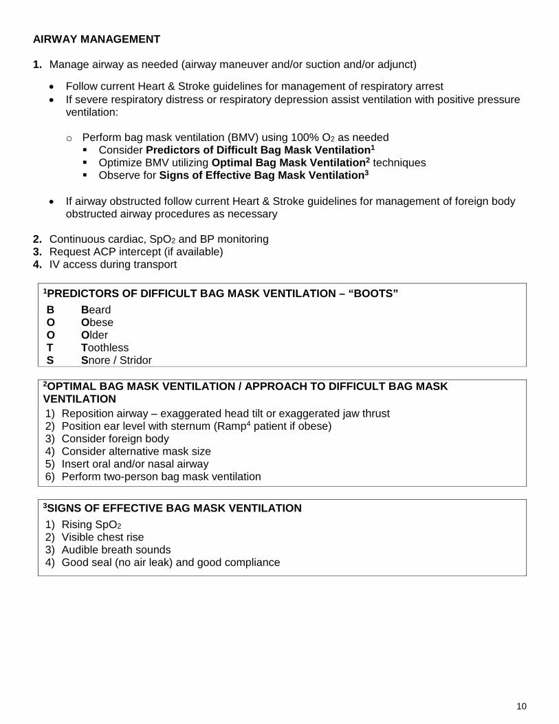
AIRWAY MANAGEMENT 1. Manage airway as needed (airway maneuver and/or suction and/or adjunct)
• Follow current Heart & Stroke guidelines for management of respiratory arrest • If severe respiratory distress or respiratory depression assist ventilation with positive pressure
ventilation:
o Perform bag mask ventilation (BMV) using 100% O2 as needed Consider Predictors of Difficult Bag Mask Ventilation1 Optimize BMV utilizing Optimal Bag Mask Ventilation2 techniques Observe for Signs of Effective Bag Mask Ventilation3
• If airway obstructed follow current Heart & Stroke guidelines for management of foreign body
obstructed airway procedures as necessary
2. Continuous cardiac, SpO2 and BP monitoring 3. Request ACP intercept (if available) 4. IV access during transport
1PREDICTORS OF DIFFICULT BAG MASK VENTILATION – “BOOTS” B O O T S
Beard Obese Older Toothless Snore / Stridor
2OPTIMAL BAG MASK VENTILATION / APPROACH TO DIFFICULT BAG MASK VENTILATION 1) Reposition airway – exaggerated head tilt or exaggerated jaw thrust 2) Position ear level with sternum (Ramp4 patient if obese) 3) Consider foreign body 4) Consider alternative mask size 5) Insert oral and/or nasal airway 6) Perform two-person bag mask ventilation
3SIGNS OF EFFECTIVE BAG MASK VENTILATION 1) Rising SpO2 2) Visible chest rise 3) Audible breath sounds 4) Good seal (no air leak) and good compliance

11
AIRWAY MANAGEMENT Cont’d
4RAMPING FOR PATIENTS WITH OBESITY
Figure A: Patient positioned without ramping
Figure B Patient ramped so that the sternum and ear line up.This position should improve ventilation
12
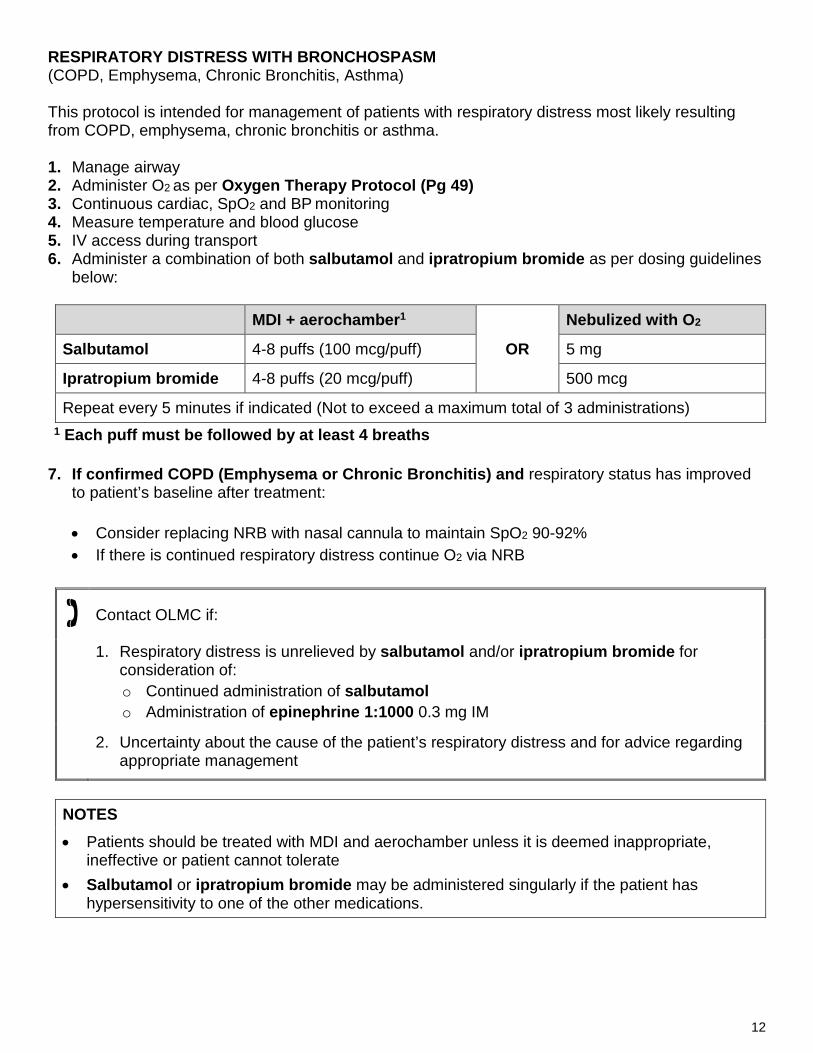
RESPIRATORY DISTRESS WITH BRONCHOSPASM (COPD, Emphysema, Chronic Bronchitis, Asthma) This protocol is intended for management of patients with respiratory distress most likely resulting from COPD, emphysema, chronic bronchitis or asthma. 1. Manage airway 2. Administer O2 as per Oxygen Therapy Protocol (Pg 49) 3. Continuous cardiac, SpO2 and BP monitoring 4. Measure temperature and blood glucose 5. IV access during transport 6. Administer a combination of both salbutamol and ipratropium bromide as per dosing guidelines
below:
MDI + aerochamber1
OR
Nebulized with O2
Salbutamol 4-8 puffs (100 mcg/puff) 5 mg
Ipratropium bromide 4-8 puffs (20 mcg/puff) 500 mcg
Repeat every 5 minutes if indicated (Not to exceed a maximum total of 3 administrations) 1 Each puff must be followed by at least 4 breaths
7. If confirmed COPD (Emphysema or Chronic Bronchitis) and respiratory status has improved
to patient’s baseline after treatment: • Consider replacing NRB with nasal cannula to maintain SpO2 90-92% • If there is continued respiratory distress continue O2 via NRB
Contact OLMC if:
1. Respiratory distress is unrelieved by salbutamol and/or ipratropium bromide for
consideration of: o Continued administration of salbutamol o Administration of epinephrine 1:1000 0.3 mg IM
2. Uncertainty about the cause of the patient’s respiratory distress and for advice regarding
appropriate management
NOTES • Patients should be treated with MDI and aerochamber unless it is deemed inappropriate,
ineffective or patient cannot tolerate • Salbutamol or ipratropium bromide may be administered singularly if the patient has
hypersensitivity to one of the other medications.

13
ALLERGY AND ANAPHYLAXIS
FINDINGS OF ANAPHYLAXIS 1) Acute onset (minutes to hours) of TWO OR MORE of the following after exposure to a LIKELY
ALLERGEN:
• Skin symptoms (hives, itching, flushing) • Oropharyngeal edema (lips, tongue, uvula) • Respiratory compromise (dyspnea, wheeze, stridor, hypoxemia) • Gastrointestinal symptoms (crampy abdominal pain, vomiting, diarrhea) • Reduced blood pressure or associated symptoms (hypotonia, collapse, syncope)
OR
2) Hypotension alone after exposure to a KNOWN ALLERGEN for patient
1. Manage airway and assist ventilations as necessary 2. Administer O2 as per Oxygen Therapy Protocol (Pg 49) 3. Continuous cardiac, SpO2 and BP monitoring 4. IV access 5. If shock present, administer a fluid bolus as per Adult Fluid Therapy Protocol (Pg 48) 6. If Findings of Anaphylaxis present administer:
• Epinephrine 1:1000 0.3 mg IM, ideally in the lateral thigh o Repeat once in 5 minutes if no improvement
AND
• DiphenhydrAMINE1 50 mg IV
7. If respiratory distress present (including wheezing), administer salbutamol: MDI + aerochamber1
OR Nebulized with O2
Salbutamol 4-8 puffs (100 mcg/puff) 5 mg
Repeat every 5 minutes if indicated (Not to exceed a maximum total of 3 administrations) 1Each puff must be followed by at least 4 breaths
1 NOTE • Patients should be treated with MDI with aerochamber unless it is deemed inappropriate,
ineffective or patient cannot tolerate • Epinephrine is relatively contraindicated in the setting of ischemic chest pain. In the rare event
that you suspect a patient has ischemic chest pain combined with anaphylaxis, contact OLMC prior to administration of epinephrine.
• May give diphenhydrAMINE 25-50 mg IV/IM alone for isolated hives. • There is NO absolute contraindication to epinephrine in a patient with anaphylaxis. • DiphenhydrAMINE DOES NOT improve angioedema or respiratory symptoms in
anaphylaxis.

14
CARDIAC ARREST If patient meets criteria outlined in the DNR Protocol (Pg 18) or Obvious Death Protocol (Pg 17) do not proceed with resuscitation 1. Confirm Vital Signs Absent (VSA) and initiate chest compressions 2. 100% O2 via BMV (15 L/min) 3. Continuous cardiac and SpO2 monitoring 4. Request ACP intercept (if available) 5. Consider and treat Reversible Causes1 6. IV access (DO NOT delay or interrupt CPR)
GENERAL GUIDELINES • Confirm absence of pulse – pulse check NOT exceeding 10 seconds • Initiate compressions immediately: C-A-B Sequence • Begin CPR while immediately attaching defibrillator – Analyze, defibrillate without delay if
indicated • Ensure high quality CPR
o Minimize interruptions in CPR o Allow full recoil of the chest between compressions o Rotate rescuers every 2 minutes (if resources allow) concurrent with pulse checks
• After third rhythm analysis determine if patient meets Termination of Resuscitation (TOR) Protocol (Pg 16) prior to initiating transport. If patient does not meet TOR, continue CPR and initiate transport.
• Analyze rhythm every 10 minutes thereafter. Continue CPR. • If return of spontaneous circulation (ROSC) proceed immediately with Post Cardiac Arrest
Care Protocol (Pg 15) • If re-arrest occurs during transport, resume Cardiac Arrest Protocol
HYPOTHERMIC CARDIAC ARREST (CORE TEMPERATURE LESS THAN 32ºC) • Hypothermic patients are to be resuscitated as per Cardiac Arrest Protocol above • Resuscitation will be continued until active re-warming has returned core temperature to
normal or there has been ROSC
1 REVERSIBLE CAUSES OF CARDIAC ARREST Hypovolemia
Hypoxia Hypothermia Hypoglycemia Drug Overdose
15
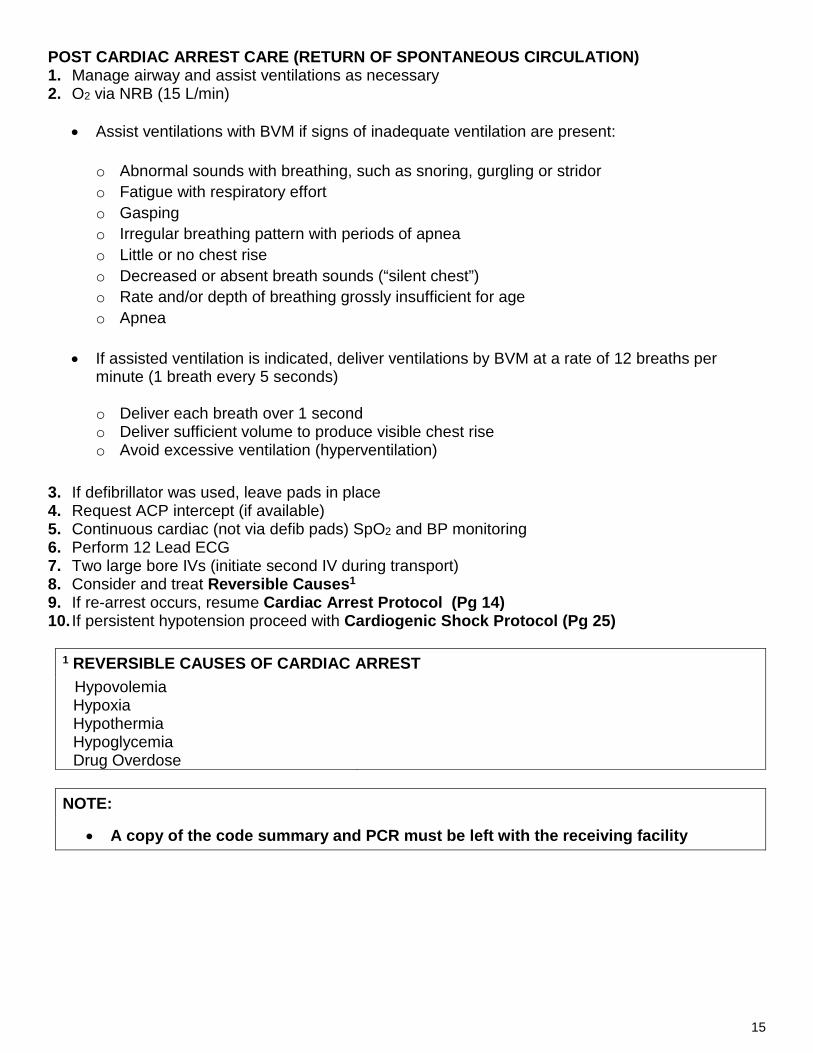
POST CARDIAC ARREST CARE (RETURN OF SPONTANEOUS CIRCULATION) 1. Manage airway and assist ventilations as necessary 2. O2 via NRB (15 L/min)
• Assist ventilations with BVM if signs of inadequate ventilation are present:
o Abnormal sounds with breathing, such as snoring, gurgling or stridor o Fatigue with respiratory effort o Gasping o Irregular breathing pattern with periods of apnea o Little or no chest rise o Decreased or absent breath sounds (“silent chest”) o Rate and/or depth of breathing grossly insufficient for age o Apnea
• If assisted ventilation is indicated, deliver ventilations by BVM at a rate of 12 breaths per minute (1 breath every 5 seconds) o Deliver each breath over 1 second o Deliver sufficient volume to produce visible chest rise o Avoid excessive ventilation (hyperventilation)
3. If defibrillator was used, leave pads in place 4. Request ACP intercept (if available) 5. Continuous cardiac (not via defib pads) SpO2 and BP monitoring 6. Perform 12 Lead ECG 7. Two large bore IVs (initiate second IV during transport) 8. Consider and treat Reversible Causes1 9. If re-arrest occurs, resume Cardiac Arrest Protocol (Pg 14) 10. If persistent hypotension proceed with Cardiogenic Shock Protocol (Pg 25)
1 REVERSIBLE CAUSES OF CARDIAC ARREST Hypovolemia
Hypoxia Hypothermia Hypoglycemia Drug Overdose
NOTE:
• A copy of the code summary and PCR must be left with the receiving facility

16
TERMINATION OF RESUSCITATION (TOR) This TOR Protocol CANNOT be utilized in situations related to: 1) Age less than 18 years 2) Pregnancy 3) Hypothermia 4) Electrocution including lightning strike 5) Trauma (Blunt or Penetrating Traumatic Cardiac Arrest Protocol Pg 46-47) 6) Poisoning or drug overdose 7) Sudden reversible event (anaphylaxis, choking, drowning with submersion less than 60 minutes,
asphyxia) In these cases resuscitation and transport must proceed as per usual cardiac arrest protocols.
CRITERIA FOR TERMINATION OF RESUSCITATION Termination of resuscitation is to be applied when resuscitation of cardiac arrest has been initiated and prior to transport The PCP can terminate resuscitative efforts when ALL of the following criteria are met: 1) Cardiac arrest unwitnessed by EMS provider 2) No ROSC has occurred after 3 full rounds of CPR by EMS Personnel
AND 3) No shock(s) advised or delivered by EMS provider or Medical First Responder If ALL requirements are met, proceed with the Management of Death Protocol (Pg 19)

17
OBVIOUS DEATH The PCP will not start resuscitation of a patient of any age that has suffered cardiac arrest (not breathing and no palpable pulse) if any of the following signs of obvious death are present:
1) Rigor mortis 2) Dependent lividity 3) Decapitation 4) Transection of the torso 5) Decomposition 6) Confirmed submersion greater than 60 minutes 7) Obvious destruction of brain, heart or lungs that is incompatible with life 8) Other catastrophic injury that is incompatible with life
NOTE • Proceed with Management of Death Protocol (Pg 19) upon recognition of cardiac arrest
meeting Obvious Death criteria
18
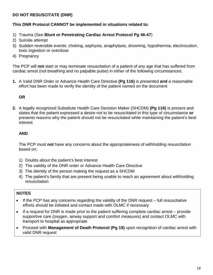
DO NOT RESUSCITATE (DNR) This DNR Protocol CANNOT be implemented in situations related to: 1) Trauma (See Blunt or Penetrating Cardiac Arrest Protocol Pg 46-47) 2) Suicide attempt 3) Sudden reversible events: choking, asphyxia, anaphylaxis, drowning, hypothermia, electrocution,
toxic ingestion or overdose 4) Pregnancy The PCP will not start or may terminate resuscitation of a patient of any age that has suffered from cardiac arrest (not breathing and no palpable pulse) in either of the following circumstances: 1. A Valid DNR Order or Advance Health Care Directive (Pg 116) is presented and a reasonable
effort has been made to verify the identity of the patient named on the document
OR 2. A legally recognized Substitute Health Care Decision Maker (SHCDM) (Pg 116) is present and
states that the patient expressed a desire not to be resuscitated in this type of circumstance or presents reasons why the patient should not be resuscitated while maintaining the patient’s best interest
AND
The PCP must not have any concerns about the appropriateness of withholding resuscitation based on:
1) Doubts about the patient’s best interest 2) The validity of the DNR order or Advance Health Care Directive 3) The identity of the person making the request as a SHCDM 4) The patient’s family that are present being unable to reach an agreement about withholding
resuscitation
NOTES • If the PCP has any concerns regarding the validity of the DNR request – full resuscitative
efforts should be initiated and contact made with OLMC if necessary • If a request for DNR is made prior to the patient suffering complete cardiac arrest – provide
supportive care (oxygen, airway support and comfort measures) and contact OLMC with transport to hospital as appropriate
• Proceed with Management of Death Protocol (Pg 19) upon recognition of cardiac arrest with valid DNR request

19
MANAGEMENT OF DEATH (Resuscitation Terminated or not indicated)
CAUTION
This protocol is not to be utilized as the initial assessment of the unconscious patient to determine if they are in cardiac arrest. The initial assessment to determine if cardiac arrest is present should be conducted in accordance with the standards outlined in the Cardiac Arrest Protocol, with a pulse check not exceeding 10 seconds duration. This protocol outlines the criteria that must be evaluated and documented in the PCR after it has been determined that resuscitation from cardiac arrest is not indicated or should be terminated when directed to do so by the Blunt or Penetrating Cardiac Arrest (Pg 46-47), DNR (Pg 18), Obvious Death (Pg 17) or Termination of Resuscitation (Pg 16) Protocol(s).
Once it is determined that resuscitation from cardiac arrest is not indicated or should be terminated, proceed with the following steps: 1. Evaluate for, confirm and document the presence of all the Documentation of Death Criteria1 2. Determine if the death meets criteria for Reportable Death2 or Expected Death3
• If the death was an Expected Death inquire whether the patient is enrolled in the “End of Life Program” and proceed as follows: o If patient enrolled in the End of Life Program, contact the health care professional that has
been identified to the family for purposes of notification of death o If the patient is not enrolled in the End of Life Program, notify the family physician or
designate. If the family physician or designate is unavailable, contact the police
• If the death meets the criteria of a Reportable Death proceed as follows: 1) Do not disturb the scene – limit access only to essential responders 2) Leave all disposable medical equipment and supplies used in the resuscitation in place –
do not remove from the scene 3) Leave defibrillation pads and airway adjuncts in position 4) Leave the deceased in position – do not move or cover the body 5) Exit the scene of the death immediately using the same pathway as was used to enter 6) Do not permit anyone entrance into the scene 7) Notify police
3. Provide comfort to the bereaved
• Disclose death simply and directly with warmth and compassion • Listen and empathize • Assist locating support – relative, friend, clergy, etc.

20
MANAGEMENT OF DEATH (RESUSCITATION TERMINATED OR NOT INDICATED) Cont’d 4. Allow the bereaved to see the body if they wish:
• If not a reportable death, prepare the deceased – clean up medical supplies, cover with blanket, place pillow under head, close eyes, wipe up body fluids, etc.
• Prepare the bereaved for what they will see and answer any questions • Do not rush the bereaved
5. Remain on-scene until appropriate supports arrive for the bereaved and/or:
• Family physician, police, medical examiner or funeral home arrive and assume control of the deceased
• Crew is requested to respond to another life-threatening time-dependent emergency call
1DOCUMENTATION OF DEATH CRITERIA Assess and document ALL of the following criteria: 1) No palpable carotid pulse (assess for 60 seconds) 2) No spontaneous respiratory effort (assess for 60 seconds) 3) No heart sounds (assess for 60 seconds) 4) Non-reactive pupils
2REPORTABLE DEATH CRITERIA When ANY ONE OR MORE of the following criteria present: 1) Death as a result of violence, accident or suicide 2) An unexpected death when the person was in good health 3) Where the person was not under the care of a physician 4) The death is obviously suspicious in nature 5) Where the cause of death is undetermined 6) Death is the result of improper or suspected negligent treatment by another person
3EXPECTED DEATH Any death that does not meet Reportable Death Criteria
NOTES • Transport of the deceased must be completed by a licensed funeral director • An ambulance may transport the deceased only if the deceased is in a public place and the
funeral director will be extensively delayed (greater than 1 hour) or as directed by police or OLMC
21
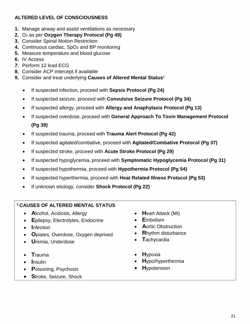
ALTERED LEVEL OF CONSCIOUSNESS 1. Manage airway and assist ventilations as necessary 2. O2 as per Oxygen Therapy Protocol (Pg 49) 3. Consider Spinal Motion Restriction 4. Continuous cardiac, SpO2 and BP monitoring 5. Measure temperature and blood glucose 6. IV Access 7. Perform 12 lead ECG 8. Consider ACP intercept if available 9. Consider and treat underlying Causes of Altered Mental Status¹
• If suspected infection, proceed with Sepsis Protocol (Pg 24) • If suspected seizure, proceed with Convulsive Seizure Protocol (Pg 34) • If suspected allergy, proceed with Allergy and Anaphylaxis Protocol (Pg 13)
• If suspected overdose, proceed with General Approach To Toxin Management Protocol (Pg 39)
• If suspected trauma, proceed with Trauma Alert Protocol (Pg 42)
• If suspected agitated/combative, proceed with Agitated/Combative Protocol (Pg 37)
• If suspected stroke, proceed with Acute Stroke Protocol (Pg 29)
• If suspected hypoglycemia, proceed with Symptomatic Hypoglycemia Protocol (Pg 31)
• If suspected hypothermia, proceed with Hypothermia Protocol (Pg 54)
• If suspected hyperthermia, proceed with Heat Related Illness Protocol (Pg 53)
• If unknown etiology, consider Shock Protocol (Pg 22)
1 CAUSES OF ALTERED MENTAL STATUS • Alcohol, Acidosis, Allergy • Epilepsy, Electrolytes, Endocrine • Infection • Opiates, Overdose, Oxygen deprived • Uremia, Underdose
• Heart Attack (MI) • Embolism • Aortic Obstruction • Rhythm disturbance • Tachycardia
• Trauma • Insulin • Poisoning, Psychosis • Stroke, Seizure, Shock
• Hypoxia • Hypo/hyperthermia • Hypotension

22
SHOCK 1. Manage airway and assist ventilations as necessary 2. Administer O2 as per Oxygen Therapy Protocol (Pg 49) 3. Control bleeding (if applicable) 4. Assess for Signs and Symptoms of Shock1 5. Continuous cardiac, SpO2 and BP monitoring 6. Measure temperature and blood glucose
7. Two large bore IVs (initiate second IV during transport) 8. Perform 12 lead ECG 9. Consider causes of shock and treat accordingly:
• If shock due to anaphylaxis, proceed with Allergy and Anaphylaxis Protocol (Pg 13) • If shock due to sepsis, proceed with Sepsis Protocol (Pg 24) • If shock due to cardiac etiology, proceed with Cardiogenic Shock Protocol (Pg 25)
10. For all other causes of shock or when the cause of shock is unknown, administer a fluid bolus as
per Adult Fluid Therapy Protocol (Pg 48)
1SIGNS AND SYMPTOMS OF SHOCK 1) Hypotension (SBP less than 90 mmHg)
AND
2) Any one or more of the following features:
• Rapid and / or shallow breathing • Cool and / or clammy skin • Rapid and / or weak pulse(s) • Near fainting and / or fainting • Weakness
Contact OLMC if patient remains hypotensive after initial fluid bolus for consideration of:
• Additional IV fluid administration

23
SHOCK Cont’d
NOTES • Shock is a life-threatening, progressive medical condition that results from the inadequate flow of
oxygenated blood to critical organs and tissues of the body. • When the blood pressure is inadequate to sustain a regular flow of oxygenated blood to the
organs and tissues of the body, end-organ damage will ensue and shock will eventually result. • Shock may result from a number of medical conditions including sepsis, trauma, blood loss,
anaphylaxis, severe dehydration and various medical conditions.
CAUTION
• Trendelenburg positioning is not indicated in the treatment of shock and is not to be utilized • Position the patient supine unless they are in severe respiratory distress • If the patient in shock is suffering from severe respiratory distress, position them semi-sitting and
assist ventilations as indicated

24
SEPSIS 1. Manage airway and assist ventilations as necessary 2. Administer O2 as per Oxygen Therapy Protocol (Pg 49) 3. Continuous cardiac, SpO2 and BP monitoring 4. Measure temperature and blood glucose 5. Two large bore IVs (initiate second IV during transport) 6. Perform 12 Lead ECG 7. If patient meets Sepsis Inclusion Criteria1 administer a fluid bolus of 20 mL/kg 0.9% NaCl
regardless of blood pressure
1SEPSIS INCLUSION CRITERIA 1) History suspicious for infection OR confirmed infection
AND
2) Any TWO OR MORE of the following clinical findings: • Temperature less than 36ºC or greater than 38ºC • Tachypnea (respiratory rate greater than 20) • Heart rate greater than 90
Contact OLMC if patient remains hypotensive after fluid bolus for consideration of:
• Additional IV fluid administration
DEFINITIONS Severe Sepsis Sepsis Inclusion Criteria + Any evidence of end-organ dysfunction
• Altered mental status, confusion or coma • Renal dysfunction or poor urine output • Respiratory distress or hypoxia • Myocardial ischemia
Septic Shock Sepsis Inclusion Criteria + SBP less than 90 mmHg despite administration of 20 mL/kg 0.9% NaCl
25
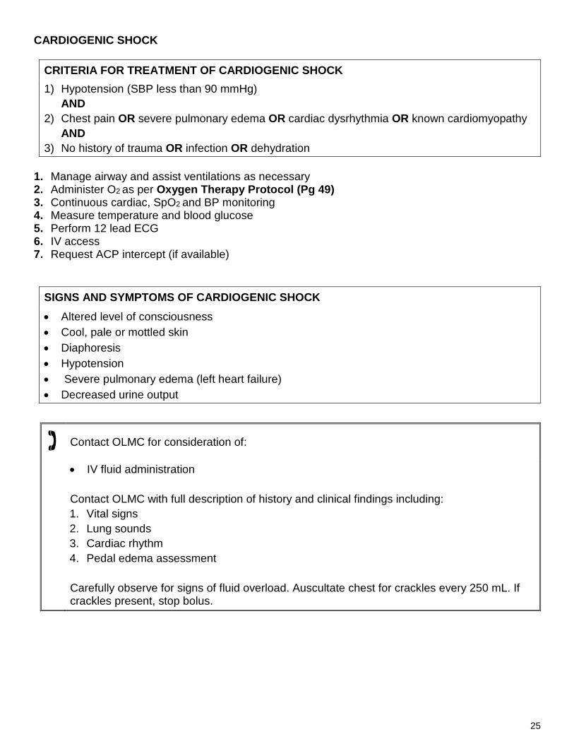
CARDIOGENIC SHOCK
CRITERIA FOR TREATMENT OF CARDIOGENIC SHOCK 1) Hypotension (SBP less than 90 mmHg)
AND 2) Chest pain OR severe pulmonary edema OR cardiac dysrhythmia OR known cardiomyopathy
AND 3) No history of trauma OR infection OR dehydration
1. Manage airway and assist ventilations as necessary 2. Administer O2 as per Oxygen Therapy Protocol (Pg 49) 3. Continuous cardiac, SpO2 and BP monitoring 4. Measure temperature and blood glucose 5. Perform 12 lead ECG 6. IV access 7. Request ACP intercept (if available)
SIGNS AND SYMPTOMS OF CARDIOGENIC SHOCK • Altered level of consciousness • Cool, pale or mottled skin • Diaphoresis • Hypotension • Severe pulmonary edema (left heart failure) • Decreased urine output
Contact OLMC for consideration of:
• IV fluid administration
Contact OLMC with full description of history and clinical findings including: 1. Vital signs 2. Lung sounds 3. Cardiac rhythm 4. Pedal edema assessment Carefully observe for signs of fluid overload. Auscultate chest for crackles every 250 mL. If crackles present, stop bolus.

26
PULMONARY EDEMA This protocol is intended for management of patients with severe and acute respiratory distress most likely resulting from pulmonary edema. 1. Manage airway and assist ventilations as necessary 2. Administer O2 as per Oxygen Therapy Protocol (Pg 49) 3. Continuous cardiac, SpO2 and BP monitoring 4. Position patient upright if SBP greater than 100 mmHg 5. Perform 12 lead ECG 6. IV access 7. Request ACP Intercept (if available) 8. Administer nitroglycerin 0.4mg SL
• Repeat every 5 minutes if indicated to a maximum of 6 sprays, until symptoms are relieved or SBP falls below 100 mmHg
• If hypotension develops or SBP falls below 100 mmHg following the administration of nitroglycerin discontinue further administration
Contact OLMC:
• For consideration of administration of nitroglycerin beyond six sprays • If there is uncertainty about the cause of the patient’s respiratory distress and for advice
regarding appropriate management
SIGNS AND SYMPTOMS OF PULMONARY EDEMA • Severe respiratory distress • Orthopnea • Crackles • Diaphoresis • Nocturnal dyspnea • Jugular vein distention • Cough that may contain foamy, blood tinged sputum • Peripheral edema

27
ISCHEMIC CHEST PAIN This protocol is intended for management of patients with chest pain suspected to be of ischemic etiology.
1. Manage airway and assist ventilations as necessary 2. Administer O2 to keep SpO2 95% or greater 3. Continuous cardiac, SpO2 and BP monitoring 4. Perform 12 lead ECG pre and post intervention and in accordance with Serial 12 Lead ECGs1
box 5. Administer ASA 160–162 mg PO chewed 6. IV access 7. Administer nitroglycerin2 0.4 mg SL
• Repeat every 5 minutes if indicated to a maximum of 6 sprays, until chest pain is relieved or SBP falls below 100 mmHg
• If patient has no response to nitroglycerin following the administration of three (3) doses, discontinue use.
8. Request ACP intercept (if available)
1 SERIAL 12 LEAD ECGs Serial 12 lead ECGs must be performed as outlined below:
1) On scene (prior to treatment) 2) In ambulance just prior to transport 3) Every 15 minutes during transport (if transport time greater than 30 minutes) 4) Just prior to arrival to receiving health care facility 5) Any time patient condition or ECG rhythm changes
If the initial 12 lead demonstrates evidence of ST elevation MI serial 12 leads are not required unless there is a change in patient condition or ECG rhythm changes
2 INFERIOR WALL MYOCARDIAL INFARCTION (MI) • Do not administer nitroglycerin if an inferior wall MI is suspected or confirmed by 12 Lead
ECG and / or patients SBP has been less than 100 mmHg at any time during current event • Fluid therapy is not to be used to increase SBP to greater than 100mmHg to aid in
nitroglycerin administration
STEMI ALERT 1) Notify receiving facility of “STEMI Alert” if ECG printout that reads “*****Acute MI***** or Left
Bundle Branch Block” in a patient experiencing chest pain 2) ASA 160-162 mg PO chewed if not already administered 3) Establish 2nd IV during transport (same arm, if possible) 4) Complete Thrombolytic Checklist for STEMI during transport
Contact OLMC:
• For consideration of administration of nitroglycerin beyond six sprays

28
SYMPTOMATIC DYSRHYTHMIAS (ADULT) (Suspected cardiac origin, non-traumatic) This protocol is intended for patients with symptomatic or clinically significant cardiac dysrhythmias. A variety of cardiac dysrhythmias may lead to symptoms or clinically significant findings including:
• Bradycardia • Wide Complex Tachycardia • Narrow Complex Tachycardia • Atrial Fibrillation with heart rate greater than 120 • Atrial Flutter
Examples of symptoms that should prompt concern for clinically significant dysrhythmia are provided below. 1. Manage airway and assist ventilations as necessary 2. Administer O2 as per Oxygen Therapy Protocol (Pg 49) 3. Continuous cardiac, SpO2 and BP monitoring 4. Perform 12 lead ECG 5. IV access 6. Request ACP intercept (if available) SIGNS AND SYMPTOMS OF CLINICALLY SIGNIFICANT DYSRHYTHMIAS
Signs Symptoms • Hypotension • Shock • Altered level of consciousness • Tachypnea • Hypoxia • Respiratory distress • Diaphoresis • Pallor or mottled skin • Vomiting
• Chest pain • Dyspnea • Syncope or presyncope • Palpitation • Nausea

29
ACUTE STROKE 1. Manage airway and assist ventilations as necessary 2. Administer O2 as per Oxygen Therapy Protocol (Pg 49) 3. Establish and document Last Seen Normal (LSN) Time1 4. Continuous cardiac, SpO2 and BP monitoring 5. Measure temperature and blood glucose
• Treat hypoglycemia as per Symptomatic Hypoglycemia Protocol (Pg 31)
6. Determine if patient is candidate for direct transport to a Stroke Centre using Paramedic Prompt
Card (Pg 30) 7. IV during transport
1 LAST SEEN NORMAL (LSN) TIME • The last time the patient was witnessed or confirmed in their usual state of health and
completely without signs or symptoms of stroke
CAUTION
• If at any time during your patient contact there is airway compromise or patient condition becomes unstable, transport to the closest Emergency Department, even if it is not a designated Stroke Centre
30
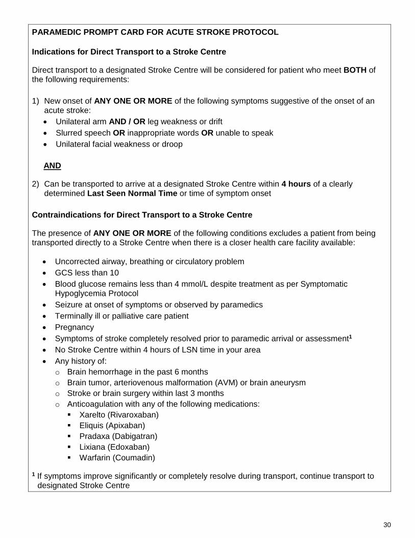
PARAMEDIC PROMPT CARD FOR ACUTE STROKE PROTOCOL
Indications for Direct Transport to a Stroke Centre
Direct transport to a designated Stroke Centre will be considered for patient who meet BOTH of the following requirements: 1) New onset of ANY ONE OR MORE of the following symptoms suggestive of the onset of an
acute stroke: • Unilateral arm AND / OR leg weakness or drift • Slurred speech OR inappropriate words OR unable to speak • Unilateral facial weakness or droop AND
2) Can be transported to arrive at a designated Stroke Centre within 4 hours of a clearly determined Last Seen Normal Time or time of symptom onset
Contraindications for Direct Transport to a Stroke Centre
The presence of ANY ONE OR MORE of the following conditions excludes a patient from being transported directly to a Stroke Centre when there is a closer health care facility available:
• Uncorrected airway, breathing or circulatory problem • GCS less than 10 • Blood glucose remains less than 4 mmol/L despite treatment as per Symptomatic
Hypoglycemia Protocol • Seizure at onset of symptoms or observed by paramedics • Terminally ill or palliative care patient • Pregnancy • Symptoms of stroke completely resolved prior to paramedic arrival or assessment1 • No Stroke Centre within 4 hours of LSN time in your area • Any history of:
o Brain hemorrhage in the past 6 months o Brain tumor, arteriovenous malformation (AVM) or brain aneurysm o Stroke or brain surgery within last 3 months o Anticoagulation with any of the following medications: Xarelto (Rivaroxaban) Eliquis (Apixaban) Pradaxa (Dabigatran) Lixiana (Edoxaban) Warfarin (Coumadin)
1 If symptoms improve significantly or completely resolve during transport, continue transport to
designated Stroke Centre

31
SYMPTOMATIC HYPOGLYCEMIA 1. Manage airway and assist ventilations as necessary 2. Administer O2 as per Oxygen Therapy Protocol (Pg 49) 3. Continuous cardiac, SpO2 and BP monitoring 4. Measure temperature and blood glucose level (BGL) 5. IV access 6. If blood glucose is less than 4 mmol/L, administer ONE of the following medications and recheck
blood glucose in accordance with table below: Patient able to maintain own airway (Awake and able to cough and swallow)
IV established Unable to establish IV
Oral glucose options: 1) Dex 4® tablets 20 g (5 tablets) 2) Insta-glucose® 1 tube (30 g) 3) 1 cup (250 mL) of juice or pop (Non-
diet) 4) 4 teaspoons (20 mL) or 4 packets of
table sugar dissolved in water
Dextrose 50% (D50%) 25 g (50 mL) IVP
Glucagon1 1 mg IM
Recheck BGL in 15 minutes Recheck BGL in 10 minutes Recheck BGL in 20 minutes
7. Repeat Step 6 once if necessary 8. If the patient expresses a wish to remain home rather than continue care to hospital evaluate for
Treat and Release inclusion and exclusion criteria (Pg 32)
Contact OLMC if blood glucose remains below 4 mmol/L after 2nd dose of dextrose or glucagon
1 NOTES • Anticipate that it could take up to 20 minutes to observe an effect from glucagon • While waiting for glucagon to take effect, manage patient’s airway as indicated and initiate
transport
CAUTION
• If head injury or stroke suspected administer half of the usual dose of dextrose, recheck blood glucose and then administer the second half dose if necessary
• The goal is to correct hypoglycemia while avoiding transient hyperglycemia that may lead to cerebral edema
32
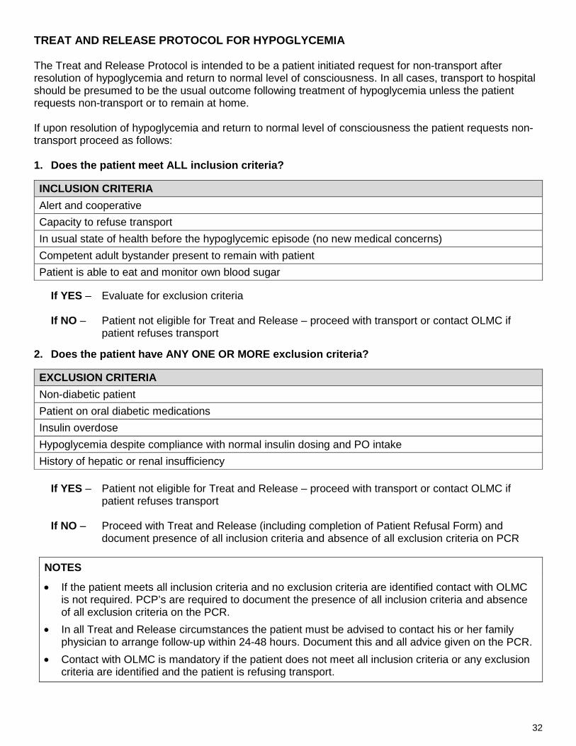
TREAT AND RELEASE PROTOCOL FOR HYPOGLYCEMIA The Treat and Release Protocol is intended to be a patient initiated request for non-transport after resolution of hypoglycemia and return to normal level of consciousness. In all cases, transport to hospital should be presumed to be the usual outcome following treatment of hypoglycemia unless the patient requests non-transport or to remain at home. If upon resolution of hypoglycemia and return to normal level of consciousness the patient requests non-transport proceed as follows:
1. Does the patient meet ALL inclusion criteria?
INCLUSION CRITERIA Alert and cooperative Capacity to refuse transport In usual state of health before the hypoglycemic episode (no new medical concerns) Competent adult bystander present to remain with patient Patient is able to eat and monitor own blood sugar
If YES – Evaluate for exclusion criteria
If NO – Patient not eligible for Treat and Release – proceed with transport or contact OLMC if
patient refuses transport 2. Does the patient have ANY ONE OR MORE exclusion criteria?
EXCLUSION CRITERIA Non-diabetic patient Patient on oral diabetic medications Insulin overdose Hypoglycemia despite compliance with normal insulin dosing and PO intake History of hepatic or renal insufficiency
If YES – Patient not eligible for Treat and Release – proceed with transport or contact OLMC if
patient refuses transport
If NO – Proceed with Treat and Release (including completion of Patient Refusal Form) and document presence of all inclusion criteria and absence of all exclusion criteria on PCR
NOTES
• If the patient meets all inclusion criteria and no exclusion criteria are identified contact with OLMC is not required. PCP’s are required to document the presence of all inclusion criteria and absence of all exclusion criteria on the PCR.
• In all Treat and Release circumstances the patient must be advised to contact his or her family physician to arrange follow-up within 24-48 hours. Document this and all advice given on the PCR.
• Contact with OLMC is mandatory if the patient does not meet all inclusion criteria or any exclusion criteria are identified and the patient is refusing transport.
33
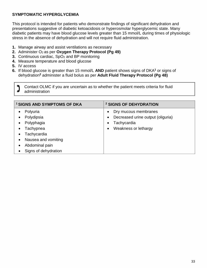
SYMPTOMATIC HYPERGLYCEMIA This protocol is intended for patients who demonstrate findings of significant dehydration and presentations suggestive of diabetic ketoacidosis or hyperosmolar hyperglycemic state. Many diabetic patients may have blood glucose levels greater than 15 mmol/L during times of physiologic stress in the absence of dehydration and will not require fluid administration. 1. Manage airway and assist ventilations as necessary 2. Administer O2 as per Oxygen Therapy Protocol (Pg 49) 3. Continuous cardiac, SpO2 and BP monitoring 4. Measure temperature and blood glucose 5. IV access 6. If blood glucose is greater than 15 mmol/L AND patient shows signs of DKA1 or signs of
dehydration2 administer a fluid bolus as per Adult Fluid Therapy Protocol (Pg 48)
Contact OLMC if you are uncertain as to whether the patient meets criteria for fluid administration
1 SIGNS AND SYMPTOMS OF DKA 2 SIGNS OF DEHYDRATION • Polyuria • Polydipsia • Polyphagia • Tachypnea • Tachycardia • Nausea and vomiting • Abdominal pain • Signs of dehydration
• Dry mucous membranes • Decreased urine output (oliguria) • Tachycardia • Weakness or lethargy

34
CONVULSIVE SEIZURES 1. Manage airway and assist ventilations as necessary 2. Administer O2 as per Oxygen Therapy Protocol (Pg 49) 3. Spinal immobilization if unprotected fall to ground and seizure has stopped if indicated by C-Spine
Assessment (Pg 43) and Spine Assessment for Backboard (Pg 44) 4. Position patient
• Actively seizing – place supine and protect from injury • Postictal – place left lateral recumbent and maintain airway
5. Continuous cardiac, SpO2 and BP monitoring 6. Measure temperature and blood glucose
• Treat hypoglycemia as per Symptomatic Hypoglycemia Protocol (Pg 31)
7. IV access 8. If available, request ACP intercept for active seizures or recurrent seizures (status epilepticus)

35
ADULT NAUSEA AND VOMITING 1. Manage airway and assist ventilations as necessary 2. O2 as per Oxygen Therapy Protocol (Pg. 49) 3. Continuous cardiac, SpO2 and BP monitoring 4. Measure temperature and blood glucose 5. IV access 6. Position the patient in a position of comfort 7. If severe nausea and vomiting administer:
• DimenhyDRINATE 25-50 mg IV/IM
o Repeat once in 15 minutes if indicated (not to exceed a maximum total dose of 50 mg) OR
• Metoclopramide 10 mg SIVP over 2 to 5 minutes if any of the following criteria apply:
o Severe nausea and vomiting refractory to dimenhyDRINATE after 15 minutes since last dose o Allergy or contraindication to dimenhyDRINATE o Altered LOC or head injury
8. If metoclopramide has been administered and acute extrapyramidal signs or symptoms1 develop,
reassure patient and administer diphenhydrAMINE 50 mg IV
1 EXTRAPYRAMIDAL SIGNS AND SYMPTOMS
• Akathisia – a severe and unpleasant sensation of restlessness in patients causing them severe anxiety and inability to sit still
• Dystonia – increased rigidity or muscle contraction that may result in twisting or abnormal postures
• Dyskinesia – abnormal or repetitive movements (e.g.: lip smacking, eye twitching, etc.) Administration of diphenhydrAMINE is not indicated for treatment of chronic extrapyramidal signs and symptoms
NOTES
• DimenhyDRINATE OR metoclopramide may be administered by IM route if indications are present and you are unable to establish an IV.

36
PAIN MANAGEMENT 1. Manage airway and assist ventilations as necessary 2. O2 as per Oxygen Therapy Protocol (Pg 49) 3. Continuous cardiac, SpO2, and BP monitoring 4. IV access 5. Administer ketorolac 15 mg SIVP/IM if severe pain1 due to one of the following:
• Acute musculoskeletal trauma • Uncomplicated renal or biliary colic if the presentation is consistent with previous episodes • Mechanical back pain • Burns
6. If severe headache2 and patient meets the Criteria for Metoclopramide in Migraine3, administer: • Metoclopramide 10 mg SIVP
o May administer 10 mg IM if indications are present AND you are unable to establish an IV o If metoclopramide has been administered and acute extrapyramidal signs or
symptoms4 develop, reassure patient and administer diphenhydrAMINE 50 mg IV 7. If patient develops nausea or vomiting proceed with Nausea and Vomiting Protocol (Pg 35)
3CRITERIA FOR METOCLOPRAMIDE IN MIGRAINE
1) Patient has all of the following: • Acute and severe unilateral headache • History of diagnosed migraine • Presentation of current migraine is consistent with previous migraines
o Any aura is consistent with previous auras AND
2) Patient has none of the following: • Recent head trauma • New onset of fever greater than 38◦C • New neurological abnormality, including acute seizure
4 EXTRAPYRAMIDAL SIGNS AND SYMPTOMS • Akathisia – a severe and unpleasant sensation of restlessness in patients causing them
severe anxiety and inability to sit still • Dystonia – increased rigidity or muscle contraction that may result in twisting or abnormal
postures • Dyskinesia – abnormal or repetitive movements (e.g.: lip smacking, eye twitching, etc.)
Administration of diphenhydrAMINE is not indicated for treatment of chronic extrapyramidal signs and symptoms
NOTES 1 Document pain severity pre and post intervention. 2 Consider migraine mimics, including stroke and pre-eclampsia.

37
AGITATED / COMBATIVE (Patient is danger to self or others) 1. Contact police and request that they attend the scene immediately 2. Manage airway and assist ventilation as necessary 3. Administer O2 as per Oxygen Therapy Protocol (Pg 49) 4. Continuous cardiac, SpO2 and BP monitoring 5. Measure temperature and blood glucose
• Treat hypoglycemia as per Symptomatic Hypoglycemia Protocol (Pg 31) 6. Consider and treat Reversible or Treatable Causes of Altered Mental Status1 7. IV access 8. Request ACP intercept (if available) 9. Attempt verbal management techniques for crisis intervention to de-escalate the situation and
calm the patient 10. If Indications for Physical Restraint2 present, apply the least amount of physical restraint
necessary to protect the patient from harming themselves or bystanders until the police arrive, as per Agitated Combative / Physical Restraint Reference (Pg 110)
1 REVERSIBLE OR TREATABLE CAUSES OF ALTERED MENTAL STATUS • Hypoxia • Hypotension • Hypoglycemia • Medications or Toxins • Sepsis
2 INDICATIONS FOR PHYSICAL RESTRAINT 1) Imminent danger3 to life or threat of physical harm to patient and/or bystanders
AND
2) Attempts at verbal de-escalation have failed
AND
3) Attempts to restrain do not place the practitioner(s) at significant risk of harm to themselves
3 NOTES Imminent Danger – an immediate threat of significant harm to one’s self or others, up to and including death
Examples of Imminent Danger: • Actively attempting suicide • Actively attempting to cause serious bodily injury to others • Attempting to jump from a building or moving vehicle

38
AGITATED / COMBATIVE Cont’d
CAUTION
• There is a high risk of positional asphyxia and/or aspiration in patients undergoing chemical or physical restraint. Close and continuous monitoring of these patients, including airway patency and adequacy of respirations is mandatory
• At NO TIME should the patient be restrained in the prone (face or chest-down) position • Always maintain an ability to escape the scene. Position yourself between the patient and the
exit at all times to maintain a safe exit should the situation escalate • Be alert for potential weapons and hazards. If the patient has a weapon, do not attempt to
disarm them. Instead, leave the scene and stage until the police declare the scene safe to re-enter
• Be aware of signs of increased agitation or aggression including, but not limited to: o Tense posture o Loud speech o Pacing o Threatening statements o Clenched hands o Hostile or aggressive body language

39
GENERAL APPROACH TO TOXIN MANAGEMENT
1. Scene safety: protect rescuers and patients from immediate danger and contamination
• Toxic exposures might require special precautions, including CBRNE precautions or decontamination, before patient treatment begins
2. Manage airway and assist ventilations as necessary 3. Administer O2 as per Oxygen Therapy Protocol (Pg 49) 4. Continuous cardiac, SpO2 and BP monitoring 5. Measure temperature and blood glucose 6. IV access 7. Perform 12 Lead ECG 8. If seizure occurs refer to Convulsive Seizure Protocol (Pg 34) 9. Request ACP intercept (if available). Do not delay transport in cases of severely symptomatic
patients
OPIOIDS This protocol is intended for management of the severely symptomatic adult patient with suspected or confirmed ingestion or use of an opioid1 agent.
Therapy is not intended to return patient to a normal level of consciousness. The goal of treatment is a respiratory rate greater than 10 per minute with adequate ventilation.
If ALL of the following criteria are met proceed with naloxone administration as outlined:
• Impaired consciousness • Respiratory rate less than 10 per minute • Pupil constriction (except in suspected meperidine (Demerol) or tramadol ingestion) • Requiring assisted ventilation
Administer naloxone2 0.2-0.4 mg IM/IV • Repeat every 2-3 minutes if indicated, titrated to improved respiratory drive
1 EXAMPLES OF OPIOIDS INCLUDE BUT ARE NOT LIMITED TO: • Morphine • Hydromorphone (Dilaudid) • Codeine • Oxycodone (Percocet, OxyContin, OxyNEO) • Fentanyl
• Tramadol (Tramacet) • Meperidine (Demerol) • Methadone • Buprenorphine (Butrans) • Heroin
2 NOTES • Return to normal alertness is not a required outcome following naloxone administration • Meperidine (Demerol) or tramadol are opioids that do not cause pupil constriction
• Examine patient for transdermal opioid patches (placed on the skin), including fentanyl and buprenorphine (Butrans) and remove with a gloved hand
Contact OLMC for guidance if required.

40
UNCONTROLLED TRAUMATIC BLEEDING 1. Manage airway and assist ventilations as necessary 2. O2 as per Oxygen Therapy Protocol (Pg 49) 3. Control Bleeding:
• Compressible site: o Apply direct pressure to site of active bleeding o If hemostasis achieved, apply pressure dressing and monitor for re-bleeding
• Non-compressible site OR Hemostasis not achieved at a compressible site: o Insert hemostatic gauze into the wound and apply direct pressure for a minimum of three
(3) minutes. Release manual pressure only when hemostasis is achieved, then apply pressure dressing over the hemostatic gauze. Monitor for re-bleeding.
• Catastrophic extremity injury with massive hemorrhage: o Rapidly apply a tourniquet at least 5 cm proximal to the injury, tighten until bleeding is
controlled • Suspected pelvic fracture:
o Apply a pelvic sling (Pg 41) and tighten until reasonably stabilized • Femur fracture:
o Apply a traction splint to mid-shaft femur fractures 4. Spinal immobilization, if indicated as per C-spine Assessment Protocol (Pg 43) or if suspected
pelvic injury 5. Continuous cardiac, SpO2 and BP monitoring 6. Measure temperature and blood glucose 7. Two large bore IVs (initiate second IV during transport) 8. Request ACP intercept (if available)
CAUTION
• Assess for both entry and exit wounds in penetrating trauma. Application of direct pressure on an entry wound while neglecting the exit wound can permit exsanguination. Remember to always assess the back of the trauma patient.

41
UNCONTROLLED TRAUMATIC BLEEDING Cont’d Improvised Pelvic Sling
• Place a sheet, folded lengthwise, across the spine board at the level of the patient’s pelvis. • Place patient on spine board, on top of the sheet. • Grab each end of the sheet and cross sheet ends across patient’s pelvis in opposing directions • Apply traction on each sheet end to increase tightness of sling without over compressing the
pelvis. The goal is to provide reasonable stability to the pelvis and reduce internal bleeding. • Hold traction on sheet ends until created sling is secured with a knot. Alternately, large surgical
clamps can be used by clamping the sheet ends to the opposing sides of the created sling. • Ensure sling is tight and prevent loosening of the sling.
Improvised Tourniquet
1 NOTES • It is essential to pre-alert the receiving health care facility as early as possible when
transporting a patient with an uncontrolled, life threatening bleed. Ensure a Trauma Alert is called when performing a radio report.
• Record time of tourniquet application. Assess and document neurological status in the distal limb every 15 minutes. Ensure the medical staff at the emergency room are fully informed of the location and time of tourniquet application.
• Do not remove a hemostatic dressing or tourniquet once applied. • If pelvic injury suspected, avoid log rolling the patient if at all possible and (if available), use a
scoop stretcher to transfer the patient to the long spine board.

42
TRAUMA ALERT Trauma Alert allows for the highest state of readiness and preparation prior to the trauma patient’s arrival to hospital. It is important that the ambulance crew identify that the situation warrants a “Trauma Alert” and notifies the receiving hospital as soon as possible. Trauma Alert Criteria Mechanism of Injury
Death occurs in same compartment of a MVC
Fall greater than 5 meters (15 feet)
Vehicle vs. pedestrian collision
Patient ejected from the vehicle
MVC greater than 100 km/hr
Motorcycle or ATV collision
Vehicle roll-over
Any time the practitioner judges the mechanism of injury to constitute a major trauma
Physical Findings
Tachycardia or bradycardia
Hypotension
Respiratory distress
Glasgow Coma Scale less than 14
Paralysis or suspected spinal cord injury
Penetrating injury
Amputation proximal to wrist or ankle
Two or more proximal long bone fractures
Suspected pelvis fracture
Burns greater than 15% of total BSA or involving face or airway
Multi-system trauma (Involves two or more body systems)
Any time the practitioner judges the physical finding(s) to constitute a major trauma
Co-Morbidities
Age less than 5 or greater than 55 years
Pregnancy
Morbid obesity
Coagulopathy

43
C-SPINE ASSESSMENT Spine assessment consists of two different decisions:
1. Does the patient require a cervical collar?
And if yes, 2. Does the patient require a backboard?
The following decision rule is based on the Canadian C-Spine Rule and will be used to determine if a cervical collar is required. It is only applicable in alert, cooperative patients with no recent history of drug or alcohol ingestion. If there is uncertainty in the interpretation of this tool or the practitioner judges the patient to be high risk for cervical spine injury, the practitioner must default towards application of a cervical collar.
Yes
Yes
Cervical Spine Decision Rule
Any high-risk feature? • Age greater than 65 or • Age less than 16 or • Dangerous Mechanism* or • Paresthesia in extremities
Any low-risk feature? • Simple rear-end MVC or • Ambulatory at any time or • Delayed onset of neck pain or • Absence of midline c-spine
tenderness
Able to rotate neck? • 45° left and right
Do not apply cervical collar
Apply cervical collar
Immobilization mandatory if: • GCS less than 15 • Acute paralysis • Unstable vitals • Known vertebral disease • Previous c-spine surgery
*Dangerous mechanism: • Fall from greater than 1m/5 stairs • Axial load to head (i.e. diving) • MVC greater than 100 km/h • Rollover MVC or ejection • Motorized recreational vehicle • Bicycle struck/collision
Simple rear-end MVC excludes:
• Pushed into oncoming traffic • Hit by bus/large truck • Rollover MVC or ejection • MVC greater than 100 km/h
Adapted from the Canadian C-Spine Rule
Yes
No
No
No
44
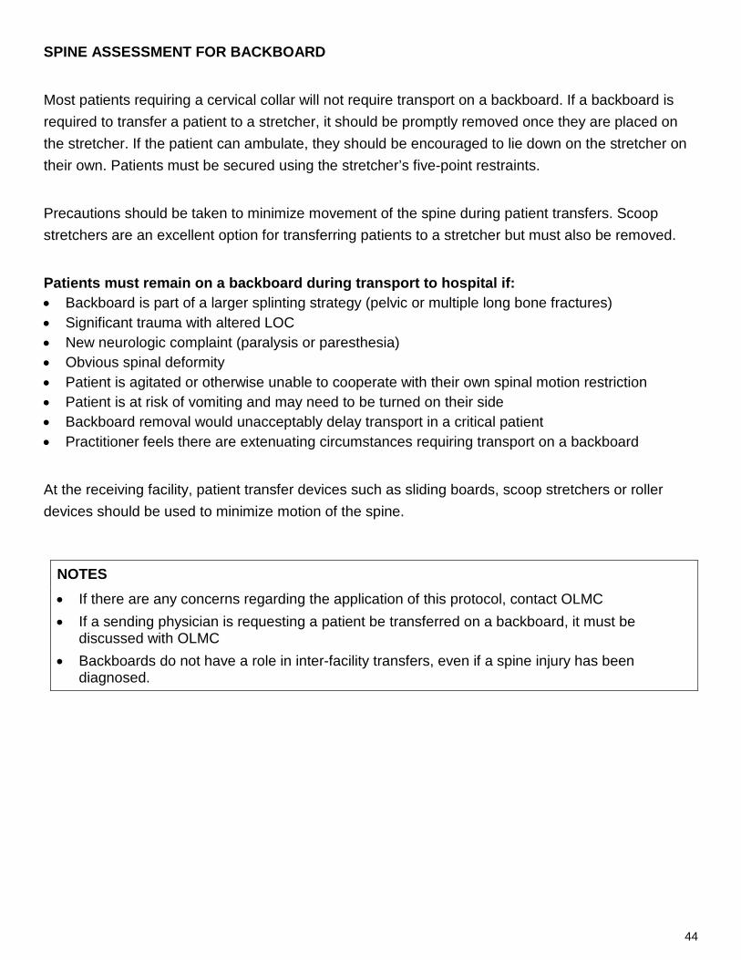
SPINE ASSESSMENT FOR BACKBOARD Most patients requiring a cervical collar will not require transport on a backboard. If a backboard is required to transfer a patient to a stretcher, it should be promptly removed once they are placed on the stretcher. If the patient can ambulate, they should be encouraged to lie down on the stretcher on their own. Patients must be secured using the stretcher’s five-point restraints. Precautions should be taken to minimize movement of the spine during patient transfers. Scoop stretchers are an excellent option for transferring patients to a stretcher but must also be removed. Patients must remain on a backboard during transport to hospital if: • Backboard is part of a larger splinting strategy (pelvic or multiple long bone fractures) • Significant trauma with altered LOC • New neurologic complaint (paralysis or paresthesia) • Obvious spinal deformity • Patient is agitated or otherwise unable to cooperate with their own spinal motion restriction • Patient is at risk of vomiting and may need to be turned on their side • Backboard removal would unacceptably delay transport in a critical patient • Practitioner feels there are extenuating circumstances requiring transport on a backboard
At the receiving facility, patient transfer devices such as sliding boards, scoop stretchers or roller devices should be used to minimize motion of the spine.
NOTES • If there are any concerns regarding the application of this protocol, contact OLMC • If a sending physician is requesting a patient be transferred on a backboard, it must be
discussed with OLMC • Backboards do not have a role in inter-facility transfers, even if a spine injury has been
diagnosed.

45
BURNS (Thermal and Chemical) 1. Manage airway and assist ventilations as necessary 2. Administer O2 as per Oxygen Therapy Protocol (Pg 49) 3. Continuous cardiac, SpO2 and BP monitoring 4. Two large bore IVs if inhalation injury1 OR greater than 20% Total Body Surface Area (TBSA)
(Initiate 2nd IV during transport) 5. Stop the burning process:
• Remove involved clothing • Brush off powdered chemicals and copious irrigation of any other chemical exposure
6. Warm ambient temperature to avoid hypothermia 7. Estimate % Total Body Surface Area (TBSA) affected using Rule of Nines (Pg 117) and provide
wound care as outlined below: Less than 5% TBSA Cover with moist or saline soaked (10-25ºC) dressing 5 – 20% TBSA Cover with clean, dry sheet or commercial dressing Greater than 20% TBSA Cover with clean, dry sheet or commercial dressing
IV fluid administration as per Parkland Formula (Pg 117) 8. Remove all items including jewelry that have the potential to become constrictive to the neck,
extremities or digits 9. Request ACP intercept (if available)
1SIGNS AND SYMPTOMS OF INHALATION INJURY • Inability to swallow • Sensation of throat swelling • Hypoxemia • Closed space fire victim • Respiratory distress
• Facial burns • Singed nasal hairs • Carbonaceous sputum • Wheezing or crackles • Voice changes
CAUTION
• Cooling with ice or ice water is contraindicated as this may increase severity of injury and lead to hypothermia.

46
BLUNT TRAUMATIC CARDIAC ARREST UNWITNESSED BLUNT CARDIAC ARREST If the following two criteria are met on arrival to patient side then no resuscitation indicated: 1) Obvious external signs of major blunt trauma consistent with Trauma Alert Activation Criteria in
the Trauma Alert Protocol (Pg 42) AND
2) Confirmed cardiac arrest by absence of spontaneous respiration and palpable pulse WITNESSED BLUNT CARDIAC ARREST
ON-SCENE • Begin CPR while attaching defibrillator • Request ACP intercept (if available) • IV access and administer 20 mL/kg 0.9% NaCl IV fluid bolus while transporting
ENROUTE TO HOSPITAL • Begin CPR while attaching defibrillator • Request ACP intercept (if available) • IV access and administer 20 mL/kg 0.9% NaCl IV fluid bolus while continuing transport • Notify receiving Emergency Department without delay that cardiac arrest has occurred and
continue transport
NOTES • If no obvious external signs of significant trauma or if unsure of mechanism of injury, consider
medical cardiac arrest and treat according to appropriate medical cardiac arrest protocol • If witnessed blunt cardiac arrest do not delay transport • Do not delay transport for IV insertion. All interventions must be performed en route to hospital • Notify receiving Emergency Department without delay of actual or impending cardiac arrest
(from the scene if possible)

47
PENETRATING TRAUMATIC CARDIAC ARREST
NOTES • If no obvious external signs of significant trauma or if unsure of mechanism of injury, consider
medical cardiac arrest and treat according to appropriate medical cardiac arrest protocol • Do not delay transport for IV insertion. All interventions must be performed en route to hospital • Notify receiving Emergency Department without delay of actual or impending cardiac arrest
(from the scene if possible)
Initiate CPR immediately • Attach defibrillator while preparing for transport • O2 via BMV • Determine rhythm
Shockable rhythm?
• Continue CPR and initiate rapid transport • Limit to ONLY ONE SHOCK ON SCENE
(unless patient entrapped or other unforeseen circumstance)
Assess for Signs of Life: 1) Pupillary response 2) Spontaneous movement 3) Organized ECG activity
If ALL Signs of Life absent: • Determine transport time to nearest
hospital
YES NO
PRESENT ABSENT
If ANY Signs of Life present: • Continue resuscitation • Initiate rapid transport • Administer 20 mL/kg 0.9% NaCl IV fluid
bolus
If transport time less than or equal to 20 minutes: • Continue resuscitation • Initiate rapid transport • Administer 20 mL/kg 0.9% NaCl IV fluid
bolus
If transport time greater than 20 minutes: • Terminate resuscitation and proceed with
Management of Death Protocol (Pg 19)
≤ 20 minutes
> 20 minutes

48
ADULT FLUID THERAPY When IV medication or fluid therapy may be required, start a peripheral IV line or lock using 0.9% NaCl solution. Unless otherwise directed by protocol or OLMC, the drip rate will be set TKVO at 30-60 mL/hr Fluid bolus should be initiated as follows unless otherwise specified by a specific treatment protocol. FLUID ADMINISTRATION IN TRAUMA CASES Bolus administration of IV fluid is to be reserved for cases of hypotension with evidence of poor perfusion. When indicated, administer IV 0.9% NaCl as outlined below: • 20 mL/kg bolus until SBP 90 mmHg achieved • If brain and/or spinal cord injury is suspected, maintain an optimal SBP of 110-120 mmHg • There is no limit to the amount of fluid a PCP may administer to achieve the desired target SBP
Routine administration of bolus IV fluids in the absence of hypotension is CONTRAINDICATED in the trauma patient. IV fluid boluses are only to be administered when above criteria is met to avoid inducing coagulopathy.
FLUID ADMINISTRATION IN MEDICAL CASES (NON-TRAUMA) When indicated by protocol, administer IV 0.9% NaCl as outlined below: • 20 mL/kg bolus • May repeat bolus administration while indications persist up to maximum 2000 mL unless
otherwise directed by protocol • If indications for additional IV fluid persist despite administration of 2000 mL IV fluids, contact
OLMC
49
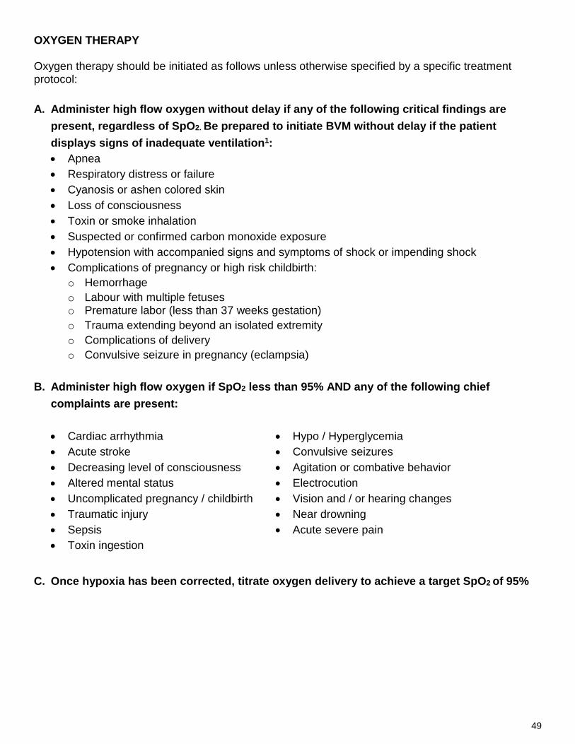
OXYGEN THERAPY Oxygen therapy should be initiated as follows unless otherwise specified by a specific treatment protocol: A. Administer high flow oxygen without delay if any of the following critical findings are
present, regardless of SpO2. Be prepared to initiate BVM without delay if the patient displays signs of inadequate ventilation1: • Apnea • Respiratory distress or failure • Cyanosis or ashen colored skin • Loss of consciousness • Toxin or smoke inhalation • Suspected or confirmed carbon monoxide exposure • Hypotension with accompanied signs and symptoms of shock or impending shock • Complications of pregnancy or high risk childbirth:
o Hemorrhage o Labour with multiple fetuses o Premature labor (less than 37 weeks gestation) o Trauma extending beyond an isolated extremity o Complications of delivery o Convulsive seizure in pregnancy (eclampsia)
B. Administer high flow oxygen if SpO2 less than 95% AND any of the following chief
complaints are present: • Cardiac arrhythmia • Acute stroke • Decreasing level of consciousness • Altered mental status • Uncomplicated pregnancy / childbirth • Traumatic injury • Sepsis • Toxin ingestion
• Hypo / Hyperglycemia • Convulsive seizures • Agitation or combative behavior • Electrocution • Vision and / or hearing changes • Near drowning • Acute severe pain
C. Once hypoxia has been corrected, titrate oxygen delivery to achieve a target SpO2 of 95%

50
OXYGEN THERAPY Cont’d
COPD If confirmed COPD (emphysema or chronic bronchitis), administer oxygen according to the following guidelines: • If the patient is in moderate to severe respiratory distress or has critical findings, administer high
flow oxygen. Be prepared to initiate BMV without delay if the patient displays signs of inadequate ventilation1. o If respiratory status has improved to patient’s baseline after treatment, consider replacing
NRB with nasal cannula to maintain SpO2 90-92% • If the patient is in mild distress, administer low flow oxygen 1 to 2 liters per minute above home
oxygen levels, titrated to a target SpO2 of 90-92% NOTES 1. If you experience any difficulty obtaining a reliable SpO2 or if at any time you obtain a low
SpO2 reading, you must administer high flow oxygen and assume the patient is hypoxic and that any low reading is accurate.
2. There may be additional circumstances beyond those contained in this protocol which will require oxygen therapy. Clinicians are advised to use sound clinical judgement to titrate oxygen therapy to balance the risk of hypoxia with concerns about hyperoxia.
1CAUTION
In order for supplementary oxygen to be effective, the patient must have adequate respiratory effort, rate and volume to ensure oxygen is delivered to the lungs. If the patient’s respiratory effort, rate or volume is inadequate to maintain oxygenation, the patient is considered to be in respiratory failure and BMV with high flow oxygen must be delivered without delay. The following signs of inadequate ventilation may be observed in patients with respiratory failure: • Abnormal sounds with breathing, such as snoring, gurgling or stridor • Fatigue with respiratory effort • Gasping • Irregular breathing pattern with periods of apnea • Little or no chest rise • Decreased or absent breath sounds (“silent chest”) • Rate and/or depth of breathing grossly insufficient for age • Apnea If there are findings of airway obstruction, such as stridor, snoring or gurgling, proceed with basic airway maneuvers to open and/or clear the airway.

51
LESS THAN LETHAL FORCE Conducted Energy Weapons (CEW) 1. If cardiac arrest present, start CPR immediately and proceed with Cardiac Arrest Protocol
(Pg 14) without delay 2. Manage airway and assist ventilation as necessary 3. Administer O2 as per Oxygen Therapy Protocol (Pg 49) 4. Continuous cardiac, SpO2 and BP monitoring 5. Measure blood glucose
• Treat hypoglycemia as per Symptomatic Hypoglycemia Protocol (Pg 31) 6. Request ACP intercept (if available) 7. Assess for secondary injuries (burns, pathological fractures, etc.) 8. If altered mental status consider the following:
• If signs of hypoglycemia, treat as per Symptomatic Hypoglycemia Protocol (Pg 31) • If severe agitation or combativeness is present proceed with Agitated/Combative Protocol
(Pg 37) • If signs of hyperthermia and Excited Delirium1 are present, initiate external cooling measures
9. Determine the event(s) preceding the use of the CEW and how many “5-second cycles of energy” were delivered to the patient
10. Inspect the impact site of the probe dart(s). If necessary, cut away clothing to view the probe darts • Do not remove any probe dart(s) • Treat dart(s) as impaled object(s) and secure in place
11. Initiate IV access • If signs of hyperthermia and Excited Delirium1 are present, initiate a second IV en route to
hospital
1 EXCITED DELIRIUM A state of excessive agitation and psychosis often brought on by overdose, drug withdrawal or non-compliance with medications used in the treatment of mental health disorders. These patients are at heightened risk of adverse outcome (cardiac and respiratory demise) and death, which is exacerbated in situations of physical restraint.
Assess the patient for the following signs of excited delirium: • Aggressive and bizarre behaviour • Dilated pupils • Extreme agitation • Shivering • Shouting • Excessive physical strength • Decreased sensitivity to pain

52
LESS THAN LETHAL FORCE Cont’d
CAUTION • Maintain police presence at all times while on-scene and request police escort during
transport. • Ensure that there is no electricity flowing through the CEW before approaching the patient. • Exercise caution when approaching a patient exposed to CEW energy as they may display
violent tendencies post-deployment. Always maintain an ability to escape the scene. Position yourself between the patient and the exit at all times to maintain a safe exit, should the situation escalate.
• At NO TIME should the patient be restrained in the prone (face or chest-down) position. • There is a high risk of positional asphyxia and/or aspiration in patients in excessively agitated
states. Close and continuous monitoring of these patients, including airway patency and adequacy of respiration, is mandatory.
• Patients with a weakened cardiac system may not tolerate exposure to CEW. Complaints of chest pain or shortness of breath must be taken seriously, evaluated and treated as appropriate.
• All patients exposed to CEW must be transported to the closest medical facility for evaluation. If police determine transport by ambulance is too dangerous, ensure that the police are clearly informed of the need for medical evaluation at a hospital and document the badge number of the police officer informed.
• Be alert for the possibility of soft tissue burns after the use of a push stun feature on the CEW.
• Be alert for the possibility of blunt force trauma after the use of a bean bag deployment device

53
HEAT RELATED ILLNESS This protocol is intended for the management of patients with exposure to high temperatures or high levels of exertion and without history of recent infection. 1. Manage airway and assist ventilations as necessary 2. O2 as per Oxygen Therapy Protocol (Pg 49) 3. Continuous cardiac, SpO2 and BP monitoring 4. Measure temperature and blood glucose
• Treat hypoglycemia as per Symptomatic Hypoglycemia Protocol (Pg 31) 5. IV access
• If signs of dehydration present administer fluid bolus as per Fluid Therapy Protocol (Pg 48) 6. Begin cooling measures1 if signs of heat exhaustion or heat stroke present2. Continue until
temperature is less than 39°C or patient starts shivering. 7. If severe agitation or combativeness is present, concurrently manage as per Agitated /
Combative Protocol (Pg 37) 8. If seizure occurs, proceed with Convulsive Seizure Protocol (Pg 34) and continue cooling. 9. Request ACP intercept (if available)
1COOLING MEASURES (STOP if patient starts shivering) 1) Remove the patient from hot environment and cool ambient temperature in the ambulance 2) Remove patient’s clothing and apply cool water to patient’s skin 3) Promote evaporation by using a fan or open window 4) Apply ice packs to the groin, neck and axilla, do not apply directly to skin
NOTES: • Patients may have normal to slightly elevated temperature with heat exhaustion • Lack of perspiration is a late sign of heat stroke • Patients with exertional heat illness may have profound sinus tachycardia as a normal
physiological response
2SIGNS OF HEAT EXHAUSTION and HEAT STROKE Patients with heat related illness may exhibit one or more of the following:
Heat Exhaustion
• Decreased coordination • Sweating • Tachycardia and hypotension
• Hyperventilation • Headache • Abdominal pain and/or nausea and vomiting
Heat Stroke 1) Temperature greater than 40°C AND 2) Altered mental status or CNS dysfunction

54
HYPOTHERMIA This protocol is intended for the management of patients with exposure to environmental conditions consistent with hypothermia.
1. Manage airway and assist ventilations as necessary 2. O2 as per Oxygen Therapy Protocol (Pg 49) 3. Continuous cardiac, SpO2 and BP monitoring 4. Measure temperature and blood glucose
• Treat hypoglycemia as per Symptomatic Hypoglycemia Protocol (Pg 31) 5. IV access
• Treat hypotension as per Adult Fluid Therapy Protocol (Pg 48) 6. If signs of frostbite present:
• Splint or pad effected area to minimize injury • Remove jewelry if required • Pad between effected digits and bandage effected tissue loosely with a soft, sterile dressing.
Do not put pressure on the effected parts. 7. If signs of hypothermia proceed with steps for rewarming1 8. Request ACP intercept (if available) 9. If Cardiac Arrest occurs proceed to Adult Cardiac Arrest Protocol (Pg 14)
1STEPS FOR REWARMING • Remove patient from cold environment • Remove wet clothing (cutting preferred) • Cover with blankets • Increase ambient temperature in ambulance • Apply radiant heat and/or warm blankets to core • Use warmed IV fluids for resuscitation
Mild Hypothermia Severe Hypothermia
1) 32°C-35°C 2) Normal mental status 3) Shivering 4) Normal to slightly elevated vital signs
1) Temperature less than 32°C 2) Decreased LOC, slurred speech and ataxia 3) Decreased heart rate and respiratory rate 4) Shivering absent below 30°C
CAUTION
• Patients in severe hypothermia often become extremely bradycardic. • Hypothermic patients are at a high risk for VF if handled roughly. Patient movement should be
limited and a horizontal position maintained whenever possible. • Severely hypothermic patients should have their core areas warmed first. Warming extremities
before core can precipitate a secondary drop in temperature. • DO NOT attempt to thaw frostbitten areas. • DO NOT ambulate patients with hypothermia.

55
ADRENAL INSUFFICIENCY 1. Manage airway and assist ventilations as necessary 2. O2 as per Oxygen Therapy Protocol (Pg 49) 3. Continuous cardiac, SpO2, and BP monitoring 4. Measure temperature AND closely monitor blood glucose
• Treat hypoglycemia as per Symptomatic Hypoglycemia Protocol (Pg 31)
5. IV access • Administer 20 mL/kg bolus NaCl 0.9% if signs or symptoms of adrenal crisis1 present
6. If patient meets Criteria for Hydrocortisone Administration2, administer:
• Hydrocortisone 100 mg IV, OR IM may be administered if IV delayed or unattainable
7. Perform 12 lead ECG
2CRITERIA FOR HYDROCORTISONE ADMINISTRATION 1) Patient has any one of the following:
• Trauma or significant physical stressor • Significant emotional crisis • Vomiting or diarrhea • Signs/symptoms of acute adrenal crisis1 • Fever of greater than or equal to 38ºC or signs of infection
AND
2) Medical history of any one of the following: • 3 weeks or more of chronic glucocorticoid use • Malcompliance or cessation of chronic glucocorticoid medication within 3 months • Addison’s Disease • Congenital Adrenal Hyperplasia • Pituitary insufficiency/hypopituitarism (i.e.: tumors, previous radiation, hypopituitary
disorders) • Bilateral adrenalectomy (removal of adrenal glands) • Patient presents a home adrenal insufficiency kit containing a glucocorticoid medication • Patient is wearing a Medic-Alert stating the patient has adrenal insufficiency
1SIGNS AND SYMPTOMS OF ADRENAL CRISIS • Nausea/Vomiting • Hypoglycemia • Abdominal pain • Arrhythmia • Hypotension (less than 100 SBP or drop of
20 mmHg from baseline)
• Weakness • Dizziness • Pallor • Confusion • Lethargy • Altered LOC

56
ADRENAL INSUFFICIENCY Cont’d
NOTES
• PMO maintains a low therapeutic threshold to administer hydrocortisone in the acutely ill or injured patient suspected to have adrenal insufficiency. There is little to no risk from a single stat dose of hydrocortisone. The risk of a low glucocorticoid level during crisis far outweighs the risk of unnecessary hydrocortisone administration.
• IV route is preferred for the administration of hydrocortisone. IM route via lateral thigh may be used if an IV is unsuccessful or otherwise delayed.
• Stress is defined as a circumstance that changes the physiological norm for the patient, and includes illness, trauma, and mental health crisis.
• Administer hydrocortisone regardless of any recent self-administration prior to EMS arrival • Glucocorticoids are used to support treatment of a multitude of medical conditions, including
but not limited to: autoimmune disorders, inflammatory bowel disease, asthma, cancer, autism, chronic allergies, and genetic enzyme deficiencies.
• Commonly prescribed glucocorticoids include, but are not limited to: prednisone, prednisolone, methylprednisolone, dexamethasone, betamethasone, triamclinolone, cortisone acetate, hydrocortisone (cortisol).
• Past history of adrenal crisis presents increased risk for repeat adrenal crisis.
CAUTION
• It is preferable to administer hydrocortisone prior to transport, as some patients in adrenal crisis may not have sufficient adrenal reserves to manage movement, even to the ambulance. These patients may deteriorate rapidly.
• Do not ambulate these patients to the ambulance. • In the rare event that a patient with adrenal insufficiency presents with anaphylaxis,
administer epinephrine first, followed immediately by hydrocortisone.

57
PAGE INTENTIALLY LEFT BLANK FOR FUTURE PROTOCOL

58
PAGE INTENTIALLY LEFT BLANK FOR FUTURE PROTOCOL

59
PART II: PEDIATRIC EMERGENCY PROTOCOLS

60
PEDIATRIC RESPIRATORY DISTRESS WITH BRONCHOSPASM 1. Manage airway and assist ventilations as necessary 2. Administer O2 as per Oxygen Therapy Protocol (Pg 49) 3. Continuous cardiac, SpO2 and BP monitoring 4. Measure temperature 5. Administer Salbutamol based on dosing below:
Age MDI + aerochamber 1
OR
Nebulized with O2
Less than 5 years 5 puffs (100 mcg/puff) 2.5 mg
Greater than or equal to 5 years 10 puffs (100 mcg/puff) 5 mg
Repeat every 5 minutes if indicated (Not to exceed a maximum total of 3 administrations) 1 Each puff must be followed by at least 4 breaths 6. Consider ipratropium bromide administration with 2nd and 3rd doses of salbutamol as per
dosing guidelines below:
Age MDI + aerochamber 1 OR
Nebulized with O2
All ages 3 puffs (20 mcg/puff) following dose of salbutamol 500 mcg (mix with salbutamol)
Repeat once in 5 minutes if indicated (Not to exceed a maximum total of 2 administrations) 1 Each puff must be followed by at least 4 breaths 7. Request ACP intercept (if available)
Contact OLMC for patients that are unrelieved by salbutamol and/or ipratropium bromide and condition is deteriorating for consideration of the following:
1. Continued administration of salbutamol 2. Epinephrine 1:1000 0.01 mg/kg (0.01 mL/kg) IM [Not to exceed a maximum single dose
of 0.3 mg (0.3 mL)]
NOTES • Patients should be treated with MDI with aerochamber unless it is deemed inappropriate,
ineffective or patient cannot tolerate • Salbutamol or ipratropium bromide may be administered singularly if the patient has
hypersensitivity to one of the other medications
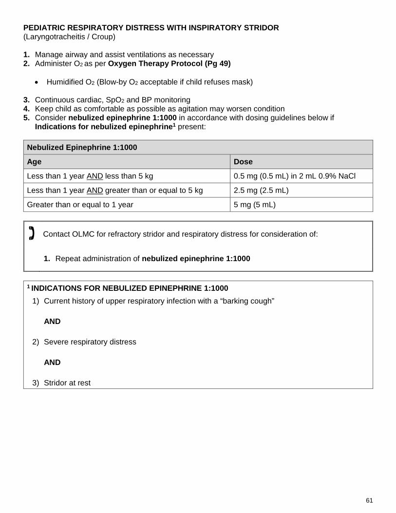
61
PEDIATRIC RESPIRATORY DISTRESS WITH INSPIRATORY STRIDOR (Laryngotracheitis / Croup) 1. Manage airway and assist ventilations as necessary 2. Administer O2 as per Oxygen Therapy Protocol (Pg 49)
• Humidified O2 (Blow-by O2 acceptable if child refuses mask)
3. Continuous cardiac, SpO2 and BP monitoring 4. Keep child as comfortable as possible as agitation may worsen condition 5. Consider nebulized epinephrine 1:1000 in accordance with dosing guidelines below if
Indications for nebulized epinephrine1 present: Nebulized Epinephrine 1:1000
Age Dose
Less than 1 year AND less than 5 kg 0.5 mg (0.5 mL) in 2 mL 0.9% NaCl
Less than 1 year AND greater than or equal to 5 kg 2.5 mg (2.5 mL)
Greater than or equal to 1 year 5 mg (5 mL)
Contact OLMC for refractory stridor and respiratory distress for consideration of:
1. Repeat administration of nebulized epinephrine 1:1000
1 INDICATIONS FOR NEBULIZED EPINEPHRINE 1:1000
1) Current history of upper respiratory infection with a “barking cough”
AND
2) Severe respiratory distress
AND
3) Stridor at rest

62
PEDIATRIC ALLERGY AND ANAPHYLAXIS FINDINGS OF ANAPHYLAXIS 1) Acute onset (minutes to hours) of TWO OR MORE of the following after exposure to a LIKELY
ALLERGEN:
• Skin symptoms (hives, itching, flushing) • Oropharyngeal edema (lips, tongue, uvula) • Respiratory compromise (dyspnea, wheeze, stridor, hypoxemia) • Gastrointestinal symptoms (crampy abdominal pain, vomiting, diarrhea) • Reduced blood pressure or associated symptoms (hypotonia, collapse, syncope)
OR
2) Age-Specific Hypotension (Pg 113) alone after exposure to a KNOWN ALLERGEN for patient 1. Manage airway and assist ventilations as necessary 2. Administer O2 as per Oxygen Therapy Protocol (Pg 49) 3. Continuous cardiac, SpO2 and BP monitoring 4. IV access 5. If age-specific hypotension (Pg 113) present, administer a fluid bolus as per Pediatric Fluid
Therapy Protocol (Pg 79) 6. If Findings of Anaphylaxis present administer:
• Epinephrine 1:1000 0.01 mg/kg (0.01 mL/kg) IM [Not to exceed a maximum single dose of 0.3 mg (0.3 mL)] o Repeat once in 5 minutes if no improvement AND
• DiphenhydrAMINE1 1 mg/kg IV (Not to exceed a maximum single dose of 50 mg) 7. If respiratory distress present (including wheezing), administer salbutamol:
Age MDI + aerochamber 2
OR
Nebulized with O2
Less than 5 years 5 puffs (100 mcg/puff) 2.5 mg
Greater than or equal to 5 years 10 puffs (100 mcg/puff) 5 mg
Repeat every 5 minutes if indicated (Not to exceed a maximum total of 3 administrations) 2Each puff must be followed by at least 4 breaths
8. Request ACP intercept (if available)

63
PEDIATRIC ALLERGY AND ANAPHYLAXIS Cont’d
Contact OLMC if severe and refractory airway compromise, respiratory failure or shock for consideration of:
• Additional IV fluid administration for refractory hypotension
1 NOTE • DiphenhydrAMINE should be avoided in pediatric patients with isolated hives,
alternative oral medications (such as cetirizine) are preferred and available in Emergency Departments.
• May administer diphenhydrAMINE 1 mg/kg IM (Not to exceed a maximum single dose of 50 mg) if anaphylaxis AND unable to establish an IV.
• There is NO absolute contraindication to epinephrine in a patient with anaphylaxis.
• DiphenhydrAMINE DOES NOT improve angioedema or respiratory symptoms in anaphylaxis.
• Patients should be treated with MDI with aerochamber unless it is deemed ineffective or patient cannot tolerate

64
PEDIATRIC CARDIAC ARREST If patient meets criteria outlined in DNR Protocol (Pg 18) or Obvious Death Protocol (Pg 17) do not proceed with resuscitation
1. Confirm Vital Signs Absent (VSA) and initiate chest compressions 2. 100% O2 via BVM (15 L/min) 3. Request ACP intercept (if available) 4. Continuous cardiac and SpO2 monitoring 5. Consider and treat Reversible Causes1 6. IV access (DO NOT delay or interrupt CPR) GENERAL GUIDELINES • Confirm absence of pulse – pulse check NOT exceeding 10 seconds • Initiate compressions immediately: C-A-B Sequence • If arrest secondary to hypoxia suspected, proceed with A-B-C Sequence • Begin CPR and immediately attach defibrillator – Analyze and defibrillate without delay if
indicated
Compressions : Ventilation Ratio Depth Rate
One Rescuer 30:2 1/3 chest depth Infants: 4 cm Child: 5 cm
At least 100 per minute Two Rescuers 15:2
• Ensure high quality CPR o Minimize interruptions in CPR o Allow full recoil of the chest between compressions o Rotate rescuers every 2 minutes (if resources allow) concurrent with pulse checks
• After third rhythm analysis initiate transport • Analyze patient every 10 minutes. Continue CPR • If return of spontaneous circulation (ROSC) proceed immediately with Pediatric Post Cardiac
Arrest Care Protocol (Pg 65) • If re-arrest occurs during transport, resume Cardiac Arrest Protocol
HYPOTHERMIC CARDIAC ARREST (CORE TEMPERATURE LESS THAN 32ºC) • Hypothermic patients are to be resuscitated as per Pediatric Cardiac Arrest Protocol • Resuscitation will be continued until active re-warming has returned core temperature to normal
or there has been ROSC
1 REVERSIBLE CAUSES OF CARDIAC ARREST Hypovolemia Hypoxia Hypothermia Hypoglycemia Drug Overdose

65
PEDIATRIC POST CARDIAC ARREST CARE (RETURN OF SPONTANEOUS CIRCULATION) 1. Manage airway 2. O2 via NRB or BVM as appropriate (15 L/min)
• Assist ventilations with BVM if signs of inadequate ventilation are present:
o Abnormal sounds with breathing, such as snoring, gurgling or stridor o Fatigue with respiratory effort o Gasping o Irregular breathing pattern with periods of apnea o Little or no chest rise o Decreased or absent breath sounds (“silent chest”) o Rate and/or depth of breathing grossly insufficient for age o Apnea
• If assisted ventilation is indicated, deliver ventilations by BVM in accordance with the following
parameters:
Patient Age Target Respiratory Rate Infants (29 days to 12 months) 20 – 30 breaths per minute (1 breath every 2-3 seconds) Children (1 year to puberty) 16 – 20 breaths per minute (1 breath every 3-4 seconds) Adolescents (Pre-puberty to adult) 12 breaths per minute (1 breath every 5 seconds)
• Inspiratory time should not exceed 1 second • Deliver only enough tidal volume to make the chest rise
3. If defibrillator was used, leave pads in place 4. Request ACP intercept (if available) 5. Continuous cardiac, SpO2 and BP monitoring 6. Perform 12 Lead ECG 7. Two peripheral IVs (initiate second IV during transport) 8. Treat Reversible Causes1 9. Adjust ventilation, oxygenation and fluid resuscitation to target values of:
• SBP – greater than age-specific hypotension (Pg 113) • SpO2 – greater than or equal to 95%
10. If re-arrest occurs, resume Pediatric Cardiac Arrest Protocol (Pg 64) and appropriate algorithm 11. If persistent hypotension, proceed with Pediatric Shock Protocol (Pg 67) 12. A copy of the code summary and PCR must be left with the receiving facility
1 REVERSIBLE CAUSES OF CARDIAC ARREST Hypovolemia
Hypoxia Hypothermia
Hypo / Hyperkalemia Hypoglycemia Drug Overdose

66
PEDIATRIC ALTERED LEVEL OF CONSCIOUSNESS 1. Manage airway and assist ventilations as necessary 2. O2 as per Oxygen Therapy Protocol (Pg 49) 3. Consider Spinal Motion Restriction 4. Continuous cardiac, SpO2 and BP monitoring 5. Measure temperature and blood glucose 6. IV Access 7. Perform 12 lead ECG 8. Consider ACP intercept if available 9. Consider and treat underlying causes of altered mental status¹
• If suspected infection, proceed with Pediatric Septic Shock Protocol (Pg 69) • If suspected seizure, proceed with Pediatric Convulsive Seizure Protocol (Pg 74) • If suspected allergy, proceed with Pediatric Allergy and Anaphylaxis Protocol (Pg 62)
• If suspected overdose, proceed with Pediatric General Approach To Toxin Management Protocol (Pg 78)
• If suspected trauma, proceed with Trauma Alert Protocol (Pg 42)
• If suspected agitated/combative, proceed with Pediatric Agitated/Combative Protocol (Pg 76)
• If suspected hypoglycemia, proceed with Pediatric Symptomatic Hypoglycemia Protocol (Pg 70)
• If suspected hypothermia, proceed with Pediatric Hypothermia Protocol (Pg 81)
• If suspected hyperthermia, proceed with Pediatric Heat Related Illness Protocol (Pg 80)
• If unknown etiology, consider Pediatric Shock Protocol (Pg 67)
1 CAUSES OF ALTERED MENTAL STATUS • Alcohol, Acidosis, Allergy • Epilepsy, Electrolytes, Endocrine • Infection • Opiates, Overdose, Oxygen deprived • Uremia, Underdose
• Heart Attack (MI) • Embolism • Aortic Obstruction • Rhythm disturbance • Tachycardia
• Trauma • Insulin • Poisoning, Psychosis • Stroke, Seizure, Shock
• Hypoxia • Hypo/hyperthermia • Hypotension
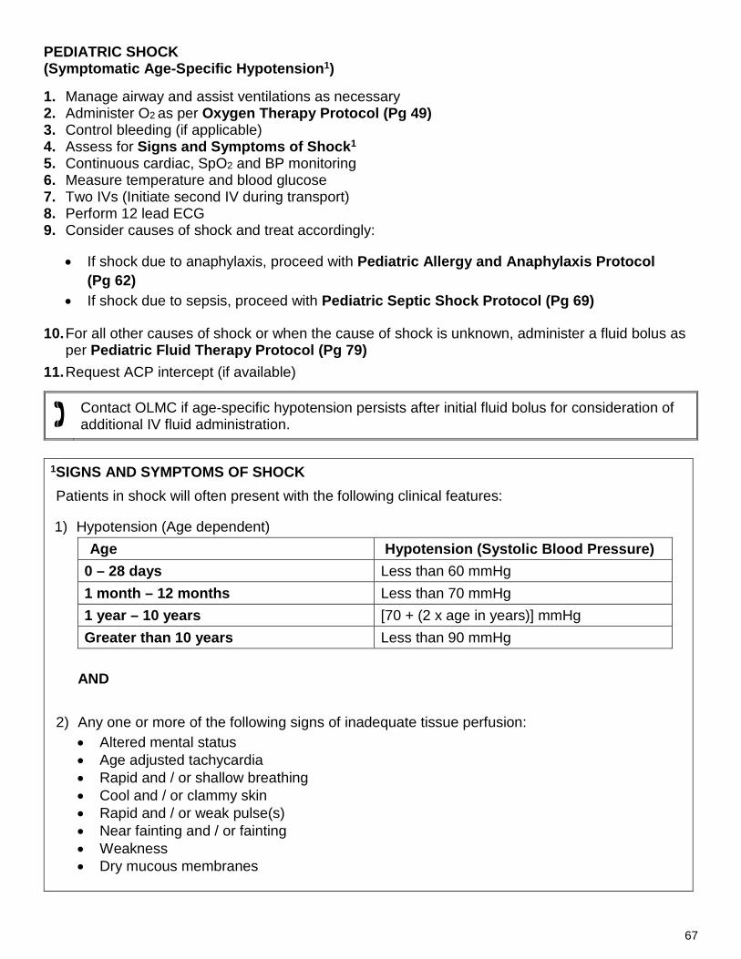
67
PEDIATRIC SHOCK (Symptomatic Age-Specific Hypotension1) 1. Manage airway and assist ventilations as necessary 2. Administer O2 as per Oxygen Therapy Protocol (Pg 49) 3. Control bleeding (if applicable) 4. Assess for Signs and Symptoms of Shock1 5. Continuous cardiac, SpO2 and BP monitoring 6. Measure temperature and blood glucose 7. Two IVs (Initiate second IV during transport) 8. Perform 12 lead ECG 9. Consider causes of shock and treat accordingly:
• If shock due to anaphylaxis, proceed with Pediatric Allergy and Anaphylaxis Protocol (Pg 62)
• If shock due to sepsis, proceed with Pediatric Septic Shock Protocol (Pg 69)
10. For all other causes of shock or when the cause of shock is unknown, administer a fluid bolus as per Pediatric Fluid Therapy Protocol (Pg 79)
11. Request ACP intercept (if available)
Contact OLMC if age-specific hypotension persists after initial fluid bolus for consideration of additional IV fluid administration.
1SIGNS AND SYMPTOMS OF SHOCK Patients in shock will often present with the following clinical features:
1) Hypotension (Age dependent) Age Hypotension (Systolic Blood Pressure)
0 – 28 days Less than 60 mmHg 1 month – 12 months Less than 70 mmHg 1 year – 10 years [70 + (2 x age in years)] mmHg Greater than 10 years Less than 90 mmHg
AND
2) Any one or more of the following signs of inadequate tissue perfusion:
• Altered mental status • Age adjusted tachycardia • Rapid and / or shallow breathing • Cool and / or clammy skin • Rapid and / or weak pulse(s) • Near fainting and / or fainting • Weakness • Dry mucous membranes

68
PEDIATRIC SHOCK Cont’d
NOTE • Shock is a life-threatening, progressive medical condition that results from the inadequate flow
of oxygenated blood to critical organs and tissues of the body • When the blood pressure is inadequate to sustain a regular flow of oxygenated blood to the
organs and tissues of the body, end-organ will ensue and shock will eventually result. • Shock may result from a number of medical conditions including sepsis, trauma, blood loss,
anaphylaxis, severe dehydration and various medical conditions.
CAUTION: • Trendelenburg positioning should not be used in the treatment of shock • Position the patient supine unless they are in severe respiratory distress • If the patient is suffering from severe respiratory distress position them semi-sitting and
assist ventilations as indicated

69
PEDIATRIC SEPTIC SHOCK 1. Manage airway and assist ventilations as necessary 2. Administer O2 as per Oxygen Therapy Protocol (Pg 49) 3. Continuous cardiac, SpO2 and BP monitoring 4. Measure temperature and blood glucose:
• Treat hypoglycemia in accordance with Pediatric Symptomatic Hypoglycemia Protocol (Pg 70)
• Treat hyperglycemia in accordance with Pediatric Symptomatic Hyperglycemia Protocol (Pg 72)
5. Two IVs (Initiate second IV during transport) 6. If patient meets Pediatric Septic Shock Inclusion Criteria1 administer fluid bolus in accordance
with Pediatric Fluid Therapy Protocol (Pg 79) 7. Request ACP intercept (if available)
1 PEDIATRIC SEPTIC SHOCK INCLUSION CRITERIA 1) History suspicious for infection OR confirmed infection
AND
2) Age-specific hypotension (Pg 113)
AND
3) Any ONE OR MORE of the following clinical findings: • Temperature less than 36ºC or greater than 38.5ºC • Altered mental status • Abnormal heart rate
o Infants – less than 90 or greater than 160 per minute o Children – less than 70 or greater than 150 per minute
8. If fever (temperature greater than 38.5ºC) present, administer acetaminophen 15 mg/kg PO. If
acetaminophen already administered within the last 4 hours, administer a “top-up” dose so total dose administered within the last 4 hours is equal to 15 mg/kg.
9. If hypotension persists after initial fluid bolus repeat IV fluid bolus to resolve age-specific hypotension (Pg 113)
Contact OLMC if age-specific hypotension persists after second fluid bolus for consideration of:
1. Additional IV fluid administration
NOTE • Age specific hypotension may be a late finding in pediatric septic shock. Suspect shock even
when normotensive if signs of inadequate tissue perfusion as per Pediatric Shock Protocol (Pg 67)

70
PEDIATRIC SYMPTOMATIC HYPOGLYCEMIA CAUTION
• This protocol is NOT intended for routine management of hypoglycemia in patients that have just been born.
• For patients that have just been born refer to Neonatal Assessment and Resuscitation Protocol (Pg 91).
• In neonatal patients (other than those who have just been born), if BGL is less than 2.6 mmol/L proceed with IV dextrose or glucagon administration as outlined in this protocol.
For neonatal patients (other than those who have just been born) with BGL between 2.6 and 4 mmol/L, contact OLMC for direction.
1. Manage airway and assist ventilations as necessary 2. Administer O2 as per Oxygen Therapy Protocol (Pg 49) 3. Continuous cardiac, SpO2 and BP monitoring 4. Measure temperature and blood glucose 5. IV access 6. If BGL is less than 4 mmol/L in the non-neonate, administer ONE of the following medications and
recheck BGL in accordance with tables below: Patient able to maintain own airway (Awake and able to cough and swallow)
IV established Unable to establish IV
Oral glucose options: 1) Dex 4® tablets 20 g (5 tablets) 2) Insta-glucose® 1 tube (30 g) 3) 1 cup (250 mL) of juice or pop (Non-
diet) 4) 4 teaspoons (20 mL) or 4 packets of
table sugar dissolved in water
IV dextrose as per dosing guidelines below
Glucagon1 as per dosing guidelines (Pg 71)
Recheck BGL in 15 minutes Recheck BGL in 10 minutes Recheck BGL in 20 minutes
DEXTROSE DOSING GUIDELINES (See Pg 132 for instructions on preparing Dextrose 10% and 25%) Weight Volume-based Dosing mL/kg (0.5 g/kg SIVP)
Less than 10 kg Dextrose 10% - 5 mL/kg SIVP
10 – 20 kg Dextrose 25% - 2 mL/kg SIVP
20 – 40 kg Dextrose 50% - 1 mL/kg SIVP [to a maximum of 50 mL (25 g)]
Greater than 40 kg Dextrose 50% - 50 mL (25 g) SIVP

71
PEDIATRIC SYMPTOMATIC HYPOGLYCEMIA Cont’d
1GLUCAGON DOSING GUIDELINES
Weight Dose
Less than 20 kg 0.5 mg IM
Greater than or equal to 20 kg 1 mg IM 7. Repeat Step 6 ONCE if necessary
All pediatric hypoglycemic patients must be transported for assessment. If a parent, guardian or mature minor is refusing transport, contact OLMC for direction
1 NOTES • Anticipate that it could take up to 20 minutes to observe an effect from glucagon • While waiting for glucagon to take effect, manage patient’s airway as indicated and initiate
transport CAUTION
• If head injury or stroke suspected administer half of the usual dose of dextrose, recheck BGL and then administer the second half dose if necessary
• The goal is to correct hypoglycemia while avoiding transient hyperglycemia that may lead to cerebral edema

72
PEDIATRIC SYMPTOMATIC HYPERGLYCEMIA This protocol is intended for patients who demonstrate findings of significant dehydration and presentations suggestive of diabetic ketoacidosis (DKA). Many diabetic patients may have blood glucose levels greater than 15 mmol/L during times of physiologic stress in the absence of dehydration and age-specific hypotension will NOT require fluid administration. 1. Manage airway and assist ventilations as necessary 2. Administer O2 as per Oxygen Therapy Protocol (Pg 49) 3. Continuous cardiac, SpO2 and BP monitoring 4. Measure temperature and blood glucose 5. IV access 6. If Indications for IV Fluid Administration in Pediatric Hyperglycemia1 present administer
10 mL/kg 0.9% NaCl IV over 1 hour4
Contact OLMC if you are uncertain as to whether the patient meets criteria for fluid administration or if age-specific hypotension persists despite administration of 10 mL/kg 0.9% NaCl IV for consideration of administration of additional IV fluids
1 INDICATIONS FOR IV FLUID ADMINISTRATION IN PEDIATRIC HYPERGLYCEMIA 1) BGL greater than 15 mmol/L
AND
2) Signs and Symptoms of DKA2 OR Signs of Dehydration3 AND
3) Age-Specific Hypotension (Pg 113)
2 SIGNS AND SYMPTOMS OF DKA 3 SIGNS OF DEHYDRATION • Polyuria • Polydipsia • Polyphagia • Tachypnea • Tachycardia • Nausea and vomiting • Abdominal pain • Signs of dehydration
• Dry mucous membranes • Absence of tears • Sunken fontanelle • Delayed capillary refill • Mottled skin • Decreased urine output (oliguria) • Tachycardia • Age-specific or postural hypotension • Weakness or lethargy

73
PEDIATRIC SYMPTOMATIC HYPERGLYCEMIA Cont’d
4 CAUTION • Rapid bolus administration of 20 mL/kg IV fluid bolus is contraindicated in pediatric patients
with DKA due to the risk of cerebral edema. SIGNS AND SYMPTOMS OF CEREBRAL EDEMA Early Warning Sign • Headache Additional Warning Signs • Drowsiness • Altered behavior • Decreasing level of consciousness MANAGEMENT OF CEREBRAL EDEMA 1) Manage airway, assist ventilations as necessary 2) O2 via NRB (15 L/min) 3) Stop IV fluids 4) Measure blood glucose and treat hypoglycemia as per Pediatric Symptomatic
Hypoglycemia Protocol (Pg 70) 5) Provide early notification to receiving facility of change in mental status

74
PEDIATRIC CONVULSIVE SEIZURES 1. Manage airway and assist ventilations as necessary 2. Administer O2 as per Oxygen Therapy Protocol (Pg 49) 3. Spinal immobilization as indicated by mechanism of injury or evidence of injury above the clavicles 4. Position patient
• Actively seizing – place supine and protect from injury • Postictal – place left lateral recumbent and maintain airway
5. Continuous cardiac, SpO2 and BP monitoring 6. Measure temperature and blood glucose
• Treat hypoglycemia as per Pediatric Symptomatic Hypoglycemia Protocol (Pg 70) 7. IV access 8. If available, request ACP intercept for active seizures or recurrent seizures (status epilepticus)

75
PEDIATRIC NAUSEA AND VOMITING 1. Manage airway and assist ventilations as necessary 2. O2 as per Oxygen Therapy Protocol (Pg 49) 3. Continuous cardiac, SpO2, and BP monitoring 4. Measure temperature and blood glucose
• Treat shock in accordance with Pediatric Shock Protocol (Pg 67) 5. Position the patient in a position of comfort 6. If symptoms severe AND the patient is actively vomiting:
• Establish IV access • Administer DimenhyDRINATE 1mg/kg IV (Not to exceed a maximum single dose of 25 mg)
OR
• Metoclopramide 0.1 mg/kg SIVP (Over 2 to 5 minutes) (Not to exceed a maximum single
dose of 10 mg) if any of the following criteria apply: o Severe nausea and vomiting refractory to dimenhyDRINATE after 15 minutes since last
dose o Allergy or contraindication to dimenhyDRINATE o Altered LOC or head injury
7. If metoclopramide has been administered and acute extrapyramidal signs or symptoms1
develop, reassure patient and administer diphenhydrAMINE 1 mg/kg IV (Not to exceed a maximum single dose of 50 mg)
NOTES
• Treatment of nausea and vomiting with parenteral anti-emetics should be reserved for cases of severe nausea and vomiting only. Current in-hospital treatment guidelines of pediatric nausea and vomiting secondary to gastroenteritis favor oral medication and rehydration without the need for IV access, except in severe cases.
1 EXTRAPYRAMIDAL SIGNS AND SYMPTOMS • Akathisia – a severe and unpleasant sensation of restlessness in patients causing them
severe anxiety and inability to sit still • Dystonia – increased rigidity or muscle contraction that may result in twisting or abnormal
postures • Dyskinesia – abnormal or repetitive movements (e.g.: lip smacking, eye twitching, etc.)
Administration of diphenhydramine is not indicated for treatment of chronic extrapyramidal signs and symptoms

76
PEDIATRIC AGITATED / COMBATIVE (Patient is danger to self or others) 1. Contact police and request that they attend the scene immediately 2. Manage airway and assist ventilations as necessary 3. Administer O2 as per Oxygen Therapy Protocol (Pg 49) 4. Continuous cardiac, SpO2 and BP monitoring 5. Measure temperature and blood glucose
• Treat hypoglycemia as per Pediatric Symptomatic Hypoglycemia Protocol (Pg 70) 6. Consider and treat Reversible or Treatable Causes of Altered Mental Status1 7. IV access 8. Attempt verbal management techniques for crisis intervention to de-escalate the situation and
calm the patient 9. If Indications for Physical Restraint2 present, apply the least amount of physical restraint
necessary to protect the patient from harming themselves or bystanders until the police arrive as per Agitated Combative / Physical Restraint Reference (Pg 110)
1 REVERSIBLE OR TREATABLE CAUSES OF ALTERED MENTAL STATUS • Hypoxia • Hypotension • Hypoglycemia • Medications or Toxins • Sepsis
2 INDICATIONS FOR PHYSICAL RESTRAINT 1) Imminent danger3 to life OR threat of physical harm to patient and/or bystanders
AND
2) Attempts at verbal de-escalation have failed AND
3) Attempts to restrain do NOT place the practitioner(s) at significant risk of harm to themselves
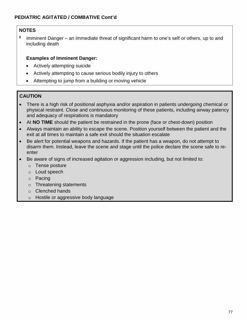
77
PEDIATRIC AGITATED / COMBATIVE Cont’d
NOTES 3 Imminent Danger – an immediate threat of significant harm to one’s self or others, up to and
including death Examples of Imminent Danger: • Actively attempting suicide • Actively attempting to cause serious bodily injury to others • Attempting to jump from a building or moving vehicle
CAUTION
• There is a high risk of positional asphyxia and/or aspiration in patients undergoing chemical or physical restraint. Close and continuous monitoring of these patients, including airway patency and adequacy of respirations is mandatory
• At NO TIME should the patient be restrained in the prone (face or chest-down) position • Always maintain an ability to escape the scene. Position yourself between the patient and the
exit at all times to maintain a safe exit should the situation escalate • Be alert for potential weapons and hazards. If the patient has a weapon, do not attempt to
disarm them. Instead, leave the scene and stage until the police declare the scene safe to re-enter
• Be aware of signs of increased agitation or aggression including, but not limited to: o Tense posture o Loud speech o Pacing o Threatening statements o Clenched hands o Hostile or aggressive body language
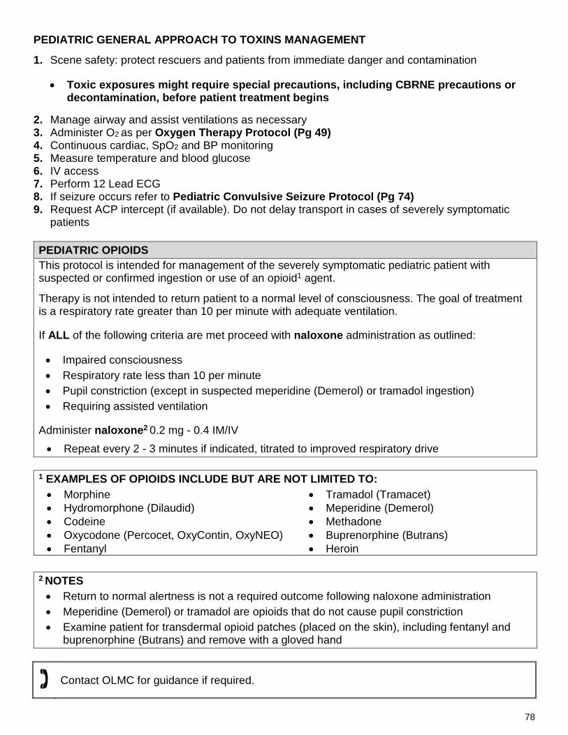
78
PEDIATRIC GENERAL APPROACH TO TOXINS MANAGEMENT
1. Scene safety: protect rescuers and patients from immediate danger and contamination
• Toxic exposures might require special precautions, including CBRNE precautions or decontamination, before patient treatment begins
2. Manage airway and assist ventilations as necessary 3. Administer O2 as per Oxygen Therapy Protocol (Pg 49) 4. Continuous cardiac, SpO2 and BP monitoring 5. Measure temperature and blood glucose 6. IV access 7. Perform 12 Lead ECG 8. If seizure occurs refer to Pediatric Convulsive Seizure Protocol (Pg 74) 9. Request ACP intercept (if available). Do not delay transport in cases of severely symptomatic
patients
PEDIATRIC OPIOIDS This protocol is intended for management of the severely symptomatic pediatric patient with suspected or confirmed ingestion or use of an opioid1 agent.
Therapy is not intended to return patient to a normal level of consciousness. The goal of treatment is a respiratory rate greater than 10 per minute with adequate ventilation.
If ALL of the following criteria are met proceed with naloxone administration as outlined:
• Impaired consciousness • Respiratory rate less than 10 per minute • Pupil constriction (except in suspected meperidine (Demerol) or tramadol ingestion) • Requiring assisted ventilation
Administer naloxone2 0.2 mg - 0.4 IM/IV • Repeat every 2 - 3 minutes if indicated, titrated to improved respiratory drive
1 EXAMPLES OF OPIOIDS INCLUDE BUT ARE NOT LIMITED TO: • Morphine • Hydromorphone (Dilaudid) • Codeine • Oxycodone (Percocet, OxyContin, OxyNEO) • Fentanyl
• Tramadol (Tramacet) • Meperidine (Demerol) • Methadone • Buprenorphine (Butrans) • Heroin
2 NOTES • Return to normal alertness is not a required outcome following naloxone administration • Meperidine (Demerol) or tramadol are opioids that do not cause pupil constriction
• Examine patient for transdermal opioid patches (placed on the skin), including fentanyl and buprenorphine (Butrans) and remove with a gloved hand
Contact OLMC for guidance if required.

79
PEDIATRIC FLUID THERAPY When IV medication or fluid therapy may be required, start a peripheral IV line or lock using 0.9% NaCl solution. Unless otherwise directed by protocol or OLMC the drip rate will be set at TKVO rate as outlined below:
• If age less than 8 years: 15 mL/hour • If age greater than or equal to 8 years: 30-60 mL/hour Fluid bolus should be initiated as follows unless otherwise specified by a specific treatment protocol.
FLUID ADMINISTRATION IN TRAUMA CASES Bolus administration of IV fluid is to be reserved for cases of hypotension AND evidence of poor perfusion. When indicated, administer IV 0.9% NaCl as outlined below:
• 20 mL/kg bolus until SBP greater than or equal to age-specific hypotension guidelines (Pg 113) • There is no limit to the amount of fluid that may administered to achieve the desired target SBP
Routine administration of bolus IV fluids in the absence of age-specific hypotension is CONTRAINDICATED in the trauma patient. IV fluid boluses are only to be administered when above criteria is met to avoid inducing coagulopathy.
FLUID ADMINISTRATION IN MEDICAL CASES (NON-TRAUMA) When indicated as per protocol administer IV 0.9% NaCl as outlined below:
• 20 mL/kg bolus until SBP greater than or equal to age-specific hypotension guidelines (Pg 113) • May repeat bolus administration once while indications persist • If indications for additional IV fluid persist despite administration of two IV fluid boluses, contact
OLMC
NOTES • Carefully observe for signs of pulmonary edema and auscultate chest for crackles after every 10
ml/kg. If crackles are detected stop IV fluid bolus • Delivery of IV fluid bolus using a buretrol or syringe is mandatory in pediatric patients less than 8
years old and should be considered in all pediatric patients greater than or equal to 8 years old
CAUTION
• Neonates and pediatric patients with hyperglycemia (Greater than 15 mmol/L) must be restricted to 10 mL/kg over 1 hour to maintain SBP greater than age-specific hypotension to avoid induction of cerebral edema
• Contact OLMC to administer additional IV fluid if age-specific hypotension persists after initial IV bolus of 10 mL/kg of 0.9% NaCl in the hyperglycemic patient

80
PEDIATRIC HEAT RELATED ILLNESS This protocol is intended for the management of patients with exposure to high temperatures or high levels of exertion and without history of recent infection 1. Manage airway and assist ventilations as necessary 2. O2 as per Oxygen Therapy Protocol (Pg 49) 3. Continuous cardiac, SpO2 and BP monitoring 4. Measure temperature and blood glucose
• Treat hypoglycemia as per Pediatric Symptomatic Hypoglycemia Protocol (Pg 70) 5. IV access
• If signs of dehydration present, administer fluid bolus as per Pediatric Fluid Therapy Protocol (Pg 79)
6. Begin cooling measures1 if signs of heat exhaustion or heat stroke present2. Continue until temperature is less than 39°C or patient starts shivering
7. If severe agitation or combativeness is present, concurrently manage as per Pediatric Agitated / Combative Protocol (Pg 76)
8. If seizure occurs, proceed with Pediatric Convulsive Seizure Protocol (Pg 74). Continue cooling 9. Request ACP intercept (if available)
1COOLING MEASURES (STOP if patient starts shivering)
1) Remove the patient from hot environment and cool ambient temperature in the ambulance. 2) Remove patient’s clothing and apply cool water to patient’s skin. 3) Promote evaporation by using a fan or open window. 4) Apply ice packs to the groin, neck and axilla. DO NOT APPLY DIRECTLY TO SKIN
NOTES: • Patients may have normal to slightly elevated temperature with heat exhaustion • Lack of perspiration is a late sign of heat stroke • Patients with exertional heat illness may have profound sinus tachycardia as a normal
physiological response • Dehydration may induce a profound tachycardic response in the pediatric patient
2SIGNS OF HEAT EXHAUSTION and HEAT STROKE Patients with heat related illness may exhibit one or more of the following:
Heat Exhaustion
• Decreased coordination • Sweating • Tachycardia and hypotension
• Hyperventilation • Headache • Abdominal pain and/or nausea and vomiting
Heat Stroke
1) Temperature greater than 40°C AND 2) Altered mental status or CNS dysfunction

81
PEDIATRIC HYPOTHERMIA This protocol is intended for the management of patients with exposure to environmental conditions consistent with hypothermia.
1. Manage airway and assist ventilations as necessary 2. O2 as per Oxygen Therapy Protocol (Pg 49) 3. Continuous cardiac, SpO2 and BP monitoring 4. Measure temperature and blood glucose
• Treat hypoglycemia as per Pediatric Symptomatic Hypoglycemia Protocol (Pg 70) 5. IV access
• Treat hypotension as per Pediatric Fluid Therapy Protocol (Pg 79) 6. If signs of frostbite present:
• Splint or pad effected area to minimize injury • Remove jewelry if required • Pad between effected digits and bandage effected tissue loosely with a soft, sterile dressing.
Do not put pressure on the effected parts. 7. If signs of hypothermia proceed with steps for rewarming1 8. Request ACP intercept (if available) 9. If Cardiac Arrest occurs proceed to Pediatric Cardiac Arrest Protocol (Pg 64)
1STEPS FOR REWARMING • Remove patient from cold environment • Remove wet clothing (cutting preferred) • Cover with blankets • Increase ambient temperature in ambulance • Apply radiant heat and/or warm blankets to core • Use warmed IV fluids for resuscitation
Mild Hypothermia Severe Hypothermia 1) 32°C-35°C 2) Normal mental status 3) Shivering 4) Normal to slightly elevated vital signs
1) Temperature less than 32°C 2) Decreased LOC, slurred speech and ataxia 3) Decreased heart rate and respiratory rate 4) Shivering absent below 30°C
CAUTION
• Patients in severe hypothermia often become extremely bradycardic. • Hypothermic patients are at a high risk for VF if handled roughly. Patient movement should be
limited and a horizontal position maintained whenever possible. • Severely hypothermic patients should have their core areas warmed first. Warming extremities
before core can precipitate a secondary drop in temperature. • DO NOT attempt to thaw frostbitten areas. • DO NOT ambulate patients with hypothermia.
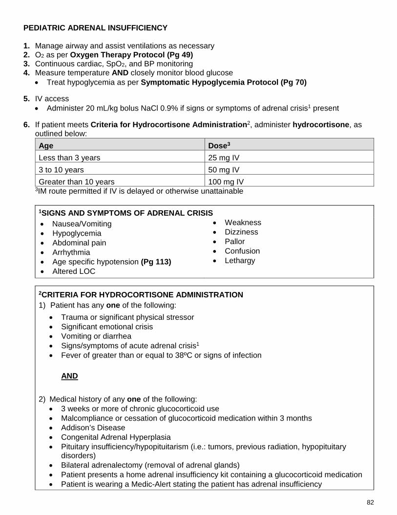
82
PEDIATRIC ADRENAL INSUFFICIENCY 1. Manage airway and assist ventilations as necessary 2. O2 as per Oxygen Therapy Protocol (Pg 49) 3. Continuous cardiac, SpO2, and BP monitoring 4. Measure temperature AND closely monitor blood glucose
• Treat hypoglycemia as per Symptomatic Hypoglycemia Protocol (Pg 70)
5. IV access • Administer 20 mL/kg bolus NaCl 0.9% if signs or symptoms of adrenal crisis1 present
6. If patient meets Criteria for Hydrocortisone Administration2, administer hydrocortisone, as
outlined below: Age Dose3
Less than 3 years 25 mg IV 3 to 10 years 50 mg IV Greater than 10 years 100 mg IV
3IM route permitted if IV is delayed or otherwise unattainable
2CRITERIA FOR HYDROCORTISONE ADMINISTRATION 1) Patient has any one of the following:
• Trauma or significant physical stressor • Significant emotional crisis • Vomiting or diarrhea • Signs/symptoms of acute adrenal crisis1 • Fever of greater than or equal to 38ºC or signs of infection
AND
2) Medical history of any one of the following: • 3 weeks or more of chronic glucocorticoid use • Malcompliance or cessation of glucocorticoid medication within 3 months • Addison’s Disease • Congenital Adrenal Hyperplasia • Pituitary insufficiency/hypopituitarism (i.e.: tumors, previous radiation, hypopituitary
disorders) • Bilateral adrenalectomy (removal of adrenal glands) • Patient presents a home adrenal insufficiency kit containing a glucocorticoid medication • Patient is wearing a Medic-Alert stating the patient has adrenal insufficiency
1SIGNS AND SYMPTOMS OF ADRENAL CRISIS • Nausea/Vomiting • Hypoglycemia • Abdominal pain • Arrhythmia • Age specific hypotension (Pg 113) • Altered LOC
• Weakness • Dizziness • Pallor • Confusion • Lethargy

83
ADRENAL INSUFFICIENCY Cont’d NOTES
• PMO maintains a low therapeutic threshold to administer hydrocortisone in the acutely ill or injured patient suspected to have adrenal insufficiency. There is little to no risk from a single stat dose of hydrocortisone. The risk of a low glucocorticoid level during crisis far outweighs the risk of unnecessary hydrocortisone administration.
• IV route is preferred for the administration of hydrocortisone. IM route via lateral thigh may be used if an IV is unsuccessful or otherwise delayed.
• Stress is defined as a circumstance that changes the physiological norm for the patient, and includes illness, trauma, and mental health crisis.
• Administer hydrocortisone regardless of any recent self-administration prior to EMS arrival • Glucocorticoids are used to support treatment of a multitude of medical conditions, including
but not limited to: autoimmune disorders, inflammatory bowel disease, asthma, cancer, autism, chronic allergies, and genetic enzyme deficiencies.
• Commonly prescribed glucocorticoids include, but are not limited to: prednisone, prednisolone, methylprednisolone, dexamethasone, betamethasone, triamclinolone, cortisone acetate, hydrocortisone (cortisol).
• Past history of adrenal crisis presents increased risk for repeat adrenal crisis. CAUTION
• It is preferable to administer hydrocortisone prior to transport, as some patients in adrenal crisis may not have sufficient adrenal reserves to manage movement, even to the ambulance. These patients may deteriorate rapidly.
• Do not ambulate these patients to the ambulance. • In the rare event that a patient with adrenal insufficiency presents with anaphylaxis,
administer epinephrine first, followed immediately by hydrocortisone.

84
PAGE INTENTIALLY LEFT BLANK FOR FUTURE PROTOCOL

85
PAGE INTENTIALLY LEFT BLANK FOR FUTURE PROTOCOL

86
PART III: OBSTETRICAL EMERGENCY PROTOCOLS

87
CHILDBIRTH 1. Manage airway and assist ventilations as necessary 2. Administer O2 as per Oxygen Therapy Protocol (Pg 49) 3. Request ACP intercept (if available) 4. Examine patient and determine if crowning2 present (Pg 88)
• If NO crowning – place in left lateral recumbent position, discourage patient from bearing down and initiate transport
• If crowning present – place supine and prepare for imminent delivery
IMMINENT DELIVERY AND POST-PARTUM CARE
1) Call for second crew or additional resources (if available) 2) Warm ambient temperature and prepare equipment
• Neonatal resuscitation equipment • Warm blankets • Clamps and scissors to cut umbilical cord • Bag for placenta
3) Apply gentle pressure to the perineum (skin stretched between the vagina and rectum) using a cupped hand and encourage a controlled (non-explosive) delivery
4) Upon delivery of the head sweep your finger around the newborn’s neck to determine if the umbilical cord is wrapped around the neck (a nuchal cord). If a nuchal cord is present: • Discourage pushing and attempt to guide the loop of cord over the newborn’s head prior to
delivery of the shoulders • If the cord is tight and you are unable to guide over the head, double clamp the cord
approximately 2.5 cm apart and cut the cord1 5) Gently guide the delivery of the anterior shoulder (shoulder up against the pubic bone) followed
by the posterior shoulder (shoulder directed towards the rectum) 6) As the delivery proceeds keep the newborn below the level of the cord 7) 30-60 seconds after delivery of the newborn clamp the umbilical cord approximately 8 cm from
the newborn and cut between the clamps 8) Proceed immediately with the Neonatal Assessment and Resuscitation Protocol (Pg 91)
• If full-term, breathing or crying spontaneously and with good tone, wrap the newborn and place on the mothers chest to encourage skin to skin contact and reduce risk of hypothermia
9) Calculate APGAR Score at 1 and 5 minutes (Pg 112) 10) Prepare for delivery of placenta
• Do not pull on umbilical cord. Allow placenta to deliver without being forced. • Upon delivery of the placenta, place in a plastic bag along with the umbilical cord
11) Perform uterine fundal massage3 (Pg 88)

88
CHILDBIRTH Cont’d 1. Null
1 NOTES • When the cord is cut be aware that supply of oxygenated blood to the baby has been
terminated • Do not delay delivery after the nuchal cord has been cut • Encourage active delivery once the nuchal cord has been cut
2 CROWNING • The phase at the end of labor in which the fetal head is
seen at the opening of the vagina
3 UTERINE FUNDAL MASSAGE
1. Place one hand horizontally across the abdomen, just above the Symphysis Pubis (Pubic bone)
2. Cup the other hand across the top of the uterus (Fundus).
3. Using a kneading or circular motion, massage the uterus between your two hands

89
POST-PARTUM HEMORRHAGE This protocol is intended for persistent and heavy vaginal bleeding post-vaginal delivery that is estimated to be greater than 500 mL and is refractory to conservative measures including firm uterine fundal massage. 1. Manage airway and assist ventilations as necessary 2. Administer O2 as per Oxygen Therapy Protocol (Pg 49) 3. Continuous cardiac, SpO2 and BP monitoring 4. IV access 5. Apply pressure to any bleeding perineal tears or lacerations of the perineum 6. Perform immediate uterine fundal massage1 7. Initiate fluid bolus as per Adult Fluid Therapy Protocol (Pg 48) 8. Request ACP Intercept (if available)
1 UTERINE FUNDAL MASSAGE 1. Place one hand horizontally across the abdomen,
just above the Symphysis Pubis (Pubic bone) 2. Cup the other hand across the top of the uterus
(Fundus). 3. Using a kneading or circular motion, massage the
uterus between your two hands
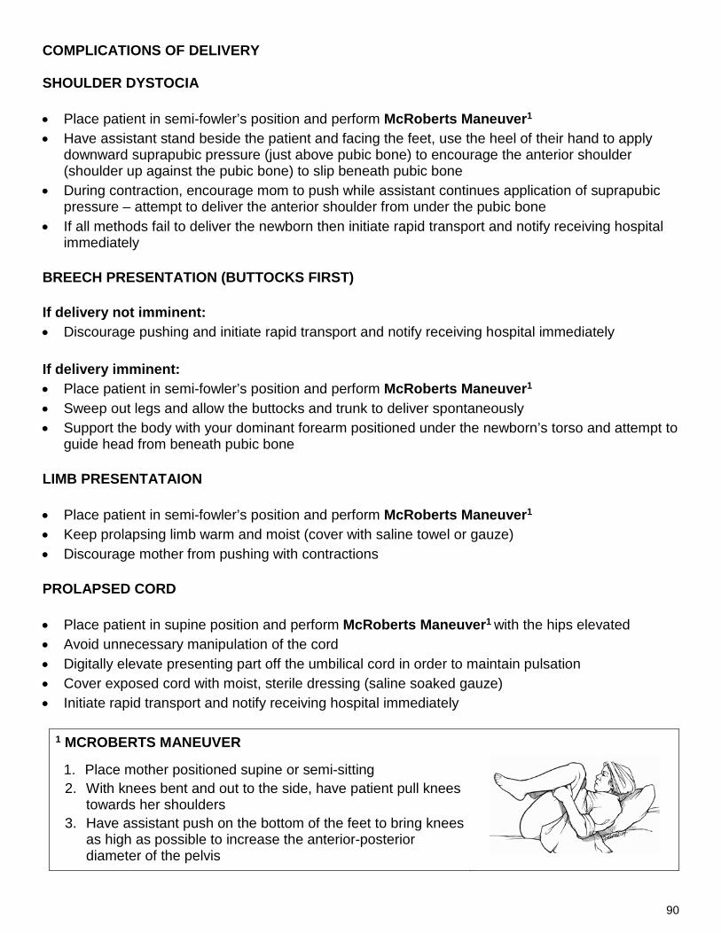
90
COMPLICATIONS OF DELIVERY SHOULDER DYSTOCIA • Place patient in semi-fowler’s position and perform McRoberts Maneuver1 • Have assistant stand beside the patient and facing the feet, use the heel of their hand to apply
downward suprapubic pressure (just above pubic bone) to encourage the anterior shoulder (shoulder up against the pubic bone) to slip beneath pubic bone
• During contraction, encourage mom to push while assistant continues application of suprapubic pressure – attempt to deliver the anterior shoulder from under the pubic bone
• If all methods fail to deliver the newborn then initiate rapid transport and notify receiving hospital immediately
BREECH PRESENTATION (BUTTOCKS FIRST) If delivery not imminent: • Discourage pushing and initiate rapid transport and notify receiving hospital immediately
If delivery imminent: • Place patient in semi-fowler’s position and perform McRoberts Maneuver1 • Sweep out legs and allow the buttocks and trunk to deliver spontaneously • Support the body with your dominant forearm positioned under the newborn’s torso and attempt to
guide head from beneath pubic bone LIMB PRESENTATAION • Place patient in semi-fowler’s position and perform McRoberts Maneuver1 • Keep prolapsing limb warm and moist (cover with saline towel or gauze) • Discourage mother from pushing with contractions PROLAPSED CORD • Place patient in supine position and perform McRoberts Maneuver1 with the hips elevated • Avoid unnecessary manipulation of the cord • Digitally elevate presenting part off the umbilical cord in order to maintain pulsation • Cover exposed cord with moist, sterile dressing (saline soaked gauze) • Initiate rapid transport and notify receiving hospital immediately
1 MCROBERTS MANEUVER
1. Place mother positioned supine or semi-sitting 2. With knees bent and out to the side, have patient pull knees
towards her shoulders 3. Have assistant push on the bottom of the feet to bring knees
as high as possible to increase the anterior-posterior diameter of the pelvis

91
NEONATAL ASSESSMENT AND RESUSCITATION 1. Determine gestational age and proceed with Neonatal Resuscitation Algorithm (Pg 93) and in
accordance with General Guidelines outlined below 2. Request ACP intercept (if available) 3. Employ strategies to prevent hypothermia in term or pre-term newborns1
GENERAL GUIDELINES • Determine heart rate by continuous cardiac monitoring if resuscitation is required NEONATAL CPR (VENTILATIONS AND COMPRESSIONS) • Use a two-thumb, encircling the chest, technique • Ensure high quality CPR
o Minimize interruptions in CPR o Allow full recoil of the chest between compressions o Deliver each breath over 1 second o Deliver only enough volume to produce visible chest rise
Compressions : Ventilation Ratio Depth Rate
3:1 1/3 chest depth 90 compressions / minute 30 breaths / minute
ASSISTED VENTILATION (WITHOUT COMPRESSIONS) • To be provided if newborn demonstrates ineffective or absent spontaneous respirations without
need for chest compressions • Rate: 40-60 breaths / minute • Deliver each breath over 1 second • Deliver only enough volume to produce visible chest rise • Avoid excessive ventilation
1 TEMPERATURE CONTROL IN THE JUST-BORN PRETERM PATIENT (LESS THAN 37
WEEKS) • Hypothermia will have significant harmful effects on the preterm patient • Warm ambient temperature in anticipation of delivery (above 26ºC where possible) • Dry the newborn • Cover newborn, from the neck down, with loose fitting plastic “wrap” (circumferential) or a
plastic bag • Cover head with a hat or part of a warmed blanket • Wrap newborn a warm blanket or place skin-to-skin with mother and cover both mother and
newborn with a warm blanket

92
NEONATAL ASSESSMENT AND RESUSCITATION Cont’d
BLOOD GLUCOSE MEASUREMENT AND HYPOGLYCEMIA IN THE JUST BORN PATIENT • Patients that have just been born will typically have blood sugars below normal adult values • Routine measurement of BGL in the just born patient is not recommended
INDICATIONS FOR BGL MEASUREMENT IN THE JUST-BORN PATIENT • BGL measurement is required ONLY if the patient is:
1) Pre-term
OR
2) Full-term AND requiring intervention or resuscitation beyond routine post-natal supportive care
• If BGL is less than 2.6 mmol/L – administer dextrose 10% 5 mL/kg SIVP
Contact OLMC if BGL between 2.6 and 4 mmol/L in the populations indicated above to discuss need for intervention and management options

93
NEONATAL RESUSCITATION \ Assess patient: • Term gestation? • Breathing or crying spontaneously? • Good tone?
Routine Post-Natal Care • Clear airway if necessary • Warm and dry • Provide ongoing evaluation
Initiate resuscitation • Provide warmth • Clear airway if necessary1 • Dry and stimulate
Evaluate heart rate and breathing • Heart rate less than 100?
OR • Gasping or apnea?
Initiate BMV, SpO2 / Cardiac monitoring
Heart rate less than 100?
Evaluate for Chest Movement, Take Ventilation Corrective Steps2 if needed, Continue BVM
Initiate CPR • Coordinate chest compressions with BMV, 100% O2
Heart rate less than 60?
Continue CPR • Continue chest compressions coordinated with BMV • Consider and treat Reversible Causes3
Post-Resuscitation Care • Keep warm • Continuous cardiac and SpO2 monitoring • Measure BGL and treat hypoglycemia (Pg 92)
2 VENTILATION CORRECTIVE STEPS
M: Mask readjustment R: Reposition airway
S: Suction mouth and nose O: Open mouth P: Pressure increase A: Airway alternative
3 REVERSIBLE CAUSES 1) Hypovolemia 2) Hypoglycemia (Pg 92) 3) Hypothermia 4) Pneumothorax
1 IF MECONIUM PRESENT AND NON-VIGOROUS (Poor respiratory effort, poor tone OR heart rate less than 100):
• Suction mouth, oropharynx and then nose
YES
YES
Proceed with Routine Post-Natal Care (Above) NO
NO
15 sec
30 sec
YES
NO
NO
YES
60 sec
YES
Heart rate less than 60?

94
PAGE INTENTIONALLY LEFT BLANK FOR FUTURE PROTOCOL

95
PART IV: REFERENCES

96
12 LEAD ECG GUIDELINES
INDICATIONS FOR 12 LEAD ECG 12 lead acquisition is required in any patient presenting with: • Chest pain • Jaw pain • Left arm pain • Epigastric pain • Non-traumatic back pain
• Shortness of breath • Syncope • Palpitation • Weakness • Nausea or vomiting • Altered LOC
• Toxic ingestion or overdose • Suspected electrolyte disturbance • Dysrhythmia on 3 lead ECG • Irregular pulse • Diaphoresis disproportionate to
environment In addition to the indications listed above a 12 lead ECG should be performed any time the paramedic feels it is indicated
SERIAL 12 LEAD ECGs If the initial 12 lead does not show evidence of ST elevation in a patient experiencing chest pain serial 12 lead ECGs must be performed as outlined below: 1) In ambulance just prior to transport 2) Every 15 minutes during transport (if transport time greater than 30 minutes) 3) Just prior to arrival to receiving health care facility 4) Any time patient condition or ECG rhythm changes If the initial 12 lead demonstrates evidence of ST elevation MI serial 12 leads are not required unless there is a change in patient condition or ECG rhythm changes
NOTES • Acquiring a 12 lead ECG should not prolong scene time or transport more than 2 minutes • 12 lead acquisition should be performed concurrent with other assessment and care and
should not interfere with treatment protocols • Any time a 12 lead has been acquired a copy of the 12 lead must be attached (stapled) to the
PCR to be left at the receiving facility and labeled with the patient’s surname and MCP number. A second copy of the 12 lead ECG must be attached (stapled) in an identical manner to the copy of the PCR to be kept by the ambulance service provider.
• You must document the device interpretation of the 12 lead ECG in the narrative portion of the PCR

97
12 LEAD ECG ACQUISITION TECHNIQUE Procedure 1. Place the patient in a supine or semi-sitting position 2. Expose chest enough to acquire a 12 lead ECG. Take all steps necessary and possible to protect
the patient’s dignity and privacy 3. Prep the skin with alcohol or other wipe. Remove excess chest hair where needed for good
contact. If patient is large breasted or obese, be sure to place leads correctly. 4. Attach the four limb and chest leads to the patient 5. Reduce causes of artifact. Stop patient movement. If en route to hospital, stop ambulance to
acquire ECG.

98
NON-EMS MEDICAL PERSONNEL ON SCENE The medical care provided at the scene is the responsibility of the highest level provider dispatched to the scene. A physician, nurse or other medical personnel will only assist in patient care with the approval of the EMS provider. Physician on Scene • If the physician provides orders in agreement with the EMS provider’s protocols, you may work
with that physician within the confines of the provider’s protocols • If the physician provides orders that are in conflict with protocols or that are outside the scope of
practice of the provider, the physician must: 1. Agree to take responsibility for the patient’s care 2. Agree to accompany the patient to the receiving facility 3. Document their involvement on the PCR and sign the PCR
• If the physician does not agree to the above or at any time terminates care they have initiated, notify the physician that you are required to discuss the case with the OLMC physician and that the OLMC physician may wish to discuss the patient care provided
• Any concerns regarding patient care must discussed with OLMC Nurse or Other Medical Personnel on Scene • The nurse or other medical personnel’s assistance must follow the EMS provider’s protocols • If the nurse or other medical personnel wish to deviate from the EMS provider’s protocols, they
shall be informed that care will not deviate from the protocols Off-duty EMS providers may participate in patient care as per the Off-Duty EMS Personnel Protocol (Page 99). If the Off duty EMS provider does not meet the criteria in this protocol, they are considered “Other Medical Personnel” for the purposes of this protocol and are advised to have valid personal errors and omissions insurance. Off-duty providers without privately obtained errors and omissions insurance will not be covered by their employer’s insurance if they provide patient care while off duty and will be held personally liable in the event of legal action resulting from the patient encounter.

99
OFF-DUTY EMS PERSONNEL Off-duty EMS personnel who are at the scene of an incident may assist members of the responding unit only if:
• Approval is granted by the lead provider • They agree to take responsibility for patient care, document their involvement on the patient
care report and accompany the patient to the hospital if administering a higher level of care than the responding provider
• They have active errors and omissions insurance through one of the following options: o A preexisting written memorandum of understanding specifically granting errors and
omissions insurance coverage during off-duty hours and for when a practitioner is placed in a humanitarian situation
o Obtaining appropriate personal errors and omissions insurance Off-duty providers without errors and omissions insurance will be held personally liable in the event of legal action resulting from the patient encounter. PMO encourages all practitioners to obtain private errors and omissions insurance, regardless of their employers insurance, to protect themselves in the event of personal litigation.

100
ACP INTERCEPT If identified by protocol or when the PCP deems it necessary, an ACP Intercept may be requested. Intercepts should be activated as early as possible. They may be cancelled by the requesting crew if reassessment determines that it is not required. The following list of conditions indicate the need for an ALS intercept:
• Abdominal pain with unstable vital signs • Accident/assault victims with multiple trauma or high-risk injuries and/or unstable vital signs • Airway compromise/potential compromise • Altered LOC: unstable or declining • Anaphylaxis: unresponsive to initial treatment • Arrest: cardiac or respiratory • Chest pain or cardiac problems • GI bleed with unstable vital signs • Hemorrhage: internal/external – uncontrollable or with signs of shock • Respiratory distress unresponsive to initial treatment • Seizures: ongoing (greater than 5 minutes) or repetitive • Abnormal vital signs
CAUTION
• Do not delay patient transport to await arrival of the intercepting unit at the scene • Initiate transport and rendezvous with intercepting crew en route to hospital
INTERCEPT DOCUMENTATION • The original responding crew must document all patient care activities up to and including, the
point of transfer of care on the initial (Primary Claim) PCR. This must also include all patient identifiers and information necessary for billing purposes.
• Once care is transferred to the intercepting crew, the intercepting crew must complete a second PCR documenting all patient care activities from the point of transfer of care until transfer of care to hospital staff. The second PCR must also include all patient identifiers and information necessary for billing purposes.
• The intercepting crew MUST document the Primary Claim ID Number of the initial PCR used by the crew that initially responded to the call.
• The original responding crew MUST document the PCR number belonging to the intercepting crew in the narrative portion of the original (Primary Claim) PCR.

101
REFUSAL OF CARE Adult patients (or a mature minor1) with medical decision-making capacity have the right to refuse pre-hospital assessment, management or transport. These patients must sign a Refusal of Care Form provided they meet the criteria outlined below. Persons that meet the criteria below can refuse treatment: 1) An adult over the age of 18 2) A mature minor1 3) A parent or legal representative2 of a minor 4) Legal representative of dependent adults3 Procedure 1. Determine the patient’s capacity4 for decision making and document on PCR. The patient has
the capacity for medical decision-making if they meet ALL of the following criteria:
The patient may be considered to have capacity for medical decision-making if they meet ALL of the following criteria:
a) GCS of 15 b) Converse spontaneously c) FULLY oriented and follow commands d) DO NOT have impairment due to drugs or alcohol e) Not in a postictal state f) Have a reasonable understanding of :
o the nature of the illness/injury o the recommendations made o the benefits that could result from treatment or transport o the risks involved in not seeking treatment or transport (as outlined and documented by the
practitioner)
g) The decision of non-transport is consistent with the patient’s normal set of values and beliefs (e.g. Is this a decision that the patient would normally make under similar circumstances?)
2. Ensure absence of medical conditions that may affect patients capacity for medical decision-making including but not limited to: • Hypoglycemia • Hypotension • Hypoxia • Delirium • Dementia • Developmental disability • Intoxication
102
REFUSAL OF CARE Cont’d
3. If it is determined that the patient has capacity for medical decision-making, contact with OLMC is NOT required. Practitioners must ensure to:
• Explain and document on the PCR the possible risks and consequences of refusal of treatment or transport
• Educate patient and bystanders to call back if patient worsens or if they change their mind regarding treatment and transport
• Recommend that contact be made with the patient’s family physician • Offer assistance in arranging alternative transportation • Have patient and witness sign the Refusal of Care Form
4. If it is determined that the patient does NOT have capacity for medical decision-making and is suicidal, poses a risk of bodily injury to themselves or others as a result of mental illness or is intoxicated and in need of medical treatment, contact Police and/or OLMC for assistance.
NOTES • A refusal of care or transport must be a patient-initiated request or inquiry. Practitioners must
never suggest or encourage patients to refuse care and/or transport. Patients have a right to access the care provided in the Emergency Department via medical transport
• Medical decision-making and refusal of care or transport must be a decision made by the patient free of fear, constraint, compulsion, coercion or duress
• Patients who are minors (under age 19 and not considered to be a mature minor1) cannot refuse care. However, if the minor’s parent or legal guardian demonstrates capacity for medical decision-making and agrees to assume responsibility for the minor, they must sign the Refusal of Care Form on behalf of the minor
DEFINITIONS 1 Mature minor: A teenager who is assessed by a health care provider to have capacity to make
a specific treatment decision based on a demonstrated ability to understand information surrounding his or her presentation including: • Nature of his or her medical condition • Proposed treatment and/or alternatives • Risk and benefits of the proposed treatment and/or alternatives • Risks or foreseeable consequences of consent to treatment or refusal of care
2 Legal representative: Court appointed individual(s) responsible for making health care related decisions for dependent adult or minors
3 Dependent adult: Any adult who is greater than or equal to 18 years of age, completely or partially dependent upon one or more other person(s) for care or support, has not established financial independence and would likely be in danger if care or support was withdrawn
4 Capacity: The patient understands the nature of his or her medical condition, risks and benefits of care and/or transport, risks or foreseeable consequences or refusal of care or transport and the patient demonstrates this understanding of the explanation(s) of these elements by the attending practitioners

103
POTENTIAL COMMUNICABLE/QUARANTINABLE DISEASE INDICATORS OF POTENTIAL COMMUNICABLE OR QUARANTINABLE DISEASE 1) Fever (Temperature greater than or equal to 38ºC)
AND
2) Any ONE OR MORE of the following: • Appearing obviously unwell • Shortness of breath (recent onset) • Multiple ill travelers aboard conveyance • Persistent cough or coughing blood • Persistent vomiting or diarrhea • Bruising or bleeding (without previous injury)
• Headache • Confusion (recent onset) • Sore throat • Muscle pains • Intense weakness • Skin rash
QUARANTINE OFFICER (ATLANTIC REGION) – 902-873-7659 1. If the patient meets the above indicators of a potential communicable disease, alert all emergency
responding agencies of appropriate personal protective equipment (PPE) requirements (to include, but not limited to: gloves, gown, goggles and N95 mask for the emergency responder; mask and appropriate draping for the patient)
2. If the patient meets the potential communicable/quarantinable indicators OR case involves a Known Quarantinable Disease1 AND is an international traveler being picked up at a port of entry (air or sea) notify the Quarantine Officer (QO) before leaving the vessel or aircraft and passing through customs (902-873-7659) for further direction
3. Notify the receiving facility of a Potential Communicable/Quarantinable Disease 4. Notify dispatch that the transport vehicle will be unavailable after transport until decontamination
has occurred (confer with local hospital Infection Control)
1 KNOWN QUARANTINABLE DISEASES Active pulmonary tuberculosis Anthrax Botulism Cholera Diphtheria Measles Pandemic Influenza Type A Plague Poliomyelitis Smallpox Severe Acute Respiratory Syndrome (SARS)
Argentine hemorrhagic fever Bolivian hemorrhagic fever Brazilian hemorrhagic fever Crimean-Congo hemorrhagic fever Ebola hemorrhagic fever Marburg hemorrhagic fever Venezuelan hemorrhagic fever Rift Valley Fever Tularemia Typhoid Fever Yellow Fever Lassa fever
QUARANTINE OFFICER (ATLANTIC REGION) – 902-873-7659
104
MASS CASUALTY INCIDENT MANAGEMENT ORGANIZATION 1. Incident Command and Triage Coordinator are established by the first arriving unit
• Roles may change as additional personnel arrive 2. Scene size up INCIDENT COMMAND 1. Estimate number of victims and notify dispatch 2. Request appropriate number of responding units, special equipment, mutual aid units and additional
resources as needed 3. Identify staging area, access and egress routes 4. Identify treatment area 5. Assign other positions as additional crews and help arrive:
• Treatment Coordinator • Transport Coordinator • Litter bearers / extrication teams • Other duties as required
TRIAGE COORDINATOR (Lowest trained personnel) 1. Direct all walking wounded to a designated area
• If possible, direct a few people to remain in the triage area and assist victims as required 2. Triage of victims should be initiated immediately using the START (Pg 105) or JumpSTART system
(Pg 106) 3. Perform only the most life saving measures (open airway, stop bleeding) 4. Oversee and direct litter bearers to transport patients from the triage area to the treatment area
according to triaged priority TREATMENT COORDINATOR (Highest trained personnel) 1. Establish treatment areas
• If incident is large, designate separate treatment areas for each triage level, including a morgue separate from other victims
2. Ensure aggressive treatment and rapid packaging of patients 3. Assign and supervise treatment teams 4. Assign transport priorities (transport highest priority first) and communicate this to transport
coordinator TRANSPORT COORDINATOR 1. Establish and supervise the Staging Area as well as access and egress routes 2. Establish and supervise the patient loading zone 3. Assign and supervise rapid and efficient loading of patients 4. Ensure smooth flow of ambulance traffic and avoid congestion of vehicles 5. Maintain a log containing the victim names, nature of injuries, time of transport, destination and triage
tag number 6. Notify the receiving facility of patient transports, including a brief description of injuries
NOTES
• There must be adequate medical personnel working in the treatment area prior to initiating transportation of the patients to receiving facilities
• All personnel are to restrict radio communications to a minimum

105
SIMPLE TRIAGE AND RAPID TREATMENT (START) TRIAGE SYSTEM The purpose of the Simple Triage And Rapid Treatment (START) system is to efficiently triage and transport adult victims of a multiple or mass casualty incident. This is used when the number of injured exceed the capabilities of the first arriving units or for large scale incidents.
GENERAL GUIDELINES
• Triage of victims should take no longer than 60 seconds per patient • Assess “R-P-M” (Respirations, Perfusion and Mental Status) for each patient • Tags of appropriate color should be placed on the upper extremity or in a visible location • Reassessments may be conducted and priority may be changed once all patients have been
triaged PROCEDURE 1. Identify “Walking Wounded”
• Voice triage should be used to direct the walking wounded to a designated area • If patient (s) able to walk to the designated area Tag GREEN – Minor • Proceed with evaluation of remaining patients as outlined in Steps 2 – 5
2. Assess R – Respirations • If respiratory rate is greater than 30 Tag Red – Immediate
• If patient is not breathing Open airway and reassess o If patient remains apneic despite airway opening Tag Black – Deceased
3. Assess P – Perfusion (Radial pulse and capillary refill)
• If absent radial pulse OR capillary refill greater than 2 seconds Tag Red – Immediate 4. Assess M – Mental Status
• If patient is unconscious, disoriented OR unable to follow simple commands Tag Red – Immediate
5. For all remaining patients Tag Yellow – Delayed

106
JUMP SIMPLE TRIAGE AND RAPID TREATMENT (JumpSTART) TRIAGE SYSTEM The purpose of the Jump Simple Triage And Rapid Treatment (JumpSTART) system is to efficiently triage and transport pediatric (Age 1-8 years) victims of a multiple or mass casualty incident. This is used when the number of injured exceed the capabilities of the first arriving units or for large scale incidents.
GENERAL GUIDELINES • The JumpSTART system is to be utilized in pediatric patients (Age 1-8 years) only • Triage of victims should take no longer than 60 seconds per patient • Assess “R-P-M” (Respirations, Perfusion and Mental Status) for each patient • Tags of appropriate color should be placed on the upper extremity or in a visible location • Reassessments may be conducted and priority may be changed once all patients have been
triaged PROCEDURE 1) Identify “Walking Wounded”
• Voice triage should be used to direct the walking wounded to a designated area • If patient (s) able to walk to the designated area Tag GREEN – Minor • The “walking wounded” categorization does not apply to pediatric patients being carried by
an adult to the designated area • Proceed with evaluation of remaining patients and those being carried, as outlined below
2) Assess R-P-M
1) R – Respirations • If respiratory rate is less than 15 or greater than 40 Tag Red – Immediate • If patient is not breathing Open airway and reassess
o If breathing resumes Tag Red – Immediate o If patient remains apneic Check pulse If no pulse Tag Black – Deceased If pulse present Perform BMV for 15 seconds (5 ventilations)
- If respirations resume Tag Red – Immediate - If no respirations Tag Black – Deceased
2) P – Perfusion (Radial and brachial pulse)
• If absent radial AND brachial pulse Tag Red – Immediate
3) M – Mental Status (AVPU) • Assess using the AVPU scale and proceed as outlined below:
o If Alert, responsive to Verbal stimulus or appropriately responsive to Pain Tag Yellow – Delayed
o If Unresponsive or demonstrates an inappropriate response to Pain Tag Red – Immediate

107
COMMUNICATIONS REFERENCE 1.
RADIO REPORT TO RECEIVING FACILITY Radio reports should be kept as concise as possible and contain essential information to ensure Emergency Department preparedness to receive the patient and provide necessary care without delay. A concise radio report should be followed by a more detailed verbal report upon arrival to the receiving facility. The purpose of the radio report is to provide an opportunity for the receiving facility to activate the appropriate resources and services to address the immediate needs of the patient.
RADIO REPORT COMPONENTS 1) Unit identification 2) Age and gender of patient 3) Level of consciousness 4) Chief complaint or primary reason for transport 5) History of present illness or injury 6) Relevant Past Medical History 7) Relevant medications (Contributing to presentation or taken by patient) 8) Relevant physical exam findings 9) Treatment rendered and response 10) Estimated time of arrival (ETA)
CONSULTATION WITH OLMC Consultation with OLMC should take place when directed to do so by a protocol OR any time the practitioner requires the advice of a physician to care for his or her patient. Be prepared to provide a comprehensive verbal report to the OLMC physician that includes all the necessary information in order for the physician to properly advise you with respect to patient care. Upon being connected with the physician you must provide the physician with the following identifying information at the start of the conversation:
1) Your name 2) Level of training 3) Registration number
This identifying information must be repeated to the physician even if you have already provided this information to the dispatcher.

108
COMMUNICATIONS REFERENCE Cont’d
OLMC PATIENT REPORT COMPONENTS When discussing the patient with the OLMC physician it is important that all necessary pieces of information are presented. This is important to ensure the physician has all the details necessary to provide you with the safest and most appropriate advice for each individual patient. Please be sure to include all of the following OLMC Report components in your case presentation:
1) Age and gender of patient 2) Chief complaint or primary reason for transport 3) History of present illness or injury 4) Past Medical History 5) Medications 6) Allergies 7) Physical exam findings 8) Complete set of vital signs 9) Treatment rendered 10) Specific questions practitioner has for the physician or request for order(s) When orders are received from the physician the practitioner must repeat the order(s) including drug name, route of administration, dose and repeats to the physician for clarification. It is essential that you state the numerical value of each vital sign rather than making a general statement such as “vital signs are normal”. What is “normal” will vary depending on the case and the physician requires the actual vital sign result to make a medical recommendation.
NULL COMMUNICATIONS FAILURE In case of a communications failure with OLMC due to equipment (radio, cell phone and/or landline) malfunction or due to incident location, the following will apply: • Practitioner(s), may within the limits of their Certification(s), perform necessary procedures,
that are contained within the protocols that would require a direct physician order under normal circumstances
• Procedures performed must be limited to the minimum amount necessary to prevent the loss of life or the critical deterioration of a patient’s condition
• All the procedures performed under this order and the conditions that contributed to the communications failure must be documented in detail on the patient care record
• Practitioner must continue to make efforts to contact OLMC during transport Practitioners are required to contact PMO the following business day to report the details of the communications failure.

109
SPECIAL PATIENT PREHOSPITAL TREATMENT Guideline Patients with high-risk diagnoses requiring special treatment may be issued individual protocols by Provincial Medical Oversight. The unique circumstances are evaluated by PMO, and an identification card is issued to identify these patients. It is important to identify these patients as soon as possible to initiate care required. The patients are instructed to have the cards with them and present to EMS upon their arrival. The protocols on the card are developed by the Provincial Medical Director, in consultation with care givers and physicians involved with the management of the patient.
Procedure
1. Manage airway 2. O2 as appropriate 3. Immediately request ACP Intercept 4. Verify special patient card information
Contact OLMC for Special Protocol Authorization.
NOTE: 1 Treatment indicated on card is to be limited to scope of practice. In these rare circumstances, practitioners are permitted to allow the patient or care givers educated in the administration of medications or interventions to provide those interventions. Ultimate responsibility for patient management lies with the practitioner.

110
AGITATED COMBATIVE / PHYSICAL RESTRAINT Physical restraint is an intervention of last resort that should only be utilized when there is an imminent danger to life or threat of physical harm to the patient and/or bystanders and reasonable attempts to defuse the situation with verbal de-escalation strategies have failed. Practitioners should utilize the least amount of restraint necessary to protect the patient and/or bystanders from harm until police arrive. Practitioners may apply physical restraint up to the point where such force would reasonably be considered to be excessive or where practitioners are no longer able to safety apply restraint as a result of imminent danger of harm to them. Provided that indications for physical restraint are present, such restraint may be applied regardless of whether or not the patient has been formally evaluated under the Mental Health Care and Treatment Act or if the patient is categorized as “voluntary” or “involuntary” under the Act.
INDICATIONS FOR PHYSICAL RESTRAINT 1) Imminent danger to life OR threat of physical harm to patient and/or bystanders
AND 2) Attempts at verbal de-escalation have failed
AND
3) Attempts to restrain do NOT place the practitioner(s) at significant risk of harm to themselves
Police attendance must be requested immediately if there is a need to physically restrain a patient or if a patient has been physically restrained based on the presence of the criteria listed above. If the estimated time of arrival for the police is anticipated to be prolonged, contact OLMC regarding the transport decision. IMMINENT DANGER Imminent danger refers to an immediate threat of significant harm to one’s self or others, up to and including death. Examples of imminent danger include, but are not limited to the following: • Actively attempting suicide • Actively attempting to cause serious bodily injury to others • Attempting to jump from a building or moving vehicle

111
AGITATED COMBATIVE / PHYSICAL RESTRAINT Cont’d SITUATIONAL CONSIDERATIONS Scene Calls
• If previous dispatch information alerts practitioners to a potentially dangerous situation and police are not on scene prior to crew arrival, crews should stage at a safe distance away from the scene and wait for police arrival prior to initiating patient contact
• If the patient becomes hostile while crew is on scene, exit the scene and remain in the ambulance a safe distance away until police arrive
• Request that police accompany the patient in the ambulance en route to hospital • If hard restraints (i.e. hand cuffs) are placed by police, police must accompany the patient in the
ambulance Inter-facility Transfers (Prior to departure)
• Type of restraint should be ordered by the attending physician and applied before departure from the facility
• Patients requiring physical restraints must be accompanied by facility escort trained in the use of the applied physical restraints. If hard restraints (i.e. hand cuffs) are in place, police must accompany the patient in the ambulance.
• If chemical restraint is used, a facility escort must accompany the patient • If practitioners feel that some sort of restraint is required and is not ordered, they should discuss
their concerns with staff at the sending facility. If the matter cannot be satisfactorily resolved practitioners are required to contact OLMC to discuss need for chemical or physical restraint for safe transport. If deemed necessary, the OLMC physician will discuss patient care needs for safe transport with the attending physician at the sending facility.
If at any time during transport the patient’s behavior escalates beyond the crew’s ability to safely manage the situation (Scene calls or Interfacility transfers):
• Immediately call for the police assistance and ask the driver to pull the ambulance over to the side of the road
• Attempt verbal management techniques to de-escalate the situation and calm the patient • If verbal management techniques are unsuccessful and indications for physical restraint are
present, both crew members should attempt to physically restrain the patient as per Agitated / Combative Protocol
• If practitioners are unable to safely apply restraint as a result of imminent danger of harm to themselves, practitioners should exit the vehicle, remove the keys from the vehicle and move to a safe location, away from the road, while waiting for police to arrive

112
PEDIATRIC REFERENCE CLASSIFICATION OF PEDIATRIC PATIENTS Pediatric patient: Pre-pubescent. Signs of puberty include breast development on the female and underarm or chest hair on the male. • Neonate 0 to 28 days • Infant 29 days to 12 months • Child 1 year to puberty • Adolescent pre-puberty to adult MATURE MINOR A teenager who is assessed by a health care provider to have capacity to make a specific treatment decision based on a demonstrated ability to understand information surrounding his or her presentation including, but not limited to: • Nature of his or her medical condition • Proposed treatment and/or alternatives • Risks and benefits of the proposed treatment and/or alternatives • Risks and foreseeable consequences of consent to treatment and/or alternatives • Risks and foreseeable consequences of refusal of care APGAR SCORE
Parameter 0 1 2
Appearance, color Blue, pale Centrally pink Completely pink
Pulse, heart rate None Less than 100 Greater than 100
Grimace, reflex No response Grimace Cough, gag, cry
Activity, attitude Flaccid or limp muscle tone Some flexion Well-flexed or active
motion Respiratory effort None Weak, irregular irritable Good, crying
PEDIATRIC HEART RATE AND RESPIRATIONS
Age Heart Rate Respirations
Less than 1 year 100 – 160 30 – 60
1 – 2 years 90 – 150 24 – 40
2 – 5 years 80 – 140 22 – 34
6 – 12 years 70 – 120 18 – 30
Greater than 12 years 60 – 100 12 – 16

113
PEDIATRIC REFERENCE Cont’d 1. null
AGE-SPECIFIC HYPOTENSION (5th PERCENTILE FOR SBP) GUIDELINES
Age Hypotension SBP (Less than 5th Percentile for SBP)
0 – 28 days Less than 60 mmHg
1 month – 12 months Less than 70 mmHg
1 year – 10 years [70 + (2 x age in years)] mmHg
Greater than 10 years Less than 90 mmHg PEDIATRIC WEIGHT ESTIMATION • Weight (Kg) = 3 x (Age in years) + 7
NOTES • Broselow tape is one of the most accurate ways to estimate pediatric parameters including but
not limited to vital signs and weight and should be used if available
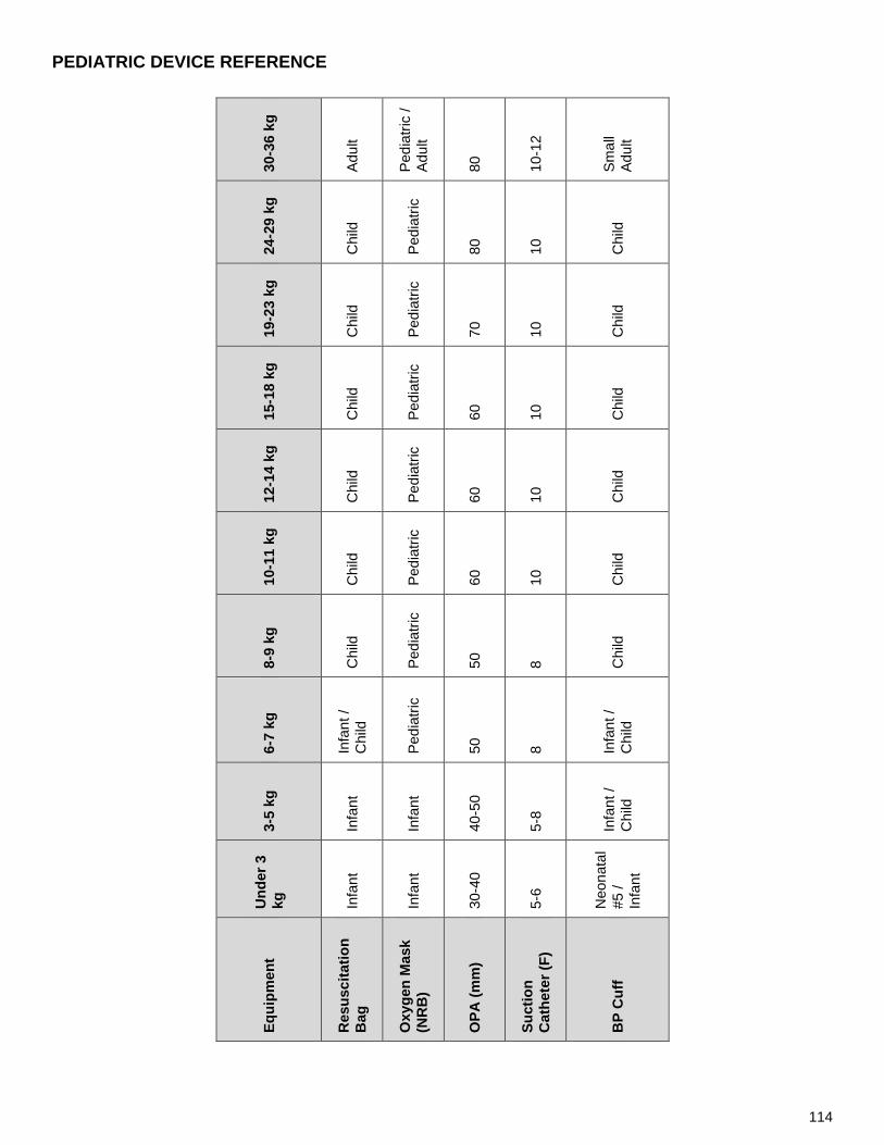
114
PEDIATRIC DEVICE REFERENCE
30-3
6 kg
Adul
t
Pedi
atric
/ Ad
ult
80
10-1
2
Smal
l Ad
ult
24-2
9 kg
Chi
ld
Pedi
atric
80
10
Chi
ld
19-2
3 kg
Chi
ld
Pedi
atric
70
10
Chi
ld
15-1
8 kg
Chi
ld
Pedi
atric
60
10
Chi
ld
12-1
4 kg
Chi
ld
Pedi
atric
60
10
Chi
ld
10-1
1 kg
Chi
ld
Pedi
atric
60
10
Chi
ld
8-9
kg
Chi
ld
Pedi
atric
50
8 Chi
ld
6-7
kg
Infa
nt /
Chi
ld
Pedi
atric
50
8 Infa
nt /
Chi
ld
3-5
kg
Infa
nt
Infa
nt
40-5
0
5-8
Infa
nt /
Chi
ld
Und
er 3
kg
Infa
nt
Infa
nt
30-4
0
5-6
Neo
nata
l #5
/ In
fant
Equi
pmen
t
Res
usci
tatio
n B
ag
Oxy
gen
Mas
k (N
RB
)
OPA
(mm
)
Suct
ion
Cat
hete
r (F)
BP
Cuf
f
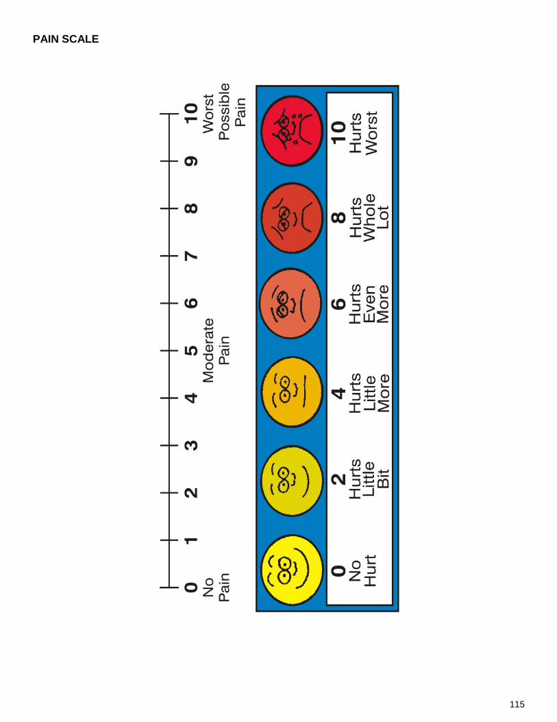
115
PAIN SCALE

116
DEFINITIONS SURROUNDING DNR, TOR AND DETERMINATION OF DEATH
SUBSTITUTE HEALTH CARE DECISION MAKER The person appointed by the Maker of an advance health care directive to make health care decisions on his or her behalf. The first named person or a member of the category of persons on the following list may, if he or she is at least 19 years of age, act as a SHCDM; the patient's:
1) Appointed substitute decision maker or a guardian that has been appointed for the purpose by a court and named on the advance health care directive
2) Spouse 3) Children 4) Parents 5) Siblings 6) Grandchildren 7) Grandparents 8) Uncles and aunts 9) Nephews or nieces 10) Another relative 11) Health care professional who is responsible for the proposed health care VALID ADVANCE HEALTH CARE DIRECTIVE (AHCD)
A document which sets out the Maker's instructions or the Maker's general principles regarding his or her health care treatment or in which a Maker appoints a substitute decision maker or both (Maker means a person who makes an advance health care directive). An Advance Health Care Directive shall be: 1) In writing 2) Witnessed by at least 2 independent persons 3) Signed by the Maker VALID DO NOT RESUSCITATE (DNR)
Is a written order issued and signed by a physician that resuscitation should not be attempted if a person suffers cardiac or respiratory arrest. Such an order may be instituted on the basis of an AHCD from a person or from a substitute health care decision maker or by a physician and it is designed to prevent unnecessary suffering.

117
BURN REFERENCES RULE OF NINES
• A method of estimating burn injury that divides the total body surface area (BSA) into sections that are multiples of 9%
• This method provides a rough estimate of burn injury size and is most accurate for adults and children greater than 10 years of age
• If burns are irregularly shaped or have a
scattered distribution, the Rule of Palms may provide a better estimate of burn surface area. The surface of the patient’s hand (palm and fingers) equals approximately 1% of the total BSA.
PARKLAND FORMULA Formula used to estimate the total volume of fluid replacement required by the burn patient in the first 24 hours post-injury
• Total fluid required in first 24 hours (mL) = [4 x (Weight in Kg) x (TBSA1) o Administer ½ of the total volume over 8 hours o Administer the second ½ of the total volume over the subsequent 16 hours
NOTES 1 TBSA should be the whole number percent estimate of the TBSA rather than the percentage
expressed as a decimal. For example – for a 40 Kg patient suffering 27% burns: • Total fluid required in first 24 hours = [4 x 40 x 27] = 4320 mL

118
GLASGOW COMA SCALE The Glasgow Coma Scale is a clinical tool used to assess the degree of consciousness and neurological functioning - and therefore severity of brain injury - by testing eye opening, verbal response and motor activity.
ADULT CHILDREN INFANT
EYE OPENING
4 Spontaneously Spontaneously Spontaneously
3 To verbal stimulus To verbal stimulus To verbal stimulus
2 To painful stimulus To painful stimulus To painful stimulus
1 No opening No opening No opening VERBAL RESPONSE
5 Completely alert, oriented and appropriate
Completely alert, oriented and appropriate
Coos, babbles and smiles as normal
4 Confused Confused Irritable cries
3 Inappropriate words Inappropriate words Inappropriate cries, screams
2 Incomprehensible Incomprehensible words or non-specific sounds
Moans in response to pain
1 No verbal response No verbal response No verbal response
MOTOR ACTIVITY
6 Obeys commands Spontaneous and appropriate
Spontaneous and appropriate
5 Localizes pain Localizes pain Localizes pain
4 Withdraws to pain Flexion withdrawal Flexion withdrawal
3 Abnormal flexion Abnormal flexion Abnormal flexion
2 Extension Extension Extension
1 No motor activity No motor activity No motor activity

119
OXYGEN TANK DURATION CHARTS Table entries represent duration of tank use in minutes
D Cylinder (Minus safe residual of 200 PSI)
PSI
1 2 3 4 5 6 7 8 9 10
11
12
13
14
15
2000
288
144
96
72
58
48
41
36
32
29
26
24
22
21
19
1900
272
136
91
68
54
45
39
34
30
27
25
23
21
19
18
1800
256
128
85
64
51
43
37
32
28
26
23
21
20
18
17
1700
240
120
80
60
48
40
34
30
27
24
22
20
18
17
16
1600
224
112
75
56
45
37
32
28
25
22
20
19
17
16
15
1500
208
104
69
52
42
35
30
26
23
21
19
17
16
15
14
1400
192
96
64
48
38
32
27
24
21
19
17
16
15
14
13
1300
176
88
59
44
35
29
25
22
20
18
16
15
14
13
12
1200
160
80
53
40
32
27
23
20
18
16
15
13
12
11
11
1100
144
72
48
36
29
24
21
18
16
14
13
12
11
10
10
1000
128
64
43
32
26
21
18
16
14
13
12
11
10
9 9
900
112
56
37
28
22
19
16
14
12
11
10
9 9 8 7
800
96
48
32
24
19
16
14
12
11
10
9 8 7 7 6
700
80
40
27
20
16
13
11
10
9 8 7 7 6 6 5
600
64
32
21
16
13
11
9 8 7 6 6 5 5 5 4
500
48
24
16
12
10
8 7 6 5 5 4 4 4 3 3
400
32
16
11
8 6 5 5 4 4 3 3 3 2 2 2
300
16
8 5 4 3 3 2 2 2 2 1 1 1 1 1
200 0 0 0 0 0 0 0 0 0 0 0 0 0 0 0
PSI
1 2 3 4 5 6 7 8 9 10
11
12
13
14
15
← Liters per minute →
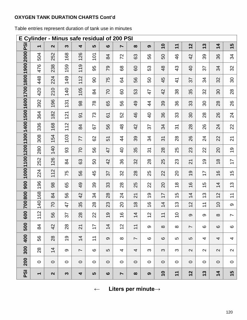
120
OXYGEN TANK DURATION CHARTS Cont’d Table entries represent duration of tank use in minutes
E Cylinder - Minus safe residual of 200 PSI
PSI
1 2 3 4 5 6 7 8 9 10
11
12
13
14
15
2000
504
252
168
126
101
84
72
63
56
50
46
42
39
36
34
1900
476
238
159
119
95
79
68
60
53
48
43
40
37
34
32
1800
448
224
149
112
90
75
64
56
50
45
41
37
34
32
30
1700
420
210
140
105
84
70
60
53
47
42
38
35
32
30
28
1600
392
196
131
98
78
65
56
49
44
39
36
33
30
28
26
1500
364
182
121
91
73
61
52
46
40
36
33
30
28
26
24
1400
336
168
112
84
67
56
48
42
37
34
31
28
26
24
22
1300
308
154
103
77
62
51
44
39
34
31
28
26
24
22
21
1200
280
140
93
70
56
47
40
35
31
28
25
23
22
20
19
1100
252
126
84
63
50
42
36
32
28
25
23
21
19
18
17
1000
224
112
75
56
45
37
32
28
25
22
20
19
17
16
15
900
196
98
65
49
39
33
28
25
22
20
18
16
15
14
13
800
168
84
56
42
34
28
24
21
19
17
15
14
13
12
11
700
140
70
47
35
28
23
20
18
16
14
13
12
11
10
9
600
112
56
37
28
22
19
16
14
12
11
10
9 9 8 7
500
84
42
28
21
17
14
12
11
9 8 8 7 6 6 6
400
56
28
19
14
11
9 8 7 6 6 5 5 4 4 4
300
28
14
9 7 6 5 4 4 3 3 3 2 2 2 2
200 0 0 0 0 0 0 0 0 0 0 0 0 0 0 0
PSI
1 2 3 4 5 6 7 8 9 10
11
12
13
14
15
← Liters per minute→

121
OXYGEN TANK DURATION CHARTS Cont’d Table entries represent duration of tank use in minutes
M Cylinder - Minus safe residual of 200 PSI
PSI
1 2 3 4 5 6 7 8 9 10
11
12
13
14
15
2000
2808
1404
936
702
562
468
401
351
312
281
255
234
216
201
187
1900
2652
1326
884
663
530
442
379
332
295
265
241
221
204
189
177
1800
2496
1248
832
624
499
416
357
312
277
250
227
208
192
178
166
1700
2340
1170
780
585
468
390
334
293
260
234
213
195
180
167
156
1600
2184
1092
728
546
437
364
312
273
243
218
199
182
168
156
146
1500
2028
1014
676
507
406
338
290
254
225
203
184
169
156
145
135
1400
1872
936
624
468
374
312
267
234
208
187
170
156
144
134
125
1300
1716
858
572
429
343
286
245
215
191
172
156
143
132
123
114
1200
1560
780
520
390
312
260
223
195
173
156
142
130
120
111
104
1100
1404
702
468
351
281
234
201
176
156
140
128
117
108
100
94
1000
1248
624
416
312
250
208
178
156
139
125
113
104
96
89
83
900
1092
546
364
273
218
182
156
137
121
109
99
91
84
78
73
800
936
468
312
234
187
156
134
117
104
94
85
78
72
67
62
700
780
390
260
195
156
130
111
98
87
78
71
65
60
56
52
600
624
312
208
156
125
104
89
78
69
62
57
52
48
45
42
500
468
234
156
117
94
78
67
59
52
47
43
39
36
33
31
400
312
156
104
78
62
52
45
39
35
31
28
26
24
22
21
300
156
78
52
39
31
26
22
20
17
16
14
13
12
11
10
200 0 0 0 0 0 0 0 0 0 0 0 0 0 0 0
PSI
1 2 3 4 5 6 7 8 9 10
11
12
13
14
15
← Liters per minute→

122
IV RATE CONVERSION CHART
Drip rate (gtts / minute) according to drop set utilized Target administration rate (mL/hour) 10 drop set 15 drop set 20 drop set 60 drop set
30 5 8 10 30
40 7 10 13 40
60 10 15 20 60
100 17 25 33 100
125 21 31 42 125
150 25 38 50 150
175 29 44 58 175
200 33 50 67 200
250 42 63 83 250
300 50 75 100 300
350 58 88 117 350
400 67 100 133 400
450 75 113 150 450
500 83 125 167 500
550 92 138 183 550
600 100 150 200 600
650 108 163 217 650
700 117 175 233 700
750 125 188 250 750
800 133 200 267 800
850 142 213 283 850
900 150 225 300 900
950 158 238 317 950
1000 167 250 333 1000 CALCULATING DRIP RATE FOR CRYSTALLOID FLUID ADMINISTRATION • Drip rate (gtts / minute) = Total amount to be administered (mL) x Drip Factor
Desired time frame (minutes)
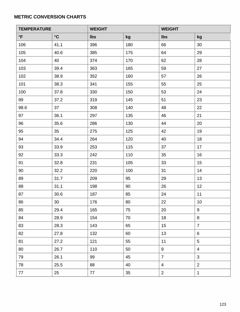
123
METRIC CONVERSION CHARTS
TEMPERATURE WEIGHT WEIGHT
°F °C lbs kg lbs kg
106 41.1 396 180 66 30
105 40.6 385 175 64 29
104 40 374 170 62 28
103 39.4 363 165 59 27
102 38.9 352 160 57 26
101 38.3 341 155 55 25
100 37.8 330 150 53 24
99 37.2 319 145 51 23
98.6 37 308 140 48 22
97 36.1 297 135 46 21
96 35.6 286 130 44 20
95 35 275 125 42 19
94 34.4 264 120 40 18
93 33.9 253 115 37 17
92 33.3 242 110 35 16
91 32.8 231 105 33 15
90 32.2 220 100 31 14
89 31.7 209 95 29 13
88 31.1 198 90 26 12
87 30.6 187 85 24 11
86 30 176 80 22 10
85 29.4 165 75 20 9
84 28.9 154 70 18 8
83 28.3 143 65 15 7
82 27.8 132 60 13 6
81 27.2 121 55 11 5
80 26.7 110 50 9 4
79 26.1 99 45 7 3
78 25.5 88 40 4 2
77 25 77 35 2 1

124
ACETAMINOPHEN DOSE CHART Only For use with 80mg/ml concentration
Weight in Kg Dose ml of 80mg/ml Concentration
3 45mg 0.5ml 4 60mg 0.75ml 5 75mg 1ml 6 90mg 1.1ml 7 105mg 1.3ml 8 120mg 1.5ml 9 135mg 1.7ml 10 150mg 1.9ml 11 165mg 2ml 12 180mg 2.25ml 13 195mg 2.4ml 14 210mg 2.6ml 15 225mg 2.8ml 16 240mg 3ml 17 255mg 3.2ml 18 270mg 3.4ml 19 285mg 3.5ml 20 300mg 3.75ml 21 315mg 4ml 22 330mg 4.1ml 23 345mg 4.3ml 24 360mg 4.5ml 25 375mg 4.7ml 26 390mg 4.9ml 27 405mg 5ml 28 420mg 5.25ml 29 435mg 5.5ml 30 450mg 5.6ml 31 465mg 5.8ml 32 480mg 6ml 33 495mg 6.2ml 34 510mg 6.4ml 35 525mg 6.6ml 36 540mg 6.75ml 37 555mg 7ml 38 570mg 7.1ml 39 585mg 7.3ml 40 600mg 7.5ml 41 615mg 7.7ml 42 630mg 7.9ml 43 645mg 8ml 44 660mg 8.25ml 45 675mg 8.5ml 46 690mg 8.6ml 47 705mg 8.8ml 48 720mg 9ml 49 735mg 9.2ml 50 750mg 9.4ml

125
ACRONYMS / ABBREVIATIONS
A-B-C Airway Breathing Circulation LPM Liters Per Minute
ACLS Advanced Cardiac Life Support LSN Last Seen Normal
AC Antecubital Lt Left
ACP Advanced Care Paramedic MCC Motorcycle Crash
AED Automated External Defibrillator mcg Micrograms
ALS Advanced Life Support MDI Metered Dose Inhaler
AMI Acute Myocardial Infarction mEq Milliequivalent
AP Anterior Posterior mL Milliliter
ASA Acetylsalicylic Acid mmHg Millimeters of Mercury
BG Blood Glucose mmol/L Millimoles Per Liter
BLS Basic Life Support MOI Mechanism of Injury
BMV Bag Mask Ventilation MVC Motor Vehicle Crash
BP Blood Pressure NIPPV Non-Invasive Positive Pressure Ventilation
BSA Body Surface Area NRB Non Rebreather Mask
BVM Bag Valve Mask NRP Neonatal Resuscitation Program
C Celsius/Centigrade NS Normal Saline
C-A-B Circulation Airway Breathing NYD Not Yet Diagnosed
CCP Critical Care Paramedic OLMC Online Medical Control
CHF Congestive Heart Failure PALS Pediatric Advanced Life Support
COPD Chronic Obstructive Pulmonary Disease PCI Percutaneous Coronary Intervention
CPAP Continuous Positive Airway Pressure PCP Primary Care Paramedic
CPR Cardiopulmonary Resuscitation PCR Patient Care Report
CVA Cerebral Vascular Accident PEA Pulseless Electrical Activity
D10/25/50 Dextrose 10,25 & 50% PMO Provincial Medical Oversight Program
DBP Diastolic Blood Pressure PO Per Os (by mouth, orally)
DHCS Department of Health and Community Services PPE Personal Protective Equipment
DKA Diabetic Ketoacidosis PPV Positive Pressure Ventilation
DNR Do Not Resuscitate PR Per Rectum
ECG Electrocardiogram PSI Pounds per Square Inch

126
EMR Emergency Medical Responder q Every
g Gram QO Quarantine Officer
GCS Glasgow Coma Scale RA Right Arm
GI Gastrointestinal RL Right Leg
gtt(s) Drop(s) ROM Range of Motion
HAZMAT Hazardous Materials ROSC Return of Spontaneous Circulation
HR Heart Rate RR Respiratory Rate
HTN Hypertension Rt Right
Hx History SBP Systolic Blood Pressure
IDDM Insulin-Dependent Diabetes Mellitus SC Subcutaneous
IM Intramuscular SHCDM Substitute Health Care Decision Maker
IN Intra Nasal SIVP Slow Intravenous Push
IO Intraosseous SL Sublingual
ITLS International Trauma Life Support SpO2 Saturation of Peripheral Oxygen
IV Intravenous STEMI ST Elevated Myocardial Infarction
IVP Intravenous Push SVT Supraventricular Tachycardia
J Joules TIA Transient Ischemic Attack
JVD Jugular Vein Distention TKVO To Keep Vein Open
kg Kilogram TOR Termination of Resuscitation
LA Left Arm VF Ventricular Fibrillation
lbs Pounds VSA Vital Signs Absent
LL Left Leg VT Ventricular Tachycardia
LOC Level Of Consciousness WPW Wolff-Parkinson-White

127
PAGE INTENTIONALLY LEFT BLANK FOR FUTURE PROTOCOL

128
PAGE INTENTIALLY LEFT BLANK FOR FUTURE PROTOCOL

129
PAGE INTENTIONALLY LEFT BLANK FOR FUTURE PROTOCOL
130
PART V: MEDICATION FORMULARY

131
FORMULARY CAUTION
• All calculations contained within this formulary are to be used as a guide and must be verified by the individual practitioner
• The information contained herein is not a substitute for clinical judgment ACETAMINOPHEN
CLASS Antipyretic, analgesic
INDICATIONS Pediatric Fever (temperature greater than 38.5ºC)
CONTRAINDICATIONS 1) Hypersensitivity 2) Nausea and vomiting (PO)
PEDIATRIC DOSE 15 mg/kg PO
NOTES • If acetaminophen already administered within the last 4 hours,
administer a “top-up” dose to so total dose administered within the last 4 hours is equal to 15 mg/kg
ACETYLSALICYLIC ACID (ASA)
CLASS Platelet aggregation inhibitor
INDICATIONS Ischemic Chest Pain
CONTRAINDICATIONS
1) Hypersensitivity to ASA or NSAIDS 2) History of active bleeding 3) Active bronchospasm or history of severe asthma with bronchospasm
related to ASA or NSAIDS 4) Age less than 16 years
PRECAUTIONS • Pregnancy • Bleeding disorders
ADULT DOSE 160-162 mg PO chewed
NOTES
• ASA should still be administered if patient has already taken their usual prescribed daily dose of ASA
• If the patient has taken ASA on the advice of the dispatcher, confirm correct identity of medication, dose and expiration date. If able to confirm appropriate self-administration do not administer additional ASA. If ASA taken is enteric coated, administer ASA as per protocol.
• Regular use of anticoagulants, such as warfarin, is not a contraindication to ASA administration.

132
DEXTROSE
CLASS Caloric Agent
INDICATIONS Adult Symptomatic Hypoglycemia Pediatric Symptomatic Hypoglycemia
CONTRAINDICATIONS 1) Hypersensitivity to corn or corn products
PRECAUTIONS
• Head injury or suspected stroke (Protocol modification) o Administer half of the usual dose, recheck BGL within recommended
time parameters and then administer the second half dose if indicated
ADULT DOSE • Dextrose 50% (D50%) 25 g (50 mL) IVP • Repeat once in 10 minutes if indicated
PEDIATRIC DOSE
WEIGHT LESS THAN 10 KG
• Dextrose 10% (D10%) 5 mL/kg SIVP • Repeat once in 10 minutes if indicated
WEIGHT 10-20 KG
• Dextrose 25% (D25%) 2 mL/kg SIVP • Repeat once in 10 minutes if indicated
WEIGHT 20-40 KG
• Dextrose 50% (D50%) 1 mL/kg SIVP [Not to exceed a maximum single dose of 50 mL (25g)]
• Repeat once in 10 minutes if indicated
WEIGHT GREATER THAN 40 KG
• Dextrose 50% (D50%) 25 g (50 mL) IVP • Repeat once in 10 minutes if indicated
NOTES
• To create dextrose 10% (D10%): o Remove 40 mL from D50% preload syringe and replace with 40 mL
0.9% NaCl to yield a final concentration of 5 g of dextrose in 50 mL (D10%).
• To create dextrose 25% (D25%): o Remove 25 mL from D50% preload syringe and replace with 25 mL
0.9% NaCl to yield a final concentration of 12.5 g of dextrose in 50 mL (D25%).
• Extravasation may cause tissue necrosis

133
DIMENHYDRINATE (GRAVOL)
CLASS Antiemetic
INDICATIONS Adult Nausea and Vomiting Pediatric Nausea and Vomiting
CONTRAINDICATIONS
1) Hypersensitivity to dimenhyDRINATE, diphenhydrAMINE or propylene glycol
2) Narrow angle glaucoma 3) Hypotension (SBP less than 100 mmHg in adults and less than age-
specific BP criteria in pediatrics) 4) Altered LOC (Including alcohol or drug intoxication)
PRECAUTIONS
• Simultaneous administration with ipratropium bromide may cause enhanced anticholinergic effects
• Elderly patients may be more sensitive to adverse effects. Consider administration of a lower dose in patients greater than 65 years of age
SIDE EFFECTS • Sedation • Hypotension • Paradoxical excitation in children
ADULT DOSE
• 25 mg IV • Repeat once in 15 minutes if indicated (Not to exceed a maximum total
dose of 50 mg) May administer 50 mg IM if indications are present AND you are unable to establish an IV
PEDIATRIC DOSE • 1 mg/kg IV (Not to exceed a maximum single dose of 25 mg)
NOTES • May cause stinging at the injection site

134
DIPHENHYDRAMINE (BENADRYL)
CLASS Antihistamine
INDICATIONS
Adult Allergy and Anaphylaxis Pediatric Allergy and Anaphylaxis Adult Nausea & Vomiting Pediatric Nausea & Vomiting
CONTRAINDICATIONS 1) Hypersensitivity to dimenhyDRINATE or diphenhydrAMINE 2) Narrow angle glaucoma
PRECAUTIONS • Simultaneous administration with ipratropium bromide may cause
enhanced anticholinergic effects • Elderly patients may be more sensitive to adverse effects.
SIDE EFFECTS • Sedation • Hypotension • Paradoxical excitation in children
ADULT DOSE
Adult Anaphylaxis • 50 mg IVP May administer 50 mg IM if indications are present AND you are unable to establish an IV Adult Isolated Hives
• 25-50 mg IV/IM
Adult Extrapyramidal Reactions
• 50 mg IV
PEDIATRIC DOSE
Pediatric Anaphylaxis • 1 mg/kg IV (Not to exceed a maximum single dose of 50 mg) May administer 1 mg/kg IM (Not to exceed a maximum single dose of 50 mg) if indications are present AND you are unable to establish an IV Pediatric Isolated Hives
• Avoid giving for this indication, oral medication (such as cetirizine) are preferred and available in Emergency Departments
Pediatric Extrapyramidal Reactions
• 1 mg/kg IV (Not to exceed a maximum single dose of 50 mg)

135
EPINEPHRINE 1:1000
CLASS Sympathomimetic
INDICATIONS
Adult Respiratory Distress With Bronchospasm (OLMC) Adult Allergy and Anaphylaxis Pediatric Respiratory Distress with Bronchospasm (OLMC) Pediatric Respiratory Distress with Inspiratory Stridor Pediatric Allergy and Anaphylaxis
RELATIVE CONTRAINDICATIONS
1) Cardiac ischemia or infarction 2) Tachydysrhythmias (Greater than 150 beats per minute in adults and
180 beats per minute in pediatrics)
PRECAUTIONS • Cardiac dysrhythmias • Cardiac valvular abnormalities
SIDE EFFECTS
ADULT DOSE
Adult Respiratory Distress With Bronchospasm (OLMC)
• 0.3 mg IM
Adult Anaphylaxis
• 0.3 mg IM • Repeat once in 5 minutes if no improvement
PEDIATRIC DOSE
Pediatric Respiratory Distress with Bronchospasm (OLMC)
• 0.01 mg/kg (0.01 mL/kg) IM [Not to exceed a maximum single dose of 0.3 mg (0.3 mL)]
Pediatric Respiratory Distress with Inspiratory Stridor
AGE LESS THAN 1 YEAR AND LESS THAN 5 KG
• 0.5 mg (0.5 mL) in 2 mL 0.9% NaCl nebulized
AGE LESS THAN 1 YEAR AND GREATER THAN OR EQUAL TO 5 KG
• 2.5 mg (2.5 mL) nebulized
AGE GREATER THAN OR EQUAL TO 1 YEAR
• 5 mg (5 mL) nebulized
Pediatric Anaphylaxis
• 0.01 mg/kg (0.01 mL/kg) IM [Not to exceed a maximum single dose of 0.3 mg (0.3 mL)]
• Repeat once in 5 minutes if no improvement

136
GLUCAGON
CLASS Hyperglycemic Agent
INDICATIONS Adult Symptomatic Hypoglycemia Pediatric Symptomatic Hypoglycemia
CONTRAINDICATIONS 1) Hypersensitivity to glucagon, glycerin or phenol 2) Anaphylaxis to lactose 3) Pheochromocytoma
PRECAUTIONS • Glycogen depleted state (Starvation, chronic hypoglycemia, alcoholism) • Adrenal insufficiency • Insulinoma
SIDE EFFECTS • Nausea and vomiting • Hypertension and hypotension • Tachycardia
ADULT DOSE
Adult Symptomatic Hypoglycemia
• 1 mg IM • Repeat once in 20 minutes if indicated
PEDIATRIC DOSE
Pediatric Symptomatic Hypoglycemia
LESS THAN 20 KG
• 0.5 mg IM • Repeat once in 20 minutes if indicated
GREATER THAN 20 KG
• 1 mg IM • Repeat once in 20 minutes if indicated
NOTES
• Glucagon is unlikely to be effective in glycogen depleted states as glucagon requires sufficient storage of glycogen in the liver to be effective.
• Inject the content of the pre-loaded syringe into the bottle of powdered glucagon and swirl to mix contents until all of the glucagon is dissolved. The resultant solution should be clear.
• Response to glucagon is not immediate; anticipate that it may take up to 20 minutes to see an effect
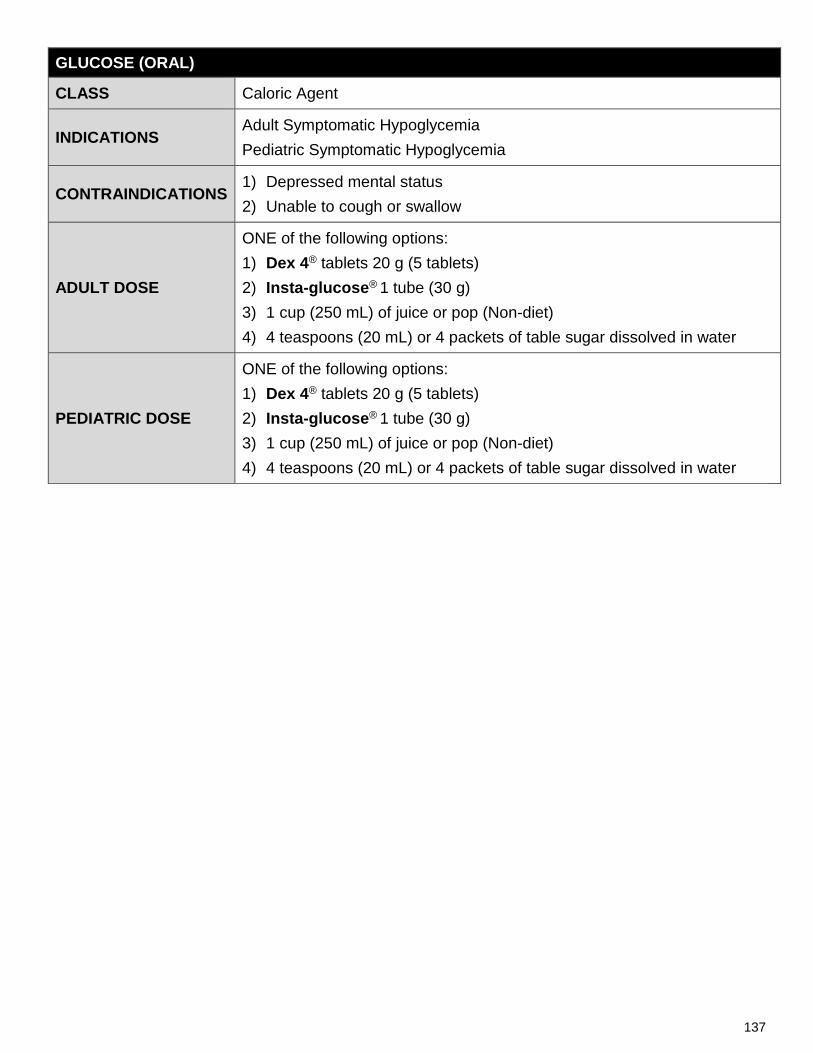
137
GLUCOSE (ORAL)
CLASS Caloric Agent
INDICATIONS Adult Symptomatic Hypoglycemia Pediatric Symptomatic Hypoglycemia
CONTRAINDICATIONS 1) Depressed mental status 2) Unable to cough or swallow
ADULT DOSE
ONE of the following options: 1) Dex 4® tablets 20 g (5 tablets) 2) Insta-glucose® 1 tube (30 g) 3) 1 cup (250 mL) of juice or pop (Non-diet) 4) 4 teaspoons (20 mL) or 4 packets of table sugar dissolved in water
PEDIATRIC DOSE
ONE of the following options: 1) Dex 4® tablets 20 g (5 tablets) 2) Insta-glucose® 1 tube (30 g) 3) 1 cup (250 mL) of juice or pop (Non-diet) 4) 4 teaspoons (20 mL) or 4 packets of table sugar dissolved in water

138
HYDROCORTISONE
CLASS Corticosteroid
INDICATIONS Adult Adrenal Insufficiency Pediatric Adrenal Insufficiency
CONTRAINDICATIONS 1) Hypersensitivity to hydrocortisone or other corticosteroids
PRECAUTIONS
• History of seizure • Peptic ulceration or inflammatory bowel disease • Diabetes (monitor blood glucose levels) • Hypertension • Renal insufficiency • Tuberculosis • Chronic psychosis • Ocular herpes simplex
SIDE EFFECTS • Hypertension • Agitation • Headache and/or vertigo
ADULT DOSE • 100 mg IV/IM
PEDIATRIC DOSE
AGE LESS THAN 3 YEARS
• 25 mg IV/IM AGE 3 TO 10 YEARS
• 50 mg IV/IM GREATER THAN 10 YEARS
• 100 mg IV/IM
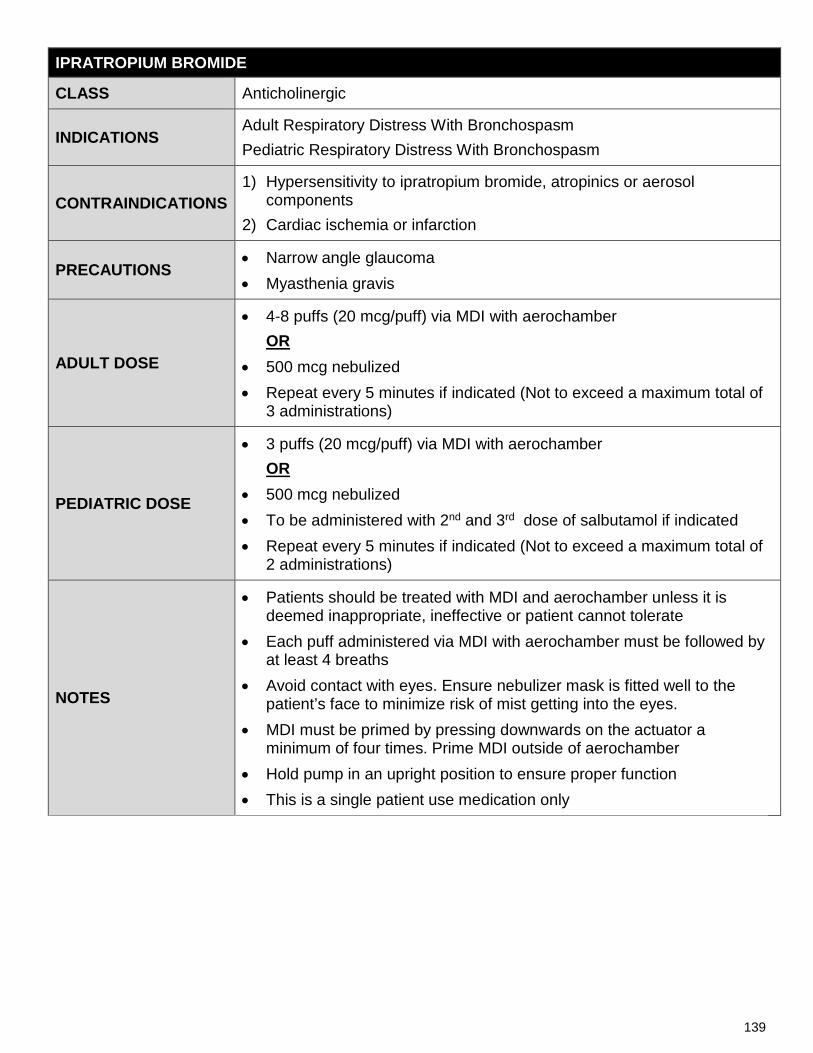
139
IPRATROPIUM BROMIDE
CLASS Anticholinergic
INDICATIONS Adult Respiratory Distress With Bronchospasm Pediatric Respiratory Distress With Bronchospasm
CONTRAINDICATIONS 1) Hypersensitivity to ipratropium bromide, atropinics or aerosol
components 2) Cardiac ischemia or infarction
PRECAUTIONS • Narrow angle glaucoma • Myasthenia gravis
ADULT DOSE
• 4-8 puffs (20 mcg/puff) via MDI with aerochamber OR
• 500 mcg nebulized • Repeat every 5 minutes if indicated (Not to exceed a maximum total of
3 administrations)
PEDIATRIC DOSE
• 3 puffs (20 mcg/puff) via MDI with aerochamber OR
• 500 mcg nebulized • To be administered with 2nd and 3rd dose of salbutamol if indicated • Repeat every 5 minutes if indicated (Not to exceed a maximum total of
2 administrations)
NOTES
• Patients should be treated with MDI and aerochamber unless it is deemed inappropriate, ineffective or patient cannot tolerate
• Each puff administered via MDI with aerochamber must be followed by at least 4 breaths
• Avoid contact with eyes. Ensure nebulizer mask is fitted well to the patient’s face to minimize risk of mist getting into the eyes.
• MDI must be primed by pressing downwards on the actuator a minimum of four times. Prime MDI outside of aerochamber
• Hold pump in an upright position to ensure proper function • This is a single patient use medication only

140
KETOROLAC
CLASS NSAID
INDICATIONS Adult Pain Management
CONTRAINDICATIONS
1) Hypersensitivity to ASA or NSAIDS 2) Active bronchospasm or history of severe asthma with bronchospasm
related to ASA or NSAIDS 3) History of active bleeding or bleeding disorder 4) History of CVA or cardiovascular disease 5) Renal or hepatic insufficiency 6) Peptic ulcer or inflammatory bowel disease 7) NSAID use, including aspirin, in previous 6 hours 8) Age less than 16 years or greater than 65 years 9) Known or suspected hyperkalemia 10) Pregnancy
PRECAUTIONS • Hypertension
SIDE EFFECTS
• Headache • Edema • GI bleeding • Hypertension
• Nausea, abdominal pain and/or dyspepsia • Drowsiness • Dizziness • Rash
ADULT DOSE • 15 mg SIVP/IM

141
METOCLOPRAMIDE
CLASS Antiemetic
INDICATIONS Adult Nausea and Vomiting Adult Pain Management (Migraine) Pediatric Nausea and Vomiting
CONTRAINDICATIONS
1) Hypersensitivity to metoclopramide or procainamide 2) Hypotension (SBP less than 100 mmHg in adults and less than age-
specific BP criteria in pediatrics) 3) Pheochromocytoma 4) Suspected GI hemorrhage, bowel obstruction or perforation 5) Seizure disorders 6) Monoamine oxidase inhibitor (MAO) therapy within the last 14 days 7) Concurrent use of antipsychotic medications
PRECAUTIONS
• Parkinson’s Disease • Altered LOC (Including alcohol or drug intoxication) • Elderly patients may be more sensitive to adverse effects. Consider
administration of a lower dose in patients greater than 65 years of age
SIDE EFFECTS
• Sedation • Hypotension or hypertension • Bronchospasm • Bradycardia • Tachycardia • Extrapyramidal signs and symptoms
ADULT DOSE
Adult Nausea and Vomiting • 10 mg SIVP (Over 2 to 5 minutes)
o May administer 10 mg IM if indications are present AND you are unable to establish an IV
Adult Pain Management (Migraine) • 10 mg SIVP (Over 2 to 5 minutes)
o May administer 10 mg IM if indications are present AND you are unable to establish an IV
PEDIATRIC DOSE • 0.1 mg/kg SIVP (Over 2 to 5 minutes) (Not to exceed a maximum single dose of 10 mg)
NOTES
• Risk of development of extrapyramidal symptoms is reduced if metoclopramide is administered slowly when using an IV route.
• If extrapyramidal signs and symptoms develop, reassure the patient and administer diphenhydrAMINE as per protocol.

142
NALOXONE HYDROCHLORIDE
CLASS Opioid antagonist
INDICATIONS Adult Toxin Management, Opioids Pediatric Toxin Management, Opioids
CONTRAINDICATIONS 1) Hypersensitivity
SIDE EFFECTS • Acute opioid withdrawal
ADULT DOSE • 0.2-0.4 mg IM/IV • Repeat every 2-3 minutes if indicated, titrated to improved respiratory
drive
PEDIATRIC DOSE • 0.2-0.4 mg IM/IV • Repeat every 2-3 minutes if indicated, titrated to improved respiratory
drive
NOTES
• Naloxone hydrochloride is light sensitive. If naloxone hydrochloride is contained in a glass ampoule it must be protected from light during storage.
• The duration of action of the opioid may be longer than the duration of action of naloxone and repeat administration of naloxone may be required if respiratory depression recurs.
• Be alert for potential acute agitation or combativeness following administration of naloxone to patients with opioid dependency. Titrate dose only to improve respiratory drive.
• Examples of shorter acting opioids include, but are not limited to: fentanyl, hydromorphone (Dilaudid), morphine (Morphine-IR), meperidine (Demerol), codeine, heroin, sufentanyl, Darvon, oxycodone
• Examples of longer acting opioids include, but are not limited to: methadone, MS-Contin, OxyNEO, OxyContin, Hydromorph-Contin, morphine-SR

143
NITROGLYCERIN
CLASS Antianginal Agent, Vasodilator
INDICATIONS Adult Ischemic Chest Pain Adult Pulmonary Edema
CONTRAINDICATIONS
1) Hypersensitivity to nitrates 2) Hypotension (SBP less than 100 mmHg in adults) 3) Heart rate less than 50 or greater than 150 beats per minute 4) Suspected right ventricular myocardial infarction or ischemia 5) Concurrent use of phosphodiesterase inhibitor(s) (Erectile dysfunction
medication) within preceding 48 hours 6) Altered LOC
PRECAUTIONS • Hypertrophic cardiomyopathy
SIDE EFFECTS • Hypotension • Headache • Dizziness
ADULT DOSE
Adult Pulmonary Edema
• 0.4 mg SL • Repeat every 5 minutes if indicated to a maximum of 6 sprays, until
symptoms are relieved or SBP less than 100 mmHg
Adult Ischemic Chest Pain
• 0.4 mg SL • Repeat every 5 minutes to a maximum of 6 sprays if indicated until
chest pain resolved or SBP less than 100 mmHg
NOTES
• Blood pressure must be monitored and recorded before and after each administration
• The patient should be sitting or lying prior to administration • Prime the pump by depressing down on the actuator, discarding the
first spray away from the patient • Hold the pump in an upright position to ensure proper function • This is a single use medication only • Phosphodiesterase inhibitors include, but are not limited to: sildenafil
(Viagra), tadalafil (Cialis), vardenafil (Levitra) • Phosphodiesterase inhibitors may be taken by both male and female
patients

144
SALBUTAMOL
CLASS Beta2 Agonist
INDICATIONS
Adult Respiratory Distress With Bronchospasm Adult Allergy and Anaphylaxis Pediatric Respiratory Distress With Bronchospasm Pediatric Allergy and Anaphylaxis
CONTRAINDICATIONS
1) Hypersensitivity 2) Cardiac ischemia or infarction 3) Tachydysrhythmias (Heart rate greater than 150 in adults and greater than
180 in pediatrics)
PRECAUTIONS • Cardiovascular disorders (Cardiac dysrhythmias, valvular heart disease)
SIDE EFFECTS
• Tachydysrhythmias • Hypertension and hypotension • Restlessness • Dizziness • Drowsiness • Headache • Nausea and vomiting • Dry mouth
ADULT DOSE
• 4-8 puffs (100 mcg/puff) via MDI with aerochamber OR
• 5 mg nebulized • Repeat every 5 minutes if indicated (Not to exceed a total of 3 administrations)
PEDIATRIC DOSE
AGE LESS THAN 5 YEARS
• 5 puffs (100 mcg/puff) via MDI with aerochamber OR
• 2.5 mg nebulized • Repeat every 5 minutes if indicated (Not to exceed a total of 3 administrations)
AGE GREATER THAN OR EQUAL TO 5 YEARS
• 10 puffs (100 mcg/puff) via MDI with aerochamber OR
• 5 mg nebulized • Repeat every 5 minutes if indicated (Not to exceed a total of 3 administrations)
NOTES
• Patients should be treated with MDI and aerochamber unless it is deemed inappropriate, ineffective or patient cannot tolerate
• Each puff administered via MDI with aerochamber must be followed by at least 4 breaths
• Avoid contact with eyes. Ensure nebulizer mask is fitted well to the patient’s face to minimize risk of mist getting into the eyes.
• MDI must be primed by pressing downwards on the actuator a minimum of four times. Prime MDI outside of aerochamber
• Hold pump in an upright position to ensure proper function • This is a single patient use medication only

145
MEDFLIGHT NL – AUTO LAUNCH CRITERIA

146
NOTES

147
NOTES

148
NOTES

149
NOTES

150
NOTES