basic neuroanatomy - univerzita...
TRANSCRIPT

Basic
neuroanatomy
© David Kachlík 30.9.2015
Characteristic of nervous tissue
• Nervous tissue is specialized for intake, workabout
and transfer of infos and that way coordinates most of the functions in the body
• cells:
– proper excitability neurons
– Support cells (astrocytes, oligodendrocytes, microglia and
ependymal cells)
• Anatomically we divide nervous system into central
(CNS – brain and medulla spinalis) and peripheral (PNS – nerves and ganglias)
• In the CNS we distinguish gray and white matter
• White matter visage is due to myelin – lipoprotein complex covering individual axons
© David Kachlík 30.9.2015
Classification of nervous tissue cells
• Nervous tissue contains two types of cells:
– neurons – major cells carrying informations
– Glial cells (neuroglia, glia) (neuroglia) – provide
support for neurons, protect them and
participate in nutrition and function
• astrocytes (astrocytus)
• oligodendrocytes (oligodendrocytus)
• mikroglia (microgliocytus)
• Ependymal cells (ependymocytus)
© David Kachlík 30.9.2015
Neurons I
• Are principal units of nervous tissue
• Responsible for intake, workabout and transfer of signal
• Composed of cell body (soma, perikaryon), dendrites(dendritum) and axon (axon)
• Size of neurons vary in interval 5 µm (granular cells of cerebellum) to 150 µm (Purkynje cells of cerebellum)
• After delivery no mitoses (not valid for hippocampal cells and bulbus olfactorius)
• Wallerian neurodegeneration and neuroregeneration rules(axon and dendrite regenerate regularly, neuronal body only in several localities – dentate gyrus of hippocampus)
© David Kachlík 30.9.2015

Neurons II
© David Kachlík 30.9.2015
Neurons classification I• According to shape we divide neurons into:
– multipolar (neuron multipolare)• More then 2 outlets (axon + dendrites)
• Vast majority of neurons
– bipolar (neuron bipolare)• Only 2 extensions (axon + dendrite)
• retina, olfactory epithelium, ganglion n. vestibulocochlearis (VIII) etc.
– pseudounipolar (neuron pseudounipolare)• Only 1 extension, subs. Splitting into dendrite and axon „T“
shaped
• Sensory ganglia of spinal and cranial nerves
– unipolar (neuron unipolare)• only 1 extension
• Amacrine and horizontal cells of retina© David Kachlík 30.9.2015

Neurons classification II
© David Kachlík 30.9.2015

Neurons classification III
• From functional point we divide them into:
– motor neurons (neuron motorium)
• Controll effector organs (skeletal and smooth
muscles; endocrine and exocrine glands)
– sensory neurons (neuron sensorium)
• Maintain infos intake from body and environment
– interneurons (interneuron, neuron internuntiale)
• Create complicated net between sensory and morot neurons
© David Kachlík 30.9.2015

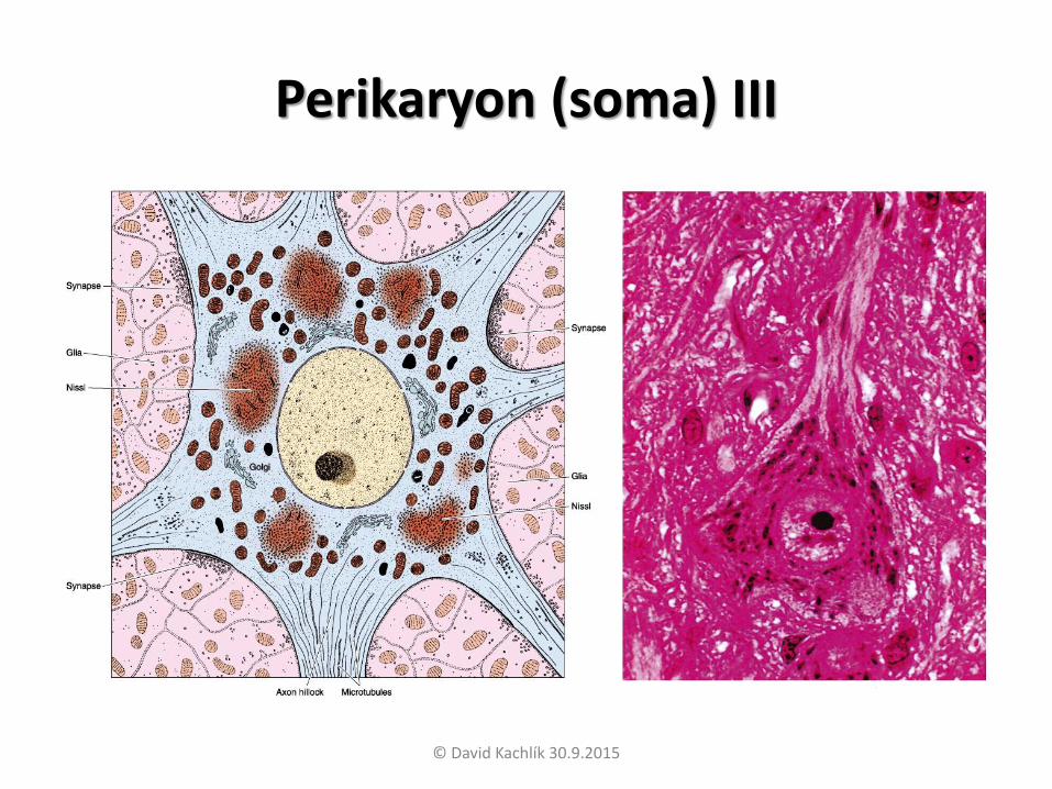
Perikaryon (soma) I
• Nucleus is big, ovoid and euchromatic with prominent nucleolus – great transcriptory activity
• GER is rich and on surface it has numerous
polyribosomes – together it forms so called Nissl substance
• GA is located only in perikaryon and it gives rise to
transport and secretory vesicules
• mitochondrias are concentrated mostly in axonal hillocks
and in pericaryon
© David Kachlík 30.9.2015
Perikaryon (soma) II
• From cytoskeletal structures are important:
– microtubules – maintain axonal transport (Hemeroff
theory of quantuum states mediating quantuum information share in the matter)
– neurofilaments – 10 nm thick intermediary filaments
specific for nervous tissue – stained by silver or gold.
• In the cytoplasm we may find also lipofuscin inclusions
(„pigment from wearing out“) or melanin
© David Kachlík 30.9.2015
Perikaryon (soma) III
© David Kachlík 30.9.2015

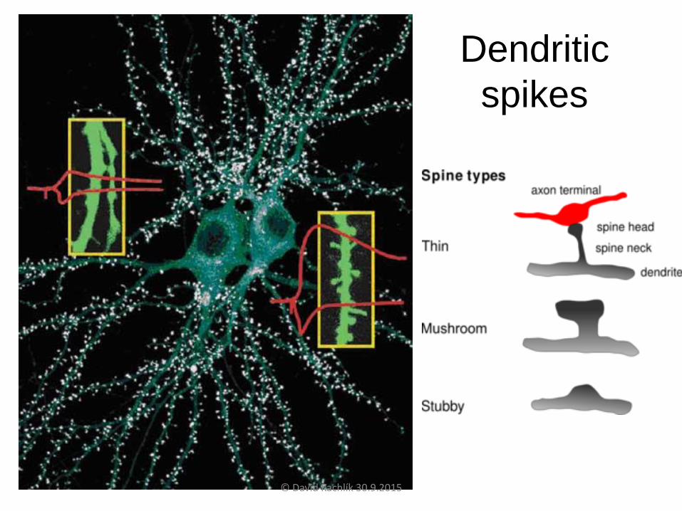
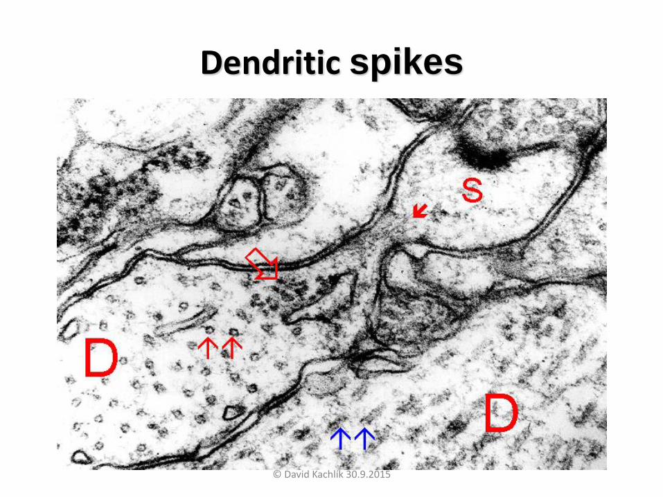
Dendrites
• Are short extensions amplifying neuron surface enabling 1 neuron to receive infos from many neurons
• In contrast to axon their width decreases with branching
• At the synapsis there are dendritic spikes (its decrease is effect of chronic stress –Sapolsky et al.)
• Cytoplasm of dendrites is almost equivalent to perikaryon, but no GA
© David Kachlík 30.9.2015
Dendritic
spikes
© David Kachlík 30.9.2015
Dendritic spikes
© David Kachlík 30.9.2015

Axon I
• Provides signal transduction from perikaryon and
its further elaboration to other neuron or effector cell
• neuron contains mostly 1 axon
• At the origin is axonal hillock with no ribosomes or GER from perikaryon
• In myelinized axons is between axonal hillock
and origin of myelin sheet inicial segment (dense layer under axolemma in EM) – here originates
action potencial
© David Kachlík 30.9.2015
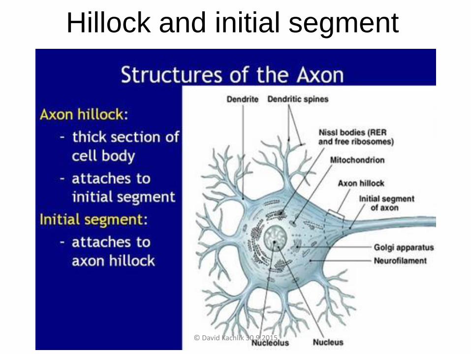
Hillock and initial segment
© David Kachlík 30.9.2015
Axon II
• axon is not branching for most of its course with
exception of collateral branches returning into perikaryon
• Branching happens at the terminal part of axon and each branch has synaptic ending – so called
bouton terminaux
• synaptic endings may be present during axonal
course as boutons en passage
© David Kachlík 30.9.2015

Bouton
terminauxVisual cortex, Macaque, Nature
2006
© David Kachlík 30.9.2015
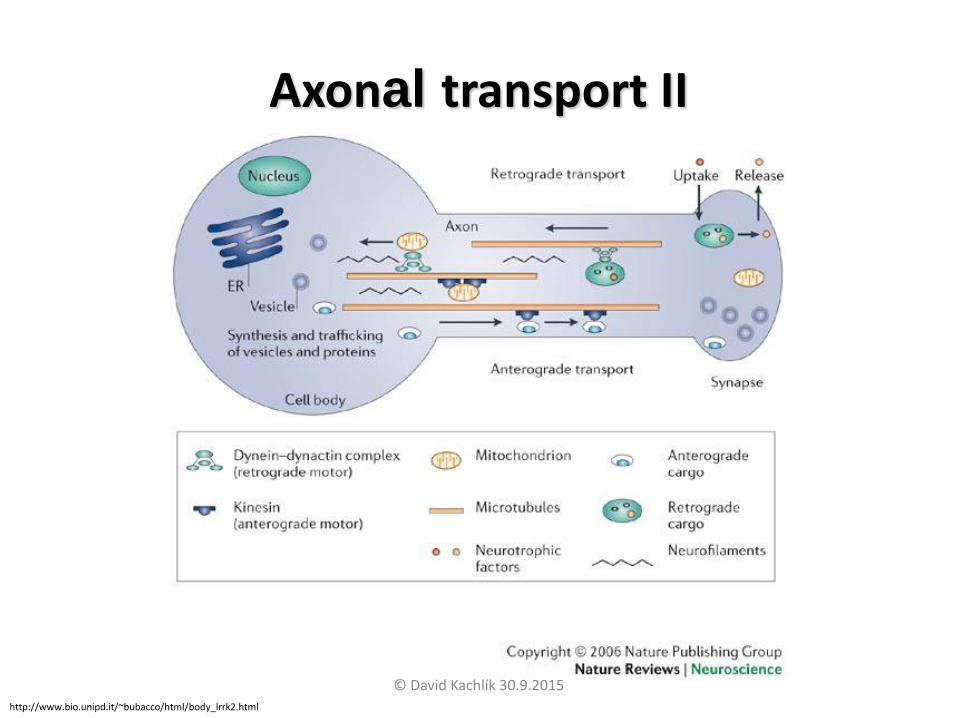
Axonal transport I• Due to missing GER is axon dependent of protein
supply from perikaryon
• Nutrition maintains axonal transport
• Function of axonal transport is granted by specific
microtubular organization – these function oas „railroad “
• „machine“ of carried particles (secretory vesicles
with neuromediators, mitochondrias etc.) is
molecular engine dynein and kinesin
• There is axonal transport anterograde (from
perikaryon) and retrograde (to perikaryon)© David Kachlík 30.9.2015
Axonal transport II
http://www.bio.unipd.it/~bubacco/html/body_lrrk2.html
© David Kachlík 30.9.2015

Synapse I
• synapse (synapsis) is specialized structure for
excitation transfer from on neuron to the other on (or on effector cell)
• According to transfer variant we divide synapses into:
– chemical (synapsis chemica): happens molecular
secretion diffusing to target cell
– electrical (synapsis electrica): cells are directly
connected by nexus – depolarization
propagates directly to target cell - rare
© David Kachlík 30.9.2015

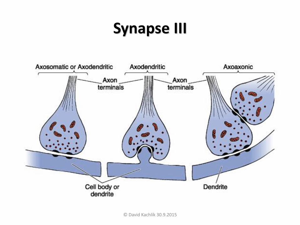
Synapse II
• According to location are synapses:
– axodendritic (synapsis axodendritica) – most
common
– axosomatic (synapsis axosomatica)
– axoaxonal – for example in presynaptic
inhibition when carrying painful stimuli
– dendritodendritic (synapsis dendritodendritica) –rare
© David Kachlík 30.9.2015
Synapse III
© David Kachlík 30.9.2015

Chemical synapse I
• Three major subparts:
– presynaptic membrane (membrana presynaptica) –cytoplasmatic membrane of axonal terminal –
cytoplasm rich in secretory (synaptic) vesicules with neurotransmitter
– synaptic cleft (fissura synaptica) – 20-30 nm wide (wider
then between neuron and glia)
– postsynaptic membrane (membrana postsynaptica) –cytoplasmatic membrane of target cell containing
receptores for neurotransmitter
© David Kachlík 30.9.2015

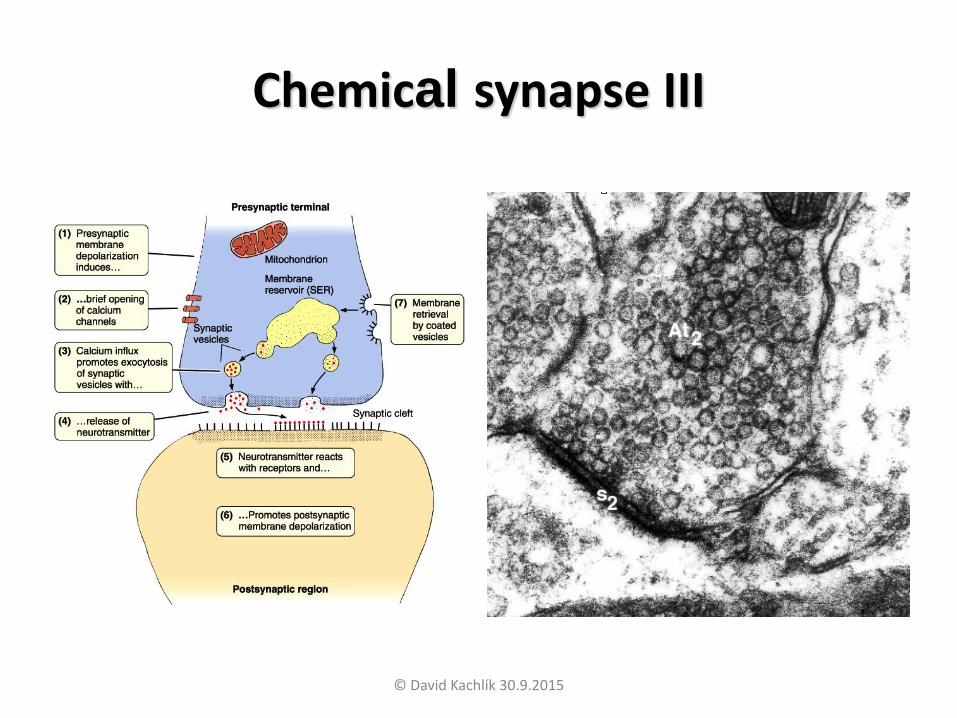
Chemical synapse II
• synaptic transfer has several phases:
– Action potential after „arriving“ into synpase opens voltage
gated Ca2+ channels – influx of Ca2+ into axonal teminal
– Increased concentration of Ca2+ starts exocytosis of
synaptic vesicles
– Ca2+ is fast inactivated – pumped into ECT
– Mediators from synaptic vesicles diffuse to target cell and
react with its receptors
– By reaction with receptors is indirectly changed
permeability of post synaptic membrane for Na+, K+, Cl- or
other ions – creation of excitatory post synaptic potential
(EPSP) or inhibitory post synaptic potential (IPSP)
• Synaptic delay is 0,3-0,5 s© David Kachlík 30.9.2015
Chemical synapse III
© David Kachlík 30.9.2015
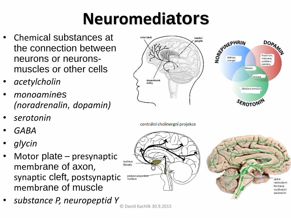
Neuromediators• Chemical substances at
the connection between neurons or neurons-muscles or other cells
• acetylcholin
• monoamines(noradrenalin, dopamin)
• serotonin
• GABA
• glycin
• Motor plate – presynaptic membrane of axon, synaptic cleft, postsynaptic membrane of muscle
• substance P, neuropeptid Y© David Kachlík 30.9.2015
Glial cells (neuroglia)
• More numerous then neurons 10-50x, but due to
smaller size form approx. 50 % of the CNS
• „cooperate“ with neurons – offer support,
nutrition, form myelin sheath of axons, phagocyte etc.
• Staining by silver or gold impregnation and histochemical methods
• Morphologically 4 types – astrocytes, oligodendrocytes, microglia and ependymal cells
• After delivery may do mitosis
© David Kachlík 30.9.2015
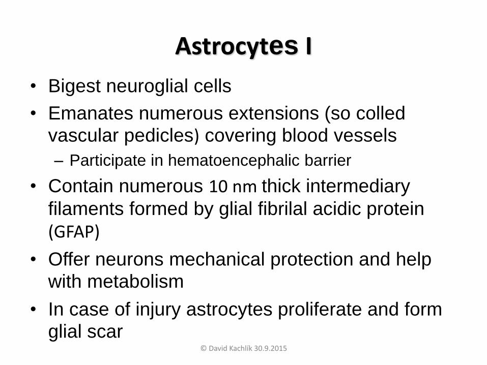
Astrocytes I
• Bigest neuroglial cells
• Emanates numerous extensions (so colled vascular pedicles) covering blood vessels
– Participate in hematoencephalic barrier
• Contain numerous 10 nm thick intermediary
filaments formed by glial fibrilal acidic protein
(GFAP)
• Offer neurons mechanical protection and help with metabolism
• In case of injury astrocytes proliferate and form glial scar
© David Kachlík 30.9.2015
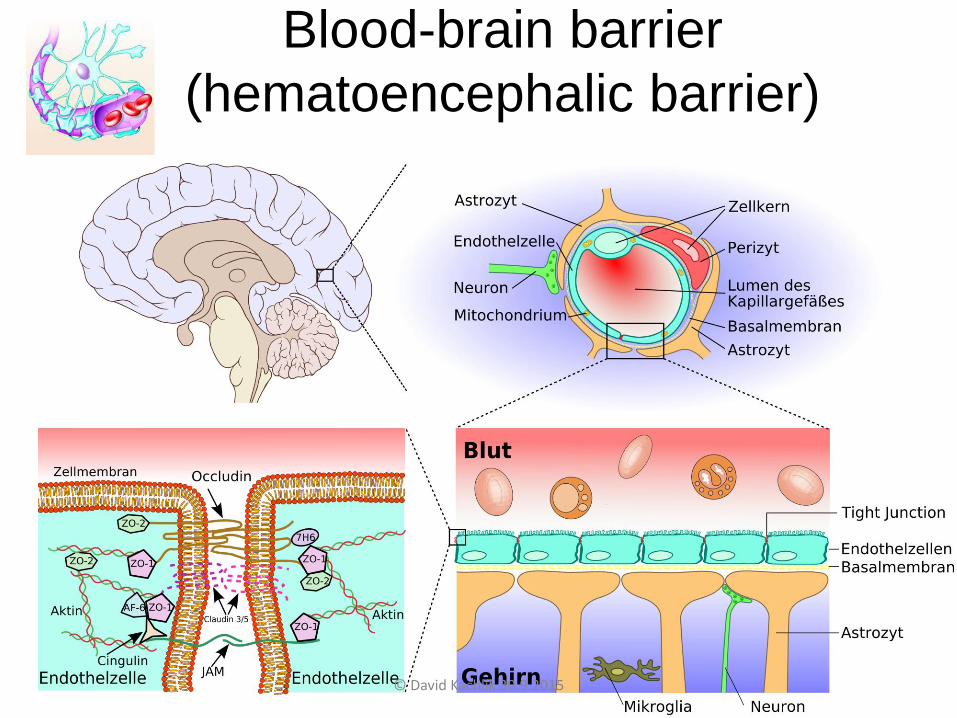
Blood-brain barrier
(hematoencephalic barrier)
© David Kachlík 30.9.2015

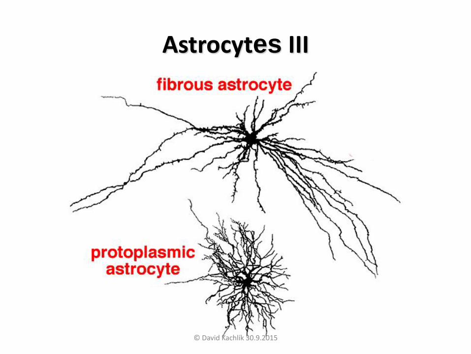
Astrocytes II
• Morphologically we recognize 2 types:
– protoplasmic astrocytes
• Numerous granulas in cytoplasm
• Extensions are shorter and richly branching
• Occur mostly in grey matter
– Fibrilary astrocytes
• Longer externsions w/o branching
• Occur mostly in white matter
© David Kachlík 30.9.2015
Astrocytes III
© David Kachlík 30.9.2015
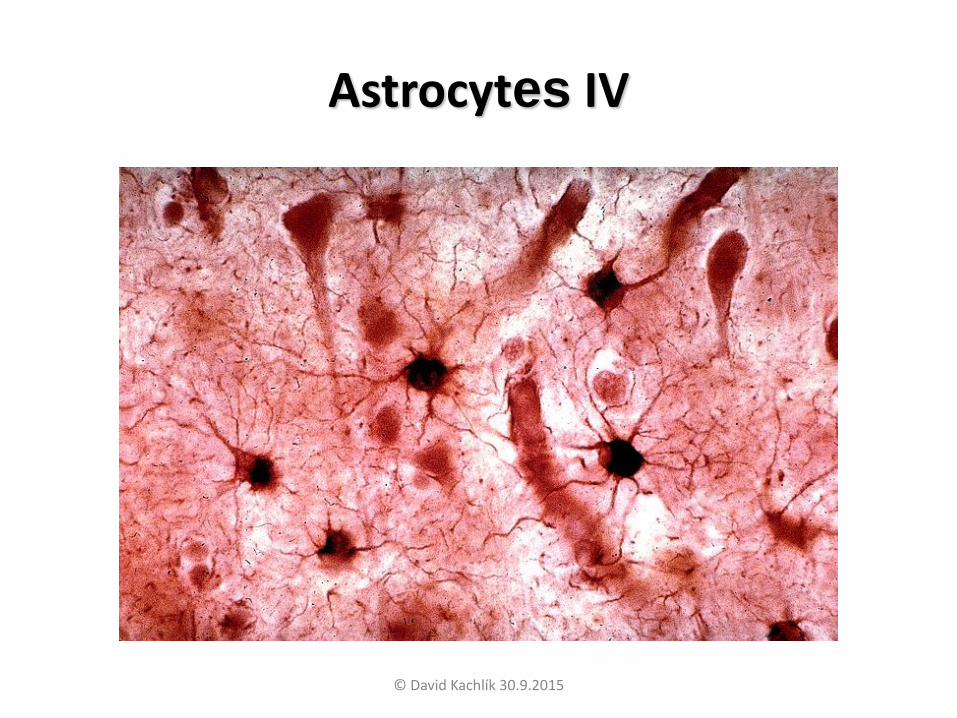
Astrocytes IV
© David Kachlík 30.9.2015
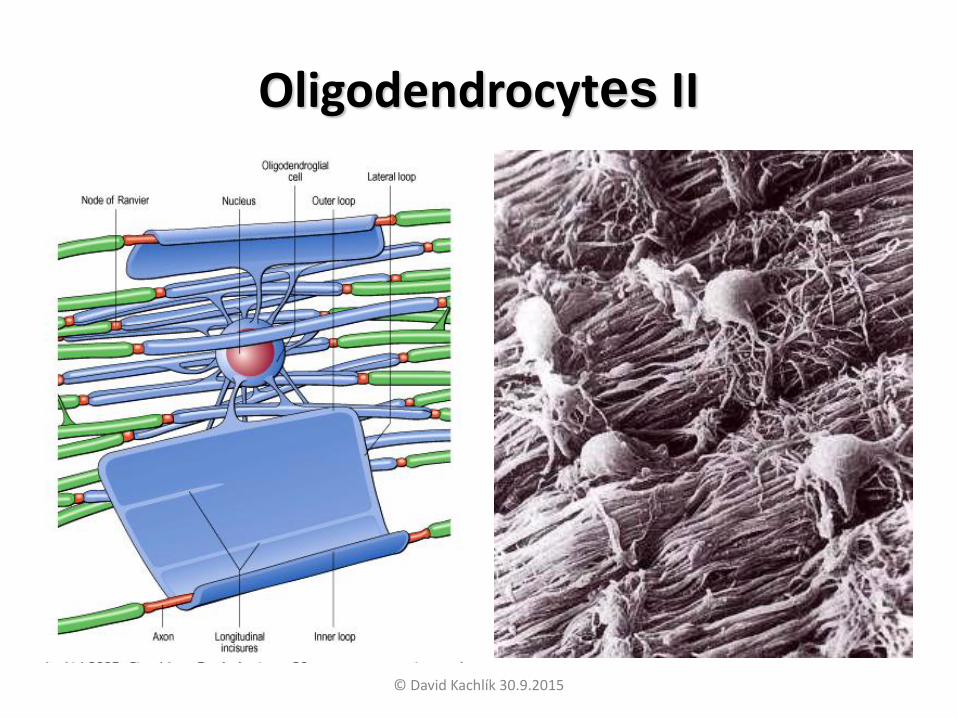
Oligodendrocytes I
• Smaller compared to astrocytes and have less intermediary filaments
• Form myelin sheath in the CNS
– Perform same function as Schwann cells in PNS
• Oligodendrocytes contain more axons at
once (in contrast to Schwann cells)
• Their number phylogenetically increases
© David Kachlík 30.9.2015
Oligodendrocytes II
© David Kachlík 30.9.2015
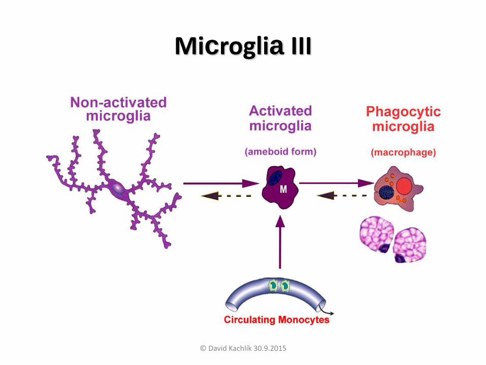
Microglia I• Smallest neuroglia
• Part of monocyto-macrophage system
– Mesodermic origin
• Movable and phagocytic
• Nuclei elongated
– As opposed to other glial cells that have round nuclei
• have „spiky“ shape
– During activation acquire shape of macrophages
© David Kachlík 30.9.2015

Microglia II
© David Kachlík 30.9.2015
Microglia III
© David Kachlík 30.9.2015
Ependymal cells I (ependymocyti)
• Originate from inner (germinal) zone of neuroepithelium
• Maintain epiteloid arrangement
• ependyme covers CNS cavities – brain ventricles, Sylvius
aqueduct, central canal of medulla spinalis
• Cells have nexus and zonulae occludentes
• Cillia at the apical pole facilitate flow of CSF
• tanycytes
– Special group o ependymal cells at the bottom of the 3rd
ventricle
– Have long extensions into nervous tissue
– May play role in chemical singal transduction from CSF
© David Kachlík 30.9.2015
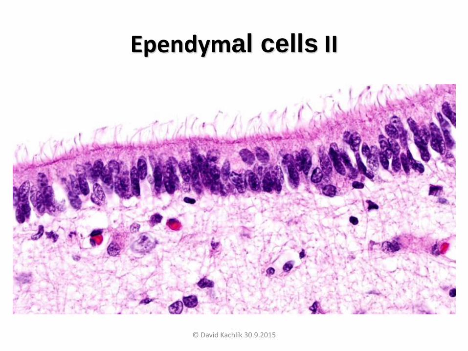
Ependymal cells II
© David Kachlík 30.9.2015
Nerve fibers (neurofibra)
• Axons or dendrites covered by special sheaths of ectodermal origin
• Nerve fiber bundles form:
– In CNS tracts (cover is formed by
oligodendrocytes)
– In PNS nerves (cover formed by Schwann
cells)
• We recognize fibers:
– Non-myelinated
– myelinated © David Kachlík 30.9.2015
Non myelinated nerve fibers I
• In the CNS located freely between neurons and glial cells
• In the PNS „invaginates“ into simple rifts in
Schwann cells
• Schwann cells (Schwannocytus) located
along nerve fiber mutually interconnected
– missing Ranvier fissures
© David Kachlík 30.9.2015
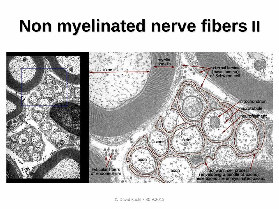
Non myelinated nerve fibers II
© David Kachlík 30.9.2015
Myelinated nerve fibers I
• Myelination occurs in several steps:
– Invagination of axon into sulcus of sheat cell
(oligodendrocyte or Schwann cell) originate so
called mesaxon (mesaxon)
– mesaxon „rotates“ around axon by 150x
– By modification of cytoplasmic membrane of
covering cell originate lipoprotein complex
myelin
© David Kachlík 30.9.2015

Myelinated nerve fibers II
© David Kachlík 30.9.2015

Myelinated nerve fibers III
• Between individual Schwann cells is myelin sheath interrupted by Ranvier fissure (nodi interruptionis myelini)
• Interval between Ranvier fissures is named internodium and has length 1-2 mm
• In the CNS are Ranvier fissures not readily visible
• Schmidt-Lanterman incisure (incisurae myelini)
– cytoplasm of Schwann cell „imprisoned“ during
myelination inside myelin sheath
– Form lighter stripes in myelin sheath
http://en.wikipedia.org/wiki/Myelin_sheath_gap
© David Kachlík 30.9.2015

Myelinated nerve fibers IV
© David Kachlík 30.9.2015

Myelin sheath – electrone microscope
© David Kachlík 30.9.2015
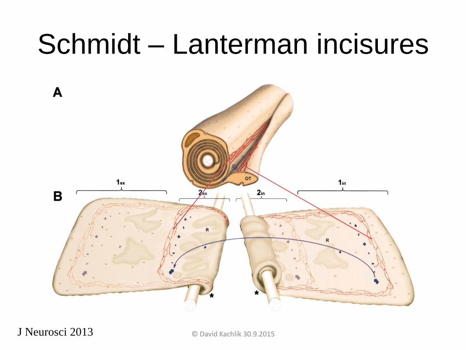
Schmidt – Lanterman incisures
J Neurosci 2013 © David Kachlík 30.9.2015

Nerves I• Formed by bundles of nerve fibers
• Nerve fibers have coverings similar to muscle
fibers:
– endoneurium
• Layer of reticular fibers around individual nerve fibers
– perineurium
• „sleeve“ covering bundles of nerve fibers formed by
layers of epitheloid cells
• Numerous zonulae occludentes – non passable barrier
protecting nerve fibers
– epineurium
• Tissue cover of whole nerve© David Kachlík 30.9.2015
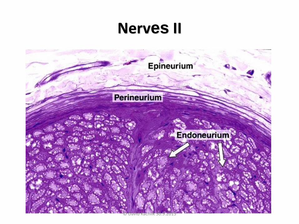
Nerves II
© David Kachlík 30.9.2015

Ganglia I• Nerve ganglia is accumulation of perikarya in
the PNS
• Have ovoid shape and their surface is covered by pouch from thick non arranged tissue
• Typical are so called satellite cells (gliocyti ganglionici) – snall cuboideal cells surrounding
neuronal perikarya
http://en.wikipedia.org/wiki/Dorsal_root_ganglion http://www.pharmainfo.net/introduction-autonomic-nervous-system/classification
© David Kachlík 30.9.2015
Ganglia II
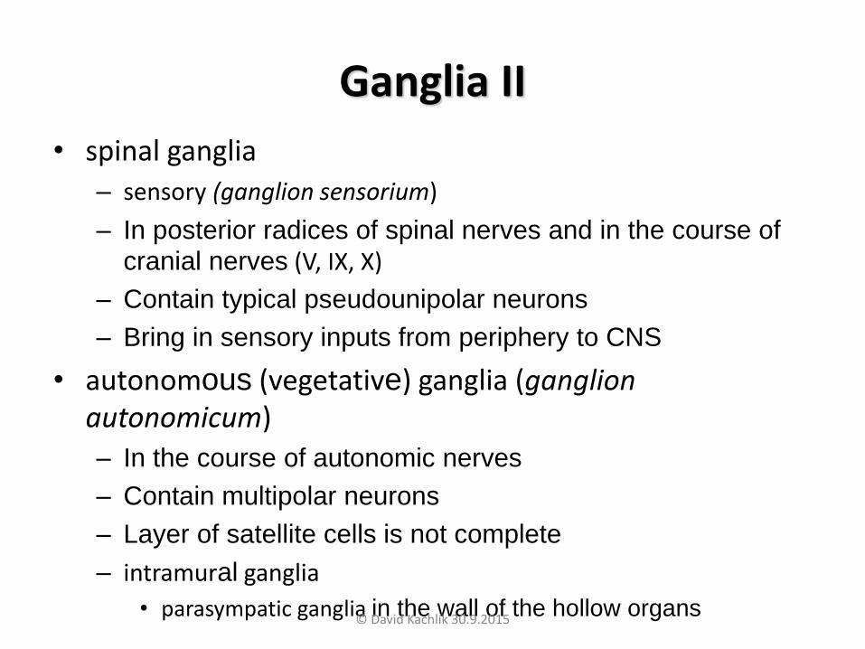
• spinal ganglia
– sensory (ganglion sensorium)
– In posterior radices of spinal nerves and in the course of
cranial nerves (V, IX, X)
– Contain typical pseudounipolar neurons
– Bring in sensory inputs from periphery to CNS
• autonomous (vegetative) ganglia (ganglion autonomicum)
– In the course of autonomic nerves
– Contain multipolar neurons
– Layer of satellite cells is not complete
– intramural ganglia
• parasympatic ganglia in the wall of the hollow organs© David Kachlík 30.9.2015

Ganglia III
© David Kachlík 30.9.2015
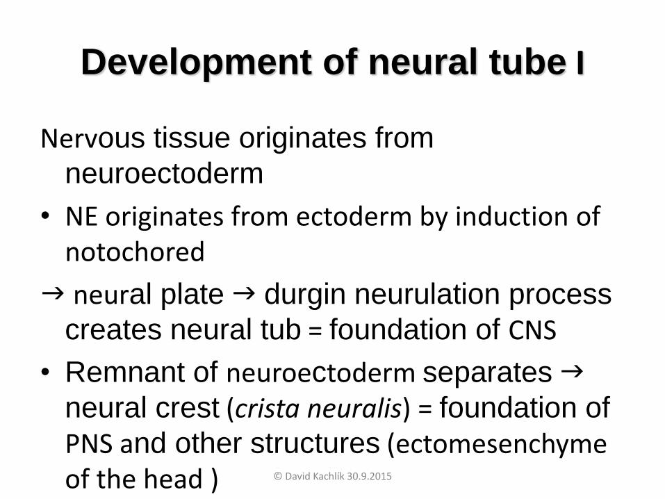
Development of neural tube I
Nervous tissue originates from
neuroectoderm
• NE originates from ectoderm by induction of notochored
neural plate durgin neurulation process
creates neural tub = foundation of CNS
• Remnant of neuroectoderm separates
neural crest (crista neuralis) = foundation of
PNS and other structures (ectomesenchyme of the head ) © David Kachlík 30.9.2015

© David Kachlík 30.9.2015
Development of neural tube II
• Primary neurulation
– Separates ectoderm into three cell types
(inside positioned nerve tube, epidermis externally and cells of the neural crest)
• Secondary neurulation
– Cells of the nerve plate form chorda dorsalis
• In the time of 35th somite formation
© David Kachlík 30.9.2015

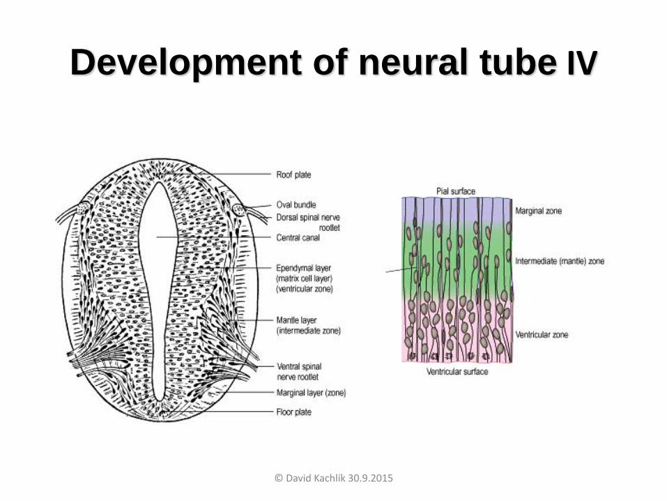
Development of neural tube III
Epitelium of nerve tube soon changes into multilayer neuroepithelium where are 3 layers:
• inner (germinal) zone (zona ventricularis; matrix germinalis)– Up to half of gravidity richly proliferates (origin of
neuroblasts)– Then reduced into ependyme
• Middle (shell) zone (zona intermedia; zona pallii)– Formed primarilly by migrating neuroblasts
– Gives rise to grey matter
• outer (marginal) zone (zona marginalis)– Formed by extensions of neuroblasts
– Gives rise to white matter
© David Kachlík 30.9.2015
Development of neural tube IV
© David Kachlík 30.9.2015
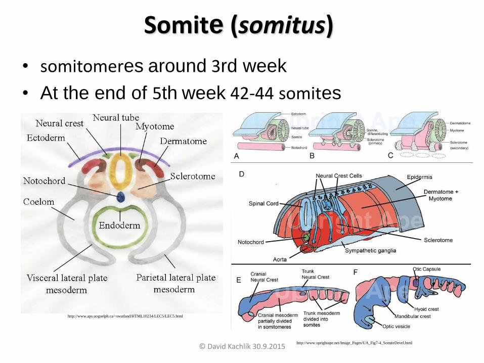
Somite (somitus)
• somitomeres around 3rd week
• At the end of 5th week 42-44 somites
http://www.aps.uoguelph.ca/~swatland/HTML10234/LEC5/LEC5.html
http://www.uprightape.net/Image_Pages/UA_Fig7-4_SomiteDevel.html
© David Kachlík 30.9.2015
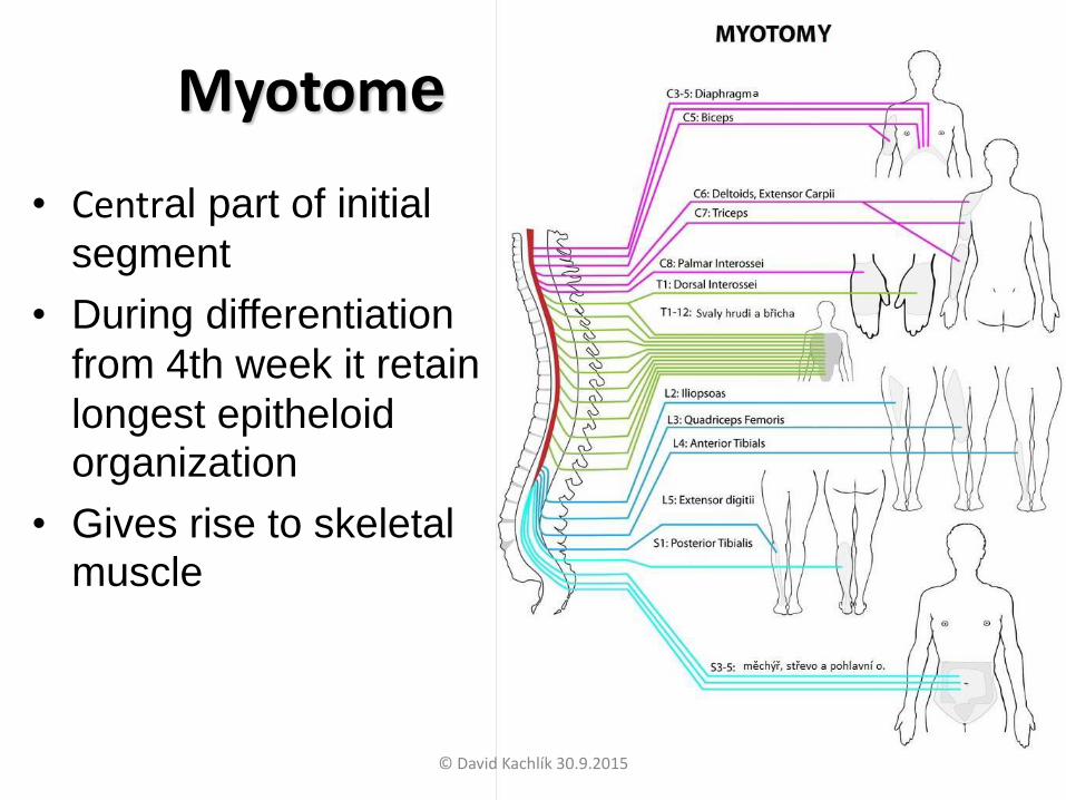
Myotome
• Central part of initial
segment
• During differentiation
from 4th week it retain
longest epitheloid organization
• Gives rise to skeletal muscle
© David Kachlík 30.9.2015
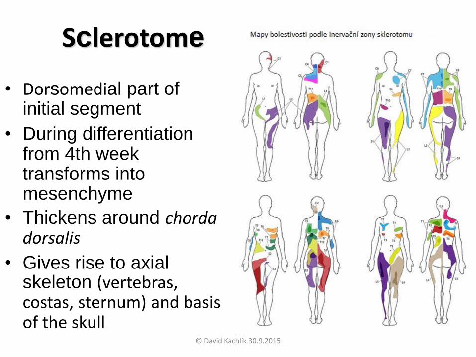
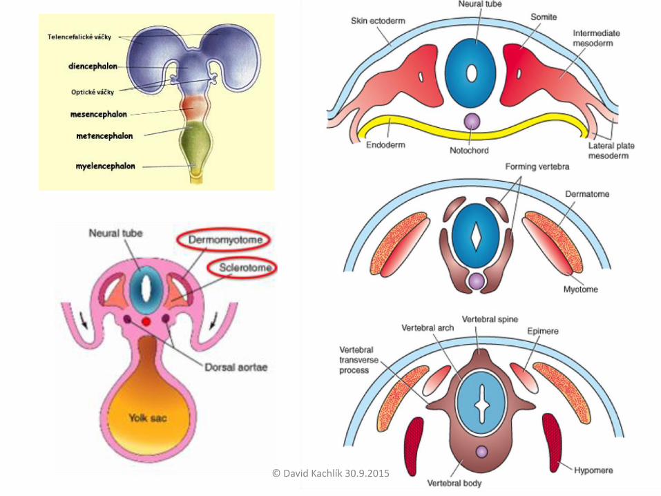
Sclerotome
• Dorsomedial part of initial segment
• During differentiation from 4th week transforms into mesenchyme
• Thickens around chorda dorsalis
• Gives rise to axial skeleton (vertebras, costas, sternum) and basis of the skull
© David Kachlík 30.9.2015

• Ventrolateral part of initial segment
• During differentiation from 4th week transforms into mesenchyme
• Migrates into somatopleura
• Gives rise to connective tissue basis of skin (dermis and tela subcutanea)
• Parts arising from the same dermatome have same innervation via spinal root
Dermatome
© David Kachlík 30.9.2015
© David Kachlík 30.9.2015
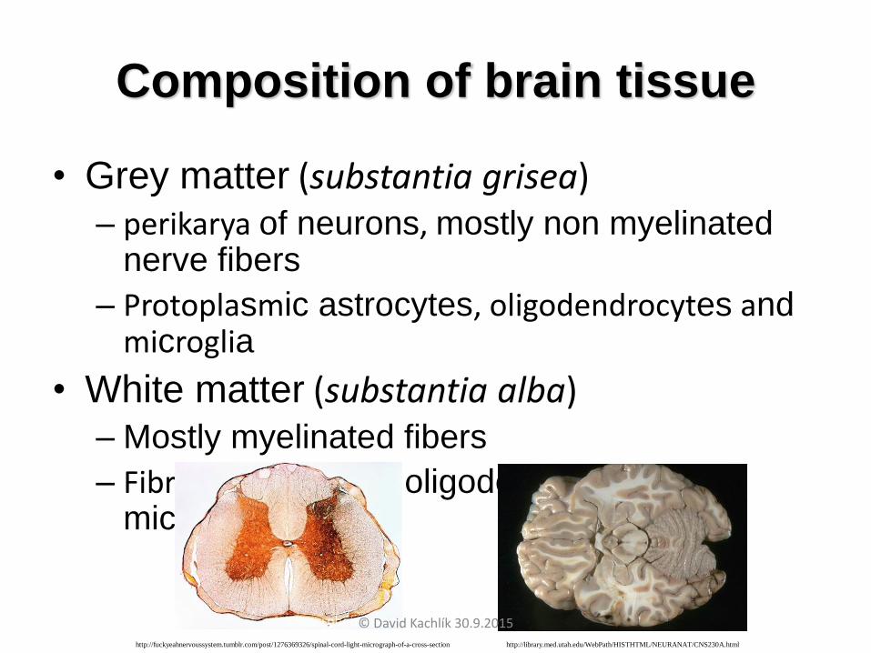
Composition of brain tissue
• Grey matter (substantia grisea)– perikarya of neurons, mostly non myelinated
nerve fibers
– Protoplasmic astrocytes, oligodendrocytes andmicroglia
• White matter (substantia alba)– Mostly myelinated fibers
– Fibrilar astrocytes, oligodendrocytes and microglia
http://library.med.utah.edu/WebPath/HISTHTML/NEURANAT/CNS230A.htmlhttp://fuckyeahnervoussystem.tumblr.com/post/1276369326/spinal-cord-light-micrograph-of-a-cross-section
© David Kachlík 30.9.2015

CNS description - parts
• Spinal cord (Medulla spinalis)• Brain stem (Truncus encephali)
– Oblongate (Medulla oblongata)– Pons (Pons) – formerly pons Varoli– Midbrain (Mesencephalon)– Hindbrain (Rhombencephalon)
• Cerebellum (Cerebellum)• Diencephalon (Diencephalon)• Terminal brain (Telencephalon)
– Bazal ganglia (nuclei basales)– Brain cortex (cortex cerebri)– Forebrain (ventral brain)
© David Kachlík 30.9.2015

Mesencephalon
Pons
Medulla oblongata
Medulla spinalis
Brain stem
© David Kachlík 30.9.2015
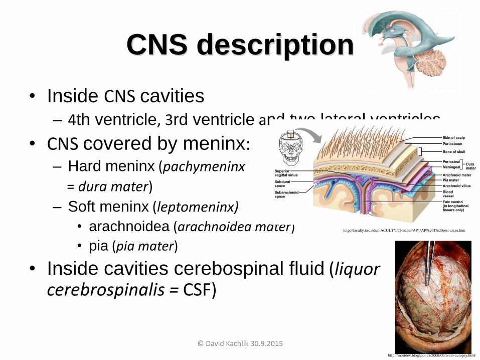
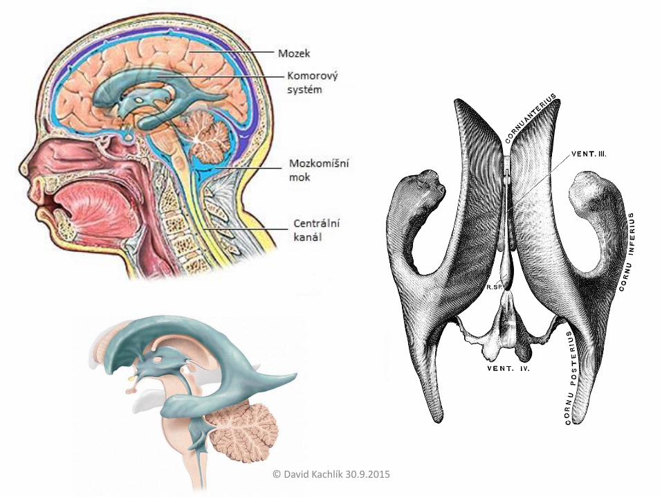
CNS description
• Inside CNS cavities
– 4th ventricle, 3rd ventricle and two lateral ventricles
• CNS covered by meninx:– Hard meninx (pachymeninx
= dura mater)
– Soft meninx (leptomeninx)
• arachnoidea (arachnoidea mater)
• pia (pia mater)
• Inside cavities cerebospinal fluid (liquor cerebrospinalis = CSF)
http://faculty.irsc.edu/FACULTY/TFischer/AP1/AP%201%20resources.htm
http://mortdev.blogspot.cz/2008/09/brain-autopsy.html
© David Kachlík 30.9.2015
© David Kachlík 30.9.2015
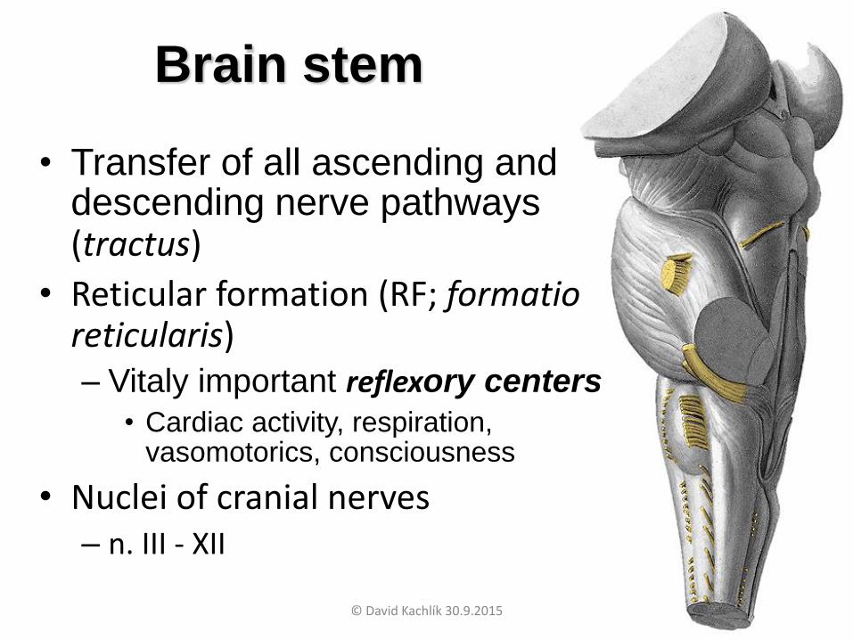
Brain stem
• Transfer of all ascending and descending nerve pathways(tractus)
• Reticular formation (RF; formatio reticularis)– Vitaly important reflexory centers
• Cardiac activity, respiration, vasomotorics, consciousness
• Nuclei of cranial nerves– n. III - XII
© David Kachlík 30.9.2015
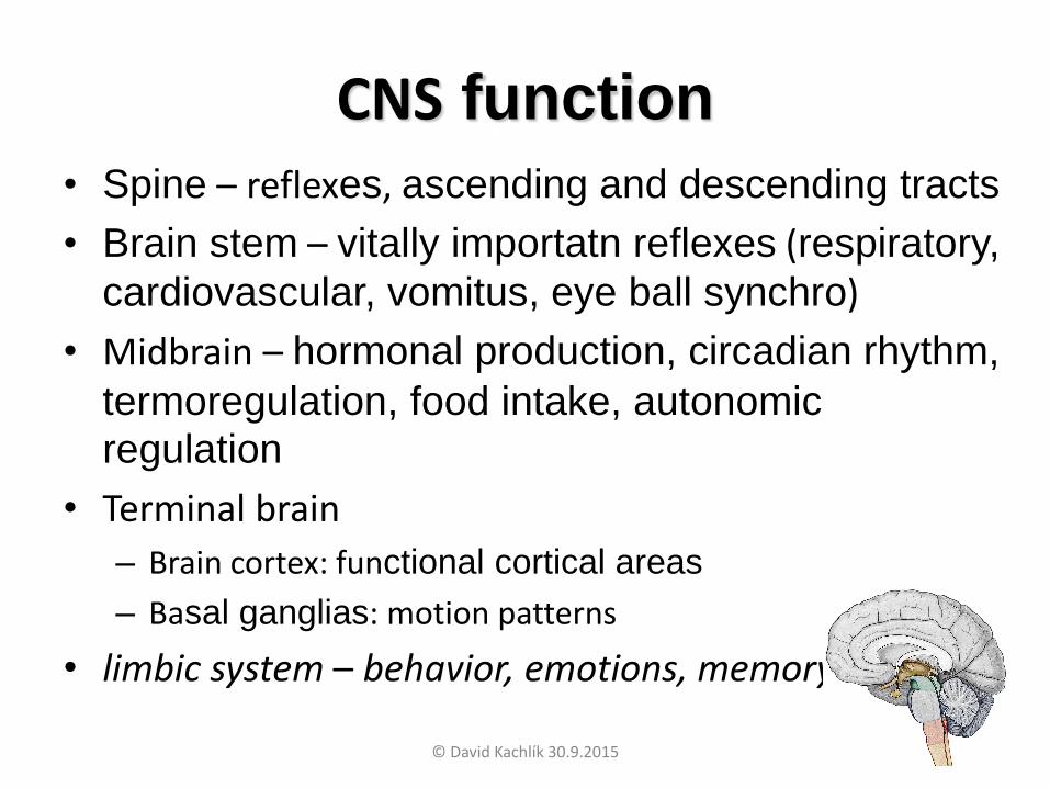
CNS function
• Spine – reflexes, ascending and descending tracts
• Brain stem – vitally importatn reflexes (respiratory,
cardiovascular, vomitus, eye ball synchro)
• Midbrain – hormonal production, circadian rhythm,
termoregulation, food intake, autonomic regulation
• Terminal brain
– Brain cortex: functional cortical areas
– Basal ganglias: motion patterns
• limbic system – behavior, emotions, memory
© David Kachlík 30.9.2015
© David Kachlík 30.9.2015

© David Kachlík 30.9.2015

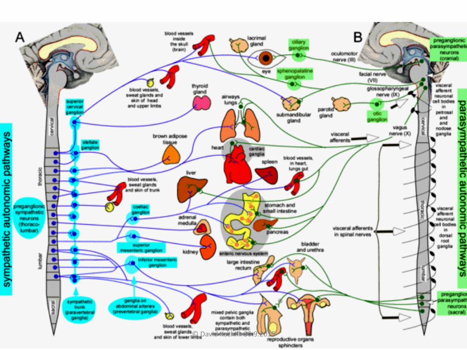
Peripheral nervous system
(Systema nervosum periphericum)
• spinal nerves (nervi spinales) – 31 pairs
• cranial nerves (nervi craniales) – 12 pairs
• autonomic nerves (systema autonomicum)
– sympathetic (pars sympathica)
– parasympathetic (pars parasympathica)
– enteric system
© David Kachlík 30.9.2015

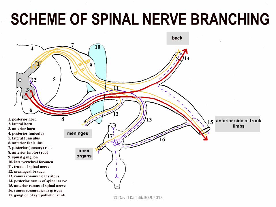
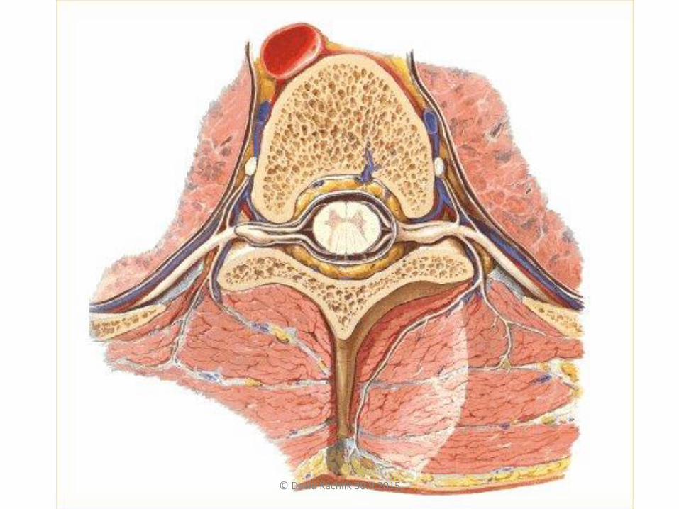
Macroscopy of spinal nerve branching
© David Kachlík 30.9.2015
© David Kachlík 30.9.2015
© David Kachlík 30.9.2015
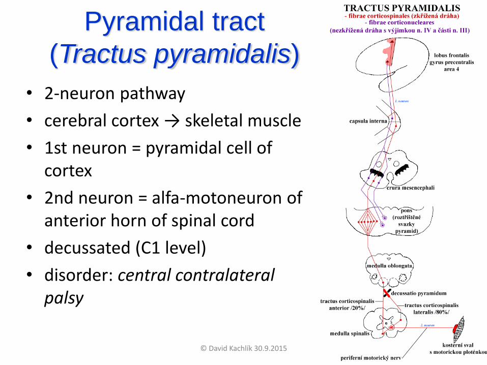
Pyramidal tract
(Tractus pyramidalis)
• 2-neuron pathway
• cerebral cortex → skeletal muscle
• 1st neuron = pyramidal cell of cortex
• 2nd neuron = alfa-motoneuron of anterior horn of spinal cord
• decussated (C1 level)
• disorder: central contralateral palsy
© David Kachlík 30.9.2015

Rami anteriores
nervorum spinalium
• plexus cervicalis (C1-4)
• plexus brachialis (C4-T1)
• nn. intercostales (T1-T12)
• plexus lumbalis (T12-L4)
• plexus sacralis (L4-S4)
• plexus coccygeus (S5-Co) © David Kachlík 30.9.2015

Plexus brachialis (C4-T1)
• truncus (trunk)superior (C4+C5+C6)medius (C7)inferior (C8+T1)
fissura scalenorum• fasciculus (cord)
lateralis medialisposterior
axilla - relation to a. axillaris• pars supraclavicularis• pars infraclavicularis
© David Kachlík 30.9.2015

Plexus brachialis - axilla
© David Kachlík 30.9.2015

© David Kachlík 30.9.2015
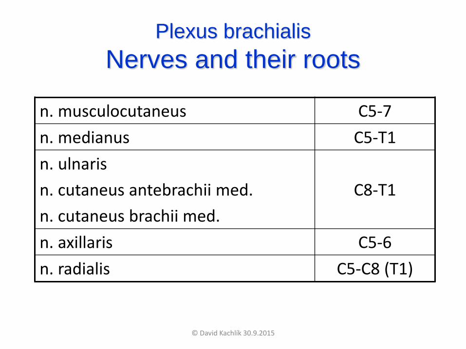
Plexus brachialis
Nerves and their roots
n. musculocutaneus C5-7
n. medianus C5-T1
n. ulnaris
n. cutaneus antebrachii med.
n. cutaneus brachii med.
C8-T1
n. axillaris C5-6
n. radialis C5-C8 (T1)
© David Kachlík 30.9.2015