bausch & lomb focus; may (spring) 1961
TRANSCRIPT

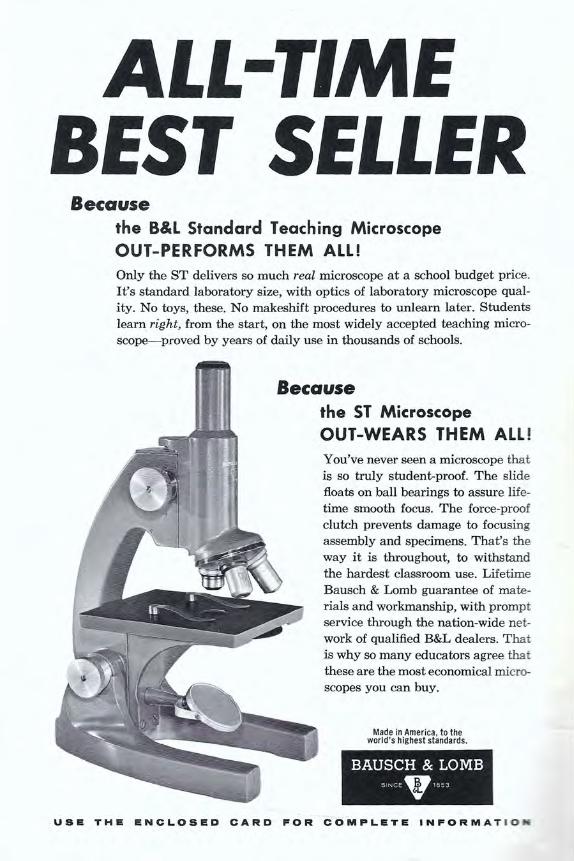
ALL-TIME BEST SELLER
Because the B&L Standard Teaching Microscope OUT-PERFORMS THEM ALL! Only the ST delivers so much real microscope at a school budget price. It's standard laboratory size, with optics of laboratory microscope qual-ity. No toys, these. No makeshift procedures to unlearn later. Students learn right, from the start, on the most widely accepted teaching micro-scope-proved by years of daily use in thousands of schools.
Because the ST Microscope OUT -WEARS THEM ALL! You've never seen a microscope that is so truly student-proof. The slide floats on ball bearings to assure life-time smooth focus. The force-proof clutch prevents damage to focusing assembly and specimens. That's the way it is throughout, to withstand the hardest classroom use. Lifetime Bausch & Lomb guarantee of mate-rials and workmanship, with prompt service through the nation-wide net-work of qualified B&L dealers. T hat is why so many educators agree that these are the most economical micro-
scopes you can buy.
Made in America , to the world 's highest standards.
BAUSCH & LOMB SINCE 1853
USE THE ENCLOSED CARD FOR COMPLETE INFORMATION
VOL. XXXII NO.1 MAY, 1961
BAUSCH & LOMB c s Published by Bausch & Lomb
Rochester 2, New York CORRADO J. BRUNO, Editor
For permission to reproduce any part of Focus please apply to the editor.
CONTENTS PREVIEW OF A NEW BOOK 2
TALL, DARK AND UPSIDE DOWN 6 DAVID CAUSEY, Zoology Department
University of Arkansas, Fayetteville, Arkansas
SEE FOR YOURSELF 10 FRED W. JOBE, Director of Ophthalmic Research
Bausch & Lomb Incorporated
WHAT'S YOUR EYE-CUE? 14
PHYSICAL PROPERTIES OF CELLS DETERMINED BY MICROSCOPE METHODS 18
HERBERT W. KRUGER, ROSEMARY FLENNIKEN
AND THOMAS HOOD Lamb-Weston, Inc., Weston, Oregon
TECHNIQUES 24 GERMAINE CRossMoN, Biological and Chemical Microscopist
Bausch & Lomb Incorporated
YOU can write for FOCUS . . . If you have information about new or novel methods in science teaching; or interesting applications of scientific optical instruments.
Before submitting material, send for our pamphlet "How to Write for the Educational Focus." It gives complete information about rates, desired article length, preferred subject matter, mechanical require-ments, etc.
OUR CovER is a photograph
of the Colorado River delta photographed from 13 ,800 feet,
courtesy of Fairchild Aerial Surveys. Did you
think it was a burning tree? See article on page 14.

We are reprinting here the introduction and just a few of the experiments from this new book. Written in easy-to-understand terms, the book will give the student a better ap-preciation of what a compound microscope is and does. The author takes the student through a number of "experiments" that viv-idly illustrate the points made. The student learns by doing.
Published by Bausch & Lomb, the book is priced at 50¢ per copy. However, we will be happy to send you a free copy if you will just check the appropriate box on the at-tached card.
GETTING ACQUAINTED WITH THE MICROSCOPE
Preview of a New Book/By Julian Corrington
Introduction The most basic and important instrument used by students of the biological sciences and many divisions of other sciences is the compound microscope. From the moment that the beginning high school or college student enrolls in the introductory course in biology, botany, or zoology, he accepts this instrument as a partner in the learning process and, should this alliance continue through advanced courses, such as plant morphology, animal histology and embry-ology, and many others, dependence upon the microscope rises to the point where it completely dominates the educational pic-ture. Later, should the student go into graduate and professional fields, he may earn his living with this king of instru-ments. There would be no cytologists, bacteriologists, or parasitologists if we had no microscope.
It is the purpose of this booklet to ac-quaint the reader with basic facts about
2
the microscope, its proper operation and care, and then to describe instances of its use in performing experiments. For uni-formity of presentation, we shall term each of the following sections "experiments," though, as will be obvious, not all are ex-periments in the correct sense, but are exercises, some of which are primarily observational.
An Experiment in Accommodation The most wonderful of all optical instru-ments, the eye, is able to alter its focal dis-tance when viewing objects at varying locations. This automatically regulated power is accommodation, and is per-formed by the lens of the eye, a transpar-ent and flexible body lying directly behind the iris.
The microscopist should learn to relax the accommodation of the eyes, to avoid eye fatigue. He should acquire the vacant stare or the unaccommodated eye of the

day dreamer when using the microscope. A famous experiment will demonstrate ac-commodation- the floating finger illusion (Figure 1). Sit at a table, facing a window, and gaze off at the horizon or the sky, re-laxing the accommodation completely, for distant vision. Without changing this con-dition, bring the two index fingers up in front of the eyes, about 8-10 inches before the face, both eyes open, the two fingers horizontal, facing each other, the two nails touching. Slowly separate these fingers about one-half to one inch, and you will observe a central, detached piece of finger floating in the air between the other two fingers, with a nail on each end. This ex-periment proves that the images in the two eyes are actually different, but can also be used, as here, to demonstrate accommoda-tion. The moment one shifts to a focus on the fingers, the illusion vanishes.
An Experiment with the Virtual Image Set up the microscope vertically, using a slide showing considerable detail, as a plant stem section of a section of stomach, intestine, or kidney. The light should come from a student gooseneck lamp or prefer-ably from a desk fluorescent lamp. Obtain a sharp, brightly illuminated image with the l0X objective. Pile magazines upon the table to the right of and against the microscope stand until the top surface is 250mm (1 0 inches) below the eyepoint of the instrument. Place a sheet of white drawing paper on the magazines and hold a sharpened drawing pencil in the right hand. Look into the microscope with the left eye and move the pencil point about with the right hand. You should be able to see both the detail of the object and the pencil point, if both eyes are kept open and accommodated for distant vision. The object is scanned with the left eye, the pencil point with the right eye, and the two images are fused in the brain as in any
form of binocular vision. Thus you will be able to sketch on the paper at least the grosser outlines of the object and get their proportions correct. Any conscious at-tempt to focus on either the object or the pencil will immediately destroy this fusion image. An observation of this sort is some-times called double vision.
An Experiment in Magnification What is magnification? When we look at the full moon we see a bright disc in the sky, and as a child we are told that it is the size of a dinner plate. Later we learn many facts about size versus distance and are told that the moon is 2.162 miles in diam-eter and a mean distance of about 238,840 miles from the earth. We can close one eye and hold a dime in front of the open eye at such a distance from the face as to obscure the full moon, yet we realize from experi-ence that the dime and the moon are not thereby of equal diameter.
Size is relative, and we must have a standard for comparison. Through many tests it has been concluded that the average person holds an object, for inspection of detail, or a book in reading, at about ten inches from the eyes. When the metric system was adopted in all scientific work, this was changed to 250mm, which is just a trifle less than 10 inches, and this dis-tance from the eye was designated the near point. Corresponding to this, the far point is at infinity. And so microscopes were designed to have the virtual image distance at the near point. Any object scanned at 250mm is natural size or 1 X. Whenever the object is brought closer to the eyes, it is seen larger and is hence mag-nified; if looked at farther away than 250mm, it is smaller or minified. You will probably be able to bring an object, such as this printed page, to within six inches of the eyes, or even closer, and still see detail clearly, but as the distance is slowly less-
3

ened, there comes a point at which the im-age blurs and detail becomes foggy or fuzzy and is then lost.
Take a piece of black photomount pa-per, as is supplied in photo albums, about postcard size and, in the center near one end, perforate it with an ordinary pin, thus making a pinhole card (Figure 2). Cut out a newspaper photograph of a person's face and mount this on another card. Now look at the photo through the pinhole, holding the pinhole card as close to one eye as possible. Bring the photo card ever closer to the face, starting at a distance of about ten inches, and you will be surprised to find that you can get it to about one inch from the face and still see detail in the picture clearly. You have in your hand the world's simplest and cheapest micro-scope, the pinhole card, achieving a mag-nification of l0X, since you are viewing
4
the card at one inch instead of ten. Magnification, then, consists in getting
the eye closer to the work. You obtain a bug's eye view, so to speak. One inch is about the shortest distance possible in this case; your head and eyelids and lashes get in the way and insufficient light is ad-mitted. If we enlarge the aperture in the card to admit more light, the image is de-stroyed by having too many rays come from the same point in the object; the im-age is fogged or, as the microscopist says, is rendered useless by glare.
Experiments in Diffraction
1 . Again take up the pinhole card. In the end opposite the pinhole, cut a two-inch vertical slit, not quite to the border, with a safety-razor blade. Hold this slit in front of the eye and look through it at a lamp (Figure 3). You will see not a single slit

but a series of them; a number of exceed-ingly fine vertical lines in the main slit. 2. Hold a fine-toothed comb in front of the eye and look through the teeth toward a lamp. You will see again some fine ver-tical lines, like extra teeth, between the actual teeth.
These experiments demonstrate diffrac-tion, a phenomenon in which light rays bend very slightly in passing through slits or past the edges of opaque objects. A pat-tern of light and dark fringes is produced.
Try to conceive of the detail in an ob-ject on a slide placed under the micro-scope at the size level approaching that of molecules. Some of the particles of matter will transmit light with little alteration, being highly transparent. Opaque particles will diffract light around their edge. Thick-er particles that are translucent will pass the light but refract the rays.
3
5

TALL, DARK AND UPSIDE DOWN
O N THOSE DAYS when the skies are leaden, the barometer is low, there
is too much dean, and ever so much too many students, barnacles are such consol-ing animals. They have done what you would like to do. A long time ago a crus-tacean got fed up the way the world was going- the wrong party (not his ) was in power, taxes were getting higher all the time, and as far as "he" was concerned Pippa could pass indefinitely - nothing was right with the world and he quit play-ing. So "he" stood on his head, altering the viewpoint but not improving much of any-thing, and gradually pulled his carapactic (there could be such a word) cloak about him. Only the hind end stuck out and the subsequent activities were somewhat rib-ald. I'm merely paraphrasing the great Huxley's statement to the effect that the barnacle stands on its head and deliber-
6
By David Causey University of Arkansas
ately kicks the food into its mouth.1 There was a time when the barnacles
were Mollusca. Now one thing which sepa-rates the Arthropoda (and consequently the Crustacea) from the Mollusca is the presence of jointed legs. How any zoolo-gist could have looked at a barnacle with its legs sticking out like tufts of feathers and thought it was a molluscan is actually a tribute to the scientific mind. I think that Aristotle taught us the mental processes for this:
The Mollusca do not have legs. The barnacle is a mollusk. Barnacles do not have legs.
1. If you think I'm telling the barnacle story too often, just remember that in time you, too, will become old and garrulous. I'm already getting letters saying, "I learned the alphabet from your early barnacle papers! " There is always a new generation .

J. Vaughan Thompson, over a hundred years ago, approached the solution by rather subversive means- he worked on the life cycle - and found a typical crus-tacean development which ended in a barnacle. Why his fellow scientists ac-cepted his work I have no idea. My old professor, Dr. C. A. Kofoid, loved to quote Bacon: "Error travels on horseback, Truth follows afoot." Too often Truth seems not to start.
Darwin and barnacles are practically synonymous. For seven weary years this clumsy, lumbering man worked at them. His letters are full of his problems and his ineptness. His volumes are classics, from a dogged plodder who kept pushing along. I have the feeling that much of what has been done since then might as well not have been done. And once recognized as an authority on the Cirripedia, he aban-doned them to write on the origin of the species, the fertilization of orchids, climb-ing plants, domestic animals, coral reefs, pangenesis and the parallel roads of Glen Roy! I like the young barnacle Darwin better than the older obsessed squire of the Downs who nursed his neuroses and sometimes worked as much as two hours a day. Don't misunderstand me- this un-brilliant country squire has changed bio-logical thought for almost a century and the tale is yet to run. His statue is placed, so my wife tells me, on one side of the Natural History Building of the British Museum, with Huxley on the other-British versions of Don Quixote and San-cho Panza, if you please.
My tall, dark barnacle is Xenobalanus globicipitis Steenstrupt, 1851. They came from Dr. Jorge Carranza, a very fine young Mexican ichthyologist, who gave them to me largely to get rid of me. I was trying, as usual, to convince him that the only excuse for fish existing is as hosts for parasitic copepods. The label reads:
Holbox, Quintana Roo Umos 40 Kms mar aquera frente a punta Mosquito
Mayo 30, 1955 J. Carranza
Xenobalanus sp. Sobre Stenella plagiodon (Tonina).
which translates as "found on a porpoise off Yucatan." Spanish, as you can readily see, is somewhat verbose!
Figure 1. Photomacrograph of the whole bar· nacle, Xenobalanus globicipitis.
7

The specimens (fig. 1) are a very dark purplish black in alcohol, with a reddish cast under photoflood lights. The length is approximately 180° F. or #3 cork- sorry, wrong side of the rule - about 3cm. The surface is iridescent, satiny and beautiful. The animal is somewhat trumpet shaped and bears a marked superficial resem-blance to the pedunculated barnacles to which it does not belong. Darwin refers to the peduncle-formed body in one para-graph and forgets himself and calls it the peduncle in the next. Even the gods nod at times. At the free end is the pseudo-capitulum (fig. 2).
Imagine a tin can which has been opened with a knife so that five pie-shaped portions of the top are sticking up more or less instead of the usual four you see in roadside parks below the sign urging you to not be a litterbug. Also imagine the body of the can as made up of five pieces which more or less overlap like shingles on a roof. If you have this in mind, you have a rough idea of the shell of a "sessile" barnacle (they all are, but I can't help that). The bottom of the can is the head end, the gashed top is the tail end. Obvi-ously a can doesn't have to be of the same dimensions always. Instead of a grocery store can, imagine it elongated quite a bit. The worms we buy come in such cans. You have imagined the situation in Tubi-cinella, a barnacle which is well named (and which I'd like to see!).
Can you imagine old sober-sided Dar-win going in for strip-teasing? "If in imag-ination we chip away (an action always in progress) the whole upper part of the shell of Tubicinella, leaving only two or three zones of growth at the base, we shall con-vert it into a Xenobalanus, with every in-ternal part and organ occupying the same relative position ... " Thusly we get our porpoise barnacle, and in the process we get one which looks so superficially like
8
the pedunculate (not "sessile") barnacles that I'm sort of glad I don't have to ex-plain it to that football player in the back row. You will note that we didn't, in im-agination, chip away all of the shell. There was a little bit left at the bottom (and don't forget that the bottom is actually the head end!). That little bit is all that is left of the shell, which Darwin describes as "like a small white irregular star, imbedded up to the top in the skin of the porpoise." You can see this in my figure 3, a somewhat oblique view because the specimen just wouldn't remain in position for a full-on photograph. Figure 2 shows the feathery legs sticking out, and the little horns which made Darwin think of the ear-like ap-pendages in Conchoderma, another bar-nacle I'd like to see. Years ago a fisherman at Grande Isle, La., asked me how the shrimping was in Arkansas, and I truth-fully told him it was very poor. Barnacle collecting in Arkansas is even worse. We have so little seashore!
The barnacle has insides (fig. 4) in which you'd probably not be interested. You can see the little horn cut through, the legs, the body, and a mass of eggs in the marsupium. One organ tempts me quite a bit, but if I yielded, Editor Bruno would be getting letters. I'll just say that the French didn't invent everything they get credit for (I forgot to mention that the dissection was done by my wife, Dr. Nell B. Causey, who would appreciate receiv-ing millipeds from your locality!). There are other aspects, too, such as the life cycle and the effect this barnacle has upon the host which I'm glad I don't have space to discuss. Strictly off the record - I wouldn't have my dean find it out for the world- I have no idea about these mat-ters. I could, however, come and give you an hour's lecture about them. Socrates was the wisest of the Greeks, but he never be-came a professor!
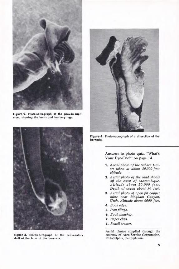
Figure 2 . Photomacrograph of the pseudo-capit-ulum, showing the horns and feathery legs_
Figure 3. Photomacrograph of the rudimentary shell at the base of the barnacle.
Figure 4. Photomacrograph of a dissection of the barnacle.
Answers to photo quiz, "What's Your Eye-Cue?" on page 14.
1. Aerial photo of the Sahara Des-ert taken at about 30,000-foot altitude.
2 . Aerial photo of the sand shoals off the coast of Mozambique. Altitude about 20,000 feet. Depth of ocean about 30 feet.
3. Aerial photo of open pit copper mine near Bingham Canyon, Utah. Altitude about 6000 feet.
4. Book edge. 5. Iron filings. 6. Book matches. 7 . Paper clips. 8 . Pencil erasers.
Aerial photos supplied through the courtesy of Aero Service Corporation, Philadelphia, Pennsylvania.
9

SEE FOR YOURSELF By Fred W. Jobe
Director of Ophthalmic Research and Development Bausch & Lomb Incorporated
Danger Signals for 51,000,000 Vision Delinquents
C HANCES ARE that there are in your classroom, right now, several stu-
dents who are "handicapped by some type of vision deficiency." It is unlikely that either you or these students are aware of their deficiency.
What is a "visual deficient"? He or she is simply someone whose vision is under the norm in one or more respects. There are 51,000,000 such persons over six years of age in the United States today! Or at least there were two years ago, when the Better Vision Institute made a thor-ough, searching study of the visual habits and characteristics of the American pub-lic. That means that one out of every four in the over-six age groups has some visual deficiency. The laws of probability neces-sarily place some of these in your classes.
The amazing fact is that vision deficien-cy is seldom intentional. This is particu-larly true of the person who has never worn glasses, has never experienced sharp vision, and as a result has no reason to sus-pect that his vision is not what it could be. Obviously, in the case of an adult, such a
10
person may have only a slight visual de-ficiency and has never considered it neces-sary to have an eye examination. A serious deficiency would make itself apparent to him simply by his observation that other people give every evidence of seeing things better than he.
On the other hand, some children, lack-ing powers of deductive reasoning, grow up in a misty, distorted or myopic world, assuming that everyone sees as they do. They see a few bright stars and never realize that there are thousands more they should be able to see. Leaves on trees are green blobs of color and the bark is an area of brownish gray with no texture.
The State of Indiana has been conduct-ing eye tests for people applying for re-newal of their drivers' licenses, and has made a rather surprising discovery. The State found that there were 3 times as many drivers over 35 passing the tests as there were young people. These youths had gone through school, reaching an age of maturity without realizing their vision was below the norm. How much the edu-
cation of these young people suffered be-cause of their visual deficiency is a matter of conjecture. But there is a wealth of

statistical evidence pointing to the fact that there is a close correlation between visual performance and school success.
In his paper "Vision and Its Relation-ship to School Achievement,"1 Dr. George D. Spache states:
"Dr. Newell C. Kephart of Purdue Uni-versity reports several studies of the im-portance of vision in schools. In a group of 250 high school students, he found that 46 percent who were reading at or above grade, had adequate visual performances. Among those who did not meet visual standards, only 28 percent were above average in reading.
"In a second study of 468 seventh grade children, Kephart found that where vision was adequate 56 percent of the children were in the upper half of the class. Where vision was inadequate, only 4 7 percent were in the upper half of the class.
"A third group in a boys' school was also studied. Two groups of children with inadequate visual skills were matched in intelligence and educational achievement. One group of 25 was given complete re-fraction and correction, plus visual train-ing where needed. The other matched group was not corrected. In a subsequent four-month period, the corrected group made 12 months' progress in educat:onal achievement, the uncorrected group only six months' growth."
A number of other similar studies have turned up the same correlation between good vision and school achievement. This, of course, should not be surprising since so much of what a student learns comes to him through his eyes. When he has diffi-culty with vision, the learning process suffers in efficiency.
How can we single out these students-the one out of every four who needs eye
1. The Journal of the American Optometric As-sociation. Vol. XXIX No. 5,. December 1957.
care? This question is important to every educator because, as we have seen, it is closely linked with the individual's ca-pacity to learn. It is vital to proper social adjustment and to a full life in school and at play. It is a question that concerns both the teacher and the student.
The best answer, of course, is a com-plete professional eye examination at least every two years, even for the person who thinks he has perfect vision. However, until such an ideal is universally achieved in the schools we must use other methods to find the students who are working with visual handicaps, notifying the parents of the condition and suggesting a profession-al eye examination.
The conventional tests commonly ad-ministered in most schools serve to uncov-er the serious cases of visual deficiency. By themselves, however, many of these tests are not adequate for the job of re-vealing the far greater number of cases of minor visual impairment of all types.
Today, thousands of schools are using instruments such as the School Vision Tester for visual examinations. These in-struments are not intended as substitutes for professional examinations. Their sole purpose is that of visual screening - to reveal deficiencies which can only be remedied by a competent professional examination.
Where such instruments are not avail-able as a screening technique, there are other danger signals which can be spotted by the alert teacher. Lack of interest and attentiveness, consistently poor marks, continued squinting, symptoms of nausea or dizziness following close work and, of course, redness of the eyes. These are some of the obvious indicators which should be considered - danger signals which may indicate visual deficiencies.
We are printing here a few simple "ex-periments" which will also serve as warn-
11

ing signals. We suggest that you try them yourself first and then ask your students to try them.
All these tests, except those that obvi-ously require the use of both eyes, should be taken first with both eyes, then each eye separately. Cover one eye with the hand or a piece of cardboard - not by squinting.
ASTIGMATISM Uneven curvature of the cornea of the eye will pro-duce astigmatism, one of the commonest visual deficiencies. To a person with astigmatism the lines in the small circles shown above, at a normal read-ing distance of 14", will appear unequally block; in the larger circle of concentric rings, there will be pie shaped segments that ore blacker and sharper than the remainder of the circle.
12
2
PRESBYOPIA Most people in their mid-forties experience a nor-mal condition called presbyopia. This simply means that the crystalline lens of the eye has lost the elasticity it enjoyed when they were young, and cannot easily "accommodate" for both near and for vision. Bifocals provide on easy and comfort-able remedy. If your most comfortable reading range is more than 15" or 16" you may be pres-byopic.
3
TUNNEL VISION In driving, one of the gravest visual deficiencies is inability to see objects approaching from the side when looking straight ahead. Hold your arms straight out (180° angle) and wiggle your fingers. If you con discern motion, you hove excellent peripheral vision . Now bring them in to a 140° angle and repeat the experiment. If you cannot see motion at this ongle1 you have a serious prob-lem for which many states would deny a driving license. You need an examination to determine the condition and possible remedies .

This is 6 point Century Schoolbook type reduced photographically to half size, or 3 po int. Anyone wit h 20/20 acuity--the a cce pted norm for good vision--should be able to recogniize each word at a norma l reading distance of 6 inches,
This is 6 point Century Schoolbook type. Anyone with 20/40 vision should be able to read it easily at a normal reading distance of 14 inches.
4
NEAR POINT TEST Do you have 20/20 vision? If so, you should be able to read the small type reproduced above. Incidentally, 20/20 vision is simply normal vision and not the best possible a cuity. If you have 20/40 vision (the criterion for many driving licenses) you should be able to read the paragraph of six point type.
T v H p R 5
FAR POINT TEST Here is a reasonably acceptable measure of visual acuity for a distance of 10 feet. Inability to distinguish the letters at this distance could be evidence of myopia.
6
BINOCULAR VISION
Here is how to tell whether your two eyes are working together as a team . Hold your two index fingers about 2" apart and 8" or 10" in front of the eyes, focusing not on the fingers but on some distant object. The fingers will be out of focus with a sausage shape object appearing between them.
u c F N c
7
DEPTH PERCEPTION This experiment will simply serve to illustrate stereoscopic vision; only a professional examination will reveal whether you have it. Stand a cigarette on end and t ry touching it with your index finger, first with one eye covered, then the other. Now do it with both eyes open.
13

WHAT'S YOUR EVE-CUE?
T HINGS AREN'T ALWAYS What they seem! When familiar objects are distorted by
magnification, miniaturization, or by be-ing viewed at peculiar angles, we "see" the objects as something entirely different from what they really are. But, assuming normal vision, the eye is an innocent dupe in the deception. It has simply picked up the light rays emanating from the object and conveyed an image, an impression really, to the brain.
Having received the information, the brain, not unlike an electronic sorter, tries to relate this information to information already "on file." As a child, for example, we saw a ball, learned that it was called "ball," and this knowledge was then stored in the brain's memory center. From-there on, every time we saw a ball in its normal size and shape, the brain unerringly in-formed us as to what it was. Now, let us take a ball and enlarge it, photographical-ly, to many, many times its normal size. The eye still continues to convey correctly what it "sees" to the brain. But the electric impulses scurrying back and forth in the brain, now bring forth a card labeled "moon," or "gasoline storage tank," or "mine," depending on the texture, color, and environment of the ball in the photo-
14
graphic enlargement. Our eyes have not "played us tricks"
but our brain has, and understandably so. After all, it had been conditioned to "see" a ball as within certain small limits in size.
Most of us have, at one time or another, looked right at an object without "seeing" it. And, conversely, there have been many instances of persons "seeing" objects that were not there, simply because they ex-pected them to be there, or urgently wanted them to be there.
The mechanics of vision are extremely complex, but the function of visual per-ception is even more so. Each individual, looking at the same "distorted" photo-graph, "sees" an image that is the culmina-tion of his own previous learning and cur-rent emotional state. Even the feel and smell of entirely unrelated objects may condition the formation of the final "im-age" in his brain.
Some of the photographs printed here have been greatly enlarged; others have been taken at great distances and are, therefore, considerably reduced. How many can you name correctly?
What's Your "Eye-Cue"? (See page 9 for the answers.)

2
15
3


BAUSCH & LOMB TRI·SIMPLEX
MICROPROJECTOR Projects mounted spec-imens or living organ-isms on screen or trac-
ing pad.
BAUSCH & LOMB STANDARD TEACHING
MICROSCOPES Standard size and op-eration with exclusive student-proof features.
BAUSCH & LOMB BUNSEN
SPECTROSCOPE Basic tool of chemical analysis; shows spectra of elements.
Why it costs far less to buy Bausch & Lomb Your investment is protected for life! B&L educa-tional instruments are built to shrug off the punish-ment of day-after-day, year-after-year use. They're made in America, to the world's highest standards. Workmanship and materials are guaranteed for life. If you should ever need service, a nation-wide net-work of B&L dealers provides it promptly and de-pendably. You save precious teaching time, too. This Tri-Simplex Microprojector lets you point out important details of microscope study to all students at the same time. They see brighter, clearer images than any other school projector can provide. They know exact-ly what to look for with their own microscopes. They understand better, learn faster.
BAUSCH & LOMB SINCF 9 '853
GET YOUR FREE COPY of this data brochure covering the world's finest teaching tools for the balanced science program.
USE THE ENCLOSED CARD FOR COMPLETE INFORMATION

PHYSICAL PROPERTIES OF CELLS DETERMINED BY MICROSCOPIC METHODS
By Herbert W. Kruger, Rosemary Flenniken and Ralph Thomas Hood
Research and Development Laboratory, Lamb-Weston, Inc., Weston , Oregon
F OR MANY YEARS one of the primary in-terests of the biologist has been the
study of individual cells. These are the building blocks of all living substances. Their origin and growth, structure and functions as well as their pathological con-dition are under constant study in hun-dreds of laboratories throughout the world. More recently, added interest has been shown in such characteristics as fragility, or the ability of the individual cell to withstand pressure or abrasive ac-tion. Much of this increased interest is being directed towards the determination of the extent to which the cells of food vegetables are weakened, or ruptured, by modern food processing methods.
A direct cause of this new interest is largely due to the m arked increase in the amount of food vegetables which are be-ing subjected to new processes before they reach the retail trade. A large portion of this increase has been in the field of de-hydrated vegetables . The production of dehydrated potatoes has increased almost ten fold between the years 1949 to 1956. Most of these dehydrated potatoes have
18
Figure 1. Micro-pressurometer ottoched to the stage of a Model STA Bausch & lomb Microscope. The tube has been removed to give an unob-structed view of the pressurometer. The pressure head shown in position over the center of the con-denser consists of an optically transparent glass disc, .063 inches in diameter and .043 inches thick. This pressure head is attached to the end of the pressure arm which in turn is secured to the steel torque wire which is .013 inch in diameter. One end is attached to the pressure adjusting dial seen on the right and the other end is attached to the tension spring and zero adjustment on the left of the pressurometer. By rotating the graduated pres-sure adjusting dial, the pressure head can be raised or lowered to any desired position, or any desired amount of pressure may be placed on any specimen which has been positioned below it .

appeared on the market in the form of flakes or granules: instant mashed pota-toes. In general the process of preparing dehydrated potato granules requires that the potato be cooked, mashed and then dehydrated and granulated. This granu-lated product consists mostly of individual potato cells. On reconstitution, by the ad-dition of hot liquids such as a combination of milk, water and butter the ideal de-hydrated potato granules should produce a food which is comparable in every re-spect with the best mashed potato pre-pared from freshly cooked potatoes.
However, we find that, due to the tor-tures of processing, cell damage can at times be so severe that a definite pastiness
Figure 2. Note method of attachment to micro· scope substage. The block to which the torque wire tension spring is attached, can be rotated to ob- tain a zero position of the pressure head with the pressure adjusting dial set at zero. The zero ad-justment is then locked in position with the lock-ing screw seen at the upper left.
is observed in the reconstituted product. Considerable literature has been published on the cause and prevention of pastiness. Some have attributed this pastiness to an excessive percentage of cells being rup-tured in the processing. R. M. Reeve, et. al. 1 have described microscopic means
literature cited: 1. Reeve, R. M. and Notter, G. K. An improved
microscopic method for counting ruptured cells in dehydrated potato products. Food Technology, 10, 574-577 (1959).
2. Severson, D. E., Cooley, A. M. and Simon, Morris. Factors affecting the texture of de-hydrated potato granules. Food Technology, 5, (1955) .
Application for patent has been filed with the U . S. Patent Office.
Figure 3. Microscope with micro·pressurometer at-tachment in position under photographic enlarger which has been converted for microphotographic use. Since this photograph was taken, a compur type shutter has been placed between the micro-scope tube and the enlarger bellows.
19

Figure 4. Pressure head with a number of indi-vidual potato cells which were reconstituted from a sample of dehydrated potato granules, 30X . By manipulation of the mechanical stage, while the pressure head remains stationary, the cells may be moved about until only a single cell remains under the pressure head for test.
for determining the percentage of rup-tured cells in dehydrated potato granules. D. E. Severson, et. al.2 found that cell breakage is not the only factor affecting the texture of the reconstituted product and suspected that, more significant than broken cell content of the dry material, is the susceptibility of this material to cell damage on reconstitution. They also found that processing markedly affects the sus-ceptibility to cell damage during reconsti-tution.
The extent of cell damage caused by a particular processing stage cannot be de-termined by any method now available. Methods are available for determining the percentage of cells which have been rup-tured in processing but there are no means for determining the effect of any one stage on the fragility of the individual cells. Thus we find, at the beginning of our research in the processing of dehy-drated potato granules, that there exists a
Figure 5. Potato cell taken from a sample of cooked potatoes which had been retrograded by freezing, lOOX. This cell showed a definite hexagonal cross-section before pressure was applied . (A) Pressure has been increased to 2.6 grams. Note that rupture has occurred at one corner and the other corners of the hexagon have become rounded. Viscosity of the fluid content is apparently quite high. (B) Pressure has been increased to 4.9 grams. During this increase in pressure the fluid flowed from the cell very slowly. This behavior is very characteristic of cells from samples of potatoes which have been cooked and then retro-graded by freezing.
20

gap in testing methods. To fill this gap the micro-pressurometer shown in the photo-graphs, Figures 1, 2 and 3, was designed and constructed in the Lamb-Weston Re-search and Development Laboratory. At this writing, it has been in use in this lab-oratory for nine months and has tested many hundreds of individual cells.
Preparation of Specimens for Examination Under the Micro-Pressurometer
Every test must have a definite objective and this must be borne in mind in the preparation of specimens for examination under the micro-pressurometer. For ex-ample; if we desire to test the ability of the cells in potato granules to withstand re-constitution in boiling liquid, then the specimen should also be prepared by re-constitution in boiling water. Cells should be kept in suspension by stirring while withdrawing a few drops of liquid in a
Figure 6. Cell from cooked pototoes which has not been retrograded; l00X. This cell ruptured ot the low value of 0.9 grom. This cell assumed a smoothly curved outline as soon as a slight pres-sure was applied. Upon rupture the fluid content was instantly disgorged to the form shown in the microphotograph. Viscosity of the fluid content is apparently quite low. This response, of cooked potato cells is characteristic of those which hove not been retrograded.
pipette for placement on the pressure plate of the microscope. If the cell density is large, there will be so many cells in the few drops placed in the slide that difficulty will be experienced in maneuvering a sin-gle cell under the pressure head for test. More liquid should be added to the test solution. If we desire to determine the change which has resulted from a certain pre-processing treatment such as retro-grading the starch content of the potato cells by freezing, then the specimen should be kept suspended in cold water. If the temperature of the suspending liquid is above the gel! point of the starch, the his-tory of its previous retrogradation will be destroyed and the test will be of no value in determining the extent to which retro-gradation has taken place in the pre-proc-essing treatment. If we were to determine the fragility of red blood cells it would seem that every effort should be made to perform the test in such a manner as will most closely approach the conditions un-der which these cells exist in the living blood stream.
Specimens may be subjected to various treatment before examination under the pressurometer. Such treatment may in-clude elevated or reduced temperatures, increased pneumatic pressure, increased pressure under liquid, vacuum treatment, partial drying, or exposure to solutions of various composition, temperature or con-centration.
Manipulation of Specimen Under The Micro-Pressurometer To examine a specimen under the micro-pressurometer, the operator may observe the response of the specimen to increasing and decreasing pressures. He may roll or slide the specimen between the pressure head and the pressure plate by manipulat-ing the mechanical stage. He may increase pressure to the point of rupture and he
21
may then decrease pressure and observe the response of the disgorged fluid. A little practice in these manipulations and obser-vation of the resultant response will con-vey much information pertaining to the physical nature of the cell wall and cell contents.
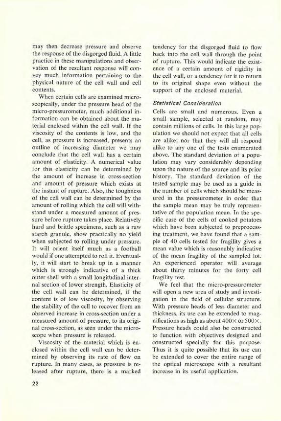
When certain cells are examined micro-scopically, under the pressure head of the micro-pressurometer, much additional in-formation can be obtained about the ma-terial enclosed within the cell wall. If the viscosity of the contents is low, and the cell, as pressure is increased, presents an outline of increasing diameter we may conclude that the cell wall has a certain amount of elasticity. A numerical value for this elasticity can be determined by the amount of increase in cross-section and amount of pressure which exists at the instant of rupture. Also, the toughness of the cell wall can be determined by the amount of rolling which the cell will with-stand under a measured amount of pres-sure before rupture takes place. Relatively hard and brittle specimens, such as a raw starch granule, show practically no yield when subjected to rolling under pressure. It will orient itself much as a football would if one attempted to roll it. Eventual-ly, it will start to break up in a manner which is strongly indicative of a thick outer shell with a smalllongitlldinal inter-nal section of lower strength. Elasticity of the cell wall can be determined, if the content is of low viscosity, by observing the stability of the cell to recover from an observed increase in cross-section under a measured amount of pressure, to its origi-nal cross-section, as seen under the micro-scope when pressure is released.
Viscosity of the material which is en-closed within the cell wall can be deter-mined by observing its rate of flow on rupture. In many cases, as pressure is re-leased after rupture, there is a marked
22
tendency for the disgorged fluid to flow back into the cell wall through the point of rupture. This would indicate the exist-ence of a certain amount of rigidity in the cell wall, or a tendency for it to return to its original shape even without the support of the enclosed material.
Statistical Consideration Cells are small and numerous. Even a small sample, selected at random, may contain millions of cells. In this large pop-ulation we should not expect that all celts are alike ; nor that they will all respond alike to any one of the tests enumerated above. The standard deviation of a popu-lation may vary considerably depending upon the nature of the source and its prior history. The standard deviation of the tested sample may be used as a guide in the number of cells which should be meas-ured in the pressurometer in order that the sample mean may be truly represen-tative of the population mean. In the spe-cific case of the cells of cooked potatoes which have been subjected to preprocess-ing treatment, we have found that a sam-ple of 40 cells tested for fragility gives a mean value which is reasonably indicative of the mean fragility of the sampled lot. An experienced operator will average about thirty minutes for the forty cell fragility test.
We feel that the micro-pressurometer will open a new area of study and investi-gation in the field of cellular structure. With pressure heads of less diameter and thickness, its use can be extended to mag-nifications as high as about 400X or 500 X. Pressure heads could also be constructed to function with objectives designed and constructed specially for this purpose. Thus it is quite possible that its use can be extended to cover the entire range of the optical microscope with a resultant increase in its useful application.

Today's best buy in a recording spectrophotometer!
... Spectronic 505® Recording Spectrophotometer
... from $3685, less than half the cost of the others No other recording spectrophotometer has ever won such prompt acceptance. In just a few short months it has become the best seller in the field. And with good reason. High accuracy and speed of analysis make it a sensible buy for recording transmittance, linear absorbance, reflectance and emission throughout the UV and visible ranges. But consider its astonishingly low price . . . add up its exclusive features ... and you'll see why so many laboratories have specified it as today's best buy.
Certified-Precision Diffraction Gratings- high resolving power and linear dispersion; constant, narrow band pass. Exclusive electronic sensor-automatically gives faster , more accurate recording. Exclusive long life air-cooled Hydrogen lamp. No water cooling lines and drainage. Precise reflectance values are easily obtained. Only $3685, visible range; $4285, UV-Visible.
BAUSCH & LOMB
Made in America, to the world's highest standards.
USE THE ENCLOSED CARD FOR COMPLETE INFORMATION

T __ E __ c __ H __ N ___ Q __ u __ By Germaine Crossman Industrial Hygienist, Biological and Chemical Microscopist Bausch & Lomb Incorporated
O NE OF THE METHODS for determining the number of bacteria in milk is to
place 1 cc. of milk diluted with a known amount of water into the bottom of a Petri dish. Melted beef extract agar is poured into the dish and by means of a rotary motion the diluted milk and agar are thor-oughly mixed. After the agar has solidi-fied, the preparation is incubated for 48 hours at approximately 35 degrees centi-grade. A similar procedure is used for de-termining the amount of bacteria in drink-ing water supplies, private and public swimming pools and other bathing areas. In this case, incubation of the plate is usu-ally done at a temperature of 35 degrees centigrade for 24 hours or at 20 degrees centigrade for 48 hours.
Counting of the colonies grown on the plate is usually done at 1.5 X magnifica-tion using a 5 inch diameter lens. An auxiliary 1.5 X lens can be used in con-junction with the regular lens when extra magnification is desired or the Petri dish can be transferred to a stereomicroscope such as the new B&L StereoZoom. The
24
references
1. Chamot E. and Mason Co., Handbook of Chemical Microscopy, Vol. I, 3rd edition 1958, John Wiley & Sons, New York, New York.
2. Crossman, G., "Dispersion Staining with Phase Contrast Microscope Accessories: The Microscopic Identification of Quartz," Science, Vol. 110, p. 237 (1949).
acknowledgment
Photographs (Figs. 1, 2 and 3) were prepared by B&L Photo Studio.
Milk and water photos for this investigation were furnished by Rochester Technical Services Incorporated and Rochester Division Laboratory of the Dairymen's League.
advantage of the StereoZoom microscope is the fact that the colony under obser-vation can be ex_amined without "black-out" during change in magnification.
We have investigated the possibility of using the Pioneer Scientific Company''' Vertical Polariscope (Fig. 1) for the ex-amination and counting of bacteria colo-nies. This instrument consists of a polar-izer and analyzer with built-in illuminator. The field of view is such that Petri dishes up to 100 mm. in diameter can be exam-ined over their entire surface. A number of milk and water plates were examined using this equipment. Magnification of 1.5 X was obtained by use of a 5-inch reading glass placed on the upper surface of the analyzer.
I was rather amazed at the success of this experiment. The colonies of bacteria, as shown in Fig. 2, appeared bright (bire-fringent) contrasting sharply with the
*Pioneer Scientific Company, located at Roches-ter, New York, is a division of Bausch & Lomb Incorporated.

dark single refracting agar. To the best of my knowledge bacteria are not considered to be birefringent. Is it possible that some bacteria, commonly found in milk and water, are intrinsically birefringent when cultured on beef extract agar or is the brightness due to form or strain birefring-ence? In answer to this question, prepa-rations of each colony on the plate were mounted in water under a cover glass and examined with our LR Research Polariz-ing Microscope. Results obtained indi-cated that all types of bacteria present on the plate were definitely single refracting and thus the brightness of the colonies was not due to intrinsic birefringence of the bacteria. One preparation examined
Figure 1. Vertical Polariscope.
showed the presence of small birefringent crystals, which may have contributed somewhat to the brightness of this colony. Following this observation, the prepara-tion was allowed to dry, flame fixed and then stained with methylene blue. It was noted that after this treatment of the preparation, the crystals had disappeared. We might conclude from this observation, that in order to examine colonies for the possible presence of crystalline material, preparations should be examined first in distilled water prerequisite to fixation and staining.
Examination of the colonies on the agar was now made at higher magnifications with the StereoZoom Microscope. The

analyzer of the polariscope was removed and placed on the stage of the microscope. The Petri dish with cover removed was centered over the analyzer (now used as a polarizer) and a second polariscope ana-lyzer used as a cover for the dish, rotating to a crossed position with reference to the polarizer located below the Petri dish. Using this equipment, it was noted that some of the spindle shaped colonies were surrounded by a bright fringe most pro-nounced at the bulge of the spindle. This birefringence was more evident by use of a full wave retardation plate (sensitive tint). It can be located either directly be-low the analyzer, thus serving as the cover for the Petri dish, or placed between the polarizer and the bottom of the dish. Us-ing this retardation plate with analyzer and polarizer in a crossed position, the background appears a purplish red color due to the interference effect of the re-tardation plate. The bright areas sur-rounding the spindle shaped colonies now appear blue or yellow changing from blue to yellow or the reverse as the Petri dish is rotated. Blue coloration indicates that at this orientation, the retardation in the
Figure 2. Colonies of Bacteria os observed with Polariscope.
26
area surrounding the colony is added to the retardation of the full wave plate, while yellow signifies an orientation as to result in subtraction.
This brightness with polarizer and ana-lyzer in a crossed position and color change using the tint plate is possibly strain birefringence. One may press the surface of the agar at any point with a teasing needle and observe a similar effect (Fig. 3). As a result of this pressure, the area appears bright or colored blue or yellow returning to dark or purplish red as the pressure is released. Is it possible that in the division of some types of bac-teria to form a colony there is enough pressure against the surrounding agar as to introduce strain birefringence? Another possibility is a change in the physical and chemical properties of the agar in and surrounding the colony due to metabolic products of the bacteria. In the case of colonies not showing the surrounding birefringent area, their brightness may be due to a large difference in refractive in-dex between the bacteria and the beef extract agar which is characteristic of form birefringence. According to Chamot
Figure 3. The area adjacent to the point of the needle appears bright due to introduction of strain birefringence in the agar as a result of the pres-sure of the needle.

& Mason,1 "If the objects have a refrac-tive index widely different from their sur-roundings, there may be sufficient depo-larization effect upon the light to render them self-luminous against the dark back-ground of the field ."
This observation of birefringence, in and surrounding bacteria colonies, sug-gests that some further investigation could be made. Do all bacteria produce bire-fringent colonies and show the same de-gree of birefringence? What results are obtained on other types of culture media? Is there a change in birefringence with aging of the colony? We have noted that one of the colonies observed contained birefringence crystals. What are these crystals and do other bacteria colonies contain crystals which may be significant for certain genus and species?
Another possible use of a Strain Po-lariscope is in the determination of anti-biotics such as penicillin in milk. Veteri-narians occasionally use antibiotics in the treatment of milk cows for such diseases as mastitis. Most health departments state that milk from these cows should not be sold for processing for a period of 72
Figure 4. l.W.D. Phase Condenser equipped with iris diaphragm and polarizer.
hours following the injection of the anti-biotic. The danger involved is that one may be allergic to the antibiotic to the point of producing anaphylactic shock.
The routine test for antibiotic as done in dairy laboratories is to mix spores of the hay bacillus (bacillus subtilis) with melted beef extract agar. After solidifica-tion of the agar, sterile circular discs are dipped in the samples of milk and then applied to the surface of the beef extract agar. The Petri dish is then incubated at 35 degrees centigrade for approximately 5 hours and examined with a colony coun-ter at the usual 1.5x magnification or if necessary at higher magnification using a stereomicroscope. Presence of antibiotic in the milk sample is indicated by a clear circular zone surrounding the test disc. This clear zone is due to the fact that anti-biotic in the milk absorbed by the disc penetrates into the surrounding agar thus inhibiting the growth of the spores of bacillus subtilis. As examined with the Strain Polariscope, the whole plate exhib-its some degree of birefringence with the exception of areas surrounding the disc containing antibiotic. These areas, having no birefringence, appear dark as com-pared to the rest of the plate.
In previous issues of the Educational Focus and in demonstration at the Bausch & Lomb Instrumentation Exhibit held yearly at Marine Biological Laboratory, Wood's Hole, Mass., we have shown the value of combining polarizing accessories including the sensitive tint plate with long working distance phase accessories. It was suggested that these accessories be em-ployed on our laboratory type microscope such as used by the biologist. Using this combination, it was possible to observe both isotropic and anisotropic structures in good contrast at the same time. Iso-tropic structures appeared in shades of gray or white dependent on their optical
27

path (refractive index X thickness) with reference to the mounting media and ani-sotropic constituents blue or orange.
This combination of polarizing and phase accessories is now possible with the new LR Research Polarizing Microscope due to the change to a standard diameter ring for holding the L.W.D. Phase Con-denser. With this polarizing microscope as compared to the laboratory type, we have the advantage of an analyzer and accessory slot incorporated in the body tube, Bertrand lens and centered circular stage. For use with phase accessories, the condenser including polarizer should be removed from the microscope and re-placed with the L.W.D. phase condenser equipped with iris diaphragm and polar-izer (Fig. 4). Phase objectives should be substituted for the bright-field. Especially good results are obtained with the 21 X (8 mm.) phase objective. The annular stop designed for the phase objective used, is inserted in the slot of the condenser. Cen-tering of the image of this annulus with the phase annulus located at the back focal plane of the objective is easily done by observation with the Bertrand lens. Cen-tering can be accomplished with polarizer and analyzer in a crossed or uncrossed position. If centered with polarizer and analyzer in a crossed position, it is best to insert the full wave plate in the accessory slot. With this combination, the image of the annular stop appears a pink color in sharp contrast to the dark phase objective annulus.
In the case of a preparation containing a large number of particles differing great-ly in refractive index from that of the mounting liquid or solid cement, it may be somewhat difficult to observe distinctly the two annuli. This is especially true in the case of ground mineral sections and unstained or stained microtome sections of biological material. It can be demon-
28
strated in the case of mineral or chemical particles by examination of a thick prepa-ration of small particle size sodium chlo-ride (index 1.544) mounted in alpha-bromonaphthalene (index 1.655). It will be noted that the annuli are difficult to observe. In order to center, it may be nec-essary to move the slide to an area con-taining less particles or outside the cover-glass area. If now a second preparation of sodium chloride is prepared of equal or even greater concentration and mounted in a liquid such as styrene, quite identical in refractive index to the sodium chloride for yellow light, it will be noted that the two annuli are clearly delineated and thus can be easily centered. This experiment can be carried further by examination of sodium chloride or other material ho-mogenous in refractive index in thicker concentrations as is possible in a cell a few millimeters in depth such as the cell fur-nished with the B&L Serum Protein Meter. As in the case of the microscope slide preparation, it will be noted that if the preparation can be rendered transparent, the two annuli can be observed in sharp contrast.
If now the microscope slide preparation of sodium chloride mounted in styrene is examined with the Bertrand lens removed from the light path, it will be observed that the sodium chloride particles are bor-dered with a red fringe. As indicated in a previous publication2 using the laboratory type microscope, the appearance of a red fringe indicates that the sample is ap-proximately equal in refractive index to the liquid for yellow light. More detailed information concerning the use of the combined polarizing phase and polariz-ing-dark-field microscopes for the identi-fication of materials by dispersion color-ation will appear in a later issue of the Educational Focus.

Reply Card
editor
POST CARD
FOCUS BAUSCH & LOMB
619 ST. PAUL ST.
ROCHESTER 2, N.Y.
Please send me the following Literature:
D ST Microscopes D- 1 0744 REMARKS,
D ESM Microscopes D-1 102
D DynaZoom Microscopes D-185
D Spectronic 505 NAME Spectrophotometer D-2009
TITLE D Tri-Simplex Micropro jector
E-20 SCHOOL O R FIRM
D Bunsen Spectroscope D-26 STREET
D Teaching Tools E-152
D Fre e copy of " Getting CITY ZONE
Acquainted With the STATE Microscope "
Vo l. 3 2, No. 1, May '6 1
PLACE STAMP HERE

Model
New! '
LOW COST ESM*
... expanding the world famous series of
Standard Teaching Microscopes
EsM-1 (shown), 1oox. * Elementary Science Microscope Model ESM-2, 40X and 100X.
Most Economical Microscope You Can Buy Compare what you get for the price you pay. Every B&L Elementary Science Microscope is made in America to the world's highest standard-with workmanship and materials guaranteed for life! Truly student-proof construction in all moving parts. Built-in safety features to protect objective, nosepiece, eyepiece and specimen slides from damage. Choice of mirror or integral Opti-lume Illuminator, at no extra charge. Will outlast comparable models two to three times. All this at the lowest cost per pupil for a full-sized microscope.
EASIEST TO TEACH AND LEARN WITH Science beginners see true, distortion-free images at 40X or 100x, the magnifications that science teachers agree are best for elementary and junior high school studies. Well-corrected achromatic objectives and Huygenian eyepiece. Students learn the right way, from the start, with no transition from "toy" to full-size microscopes. See for yourself, in your own classroom, how B&L gives you the most for your budget dollar.
PRICES START AT ONLY
$71 in lots of five or more
BAUSCH & LOMB , SINCE 1853
Made in America, to the world's highest standards.
USE THE ENCLOSED CARD FOR COMPLETE INFORMATION
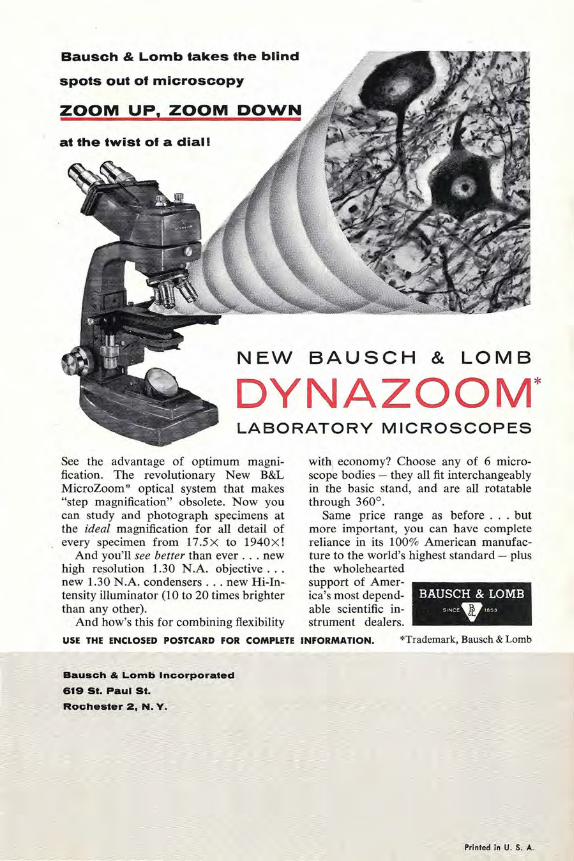
Bausch & Lomb takes the blind
spots out of microscopy
ZOOM UP, ZOOM DOWN
at the twist of a dial!
NEW BAUSCH & LOMB
DYNAZOOM* LABORATORY MICROSCOPES
See the advantage of optimum magni-fication. The revolutionary New B&L MicroZoom * optical system that makes "step magnification" obsolete. Now you can study and photograph specimens at the ideal magnification for all detail of every specimen from 17.5 X to 1940X !
And you'll see better than ever . .. new high resolution 1.30 N.A. objective .. . new 1.30 N.A. condensers . .. new Hi-In-tensity illuminator (1 0 to 20 times brighter than any other).
And how's this for combining flexibility
with economy? Choose any of 6 micro-scope bodies - they all fit interchangeably in the basic stand, and are all rotatable through 360° .
Same price range as before . . . but more important, you can have complete reliance in its 100% American manufac-ture to the world's highest standard- plus the wholehearted support of Amer-ica's most depend-able scientific in-strument dealers.
BAUSCH & LOMB SNCE . 1653
USE THE ENCLOSED POSTCARD FOR COMPLETE INFORMATION. *Trademark, Bausch & Lomb
Bausch & Lomb Incorporated
619 St. Paul St. Rochester 2, N.Y.
Printed in U. S. A.